regulation of seed germination in lepidium sativum - plant physiology
TRANSCRIPT

Regulation of seed germination in Lepidium sativum
William E Finch-Savage
School of Life Sciences Warwick University Wellesbourne
Warwick CV35 9EF UK
Tel no +44 (0)24 76574968
e-mail billfinch-savagewarwickacuk
Journal Research Area Development and hormone action
Plant Physiology Preview Published on February 14 2011 as DOI101104pp110169706
Copyright 2011 by the American Society of Plant Biologists
wwwplantphysiolorgon February 1 2018 - Published by Downloaded from Copyright copy 2011 American Society of Plant Biologists All rights reserved
2
Regulation of seed germination in the close Arabidopsis relative Lepidium sativum A global tissue specific transcript analysis Karl Morrisa Ada Linkiesb Kerstin Muumlllerb Krystyna Oraczb Xiaofeng Wangc James R Lynna and Gerhard Leubner-Metzgerb and William E Finch-Savagea a School of Life Sciences Warwick University Wellesbourne Warwick CV35 9EF UK b University of Freiburg Faculty of Biology Institute for Biology II Botany Plant Physiology Schaumlnzlestr 1 D-79104 Freiburg Germany c College of Life Sciences South China Agricultural University Guangzhou 510642 China
wwwplantphysiolorgon February 1 2018 - Published by Downloaded from Copyright copy 2011 American Society of Plant Biologists All rights reserved
3
Work was supported by the Biotechnology and Biological Sciences Research Council (Grant
BBE0064181) to WF-S the Deutsche Forschungsgemeinschaft (grant no DFG LE7206) and
the Deutscher Akademischer Austauschdienst (grant no DAAD D0628197) to GL-M the
Wissenschaftliche Gesellschaft Freiburg to GL-M and AL the Guangdong Natural Science
Fundation (07006658) to XW and an Alexander von Humboldt-foundation Research Fellowship
to KO
Kerstin Muumlller present address Department of Biological Sciences Simon Fraser University 8888 University Drive Burnaby BC V5A 1S6 Canada Corresponding author e-mail billfinch-savagewarwickacuk
wwwplantphysiolorgon February 1 2018 - Published by Downloaded from Copyright copy 2011 American Society of Plant Biologists All rights reserved
4
The completion of germination in Lepidium sativum and other endospermic seeds (eg
Arabidopsis) is regulated by two opposing forces the growth potential of the radicle (RAD) and
the resistance to this growth from the micropylar endosperm cap (CAP) surrounding it We show
by puncture force measurement that the CAP progressively weakens during germination and we
have conducted a timecourse transcript analysis of RAD and CAP tissues throughout this
process We have also used specific inhibitors to investigate the importance of transcription
translation and posttranslation levels of regulation of endosperm weakening in isolated CAPs
Although the impact of inhibiting translation is greater both transcription and translation are
required for completion of endosperm weakening in the whole seed population The majority of
genes expressed during this process occur in both tissues but where they are uniquely
expressed or significantly differentially expressed between tissues this relates to the functions
of the RAD as growing tissue and the CAP as a regulator of germination through weakening
More detailed analysis showed that putative orthologs of cell-wall remodelling genes are
expressed in a complex manner during CAP weakening suggesting distinct roles in the RAD and
CAP Expression patterns are also consistent with the CAP being a receptor for environmental
signals influencing germination Inhibitors of the aspartic serine and cysteine proteases
reduced the number of isolated CAPs in which weakening developed and inhibition of the 26S
proteasome resulted in its complete cessation This indicates that targeted protein degradation is
a major control point for endosperm weakening
wwwplantphysiolorgon February 1 2018 - Published by Downloaded from Copyright copy 2011 American Society of Plant Biologists All rights reserved
5
The seed germination process begins with imbibition of the dry seed and is completed when the
radicle has emerged through all the layers enveloping the embryo (Finch-Savage and Leubner-
Metzger 2006) In both Arabidopsis thaliana (Arabidopsis) and Lepidium sativum (Lepidium)
there are two such layers an outer dead testa (seed coat) and beneath that a layer of living
endosperm cells (aleurone layer) Germination in these species has two separate visible stages
firstly the testa ruptures and then the lower hypocotylradicle (RAD) extends to complete
germination by rupturing the micropylar endosperm layer (CAP) that covers it A recent
publication by Sliwinska et al (2009) describes how embryo elongation during Arabidopsis seed
germination is due to cell expansion growth in a specific zone in the lower hypocotylradicle
transition region During the latter process the CAP weakens through autolysis to reduce the
mechanical resistance to radicle protrusion Biomechanical measurements have been used to
record such weakening in species from a variety of different families (eg Bewley 1997 Toorop
et al 2000 da Silva et al 2004 Finch-Savage and Leubner-Metzger 2006) However
Arabidopsis seeds are too small for such measurements with the techniques used to date and
this has limited progress in linking biomechanical and molecular studies To overcome this
obstacle we have demonstrated that the larger seeds of Lepidium can be used as a model
system for studying both the molecular and biomechanical mechanisms of endosperm cap
weakening (Muumlller et al 2006 Muumlller et al 2009 Linkies et al 2009) In this work direct
biomechanical measurement has shown that endosperm cap weakening is promoted by
gibberellins (GA) and inhibited by abscisic acid (ABA) This endosperm weakening is induced by
an early signal from the embryo after which weakening and lysis proceed as an organ-
autonomous process Further experimentation has shown that in isolated endosperm caps GA
can replace the embryo signal that de novo GA biosynthesis occurs in the endosperm and that
the weakening is regulated at least in part by the GAABA ratio
The genera Lepidium and Arabidopsis both belong to the lineage I clade of the Brassicaceae
family and are therefore closely related (Franzke et al 2009) As may be expected from this
close relationship the above findings in Lepidium are consistent with the known spatial
temporal and GA-mediated regulation of genes during Arabidopsis seed germination
(Yamaguchi et al 2001 Ogawa et al 2003 Yamauchi et al 2004) Separate global expression
profiles of the whole embryo and endosperm shortly after radicle emergence in Arabidopsis are
also consistent with this pattern of regulation (Penfield et al 2006) Comparison of the
transcriptomes of endosperm and embryo tissues at a single time point of 24h also showed large
differences in expression between the tissues (Okamoto et al 2010) However to date there has
wwwplantphysiolorgon February 1 2018 - Published by Downloaded from Copyright copy 2011 American Society of Plant Biologists All rights reserved
6
been no similar analysis of the changes in these tissues leading to the completion of
germination
To take advantage of their close relationship we carried out a global transcript analysis of the
interaction between individual seed tissues in a time course during germination of Lepidium by
cross species hybridization to a full genome Arabidopsis array (Linkies et al 2009) The larger
size of Lepidium enabled us to use RNA samples collected specifically from the CAP and RAD
to avoid confounding the results with other tissues in the embryo and other regions of the
endosperm This work demonstrated that the CATMA 25K microarrays (Hilson et al 2004
Allemeersch et al 2005) which are spotted with PCR-amplified Arabidopsis gene-specific tags
(GST 150-500 bp) were effective for comparative genomics by cross-species microarray
hybrization with Lepidium Such cross-species hybridizations for closely related species using
several array platforms have become an accepted approach where no species specific arrays
are available (reviewed by Van de Mortel and Aarts 2006 Bar-Or et al 2007 Broadley et al
2008) CATMA microarrays have also been shown to be effective for cross-species microarray
hybridization in work by Slotte et al (2007) in which Capsella bursa-pastoris (Capsella)
accessions differing in flowering time were compared at the transcriptome level Capsella like
Lepidium and Arabidopsis is from the lineage-I clade of the Brassicaceae (Franzke et al 2009)
In Linkies et al (2009) a preliminary analysis of these cross-species Lepidium arrays indicated
that ethylene-related transcripts were over-represented in the lists of regulated genes The array
data was therefore used to complement an investigation of the interaction of ethylene with ABA
which resulted in a model for the hormonal regulation of endosperm cap weakening and rupture
In the present work we investigate the importance of the transcription translation and
posttranslation stages in the regulation of germination through endosperm weakening We also
carry out a full global transcript analysis of the RAD and CAP tissues during the germination
process
RESULTS AND DISCUSSION
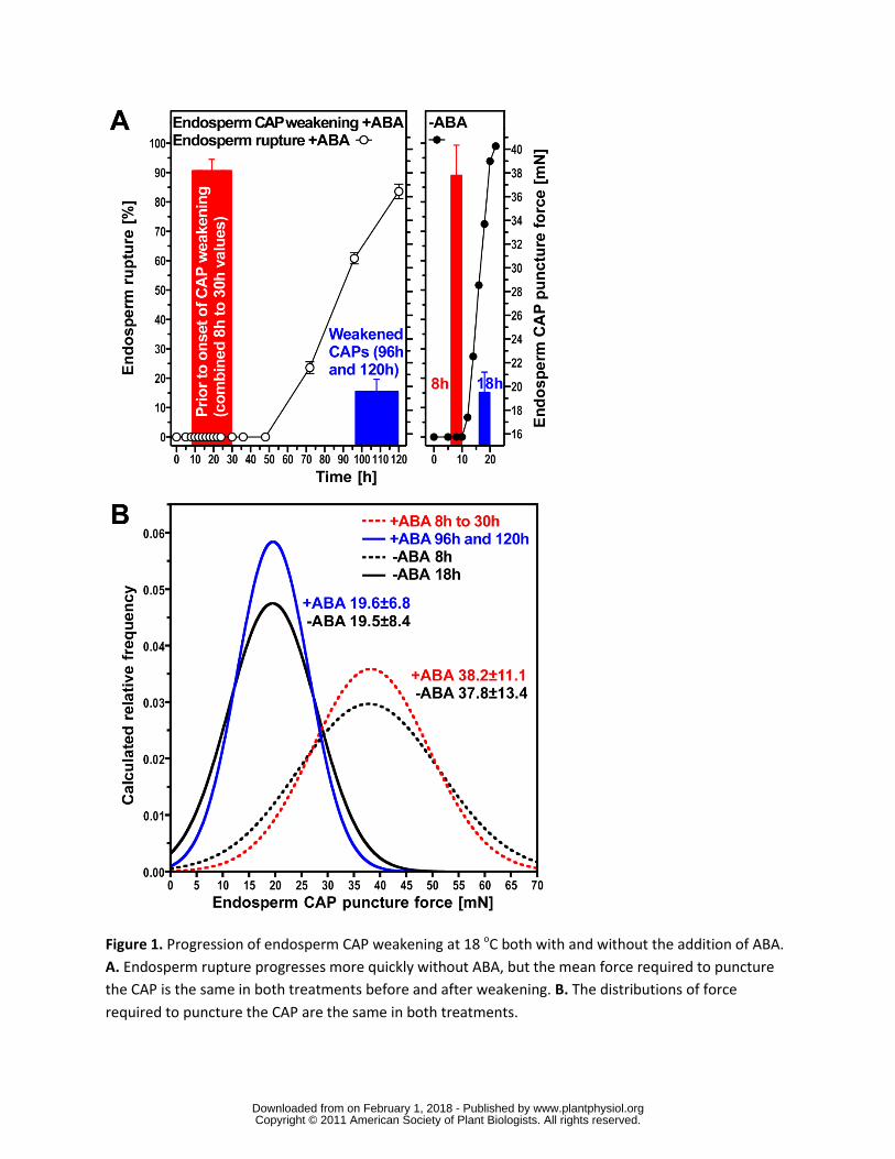
The progression of germination is clearly linked to endosperm weakening that requires
both transcription and translation for completion in the whole seed population
wwwplantphysiolorgon February 1 2018 - Published by Downloaded from Copyright copy 2011 American Society of Plant Biologists All rights reserved
7
The completion of germination (radicle emergence) in Lepidium is regulated by two opposing
forces the growth potential of the RAD and the resistance to this growth from the seed covering
layers (testa and CAP) After testa rupture the latter is determined by the strength of the
endosperm which can be determined by puncture force measurement and this progressively
decreases towards germination completion (Fig 1) Onset of endosperm weakening occurs after
8 h of imbibition on medium without hormonal addition (-ABA in Fig 1) and its progression
results in the occurrence of endosperm rupture and germination completion in an increasing
proportion of the seed population up to 18 h (Fig 1A) Both the onset and the completion of
endosperm weakening are delayed by the addition of ABA shifting its onset to more than 30 h
imbibition and its completion to 96120 h At the onset of endosperm weakening there is a high
variance in the force required to puncture the endosperm (Fig 1B) This variance declines as the
endosperm weakens in an identical fashion with and without ABA (Fig 1C) indicating that ABA
has an effect only on the timing of this process ABA therefore provides a means of spreading
out the process of endosperm weakening enabling samples to be taken at several stages both
before and during the process Overall these results show that endosperm weakening is not just
an imbibition-effect but clearly related to the progression of the germination process Single-
tissue analyses of the RAD and CAP should therefore provide a means to identify mechanisms
underlying the germination process
Dry seeds store mRNAs which are assumed to contain transcripts for genes that are important
for both late embryogenesis and for early seed germination (Comai 1989 Hughes and Galau
1989 1991) Upon imbibition transcriptional changes take place and after the first 3 h huge
changes in transcript abundance are already evident in seeds of Arabidopsis (Nakabayashi et al
2005 Preston et al 2009 Kimura and Nambara 2010) Rajjou et al (2004) have shown that
inhibiting this transcription with α-amanitin delays the germination process of whole seeds and
inhibits seedling development in Arabidopsis Transcription inhibitors have also delayed
germination in wheat embryos (Jendrisak 1980) and endosperm rupture in tobacco seeds
(Arcila and Mohapatra 1992) In contrast inhibition of translation by cycoheximide entirely
blocks the completion of germination in whole seeds (Rajjou et al 2004) We have utilized the
bigger seeds of Lepidium to investigate in a similar way the necessity of transcription and
translation during endosperm weakening in individual seed tissues When Lepidium CAPs were
dissected from -ABA-imbibed seeds (Fig 2A B) and incubated individually the initial autolysis
caused either hole formation close to where the radicle in an intact seed would penetrate
through the endosperm andor abscission of the CAP tip (Fig 2C) Subsequent progressive
wwwplantphysiolorgon February 1 2018 - Published by Downloaded from Copyright copy 2011 American Society of Plant Biologists All rights reserved
8
autolysis later disrupts the whole CAP (Fig 2D) We exploited this situation to observe the
influence of the inhibitors α-amanitin and cycloheximide on the progression of endosperm
weakening Unlike when whole seeds are used there are no problems with uptake of inhibitors
into the tissues using this system
Incubation on α-amanitin following dissection slowed the progress of autolysis and prevents the
completion of the process in a proportion of the CAPs (Fig 2E) In contrast cycloheximide
completely blocks autolysis of more than 90 of CAPs (Fig 2E) even during the very late stages
of germination ie following dissection at 18 h when some seeds in the population have already
begun autolysis in situ Comparison of the progression of autolysis in control CAPs dissected at
10 and 18 h suggests that the process occurs more quickly in the presence of the RAD in whole
seeds than it does after dissection ie CAPs dissected at 18 h are further progressed than
CAPs dissected at 10 h plus 8 h further incubation following dissection (Fig 2F) If the same
treatments are applied to the RAD dissected after 18 h no inhibition of growth was observed on
α-amanitin but cycloheximide significantly inhibited radicle growth (Supplemental Table S1)
These findings show that in addition to translation transcription is very important in the CAP
and comparative global transcriptome analysis of both tissues will be very informative
Arabidopis CATMA microarrays can be used effectively to investigate patterns in
Lepidium transcript expression
To investigate how Lepidium gene transcripts in specific seed-tissues are regulated temporally
and spatially we hybridized Lepidium RNA samples to Arabidopsis CATMA 25k microarrays
(Complete Arabidopsis Transcriptome Microarray wwwcatmaorg Hilson et al 2004
Allemeersch et al 2005) The RNA was extracted from specific Lepidium seed tissues (RAD
CAP and non-micropylar endosperm (NME)) at defined time points during germination These
tissues were collected after testa rupture before and during endosperm weakening but prior to
endosperm rupture ie only seeds with intact endosperm were used
The principal experiment (+ABA-arrays) produced samples from seeds imbibed on medium with
ABA (10 microM) as this slows the germination process allowing the dissection at earlier
developmental stages ie dissection is not possible before 8 h of imbibition but without ABA (-
ABA-arrays) changes that lead to endosperm weakening have already occurred (Linkies et al
2009) We therefore compared RAD and CAP from seeds incubated in medium containing 10
wwwplantphysiolorgon February 1 2018 - Published by Downloaded from Copyright copy 2011 American Society of Plant Biologists All rights reserved
9
microM ABA at 8 18 and 30 h leading up to the onset of endosperm weakening and later at 96h
just prior to endosperm rupture (Fig 1) In this experiment 10 microM ABA slows the germination
process but importantly does not prevent the completion of germination (radicle emergence)
Indeed the relationship between decreasing endosperm cap puncture force and increasing
percentage of seeds showing endosperm rupture was almost identical with and without ABA
despite the very different rates of this process on these solutions (Linkies et al 2009 Fig 1) In
a further smaller experiment (-ABA-arrays) seeds were imbibed on medium without ABA and
samples were prepared at 8 and 18 h from RAD CAP and NME These data are used to confirm
results collected in the first experiment and to help aid the identification of CAP specific gene
expression
Normalized expression values for Lepidium were obtained in the +ABA-arrays for 19794
CATMA probes to which there was significant transcript hybridisation (Supplemental Table S2)
and in the -ABA-arrays for 22025 probes (Supplemental Table S3) Lepidium gene transcripts
that hybridised to these probes were assigned as putative Arabidopsis orthologs defined by
having an Arabidopsis Genome Initiative (AGI) identifier such as At1g62380 and a gene
ontology (GO) annotation associated with this AGI (wwwarabidopsisorg) Henceforth to avoid
repetitive use of the term putative orthologs in Lepidium will be referred to using AGI annotation
All microarray data including the normalised intensity values for each microarray were deposited
in ArrayExpress (wwwebiacukmicroarray +ABA-arrays accession no E-TABM-743 -ABA-
arrays accession no E-TABM-745) To support the use of these cross species hybridizations
Linkies et al (2009) verified the transcript expression pattern of the arrays by comparing them to
corresponding qRT-PCR results obtained with independent biological RNA samples from a
separate experiment They concluded that cross-species microarray hybridization with the
CATMA platform is a useful and effective tool for heterologous transcriptomics with Lepidium
There are differences in the pattern of transcription between the radicle tip and the
endosperm cap but much of the temporal change is common to both tissues
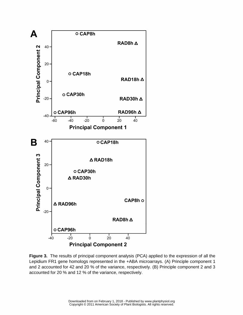
Principal component analysis (PCA) was used to look for global patterns in the Lepidium
expression data across all the gene transcripts (Fig 3) The two components PC1 and PC2
accounted for more than 60 of the variance in gene expression PC1 clearly separated RAD
and CAP (Fig 3A) PC2 then separated the times in a continuous temporal order These clear
patterns indicate that the data behave in an expected fashion with greatest differences occurring
wwwplantphysiolorgon February 1 2018 - Published by Downloaded from Copyright copy 2011 American Society of Plant Biologists All rights reserved
10
between the tissues The comparison indicates that the majority of change in transcript numbers
occurs before endosperm weakening (ie 8-18 h) The very similar ordering of the time course
suggests that much of this change in the earlier stages of germination is common to the two
tissues PC3 confirms the step change between 8 h and 18 h with a subsequent smaller
progressive change 18-96 h (Fig 3B) Distances between RAD and CAP are similar at 8 h and
18 h least at 30 h and then greatest at 96 h coinciding with the period of endosperm weakening
from 30 to 96 h (Fig 1) and preparation for radicle expansion and emergence
The majority of the genes expressed (ldquopresentrdquo) during germination occur in both
tissues but where they are uniquely expressed this relates to their specific functions
To determine whether individual genes were expressed or not the normalised values for each
probe were compared to those for the 912 empty spots on the arrays with a one-sided t-test
Probes for which the normalised values were significantly greater than the empty spots (plt005)
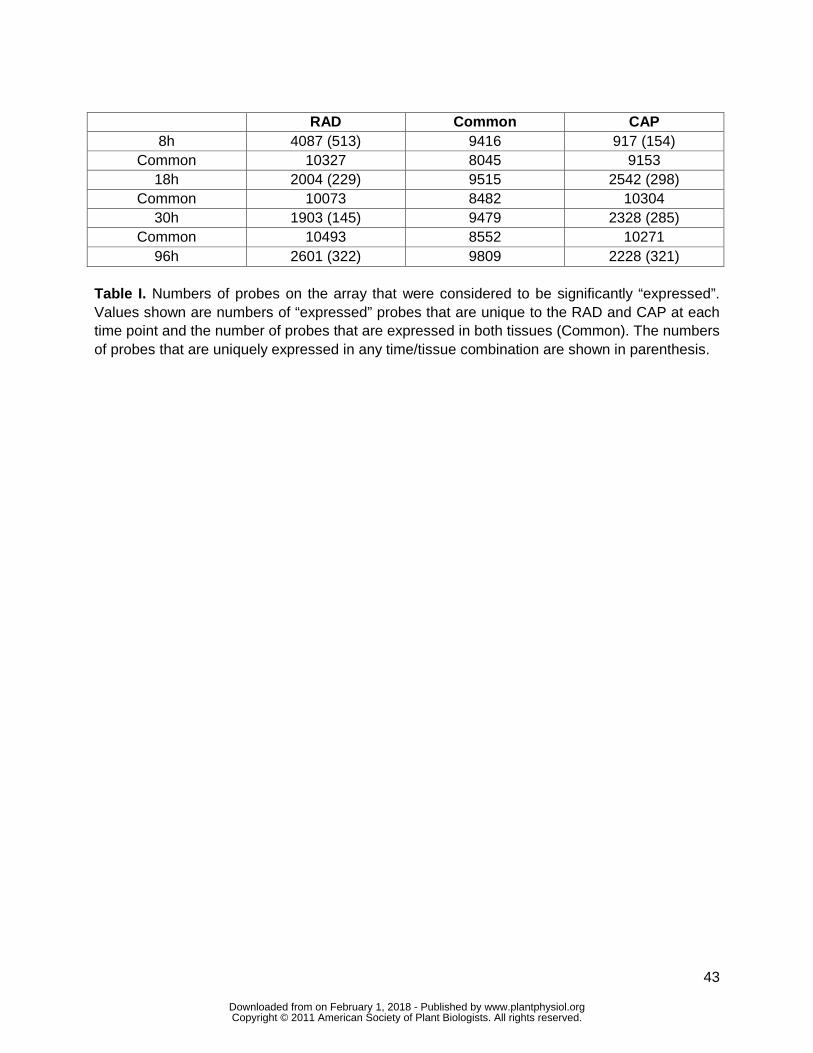
were considered to be ldquoexpressedrdquo The data shown in Table I are the number of probes on the
array that indicate expression based on this criterion In agreement with the PCA the majority of
genes expressed at any time point are expressed in both the CAP and RAD (common Table I)
Similarly the majority of genes expressed in successive time points in the same tissue are
common The number of commonly expressed genes range from 8045 to 10493 This is a very
similar number to that found by Penfield et al (2006) shortly after radicle emergence in
Arabidopsis seeds They found 9650 genes in common with approximately 4000 that were
expressed differentially in the embryo or the endosperm They concluded that patterns of gene
expression are broadly similar between the two organs suggesting similar post-germinative
metabolism occurring in these tissues We show here that this similarity extends to the
germination process
As Penfield et al (2006) found post-germination in Arabidopsis there are a number of genes
that are expressed in one tissue but not the other at all time points leading to germination
completion At 8 h the number of genes uniquely expressed in the RAD (4087) is much greater
than in the CAP (917) This suggests much greater early transcriptional activity in the RAD than
the CAP In barley Barrero et al (2009) suggest that the coleorhiza is functionally related to the
endosperm cap in Arabidopsis and Lepidium since it appears to regulate germination by
restricting radicle elongation In this species they show that 23 of genes are differentially
expressed during first 8 h in the coleorhiza but only 16 in the radicle suggesting a more active
wwwplantphysiolorgon February 1 2018 - Published by Downloaded from Copyright copy 2011 American Society of Plant Biologists All rights reserved
11
role for the former This is opposite to the results shown here however the coleorhiza initially
elongates (it grows) with the root before the root penetrates it This may explain the different
pattern of gene expression in the two species since the CAP of Lepidium shows no such growth
By 18 and 30 h this ratio has changed so that the numbers of genes uniquely expressed is 27
and 22 greater respectively in the CAP than the RAD by 96 h the number of genes expressed
in the two tissues is more similar To investigate whether these differences were linked to
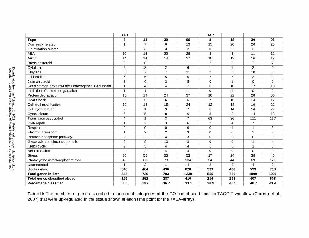
functional specialisation of the CAP and RAD tissues we applied the GO-based seed-specific
TAGGIT workflow (Carrera et al 2007) to identify proportional representations of genes into
functional categories (Supplemental Table S4) There are also a number of genes whose
expression is unique to each tissuetime combination (Table I in parenthesis) these range from
145 (30 h RAD) to 513 (8 h RAD) Again the GO-based seed-specific TAGGIT workflow (Carrera
et al 2007) was used to categorise these genes (Supplemental Table S5)
At 8 h the numbers of genes represented in TAGGIT categories is greater in the RAD than CAP
(Supplemental Table S4) which in general reflects the pattern in the total numbers expressed
(Table I) There are also tissue-specific differences in the numbers of genes uniquely expressed
at each time point In general for the RAD the numbers of genes are greater than in the CAP in
the following categories dormancy related brassinosteroid ethylene cell cycle related
cytoskeleton and translation associated This reflects the radicle as growing tissue Whereas for
the CAP after 8 h the numbers of genes are greater than for the RAD in the following categories
gibberellins jasmonic acid glycolysis and gluconeogenesis Krebs cycle beta oxidation and
stress This pattern may reflect the function of the endosperm to regulate germination through its
autolysis and subsequent death These differences in the two tissues are broadly similar to
those found by Penfield et al (2006) in post-germinative Arabidopsis tissues In contrast there is
little difference in the numbers of common genes that are expressed by both tissues in any
TAGGIT category between time points and little pattern is shown in the data (Supplemental
Table S4) Similarly there is little difference and pattern in other genes expressed in common
between time points and tissues (data not shown)
There are differences in the numbers of gene transcripts that are differentially regulated
in the RAD and CAP
The transcript abundance of individual genes in the +ABA-array data were compared between
wwwplantphysiolorgon February 1 2018 - Published by Downloaded from Copyright copy 2011 American Society of Plant Biologists All rights reserved
12
the RAD and the CAP at each time point using t-Tests to identify which genes showed
differential expression relative to each other P-values were adjusted for false discovery rate
(Benjamini and Hochberg 1995) and the resulting gene lists (p-values le 010) are given in
Supplemental Table S6 These genes were considered to be up- or down-regulated between
tissues at the time point specified The total number of genes that are differentially regulated
between the RAD and the CAP increases as the seeds progress towards germination and
presumably the functional specialisation of the tissues develop (Table II) The numbers of genes
that are up-regulated in the RAD and CAP are similar at 8 18 and 96 h but at 30 h the number
is higher in RAD (1000) compared to Cap (783) If higher stringency is applied to the analysis
the overall pattern shown in Table II remains the same but with fewer genes (eg for 96 h at
Plt010 2464 at Plt005 1600 data not shown)
To further investigate the functional specialisation of the tissue we applied the seed-specific
TAGGIT workflow (Carrera et al 2007) to the genes up-regulated in either the RAD or the CAP
relative to the other (Table II) Although the number of up-regulated genes in both tissues is
similar there are differences in the categories of genes that are over-represented at all time
points In general the categories with gene numbers over-represented in the CAP are related to
hormones aspects of metabolism and reserve mobilisation and stress Whereas the categories
with gene numbers over-represented in the RAD are related to dormancy late embryogenesis
abundant proteins aspects of growth and DNA repair The most over-represented category in
RAD is translation associated proteins with a 26 fold higher number of genes across the four
time points than the CAP (397 and 15 respectively) However when viewing these data it should
be remembered that the actual number of different genes is less than this since genes can be
represented at more than one time point Protein synthesis and ribosomal protein genes were
also highly expressed in the embryo relative to the endosperm in Arabidopsis shortly after
germination and this was linked to a fivefold higher number of ribosomes in the embryo than the
endosperm (Penfield et al 2006)
There are similar numbers of genes up-regulated in both tissues in the following categories cell-
wall modification and protein degradation This superficial similarity obscures important
differences in the detail that are explored below TAGGIT effectively summarises the
proportional representation of seed-specific genes into functional categories however the gene
lists used are no longer entirely current In the following sections we investigate categories
identified with TAGGIT but use comprehensive gene lists that extend beyond those known to be
wwwplantphysiolorgon February 1 2018 - Published by Downloaded from Copyright copy 2011 American Society of Plant Biologists All rights reserved
13
seed-specific
Genes involved in gibberellin (GA) and abscisic acid (ABA) signalling networks have
different temporal and spatial patterns consistent with regulation through subtle changes
in hormone sensitivity
In Linkies et al (2009) a model for the hormonal regulation of endosperm cap weakening and
rupture was constructed for Lepidium In this model GA is an embryo signal that releases coat
dormancy (if present) and induces the CAP weakening process Thereafter weakening is a cap-
autonomous process with the rate regulated by GA-ABA and ethylene-ABA antagonisms that
result in the completion of germination There are many proteins involved in the regulatory
networks controlling this process and current understanding can be found in several recent
comprehensive reviews (Holdsworth et al 2008a Finkelstein et al 2008 Penfield and King
2009) Hormone signalling especially that resulting from the dynamic balance of GA and ABA is
a key component of these networks which are thought to have significant interactions (Kucera
et al 2005 Holdworth etal 2008a) Understanding of ABA signal transduction is developing
rapidly and a model has recently emerged in which PYRPYLRCAR receptors bind to ABA to
remove the repression by PP2Cs of downstream signalling via SnRK2s to ABRE-regulated gene
expression by the transcription factors ABI3 ABI4 and ABI5 (Cutler et al 2010 Fig 5A) On
the other side of this balance DELLA proteins repress GA responses and therefore germination
(Sun and Gubler 2004) DELLAs are degraded to remove this repression when they form a
complex with GA and GID1 receptors (Hartweck 2008 Fig 5A) These signalling networks are
influenced by a diverse range of environmental signals during germination principally
temperature and light Key components of the interaction between these two environmental
signals and GA are the two phytochrome-interacting bHLH transcription factors PIL5 and SPT5
these both repress germination in the dark (Penfield et al 2005)
In the Lepidium after-ripened seed used in this work the kinetics of endosperm rupture is strongly
dependant on temperature but not light and there is no obvious evidence of residual dormancy
This situation contrasts with Arabidopsis seeds that are light sensitive and so expression patterns
may differ from those anticipated from work on Arabidopsis Transcript levels of genes encoding
the components of the hormone signalling networks in the CAP and RAD during Lepidium
germination are shown in Fig 5B Although germination of our Lepidium seed batch was not
responsive to light phytochrome genes are surprisingly shown to be some of the most highly
wwwplantphysiolorgon February 1 2018 - Published by Downloaded from Copyright copy 2011 American Society of Plant Biologists All rights reserved
14
expressed in both tissues These genes are up-regulated in the CAP relative to the RAD in
particular PHYA ndashABA-array results indicate that expression of PHYA is higher in the CAP than
the NME and is therefore CAP specific In Arabidopsis SOM is thought to encode a component of
the phytochrome signal transduction pathway that regulates genes in hormone metabolism and
acts as negative regulator in PHYA-mediated promotion of germination (Kim et al 2008)
However in Lepidium SOM is expressed very highly in both tissues which from its function in
Arabidopsis appears counter intuitive in these actively germinating seeds The reason for these
very clear expression patterns with PHYA and SOM in these light insensitive Lepidium seeds is
not clear PIL5 expression is low as expected for a negative regulator of germination in this
situation SPT tends to be more highly expressed in the CAP but transcript levels are low These
results are consistent with the CAP being the principal receptor for environmental signals
influencing germination
In general genes relating to GA signalling are more highly expressed than those relating to ABA
signalling (Fig 5B) and this is consistent with expectations for non-dormant seeds progressing
towards the completion of germination Nevertheless it is interesting to note that ABI4
expression is significantly up-regulated in the RAD whereas ABI5 expression tends to be higher
in the CAP and ABI3 is similarly expressed in both tissues at a low level This is entirely
consistent with the results of Penfield et al (2006) who showed using GUS fusions that in
Arabidopsis ABI3 is expressed in embryo and endosperm ABI4 expression was specific to the
embryo and although ABI5 was expressed in the embryo and endosperm expression in the
latter was CAP specific ABI4 is thought to repress lipid breakdown in the seed (Penfield et al
2006) Another note of interest with ABA signalling is that genes encoding for ABA receptors
each exhibit distinct patterns but where there is a significant differential expression between
tissues for example PYL4 PYL5 PYL 6 and PYR1 they are up-regulated in the CAP
Seeds with an absence of GA receptors fail to germinate (Griffiths et al 2006 Willige et al
2007) and by binding to GA and DELLAs the latter are degraded to de-repress germination (Fig
5A) Genes encoding these receptors are up-regulated in the CAP in particular GID1A and
GID1C the latter late in the germination process Interestingly the reverse is true with DELLA
repressor genes which are expressed more highly in the radicle in particular RGL3 early in the
germination process In Arabidopsis RGL3 represses testa rupture in response to changes in
GA and ABA levels (Piskurewicz and Lopez-Molina 2009) As with the ABA receptors each of
these genes displays a different temporal pattern suggesting regulation occurs through a
wwwplantphysiolorgon February 1 2018 - Published by Downloaded from Copyright copy 2011 American Society of Plant Biologists All rights reserved
15
complex mix of subtle controls with the potential to be highly responsive to the prevailing
conditions Regulation clearly does not result solely from a simple hormone balance but
additionally through differing spatial and temporal sensitivity to these hormones generated in the
hormone signalling networks
There is a complex pattern of gene expression linked to proteins associated with cell-wall
modification that underlies CAP weakening
Much of the differences in gene expression between the CAP and RAD are likely to result from
the functional specialisation of the whole endosperm as an embryo nutritional tissue during seed
development and the RAD as a growing tissue in the germinating seed However the CAP in
Lepidium is specifically associated with the regulation of germination through endosperm
weakening (above and Muumlller et al 2006 Linkies et al 2009) which requires cell-wall
modification by cell-wall remodelling proteins (CWRPs) Table II indicated that a similar number
of genes in TAGGIT cell-wall modification category are up-regulated in the two tissues at
different time points The total number of genes at all time points is 76 and 71 for CAP and RAD
respectively However on closer inspection there are very different patterns to the expression of
CWRP genes in the two tissues (Fig 5 Supplemental Fig S1)
Plant cell expansion growth is driven by water uptake and restricted by the cell wall The
mechanical strength of the plant cell wall determines the shape and the rate and direction of
growth of individual cells as well as the mechanical resistance of whole tissues (Fry 2004
Cosgrove 2005 Schopfer 2006 Knox 2008) The primary cell wall has a fibreglass-like
structure with crystalline cellulose microfibrills that are embedded in a matrix of complex
polysaccharides which are divided into two classes hemicelluloses and pectins Hemicelluloses
are cellulose-binding polysaccharides which together with cellulose form a network that is
strong yet resilient Pectins form hydrated gels that push microfibrils apart easing their sideways
slippage during cell growth while also locking them in place when growth ceases They are
important determinants of wall porosity and wall thickness and they glue cells together in an
adhesive layer called the middle lamella Known wall-remodelling mechanisms include reactive
oxygen species (ROS)-mediated polysaccharide scission and CWRP actions include enzymatic
hydrolysis transglycosylation and expansin action Cell wall loosening is an important
developmental process in all stages of plant development requiring elongation growth or tissue
weakening Examples include seed germination (Bewley 1997 Nonogaki et al 2007 Muumlller et
wwwplantphysiolorgon February 1 2018 - Published by Downloaded from Copyright copy 2011 American Society of Plant Biologists All rights reserved
16
al 2009) seedling elongation growth (Schopfer 2006 Schopfer and Liszkay 2006) and fruit
ripening (Fry et al 2001 Saladie et al 2007)
Ikuma and Thimann (1963) in their hatching hypothesis of seed biology suggested that the
final step in the germination control process is the production of an enzyme whose action
enables the tip of the radicle to penetrate through the coat In searching for this hatching
enzyme evidence has been uncovered for the contribution of various CWRPs including endo-
szlig-14-mannanases (eg Bewley 1997 Nonogaki et al 2000 Iglesias-Fernandez et al 2011)
and endo-szlig-13-glucanases (eg Leubner-Metzger 2002 Leubner-Metzger 2003 Petruzzelli et
al 2003) as well as for ROS (Muumlller et al 2009) but most of this work was in Solanaceous
seeds However endosperm weakening is also evident in Brassicaceae seeds where it is
promoted by GA and ethylene and inhibited by ABA (Debeaujon and Koornneef 2000
Debeaujon et al 2000 Muumlller et al 2006 Bethke et al 2007 Linkies et al 2009 Iglesias-
Fernandez et al 2011) Based on the timing of GA-inducible transcripts in whole seeds of
Arabidopsis many CWRP genes that remodel hemicellulose are expressed during the early
germination phase (Ogawa et al 2003 Nonogaki et al 2007) Our tissue-specific transcriptome
analysis with Lepidium (this work and Linkies et al 2009) shows that many of the bigger CWRP
families exhibit complex temporal and spatial expression patterns that are presented in the
Supplemental FiguresTables We therefore restrict our subsequent discussion to a selection of
early-expressed hemicellulose-related genes that are abundant during CAP weakening
Expansins are plant cell-wall loosening proteins which disrupt noncovalent bonds that tether
matrix polysaccharides to the surface of cellulose microfibrils or to each other (Sampedro and
Cosgrove 2005 Choi et al 2006) Whatever their biochemical mechanism of action expansins
act in catalytic amounts to stimulate wall polymer creep without causing major covalent
alterations of the cell wall The α-expansins (EXPA) act with a pH optimum around 4 They have
possible roles in developmental processes like organ size and elongation growth fruit tissue
softening and seed germination (Sampedro and Cosgrove 2005 Choi et al 2006 Gaete-
Eastman et al 2009 Lizana et al 2010) Transcripts of the tomato α-expansin LeEXPA4 and
its putative ortholog in Datura ferox were specifically expressed in the micropylar endosperm in
association with endosperm weakening (Chen and Bradford 2000 Mella et al 2004) This
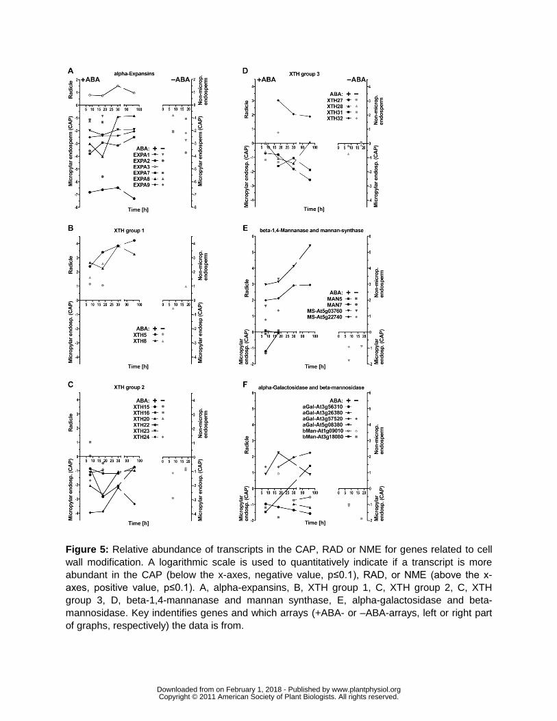
transcript expression was promoted by GA but not inhibited by ABA Where there is a significant
difference in the level of expression between the tissues the majority of Lepidium expansin
genes (Fig 5A Supplemental Fig S1A) are expressed more highly in the CAP than the RAD
wwwplantphysiolorgon February 1 2018 - Published by Downloaded from Copyright copy 2011 American Society of Plant Biologists All rights reserved
17
This is particularly true for the α-expansins EXPA1 2 7 8 9 for which in the +ABA-arrays the
transcript levels are higher in the CAP compared to the RAD at all time points (Fig 5A) only
EXPA3 was RAD-specific The ndashABA-arrays show that EXPA1 2 9 are specifically expressed in
the CAP but not in the NME ABA does not down-regulate any of these CAP-specific α-
expansin genes During the early phase of Arabidopsis seed germination transcripts of
AtEXPA1 2 8 9 accumulate 100- to 500-fold (from 0 h to 12 h in whole unstratified seeds) and
this induction is promoted by GA not inhibited by ABA and mainly localized in the endosperm
(Nakabayashi et al 2005 Penfield et al 2006 Carrera et al 2008 Holdsworth et al 2008a
Preston et al 2009) In summary transcript expression analysis during germination of both
Lepidium and Arabidopis shows that α-expansin genes in particular EXPA2 are induced early
in the endosperm cap prior to the onset of weakening and are involved in ABA-insensitive
processes that lead to testa rupture and cap weakening (this work and Linkies et al 2009)
Based on their temporal spatial and hormonal expression pattern α-expansins are likely to
contribute to processes in the CAP prior to and during endosperm weakening and rupture but
they do not confer the ABA-regulation of these processes
Xyloglucan endotransglycosylasehydrolase (XTHs) modify xyloglucans which are part of
the hemicellulose network believed to crosslink cellulose microfibrils (Fry 2004 Cosgrove
2005) Xyloglucan is the primary XTH substrate and most XTHs exhibit XET (transferase)
activity that breaks and remakes glycosidic bonds in the backbone of xyloglucan Some XHTs
also exhibit XEH (hydrolase) activity and mediate xyloglucan strand breaks and a few exhibit
only XEH activity Direct unambiguous proof of XTHs inducing wall stress relaxation and
extension is still lacking However XTHs are implicated in cell-wall hemicellulose remodelling
leading to loosening (Van Sandt et al 2007) or stiffening (Maris et al 2009) XTHs are
proposed to have roles in many developmental processes including cell growth fruit ripening
and reserve mobilisation following germination of xyloglucan storing seeds (Tine et al 2000
Fry 2004 Nonogaki et al 2007 Van Sandt et al 2007) During tomato seed germination
transcript accumulation of LeXET4 was induced in the micropylar endosperm promoted by GA
but not inhibited by ABA (Chen et al 2002) The phylogenetic relationship between the 33
members of the Arabidopsis XTH gene family reveal 3 groups
(httplabsplantbiocornelleduXTHarabidopsishtml Becnel et al 2006) Several XTH genes
of Lepidium showed tissue-specific expression during seed germination (Fig 5B 5C 5D
Supplemental Fig S1B) The group 2 XTHs 15 16 20 22 23 24 and group 3 XTHs 27 28 and
31 exhibit stronger expression in the CAP compared to the RAD In contrast the group 1 XTHs 5
wwwplantphysiolorgon February 1 2018 - Published by Downloaded from Copyright copy 2011 American Society of Plant Biologists All rights reserved
18
and 8 and the group 3 XTH32 exhibit stronger expression in the RAD compared to the CAP
During the early phase of Arabidopsis seed germination of the above mentioned genes only
transcripts of AtXTH5 16 and 27 accumulated significantly at 6 h and AtXTH15 22 28 and 31
at 12 h while AtXTH20 23 and 24 were not induced Only XTH15 16 5 31 were induced by
GA and only XTH24 was down-regulated by ABA (Nakabayashi et al 2005 Preston et al
2009) In summary transcript expression analysis during seed germination of both Lepidium and
Arabidopsis shows that XTH genes are expressed in a complex manner suggesting distinct roles
in the RAD and CAP
Mannans are rigidity and mechanical strength conferring hemicellulosic polysaccharides present
in the endosperm of many seeds (Bewley 1997 Reid et al 2003 Nonogaki et al 2007) The
endosperm cell walls of Solanaceous seeds contain ca 60 mannose and ca 10 galactose
as galactomannans Coffee galactomannan contains only ca 2 galactose which results in hard
and brittle endosperm properties Mannan polysaccharides could be masked and this may have
prevented the detection of mannan epitopes in Arabidopsis seeds but genetic evidence has
strongly indicated a functional role for mannan in seed development and germination of this
species (Marcus et al 2010 Iglesias-Fernandez et al 2011) Galactomannan biosynthesis in
seed endosperms involves β-1-4-mannan synthase and galactomannan galactosyltransferase
(Reid et al 2003 Edwards et al 2004) The β-14-mannan synthases are encoded by the
cellulose synthase-like A (CslA) gene family (Dhugga et al 2004 Liepman et al 2005)
AtCslA2 transcripts accumulated in germinating Arabidopsis seeds in a GA-promoted and ABA-
unaffected manner (Nakabayashi et al 2005 Preston et al 2009) In Lepidium CSL2 and CSL9
showed a radicle-specific expression during seed germination (Fig 5E Supplemental Fig S1C)
Degradation of mannan and galactomannan polymers involves endo-β-14-mannanase α-
galactosidase and β-mannosidase all of which have been identified in germinating seeds
several endo-β-14-mannanases have hydrolase and endotransglycosylase activity (Schroder et
al 2009) Among the many endo-β-14-mannanase isoforms of tomato the LeMAN2 gene is
expressed specifically in the micropylar endosperm prior to radicle emergence in association
with enzyme activity accumulation (Nonogaki et al 2000 Toorop et al 2000 Gong and
Bewley 2007) This induction is promoted by GA but not inhibited by ABA Endo-β-14-
mannanase also accumulates in the micropylar endosperm of Solanum lycocarpum Datura
ferox and coffee and is thought to contribute to endosperm weakening (Bewley 1997 Nonogaki
et al 2000 Toorop et al 2000 da Silva et al 2004 Arana et al 2006 Pinto et al 2007)
wwwplantphysiolorgon February 1 2018 - Published by Downloaded from Copyright copy 2011 American Society of Plant Biologists All rights reserved
19
Endo-β-14-mannanase enzyme activities of individual tomato micropylar endosperm caps
varies at least 100-fold (Still and Bradford 1997) Although the presence of endo-β-14-
mannanase enzyme activity in the tomato endosperm cap is consistently associated with radicle
emergence it is not the sole or limiting factor under all conditions Seed germination of tomato
lines over-expressing an endo-β-14-mannanase was not promoted (Belotserkovsky et al
2007) Seed-specific regulation of several endo-β-14-mannanases are also known from rice
(Yuan et al 2007 Ren et al 2008) Seven endo-β-14-mannanase genes are known in
Arabidopsis (Yuan et al 2007) but of these only AtMan7 (At5g66460) transcripts accumulated
in whole unstratified seeds and this induction is promoted by GA but not inhibited by ABA
(Nakabayashi et al 2005 Preston et al 2009) In agreement with this transcripts of the
Lepidium MAN7 accumulated in the CAP and to a lesser extent in the RAD during seed
germination (Fig 5E Supplemental Fig S6C) This induction was not inhibited by ABA and at 8
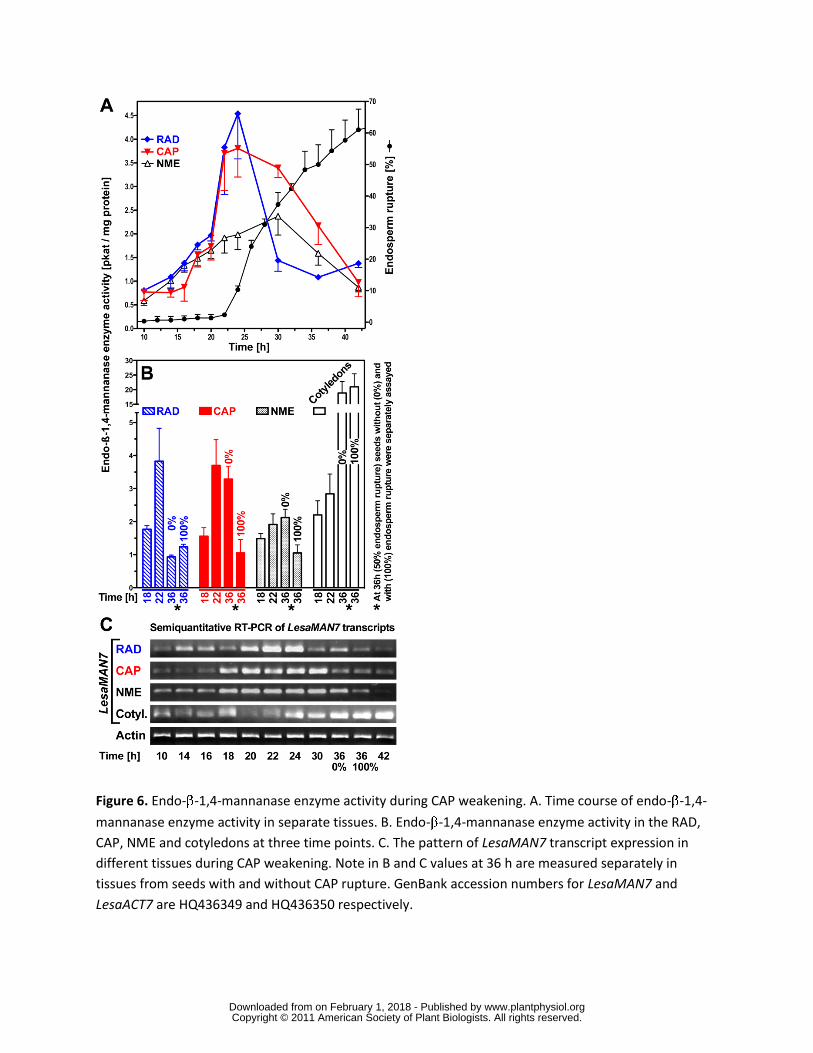
h was stronger in CAP than the RAD and the NME (Fig 5E) Figure 6A and 6B show that endo-
β-14-mannanase enzyme activity accumulated in the CAP and the RAD but less in the NME
prior to endosperm rupture This increasing activity is at least in part due to LesaMAN7 as the
transcript expression pattern is regulated in a similar manner (Fig 6C) Late during germination
and after endosperm rupture endo-β-14-mannanase enzyme activity and LesaMAN7
transcripts also accumulate in the cotyledons (Fig 6) In agreement with a role for LesaMAN7 in
germination a recent study shows that Arabidopsis knockout mutants for AtMAN7 6 and 5 had
slower germination than the wild-type (Iglesias-Fernandez et al 2011) In seeds of Sisymbrium
officinale which is also a Brassicaceae endo-β-14-mannanase enzyme activity accumulated in
an ethylene- and GA-promoted manner (Iglesias-Fernandez and Matilla 2009)
α-Galactosidases and β-mannosidases contribute to seed galactomannan degradation (Bewley
1997 Feurtado et al 2001 Nonogaki et al 2007) and β-mannosidase enzyme activity has
been detected in the micropylar endosperm of tomato seeds Datura and coffee (de Miguel et
al 2000 Mo and Bewley 2002 da Silva et al 2005) In Arabidopsis seeds At3g18080 β-
mannosidase transcripts accumulate mainly in embryo and At3g57520 α-galactosidase
transcripts were abundant between 3 h and 24 h in whole seeds (Nakabayashi et al 2005
Penfield et al 2006 Preston et al 2009) Transcripts of Lepidium α-galactosidases and β-
mannosidases had complex tissue-specific patterns (Fig 5F) Taken together these results
support a role for endo-β-14-mannanase during the germination of endospermic Brassicaceae
seeds
wwwplantphysiolorgon February 1 2018 - Published by Downloaded from Copyright copy 2011 American Society of Plant Biologists All rights reserved
20
Cellulase (endo-β-14-glucanase) activity was detected in tomato Datura and coffee seeds
(Sanchez et al 1986 da Silva et al 2004 Nonogaki et al 2007) In Datura and coffee but not
in tomato this was in association with endosperm weakening and germination Several putative
orthologs of Arabidopsis showed a CAP-specific expression pattern during seed germination of
Lepidium while orthologs of At1g70710 and At1g64390 showed a RAD-specific expression
(Supplemental Fig 6D) During the early phase of Arabidopsis seed germination transcripts of
At1g70710 and At1g64390 accumulate ca 100-fold (from 0 h to 24 h of whole unstratified
seeds) and this induction is promoted by GA but not appreciably inhibited by ABA (Nakabayashi
et al 2005 Preston et al 2009) Tomato endosperm cell walls contain up to 10 arabinose but
little xylose Transcripts of a β-D-xylosidase accumulated in the embryo of germinating tomato
seeds (Itai et al 2003 Nonogaki et al 2007) In Lepidium β-D-xylosidases (At1g02640
At5g64570 At1g78060 At5g10560) and α-D-xylosidases showed RAD-specific expression
during seed germination while the β-D-xylosidase At5g49360 was higher in the CAP
(Supplemental Fig 1D) In Arabidopsis transcripts of β-D-xylosidases accumulated gt100-fold
(from 0 h to 24 h of whole unstratified seeds) while the β-D-xylosidase At5g49360 was gt20-fold
induced in the endosperm and these inductions were promoted by GA but not inhibited by ABA
(Nakabayashi et al 2005 Preston et al 2009) The transcript expression pattern of pectin-
related enzymes in Lepidium has already been discussed in Linkies et al (2009)
Genes relating to protein degradation and post-translational modification are important in
the regulation of cell wall modification
From a review of recent post-genomic data Holdsworth et al (2008b) concluded that RNA
translation and posttranslational modification are major levels of control for germination
completion However there are similar numbers of genes up-regulated in both tissues in the
TAGGIT category protein degradation (Table III) but as discussed above this similarity obscures
important differences in the detail There are 620 genes tagged in this category of which 76 are
significantly differentially expressed between the two tissues with 34 and 42 expressed more
highly in the CAP and RAD respectively (Supplemental Figs S2A and B) Closer inspection of
this cohort of genes reveals a prominent role for the aspartic acid and subtilase families of plant
proteases We therefore looked at genes from all members of these two families of plant
proteases and included the cysteine protease family of enzymes Members of these classes of
proteases are reported in Beers et al (2004) Fig 7 summarises those members that are
wwwplantphysiolorgon February 1 2018 - Published by Downloaded from Copyright copy 2011 American Society of Plant Biologists All rights reserved
21
significantly (Plt010) differentially expressed between the two tissues The SBT protease
members are predominantly over-represented in the RAD whilst the significant aspartic
proteases are mainly overrepresented in the CAP We have also investigated the expression of
genes encoding key enzymes in protein modification involving the ubiquitin26S proteasome E3
ligases specifically the F box and RING finger proteins (Supplemental Figs 2C and D)
SBT proteases Subtilases are a diverse family of serine proteases which number 54 in
Arabidopsis and have a high degree of gene duplication and associated redundancy
(Rautengarten et al 2005) This functional redundancy has made it difficult to associate
biological function to individual genes with only two knock out mutants stomatal density and
development 1 (sdd1) and Abnormal leaf shape (ale1) having recognisable phenotypes It has
been postulated that these encode proteins (SDD1 and ALE1) that act as proprotein
convertases yielding bioactive peptides (Berger and Altmann 2000 Tanaka et al 2001) In
Lepidium Fig 7 shows predominantly greater expression of the subtilase gene family members
in the RAD than in the CAP one such transcript in Arabidopsis AIR3 (At2g04160) has been
linked to lateral root emergence (Neuteboom et al 1999) In Arabidopsis more than 20 of the
family members have been shown to be transcriptionally regulated by light with the expression
of At2g39850 and At5g59130 demonstrating sole dependence on PHYA under far red light for
induction (Zhou 2009) In Lepidium both these subtilases are expressed significantly higher in
the RAD whereas PhyA is expressed significantly more highly in the CAP (Fig 5B) suggesting
the possibility of signalling between the two tissues regulated by light
Aspartate proteases There are 59 aspartic proteases identified amongst the annotated
Arabidopsis genes and little is known about their biological role (Beers et al 2004) Sub-cellular
localisation may help to elucidate their physiological function and a number have been located in
the intracellular fluid of the apoplast with a role in disease resistance signalling (Xia et al 2004)
There is also evidence for a role in seeds Mutlu et al (1999) characterised an aspartic protease
from dry seeds of Arabidopsis and collocated it with the seed storage protein 2S albumin and the
vacuolar marker enzyme α-mannosidase Molecular studies of osmoprimed seeds of cauliflower
(Fujikura and Karssen 1995) identified two aspartic proteases with enhanced expression upon
priming A proteomic analysis of Lepidium CAP tissue (Mϋller et al 2010) identified aspartic
proteases as a main class of proteins involved in storage protein degradation The abundance
of one aspartic protease was shown to increase from 8h to 18h during the period of CAP
weakening The authors concluded that this early mobilization of protein bodies in the cap is
wwwplantphysiolorgon February 1 2018 - Published by Downloaded from Copyright copy 2011 American Society of Plant Biologists All rights reserved
22
likely to serve a non-nutritional function in the control of germination (Mϋller et al 2010) These
observations are consistent with the CAP specific expression of a number of putative aspartic
protease transcripts within our data set (Fig 7)
Cysteine proteases A number of cysteine proteases have been described in the literature that
are involved in seed germination (Helm et al 2008 Cervantes et al 1994) Ethylene was
shown to induce the expression of a cysteine protease responsible for the catabolism of major
reserve proteins (Cervantes et al 1994) Helm et al (2008) have reported a number of KDEL-
cysteine proteases involved in programmed cell death (PCD) and the dismantling of extension
scaffolds This led the authors to the hypothesis that the KDEL-tailed cysteine proteases they
identified participate in the final cell collapse during PCD by attacking the structural
hydroxyproline-rich glycoproteins of the cell wall In Lepidium transcript numbers of one of
these KDEL-tailed cysteine proteases At3g48340 was significantly up-regulated in the CAP
(Fig 7)
RING finger E3 ligases and F box proteins E3 ligases are the components of the 26S
proteasome that confer substrate specificity to the system The ubiquitin26S proteasome
pathway is important to most aspects of plant biology (Vierstra 2009) including hormonal
signalling (Frugis and Chua 2002) There are 697 F box proteins (Gagne et al 2002) and 469
RING finger proteins (Stone et al 2005) in the Arabidopsis genome of which we have
transcriptional data for 333 and 327 putative orthologs respectively in our Lepidium data set
Transcript abundances from this set that are significantly differentially expressed between the
two tissues is shown in Supplemental Figure 2C and D There was a greater proportion of both
F box and E3 ligase encoding genes up-regulated in the CAP compared to the RAD (23 and 27
fold respectively) This suggests that posttranslational modification in the form of selected
proteolysis performs a more significant role in CAP weakening than in the developing RAD
The role of the 26S proteasome pathway in light and hormonal signalling is well characterized in
plants (Vierstra 2009) and this apparent enrichment of E3 ligase mRNAs may strengthen the
argument for these environmental cues playing a substantial role in endosperm weakening and
signalling to the developing seedling
The effect of protease inhibitors To investigate the role of these proteins in protein
degradation we have monitored the progression of CAP hole formation as described above
when incubated upon specific protease inhibitors The CAPs isolated after 12 h imbibition were
wwwplantphysiolorgon February 1 2018 - Published by Downloaded from Copyright copy 2011 American Society of Plant Biologists All rights reserved
23
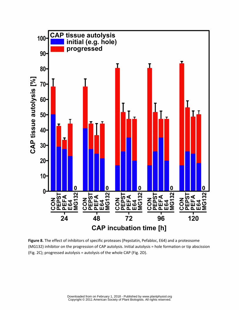
then incubated upon 1 microM Pepsatin 4 mM Pefabloc 28 microM E-64 and 60 microM MG132 which
inhibit aspartic serine and cysteine proteases and the 26S proteasome respectively (Fig 8) It
is clear from these data that all 4 classes of proteases investigated have a pronounced affect on
endosperm weakening with the most dramatic affect being the complete cessation of autolysis
by the proteasomal inhibitor MG132 The other three inhibitors reduced the number of CAPs
exhibiting autolysis to approximately 50 of that shown in the control (84) over the same time
period This suggests that each class of protease has a specific protein target and numerous
protein targets may be required for complete lysis
The complete inhibition of hole formation and tissue autolysis in those CAPs treated with MG132
suggests that targeted protein degradation is a major control point for endosperm weakening It
is now well established that the ubiquitination pathway plays a role in various hormonal
signalling pathways and is involved in the regulation of germination through the degradation of
DELLA proteins (Dill et al 2001) it could therefore be hypothesised that inhibition of the
degradation of an important transcription repressor prevents the cascade of transcriptional
activity that we show in our array data It was recently demonstrated that ABA inhibits CAP hole
formation (Linkies et al 2009) and we show here that the proteasome inhibitor MG132
completely blocks cap hole formation and autolysis This suggests the involvement of a key
transcriptional repressor implicated in several signalling pathways as well as in ABA signalling
Several ubiquitin ligases have been linked to ABA signalling and have the transcription factors
ABI3 and ABI5 both important regulators of seed germination as targets for proteolysis
(Santner and Estelle 2010 Lopez-Molina et al 2003 Holdsworth et al 2008) ABI5 and ABI5
BINDING PROTEIN (AFP) mRNA and protein levels increase when seeds are treated with ABA
and mutants for ABI5 and ABF exhibit seed phenotypes NINJA and AFP are related proteins
and it has been proposed in recent work that the GrouchoTup1-type family co-repressors
including TOPLESS (TLP) are part of a general repressor machinery implicated in several
signaling pathways (Liu and Karmarkar 2008 Pauwels et al 2010) For jasmonic acid and ABA
signalling NINJA and AFP are proposed to mediate the interaction between transcription
factors ABI5 for ABA and TLP The TPL-type co-repressors have general functions in plant
hormone signalling that are related to transcription factor proteolysis Supplemental Figure S2C
emphasises the influence of the proteasomal degradation pathway on hormonal signalling
There was significant differential expression between CAP and RAD of genes related to auxin
(AFB3 TIR1 and MAX2) jasmonic acid (COI1) and ethylene (EBF1) All these except TIR1
showed elevated levels of expression in the CAP
wwwplantphysiolorgon February 1 2018 - Published by Downloaded from Copyright copy 2011 American Society of Plant Biologists All rights reserved
24
CONCLUDING DISCUSSION
We have shown that following rupture of the testa germination in Lepidium is regulated by the
opposing forces of RAD (radicle plus lower hypocotyl) extension and the resistance to this by the
surrounding CAP (micropylar endosperm) which progressively declines through autolysis By 18
h some seeds have completed autolysis (germination has occurred) but even at this late stage
progress in some seeds can be stopped by inhibiting transcription and the remainder can be
stopped by blocking translation and posttranslational changes Taken together these results
suggest that this is a control point for germination completion that is very late on in the
germination process This late control point therefore acts as a gateway to seedling
development but the rate of germination must be determined earlier in the process as seeds
reach this control point at different times and thus weakening does not determine vigour Late
control is a necessary feature that prevents inappropriate germination when environmental
conditions change Expression of genes involved in hormone signalling networks was shown to
have different temporal and spatial patterns consistent with establishing a complex responsive
regulation through subtle changes in hormone sensitivity rather than through a crude hormone
balance Genes encoding cell-wall remodelling proteins were also expressed in a complex
tissue specific manner during endosperm weakening that would allow subtle regulation of
weakening and therefore germination completion in response to hormone signals driven by the
current ambient environment
MATERIALS AND METHODS
Plant material germination and puncture-force measurements
After-ripened Lepidium sativum L FR1 (Gartenkresse einfache) and FR14 (Keimsprossen)
seeds (Juliwa Heidelberg Germany) were incubated in petri dishes on two layers of filter paper
with 6 ml 110 Murashige-Skoog salts as medium in continuous white light (ca 100 micromolbulls-1bullm-2)
as described by Muumlller et al (2006) at the temperatures indicated Testa rupture and endosperm
rupture were scored using a binocular microscope Puncture-force measurements were performed
as described by Muumlller et al (2006)
Inhibitor studies on endosperm hole formation and autolysis
Afterripened seeds of Lepidium sativum (Lepidium) `FR1acute were incubated in Petri dishes on two
layers of filter paper with 6 ml 110 Murashige and Skoog (MS)-salts as medium in continuous
wwwplantphysiolorgon February 1 2018 - Published by Downloaded from Copyright copy 2011 American Society of Plant Biologists All rights reserved
25
white light (approx 100 micromols-1m-2) at 18 degC After 10 12 and 18 h the micropylar endosperm
was dissected from the seeds for further incubation on 500 microM cycloheximide (Sigma) or 1 microgml
α-amanitin (Sigma) Cycloheximide was dissolved in 50 acetone Following dilution 01
acetone remained and so this same amount was added to all treatments and the control
Preliminary work determined that this concentration had no influence on germination hole
formation or radicle growth In a second experiment dissection at 12 h was followed by
incubation on 1 microM pepstatin (Roche) 4 mM Pefabloc (Roche) 28 microM E64 (Roche) and 60 microM
MG132 (Merck) The concentrations used were those recommended by the manufacturer In
preliminary work ten-fold lower concentrations were also used to test for a lower dose response
The inhibitors were dissolved in methanol water waterethanol and DMSO respectively
Controls for each inhibitor differed and contained the appropriate chemical at lt005 For every
inhibitor and control at least 3 replicates of 20 micropylar endosperm caps each were incubated
in small Petri dishes on two layers of filter paper with 25 ml 110 MS-salts as medium with the
indicated inhibitor in continuous white light (approx 100 micromols-1m-2) at 18 degC Experiments
were repeated to confirm results Analysis of endosperm autolysis was determined at the times
indicated by 2 categories beginning of autolysis (initial autolysis) was recorded as soon as 1
hole was visible in nearly all cases that happened just below the tip progression of autolysis
was recorded when more than 1 hole was visible which later led to autolysis resulting in
digestion of whole parts of the endosperm
Endo- β -14-mannanase enzyme activity assay
Seed tissues (RAD CAP NME and cotyledons) were ground in 01 M Hepes-05 M NaCl buffer
(pH 75) using an ice-cold mortar The volume of the Hepes buffer was added at the ratio of
fresh weight of tissues (mg)buffer volume (mL) = 13 The extract was centrifuged at 4 ordmC for 10
min at 10000 rpm and the supernatant was used to assay the activity of endo-β-mannanase as
described by Bourgault and Bewley (2002)
Semiquantitative RT-PCR
One microgram of RNA was reverse transcribed using oligo(dT) primer according to the
PrimerScriptTM Reverse Transcriptase Kit instructions (TaKaRa) Aliquots of these first-strand
wwwplantphysiolorgon February 1 2018 - Published by Downloaded from Copyright copy 2011 American Society of Plant Biologists All rights reserved
26
cDNAs as templates were used in subsequent PCR reactions For the semi-quantitative PCR
analysis template volumes were determined that result in equal amplification for the actin
reference gene for each sample For actin optimal conditions were 27 amplification cycles with
52 ordmC as annealing temperature forward primer 5rsquo- CTAAAGCCAACAGGGAGA-3rsquo reverse
primer 5rsquo-TTGGTGCGAGTGCGGTGA-3rsquo The template volumes determined for actin were used
for the semi-quantitative PCR analysis of the endo--14-mannanase (52 ordmC annealing
temperature 35 amplification cycles) forward primer 5rsquo-ACCGATTTCATTGCCAATAACCG-3rsquo
reverse primer 5rsquo-TGTCGACTTTGTGGCATCAGAGA-3rsquo
RNA isolation from Lepidium seed tissues
For each sample ca 1000 Lepidium endosperm caps (CAP) ca 1000 non-micropylar
endosperms (NME) or ca 100 radicles (RAD) were collected at the times indicated frozen in
liquid nitrogen and stored at -80 ordmC Total RNA extraction was carried out by the CTAB-method
followed by quantity and quality control analyses as described (Chang et al 1993) Four
biological replica RNA samples were used for downstream applications
Microarray experimental design
We carried out two separate microarray experiments The first compared CAP and RAD at 8 18
30 and 96 h of imbibition on 10 microM ABA and were termed +ABA-arrays The second compared
CAP NME and RAD at 8 and 18 h of imbibition on germination medium without ABA and were
termed ndashABA-arrays Each experiment used 4 biological replicates Hybridisations were carried
out according to the description below and Linkies et al (2009) For the -ABA-array experiment
the two time points for each tissue were directly compared on four microarrays balanced for
colour For each tissue in the +ABA-array experiment all time points were directly compared to
each other on one microarray each and for each time point the two tissues were compared on
one microarray Each treatment was balanced for colour This design can be thought of as four
interlinked loops
Cross-species CATMA microarrays and Lepidium RNA hybridisation
RNA was prepared in the following way for microarray hybridization The Ambion
MessageAmptrade II aRNA Amplification Kit (AM1751 Applied Biosystems Darmstadt Germany)
was used according to the manual with 2 microg of Lepidium FR1 total RNA as template to generate
antisense amplified RNA called aRNA (Van Gelder et al 1990) The quality and quantity of the
wwwplantphysiolorgon February 1 2018 - Published by Downloaded from Copyright copy 2011 American Society of Plant Biologists All rights reserved
27
aRNA was checked by running an aliquot on a 2100 Bioanalyzer (Agilent UK) The microarrays
used carried genome sequence tag (GST) fragments generated using gene-specific primers
identified by the CATMA Consortium (httpwwwcatmaorg Hilson et al 2004 Allemeersch et
al 2005) CATMA version 2 arrays with 24576 GST were used for the ndashABA-array experiment
whilst CATMA version 3 arrays with 30343 GST were used for the +ABA-array experiment The
aRNA was labelled and the CATMA microarrays were hybridized according to the method
described in Lim et al (2007) and Linkies et al (2009) The microarrays were scanned using an
Affymetrix 428 array scanner at 532 nm (Cy3) and 635 nm (Cy5) Scanned data were quantified
using Imagene version 42 software (BioDiscovery httpwwwbiodiscoverycom) Microarray
data were deposited in ArrayExpress (httpwwwebiacukmicroarray) under accession number
E-TABM-745 (-ABA-arrays) and E-TABM-743 (+ABA-arrays)
RNA microarray data handling and analysis
Data from the two experiments (+ABA-arrays and -ABA-arrays) were analysed separately using
a similar approach but differing in line with the different experimental designs used and the
availability of genomic DNA hybridisation data for CATMA v3 arrays used in the ABA experiment
(Linkies et al 2009) In both cases spot intensity data from Imagene were analysed using the
limma package in Bioconductor (Smyth 2005) There was an initial screen to the data which
removed all probes that could not be assigned to an Arabidopsis gene defined by having an
Arabidopsis Genome Initiative (AGI) identifier and associated with this a TAIR 7 gene ontology
(GO httpwwwarabidopsisorg) Background correction was performed using the normexp
method which is analogous to RMA Within array normalisation (Smyth and Speed 2003) was
performed using print tip loess In the -ABA-array experiment between array normalisation was
performed using quantile normalisation on the A values For the +ABA-array experiment
probes which had shown no significant response in the genomic DNA microarrays (Linkies et al
2009) were weighted out of the normalisation and analysis The two filtering steps resulted in
lists (Supplemental Tables S1 and S2) containing 19794 and 22025 genes for the +ABA-arrays
and -ABA-arrays respectively These gene lists were then used in all downstream analyses For
both experiments the data were analysed as a linear model (Smyth 2004) for the -ABA-arrays
experiment the analysis was adjusted for the intraspot correlation The variance estimates were
adjusted using empirical Bayes estimates of the per-spot variability for use in differential
expression analyses Estimates of the transcript numbers (intensities) for individual spots
(genes) on the +ABA- and -ABA-arrays (Tables S1 and S2) were compared across sample time
points and tissues using F-Tests to identify those whose intensity had significantly changed
wwwplantphysiolorgon February 1 2018 - Published by Downloaded from Copyright copy 2011 American Society of Plant Biologists All rights reserved
28
Statistical significance of differences was assessed using the approach of Benjimini and
Hochberg (1995) to control the false discovery rate at the level of 10 The genes identified
were considered to be up- or down-regulated between tissues at the same time or different time
points PCA was performed in R for each experiment to compare the tissues and time points
across the probes
Functional categorisation of genes
Gene lists were created for genes that were significantly expressed (ldquopresentrdquo) on the arrays and
significantly up- or down-regulated between tissues using procedures described at appropriate
points in the text In order to investigate whether these genes were linked to functional
specialisation categories the lists were subjected to the GO-based established seed-specific
TAGGIT workflow (Carrera et al 2007) to identify proportional representations of genes in
functional categories
Supplemental Data
The following materials are available in the online version of this article
Supplemental Table S1 Effect of α-amanitin and cycloheximide on radicle growth
Supplemental Table S2 Normalised mean abundance of transcripts from genes identified as
ldquopresentrdquo and used in the analysis of +ABA-arrays (19794 genes)
Supplemental Table S3 Normalised mean abundance of transcripts from genes indentified as
ldquopresentrdquo and used in the analysis of ndashABA-arrays (22025 genes)
Supplemental Table S4 Proportional representation in functional categories of expressed
(ldquopresentrdquo) genes on +ABA-arrays
Supplemental Table S5 Proportional representation in functional categories of genes with
expression on +ABA-arrays that is unique to each tissuetime combination
Supplemental Table S6 Genes on +ABA-arrays that show differential expression between
tissues
wwwplantphysiolorgon February 1 2018 - Published by Downloaded from Copyright copy 2011 American Society of Plant Biologists All rights reserved
29
Supplemental Figure S1 Heat maps showing the relative abundance of transcripts from genes
encoding cell wall modification proteins
Supplemental Figure S2 Heat maps showing the relative abundance of transcripts from genes
encoding proteins associated with posttranslational modification
ACKNOWLEDGEMENTS
We thank Cassandra Cadman (Warwick University UK) Meike Wenk and Anita Rott (University
Freiburg Germany) and Jianqing Zhang (South China Agricultural University China) for expert
technical help Alex Tabrett and Jim Beynon for enabling access to CATMA arrays at Warwick
University
wwwplantphysiolorgon February 1 2018 - Published by Downloaded from Copyright copy 2011 American Society of Plant Biologists All rights reserved
30
LITERATURE CITED
Allemeersch J Durinck S Vanderhaeghen R Alard P Maes R Seeuws K Bogaert T
Coddens K Deschouwer K Van Hummelen P Vuylsteke M Moreau Y
Kwekkeboom J Wijfjes AHM May S Beynon J Hilson P Kuiper MTR (2005)
Benchmarking the CATMA microarray A novel tool for Arabidopsis transcriptome
analysis Plant Physiol 137 588-601
Arana MV de Miguel LC Sanchez RA (2006) A phytochrome-dependent embryonic factor
modulates gibberellin responses in the embryo and micropylar endosperm of Datura
ferox seeds Planta 223 847-857
Arcila J Mohapatra SC (1992) Effect of protein-synthesis inhibitors on tobacco seed-
germination and seedling emergence J Plant Physiol 139 460-466
Bar-Or C Czosnek H Koltai H (2007) Cross-species microarray hybridizations a developing
tool for studying species diversity Trends Genet 23 200-207
Barrero J Talbot M White R Jacobsen J Gubler F (2009) Anatomical and transcriptomic
studies of the coleorhiza reveal the importance of this tissue in regulating dormancy in
barley Plant Physiol 150 1006
Becnel J Natarajan M Kipp A Braam J (2006) Developmental expression patterns of
Arabidopsis XTH genes reported by transgenes and Genevestigator Plant Mol Biol 61
451-467
Beers EP Jones AM Dickerman AW (2004) The S8 serine C1A cysteine and Al aspartic
protease families in Arabidopsis Phytochem 65 43-58
Belotserkovsky H Berger Y Shahar R Wolf S (2007) Specific role of LeMAN2 in the control
of seed germination exposed by overexpression of the LeMAN3 gene in tomato plants
Planta 227 199-209
Benjamini Y Hochberg Y (1995) Controlling the false discovery rate - a practical and powerful
approach to multiple testing Journal of the Royal Statistical Society Series B-
Methodological 57 289-300
Berger D Altmann T (2000) A subtilisin-like serine protease involved in the regulation of
stomatal density and distribution in Arabidopsis thaliana Genes amp Development 14
1119-1131
Bethke PC Libourel IGL Aoyama N Chung YY Still DW Jones RL (2007) The Arabidopsis
aleurone layer responds to nitric oxide gibberellin and abscisic acid and is sufficient and
necessary for seed dormancy Plant Physiol 143 1173-1188
wwwplantphysiolorgon February 1 2018 - Published by Downloaded from Copyright copy 2011 American Society of Plant Biologists All rights reserved
31
Bewley JD (1997) Breaking down the walls - a role for endo-β-mannanase in release from seed
dormancy Trends Plant Sci 2 464-469
Bourgault R Bewley JD (2002) Gel diffusion assays for endo-β-mannanase and pectin
methylesterase can underestimate enzyme activity due to proteolytic degradation a
remedy Anal Biochem 300 87-93
Broadley MR White PJ Hammond JP Graham NS Bowen HC Emmerson ZF Fray RG
Iannetta PPM McNicol JW May ST (2008) Evidence of neutral transcriptome evolution
in plants New Phytol 180 587-593
Carrera E Holman T Medhurst A Dietrich D Footitt S Theodoulou FL Holdsworth MJ
(2008) Seed after-ripening is a discrete developmental pathway associated with specific
gene networks in Arabidopsis Plant J 53 214-224
Carrera E Holman T Medhurst A Peer W Schmuths H Footitt S Theodoulou FL
Holdsworth MJ (2007) Gene expression profiling reveals defined functions of the ATP-
binding cassette transporter COMATOSE late in phase II of germination Plant Physiol
143 1669-1679
Cervantes E Rodriguez A Nicolas G (1994) Ethylene regulates the expression of a cysteine
proteinase gene during germination of chickpea (Cicer arietinum L) Plant Mol Biol 25
207-215
Chang S Puryear J Cairney J (1993) A simple and efficient method for isolating RNA from
pine trees Plant Mol Biol Rep 11 113-116
Chen F Bradford KJ (2000) Expression of an expansin is associated with endosperm
weakening during tomato seed germination Plant Physiol 124 1265-1274
Chen F Nonogaki H Bradford KJ (2002) A gibberellin-regulated xyloglucan
endotransglycosylase gene is expressed in the endosperm cap during tomato seed
germination J Exptl Bot 53 215-223
Choi D Cho HT Lee Y (2006) Expansins expanding importance in plant growth and
development Physiol Plant 126 511-518
Comai L Dietrich RA Maslyar DJ Baden CS Harada JJ (1989) Coordinate expression of
transcriptionally regulated isocitrate lyase and malate synthase genes in Brassica napus
L Plant Cell 1 293-300
Cosgrove DJ (2005) Growth of the plant cell wall Nature Rev Mol Cell Biol 6 850-861
Cutler SR Rodriguez PL Finkelstein RR Abrams SR (2010) Abscisic acid emergence of a
core signaling network Annual Rev Plant Biol 61 651-679
wwwplantphysiolorgon February 1 2018 - Published by Downloaded from Copyright copy 2011 American Society of Plant Biologists All rights reserved
32
da Silva EAA Toorop PE Nijsse J Bewley JD Hilhorst HWM (2005) Exogenous gibberellins
inhibit coffee (Coffea arabica cv Rubi) seed germination and cause cell death in the
embryo J Exptl Bot 56 1029-1038
da Silva EAA Toorop PE van Aelst AC Hilhorst HWM (2004) Abscisic acid controls embryo
growth potential and endosperm cap weakening during coffee (Coffea arabica cv Rubi)
seed germination Planta 220 251-261
de Miguel L Burgin MJ Casal JJ Sanchez RA (2000) Antagonistic action of low-fluence and
high-irradiance modes of response of phytochrome on germination and beta-mannanase
activity in Datura ferox seeds J Exptl Bot 51 1127-1133
Debeaujon I Koornneef M (2000) Gibberellin requirement for Arabidopsis seed germination is
determined both by testa characteristics and embryonic abscisic acid Plant Physiol 122
415-424
Debeaujon I Leon-Kloosterziel KM Koornneef M (2000) Influence of the testa on seed
dormancy germination and longevity in Arabidopsis Plant Physiol 122 403-413
Dhugga KS Barreiro R Whitten B Stecca K Hazebroek J Randhawa GS Dolan M
Kinney AJ Tomes D Nichols S Anderson P (2004) Guar seed beta-mannan
synthase is a member of the cellulose synthase super gene family Science 303 363-
366
Dill A Jung HS Sun TP (2001) The DELLA motif is essential for gibberellin-induced
degradation of RGA Proc Natl Acad Sci USA 98 14162-14167
Edwards ME Choo TS Dickson CA Scott C Gidley MJ Reid JSG (2004) The seeds of
Lotus japonicus lines transformed with sense antisense and senseantisense
galactomannan galactosyltransferase constructs have structurally altered
galactomannans in their endosperm cell walls Plant Physiol 134 1153-1162
Feurtado JA Banik M Bewley JD (2001) The cloning and characterization of alpha-
galactosidase present during and following germination of tomato (Lycopersicon
esculentum Mill) seed J Exptl Bot 52 1239-1249
Finch-Savage WE Leubner-Metzger G (2006) Seed dormancy and the control of germination
New Phytol 171 501-523
Finkelstein R Reeves W Ariizumi T Steber C (2008) Molecular aspects of seed dormancy
Annual Rev Plant Biol 59 387-415
Franzke A German D Al-Shehbaz IA Mummenhoff K (2009) Arabidopsis family ties
molecular phylogeny and age estimates in Brassicaceae Taxon 58 425-437
wwwplantphysiolorgon February 1 2018 - Published by Downloaded from Copyright copy 2011 American Society of Plant Biologists All rights reserved
33
Frugis G Chua NH (2002) Ubiquitin-mediated proteolysis in plant hormone signal transduction
Trends Cell Biol 12 308-311
Fry SC (2004) Primary cell wall metabolism tracking the careers of wall polymers in living plant
cells New Phytol 161 641-675
Fry SC Dumville JC Miller JG (2001) Fingerprinting of polysaccharides attacked by hydroxyl
radicals in vitro and in the cell walls of ripening pear fruit Biochem J 357 729-737
Fujikura Y Karssen CM (1995) Molecular studies on osmoprimed seeds of cauliflower - a
partial amino-acid-sequence of a vigor-related protein and osmopriming-enhanced
expression of putative aspartic protease Seed Sci Res 5 177-181
Gaete-Eastman C Figueroa CR Balbontin C Moya M Atkinson RG Herrera R Moya-Leon
MA (2009) Expression of an ethylene-related expansin gene during softening of
mountain papaya fruit (Vasconcellea pubescens) Postharvest Biol Technol 53 58-65
Gagne JM Downes BP Shiu SH Durski AM Vierstra RD (2002) The F-box subunit of the
SCF E3 complex is encoded by a diverse superfamily of genes in Arabidopsis Proc Natl
Acad Sci USA 99 11519-11524
Gong XM Bewley JD (2007) Sorting out the LeMANs endo-beta-mannanase genes and their
encoded proteins in tomato Seed Sci Res 17 143-154
Griffiths J Murase K Rieu I Zentella R Zhang ZL Powers SJ Gong F Phillips AL
Hedden P Sun TP Thomas SG (2006) Genetic characterization and functional analysis
of the GID1 gibberellin receptors in Arabidopsis Plant Cell 18 3399-3414
Hartweck LM (2008) Gibberellin signaling Planta 229 1-13
Helm M Schmid M Hierl G Terneus K Tan L Lottspeich F Kieliszewski MJ Gietl C
(2008) KDEL-tailed cysteine endopeptidases involved in programmed cell death
intercalation of new cells and dismantling of extensin scaffolds Amer J Bot 95 1049-
1062
Hilson P Allemeersch J Altmann T Aubourg S Avon A Beynon J Bhalerao RP Bitton F
Caboche M Cannoot B Chardakov V Cognet-Holliger C Colot V Crowe M
Darimont C Durinck S Eickhoff H de Longevialle AF Farmer EE Grant M Kuiper
MTR Lehrach H Leon C Leyva A Lundeberg J Lurin C Moreau Y Nietfeld W
Paz-Ares J Reymond P Rouze P Sandberg G Segura MD Serizet C Tabrett A
Taconnat L Thareau V Van Hummelen P Vercruysse S Vuylsteke M Weingartner
M Weisbeek PJ Wirta V Wittink FRA Zabeau M Small I (2004) Versatile gene-
specific sequence tags for Arabidopsis functional genomics trancript profiling and
reverse genetics applications Genome Res 14 2176-2189
wwwplantphysiolorgon February 1 2018 - Published by Downloaded from Copyright copy 2011 American Society of Plant Biologists All rights reserved
34
Holdsworth MJ Bentsink L Soppe WJJ (2008) Molecular networks regulating Arabidopsis
seed maturation after-ripening dormancy and germination New Phytol 179 33-54
Holdsworth MJ Finch-Savage WE Grappin P Job D (2008) Post-genomics dissection of
seed dormancy and germination Trends Plant Sci 13 7-13
Hughes DW Galau GA (1989) Temporally modular gene-expression during cotyledon
development Genes Devel 3 358-369
Hughes DW Galau GA (1991) Developmental and environmental induction of Lea and LeaA
mRNAs and the postabscission program during embryo culture Plant Cell 3 605-618
Iglesias-Fernandez R Matilla A (2009) After-ripening alters the gene expression pattern of
oxidases involved in the ethylene and gibberellin pathways during early imbibition of
Sisymbrium officinale L seeds J Exptl Bot 60 1645-1661
Iglesias-Fernandez R Rodriguez-Gacio MdC Barrero-Sicilia C Carbonero P Matilla A
(2011) Three endo-beta-mananase genes expressed in the micropylar endosperm and in
the radicle influence germination of Arabidopsis thaliana seeds Planta 233 25-36
Itai A Ishihara K Bewley JD (2003) Characterization of expression and cloning of beta-D-
xylosidase and alpha-L-arabinofuranosidase in developing and ripening tomato
(Lycopersicon esculentum Mill) fruit J Exptl Bot 54 2615-2622
Ikuma H Thimann KV (1963) The role of the seed-coats in germination of photosensitive
lettuce seeds Plant Cell Physiol 4 169-185
Jendrisak J (1980) The use of alpha-amanitin to inhibit in vivo RNA-synthesis and germination
in wheat embryos J Biol Chem 255 8529-8533
Kimura M Nambara E (2010) Stored and neosynthesized mRNA in Arabidopsis seeds effects
of cycloheximide and controlled deterioration treatment on the resumption of transcription
during imbibition Plant Mol Biol 73 119-129
Kim DH Yamaguchi S Lim S Oh E Park J Hanada A Kamiya Y Choi G (2008) SOMNUS
a CCCH-type zinc finger protein in Arabidopsis negatively regulates light-dependent
seed germination downstream of PIL5 Plant Cell 20 1260-1277
Knox JP (2008) Mapping the walls of the kingdom the view from the horsetails New Phytol
179 1-3
Kucera B Cohn MA Leubner-Metzger G (2005) Plant hormone interactions during seed
dormancy release and germination Seed Sci Res 15 281-307
Leubner-Metzger G (2002) Seed after-ripening and over-expression of class I beta-13-
glucanase confer maternal effects on tobacco testa rupture and dormancy release
Planta 215 959-968
wwwplantphysiolorgon February 1 2018 - Published by Downloaded from Copyright copy 2011 American Society of Plant Biologists All rights reserved
35
Leubner-Metzger G (2003) Functions and regulation of beta-13-glucanases during seed
germination dormancy release and after-ripening Seed Sci Res 13 17-34
Liepman AH Wilkerson CG Keegstra K (2005) Expression of cellulose synthase-like (Csl)
genes in insect cells reveals that CslA family members encode mannan synthases Proc
Natl Acad Sci USA 102 2221-2226
Lim PO Kim Y Breeze E Koo JC Woo HR Ryu JS Park DH Beynon J Tabrett A
Buchanan-Wollaston V Nam HG (2007) Overexpression of a chromatin architecture-
controlling AT-hook protein extends leaf longevity and increases the post-harvest storage
life of plants Plant J 52 1140-1153
Linkies A Muumlller K Morris K Turečkovaacute V Wenk M Cadman CSC Corbineau F Strnad
M Lynn JR Finch-Savage WE Leubner-Metzger G (2009) Ethylene interacts with
abscisic acid to regulate endosperm rupture during germination a comparative approach
using Lepidium sativum and Arabidopsis thaliana Plant Cell 21 3803-3822
Liu Z Karmarkar V (2008) GrouchoTup1 family co-repressoprs in plant development Trends
Plant Sci 13 137-144
Lopez-Molina L Mongrand S Kinoshita N Chua N-H (2003) AFP is a novel negative
regulator of ABA signaling that promotes ABI5 protein degradation Genes Devel 17
410-418
Lizana XC Riegel R Gomez LD Herrera J Isla A McQueen-Mason SJ Calderini DF (2010)
Expansins expression is associated with grain size dynamics in wheat (Triticum aestivum
L) J Exptl Bot 61 1147-1157
Marcus SE Blake AW Benians TAS Lee KJD Poyser C Donaldson L Leroux O
Rogowski A Petersen HL Boraston A Gilbert HJ Willats WGT Knox JP (2010)
Restricted access of proteins to mannan polysaccharides in intact plant cell walls Plant J
64 191-203
Maris A Suslov D Fry SC Verbelen JP Vissenberg K (2009) Enzymic characterization of
two recombinant xyloglucan endotransglucosylasehydrolase (XTH) proteins of
Arabidopsis and their effect on root growth and cell wall extension J Exptl Bot 60 3959-
3972
Mella RA Burgin MJ Sanchez RA (2004) Expansin gene expression in Datura ferox L seeds
is regulated by the low-fluence response but not by the high-irradiance response of
phytochromes Seed Sci Res 14 61-71
wwwplantphysiolorgon February 1 2018 - Published by Downloaded from Copyright copy 2011 American Society of Plant Biologists All rights reserved
36
Mo BX Bewley JD (2002) beta-Mannosidase (EC 32125) activity during and following
germination of tomato (Lycopersicon esculentum Mill) seeds Purification cloning and
characterization Planta 215 141-152
Muumlller K Job C Belghazi M Job D Leubner-Metzger G (2010) Proteomics reveal tissue-
specific features of the cress (Lepidium sativum L) endosperm cap proteome and its
hormone-induced changes during seed germination Proteomics 10 406-416
Muumlller K Linkies A Vreeburg RAM Fry SC Krieger-Liszkay A Leubner-Metzger G (2009)
In vivo cell wall loosening by hydroxyl radicals during cress seed germination and
elongation growth Plant Physiol 150 1855-1865
Muumlller K Tintelnot S Leubner-Metzger G (2006) Endosperm-limited Brassicaceae seed
germination abscisic acid inhibits embryo-induced endosperm weakening of Lepidium
sativum (cress) and endosperm rupture of cress and Arabidopsis thaliana Plant Cell
Physiol 47 864-877
Mutlu A Chen X Reddy SM Gal S (1999) The aspartic proteinase is expressed in Arabidopsis
thaliana seeds and localized in the protein bodies Seed Sci Res 9 75-84
Nakabayashi K Okamoto M Koshiba T Kamiya Y Nambara E (2005) Genome-wide
profiling of stored mRNA in Arabidopsis thaliana seed germination epigenetic and
genetic regulation of transcription in seed Plant J 41 697-709
Neuteboom LW Veth-Tello LM Clijdesdale OR Hooykaas PJ van der Zaal BJ (1999) A
novel subtilisin-like protease gene from Arabidopsis thaliana is expressed at sites of
lateral root emergence DNA Res 6 13-19
Nonogaki H Chen F Bradford K (2007) Mechanisms and genes involved in germination sensu
stricto In K Bradford H Nonogaki eds Seed development dormancy and germination
Blackwell Oxford pp 264-304
Nonogaki H Gee OH Bradford KJ (2000) A germination-specific endo-beta-mannanase gene
is expressed in the micropylar endosperm cap of tomato seeds Plant Physiol 123 1235-
1245
Ogawa M Hanada A Yamauchi Y Kuwalhara A Kamiya Y Yamaguchi S (2003) Gibberellin
biosynthesis and response during Arabidopsis seed germination Plant Cell 15 1591-
1604
Okamoto M Tatematsu K Matsui A Morosawa T Ishida J Tanaka M Endo TA Mochizuki
Y Toyoda T Kamiya Y Shinozaki K Nambara E Seki M (2010) Genome-wide
analysis of endogenous abscisic acid-mediated transcription in dry and imbibed seeds of
Arabidopsis using tiling arrays Plant J 62 39-51
wwwplantphysiolorgon February 1 2018 - Published by Downloaded from Copyright copy 2011 American Society of Plant Biologists All rights reserved
37
Pauwels L Barbero GF Geerinck J Tilleman S Grunewald W Perez AC Chico JM
Vanden Bossche R Sewell J Gil E Garcia-Casado G Witters E Inze D Long JA
De Jaeger G Solano R Goossens A (2010) NINJA connects the co-repressor
TOPLESS to jasmonate signalling Nature 464 788-791
Penfield S Josse EM Kannangara R Gilday AD Halliday KJ Graham IA (2005) Cold and
light control seed germination through the bHLH transcription factor SPATULA Curr Biol
15 1998-2006
Penfield S King J (2009) Towards a systems biology approach to understanding seed
dormancy and germination Proceedings of the Royal Society B-Biological Sciences 276
3561-3569
Penfield S Li Y Gilday AD Graham S Graham IA (2006) Arabidopsis ABA INSENSITIVE4
regulates lipid mobilization in the embryo and reveals repression of seed germination by
the endosperm Plant Cell 18 1887-1899
Petruzzelli L Muumlller K Hermann K Leubner-Metzger G (2003) Distinct expression patterns of
beta-13-glucanases and chitinases during the germination of Solanaceous seeds Seed
Sci Res 13 139-153
Pinto LVA Da Silva EAA Davide AC De Jesus VAM Toorop PE Hilhorst HWM (2007)
Mechanism and control of Solanum lycocarpum seed germination Ann Bot 100 1175-
1187
Preston J Tatematsu K Kanno Y Hobo T Kimura M Jikumaru Y Yano R Kamiya Y
Nambara E (2009) Temporal expression patterns of hormone metabolism genes during
imbibition of Arabidopsis thaliana seeds a comparative study on dormant and non-
dormant accessions Plant Cell Physiol 50 1786-1800
Rajjou L Gallardo K Debeaujon I Vandekerckhove J Job C Job D (2004) The effect of
alpha-amanitin on the Arabidopsis seed proteome highlights the distinct roles of stored
and neosynthesized mRNAs during germination Plant Physiol 134 1598-1613
Rautengarten C Steinhauser D Bussis D Stintzi A Schaller A Kopka J Altmann T (2005)
Inferring hypotheses on functional relationships of genes analysis of the Arabidopsis
thaliana subtilase gene family Plos Comput Biol 1 297-312
Reid JSG Edwards ME Dickson CA Scott C Gidley MJ (2003) Tobacco transgenic lines
that express fenugreek galactomannan galactosyltransferase constitutively have
structurally altered galactomannans in their seed endosperm cell walls Plant Physiol
131 1487-1495
wwwplantphysiolorgon February 1 2018 - Published by Downloaded from Copyright copy 2011 American Society of Plant Biologists All rights reserved
38
Ren YF Bewley JD Wang XF (2008) Protein and gene expression patterns of endo-beta-
mannanase following germination of rice Seed Sci Res 18 139-149
Saladie M Matas AJ Isaacson T Jenks MA Goodwin SM Niklas KJ Ren XL Labavitch
JM Shackel KA Fernie AR Lytovchenko A ONeill MA Watkins CB Rose JKC
(2007) A reevaluation of the key factors that influence tomato fruit softening and integrity
Plant Physiol 144 1012-1028
Sampedro J Cosgrove DJ (2005) The expansin superfamily Genome Biol 6
Sanchez RA Demiguel L Mercuri O (1986) Phytochrome control of cellulase activity in Datura
ferox L seeds and its relationship with germination J Exptl Bot 37 1574-1580
Santner A Estelle M (2010) The ubiquitin-proteasome system regulates plant hormone
signaling Plant J 61 1029-1040
Schopfer P (2006) Biomechanics of plant growth Amer J Botany 93 1415-1425
Schopfer P Liszkay A (2006) Plasma membrane-generated reactive oxygen intermediates and
their role in cell growth of plants Biofactors 28 73-81
Schroder R Atkinson RG Redgwell RJ (2009) Re-interpreting the role of endo-beta-
mannanases as mannan endotransglycosylasehydrolases in the plant cell wall Ann Bot
104 197-204
Sliwinska E Bassel GW Bewley JD (2009) Germination of Arabidopsis thaliana seeds is not
completed as a result of elongation of the radicle but of the adjacent transition zone and
lower hypocotyl J Exptl Bot 60 3587-3594
Slotte T Holm K McIntyre LM Lagercrantz U Lascoux M (2007) Differential expression of
genes important for adaptation in Capsella bursa-pastoris (Brassicaceae) Plant Physiol
145 160-173
Smyth G (2004) Linear models and empirical Bayes methods for assessing differential
expression in microarray experiments Stat Appl Genet Mol Biol 3 Article 3
Smyth G (2005) Limma linear models for microarray data In R Gentleman V Carey S Dudoit
R Irizarry W Huber eds Bioinformatics and computational biology solutions using R and
Bioconductor Springer New York pp 397-420
Smyth GK Speed T (2003) Normalization of cDNA microarray data Methods 31 265-273
Still DW Bradford KJ (1997) Endo-beta-mannanase activity from individual tomato endosperm
caps and radicle tips in relation to germination rates Plant Physiol 113 21-29
Stone SL Hauksdottir H Troy A Herschleb J Kraft E Callis J (2005) Functional analysis of
the RING-type ubiquitin ligase family of Arabidopsis Plant Physiol 137 13-30
wwwplantphysiolorgon February 1 2018 - Published by Downloaded from Copyright copy 2011 American Society of Plant Biologists All rights reserved
39
Sun TP Gubler F (2004) Molecular mechanism of gibberellin signaling in plants Ann Rev Plant
Biol 55 197-223
Tanaka H Onouchi H Kondo M Hara-Nishimura I Nishimura M Machida C Machida Y
(2001) A subtilisin-like serine protease is required for epidermal surface formation in
Arabidopsis embryos and juvenile plants Develop 128 4681-4689
Tine MAS Cortelazzo AL Buckeridge MS (2000) Xyloglucan mobilisation in cotyledons of
developing plantlets of Hymenaea courbaril L (Leguminosae-Caesalpinoideae) Plant
Sci 154 117-126
Toorop PE van Aelst AC Hilhorst HWM (2000) The second step of the biphasic endosperm
cap weakening that mediates tomato (Lycopersicon esculentum) seed germination is
under control of ABA J Exptl Bot 51 1371-1379
van de Mortel JE Aarts MGM (2006) Comparative transcriptomics - model species lead the
way New Phytol 170 199-201
Van Sandt VST Suslov D Verbelen JP Vissenberg K (2007) Xyloglucan
endotransglucosylase activity loosens a plant cell wall Ann Bot 100 1467-1473
Van Gelder RN Vonzastrow ME Yool A Dement WC Barchas JD Eberwine JH (1990)
Amplified rna synthesized from limited quantities of heterogeneous cDNA Proc Natl
Acad Sci USA 87 1663-1667
Vierstra RD (2009) The ubiquitin-26S proteasome system at the nexus of plant biology Nature
Rev Mol Cell Biol 10 385-397
Willige BC Ghosh S Nill C Zourelidou M Dohmann EMN Maier A Schwechheimer C
(2007) The DELLA domain of GA INSENSITIVE mediates the interaction with the GA
INSENSITIVE DWARF1A gibberellin receptor of Arabidopsis Plant Cell 19 1209-1220
Xia YJ Suzuki H Borevitz J Blount J Guo ZJ Patel K Dixon RA Lamb C (2004) An
extracellular aspartic protease functions in Arabidopsis disease resistance signaling
Embo J 23 980-988
Yamaguchi S Kamiya Y Sun TP (2001) Distinct cell-specific expression patterns of early and
late gibberellin biosynthetic genes during Arabidopsis seed germination Plant J 28 443-
453
Yamauchi Y Ogawa M Kuwahara A Hanada A Kamiya Y Yamaguchi S (2004) Activation
of Gibberellin biosynthesis and response pathways by low temperature during imbibition
of Arabidopsis thaliana seeds Plant Cell 16 367-378
wwwplantphysiolorgon February 1 2018 - Published by Downloaded from Copyright copy 2011 American Society of Plant Biologists All rights reserved
40
Yuan JS Yang XH Lai JR Lin H Cheng ZM Nonogaki H Chen F (2007) The endo-beta-
mannanase gene families in Arabidopsis rice and poplar Funct Integr Genomics 7 1-
16
Zhou Z (2009) Study of light dependent Arabidopsis phytochrome A signal transduction through
FHY1 and its downstream gene expression regulation PhD Thesis State University of
New York at Binghampton 136 pages
wwwplantphysiolorgon February 1 2018 - Published by Downloaded from Copyright copy 2011 American Society of Plant Biologists All rights reserved
41
Figure legends
Figure 1 Progression of endosperm CAP weakening both with and without the addition of ABA
A Endosperm rupture progresses more quickly without ABA but the mean force required to
puncture the CAP is the same in both treatments before and after weakening B The
distributions of force required to puncture the CAP are the same in both treatments
Figure 2 The effect of transcription (α-amanitin) and translation (cycloheximide) inhibitors on
the progress of autolysis in isolated CAPs A schematic cross-section of a Lepidium sativum
seed B Isolated CAP (micropylar endosperm) C Examples of initial autolysis CAP tip
abscission (upper) and hole formation (lower) D Progressed autolysis E The effect of
inhibitors on the progress of autolysis in a population of CAPs isolated at 18h imbibition Isolated
CAPs were incubated for the times indicated without or with inhibitors (α-AM = α-amanitin CHX
= cycloheximide) F The effect of inhibitors on the progress of autolysis (initial + progressed
lysis) in a population of CAPs isolated after 10 and 18 h of seed imbibition subsequent
incubation as indicated
Figure 3 The results of principal component analysis (PCA) applied to the expression of all the
Lepidium FR1 gene homologs represented in the +ABA microarrays (A) Principle component 1
and 2 accounted for 42 and 20 of the variance respectively (B) Principle component 2 and 3
accounted for 20 and 12 of the variance respectively
Figure 4 Hormone signalling in Lepidium sativum during CAP weakening A Schematic to
illustrate ABA and GA signalling pathways B Heat maps showing the relative abundance of
transcripts from genes involved in ABA GA and environmental signalling lsquo indicate that
transcript numbers are significantly different between the tissues on +ABA-arrays at Plt01
lt005 lt001 lt0001 respectively Genes not present in the data sets are coloured grey
Figure 5 Relative abundance of transcripts in the CAP RAD or NME for genes related to cell
wall modification A logarithmic scale is used to quantitatively indicate if a transcript is more
abundant in the CAP (below the x-axes negative value ple01) RAD or NME (above the x-
axes positive value ple01) A alpha-expansins B XTH group 1 C XTH group 2 C XTH
group 3 D beta-14-mannanase and mannan synthase E alpha-galactosidase and beta-
mannosidase Key indentifies genes and which arrays (+ABA- or ndashABA-arrays left or right part
of graphs respectively) the data is from
wwwplantphysiolorgon February 1 2018 - Published by Downloaded from Copyright copy 2011 American Society of Plant Biologists All rights reserved
42
Figure 6 Endo-β-14-mannanase enzyme activity during CAP weakening A Time course of
endo-β-14-mannanase enzyme activity in separate tissues B Endo-β-14-mannanase enzyme
activity in the RAD CAP NME and cotyledons at three time points C The pattern of LesaMAN7
transcript expression in different tissues during CAP weakening Note in B and C values at 36 h
are measured separately in tissues from seeds with and without CAP rupture GenBank
accession numbers for LesaMAN7 and LesaACT7 are HQ436349 and HQ436350 respectively
Figure 7 Heat maps showing relative abundance of transcripts from genes encoding proteases
that are differentially expressed between seed tissues lsquo indicate that transcript
numbers are significantly different between the tissues on +ABA-arrays at Plt01 lt005 lt001
lt0001 respectively
Figure 8 The effect of inhibitors of specific proteases (Pepstatin Pefabloc E64) and a
proteosome (MG132) inhibitor on the progression of CAP autolysis Initial autolysis = hole
formation or tip abcission (Fig 2C) progressed autolysis = autolysis of the whole CAP (Fig 2D)
wwwplantphysiolorgon February 1 2018 - Published by Downloaded from Copyright copy 2011 American Society of Plant Biologists All rights reserved
43
RAD Common CAP
8h 4087 (513) 9416 917 (154) Common 10327 8045 9153
18h 2004 (229) 9515 2542 (298) Common 10073 8482 10304
30h 1903 (145) 9479 2328 (285) Common 10493 8552 10271
96h 2601 (322) 9809 2228 (321) Table I Numbers of probes on the array that were considered to be significantly ldquoexpressedrdquo Values shown are numbers of ldquoexpressedrdquo probes that are unique to the RAD and CAP at each time point and the number of probes that are expressed in both tissues (Common) The numbers of probes that are uniquely expressed in any timetissue combination are shown in parenthesis
wwwplantphysiolorgon February 1 2018 - Published by Downloaded from Copyright copy 2011 American Society of Plant Biologists All rights reserved
RAD CAP Tags 8 18 30 96 8 18 30 96 Dormancy related 1 7 6 13 15 20 26 25 Germination related 2 3 3 2 0 0 2 3 ABA 10 16 22 26 8 6 11 12 Auxin 14 14 14 27 10 13 16 12 Brassinosteroid 0 0 1 1 2 3 3 2 Cytokinin 6 3 2 6 1 1 2 2 Ethylene 6 7 7 11 2 5 10 8 Gibberellin 6 5 5 5 2 5 3 3 Jasmonic acid 2 6 3 5 0 1 0 1 Seed storage proteinsLate Embryogenesis Abundant 1 4 4 7 6 10 12 10 Inhibition of protein degradation 1 1 1 1 0 1 0 0 Protein degradation 13 19 24 37 18 22 28 35 Heat Shock 2 5 6 6 7 10 14 17 Cell-wall modification 19 18 15 24 12 18 19 22 Cell cycle related 7 1 9 7 6 14 14 22 Cytoskeleton 6 5 8 6 8 8 14 13 Translation associated 4 1 3 7 63 86 111 137 DNA repair 1 2 3 6 2 4 7 5 Respiration 0 0 0 0 0 1 1 3 Electron Transport 1 2 2 3 0 0 1 2 Pentose phosphate pathway 2 2 4 3 0 0 0 0 Glycolysis and gluconeogenesis 6 9 10 8 0 0 1 4 Krebs cycle 2 3 4 4 1 0 1 1 Beta oxidation 2 2 4 4 0 0 0 0 Stress 36 55 53 53 17 24 38 45 Photosynthesischloroplast related 48 60 73 134 34 44 69 121 Unannotated 1 2 1 4 2 2 4 3 Unclassified 346 484 496 828 339 438 593 718 Total genes in lists 545 736 783 1238 555 736 1000 1226 Total genes classified above 199 252 287 410 216 298 407 508 Percentage classified 365 342 367 331 389 405 407 414
Table II The numbers of genes classified in functional categories of the GO-based seed-specific TAGGIT workflow (Carrera et al 2007) that were up-regulated in the tissue shown at each time point for the +ABA-arrays
w
ww
plantphysiolorgon F
ebruary 1 2018 - Published by
Dow
nloaded from
Copyright copy
2011 Am
erican Society of P
lant Biologists A
ll rights reserved
Figure 1 Progression of endosperm CAP weakening at 18 oC both with and without the addition of ABA
A Endosperm rupture progresses more quickly without ABA but the mean force required to puncture
the CAP is the same in both treatments before and after weakening B The distributions of force
required to puncture the CAP are the same in both treatments
wwwplantphysiolorgon February 1 2018 - Published by Downloaded from Copyright copy 2011 American Society of Plant Biologists All rights reserved
Figure 2 The effect of transcription (α-amanitin) and translation (cycloheximide) inhibitors on the
progress of autolysis in isolated CAPs A schematic cross-section of a Lepidium sativum seed B Isolated
CAP (micropylar endosperm) C Examples of initial autolysis CAP tip abscission (upper) and hole
formation (lower) D Progressed autolysis E The effect of inhibitors on the progress of autolysis at 18 oC in a population of CAPs isolated at 18h imbibition Isolated CAPs were incubated for the times
indicated without or with inhibitors (α-AM = α-amanitin CHX = cycloheximide) F The effect of
inhibitors on the progress of autolysis (initial + progressed lysis) at 18 oC in a population of CAPs isolated
after 10 and 18 h of seed imbibition subsequent incubation as indicated
wwwplantphysiolorgon February 1 2018 - Published by Downloaded from Copyright copy 2011 American Society of Plant Biologists All rights reserved
Figure 3 The results of principal component analysis (PCA) applied to the expression of all the
Lepidium FR1 gene homologs represented in the +ABA microarrays (A) Principle component 1
and 2 accounted for 42 and 20 of the variance respectively (B) Principle component 2 and 3
accounted for 20 and 12 of the variance respectively
wwwplantphysiolorgon February 1 2018 - Published by Downloaded from Copyright copy 2011 American Society of Plant Biologists All rights reserved
A
B
Figure 4 Hormone signalling in Lepidium during CAP weakening A Schematic to illustrate ABA
and GA signalling pathways B Heat maps showing the relative expression levels of genes
involved in ABA GA and environmental signalling lsquo indicate that transcript numbers are
significantly different between the tissues on +ABA-arrays at Plt01 lt005 lt001 lt0001
respectively Genes not present in the data sets are coloured grey
ABA
SnRK2
ABI4
ABI5
ABI3
ABA regulated transcription
GA GID1abc
DELLAs
GA regulated transcription
PP2c [ABI1ABI2 etc]
PYRPYLRCAR
SPT
PIL5
Genes encoding cell wall modifiers
wwwplantphysiolorgon February 1 2018 - Published by Downloaded from Copyright copy 2011 American Society of Plant Biologists All rights reserved
Figure 5 Relative abundance of transcripts in the CAP RAD or NME for genes related to cell
wall modification A logarithmic scale is used to quantitatively indicate if a transcript is more
abundant in the CAP (below the x-axes negative value ple01) RAD or NME (above the x-
axes positive value ple01) A alpha-expansins B XTH group 1 C XTH group 2 C XTH
group 3 D beta-14-mannanase and mannan synthase E alpha-galactosidase and beta-
mannosidase Key indentifies genes and which arrays (+ABA- or ndashABA-arrays left or right part
of graphs respectively) the data is from
wwwplantphysiolorgon February 1 2018 - Published by Downloaded from Copyright copy 2011 American Society of Plant Biologists All rights reserved
Figure 6 Endo- -14-mannanase enzyme activity during CAP weakening A Time course of endo- -14-
mannanase enzyme activity in separate tissues B Endo- -14-mannanase enzyme activity in the RAD
CAP NME and cotyledons at three time points C The pattern of LesaMAN7 transcript expression in
different tissues during CAP weakening Note in B and C values at 36 h are measured separately in
tissues from seeds with and without CAP rupture GenBank accession numbers for LesaMAN7 and
LesaACT7 are HQ436349 and HQ436350 respectively
wwwplantphysiolorgon February 1 2018 - Published by Downloaded from Copyright copy 2011 American Society of Plant Biologists All rights reserved
Figure 7 Heat maps showing relative expression level of genes encoding proteases that are
differentially expressed between seed tissues lsquo indicate that transcript numbers are
significantly different between the tissues on +ABA-arrays at Plt01 lt005 lt001 lt0001
respectively
Aspartic Protease
Cysteine Protease
SBT Protease
wwwplantphysiolorgon February 1 2018 - Published by Downloaded from Copyright copy 2011 American Society of Plant Biologists All rights reserved
Figure 8 The effect of inhibitors of specific proteases (Pepstatin Pefabloc E64) and a proteosome
(MG132) inhibitor on the progression of CAP autolysis Initial autolysis = hole formation or tip abscission
(Fig 2C) progressed autolysis = autolysis of the whole CAP (Fig 2D)
wwwplantphysiolorgon February 1 2018 - Published by Downloaded from Copyright copy 2011 American Society of Plant Biologists All rights reserved

2
Regulation of seed germination in the close Arabidopsis relative Lepidium sativum A global tissue specific transcript analysis Karl Morrisa Ada Linkiesb Kerstin Muumlllerb Krystyna Oraczb Xiaofeng Wangc James R Lynna and Gerhard Leubner-Metzgerb and William E Finch-Savagea a School of Life Sciences Warwick University Wellesbourne Warwick CV35 9EF UK b University of Freiburg Faculty of Biology Institute for Biology II Botany Plant Physiology Schaumlnzlestr 1 D-79104 Freiburg Germany c College of Life Sciences South China Agricultural University Guangzhou 510642 China
wwwplantphysiolorgon February 1 2018 - Published by Downloaded from Copyright copy 2011 American Society of Plant Biologists All rights reserved
3
Work was supported by the Biotechnology and Biological Sciences Research Council (Grant
BBE0064181) to WF-S the Deutsche Forschungsgemeinschaft (grant no DFG LE7206) and
the Deutscher Akademischer Austauschdienst (grant no DAAD D0628197) to GL-M the
Wissenschaftliche Gesellschaft Freiburg to GL-M and AL the Guangdong Natural Science
Fundation (07006658) to XW and an Alexander von Humboldt-foundation Research Fellowship
to KO
Kerstin Muumlller present address Department of Biological Sciences Simon Fraser University 8888 University Drive Burnaby BC V5A 1S6 Canada Corresponding author e-mail billfinch-savagewarwickacuk
wwwplantphysiolorgon February 1 2018 - Published by Downloaded from Copyright copy 2011 American Society of Plant Biologists All rights reserved
4
The completion of germination in Lepidium sativum and other endospermic seeds (eg
Arabidopsis) is regulated by two opposing forces the growth potential of the radicle (RAD) and
the resistance to this growth from the micropylar endosperm cap (CAP) surrounding it We show
by puncture force measurement that the CAP progressively weakens during germination and we
have conducted a timecourse transcript analysis of RAD and CAP tissues throughout this
process We have also used specific inhibitors to investigate the importance of transcription
translation and posttranslation levels of regulation of endosperm weakening in isolated CAPs
Although the impact of inhibiting translation is greater both transcription and translation are
required for completion of endosperm weakening in the whole seed population The majority of
genes expressed during this process occur in both tissues but where they are uniquely
expressed or significantly differentially expressed between tissues this relates to the functions
of the RAD as growing tissue and the CAP as a regulator of germination through weakening
More detailed analysis showed that putative orthologs of cell-wall remodelling genes are
expressed in a complex manner during CAP weakening suggesting distinct roles in the RAD and
CAP Expression patterns are also consistent with the CAP being a receptor for environmental
signals influencing germination Inhibitors of the aspartic serine and cysteine proteases
reduced the number of isolated CAPs in which weakening developed and inhibition of the 26S
proteasome resulted in its complete cessation This indicates that targeted protein degradation is
a major control point for endosperm weakening
wwwplantphysiolorgon February 1 2018 - Published by Downloaded from Copyright copy 2011 American Society of Plant Biologists All rights reserved
5
The seed germination process begins with imbibition of the dry seed and is completed when the
radicle has emerged through all the layers enveloping the embryo (Finch-Savage and Leubner-
Metzger 2006) In both Arabidopsis thaliana (Arabidopsis) and Lepidium sativum (Lepidium)
there are two such layers an outer dead testa (seed coat) and beneath that a layer of living
endosperm cells (aleurone layer) Germination in these species has two separate visible stages
firstly the testa ruptures and then the lower hypocotylradicle (RAD) extends to complete
germination by rupturing the micropylar endosperm layer (CAP) that covers it A recent
publication by Sliwinska et al (2009) describes how embryo elongation during Arabidopsis seed
germination is due to cell expansion growth in a specific zone in the lower hypocotylradicle
transition region During the latter process the CAP weakens through autolysis to reduce the
mechanical resistance to radicle protrusion Biomechanical measurements have been used to
record such weakening in species from a variety of different families (eg Bewley 1997 Toorop
et al 2000 da Silva et al 2004 Finch-Savage and Leubner-Metzger 2006) However
Arabidopsis seeds are too small for such measurements with the techniques used to date and
this has limited progress in linking biomechanical and molecular studies To overcome this
obstacle we have demonstrated that the larger seeds of Lepidium can be used as a model
system for studying both the molecular and biomechanical mechanisms of endosperm cap
weakening (Muumlller et al 2006 Muumlller et al 2009 Linkies et al 2009) In this work direct
biomechanical measurement has shown that endosperm cap weakening is promoted by
gibberellins (GA) and inhibited by abscisic acid (ABA) This endosperm weakening is induced by
an early signal from the embryo after which weakening and lysis proceed as an organ-
autonomous process Further experimentation has shown that in isolated endosperm caps GA
can replace the embryo signal that de novo GA biosynthesis occurs in the endosperm and that
the weakening is regulated at least in part by the GAABA ratio
The genera Lepidium and Arabidopsis both belong to the lineage I clade of the Brassicaceae
family and are therefore closely related (Franzke et al 2009) As may be expected from this
close relationship the above findings in Lepidium are consistent with the known spatial
temporal and GA-mediated regulation of genes during Arabidopsis seed germination
(Yamaguchi et al 2001 Ogawa et al 2003 Yamauchi et al 2004) Separate global expression
profiles of the whole embryo and endosperm shortly after radicle emergence in Arabidopsis are
also consistent with this pattern of regulation (Penfield et al 2006) Comparison of the
transcriptomes of endosperm and embryo tissues at a single time point of 24h also showed large
differences in expression between the tissues (Okamoto et al 2010) However to date there has
wwwplantphysiolorgon February 1 2018 - Published by Downloaded from Copyright copy 2011 American Society of Plant Biologists All rights reserved
6
been no similar analysis of the changes in these tissues leading to the completion of
germination
To take advantage of their close relationship we carried out a global transcript analysis of the
interaction between individual seed tissues in a time course during germination of Lepidium by
cross species hybridization to a full genome Arabidopsis array (Linkies et al 2009) The larger
size of Lepidium enabled us to use RNA samples collected specifically from the CAP and RAD
to avoid confounding the results with other tissues in the embryo and other regions of the
endosperm This work demonstrated that the CATMA 25K microarrays (Hilson et al 2004
Allemeersch et al 2005) which are spotted with PCR-amplified Arabidopsis gene-specific tags
(GST 150-500 bp) were effective for comparative genomics by cross-species microarray
hybrization with Lepidium Such cross-species hybridizations for closely related species using
several array platforms have become an accepted approach where no species specific arrays
are available (reviewed by Van de Mortel and Aarts 2006 Bar-Or et al 2007 Broadley et al
2008) CATMA microarrays have also been shown to be effective for cross-species microarray
hybridization in work by Slotte et al (2007) in which Capsella bursa-pastoris (Capsella)
accessions differing in flowering time were compared at the transcriptome level Capsella like
Lepidium and Arabidopsis is from the lineage-I clade of the Brassicaceae (Franzke et al 2009)
In Linkies et al (2009) a preliminary analysis of these cross-species Lepidium arrays indicated
that ethylene-related transcripts were over-represented in the lists of regulated genes The array
data was therefore used to complement an investigation of the interaction of ethylene with ABA
which resulted in a model for the hormonal regulation of endosperm cap weakening and rupture
In the present work we investigate the importance of the transcription translation and
posttranslation stages in the regulation of germination through endosperm weakening We also
carry out a full global transcript analysis of the RAD and CAP tissues during the germination
process
RESULTS AND DISCUSSION
The progression of germination is clearly linked to endosperm weakening that requires
both transcription and translation for completion in the whole seed population
wwwplantphysiolorgon February 1 2018 - Published by Downloaded from Copyright copy 2011 American Society of Plant Biologists All rights reserved
7
The completion of germination (radicle emergence) in Lepidium is regulated by two opposing
forces the growth potential of the RAD and the resistance to this growth from the seed covering
layers (testa and CAP) After testa rupture the latter is determined by the strength of the
endosperm which can be determined by puncture force measurement and this progressively
decreases towards germination completion (Fig 1) Onset of endosperm weakening occurs after
8 h of imbibition on medium without hormonal addition (-ABA in Fig 1) and its progression
results in the occurrence of endosperm rupture and germination completion in an increasing
proportion of the seed population up to 18 h (Fig 1A) Both the onset and the completion of
endosperm weakening are delayed by the addition of ABA shifting its onset to more than 30 h
imbibition and its completion to 96120 h At the onset of endosperm weakening there is a high
variance in the force required to puncture the endosperm (Fig 1B) This variance declines as the
endosperm weakens in an identical fashion with and without ABA (Fig 1C) indicating that ABA
has an effect only on the timing of this process ABA therefore provides a means of spreading
out the process of endosperm weakening enabling samples to be taken at several stages both
before and during the process Overall these results show that endosperm weakening is not just
an imbibition-effect but clearly related to the progression of the germination process Single-
tissue analyses of the RAD and CAP should therefore provide a means to identify mechanisms
underlying the germination process
Dry seeds store mRNAs which are assumed to contain transcripts for genes that are important
for both late embryogenesis and for early seed germination (Comai 1989 Hughes and Galau
1989 1991) Upon imbibition transcriptional changes take place and after the first 3 h huge
changes in transcript abundance are already evident in seeds of Arabidopsis (Nakabayashi et al
2005 Preston et al 2009 Kimura and Nambara 2010) Rajjou et al (2004) have shown that
inhibiting this transcription with α-amanitin delays the germination process of whole seeds and
inhibits seedling development in Arabidopsis Transcription inhibitors have also delayed
germination in wheat embryos (Jendrisak 1980) and endosperm rupture in tobacco seeds
(Arcila and Mohapatra 1992) In contrast inhibition of translation by cycoheximide entirely
blocks the completion of germination in whole seeds (Rajjou et al 2004) We have utilized the
bigger seeds of Lepidium to investigate in a similar way the necessity of transcription and
translation during endosperm weakening in individual seed tissues When Lepidium CAPs were
dissected from -ABA-imbibed seeds (Fig 2A B) and incubated individually the initial autolysis
caused either hole formation close to where the radicle in an intact seed would penetrate
through the endosperm andor abscission of the CAP tip (Fig 2C) Subsequent progressive
wwwplantphysiolorgon February 1 2018 - Published by Downloaded from Copyright copy 2011 American Society of Plant Biologists All rights reserved
8
autolysis later disrupts the whole CAP (Fig 2D) We exploited this situation to observe the
influence of the inhibitors α-amanitin and cycloheximide on the progression of endosperm
weakening Unlike when whole seeds are used there are no problems with uptake of inhibitors
into the tissues using this system
Incubation on α-amanitin following dissection slowed the progress of autolysis and prevents the
completion of the process in a proportion of the CAPs (Fig 2E) In contrast cycloheximide
completely blocks autolysis of more than 90 of CAPs (Fig 2E) even during the very late stages
of germination ie following dissection at 18 h when some seeds in the population have already
begun autolysis in situ Comparison of the progression of autolysis in control CAPs dissected at
10 and 18 h suggests that the process occurs more quickly in the presence of the RAD in whole
seeds than it does after dissection ie CAPs dissected at 18 h are further progressed than
CAPs dissected at 10 h plus 8 h further incubation following dissection (Fig 2F) If the same
treatments are applied to the RAD dissected after 18 h no inhibition of growth was observed on
α-amanitin but cycloheximide significantly inhibited radicle growth (Supplemental Table S1)
These findings show that in addition to translation transcription is very important in the CAP
and comparative global transcriptome analysis of both tissues will be very informative
Arabidopis CATMA microarrays can be used effectively to investigate patterns in
Lepidium transcript expression
To investigate how Lepidium gene transcripts in specific seed-tissues are regulated temporally
and spatially we hybridized Lepidium RNA samples to Arabidopsis CATMA 25k microarrays
(Complete Arabidopsis Transcriptome Microarray wwwcatmaorg Hilson et al 2004
Allemeersch et al 2005) The RNA was extracted from specific Lepidium seed tissues (RAD
CAP and non-micropylar endosperm (NME)) at defined time points during germination These
tissues were collected after testa rupture before and during endosperm weakening but prior to
endosperm rupture ie only seeds with intact endosperm were used
The principal experiment (+ABA-arrays) produced samples from seeds imbibed on medium with
ABA (10 microM) as this slows the germination process allowing the dissection at earlier
developmental stages ie dissection is not possible before 8 h of imbibition but without ABA (-
ABA-arrays) changes that lead to endosperm weakening have already occurred (Linkies et al
2009) We therefore compared RAD and CAP from seeds incubated in medium containing 10
wwwplantphysiolorgon February 1 2018 - Published by Downloaded from Copyright copy 2011 American Society of Plant Biologists All rights reserved
9
microM ABA at 8 18 and 30 h leading up to the onset of endosperm weakening and later at 96h
just prior to endosperm rupture (Fig 1) In this experiment 10 microM ABA slows the germination
process but importantly does not prevent the completion of germination (radicle emergence)
Indeed the relationship between decreasing endosperm cap puncture force and increasing
percentage of seeds showing endosperm rupture was almost identical with and without ABA
despite the very different rates of this process on these solutions (Linkies et al 2009 Fig 1) In
a further smaller experiment (-ABA-arrays) seeds were imbibed on medium without ABA and
samples were prepared at 8 and 18 h from RAD CAP and NME These data are used to confirm
results collected in the first experiment and to help aid the identification of CAP specific gene
expression
Normalized expression values for Lepidium were obtained in the +ABA-arrays for 19794
CATMA probes to which there was significant transcript hybridisation (Supplemental Table S2)
and in the -ABA-arrays for 22025 probes (Supplemental Table S3) Lepidium gene transcripts
that hybridised to these probes were assigned as putative Arabidopsis orthologs defined by
having an Arabidopsis Genome Initiative (AGI) identifier such as At1g62380 and a gene
ontology (GO) annotation associated with this AGI (wwwarabidopsisorg) Henceforth to avoid
repetitive use of the term putative orthologs in Lepidium will be referred to using AGI annotation
All microarray data including the normalised intensity values for each microarray were deposited
in ArrayExpress (wwwebiacukmicroarray +ABA-arrays accession no E-TABM-743 -ABA-
arrays accession no E-TABM-745) To support the use of these cross species hybridizations
Linkies et al (2009) verified the transcript expression pattern of the arrays by comparing them to
corresponding qRT-PCR results obtained with independent biological RNA samples from a
separate experiment They concluded that cross-species microarray hybridization with the
CATMA platform is a useful and effective tool for heterologous transcriptomics with Lepidium
There are differences in the pattern of transcription between the radicle tip and the
endosperm cap but much of the temporal change is common to both tissues
Principal component analysis (PCA) was used to look for global patterns in the Lepidium
expression data across all the gene transcripts (Fig 3) The two components PC1 and PC2
accounted for more than 60 of the variance in gene expression PC1 clearly separated RAD
and CAP (Fig 3A) PC2 then separated the times in a continuous temporal order These clear
patterns indicate that the data behave in an expected fashion with greatest differences occurring
wwwplantphysiolorgon February 1 2018 - Published by Downloaded from Copyright copy 2011 American Society of Plant Biologists All rights reserved
10
between the tissues The comparison indicates that the majority of change in transcript numbers
occurs before endosperm weakening (ie 8-18 h) The very similar ordering of the time course
suggests that much of this change in the earlier stages of germination is common to the two
tissues PC3 confirms the step change between 8 h and 18 h with a subsequent smaller
progressive change 18-96 h (Fig 3B) Distances between RAD and CAP are similar at 8 h and
18 h least at 30 h and then greatest at 96 h coinciding with the period of endosperm weakening
from 30 to 96 h (Fig 1) and preparation for radicle expansion and emergence
The majority of the genes expressed (ldquopresentrdquo) during germination occur in both
tissues but where they are uniquely expressed this relates to their specific functions
To determine whether individual genes were expressed or not the normalised values for each
probe were compared to those for the 912 empty spots on the arrays with a one-sided t-test
Probes for which the normalised values were significantly greater than the empty spots (plt005)
were considered to be ldquoexpressedrdquo The data shown in Table I are the number of probes on the
array that indicate expression based on this criterion In agreement with the PCA the majority of
genes expressed at any time point are expressed in both the CAP and RAD (common Table I)
Similarly the majority of genes expressed in successive time points in the same tissue are
common The number of commonly expressed genes range from 8045 to 10493 This is a very
similar number to that found by Penfield et al (2006) shortly after radicle emergence in
Arabidopsis seeds They found 9650 genes in common with approximately 4000 that were
expressed differentially in the embryo or the endosperm They concluded that patterns of gene
expression are broadly similar between the two organs suggesting similar post-germinative
metabolism occurring in these tissues We show here that this similarity extends to the
germination process
As Penfield et al (2006) found post-germination in Arabidopsis there are a number of genes
that are expressed in one tissue but not the other at all time points leading to germination
completion At 8 h the number of genes uniquely expressed in the RAD (4087) is much greater
than in the CAP (917) This suggests much greater early transcriptional activity in the RAD than
the CAP In barley Barrero et al (2009) suggest that the coleorhiza is functionally related to the
endosperm cap in Arabidopsis and Lepidium since it appears to regulate germination by
restricting radicle elongation In this species they show that 23 of genes are differentially
expressed during first 8 h in the coleorhiza but only 16 in the radicle suggesting a more active
wwwplantphysiolorgon February 1 2018 - Published by Downloaded from Copyright copy 2011 American Society of Plant Biologists All rights reserved
11
role for the former This is opposite to the results shown here however the coleorhiza initially
elongates (it grows) with the root before the root penetrates it This may explain the different
pattern of gene expression in the two species since the CAP of Lepidium shows no such growth
By 18 and 30 h this ratio has changed so that the numbers of genes uniquely expressed is 27
and 22 greater respectively in the CAP than the RAD by 96 h the number of genes expressed
in the two tissues is more similar To investigate whether these differences were linked to
functional specialisation of the CAP and RAD tissues we applied the GO-based seed-specific
TAGGIT workflow (Carrera et al 2007) to identify proportional representations of genes into
functional categories (Supplemental Table S4) There are also a number of genes whose
expression is unique to each tissuetime combination (Table I in parenthesis) these range from
145 (30 h RAD) to 513 (8 h RAD) Again the GO-based seed-specific TAGGIT workflow (Carrera
et al 2007) was used to categorise these genes (Supplemental Table S5)
At 8 h the numbers of genes represented in TAGGIT categories is greater in the RAD than CAP
(Supplemental Table S4) which in general reflects the pattern in the total numbers expressed
(Table I) There are also tissue-specific differences in the numbers of genes uniquely expressed
at each time point In general for the RAD the numbers of genes are greater than in the CAP in
the following categories dormancy related brassinosteroid ethylene cell cycle related
cytoskeleton and translation associated This reflects the radicle as growing tissue Whereas for
the CAP after 8 h the numbers of genes are greater than for the RAD in the following categories
gibberellins jasmonic acid glycolysis and gluconeogenesis Krebs cycle beta oxidation and
stress This pattern may reflect the function of the endosperm to regulate germination through its
autolysis and subsequent death These differences in the two tissues are broadly similar to
those found by Penfield et al (2006) in post-germinative Arabidopsis tissues In contrast there is
little difference in the numbers of common genes that are expressed by both tissues in any
TAGGIT category between time points and little pattern is shown in the data (Supplemental
Table S4) Similarly there is little difference and pattern in other genes expressed in common
between time points and tissues (data not shown)
There are differences in the numbers of gene transcripts that are differentially regulated
in the RAD and CAP
The transcript abundance of individual genes in the +ABA-array data were compared between
wwwplantphysiolorgon February 1 2018 - Published by Downloaded from Copyright copy 2011 American Society of Plant Biologists All rights reserved
12
the RAD and the CAP at each time point using t-Tests to identify which genes showed
differential expression relative to each other P-values were adjusted for false discovery rate
(Benjamini and Hochberg 1995) and the resulting gene lists (p-values le 010) are given in
Supplemental Table S6 These genes were considered to be up- or down-regulated between
tissues at the time point specified The total number of genes that are differentially regulated
between the RAD and the CAP increases as the seeds progress towards germination and
presumably the functional specialisation of the tissues develop (Table II) The numbers of genes
that are up-regulated in the RAD and CAP are similar at 8 18 and 96 h but at 30 h the number
is higher in RAD (1000) compared to Cap (783) If higher stringency is applied to the analysis
the overall pattern shown in Table II remains the same but with fewer genes (eg for 96 h at
Plt010 2464 at Plt005 1600 data not shown)
To further investigate the functional specialisation of the tissue we applied the seed-specific
TAGGIT workflow (Carrera et al 2007) to the genes up-regulated in either the RAD or the CAP
relative to the other (Table II) Although the number of up-regulated genes in both tissues is
similar there are differences in the categories of genes that are over-represented at all time
points In general the categories with gene numbers over-represented in the CAP are related to
hormones aspects of metabolism and reserve mobilisation and stress Whereas the categories
with gene numbers over-represented in the RAD are related to dormancy late embryogenesis
abundant proteins aspects of growth and DNA repair The most over-represented category in
RAD is translation associated proteins with a 26 fold higher number of genes across the four
time points than the CAP (397 and 15 respectively) However when viewing these data it should
be remembered that the actual number of different genes is less than this since genes can be
represented at more than one time point Protein synthesis and ribosomal protein genes were
also highly expressed in the embryo relative to the endosperm in Arabidopsis shortly after
germination and this was linked to a fivefold higher number of ribosomes in the embryo than the
endosperm (Penfield et al 2006)
There are similar numbers of genes up-regulated in both tissues in the following categories cell-
wall modification and protein degradation This superficial similarity obscures important
differences in the detail that are explored below TAGGIT effectively summarises the
proportional representation of seed-specific genes into functional categories however the gene
lists used are no longer entirely current In the following sections we investigate categories
identified with TAGGIT but use comprehensive gene lists that extend beyond those known to be
wwwplantphysiolorgon February 1 2018 - Published by Downloaded from Copyright copy 2011 American Society of Plant Biologists All rights reserved
13
seed-specific
Genes involved in gibberellin (GA) and abscisic acid (ABA) signalling networks have
different temporal and spatial patterns consistent with regulation through subtle changes
in hormone sensitivity
In Linkies et al (2009) a model for the hormonal regulation of endosperm cap weakening and
rupture was constructed for Lepidium In this model GA is an embryo signal that releases coat
dormancy (if present) and induces the CAP weakening process Thereafter weakening is a cap-
autonomous process with the rate regulated by GA-ABA and ethylene-ABA antagonisms that
result in the completion of germination There are many proteins involved in the regulatory
networks controlling this process and current understanding can be found in several recent
comprehensive reviews (Holdsworth et al 2008a Finkelstein et al 2008 Penfield and King
2009) Hormone signalling especially that resulting from the dynamic balance of GA and ABA is
a key component of these networks which are thought to have significant interactions (Kucera
et al 2005 Holdworth etal 2008a) Understanding of ABA signal transduction is developing
rapidly and a model has recently emerged in which PYRPYLRCAR receptors bind to ABA to
remove the repression by PP2Cs of downstream signalling via SnRK2s to ABRE-regulated gene
expression by the transcription factors ABI3 ABI4 and ABI5 (Cutler et al 2010 Fig 5A) On
the other side of this balance DELLA proteins repress GA responses and therefore germination
(Sun and Gubler 2004) DELLAs are degraded to remove this repression when they form a
complex with GA and GID1 receptors (Hartweck 2008 Fig 5A) These signalling networks are
influenced by a diverse range of environmental signals during germination principally
temperature and light Key components of the interaction between these two environmental
signals and GA are the two phytochrome-interacting bHLH transcription factors PIL5 and SPT5
these both repress germination in the dark (Penfield et al 2005)
In the Lepidium after-ripened seed used in this work the kinetics of endosperm rupture is strongly
dependant on temperature but not light and there is no obvious evidence of residual dormancy
This situation contrasts with Arabidopsis seeds that are light sensitive and so expression patterns
may differ from those anticipated from work on Arabidopsis Transcript levels of genes encoding
the components of the hormone signalling networks in the CAP and RAD during Lepidium
germination are shown in Fig 5B Although germination of our Lepidium seed batch was not
responsive to light phytochrome genes are surprisingly shown to be some of the most highly
wwwplantphysiolorgon February 1 2018 - Published by Downloaded from Copyright copy 2011 American Society of Plant Biologists All rights reserved
14
expressed in both tissues These genes are up-regulated in the CAP relative to the RAD in
particular PHYA ndashABA-array results indicate that expression of PHYA is higher in the CAP than
the NME and is therefore CAP specific In Arabidopsis SOM is thought to encode a component of
the phytochrome signal transduction pathway that regulates genes in hormone metabolism and
acts as negative regulator in PHYA-mediated promotion of germination (Kim et al 2008)
However in Lepidium SOM is expressed very highly in both tissues which from its function in
Arabidopsis appears counter intuitive in these actively germinating seeds The reason for these
very clear expression patterns with PHYA and SOM in these light insensitive Lepidium seeds is
not clear PIL5 expression is low as expected for a negative regulator of germination in this
situation SPT tends to be more highly expressed in the CAP but transcript levels are low These
results are consistent with the CAP being the principal receptor for environmental signals
influencing germination
In general genes relating to GA signalling are more highly expressed than those relating to ABA
signalling (Fig 5B) and this is consistent with expectations for non-dormant seeds progressing
towards the completion of germination Nevertheless it is interesting to note that ABI4
expression is significantly up-regulated in the RAD whereas ABI5 expression tends to be higher
in the CAP and ABI3 is similarly expressed in both tissues at a low level This is entirely
consistent with the results of Penfield et al (2006) who showed using GUS fusions that in
Arabidopsis ABI3 is expressed in embryo and endosperm ABI4 expression was specific to the
embryo and although ABI5 was expressed in the embryo and endosperm expression in the
latter was CAP specific ABI4 is thought to repress lipid breakdown in the seed (Penfield et al
2006) Another note of interest with ABA signalling is that genes encoding for ABA receptors
each exhibit distinct patterns but where there is a significant differential expression between
tissues for example PYL4 PYL5 PYL 6 and PYR1 they are up-regulated in the CAP
Seeds with an absence of GA receptors fail to germinate (Griffiths et al 2006 Willige et al
2007) and by binding to GA and DELLAs the latter are degraded to de-repress germination (Fig
5A) Genes encoding these receptors are up-regulated in the CAP in particular GID1A and
GID1C the latter late in the germination process Interestingly the reverse is true with DELLA
repressor genes which are expressed more highly in the radicle in particular RGL3 early in the
germination process In Arabidopsis RGL3 represses testa rupture in response to changes in
GA and ABA levels (Piskurewicz and Lopez-Molina 2009) As with the ABA receptors each of
these genes displays a different temporal pattern suggesting regulation occurs through a
wwwplantphysiolorgon February 1 2018 - Published by Downloaded from Copyright copy 2011 American Society of Plant Biologists All rights reserved
15
complex mix of subtle controls with the potential to be highly responsive to the prevailing
conditions Regulation clearly does not result solely from a simple hormone balance but
additionally through differing spatial and temporal sensitivity to these hormones generated in the
hormone signalling networks
There is a complex pattern of gene expression linked to proteins associated with cell-wall
modification that underlies CAP weakening
Much of the differences in gene expression between the CAP and RAD are likely to result from
the functional specialisation of the whole endosperm as an embryo nutritional tissue during seed
development and the RAD as a growing tissue in the germinating seed However the CAP in
Lepidium is specifically associated with the regulation of germination through endosperm
weakening (above and Muumlller et al 2006 Linkies et al 2009) which requires cell-wall
modification by cell-wall remodelling proteins (CWRPs) Table II indicated that a similar number
of genes in TAGGIT cell-wall modification category are up-regulated in the two tissues at
different time points The total number of genes at all time points is 76 and 71 for CAP and RAD
respectively However on closer inspection there are very different patterns to the expression of
CWRP genes in the two tissues (Fig 5 Supplemental Fig S1)
Plant cell expansion growth is driven by water uptake and restricted by the cell wall The
mechanical strength of the plant cell wall determines the shape and the rate and direction of
growth of individual cells as well as the mechanical resistance of whole tissues (Fry 2004
Cosgrove 2005 Schopfer 2006 Knox 2008) The primary cell wall has a fibreglass-like
structure with crystalline cellulose microfibrills that are embedded in a matrix of complex
polysaccharides which are divided into two classes hemicelluloses and pectins Hemicelluloses
are cellulose-binding polysaccharides which together with cellulose form a network that is
strong yet resilient Pectins form hydrated gels that push microfibrils apart easing their sideways
slippage during cell growth while also locking them in place when growth ceases They are
important determinants of wall porosity and wall thickness and they glue cells together in an
adhesive layer called the middle lamella Known wall-remodelling mechanisms include reactive
oxygen species (ROS)-mediated polysaccharide scission and CWRP actions include enzymatic
hydrolysis transglycosylation and expansin action Cell wall loosening is an important
developmental process in all stages of plant development requiring elongation growth or tissue
weakening Examples include seed germination (Bewley 1997 Nonogaki et al 2007 Muumlller et
wwwplantphysiolorgon February 1 2018 - Published by Downloaded from Copyright copy 2011 American Society of Plant Biologists All rights reserved
16
al 2009) seedling elongation growth (Schopfer 2006 Schopfer and Liszkay 2006) and fruit
ripening (Fry et al 2001 Saladie et al 2007)
Ikuma and Thimann (1963) in their hatching hypothesis of seed biology suggested that the
final step in the germination control process is the production of an enzyme whose action
enables the tip of the radicle to penetrate through the coat In searching for this hatching
enzyme evidence has been uncovered for the contribution of various CWRPs including endo-
szlig-14-mannanases (eg Bewley 1997 Nonogaki et al 2000 Iglesias-Fernandez et al 2011)
and endo-szlig-13-glucanases (eg Leubner-Metzger 2002 Leubner-Metzger 2003 Petruzzelli et
al 2003) as well as for ROS (Muumlller et al 2009) but most of this work was in Solanaceous
seeds However endosperm weakening is also evident in Brassicaceae seeds where it is
promoted by GA and ethylene and inhibited by ABA (Debeaujon and Koornneef 2000
Debeaujon et al 2000 Muumlller et al 2006 Bethke et al 2007 Linkies et al 2009 Iglesias-
Fernandez et al 2011) Based on the timing of GA-inducible transcripts in whole seeds of
Arabidopsis many CWRP genes that remodel hemicellulose are expressed during the early
germination phase (Ogawa et al 2003 Nonogaki et al 2007) Our tissue-specific transcriptome
analysis with Lepidium (this work and Linkies et al 2009) shows that many of the bigger CWRP
families exhibit complex temporal and spatial expression patterns that are presented in the
Supplemental FiguresTables We therefore restrict our subsequent discussion to a selection of
early-expressed hemicellulose-related genes that are abundant during CAP weakening
Expansins are plant cell-wall loosening proteins which disrupt noncovalent bonds that tether
matrix polysaccharides to the surface of cellulose microfibrils or to each other (Sampedro and
Cosgrove 2005 Choi et al 2006) Whatever their biochemical mechanism of action expansins
act in catalytic amounts to stimulate wall polymer creep without causing major covalent
alterations of the cell wall The α-expansins (EXPA) act with a pH optimum around 4 They have
possible roles in developmental processes like organ size and elongation growth fruit tissue
softening and seed germination (Sampedro and Cosgrove 2005 Choi et al 2006 Gaete-
Eastman et al 2009 Lizana et al 2010) Transcripts of the tomato α-expansin LeEXPA4 and
its putative ortholog in Datura ferox were specifically expressed in the micropylar endosperm in
association with endosperm weakening (Chen and Bradford 2000 Mella et al 2004) This
transcript expression was promoted by GA but not inhibited by ABA Where there is a significant
difference in the level of expression between the tissues the majority of Lepidium expansin
genes (Fig 5A Supplemental Fig S1A) are expressed more highly in the CAP than the RAD
wwwplantphysiolorgon February 1 2018 - Published by Downloaded from Copyright copy 2011 American Society of Plant Biologists All rights reserved
17
This is particularly true for the α-expansins EXPA1 2 7 8 9 for which in the +ABA-arrays the
transcript levels are higher in the CAP compared to the RAD at all time points (Fig 5A) only
EXPA3 was RAD-specific The ndashABA-arrays show that EXPA1 2 9 are specifically expressed in
the CAP but not in the NME ABA does not down-regulate any of these CAP-specific α-
expansin genes During the early phase of Arabidopsis seed germination transcripts of
AtEXPA1 2 8 9 accumulate 100- to 500-fold (from 0 h to 12 h in whole unstratified seeds) and
this induction is promoted by GA not inhibited by ABA and mainly localized in the endosperm
(Nakabayashi et al 2005 Penfield et al 2006 Carrera et al 2008 Holdsworth et al 2008a
Preston et al 2009) In summary transcript expression analysis during germination of both
Lepidium and Arabidopis shows that α-expansin genes in particular EXPA2 are induced early
in the endosperm cap prior to the onset of weakening and are involved in ABA-insensitive
processes that lead to testa rupture and cap weakening (this work and Linkies et al 2009)
Based on their temporal spatial and hormonal expression pattern α-expansins are likely to
contribute to processes in the CAP prior to and during endosperm weakening and rupture but
they do not confer the ABA-regulation of these processes
Xyloglucan endotransglycosylasehydrolase (XTHs) modify xyloglucans which are part of
the hemicellulose network believed to crosslink cellulose microfibrils (Fry 2004 Cosgrove
2005) Xyloglucan is the primary XTH substrate and most XTHs exhibit XET (transferase)
activity that breaks and remakes glycosidic bonds in the backbone of xyloglucan Some XHTs
also exhibit XEH (hydrolase) activity and mediate xyloglucan strand breaks and a few exhibit
only XEH activity Direct unambiguous proof of XTHs inducing wall stress relaxation and
extension is still lacking However XTHs are implicated in cell-wall hemicellulose remodelling
leading to loosening (Van Sandt et al 2007) or stiffening (Maris et al 2009) XTHs are
proposed to have roles in many developmental processes including cell growth fruit ripening
and reserve mobilisation following germination of xyloglucan storing seeds (Tine et al 2000
Fry 2004 Nonogaki et al 2007 Van Sandt et al 2007) During tomato seed germination
transcript accumulation of LeXET4 was induced in the micropylar endosperm promoted by GA
but not inhibited by ABA (Chen et al 2002) The phylogenetic relationship between the 33
members of the Arabidopsis XTH gene family reveal 3 groups
(httplabsplantbiocornelleduXTHarabidopsishtml Becnel et al 2006) Several XTH genes
of Lepidium showed tissue-specific expression during seed germination (Fig 5B 5C 5D
Supplemental Fig S1B) The group 2 XTHs 15 16 20 22 23 24 and group 3 XTHs 27 28 and
31 exhibit stronger expression in the CAP compared to the RAD In contrast the group 1 XTHs 5
wwwplantphysiolorgon February 1 2018 - Published by Downloaded from Copyright copy 2011 American Society of Plant Biologists All rights reserved
18
and 8 and the group 3 XTH32 exhibit stronger expression in the RAD compared to the CAP
During the early phase of Arabidopsis seed germination of the above mentioned genes only
transcripts of AtXTH5 16 and 27 accumulated significantly at 6 h and AtXTH15 22 28 and 31
at 12 h while AtXTH20 23 and 24 were not induced Only XTH15 16 5 31 were induced by
GA and only XTH24 was down-regulated by ABA (Nakabayashi et al 2005 Preston et al
2009) In summary transcript expression analysis during seed germination of both Lepidium and
Arabidopsis shows that XTH genes are expressed in a complex manner suggesting distinct roles
in the RAD and CAP
Mannans are rigidity and mechanical strength conferring hemicellulosic polysaccharides present
in the endosperm of many seeds (Bewley 1997 Reid et al 2003 Nonogaki et al 2007) The
endosperm cell walls of Solanaceous seeds contain ca 60 mannose and ca 10 galactose
as galactomannans Coffee galactomannan contains only ca 2 galactose which results in hard
and brittle endosperm properties Mannan polysaccharides could be masked and this may have
prevented the detection of mannan epitopes in Arabidopsis seeds but genetic evidence has
strongly indicated a functional role for mannan in seed development and germination of this
species (Marcus et al 2010 Iglesias-Fernandez et al 2011) Galactomannan biosynthesis in
seed endosperms involves β-1-4-mannan synthase and galactomannan galactosyltransferase
(Reid et al 2003 Edwards et al 2004) The β-14-mannan synthases are encoded by the
cellulose synthase-like A (CslA) gene family (Dhugga et al 2004 Liepman et al 2005)
AtCslA2 transcripts accumulated in germinating Arabidopsis seeds in a GA-promoted and ABA-
unaffected manner (Nakabayashi et al 2005 Preston et al 2009) In Lepidium CSL2 and CSL9
showed a radicle-specific expression during seed germination (Fig 5E Supplemental Fig S1C)
Degradation of mannan and galactomannan polymers involves endo-β-14-mannanase α-
galactosidase and β-mannosidase all of which have been identified in germinating seeds
several endo-β-14-mannanases have hydrolase and endotransglycosylase activity (Schroder et
al 2009) Among the many endo-β-14-mannanase isoforms of tomato the LeMAN2 gene is
expressed specifically in the micropylar endosperm prior to radicle emergence in association
with enzyme activity accumulation (Nonogaki et al 2000 Toorop et al 2000 Gong and
Bewley 2007) This induction is promoted by GA but not inhibited by ABA Endo-β-14-
mannanase also accumulates in the micropylar endosperm of Solanum lycocarpum Datura
ferox and coffee and is thought to contribute to endosperm weakening (Bewley 1997 Nonogaki
et al 2000 Toorop et al 2000 da Silva et al 2004 Arana et al 2006 Pinto et al 2007)
wwwplantphysiolorgon February 1 2018 - Published by Downloaded from Copyright copy 2011 American Society of Plant Biologists All rights reserved
19
Endo-β-14-mannanase enzyme activities of individual tomato micropylar endosperm caps
varies at least 100-fold (Still and Bradford 1997) Although the presence of endo-β-14-
mannanase enzyme activity in the tomato endosperm cap is consistently associated with radicle
emergence it is not the sole or limiting factor under all conditions Seed germination of tomato
lines over-expressing an endo-β-14-mannanase was not promoted (Belotserkovsky et al
2007) Seed-specific regulation of several endo-β-14-mannanases are also known from rice
(Yuan et al 2007 Ren et al 2008) Seven endo-β-14-mannanase genes are known in
Arabidopsis (Yuan et al 2007) but of these only AtMan7 (At5g66460) transcripts accumulated
in whole unstratified seeds and this induction is promoted by GA but not inhibited by ABA
(Nakabayashi et al 2005 Preston et al 2009) In agreement with this transcripts of the
Lepidium MAN7 accumulated in the CAP and to a lesser extent in the RAD during seed
germination (Fig 5E Supplemental Fig S6C) This induction was not inhibited by ABA and at 8
h was stronger in CAP than the RAD and the NME (Fig 5E) Figure 6A and 6B show that endo-
β-14-mannanase enzyme activity accumulated in the CAP and the RAD but less in the NME
prior to endosperm rupture This increasing activity is at least in part due to LesaMAN7 as the
transcript expression pattern is regulated in a similar manner (Fig 6C) Late during germination
and after endosperm rupture endo-β-14-mannanase enzyme activity and LesaMAN7
transcripts also accumulate in the cotyledons (Fig 6) In agreement with a role for LesaMAN7 in
germination a recent study shows that Arabidopsis knockout mutants for AtMAN7 6 and 5 had
slower germination than the wild-type (Iglesias-Fernandez et al 2011) In seeds of Sisymbrium
officinale which is also a Brassicaceae endo-β-14-mannanase enzyme activity accumulated in
an ethylene- and GA-promoted manner (Iglesias-Fernandez and Matilla 2009)
α-Galactosidases and β-mannosidases contribute to seed galactomannan degradation (Bewley
1997 Feurtado et al 2001 Nonogaki et al 2007) and β-mannosidase enzyme activity has
been detected in the micropylar endosperm of tomato seeds Datura and coffee (de Miguel et
al 2000 Mo and Bewley 2002 da Silva et al 2005) In Arabidopsis seeds At3g18080 β-
mannosidase transcripts accumulate mainly in embryo and At3g57520 α-galactosidase
transcripts were abundant between 3 h and 24 h in whole seeds (Nakabayashi et al 2005
Penfield et al 2006 Preston et al 2009) Transcripts of Lepidium α-galactosidases and β-
mannosidases had complex tissue-specific patterns (Fig 5F) Taken together these results
support a role for endo-β-14-mannanase during the germination of endospermic Brassicaceae
seeds
wwwplantphysiolorgon February 1 2018 - Published by Downloaded from Copyright copy 2011 American Society of Plant Biologists All rights reserved
20
Cellulase (endo-β-14-glucanase) activity was detected in tomato Datura and coffee seeds
(Sanchez et al 1986 da Silva et al 2004 Nonogaki et al 2007) In Datura and coffee but not
in tomato this was in association with endosperm weakening and germination Several putative
orthologs of Arabidopsis showed a CAP-specific expression pattern during seed germination of
Lepidium while orthologs of At1g70710 and At1g64390 showed a RAD-specific expression
(Supplemental Fig 6D) During the early phase of Arabidopsis seed germination transcripts of
At1g70710 and At1g64390 accumulate ca 100-fold (from 0 h to 24 h of whole unstratified
seeds) and this induction is promoted by GA but not appreciably inhibited by ABA (Nakabayashi
et al 2005 Preston et al 2009) Tomato endosperm cell walls contain up to 10 arabinose but
little xylose Transcripts of a β-D-xylosidase accumulated in the embryo of germinating tomato
seeds (Itai et al 2003 Nonogaki et al 2007) In Lepidium β-D-xylosidases (At1g02640
At5g64570 At1g78060 At5g10560) and α-D-xylosidases showed RAD-specific expression
during seed germination while the β-D-xylosidase At5g49360 was higher in the CAP
(Supplemental Fig 1D) In Arabidopsis transcripts of β-D-xylosidases accumulated gt100-fold
(from 0 h to 24 h of whole unstratified seeds) while the β-D-xylosidase At5g49360 was gt20-fold
induced in the endosperm and these inductions were promoted by GA but not inhibited by ABA
(Nakabayashi et al 2005 Preston et al 2009) The transcript expression pattern of pectin-
related enzymes in Lepidium has already been discussed in Linkies et al (2009)
Genes relating to protein degradation and post-translational modification are important in
the regulation of cell wall modification
From a review of recent post-genomic data Holdsworth et al (2008b) concluded that RNA
translation and posttranslational modification are major levels of control for germination
completion However there are similar numbers of genes up-regulated in both tissues in the
TAGGIT category protein degradation (Table III) but as discussed above this similarity obscures
important differences in the detail There are 620 genes tagged in this category of which 76 are
significantly differentially expressed between the two tissues with 34 and 42 expressed more
highly in the CAP and RAD respectively (Supplemental Figs S2A and B) Closer inspection of
this cohort of genes reveals a prominent role for the aspartic acid and subtilase families of plant
proteases We therefore looked at genes from all members of these two families of plant
proteases and included the cysteine protease family of enzymes Members of these classes of
proteases are reported in Beers et al (2004) Fig 7 summarises those members that are
wwwplantphysiolorgon February 1 2018 - Published by Downloaded from Copyright copy 2011 American Society of Plant Biologists All rights reserved
21
significantly (Plt010) differentially expressed between the two tissues The SBT protease
members are predominantly over-represented in the RAD whilst the significant aspartic
proteases are mainly overrepresented in the CAP We have also investigated the expression of
genes encoding key enzymes in protein modification involving the ubiquitin26S proteasome E3
ligases specifically the F box and RING finger proteins (Supplemental Figs 2C and D)
SBT proteases Subtilases are a diverse family of serine proteases which number 54 in
Arabidopsis and have a high degree of gene duplication and associated redundancy
(Rautengarten et al 2005) This functional redundancy has made it difficult to associate
biological function to individual genes with only two knock out mutants stomatal density and
development 1 (sdd1) and Abnormal leaf shape (ale1) having recognisable phenotypes It has
been postulated that these encode proteins (SDD1 and ALE1) that act as proprotein
convertases yielding bioactive peptides (Berger and Altmann 2000 Tanaka et al 2001) In
Lepidium Fig 7 shows predominantly greater expression of the subtilase gene family members
in the RAD than in the CAP one such transcript in Arabidopsis AIR3 (At2g04160) has been
linked to lateral root emergence (Neuteboom et al 1999) In Arabidopsis more than 20 of the
family members have been shown to be transcriptionally regulated by light with the expression
of At2g39850 and At5g59130 demonstrating sole dependence on PHYA under far red light for
induction (Zhou 2009) In Lepidium both these subtilases are expressed significantly higher in
the RAD whereas PhyA is expressed significantly more highly in the CAP (Fig 5B) suggesting
the possibility of signalling between the two tissues regulated by light
Aspartate proteases There are 59 aspartic proteases identified amongst the annotated
Arabidopsis genes and little is known about their biological role (Beers et al 2004) Sub-cellular
localisation may help to elucidate their physiological function and a number have been located in
the intracellular fluid of the apoplast with a role in disease resistance signalling (Xia et al 2004)
There is also evidence for a role in seeds Mutlu et al (1999) characterised an aspartic protease
from dry seeds of Arabidopsis and collocated it with the seed storage protein 2S albumin and the
vacuolar marker enzyme α-mannosidase Molecular studies of osmoprimed seeds of cauliflower
(Fujikura and Karssen 1995) identified two aspartic proteases with enhanced expression upon
priming A proteomic analysis of Lepidium CAP tissue (Mϋller et al 2010) identified aspartic
proteases as a main class of proteins involved in storage protein degradation The abundance
of one aspartic protease was shown to increase from 8h to 18h during the period of CAP
weakening The authors concluded that this early mobilization of protein bodies in the cap is
wwwplantphysiolorgon February 1 2018 - Published by Downloaded from Copyright copy 2011 American Society of Plant Biologists All rights reserved
22
likely to serve a non-nutritional function in the control of germination (Mϋller et al 2010) These
observations are consistent with the CAP specific expression of a number of putative aspartic
protease transcripts within our data set (Fig 7)
Cysteine proteases A number of cysteine proteases have been described in the literature that
are involved in seed germination (Helm et al 2008 Cervantes et al 1994) Ethylene was
shown to induce the expression of a cysteine protease responsible for the catabolism of major
reserve proteins (Cervantes et al 1994) Helm et al (2008) have reported a number of KDEL-
cysteine proteases involved in programmed cell death (PCD) and the dismantling of extension
scaffolds This led the authors to the hypothesis that the KDEL-tailed cysteine proteases they
identified participate in the final cell collapse during PCD by attacking the structural
hydroxyproline-rich glycoproteins of the cell wall In Lepidium transcript numbers of one of
these KDEL-tailed cysteine proteases At3g48340 was significantly up-regulated in the CAP
(Fig 7)
RING finger E3 ligases and F box proteins E3 ligases are the components of the 26S
proteasome that confer substrate specificity to the system The ubiquitin26S proteasome
pathway is important to most aspects of plant biology (Vierstra 2009) including hormonal
signalling (Frugis and Chua 2002) There are 697 F box proteins (Gagne et al 2002) and 469
RING finger proteins (Stone et al 2005) in the Arabidopsis genome of which we have
transcriptional data for 333 and 327 putative orthologs respectively in our Lepidium data set
Transcript abundances from this set that are significantly differentially expressed between the
two tissues is shown in Supplemental Figure 2C and D There was a greater proportion of both
F box and E3 ligase encoding genes up-regulated in the CAP compared to the RAD (23 and 27
fold respectively) This suggests that posttranslational modification in the form of selected
proteolysis performs a more significant role in CAP weakening than in the developing RAD
The role of the 26S proteasome pathway in light and hormonal signalling is well characterized in
plants (Vierstra 2009) and this apparent enrichment of E3 ligase mRNAs may strengthen the
argument for these environmental cues playing a substantial role in endosperm weakening and
signalling to the developing seedling
The effect of protease inhibitors To investigate the role of these proteins in protein
degradation we have monitored the progression of CAP hole formation as described above
when incubated upon specific protease inhibitors The CAPs isolated after 12 h imbibition were
wwwplantphysiolorgon February 1 2018 - Published by Downloaded from Copyright copy 2011 American Society of Plant Biologists All rights reserved
23
then incubated upon 1 microM Pepsatin 4 mM Pefabloc 28 microM E-64 and 60 microM MG132 which
inhibit aspartic serine and cysteine proteases and the 26S proteasome respectively (Fig 8) It
is clear from these data that all 4 classes of proteases investigated have a pronounced affect on
endosperm weakening with the most dramatic affect being the complete cessation of autolysis
by the proteasomal inhibitor MG132 The other three inhibitors reduced the number of CAPs
exhibiting autolysis to approximately 50 of that shown in the control (84) over the same time
period This suggests that each class of protease has a specific protein target and numerous
protein targets may be required for complete lysis
The complete inhibition of hole formation and tissue autolysis in those CAPs treated with MG132
suggests that targeted protein degradation is a major control point for endosperm weakening It
is now well established that the ubiquitination pathway plays a role in various hormonal
signalling pathways and is involved in the regulation of germination through the degradation of
DELLA proteins (Dill et al 2001) it could therefore be hypothesised that inhibition of the
degradation of an important transcription repressor prevents the cascade of transcriptional
activity that we show in our array data It was recently demonstrated that ABA inhibits CAP hole
formation (Linkies et al 2009) and we show here that the proteasome inhibitor MG132
completely blocks cap hole formation and autolysis This suggests the involvement of a key
transcriptional repressor implicated in several signalling pathways as well as in ABA signalling
Several ubiquitin ligases have been linked to ABA signalling and have the transcription factors
ABI3 and ABI5 both important regulators of seed germination as targets for proteolysis
(Santner and Estelle 2010 Lopez-Molina et al 2003 Holdsworth et al 2008) ABI5 and ABI5
BINDING PROTEIN (AFP) mRNA and protein levels increase when seeds are treated with ABA
and mutants for ABI5 and ABF exhibit seed phenotypes NINJA and AFP are related proteins
and it has been proposed in recent work that the GrouchoTup1-type family co-repressors
including TOPLESS (TLP) are part of a general repressor machinery implicated in several
signaling pathways (Liu and Karmarkar 2008 Pauwels et al 2010) For jasmonic acid and ABA
signalling NINJA and AFP are proposed to mediate the interaction between transcription
factors ABI5 for ABA and TLP The TPL-type co-repressors have general functions in plant
hormone signalling that are related to transcription factor proteolysis Supplemental Figure S2C
emphasises the influence of the proteasomal degradation pathway on hormonal signalling
There was significant differential expression between CAP and RAD of genes related to auxin
(AFB3 TIR1 and MAX2) jasmonic acid (COI1) and ethylene (EBF1) All these except TIR1
showed elevated levels of expression in the CAP
wwwplantphysiolorgon February 1 2018 - Published by Downloaded from Copyright copy 2011 American Society of Plant Biologists All rights reserved
24
CONCLUDING DISCUSSION
We have shown that following rupture of the testa germination in Lepidium is regulated by the
opposing forces of RAD (radicle plus lower hypocotyl) extension and the resistance to this by the
surrounding CAP (micropylar endosperm) which progressively declines through autolysis By 18
h some seeds have completed autolysis (germination has occurred) but even at this late stage
progress in some seeds can be stopped by inhibiting transcription and the remainder can be
stopped by blocking translation and posttranslational changes Taken together these results
suggest that this is a control point for germination completion that is very late on in the
germination process This late control point therefore acts as a gateway to seedling
development but the rate of germination must be determined earlier in the process as seeds
reach this control point at different times and thus weakening does not determine vigour Late
control is a necessary feature that prevents inappropriate germination when environmental
conditions change Expression of genes involved in hormone signalling networks was shown to
have different temporal and spatial patterns consistent with establishing a complex responsive
regulation through subtle changes in hormone sensitivity rather than through a crude hormone
balance Genes encoding cell-wall remodelling proteins were also expressed in a complex
tissue specific manner during endosperm weakening that would allow subtle regulation of
weakening and therefore germination completion in response to hormone signals driven by the
current ambient environment
MATERIALS AND METHODS
Plant material germination and puncture-force measurements
After-ripened Lepidium sativum L FR1 (Gartenkresse einfache) and FR14 (Keimsprossen)
seeds (Juliwa Heidelberg Germany) were incubated in petri dishes on two layers of filter paper
with 6 ml 110 Murashige-Skoog salts as medium in continuous white light (ca 100 micromolbulls-1bullm-2)
as described by Muumlller et al (2006) at the temperatures indicated Testa rupture and endosperm
rupture were scored using a binocular microscope Puncture-force measurements were performed
as described by Muumlller et al (2006)
Inhibitor studies on endosperm hole formation and autolysis
Afterripened seeds of Lepidium sativum (Lepidium) `FR1acute were incubated in Petri dishes on two
layers of filter paper with 6 ml 110 Murashige and Skoog (MS)-salts as medium in continuous
wwwplantphysiolorgon February 1 2018 - Published by Downloaded from Copyright copy 2011 American Society of Plant Biologists All rights reserved
25
white light (approx 100 micromols-1m-2) at 18 degC After 10 12 and 18 h the micropylar endosperm
was dissected from the seeds for further incubation on 500 microM cycloheximide (Sigma) or 1 microgml
α-amanitin (Sigma) Cycloheximide was dissolved in 50 acetone Following dilution 01
acetone remained and so this same amount was added to all treatments and the control
Preliminary work determined that this concentration had no influence on germination hole
formation or radicle growth In a second experiment dissection at 12 h was followed by
incubation on 1 microM pepstatin (Roche) 4 mM Pefabloc (Roche) 28 microM E64 (Roche) and 60 microM
MG132 (Merck) The concentrations used were those recommended by the manufacturer In
preliminary work ten-fold lower concentrations were also used to test for a lower dose response
The inhibitors were dissolved in methanol water waterethanol and DMSO respectively
Controls for each inhibitor differed and contained the appropriate chemical at lt005 For every
inhibitor and control at least 3 replicates of 20 micropylar endosperm caps each were incubated
in small Petri dishes on two layers of filter paper with 25 ml 110 MS-salts as medium with the
indicated inhibitor in continuous white light (approx 100 micromols-1m-2) at 18 degC Experiments
were repeated to confirm results Analysis of endosperm autolysis was determined at the times
indicated by 2 categories beginning of autolysis (initial autolysis) was recorded as soon as 1
hole was visible in nearly all cases that happened just below the tip progression of autolysis
was recorded when more than 1 hole was visible which later led to autolysis resulting in
digestion of whole parts of the endosperm
Endo- β -14-mannanase enzyme activity assay
Seed tissues (RAD CAP NME and cotyledons) were ground in 01 M Hepes-05 M NaCl buffer
(pH 75) using an ice-cold mortar The volume of the Hepes buffer was added at the ratio of
fresh weight of tissues (mg)buffer volume (mL) = 13 The extract was centrifuged at 4 ordmC for 10
min at 10000 rpm and the supernatant was used to assay the activity of endo-β-mannanase as
described by Bourgault and Bewley (2002)
Semiquantitative RT-PCR
One microgram of RNA was reverse transcribed using oligo(dT) primer according to the
PrimerScriptTM Reverse Transcriptase Kit instructions (TaKaRa) Aliquots of these first-strand
wwwplantphysiolorgon February 1 2018 - Published by Downloaded from Copyright copy 2011 American Society of Plant Biologists All rights reserved
26
cDNAs as templates were used in subsequent PCR reactions For the semi-quantitative PCR
analysis template volumes were determined that result in equal amplification for the actin
reference gene for each sample For actin optimal conditions were 27 amplification cycles with
52 ordmC as annealing temperature forward primer 5rsquo- CTAAAGCCAACAGGGAGA-3rsquo reverse
primer 5rsquo-TTGGTGCGAGTGCGGTGA-3rsquo The template volumes determined for actin were used
for the semi-quantitative PCR analysis of the endo--14-mannanase (52 ordmC annealing
temperature 35 amplification cycles) forward primer 5rsquo-ACCGATTTCATTGCCAATAACCG-3rsquo
reverse primer 5rsquo-TGTCGACTTTGTGGCATCAGAGA-3rsquo
RNA isolation from Lepidium seed tissues
For each sample ca 1000 Lepidium endosperm caps (CAP) ca 1000 non-micropylar
endosperms (NME) or ca 100 radicles (RAD) were collected at the times indicated frozen in
liquid nitrogen and stored at -80 ordmC Total RNA extraction was carried out by the CTAB-method
followed by quantity and quality control analyses as described (Chang et al 1993) Four
biological replica RNA samples were used for downstream applications
Microarray experimental design
We carried out two separate microarray experiments The first compared CAP and RAD at 8 18
30 and 96 h of imbibition on 10 microM ABA and were termed +ABA-arrays The second compared
CAP NME and RAD at 8 and 18 h of imbibition on germination medium without ABA and were
termed ndashABA-arrays Each experiment used 4 biological replicates Hybridisations were carried
out according to the description below and Linkies et al (2009) For the -ABA-array experiment
the two time points for each tissue were directly compared on four microarrays balanced for
colour For each tissue in the +ABA-array experiment all time points were directly compared to
each other on one microarray each and for each time point the two tissues were compared on
one microarray Each treatment was balanced for colour This design can be thought of as four
interlinked loops
Cross-species CATMA microarrays and Lepidium RNA hybridisation
RNA was prepared in the following way for microarray hybridization The Ambion
MessageAmptrade II aRNA Amplification Kit (AM1751 Applied Biosystems Darmstadt Germany)
was used according to the manual with 2 microg of Lepidium FR1 total RNA as template to generate
antisense amplified RNA called aRNA (Van Gelder et al 1990) The quality and quantity of the
wwwplantphysiolorgon February 1 2018 - Published by Downloaded from Copyright copy 2011 American Society of Plant Biologists All rights reserved
27
aRNA was checked by running an aliquot on a 2100 Bioanalyzer (Agilent UK) The microarrays
used carried genome sequence tag (GST) fragments generated using gene-specific primers
identified by the CATMA Consortium (httpwwwcatmaorg Hilson et al 2004 Allemeersch et
al 2005) CATMA version 2 arrays with 24576 GST were used for the ndashABA-array experiment
whilst CATMA version 3 arrays with 30343 GST were used for the +ABA-array experiment The
aRNA was labelled and the CATMA microarrays were hybridized according to the method
described in Lim et al (2007) and Linkies et al (2009) The microarrays were scanned using an
Affymetrix 428 array scanner at 532 nm (Cy3) and 635 nm (Cy5) Scanned data were quantified
using Imagene version 42 software (BioDiscovery httpwwwbiodiscoverycom) Microarray
data were deposited in ArrayExpress (httpwwwebiacukmicroarray) under accession number
E-TABM-745 (-ABA-arrays) and E-TABM-743 (+ABA-arrays)
RNA microarray data handling and analysis
Data from the two experiments (+ABA-arrays and -ABA-arrays) were analysed separately using
a similar approach but differing in line with the different experimental designs used and the
availability of genomic DNA hybridisation data for CATMA v3 arrays used in the ABA experiment
(Linkies et al 2009) In both cases spot intensity data from Imagene were analysed using the
limma package in Bioconductor (Smyth 2005) There was an initial screen to the data which
removed all probes that could not be assigned to an Arabidopsis gene defined by having an
Arabidopsis Genome Initiative (AGI) identifier and associated with this a TAIR 7 gene ontology
(GO httpwwwarabidopsisorg) Background correction was performed using the normexp
method which is analogous to RMA Within array normalisation (Smyth and Speed 2003) was
performed using print tip loess In the -ABA-array experiment between array normalisation was
performed using quantile normalisation on the A values For the +ABA-array experiment
probes which had shown no significant response in the genomic DNA microarrays (Linkies et al
2009) were weighted out of the normalisation and analysis The two filtering steps resulted in
lists (Supplemental Tables S1 and S2) containing 19794 and 22025 genes for the +ABA-arrays
and -ABA-arrays respectively These gene lists were then used in all downstream analyses For
both experiments the data were analysed as a linear model (Smyth 2004) for the -ABA-arrays
experiment the analysis was adjusted for the intraspot correlation The variance estimates were
adjusted using empirical Bayes estimates of the per-spot variability for use in differential
expression analyses Estimates of the transcript numbers (intensities) for individual spots
(genes) on the +ABA- and -ABA-arrays (Tables S1 and S2) were compared across sample time
points and tissues using F-Tests to identify those whose intensity had significantly changed
wwwplantphysiolorgon February 1 2018 - Published by Downloaded from Copyright copy 2011 American Society of Plant Biologists All rights reserved
28
Statistical significance of differences was assessed using the approach of Benjimini and
Hochberg (1995) to control the false discovery rate at the level of 10 The genes identified
were considered to be up- or down-regulated between tissues at the same time or different time
points PCA was performed in R for each experiment to compare the tissues and time points
across the probes
Functional categorisation of genes
Gene lists were created for genes that were significantly expressed (ldquopresentrdquo) on the arrays and
significantly up- or down-regulated between tissues using procedures described at appropriate
points in the text In order to investigate whether these genes were linked to functional
specialisation categories the lists were subjected to the GO-based established seed-specific
TAGGIT workflow (Carrera et al 2007) to identify proportional representations of genes in
functional categories
Supplemental Data
The following materials are available in the online version of this article
Supplemental Table S1 Effect of α-amanitin and cycloheximide on radicle growth
Supplemental Table S2 Normalised mean abundance of transcripts from genes identified as
ldquopresentrdquo and used in the analysis of +ABA-arrays (19794 genes)
Supplemental Table S3 Normalised mean abundance of transcripts from genes indentified as
ldquopresentrdquo and used in the analysis of ndashABA-arrays (22025 genes)
Supplemental Table S4 Proportional representation in functional categories of expressed
(ldquopresentrdquo) genes on +ABA-arrays
Supplemental Table S5 Proportional representation in functional categories of genes with
expression on +ABA-arrays that is unique to each tissuetime combination
Supplemental Table S6 Genes on +ABA-arrays that show differential expression between
tissues
wwwplantphysiolorgon February 1 2018 - Published by Downloaded from Copyright copy 2011 American Society of Plant Biologists All rights reserved
29
Supplemental Figure S1 Heat maps showing the relative abundance of transcripts from genes
encoding cell wall modification proteins
Supplemental Figure S2 Heat maps showing the relative abundance of transcripts from genes
encoding proteins associated with posttranslational modification
ACKNOWLEDGEMENTS
We thank Cassandra Cadman (Warwick University UK) Meike Wenk and Anita Rott (University
Freiburg Germany) and Jianqing Zhang (South China Agricultural University China) for expert
technical help Alex Tabrett and Jim Beynon for enabling access to CATMA arrays at Warwick
University
wwwplantphysiolorgon February 1 2018 - Published by Downloaded from Copyright copy 2011 American Society of Plant Biologists All rights reserved
30
LITERATURE CITED
Allemeersch J Durinck S Vanderhaeghen R Alard P Maes R Seeuws K Bogaert T
Coddens K Deschouwer K Van Hummelen P Vuylsteke M Moreau Y
Kwekkeboom J Wijfjes AHM May S Beynon J Hilson P Kuiper MTR (2005)
Benchmarking the CATMA microarray A novel tool for Arabidopsis transcriptome
analysis Plant Physiol 137 588-601
Arana MV de Miguel LC Sanchez RA (2006) A phytochrome-dependent embryonic factor
modulates gibberellin responses in the embryo and micropylar endosperm of Datura
ferox seeds Planta 223 847-857
Arcila J Mohapatra SC (1992) Effect of protein-synthesis inhibitors on tobacco seed-
germination and seedling emergence J Plant Physiol 139 460-466
Bar-Or C Czosnek H Koltai H (2007) Cross-species microarray hybridizations a developing
tool for studying species diversity Trends Genet 23 200-207
Barrero J Talbot M White R Jacobsen J Gubler F (2009) Anatomical and transcriptomic
studies of the coleorhiza reveal the importance of this tissue in regulating dormancy in
barley Plant Physiol 150 1006
Becnel J Natarajan M Kipp A Braam J (2006) Developmental expression patterns of
Arabidopsis XTH genes reported by transgenes and Genevestigator Plant Mol Biol 61
451-467
Beers EP Jones AM Dickerman AW (2004) The S8 serine C1A cysteine and Al aspartic
protease families in Arabidopsis Phytochem 65 43-58
Belotserkovsky H Berger Y Shahar R Wolf S (2007) Specific role of LeMAN2 in the control
of seed germination exposed by overexpression of the LeMAN3 gene in tomato plants
Planta 227 199-209
Benjamini Y Hochberg Y (1995) Controlling the false discovery rate - a practical and powerful
approach to multiple testing Journal of the Royal Statistical Society Series B-
Methodological 57 289-300
Berger D Altmann T (2000) A subtilisin-like serine protease involved in the regulation of
stomatal density and distribution in Arabidopsis thaliana Genes amp Development 14
1119-1131
Bethke PC Libourel IGL Aoyama N Chung YY Still DW Jones RL (2007) The Arabidopsis
aleurone layer responds to nitric oxide gibberellin and abscisic acid and is sufficient and
necessary for seed dormancy Plant Physiol 143 1173-1188
wwwplantphysiolorgon February 1 2018 - Published by Downloaded from Copyright copy 2011 American Society of Plant Biologists All rights reserved
31
Bewley JD (1997) Breaking down the walls - a role for endo-β-mannanase in release from seed
dormancy Trends Plant Sci 2 464-469
Bourgault R Bewley JD (2002) Gel diffusion assays for endo-β-mannanase and pectin
methylesterase can underestimate enzyme activity due to proteolytic degradation a
remedy Anal Biochem 300 87-93
Broadley MR White PJ Hammond JP Graham NS Bowen HC Emmerson ZF Fray RG
Iannetta PPM McNicol JW May ST (2008) Evidence of neutral transcriptome evolution
in plants New Phytol 180 587-593
Carrera E Holman T Medhurst A Dietrich D Footitt S Theodoulou FL Holdsworth MJ
(2008) Seed after-ripening is a discrete developmental pathway associated with specific
gene networks in Arabidopsis Plant J 53 214-224
Carrera E Holman T Medhurst A Peer W Schmuths H Footitt S Theodoulou FL
Holdsworth MJ (2007) Gene expression profiling reveals defined functions of the ATP-
binding cassette transporter COMATOSE late in phase II of germination Plant Physiol
143 1669-1679
Cervantes E Rodriguez A Nicolas G (1994) Ethylene regulates the expression of a cysteine
proteinase gene during germination of chickpea (Cicer arietinum L) Plant Mol Biol 25
207-215
Chang S Puryear J Cairney J (1993) A simple and efficient method for isolating RNA from
pine trees Plant Mol Biol Rep 11 113-116
Chen F Bradford KJ (2000) Expression of an expansin is associated with endosperm
weakening during tomato seed germination Plant Physiol 124 1265-1274
Chen F Nonogaki H Bradford KJ (2002) A gibberellin-regulated xyloglucan
endotransglycosylase gene is expressed in the endosperm cap during tomato seed
germination J Exptl Bot 53 215-223
Choi D Cho HT Lee Y (2006) Expansins expanding importance in plant growth and
development Physiol Plant 126 511-518
Comai L Dietrich RA Maslyar DJ Baden CS Harada JJ (1989) Coordinate expression of
transcriptionally regulated isocitrate lyase and malate synthase genes in Brassica napus
L Plant Cell 1 293-300
Cosgrove DJ (2005) Growth of the plant cell wall Nature Rev Mol Cell Biol 6 850-861
Cutler SR Rodriguez PL Finkelstein RR Abrams SR (2010) Abscisic acid emergence of a
core signaling network Annual Rev Plant Biol 61 651-679
wwwplantphysiolorgon February 1 2018 - Published by Downloaded from Copyright copy 2011 American Society of Plant Biologists All rights reserved
32
da Silva EAA Toorop PE Nijsse J Bewley JD Hilhorst HWM (2005) Exogenous gibberellins
inhibit coffee (Coffea arabica cv Rubi) seed germination and cause cell death in the
embryo J Exptl Bot 56 1029-1038
da Silva EAA Toorop PE van Aelst AC Hilhorst HWM (2004) Abscisic acid controls embryo
growth potential and endosperm cap weakening during coffee (Coffea arabica cv Rubi)
seed germination Planta 220 251-261
de Miguel L Burgin MJ Casal JJ Sanchez RA (2000) Antagonistic action of low-fluence and
high-irradiance modes of response of phytochrome on germination and beta-mannanase
activity in Datura ferox seeds J Exptl Bot 51 1127-1133
Debeaujon I Koornneef M (2000) Gibberellin requirement for Arabidopsis seed germination is
determined both by testa characteristics and embryonic abscisic acid Plant Physiol 122
415-424
Debeaujon I Leon-Kloosterziel KM Koornneef M (2000) Influence of the testa on seed
dormancy germination and longevity in Arabidopsis Plant Physiol 122 403-413
Dhugga KS Barreiro R Whitten B Stecca K Hazebroek J Randhawa GS Dolan M
Kinney AJ Tomes D Nichols S Anderson P (2004) Guar seed beta-mannan
synthase is a member of the cellulose synthase super gene family Science 303 363-
366
Dill A Jung HS Sun TP (2001) The DELLA motif is essential for gibberellin-induced
degradation of RGA Proc Natl Acad Sci USA 98 14162-14167
Edwards ME Choo TS Dickson CA Scott C Gidley MJ Reid JSG (2004) The seeds of
Lotus japonicus lines transformed with sense antisense and senseantisense
galactomannan galactosyltransferase constructs have structurally altered
galactomannans in their endosperm cell walls Plant Physiol 134 1153-1162
Feurtado JA Banik M Bewley JD (2001) The cloning and characterization of alpha-
galactosidase present during and following germination of tomato (Lycopersicon
esculentum Mill) seed J Exptl Bot 52 1239-1249
Finch-Savage WE Leubner-Metzger G (2006) Seed dormancy and the control of germination
New Phytol 171 501-523
Finkelstein R Reeves W Ariizumi T Steber C (2008) Molecular aspects of seed dormancy
Annual Rev Plant Biol 59 387-415
Franzke A German D Al-Shehbaz IA Mummenhoff K (2009) Arabidopsis family ties
molecular phylogeny and age estimates in Brassicaceae Taxon 58 425-437
wwwplantphysiolorgon February 1 2018 - Published by Downloaded from Copyright copy 2011 American Society of Plant Biologists All rights reserved
33
Frugis G Chua NH (2002) Ubiquitin-mediated proteolysis in plant hormone signal transduction
Trends Cell Biol 12 308-311
Fry SC (2004) Primary cell wall metabolism tracking the careers of wall polymers in living plant
cells New Phytol 161 641-675
Fry SC Dumville JC Miller JG (2001) Fingerprinting of polysaccharides attacked by hydroxyl
radicals in vitro and in the cell walls of ripening pear fruit Biochem J 357 729-737
Fujikura Y Karssen CM (1995) Molecular studies on osmoprimed seeds of cauliflower - a
partial amino-acid-sequence of a vigor-related protein and osmopriming-enhanced
expression of putative aspartic protease Seed Sci Res 5 177-181
Gaete-Eastman C Figueroa CR Balbontin C Moya M Atkinson RG Herrera R Moya-Leon
MA (2009) Expression of an ethylene-related expansin gene during softening of
mountain papaya fruit (Vasconcellea pubescens) Postharvest Biol Technol 53 58-65
Gagne JM Downes BP Shiu SH Durski AM Vierstra RD (2002) The F-box subunit of the
SCF E3 complex is encoded by a diverse superfamily of genes in Arabidopsis Proc Natl
Acad Sci USA 99 11519-11524
Gong XM Bewley JD (2007) Sorting out the LeMANs endo-beta-mannanase genes and their
encoded proteins in tomato Seed Sci Res 17 143-154
Griffiths J Murase K Rieu I Zentella R Zhang ZL Powers SJ Gong F Phillips AL
Hedden P Sun TP Thomas SG (2006) Genetic characterization and functional analysis
of the GID1 gibberellin receptors in Arabidopsis Plant Cell 18 3399-3414
Hartweck LM (2008) Gibberellin signaling Planta 229 1-13
Helm M Schmid M Hierl G Terneus K Tan L Lottspeich F Kieliszewski MJ Gietl C
(2008) KDEL-tailed cysteine endopeptidases involved in programmed cell death
intercalation of new cells and dismantling of extensin scaffolds Amer J Bot 95 1049-
1062
Hilson P Allemeersch J Altmann T Aubourg S Avon A Beynon J Bhalerao RP Bitton F
Caboche M Cannoot B Chardakov V Cognet-Holliger C Colot V Crowe M
Darimont C Durinck S Eickhoff H de Longevialle AF Farmer EE Grant M Kuiper
MTR Lehrach H Leon C Leyva A Lundeberg J Lurin C Moreau Y Nietfeld W
Paz-Ares J Reymond P Rouze P Sandberg G Segura MD Serizet C Tabrett A
Taconnat L Thareau V Van Hummelen P Vercruysse S Vuylsteke M Weingartner
M Weisbeek PJ Wirta V Wittink FRA Zabeau M Small I (2004) Versatile gene-
specific sequence tags for Arabidopsis functional genomics trancript profiling and
reverse genetics applications Genome Res 14 2176-2189
wwwplantphysiolorgon February 1 2018 - Published by Downloaded from Copyright copy 2011 American Society of Plant Biologists All rights reserved
34
Holdsworth MJ Bentsink L Soppe WJJ (2008) Molecular networks regulating Arabidopsis
seed maturation after-ripening dormancy and germination New Phytol 179 33-54
Holdsworth MJ Finch-Savage WE Grappin P Job D (2008) Post-genomics dissection of
seed dormancy and germination Trends Plant Sci 13 7-13
Hughes DW Galau GA (1989) Temporally modular gene-expression during cotyledon
development Genes Devel 3 358-369
Hughes DW Galau GA (1991) Developmental and environmental induction of Lea and LeaA
mRNAs and the postabscission program during embryo culture Plant Cell 3 605-618
Iglesias-Fernandez R Matilla A (2009) After-ripening alters the gene expression pattern of
oxidases involved in the ethylene and gibberellin pathways during early imbibition of
Sisymbrium officinale L seeds J Exptl Bot 60 1645-1661
Iglesias-Fernandez R Rodriguez-Gacio MdC Barrero-Sicilia C Carbonero P Matilla A
(2011) Three endo-beta-mananase genes expressed in the micropylar endosperm and in
the radicle influence germination of Arabidopsis thaliana seeds Planta 233 25-36
Itai A Ishihara K Bewley JD (2003) Characterization of expression and cloning of beta-D-
xylosidase and alpha-L-arabinofuranosidase in developing and ripening tomato
(Lycopersicon esculentum Mill) fruit J Exptl Bot 54 2615-2622
Ikuma H Thimann KV (1963) The role of the seed-coats in germination of photosensitive
lettuce seeds Plant Cell Physiol 4 169-185
Jendrisak J (1980) The use of alpha-amanitin to inhibit in vivo RNA-synthesis and germination
in wheat embryos J Biol Chem 255 8529-8533
Kimura M Nambara E (2010) Stored and neosynthesized mRNA in Arabidopsis seeds effects
of cycloheximide and controlled deterioration treatment on the resumption of transcription
during imbibition Plant Mol Biol 73 119-129
Kim DH Yamaguchi S Lim S Oh E Park J Hanada A Kamiya Y Choi G (2008) SOMNUS
a CCCH-type zinc finger protein in Arabidopsis negatively regulates light-dependent
seed germination downstream of PIL5 Plant Cell 20 1260-1277
Knox JP (2008) Mapping the walls of the kingdom the view from the horsetails New Phytol
179 1-3
Kucera B Cohn MA Leubner-Metzger G (2005) Plant hormone interactions during seed
dormancy release and germination Seed Sci Res 15 281-307
Leubner-Metzger G (2002) Seed after-ripening and over-expression of class I beta-13-
glucanase confer maternal effects on tobacco testa rupture and dormancy release
Planta 215 959-968
wwwplantphysiolorgon February 1 2018 - Published by Downloaded from Copyright copy 2011 American Society of Plant Biologists All rights reserved
35
Leubner-Metzger G (2003) Functions and regulation of beta-13-glucanases during seed
germination dormancy release and after-ripening Seed Sci Res 13 17-34
Liepman AH Wilkerson CG Keegstra K (2005) Expression of cellulose synthase-like (Csl)
genes in insect cells reveals that CslA family members encode mannan synthases Proc
Natl Acad Sci USA 102 2221-2226
Lim PO Kim Y Breeze E Koo JC Woo HR Ryu JS Park DH Beynon J Tabrett A
Buchanan-Wollaston V Nam HG (2007) Overexpression of a chromatin architecture-
controlling AT-hook protein extends leaf longevity and increases the post-harvest storage
life of plants Plant J 52 1140-1153
Linkies A Muumlller K Morris K Turečkovaacute V Wenk M Cadman CSC Corbineau F Strnad
M Lynn JR Finch-Savage WE Leubner-Metzger G (2009) Ethylene interacts with
abscisic acid to regulate endosperm rupture during germination a comparative approach
using Lepidium sativum and Arabidopsis thaliana Plant Cell 21 3803-3822
Liu Z Karmarkar V (2008) GrouchoTup1 family co-repressoprs in plant development Trends
Plant Sci 13 137-144
Lopez-Molina L Mongrand S Kinoshita N Chua N-H (2003) AFP is a novel negative
regulator of ABA signaling that promotes ABI5 protein degradation Genes Devel 17
410-418
Lizana XC Riegel R Gomez LD Herrera J Isla A McQueen-Mason SJ Calderini DF (2010)
Expansins expression is associated with grain size dynamics in wheat (Triticum aestivum
L) J Exptl Bot 61 1147-1157
Marcus SE Blake AW Benians TAS Lee KJD Poyser C Donaldson L Leroux O
Rogowski A Petersen HL Boraston A Gilbert HJ Willats WGT Knox JP (2010)
Restricted access of proteins to mannan polysaccharides in intact plant cell walls Plant J
64 191-203
Maris A Suslov D Fry SC Verbelen JP Vissenberg K (2009) Enzymic characterization of
two recombinant xyloglucan endotransglucosylasehydrolase (XTH) proteins of
Arabidopsis and their effect on root growth and cell wall extension J Exptl Bot 60 3959-
3972
Mella RA Burgin MJ Sanchez RA (2004) Expansin gene expression in Datura ferox L seeds
is regulated by the low-fluence response but not by the high-irradiance response of
phytochromes Seed Sci Res 14 61-71
wwwplantphysiolorgon February 1 2018 - Published by Downloaded from Copyright copy 2011 American Society of Plant Biologists All rights reserved
36
Mo BX Bewley JD (2002) beta-Mannosidase (EC 32125) activity during and following
germination of tomato (Lycopersicon esculentum Mill) seeds Purification cloning and
characterization Planta 215 141-152
Muumlller K Job C Belghazi M Job D Leubner-Metzger G (2010) Proteomics reveal tissue-
specific features of the cress (Lepidium sativum L) endosperm cap proteome and its
hormone-induced changes during seed germination Proteomics 10 406-416
Muumlller K Linkies A Vreeburg RAM Fry SC Krieger-Liszkay A Leubner-Metzger G (2009)
In vivo cell wall loosening by hydroxyl radicals during cress seed germination and
elongation growth Plant Physiol 150 1855-1865
Muumlller K Tintelnot S Leubner-Metzger G (2006) Endosperm-limited Brassicaceae seed
germination abscisic acid inhibits embryo-induced endosperm weakening of Lepidium
sativum (cress) and endosperm rupture of cress and Arabidopsis thaliana Plant Cell
Physiol 47 864-877
Mutlu A Chen X Reddy SM Gal S (1999) The aspartic proteinase is expressed in Arabidopsis
thaliana seeds and localized in the protein bodies Seed Sci Res 9 75-84
Nakabayashi K Okamoto M Koshiba T Kamiya Y Nambara E (2005) Genome-wide
profiling of stored mRNA in Arabidopsis thaliana seed germination epigenetic and
genetic regulation of transcription in seed Plant J 41 697-709
Neuteboom LW Veth-Tello LM Clijdesdale OR Hooykaas PJ van der Zaal BJ (1999) A
novel subtilisin-like protease gene from Arabidopsis thaliana is expressed at sites of
lateral root emergence DNA Res 6 13-19
Nonogaki H Chen F Bradford K (2007) Mechanisms and genes involved in germination sensu
stricto In K Bradford H Nonogaki eds Seed development dormancy and germination
Blackwell Oxford pp 264-304
Nonogaki H Gee OH Bradford KJ (2000) A germination-specific endo-beta-mannanase gene
is expressed in the micropylar endosperm cap of tomato seeds Plant Physiol 123 1235-
1245
Ogawa M Hanada A Yamauchi Y Kuwalhara A Kamiya Y Yamaguchi S (2003) Gibberellin
biosynthesis and response during Arabidopsis seed germination Plant Cell 15 1591-
1604
Okamoto M Tatematsu K Matsui A Morosawa T Ishida J Tanaka M Endo TA Mochizuki
Y Toyoda T Kamiya Y Shinozaki K Nambara E Seki M (2010) Genome-wide
analysis of endogenous abscisic acid-mediated transcription in dry and imbibed seeds of
Arabidopsis using tiling arrays Plant J 62 39-51
wwwplantphysiolorgon February 1 2018 - Published by Downloaded from Copyright copy 2011 American Society of Plant Biologists All rights reserved
37
Pauwels L Barbero GF Geerinck J Tilleman S Grunewald W Perez AC Chico JM
Vanden Bossche R Sewell J Gil E Garcia-Casado G Witters E Inze D Long JA
De Jaeger G Solano R Goossens A (2010) NINJA connects the co-repressor
TOPLESS to jasmonate signalling Nature 464 788-791
Penfield S Josse EM Kannangara R Gilday AD Halliday KJ Graham IA (2005) Cold and
light control seed germination through the bHLH transcription factor SPATULA Curr Biol
15 1998-2006
Penfield S King J (2009) Towards a systems biology approach to understanding seed
dormancy and germination Proceedings of the Royal Society B-Biological Sciences 276
3561-3569
Penfield S Li Y Gilday AD Graham S Graham IA (2006) Arabidopsis ABA INSENSITIVE4
regulates lipid mobilization in the embryo and reveals repression of seed germination by
the endosperm Plant Cell 18 1887-1899
Petruzzelli L Muumlller K Hermann K Leubner-Metzger G (2003) Distinct expression patterns of
beta-13-glucanases and chitinases during the germination of Solanaceous seeds Seed
Sci Res 13 139-153
Pinto LVA Da Silva EAA Davide AC De Jesus VAM Toorop PE Hilhorst HWM (2007)
Mechanism and control of Solanum lycocarpum seed germination Ann Bot 100 1175-
1187
Preston J Tatematsu K Kanno Y Hobo T Kimura M Jikumaru Y Yano R Kamiya Y
Nambara E (2009) Temporal expression patterns of hormone metabolism genes during
imbibition of Arabidopsis thaliana seeds a comparative study on dormant and non-
dormant accessions Plant Cell Physiol 50 1786-1800
Rajjou L Gallardo K Debeaujon I Vandekerckhove J Job C Job D (2004) The effect of
alpha-amanitin on the Arabidopsis seed proteome highlights the distinct roles of stored
and neosynthesized mRNAs during germination Plant Physiol 134 1598-1613
Rautengarten C Steinhauser D Bussis D Stintzi A Schaller A Kopka J Altmann T (2005)
Inferring hypotheses on functional relationships of genes analysis of the Arabidopsis
thaliana subtilase gene family Plos Comput Biol 1 297-312
Reid JSG Edwards ME Dickson CA Scott C Gidley MJ (2003) Tobacco transgenic lines
that express fenugreek galactomannan galactosyltransferase constitutively have
structurally altered galactomannans in their seed endosperm cell walls Plant Physiol
131 1487-1495
wwwplantphysiolorgon February 1 2018 - Published by Downloaded from Copyright copy 2011 American Society of Plant Biologists All rights reserved
38
Ren YF Bewley JD Wang XF (2008) Protein and gene expression patterns of endo-beta-
mannanase following germination of rice Seed Sci Res 18 139-149
Saladie M Matas AJ Isaacson T Jenks MA Goodwin SM Niklas KJ Ren XL Labavitch
JM Shackel KA Fernie AR Lytovchenko A ONeill MA Watkins CB Rose JKC
(2007) A reevaluation of the key factors that influence tomato fruit softening and integrity
Plant Physiol 144 1012-1028
Sampedro J Cosgrove DJ (2005) The expansin superfamily Genome Biol 6
Sanchez RA Demiguel L Mercuri O (1986) Phytochrome control of cellulase activity in Datura
ferox L seeds and its relationship with germination J Exptl Bot 37 1574-1580
Santner A Estelle M (2010) The ubiquitin-proteasome system regulates plant hormone
signaling Plant J 61 1029-1040
Schopfer P (2006) Biomechanics of plant growth Amer J Botany 93 1415-1425
Schopfer P Liszkay A (2006) Plasma membrane-generated reactive oxygen intermediates and
their role in cell growth of plants Biofactors 28 73-81
Schroder R Atkinson RG Redgwell RJ (2009) Re-interpreting the role of endo-beta-
mannanases as mannan endotransglycosylasehydrolases in the plant cell wall Ann Bot
104 197-204
Sliwinska E Bassel GW Bewley JD (2009) Germination of Arabidopsis thaliana seeds is not
completed as a result of elongation of the radicle but of the adjacent transition zone and
lower hypocotyl J Exptl Bot 60 3587-3594
Slotte T Holm K McIntyre LM Lagercrantz U Lascoux M (2007) Differential expression of
genes important for adaptation in Capsella bursa-pastoris (Brassicaceae) Plant Physiol
145 160-173
Smyth G (2004) Linear models and empirical Bayes methods for assessing differential
expression in microarray experiments Stat Appl Genet Mol Biol 3 Article 3
Smyth G (2005) Limma linear models for microarray data In R Gentleman V Carey S Dudoit
R Irizarry W Huber eds Bioinformatics and computational biology solutions using R and
Bioconductor Springer New York pp 397-420
Smyth GK Speed T (2003) Normalization of cDNA microarray data Methods 31 265-273
Still DW Bradford KJ (1997) Endo-beta-mannanase activity from individual tomato endosperm
caps and radicle tips in relation to germination rates Plant Physiol 113 21-29
Stone SL Hauksdottir H Troy A Herschleb J Kraft E Callis J (2005) Functional analysis of
the RING-type ubiquitin ligase family of Arabidopsis Plant Physiol 137 13-30
wwwplantphysiolorgon February 1 2018 - Published by Downloaded from Copyright copy 2011 American Society of Plant Biologists All rights reserved
39
Sun TP Gubler F (2004) Molecular mechanism of gibberellin signaling in plants Ann Rev Plant
Biol 55 197-223
Tanaka H Onouchi H Kondo M Hara-Nishimura I Nishimura M Machida C Machida Y
(2001) A subtilisin-like serine protease is required for epidermal surface formation in
Arabidopsis embryos and juvenile plants Develop 128 4681-4689
Tine MAS Cortelazzo AL Buckeridge MS (2000) Xyloglucan mobilisation in cotyledons of
developing plantlets of Hymenaea courbaril L (Leguminosae-Caesalpinoideae) Plant
Sci 154 117-126
Toorop PE van Aelst AC Hilhorst HWM (2000) The second step of the biphasic endosperm
cap weakening that mediates tomato (Lycopersicon esculentum) seed germination is
under control of ABA J Exptl Bot 51 1371-1379
van de Mortel JE Aarts MGM (2006) Comparative transcriptomics - model species lead the
way New Phytol 170 199-201
Van Sandt VST Suslov D Verbelen JP Vissenberg K (2007) Xyloglucan
endotransglucosylase activity loosens a plant cell wall Ann Bot 100 1467-1473
Van Gelder RN Vonzastrow ME Yool A Dement WC Barchas JD Eberwine JH (1990)
Amplified rna synthesized from limited quantities of heterogeneous cDNA Proc Natl
Acad Sci USA 87 1663-1667
Vierstra RD (2009) The ubiquitin-26S proteasome system at the nexus of plant biology Nature
Rev Mol Cell Biol 10 385-397
Willige BC Ghosh S Nill C Zourelidou M Dohmann EMN Maier A Schwechheimer C
(2007) The DELLA domain of GA INSENSITIVE mediates the interaction with the GA
INSENSITIVE DWARF1A gibberellin receptor of Arabidopsis Plant Cell 19 1209-1220
Xia YJ Suzuki H Borevitz J Blount J Guo ZJ Patel K Dixon RA Lamb C (2004) An
extracellular aspartic protease functions in Arabidopsis disease resistance signaling
Embo J 23 980-988
Yamaguchi S Kamiya Y Sun TP (2001) Distinct cell-specific expression patterns of early and
late gibberellin biosynthetic genes during Arabidopsis seed germination Plant J 28 443-
453
Yamauchi Y Ogawa M Kuwahara A Hanada A Kamiya Y Yamaguchi S (2004) Activation
of Gibberellin biosynthesis and response pathways by low temperature during imbibition
of Arabidopsis thaliana seeds Plant Cell 16 367-378
wwwplantphysiolorgon February 1 2018 - Published by Downloaded from Copyright copy 2011 American Society of Plant Biologists All rights reserved
40
Yuan JS Yang XH Lai JR Lin H Cheng ZM Nonogaki H Chen F (2007) The endo-beta-
mannanase gene families in Arabidopsis rice and poplar Funct Integr Genomics 7 1-
16
Zhou Z (2009) Study of light dependent Arabidopsis phytochrome A signal transduction through
FHY1 and its downstream gene expression regulation PhD Thesis State University of
New York at Binghampton 136 pages
wwwplantphysiolorgon February 1 2018 - Published by Downloaded from Copyright copy 2011 American Society of Plant Biologists All rights reserved
41
Figure legends
Figure 1 Progression of endosperm CAP weakening both with and without the addition of ABA
A Endosperm rupture progresses more quickly without ABA but the mean force required to
puncture the CAP is the same in both treatments before and after weakening B The
distributions of force required to puncture the CAP are the same in both treatments
Figure 2 The effect of transcription (α-amanitin) and translation (cycloheximide) inhibitors on
the progress of autolysis in isolated CAPs A schematic cross-section of a Lepidium sativum
seed B Isolated CAP (micropylar endosperm) C Examples of initial autolysis CAP tip
abscission (upper) and hole formation (lower) D Progressed autolysis E The effect of
inhibitors on the progress of autolysis in a population of CAPs isolated at 18h imbibition Isolated
CAPs were incubated for the times indicated without or with inhibitors (α-AM = α-amanitin CHX
= cycloheximide) F The effect of inhibitors on the progress of autolysis (initial + progressed
lysis) in a population of CAPs isolated after 10 and 18 h of seed imbibition subsequent
incubation as indicated
Figure 3 The results of principal component analysis (PCA) applied to the expression of all the
Lepidium FR1 gene homologs represented in the +ABA microarrays (A) Principle component 1
and 2 accounted for 42 and 20 of the variance respectively (B) Principle component 2 and 3
accounted for 20 and 12 of the variance respectively
Figure 4 Hormone signalling in Lepidium sativum during CAP weakening A Schematic to
illustrate ABA and GA signalling pathways B Heat maps showing the relative abundance of
transcripts from genes involved in ABA GA and environmental signalling lsquo indicate that
transcript numbers are significantly different between the tissues on +ABA-arrays at Plt01
lt005 lt001 lt0001 respectively Genes not present in the data sets are coloured grey
Figure 5 Relative abundance of transcripts in the CAP RAD or NME for genes related to cell
wall modification A logarithmic scale is used to quantitatively indicate if a transcript is more
abundant in the CAP (below the x-axes negative value ple01) RAD or NME (above the x-
axes positive value ple01) A alpha-expansins B XTH group 1 C XTH group 2 C XTH
group 3 D beta-14-mannanase and mannan synthase E alpha-galactosidase and beta-
mannosidase Key indentifies genes and which arrays (+ABA- or ndashABA-arrays left or right part
of graphs respectively) the data is from
wwwplantphysiolorgon February 1 2018 - Published by Downloaded from Copyright copy 2011 American Society of Plant Biologists All rights reserved
42
Figure 6 Endo-β-14-mannanase enzyme activity during CAP weakening A Time course of
endo-β-14-mannanase enzyme activity in separate tissues B Endo-β-14-mannanase enzyme
activity in the RAD CAP NME and cotyledons at three time points C The pattern of LesaMAN7
transcript expression in different tissues during CAP weakening Note in B and C values at 36 h
are measured separately in tissues from seeds with and without CAP rupture GenBank
accession numbers for LesaMAN7 and LesaACT7 are HQ436349 and HQ436350 respectively
Figure 7 Heat maps showing relative abundance of transcripts from genes encoding proteases
that are differentially expressed between seed tissues lsquo indicate that transcript
numbers are significantly different between the tissues on +ABA-arrays at Plt01 lt005 lt001
lt0001 respectively
Figure 8 The effect of inhibitors of specific proteases (Pepstatin Pefabloc E64) and a
proteosome (MG132) inhibitor on the progression of CAP autolysis Initial autolysis = hole
formation or tip abcission (Fig 2C) progressed autolysis = autolysis of the whole CAP (Fig 2D)
wwwplantphysiolorgon February 1 2018 - Published by Downloaded from Copyright copy 2011 American Society of Plant Biologists All rights reserved
43
RAD Common CAP
8h 4087 (513) 9416 917 (154) Common 10327 8045 9153
18h 2004 (229) 9515 2542 (298) Common 10073 8482 10304
30h 1903 (145) 9479 2328 (285) Common 10493 8552 10271
96h 2601 (322) 9809 2228 (321) Table I Numbers of probes on the array that were considered to be significantly ldquoexpressedrdquo Values shown are numbers of ldquoexpressedrdquo probes that are unique to the RAD and CAP at each time point and the number of probes that are expressed in both tissues (Common) The numbers of probes that are uniquely expressed in any timetissue combination are shown in parenthesis
wwwplantphysiolorgon February 1 2018 - Published by Downloaded from Copyright copy 2011 American Society of Plant Biologists All rights reserved
RAD CAP Tags 8 18 30 96 8 18 30 96 Dormancy related 1 7 6 13 15 20 26 25 Germination related 2 3 3 2 0 0 2 3 ABA 10 16 22 26 8 6 11 12 Auxin 14 14 14 27 10 13 16 12 Brassinosteroid 0 0 1 1 2 3 3 2 Cytokinin 6 3 2 6 1 1 2 2 Ethylene 6 7 7 11 2 5 10 8 Gibberellin 6 5 5 5 2 5 3 3 Jasmonic acid 2 6 3 5 0 1 0 1 Seed storage proteinsLate Embryogenesis Abundant 1 4 4 7 6 10 12 10 Inhibition of protein degradation 1 1 1 1 0 1 0 0 Protein degradation 13 19 24 37 18 22 28 35 Heat Shock 2 5 6 6 7 10 14 17 Cell-wall modification 19 18 15 24 12 18 19 22 Cell cycle related 7 1 9 7 6 14 14 22 Cytoskeleton 6 5 8 6 8 8 14 13 Translation associated 4 1 3 7 63 86 111 137 DNA repair 1 2 3 6 2 4 7 5 Respiration 0 0 0 0 0 1 1 3 Electron Transport 1 2 2 3 0 0 1 2 Pentose phosphate pathway 2 2 4 3 0 0 0 0 Glycolysis and gluconeogenesis 6 9 10 8 0 0 1 4 Krebs cycle 2 3 4 4 1 0 1 1 Beta oxidation 2 2 4 4 0 0 0 0 Stress 36 55 53 53 17 24 38 45 Photosynthesischloroplast related 48 60 73 134 34 44 69 121 Unannotated 1 2 1 4 2 2 4 3 Unclassified 346 484 496 828 339 438 593 718 Total genes in lists 545 736 783 1238 555 736 1000 1226 Total genes classified above 199 252 287 410 216 298 407 508 Percentage classified 365 342 367 331 389 405 407 414
Table II The numbers of genes classified in functional categories of the GO-based seed-specific TAGGIT workflow (Carrera et al 2007) that were up-regulated in the tissue shown at each time point for the +ABA-arrays
w
ww
plantphysiolorgon F
ebruary 1 2018 - Published by
Dow
nloaded from
Copyright copy
2011 Am
erican Society of P
lant Biologists A
ll rights reserved
Figure 1 Progression of endosperm CAP weakening at 18 oC both with and without the addition of ABA
A Endosperm rupture progresses more quickly without ABA but the mean force required to puncture
the CAP is the same in both treatments before and after weakening B The distributions of force
required to puncture the CAP are the same in both treatments
wwwplantphysiolorgon February 1 2018 - Published by Downloaded from Copyright copy 2011 American Society of Plant Biologists All rights reserved
Figure 2 The effect of transcription (α-amanitin) and translation (cycloheximide) inhibitors on the
progress of autolysis in isolated CAPs A schematic cross-section of a Lepidium sativum seed B Isolated
CAP (micropylar endosperm) C Examples of initial autolysis CAP tip abscission (upper) and hole
formation (lower) D Progressed autolysis E The effect of inhibitors on the progress of autolysis at 18 oC in a population of CAPs isolated at 18h imbibition Isolated CAPs were incubated for the times
indicated without or with inhibitors (α-AM = α-amanitin CHX = cycloheximide) F The effect of
inhibitors on the progress of autolysis (initial + progressed lysis) at 18 oC in a population of CAPs isolated
after 10 and 18 h of seed imbibition subsequent incubation as indicated
wwwplantphysiolorgon February 1 2018 - Published by Downloaded from Copyright copy 2011 American Society of Plant Biologists All rights reserved
Figure 3 The results of principal component analysis (PCA) applied to the expression of all the
Lepidium FR1 gene homologs represented in the +ABA microarrays (A) Principle component 1
and 2 accounted for 42 and 20 of the variance respectively (B) Principle component 2 and 3
accounted for 20 and 12 of the variance respectively
wwwplantphysiolorgon February 1 2018 - Published by Downloaded from Copyright copy 2011 American Society of Plant Biologists All rights reserved
A
B
Figure 4 Hormone signalling in Lepidium during CAP weakening A Schematic to illustrate ABA
and GA signalling pathways B Heat maps showing the relative expression levels of genes
involved in ABA GA and environmental signalling lsquo indicate that transcript numbers are
significantly different between the tissues on +ABA-arrays at Plt01 lt005 lt001 lt0001
respectively Genes not present in the data sets are coloured grey
ABA
SnRK2
ABI4
ABI5
ABI3
ABA regulated transcription
GA GID1abc
DELLAs
GA regulated transcription
PP2c [ABI1ABI2 etc]
PYRPYLRCAR
SPT
PIL5
Genes encoding cell wall modifiers
wwwplantphysiolorgon February 1 2018 - Published by Downloaded from Copyright copy 2011 American Society of Plant Biologists All rights reserved
Figure 5 Relative abundance of transcripts in the CAP RAD or NME for genes related to cell
wall modification A logarithmic scale is used to quantitatively indicate if a transcript is more
abundant in the CAP (below the x-axes negative value ple01) RAD or NME (above the x-
axes positive value ple01) A alpha-expansins B XTH group 1 C XTH group 2 C XTH
group 3 D beta-14-mannanase and mannan synthase E alpha-galactosidase and beta-
mannosidase Key indentifies genes and which arrays (+ABA- or ndashABA-arrays left or right part
of graphs respectively) the data is from
wwwplantphysiolorgon February 1 2018 - Published by Downloaded from Copyright copy 2011 American Society of Plant Biologists All rights reserved
Figure 6 Endo- -14-mannanase enzyme activity during CAP weakening A Time course of endo- -14-
mannanase enzyme activity in separate tissues B Endo- -14-mannanase enzyme activity in the RAD
CAP NME and cotyledons at three time points C The pattern of LesaMAN7 transcript expression in
different tissues during CAP weakening Note in B and C values at 36 h are measured separately in
tissues from seeds with and without CAP rupture GenBank accession numbers for LesaMAN7 and
LesaACT7 are HQ436349 and HQ436350 respectively
wwwplantphysiolorgon February 1 2018 - Published by Downloaded from Copyright copy 2011 American Society of Plant Biologists All rights reserved
Figure 7 Heat maps showing relative expression level of genes encoding proteases that are
differentially expressed between seed tissues lsquo indicate that transcript numbers are
significantly different between the tissues on +ABA-arrays at Plt01 lt005 lt001 lt0001
respectively
Aspartic Protease
Cysteine Protease
SBT Protease
wwwplantphysiolorgon February 1 2018 - Published by Downloaded from Copyright copy 2011 American Society of Plant Biologists All rights reserved
Figure 8 The effect of inhibitors of specific proteases (Pepstatin Pefabloc E64) and a proteosome
(MG132) inhibitor on the progression of CAP autolysis Initial autolysis = hole formation or tip abscission
(Fig 2C) progressed autolysis = autolysis of the whole CAP (Fig 2D)
wwwplantphysiolorgon February 1 2018 - Published by Downloaded from Copyright copy 2011 American Society of Plant Biologists All rights reserved
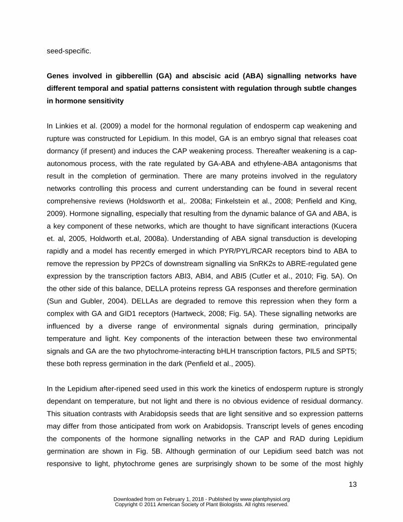
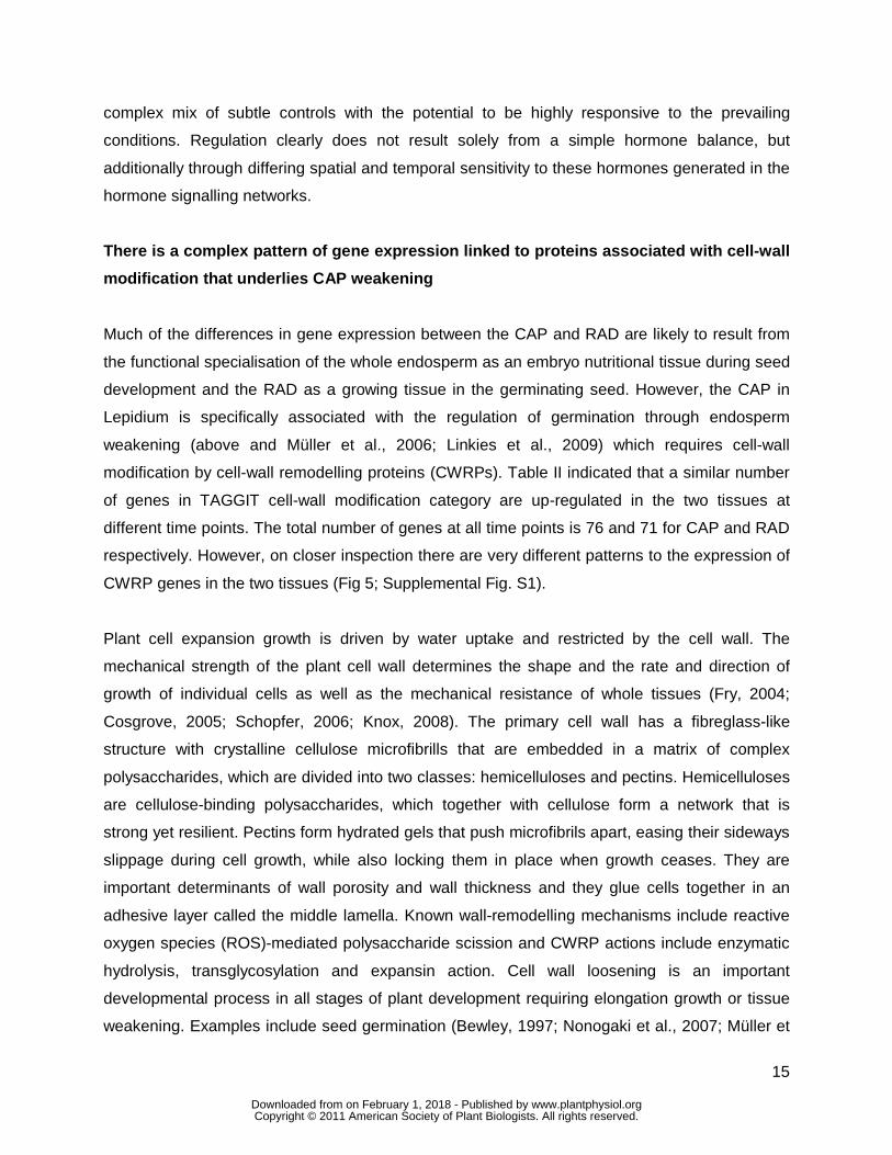

16
al 2009) seedling elongation growth (Schopfer 2006 Schopfer and Liszkay 2006) and fruit
ripening (Fry et al 2001 Saladie et al 2007)
Ikuma and Thimann (1963) in their hatching hypothesis of seed biology suggested that the
final step in the germination control process is the production of an enzyme whose action
enables the tip of the radicle to penetrate through the coat In searching for this hatching
enzyme evidence has been uncovered for the contribution of various CWRPs including endo-
szlig-14-mannanases (eg Bewley 1997 Nonogaki et al 2000 Iglesias-Fernandez et al 2011)
and endo-szlig-13-glucanases (eg Leubner-Metzger 2002 Leubner-Metzger 2003 Petruzzelli et
al 2003) as well as for ROS (Muumlller et al 2009) but most of this work was in Solanaceous
seeds However endosperm weakening is also evident in Brassicaceae seeds where it is
promoted by GA and ethylene and inhibited by ABA (Debeaujon and Koornneef 2000
Debeaujon et al 2000 Muumlller et al 2006 Bethke et al 2007 Linkies et al 2009 Iglesias-
Fernandez et al 2011) Based on the timing of GA-inducible transcripts in whole seeds of
Arabidopsis many CWRP genes that remodel hemicellulose are expressed during the early
germination phase (Ogawa et al 2003 Nonogaki et al 2007) Our tissue-specific transcriptome
analysis with Lepidium (this work and Linkies et al 2009) shows that many of the bigger CWRP
families exhibit complex temporal and spatial expression patterns that are presented in the
Supplemental FiguresTables We therefore restrict our subsequent discussion to a selection of
early-expressed hemicellulose-related genes that are abundant during CAP weakening
Expansins are plant cell-wall loosening proteins which disrupt noncovalent bonds that tether
matrix polysaccharides to the surface of cellulose microfibrils or to each other (Sampedro and
Cosgrove 2005 Choi et al 2006) Whatever their biochemical mechanism of action expansins
act in catalytic amounts to stimulate wall polymer creep without causing major covalent
alterations of the cell wall The α-expansins (EXPA) act with a pH optimum around 4 They have
possible roles in developmental processes like organ size and elongation growth fruit tissue
softening and seed germination (Sampedro and Cosgrove 2005 Choi et al 2006 Gaete-
Eastman et al 2009 Lizana et al 2010) Transcripts of the tomato α-expansin LeEXPA4 and
its putative ortholog in Datura ferox were specifically expressed in the micropylar endosperm in
association with endosperm weakening (Chen and Bradford 2000 Mella et al 2004) This
transcript expression was promoted by GA but not inhibited by ABA Where there is a significant
difference in the level of expression between the tissues the majority of Lepidium expansin
genes (Fig 5A Supplemental Fig S1A) are expressed more highly in the CAP than the RAD
wwwplantphysiolorgon February 1 2018 - Published by Downloaded from Copyright copy 2011 American Society of Plant Biologists All rights reserved
17
This is particularly true for the α-expansins EXPA1 2 7 8 9 for which in the +ABA-arrays the
transcript levels are higher in the CAP compared to the RAD at all time points (Fig 5A) only
EXPA3 was RAD-specific The ndashABA-arrays show that EXPA1 2 9 are specifically expressed in
the CAP but not in the NME ABA does not down-regulate any of these CAP-specific α-
expansin genes During the early phase of Arabidopsis seed germination transcripts of
AtEXPA1 2 8 9 accumulate 100- to 500-fold (from 0 h to 12 h in whole unstratified seeds) and
this induction is promoted by GA not inhibited by ABA and mainly localized in the endosperm
(Nakabayashi et al 2005 Penfield et al 2006 Carrera et al 2008 Holdsworth et al 2008a
Preston et al 2009) In summary transcript expression analysis during germination of both
Lepidium and Arabidopis shows that α-expansin genes in particular EXPA2 are induced early
in the endosperm cap prior to the onset of weakening and are involved in ABA-insensitive
processes that lead to testa rupture and cap weakening (this work and Linkies et al 2009)
Based on their temporal spatial and hormonal expression pattern α-expansins are likely to
contribute to processes in the CAP prior to and during endosperm weakening and rupture but
they do not confer the ABA-regulation of these processes
Xyloglucan endotransglycosylasehydrolase (XTHs) modify xyloglucans which are part of
the hemicellulose network believed to crosslink cellulose microfibrils (Fry 2004 Cosgrove
2005) Xyloglucan is the primary XTH substrate and most XTHs exhibit XET (transferase)
activity that breaks and remakes glycosidic bonds in the backbone of xyloglucan Some XHTs
also exhibit XEH (hydrolase) activity and mediate xyloglucan strand breaks and a few exhibit
only XEH activity Direct unambiguous proof of XTHs inducing wall stress relaxation and
extension is still lacking However XTHs are implicated in cell-wall hemicellulose remodelling
leading to loosening (Van Sandt et al 2007) or stiffening (Maris et al 2009) XTHs are
proposed to have roles in many developmental processes including cell growth fruit ripening
and reserve mobilisation following germination of xyloglucan storing seeds (Tine et al 2000
Fry 2004 Nonogaki et al 2007 Van Sandt et al 2007) During tomato seed germination
transcript accumulation of LeXET4 was induced in the micropylar endosperm promoted by GA
but not inhibited by ABA (Chen et al 2002) The phylogenetic relationship between the 33
members of the Arabidopsis XTH gene family reveal 3 groups
(httplabsplantbiocornelleduXTHarabidopsishtml Becnel et al 2006) Several XTH genes
of Lepidium showed tissue-specific expression during seed germination (Fig 5B 5C 5D
Supplemental Fig S1B) The group 2 XTHs 15 16 20 22 23 24 and group 3 XTHs 27 28 and
31 exhibit stronger expression in the CAP compared to the RAD In contrast the group 1 XTHs 5
wwwplantphysiolorgon February 1 2018 - Published by Downloaded from Copyright copy 2011 American Society of Plant Biologists All rights reserved
18
and 8 and the group 3 XTH32 exhibit stronger expression in the RAD compared to the CAP
During the early phase of Arabidopsis seed germination of the above mentioned genes only
transcripts of AtXTH5 16 and 27 accumulated significantly at 6 h and AtXTH15 22 28 and 31
at 12 h while AtXTH20 23 and 24 were not induced Only XTH15 16 5 31 were induced by
GA and only XTH24 was down-regulated by ABA (Nakabayashi et al 2005 Preston et al
2009) In summary transcript expression analysis during seed germination of both Lepidium and
Arabidopsis shows that XTH genes are expressed in a complex manner suggesting distinct roles
in the RAD and CAP
Mannans are rigidity and mechanical strength conferring hemicellulosic polysaccharides present
in the endosperm of many seeds (Bewley 1997 Reid et al 2003 Nonogaki et al 2007) The
endosperm cell walls of Solanaceous seeds contain ca 60 mannose and ca 10 galactose
as galactomannans Coffee galactomannan contains only ca 2 galactose which results in hard
and brittle endosperm properties Mannan polysaccharides could be masked and this may have
prevented the detection of mannan epitopes in Arabidopsis seeds but genetic evidence has
strongly indicated a functional role for mannan in seed development and germination of this
species (Marcus et al 2010 Iglesias-Fernandez et al 2011) Galactomannan biosynthesis in
seed endosperms involves β-1-4-mannan synthase and galactomannan galactosyltransferase
(Reid et al 2003 Edwards et al 2004) The β-14-mannan synthases are encoded by the
cellulose synthase-like A (CslA) gene family (Dhugga et al 2004 Liepman et al 2005)
AtCslA2 transcripts accumulated in germinating Arabidopsis seeds in a GA-promoted and ABA-
unaffected manner (Nakabayashi et al 2005 Preston et al 2009) In Lepidium CSL2 and CSL9
showed a radicle-specific expression during seed germination (Fig 5E Supplemental Fig S1C)
Degradation of mannan and galactomannan polymers involves endo-β-14-mannanase α-
galactosidase and β-mannosidase all of which have been identified in germinating seeds
several endo-β-14-mannanases have hydrolase and endotransglycosylase activity (Schroder et
al 2009) Among the many endo-β-14-mannanase isoforms of tomato the LeMAN2 gene is
expressed specifically in the micropylar endosperm prior to radicle emergence in association
with enzyme activity accumulation (Nonogaki et al 2000 Toorop et al 2000 Gong and
Bewley 2007) This induction is promoted by GA but not inhibited by ABA Endo-β-14-
mannanase also accumulates in the micropylar endosperm of Solanum lycocarpum Datura
ferox and coffee and is thought to contribute to endosperm weakening (Bewley 1997 Nonogaki
et al 2000 Toorop et al 2000 da Silva et al 2004 Arana et al 2006 Pinto et al 2007)
wwwplantphysiolorgon February 1 2018 - Published by Downloaded from Copyright copy 2011 American Society of Plant Biologists All rights reserved
19
Endo-β-14-mannanase enzyme activities of individual tomato micropylar endosperm caps
varies at least 100-fold (Still and Bradford 1997) Although the presence of endo-β-14-
mannanase enzyme activity in the tomato endosperm cap is consistently associated with radicle
emergence it is not the sole or limiting factor under all conditions Seed germination of tomato
lines over-expressing an endo-β-14-mannanase was not promoted (Belotserkovsky et al
2007) Seed-specific regulation of several endo-β-14-mannanases are also known from rice
(Yuan et al 2007 Ren et al 2008) Seven endo-β-14-mannanase genes are known in
Arabidopsis (Yuan et al 2007) but of these only AtMan7 (At5g66460) transcripts accumulated
in whole unstratified seeds and this induction is promoted by GA but not inhibited by ABA
(Nakabayashi et al 2005 Preston et al 2009) In agreement with this transcripts of the
Lepidium MAN7 accumulated in the CAP and to a lesser extent in the RAD during seed
germination (Fig 5E Supplemental Fig S6C) This induction was not inhibited by ABA and at 8
h was stronger in CAP than the RAD and the NME (Fig 5E) Figure 6A and 6B show that endo-
β-14-mannanase enzyme activity accumulated in the CAP and the RAD but less in the NME
prior to endosperm rupture This increasing activity is at least in part due to LesaMAN7 as the
transcript expression pattern is regulated in a similar manner (Fig 6C) Late during germination
and after endosperm rupture endo-β-14-mannanase enzyme activity and LesaMAN7
transcripts also accumulate in the cotyledons (Fig 6) In agreement with a role for LesaMAN7 in
germination a recent study shows that Arabidopsis knockout mutants for AtMAN7 6 and 5 had
slower germination than the wild-type (Iglesias-Fernandez et al 2011) In seeds of Sisymbrium
officinale which is also a Brassicaceae endo-β-14-mannanase enzyme activity accumulated in
an ethylene- and GA-promoted manner (Iglesias-Fernandez and Matilla 2009)
α-Galactosidases and β-mannosidases contribute to seed galactomannan degradation (Bewley
1997 Feurtado et al 2001 Nonogaki et al 2007) and β-mannosidase enzyme activity has
been detected in the micropylar endosperm of tomato seeds Datura and coffee (de Miguel et
al 2000 Mo and Bewley 2002 da Silva et al 2005) In Arabidopsis seeds At3g18080 β-
mannosidase transcripts accumulate mainly in embryo and At3g57520 α-galactosidase
transcripts were abundant between 3 h and 24 h in whole seeds (Nakabayashi et al 2005
Penfield et al 2006 Preston et al 2009) Transcripts of Lepidium α-galactosidases and β-
mannosidases had complex tissue-specific patterns (Fig 5F) Taken together these results
support a role for endo-β-14-mannanase during the germination of endospermic Brassicaceae
seeds
wwwplantphysiolorgon February 1 2018 - Published by Downloaded from Copyright copy 2011 American Society of Plant Biologists All rights reserved
20
Cellulase (endo-β-14-glucanase) activity was detected in tomato Datura and coffee seeds
(Sanchez et al 1986 da Silva et al 2004 Nonogaki et al 2007) In Datura and coffee but not
in tomato this was in association with endosperm weakening and germination Several putative
orthologs of Arabidopsis showed a CAP-specific expression pattern during seed germination of
Lepidium while orthologs of At1g70710 and At1g64390 showed a RAD-specific expression
(Supplemental Fig 6D) During the early phase of Arabidopsis seed germination transcripts of
At1g70710 and At1g64390 accumulate ca 100-fold (from 0 h to 24 h of whole unstratified
seeds) and this induction is promoted by GA but not appreciably inhibited by ABA (Nakabayashi
et al 2005 Preston et al 2009) Tomato endosperm cell walls contain up to 10 arabinose but
little xylose Transcripts of a β-D-xylosidase accumulated in the embryo of germinating tomato
seeds (Itai et al 2003 Nonogaki et al 2007) In Lepidium β-D-xylosidases (At1g02640
At5g64570 At1g78060 At5g10560) and α-D-xylosidases showed RAD-specific expression
during seed germination while the β-D-xylosidase At5g49360 was higher in the CAP
(Supplemental Fig 1D) In Arabidopsis transcripts of β-D-xylosidases accumulated gt100-fold
(from 0 h to 24 h of whole unstratified seeds) while the β-D-xylosidase At5g49360 was gt20-fold
induced in the endosperm and these inductions were promoted by GA but not inhibited by ABA
(Nakabayashi et al 2005 Preston et al 2009) The transcript expression pattern of pectin-
related enzymes in Lepidium has already been discussed in Linkies et al (2009)
Genes relating to protein degradation and post-translational modification are important in
the regulation of cell wall modification
From a review of recent post-genomic data Holdsworth et al (2008b) concluded that RNA
translation and posttranslational modification are major levels of control for germination
completion However there are similar numbers of genes up-regulated in both tissues in the
TAGGIT category protein degradation (Table III) but as discussed above this similarity obscures
important differences in the detail There are 620 genes tagged in this category of which 76 are
significantly differentially expressed between the two tissues with 34 and 42 expressed more
highly in the CAP and RAD respectively (Supplemental Figs S2A and B) Closer inspection of
this cohort of genes reveals a prominent role for the aspartic acid and subtilase families of plant
proteases We therefore looked at genes from all members of these two families of plant
proteases and included the cysteine protease family of enzymes Members of these classes of
proteases are reported in Beers et al (2004) Fig 7 summarises those members that are
wwwplantphysiolorgon February 1 2018 - Published by Downloaded from Copyright copy 2011 American Society of Plant Biologists All rights reserved
21
significantly (Plt010) differentially expressed between the two tissues The SBT protease
members are predominantly over-represented in the RAD whilst the significant aspartic
proteases are mainly overrepresented in the CAP We have also investigated the expression of
genes encoding key enzymes in protein modification involving the ubiquitin26S proteasome E3
ligases specifically the F box and RING finger proteins (Supplemental Figs 2C and D)
SBT proteases Subtilases are a diverse family of serine proteases which number 54 in
Arabidopsis and have a high degree of gene duplication and associated redundancy
(Rautengarten et al 2005) This functional redundancy has made it difficult to associate
biological function to individual genes with only two knock out mutants stomatal density and
development 1 (sdd1) and Abnormal leaf shape (ale1) having recognisable phenotypes It has
been postulated that these encode proteins (SDD1 and ALE1) that act as proprotein
convertases yielding bioactive peptides (Berger and Altmann 2000 Tanaka et al 2001) In
Lepidium Fig 7 shows predominantly greater expression of the subtilase gene family members
in the RAD than in the CAP one such transcript in Arabidopsis AIR3 (At2g04160) has been
linked to lateral root emergence (Neuteboom et al 1999) In Arabidopsis more than 20 of the
family members have been shown to be transcriptionally regulated by light with the expression
of At2g39850 and At5g59130 demonstrating sole dependence on PHYA under far red light for
induction (Zhou 2009) In Lepidium both these subtilases are expressed significantly higher in
the RAD whereas PhyA is expressed significantly more highly in the CAP (Fig 5B) suggesting
the possibility of signalling between the two tissues regulated by light
Aspartate proteases There are 59 aspartic proteases identified amongst the annotated
Arabidopsis genes and little is known about their biological role (Beers et al 2004) Sub-cellular
localisation may help to elucidate their physiological function and a number have been located in
the intracellular fluid of the apoplast with a role in disease resistance signalling (Xia et al 2004)
There is also evidence for a role in seeds Mutlu et al (1999) characterised an aspartic protease
from dry seeds of Arabidopsis and collocated it with the seed storage protein 2S albumin and the
vacuolar marker enzyme α-mannosidase Molecular studies of osmoprimed seeds of cauliflower
(Fujikura and Karssen 1995) identified two aspartic proteases with enhanced expression upon
priming A proteomic analysis of Lepidium CAP tissue (Mϋller et al 2010) identified aspartic
proteases as a main class of proteins involved in storage protein degradation The abundance
of one aspartic protease was shown to increase from 8h to 18h during the period of CAP
weakening The authors concluded that this early mobilization of protein bodies in the cap is
wwwplantphysiolorgon February 1 2018 - Published by Downloaded from Copyright copy 2011 American Society of Plant Biologists All rights reserved
22
likely to serve a non-nutritional function in the control of germination (Mϋller et al 2010) These
observations are consistent with the CAP specific expression of a number of putative aspartic
protease transcripts within our data set (Fig 7)
Cysteine proteases A number of cysteine proteases have been described in the literature that
are involved in seed germination (Helm et al 2008 Cervantes et al 1994) Ethylene was
shown to induce the expression of a cysteine protease responsible for the catabolism of major
reserve proteins (Cervantes et al 1994) Helm et al (2008) have reported a number of KDEL-
cysteine proteases involved in programmed cell death (PCD) and the dismantling of extension
scaffolds This led the authors to the hypothesis that the KDEL-tailed cysteine proteases they
identified participate in the final cell collapse during PCD by attacking the structural
hydroxyproline-rich glycoproteins of the cell wall In Lepidium transcript numbers of one of
these KDEL-tailed cysteine proteases At3g48340 was significantly up-regulated in the CAP
(Fig 7)
RING finger E3 ligases and F box proteins E3 ligases are the components of the 26S
proteasome that confer substrate specificity to the system The ubiquitin26S proteasome
pathway is important to most aspects of plant biology (Vierstra 2009) including hormonal
signalling (Frugis and Chua 2002) There are 697 F box proteins (Gagne et al 2002) and 469
RING finger proteins (Stone et al 2005) in the Arabidopsis genome of which we have
transcriptional data for 333 and 327 putative orthologs respectively in our Lepidium data set
Transcript abundances from this set that are significantly differentially expressed between the
two tissues is shown in Supplemental Figure 2C and D There was a greater proportion of both
F box and E3 ligase encoding genes up-regulated in the CAP compared to the RAD (23 and 27
fold respectively) This suggests that posttranslational modification in the form of selected
proteolysis performs a more significant role in CAP weakening than in the developing RAD
The role of the 26S proteasome pathway in light and hormonal signalling is well characterized in
plants (Vierstra 2009) and this apparent enrichment of E3 ligase mRNAs may strengthen the
argument for these environmental cues playing a substantial role in endosperm weakening and
signalling to the developing seedling
The effect of protease inhibitors To investigate the role of these proteins in protein
degradation we have monitored the progression of CAP hole formation as described above
when incubated upon specific protease inhibitors The CAPs isolated after 12 h imbibition were
wwwplantphysiolorgon February 1 2018 - Published by Downloaded from Copyright copy 2011 American Society of Plant Biologists All rights reserved
23
then incubated upon 1 microM Pepsatin 4 mM Pefabloc 28 microM E-64 and 60 microM MG132 which
inhibit aspartic serine and cysteine proteases and the 26S proteasome respectively (Fig 8) It
is clear from these data that all 4 classes of proteases investigated have a pronounced affect on
endosperm weakening with the most dramatic affect being the complete cessation of autolysis
by the proteasomal inhibitor MG132 The other three inhibitors reduced the number of CAPs
exhibiting autolysis to approximately 50 of that shown in the control (84) over the same time
period This suggests that each class of protease has a specific protein target and numerous
protein targets may be required for complete lysis
The complete inhibition of hole formation and tissue autolysis in those CAPs treated with MG132
suggests that targeted protein degradation is a major control point for endosperm weakening It
is now well established that the ubiquitination pathway plays a role in various hormonal
signalling pathways and is involved in the regulation of germination through the degradation of
DELLA proteins (Dill et al 2001) it could therefore be hypothesised that inhibition of the
degradation of an important transcription repressor prevents the cascade of transcriptional
activity that we show in our array data It was recently demonstrated that ABA inhibits CAP hole
formation (Linkies et al 2009) and we show here that the proteasome inhibitor MG132
completely blocks cap hole formation and autolysis This suggests the involvement of a key
transcriptional repressor implicated in several signalling pathways as well as in ABA signalling
Several ubiquitin ligases have been linked to ABA signalling and have the transcription factors
ABI3 and ABI5 both important regulators of seed germination as targets for proteolysis
(Santner and Estelle 2010 Lopez-Molina et al 2003 Holdsworth et al 2008) ABI5 and ABI5
BINDING PROTEIN (AFP) mRNA and protein levels increase when seeds are treated with ABA
and mutants for ABI5 and ABF exhibit seed phenotypes NINJA and AFP are related proteins
and it has been proposed in recent work that the GrouchoTup1-type family co-repressors
including TOPLESS (TLP) are part of a general repressor machinery implicated in several
signaling pathways (Liu and Karmarkar 2008 Pauwels et al 2010) For jasmonic acid and ABA
signalling NINJA and AFP are proposed to mediate the interaction between transcription
factors ABI5 for ABA and TLP The TPL-type co-repressors have general functions in plant
hormone signalling that are related to transcription factor proteolysis Supplemental Figure S2C
emphasises the influence of the proteasomal degradation pathway on hormonal signalling
There was significant differential expression between CAP and RAD of genes related to auxin
(AFB3 TIR1 and MAX2) jasmonic acid (COI1) and ethylene (EBF1) All these except TIR1
showed elevated levels of expression in the CAP
wwwplantphysiolorgon February 1 2018 - Published by Downloaded from Copyright copy 2011 American Society of Plant Biologists All rights reserved
24
CONCLUDING DISCUSSION
We have shown that following rupture of the testa germination in Lepidium is regulated by the
opposing forces of RAD (radicle plus lower hypocotyl) extension and the resistance to this by the
surrounding CAP (micropylar endosperm) which progressively declines through autolysis By 18
h some seeds have completed autolysis (germination has occurred) but even at this late stage
progress in some seeds can be stopped by inhibiting transcription and the remainder can be
stopped by blocking translation and posttranslational changes Taken together these results
suggest that this is a control point for germination completion that is very late on in the
germination process This late control point therefore acts as a gateway to seedling
development but the rate of germination must be determined earlier in the process as seeds
reach this control point at different times and thus weakening does not determine vigour Late
control is a necessary feature that prevents inappropriate germination when environmental
conditions change Expression of genes involved in hormone signalling networks was shown to
have different temporal and spatial patterns consistent with establishing a complex responsive
regulation through subtle changes in hormone sensitivity rather than through a crude hormone
balance Genes encoding cell-wall remodelling proteins were also expressed in a complex
tissue specific manner during endosperm weakening that would allow subtle regulation of
weakening and therefore germination completion in response to hormone signals driven by the
current ambient environment
MATERIALS AND METHODS
Plant material germination and puncture-force measurements
After-ripened Lepidium sativum L FR1 (Gartenkresse einfache) and FR14 (Keimsprossen)
seeds (Juliwa Heidelberg Germany) were incubated in petri dishes on two layers of filter paper
with 6 ml 110 Murashige-Skoog salts as medium in continuous white light (ca 100 micromolbulls-1bullm-2)
as described by Muumlller et al (2006) at the temperatures indicated Testa rupture and endosperm
rupture were scored using a binocular microscope Puncture-force measurements were performed
as described by Muumlller et al (2006)
Inhibitor studies on endosperm hole formation and autolysis
Afterripened seeds of Lepidium sativum (Lepidium) `FR1acute were incubated in Petri dishes on two
layers of filter paper with 6 ml 110 Murashige and Skoog (MS)-salts as medium in continuous
wwwplantphysiolorgon February 1 2018 - Published by Downloaded from Copyright copy 2011 American Society of Plant Biologists All rights reserved
25
white light (approx 100 micromols-1m-2) at 18 degC After 10 12 and 18 h the micropylar endosperm
was dissected from the seeds for further incubation on 500 microM cycloheximide (Sigma) or 1 microgml
α-amanitin (Sigma) Cycloheximide was dissolved in 50 acetone Following dilution 01
acetone remained and so this same amount was added to all treatments and the control
Preliminary work determined that this concentration had no influence on germination hole
formation or radicle growth In a second experiment dissection at 12 h was followed by
incubation on 1 microM pepstatin (Roche) 4 mM Pefabloc (Roche) 28 microM E64 (Roche) and 60 microM
MG132 (Merck) The concentrations used were those recommended by the manufacturer In
preliminary work ten-fold lower concentrations were also used to test for a lower dose response
The inhibitors were dissolved in methanol water waterethanol and DMSO respectively
Controls for each inhibitor differed and contained the appropriate chemical at lt005 For every
inhibitor and control at least 3 replicates of 20 micropylar endosperm caps each were incubated
in small Petri dishes on two layers of filter paper with 25 ml 110 MS-salts as medium with the
indicated inhibitor in continuous white light (approx 100 micromols-1m-2) at 18 degC Experiments
were repeated to confirm results Analysis of endosperm autolysis was determined at the times
indicated by 2 categories beginning of autolysis (initial autolysis) was recorded as soon as 1
hole was visible in nearly all cases that happened just below the tip progression of autolysis
was recorded when more than 1 hole was visible which later led to autolysis resulting in
digestion of whole parts of the endosperm
Endo- β -14-mannanase enzyme activity assay
Seed tissues (RAD CAP NME and cotyledons) were ground in 01 M Hepes-05 M NaCl buffer
(pH 75) using an ice-cold mortar The volume of the Hepes buffer was added at the ratio of
fresh weight of tissues (mg)buffer volume (mL) = 13 The extract was centrifuged at 4 ordmC for 10
min at 10000 rpm and the supernatant was used to assay the activity of endo-β-mannanase as
described by Bourgault and Bewley (2002)
Semiquantitative RT-PCR
One microgram of RNA was reverse transcribed using oligo(dT) primer according to the
PrimerScriptTM Reverse Transcriptase Kit instructions (TaKaRa) Aliquots of these first-strand
wwwplantphysiolorgon February 1 2018 - Published by Downloaded from Copyright copy 2011 American Society of Plant Biologists All rights reserved
26
cDNAs as templates were used in subsequent PCR reactions For the semi-quantitative PCR
analysis template volumes were determined that result in equal amplification for the actin
reference gene for each sample For actin optimal conditions were 27 amplification cycles with
52 ordmC as annealing temperature forward primer 5rsquo- CTAAAGCCAACAGGGAGA-3rsquo reverse
primer 5rsquo-TTGGTGCGAGTGCGGTGA-3rsquo The template volumes determined for actin were used
for the semi-quantitative PCR analysis of the endo--14-mannanase (52 ordmC annealing
temperature 35 amplification cycles) forward primer 5rsquo-ACCGATTTCATTGCCAATAACCG-3rsquo
reverse primer 5rsquo-TGTCGACTTTGTGGCATCAGAGA-3rsquo
RNA isolation from Lepidium seed tissues
For each sample ca 1000 Lepidium endosperm caps (CAP) ca 1000 non-micropylar
endosperms (NME) or ca 100 radicles (RAD) were collected at the times indicated frozen in
liquid nitrogen and stored at -80 ordmC Total RNA extraction was carried out by the CTAB-method
followed by quantity and quality control analyses as described (Chang et al 1993) Four
biological replica RNA samples were used for downstream applications
Microarray experimental design
We carried out two separate microarray experiments The first compared CAP and RAD at 8 18
30 and 96 h of imbibition on 10 microM ABA and were termed +ABA-arrays The second compared
CAP NME and RAD at 8 and 18 h of imbibition on germination medium without ABA and were
termed ndashABA-arrays Each experiment used 4 biological replicates Hybridisations were carried
out according to the description below and Linkies et al (2009) For the -ABA-array experiment
the two time points for each tissue were directly compared on four microarrays balanced for
colour For each tissue in the +ABA-array experiment all time points were directly compared to
each other on one microarray each and for each time point the two tissues were compared on
one microarray Each treatment was balanced for colour This design can be thought of as four
interlinked loops
Cross-species CATMA microarrays and Lepidium RNA hybridisation
RNA was prepared in the following way for microarray hybridization The Ambion
MessageAmptrade II aRNA Amplification Kit (AM1751 Applied Biosystems Darmstadt Germany)
was used according to the manual with 2 microg of Lepidium FR1 total RNA as template to generate
antisense amplified RNA called aRNA (Van Gelder et al 1990) The quality and quantity of the
wwwplantphysiolorgon February 1 2018 - Published by Downloaded from Copyright copy 2011 American Society of Plant Biologists All rights reserved
27
aRNA was checked by running an aliquot on a 2100 Bioanalyzer (Agilent UK) The microarrays
used carried genome sequence tag (GST) fragments generated using gene-specific primers
identified by the CATMA Consortium (httpwwwcatmaorg Hilson et al 2004 Allemeersch et
al 2005) CATMA version 2 arrays with 24576 GST were used for the ndashABA-array experiment
whilst CATMA version 3 arrays with 30343 GST were used for the +ABA-array experiment The
aRNA was labelled and the CATMA microarrays were hybridized according to the method
described in Lim et al (2007) and Linkies et al (2009) The microarrays were scanned using an
Affymetrix 428 array scanner at 532 nm (Cy3) and 635 nm (Cy5) Scanned data were quantified
using Imagene version 42 software (BioDiscovery httpwwwbiodiscoverycom) Microarray
data were deposited in ArrayExpress (httpwwwebiacukmicroarray) under accession number
E-TABM-745 (-ABA-arrays) and E-TABM-743 (+ABA-arrays)
RNA microarray data handling and analysis
Data from the two experiments (+ABA-arrays and -ABA-arrays) were analysed separately using
a similar approach but differing in line with the different experimental designs used and the
availability of genomic DNA hybridisation data for CATMA v3 arrays used in the ABA experiment
(Linkies et al 2009) In both cases spot intensity data from Imagene were analysed using the
limma package in Bioconductor (Smyth 2005) There was an initial screen to the data which
removed all probes that could not be assigned to an Arabidopsis gene defined by having an
Arabidopsis Genome Initiative (AGI) identifier and associated with this a TAIR 7 gene ontology
(GO httpwwwarabidopsisorg) Background correction was performed using the normexp
method which is analogous to RMA Within array normalisation (Smyth and Speed 2003) was
performed using print tip loess In the -ABA-array experiment between array normalisation was
performed using quantile normalisation on the A values For the +ABA-array experiment
probes which had shown no significant response in the genomic DNA microarrays (Linkies et al
2009) were weighted out of the normalisation and analysis The two filtering steps resulted in
lists (Supplemental Tables S1 and S2) containing 19794 and 22025 genes for the +ABA-arrays
and -ABA-arrays respectively These gene lists were then used in all downstream analyses For
both experiments the data were analysed as a linear model (Smyth 2004) for the -ABA-arrays
experiment the analysis was adjusted for the intraspot correlation The variance estimates were
adjusted using empirical Bayes estimates of the per-spot variability for use in differential
expression analyses Estimates of the transcript numbers (intensities) for individual spots
(genes) on the +ABA- and -ABA-arrays (Tables S1 and S2) were compared across sample time
points and tissues using F-Tests to identify those whose intensity had significantly changed
wwwplantphysiolorgon February 1 2018 - Published by Downloaded from Copyright copy 2011 American Society of Plant Biologists All rights reserved
28
Statistical significance of differences was assessed using the approach of Benjimini and
Hochberg (1995) to control the false discovery rate at the level of 10 The genes identified
were considered to be up- or down-regulated between tissues at the same time or different time
points PCA was performed in R for each experiment to compare the tissues and time points
across the probes
Functional categorisation of genes
Gene lists were created for genes that were significantly expressed (ldquopresentrdquo) on the arrays and
significantly up- or down-regulated between tissues using procedures described at appropriate
points in the text In order to investigate whether these genes were linked to functional
specialisation categories the lists were subjected to the GO-based established seed-specific
TAGGIT workflow (Carrera et al 2007) to identify proportional representations of genes in
functional categories
Supplemental Data
The following materials are available in the online version of this article
Supplemental Table S1 Effect of α-amanitin and cycloheximide on radicle growth
Supplemental Table S2 Normalised mean abundance of transcripts from genes identified as
ldquopresentrdquo and used in the analysis of +ABA-arrays (19794 genes)
Supplemental Table S3 Normalised mean abundance of transcripts from genes indentified as
ldquopresentrdquo and used in the analysis of ndashABA-arrays (22025 genes)
Supplemental Table S4 Proportional representation in functional categories of expressed
(ldquopresentrdquo) genes on +ABA-arrays
Supplemental Table S5 Proportional representation in functional categories of genes with
expression on +ABA-arrays that is unique to each tissuetime combination
Supplemental Table S6 Genes on +ABA-arrays that show differential expression between
tissues
wwwplantphysiolorgon February 1 2018 - Published by Downloaded from Copyright copy 2011 American Society of Plant Biologists All rights reserved
29
Supplemental Figure S1 Heat maps showing the relative abundance of transcripts from genes
encoding cell wall modification proteins
Supplemental Figure S2 Heat maps showing the relative abundance of transcripts from genes
encoding proteins associated with posttranslational modification
ACKNOWLEDGEMENTS
We thank Cassandra Cadman (Warwick University UK) Meike Wenk and Anita Rott (University
Freiburg Germany) and Jianqing Zhang (South China Agricultural University China) for expert
technical help Alex Tabrett and Jim Beynon for enabling access to CATMA arrays at Warwick
University
wwwplantphysiolorgon February 1 2018 - Published by Downloaded from Copyright copy 2011 American Society of Plant Biologists All rights reserved
30
LITERATURE CITED
Allemeersch J Durinck S Vanderhaeghen R Alard P Maes R Seeuws K Bogaert T
Coddens K Deschouwer K Van Hummelen P Vuylsteke M Moreau Y
Kwekkeboom J Wijfjes AHM May S Beynon J Hilson P Kuiper MTR (2005)
Benchmarking the CATMA microarray A novel tool for Arabidopsis transcriptome
analysis Plant Physiol 137 588-601
Arana MV de Miguel LC Sanchez RA (2006) A phytochrome-dependent embryonic factor
modulates gibberellin responses in the embryo and micropylar endosperm of Datura
ferox seeds Planta 223 847-857
Arcila J Mohapatra SC (1992) Effect of protein-synthesis inhibitors on tobacco seed-
germination and seedling emergence J Plant Physiol 139 460-466
Bar-Or C Czosnek H Koltai H (2007) Cross-species microarray hybridizations a developing
tool for studying species diversity Trends Genet 23 200-207
Barrero J Talbot M White R Jacobsen J Gubler F (2009) Anatomical and transcriptomic
studies of the coleorhiza reveal the importance of this tissue in regulating dormancy in
barley Plant Physiol 150 1006
Becnel J Natarajan M Kipp A Braam J (2006) Developmental expression patterns of
Arabidopsis XTH genes reported by transgenes and Genevestigator Plant Mol Biol 61
451-467
Beers EP Jones AM Dickerman AW (2004) The S8 serine C1A cysteine and Al aspartic
protease families in Arabidopsis Phytochem 65 43-58
Belotserkovsky H Berger Y Shahar R Wolf S (2007) Specific role of LeMAN2 in the control
of seed germination exposed by overexpression of the LeMAN3 gene in tomato plants
Planta 227 199-209
Benjamini Y Hochberg Y (1995) Controlling the false discovery rate - a practical and powerful
approach to multiple testing Journal of the Royal Statistical Society Series B-
Methodological 57 289-300
Berger D Altmann T (2000) A subtilisin-like serine protease involved in the regulation of
stomatal density and distribution in Arabidopsis thaliana Genes amp Development 14
1119-1131
Bethke PC Libourel IGL Aoyama N Chung YY Still DW Jones RL (2007) The Arabidopsis
aleurone layer responds to nitric oxide gibberellin and abscisic acid and is sufficient and
necessary for seed dormancy Plant Physiol 143 1173-1188
wwwplantphysiolorgon February 1 2018 - Published by Downloaded from Copyright copy 2011 American Society of Plant Biologists All rights reserved
31
Bewley JD (1997) Breaking down the walls - a role for endo-β-mannanase in release from seed
dormancy Trends Plant Sci 2 464-469
Bourgault R Bewley JD (2002) Gel diffusion assays for endo-β-mannanase and pectin
methylesterase can underestimate enzyme activity due to proteolytic degradation a
remedy Anal Biochem 300 87-93
Broadley MR White PJ Hammond JP Graham NS Bowen HC Emmerson ZF Fray RG
Iannetta PPM McNicol JW May ST (2008) Evidence of neutral transcriptome evolution
in plants New Phytol 180 587-593
Carrera E Holman T Medhurst A Dietrich D Footitt S Theodoulou FL Holdsworth MJ
(2008) Seed after-ripening is a discrete developmental pathway associated with specific
gene networks in Arabidopsis Plant J 53 214-224
Carrera E Holman T Medhurst A Peer W Schmuths H Footitt S Theodoulou FL
Holdsworth MJ (2007) Gene expression profiling reveals defined functions of the ATP-
binding cassette transporter COMATOSE late in phase II of germination Plant Physiol
143 1669-1679
Cervantes E Rodriguez A Nicolas G (1994) Ethylene regulates the expression of a cysteine
proteinase gene during germination of chickpea (Cicer arietinum L) Plant Mol Biol 25
207-215
Chang S Puryear J Cairney J (1993) A simple and efficient method for isolating RNA from
pine trees Plant Mol Biol Rep 11 113-116
Chen F Bradford KJ (2000) Expression of an expansin is associated with endosperm
weakening during tomato seed germination Plant Physiol 124 1265-1274
Chen F Nonogaki H Bradford KJ (2002) A gibberellin-regulated xyloglucan
endotransglycosylase gene is expressed in the endosperm cap during tomato seed
germination J Exptl Bot 53 215-223
Choi D Cho HT Lee Y (2006) Expansins expanding importance in plant growth and
development Physiol Plant 126 511-518
Comai L Dietrich RA Maslyar DJ Baden CS Harada JJ (1989) Coordinate expression of
transcriptionally regulated isocitrate lyase and malate synthase genes in Brassica napus
L Plant Cell 1 293-300
Cosgrove DJ (2005) Growth of the plant cell wall Nature Rev Mol Cell Biol 6 850-861
Cutler SR Rodriguez PL Finkelstein RR Abrams SR (2010) Abscisic acid emergence of a
core signaling network Annual Rev Plant Biol 61 651-679
wwwplantphysiolorgon February 1 2018 - Published by Downloaded from Copyright copy 2011 American Society of Plant Biologists All rights reserved
32
da Silva EAA Toorop PE Nijsse J Bewley JD Hilhorst HWM (2005) Exogenous gibberellins
inhibit coffee (Coffea arabica cv Rubi) seed germination and cause cell death in the
embryo J Exptl Bot 56 1029-1038
da Silva EAA Toorop PE van Aelst AC Hilhorst HWM (2004) Abscisic acid controls embryo
growth potential and endosperm cap weakening during coffee (Coffea arabica cv Rubi)
seed germination Planta 220 251-261
de Miguel L Burgin MJ Casal JJ Sanchez RA (2000) Antagonistic action of low-fluence and
high-irradiance modes of response of phytochrome on germination and beta-mannanase
activity in Datura ferox seeds J Exptl Bot 51 1127-1133
Debeaujon I Koornneef M (2000) Gibberellin requirement for Arabidopsis seed germination is
determined both by testa characteristics and embryonic abscisic acid Plant Physiol 122
415-424
Debeaujon I Leon-Kloosterziel KM Koornneef M (2000) Influence of the testa on seed
dormancy germination and longevity in Arabidopsis Plant Physiol 122 403-413
Dhugga KS Barreiro R Whitten B Stecca K Hazebroek J Randhawa GS Dolan M
Kinney AJ Tomes D Nichols S Anderson P (2004) Guar seed beta-mannan
synthase is a member of the cellulose synthase super gene family Science 303 363-
366
Dill A Jung HS Sun TP (2001) The DELLA motif is essential for gibberellin-induced
degradation of RGA Proc Natl Acad Sci USA 98 14162-14167
Edwards ME Choo TS Dickson CA Scott C Gidley MJ Reid JSG (2004) The seeds of
Lotus japonicus lines transformed with sense antisense and senseantisense
galactomannan galactosyltransferase constructs have structurally altered
galactomannans in their endosperm cell walls Plant Physiol 134 1153-1162
Feurtado JA Banik M Bewley JD (2001) The cloning and characterization of alpha-
galactosidase present during and following germination of tomato (Lycopersicon
esculentum Mill) seed J Exptl Bot 52 1239-1249
Finch-Savage WE Leubner-Metzger G (2006) Seed dormancy and the control of germination
New Phytol 171 501-523
Finkelstein R Reeves W Ariizumi T Steber C (2008) Molecular aspects of seed dormancy
Annual Rev Plant Biol 59 387-415
Franzke A German D Al-Shehbaz IA Mummenhoff K (2009) Arabidopsis family ties
molecular phylogeny and age estimates in Brassicaceae Taxon 58 425-437
wwwplantphysiolorgon February 1 2018 - Published by Downloaded from Copyright copy 2011 American Society of Plant Biologists All rights reserved
33
Frugis G Chua NH (2002) Ubiquitin-mediated proteolysis in plant hormone signal transduction
Trends Cell Biol 12 308-311
Fry SC (2004) Primary cell wall metabolism tracking the careers of wall polymers in living plant
cells New Phytol 161 641-675
Fry SC Dumville JC Miller JG (2001) Fingerprinting of polysaccharides attacked by hydroxyl
radicals in vitro and in the cell walls of ripening pear fruit Biochem J 357 729-737
Fujikura Y Karssen CM (1995) Molecular studies on osmoprimed seeds of cauliflower - a
partial amino-acid-sequence of a vigor-related protein and osmopriming-enhanced
expression of putative aspartic protease Seed Sci Res 5 177-181
Gaete-Eastman C Figueroa CR Balbontin C Moya M Atkinson RG Herrera R Moya-Leon
MA (2009) Expression of an ethylene-related expansin gene during softening of
mountain papaya fruit (Vasconcellea pubescens) Postharvest Biol Technol 53 58-65
Gagne JM Downes BP Shiu SH Durski AM Vierstra RD (2002) The F-box subunit of the
SCF E3 complex is encoded by a diverse superfamily of genes in Arabidopsis Proc Natl
Acad Sci USA 99 11519-11524
Gong XM Bewley JD (2007) Sorting out the LeMANs endo-beta-mannanase genes and their
encoded proteins in tomato Seed Sci Res 17 143-154
Griffiths J Murase K Rieu I Zentella R Zhang ZL Powers SJ Gong F Phillips AL
Hedden P Sun TP Thomas SG (2006) Genetic characterization and functional analysis
of the GID1 gibberellin receptors in Arabidopsis Plant Cell 18 3399-3414
Hartweck LM (2008) Gibberellin signaling Planta 229 1-13
Helm M Schmid M Hierl G Terneus K Tan L Lottspeich F Kieliszewski MJ Gietl C
(2008) KDEL-tailed cysteine endopeptidases involved in programmed cell death
intercalation of new cells and dismantling of extensin scaffolds Amer J Bot 95 1049-
1062
Hilson P Allemeersch J Altmann T Aubourg S Avon A Beynon J Bhalerao RP Bitton F
Caboche M Cannoot B Chardakov V Cognet-Holliger C Colot V Crowe M
Darimont C Durinck S Eickhoff H de Longevialle AF Farmer EE Grant M Kuiper
MTR Lehrach H Leon C Leyva A Lundeberg J Lurin C Moreau Y Nietfeld W
Paz-Ares J Reymond P Rouze P Sandberg G Segura MD Serizet C Tabrett A
Taconnat L Thareau V Van Hummelen P Vercruysse S Vuylsteke M Weingartner
M Weisbeek PJ Wirta V Wittink FRA Zabeau M Small I (2004) Versatile gene-
specific sequence tags for Arabidopsis functional genomics trancript profiling and
reverse genetics applications Genome Res 14 2176-2189
wwwplantphysiolorgon February 1 2018 - Published by Downloaded from Copyright copy 2011 American Society of Plant Biologists All rights reserved
34
Holdsworth MJ Bentsink L Soppe WJJ (2008) Molecular networks regulating Arabidopsis
seed maturation after-ripening dormancy and germination New Phytol 179 33-54
Holdsworth MJ Finch-Savage WE Grappin P Job D (2008) Post-genomics dissection of
seed dormancy and germination Trends Plant Sci 13 7-13
Hughes DW Galau GA (1989) Temporally modular gene-expression during cotyledon
development Genes Devel 3 358-369
Hughes DW Galau GA (1991) Developmental and environmental induction of Lea and LeaA
mRNAs and the postabscission program during embryo culture Plant Cell 3 605-618
Iglesias-Fernandez R Matilla A (2009) After-ripening alters the gene expression pattern of
oxidases involved in the ethylene and gibberellin pathways during early imbibition of
Sisymbrium officinale L seeds J Exptl Bot 60 1645-1661
Iglesias-Fernandez R Rodriguez-Gacio MdC Barrero-Sicilia C Carbonero P Matilla A
(2011) Three endo-beta-mananase genes expressed in the micropylar endosperm and in
the radicle influence germination of Arabidopsis thaliana seeds Planta 233 25-36
Itai A Ishihara K Bewley JD (2003) Characterization of expression and cloning of beta-D-
xylosidase and alpha-L-arabinofuranosidase in developing and ripening tomato
(Lycopersicon esculentum Mill) fruit J Exptl Bot 54 2615-2622
Ikuma H Thimann KV (1963) The role of the seed-coats in germination of photosensitive
lettuce seeds Plant Cell Physiol 4 169-185
Jendrisak J (1980) The use of alpha-amanitin to inhibit in vivo RNA-synthesis and germination
in wheat embryos J Biol Chem 255 8529-8533
Kimura M Nambara E (2010) Stored and neosynthesized mRNA in Arabidopsis seeds effects
of cycloheximide and controlled deterioration treatment on the resumption of transcription
during imbibition Plant Mol Biol 73 119-129
Kim DH Yamaguchi S Lim S Oh E Park J Hanada A Kamiya Y Choi G (2008) SOMNUS
a CCCH-type zinc finger protein in Arabidopsis negatively regulates light-dependent
seed germination downstream of PIL5 Plant Cell 20 1260-1277
Knox JP (2008) Mapping the walls of the kingdom the view from the horsetails New Phytol
179 1-3
Kucera B Cohn MA Leubner-Metzger G (2005) Plant hormone interactions during seed
dormancy release and germination Seed Sci Res 15 281-307
Leubner-Metzger G (2002) Seed after-ripening and over-expression of class I beta-13-
glucanase confer maternal effects on tobacco testa rupture and dormancy release
Planta 215 959-968
wwwplantphysiolorgon February 1 2018 - Published by Downloaded from Copyright copy 2011 American Society of Plant Biologists All rights reserved
35
Leubner-Metzger G (2003) Functions and regulation of beta-13-glucanases during seed
germination dormancy release and after-ripening Seed Sci Res 13 17-34
Liepman AH Wilkerson CG Keegstra K (2005) Expression of cellulose synthase-like (Csl)
genes in insect cells reveals that CslA family members encode mannan synthases Proc
Natl Acad Sci USA 102 2221-2226
Lim PO Kim Y Breeze E Koo JC Woo HR Ryu JS Park DH Beynon J Tabrett A
Buchanan-Wollaston V Nam HG (2007) Overexpression of a chromatin architecture-
controlling AT-hook protein extends leaf longevity and increases the post-harvest storage
life of plants Plant J 52 1140-1153
Linkies A Muumlller K Morris K Turečkovaacute V Wenk M Cadman CSC Corbineau F Strnad
M Lynn JR Finch-Savage WE Leubner-Metzger G (2009) Ethylene interacts with
abscisic acid to regulate endosperm rupture during germination a comparative approach
using Lepidium sativum and Arabidopsis thaliana Plant Cell 21 3803-3822
Liu Z Karmarkar V (2008) GrouchoTup1 family co-repressoprs in plant development Trends
Plant Sci 13 137-144
Lopez-Molina L Mongrand S Kinoshita N Chua N-H (2003) AFP is a novel negative
regulator of ABA signaling that promotes ABI5 protein degradation Genes Devel 17
410-418
Lizana XC Riegel R Gomez LD Herrera J Isla A McQueen-Mason SJ Calderini DF (2010)
Expansins expression is associated with grain size dynamics in wheat (Triticum aestivum
L) J Exptl Bot 61 1147-1157
Marcus SE Blake AW Benians TAS Lee KJD Poyser C Donaldson L Leroux O
Rogowski A Petersen HL Boraston A Gilbert HJ Willats WGT Knox JP (2010)
Restricted access of proteins to mannan polysaccharides in intact plant cell walls Plant J
64 191-203
Maris A Suslov D Fry SC Verbelen JP Vissenberg K (2009) Enzymic characterization of
two recombinant xyloglucan endotransglucosylasehydrolase (XTH) proteins of
Arabidopsis and their effect on root growth and cell wall extension J Exptl Bot 60 3959-
3972
Mella RA Burgin MJ Sanchez RA (2004) Expansin gene expression in Datura ferox L seeds
is regulated by the low-fluence response but not by the high-irradiance response of
phytochromes Seed Sci Res 14 61-71
wwwplantphysiolorgon February 1 2018 - Published by Downloaded from Copyright copy 2011 American Society of Plant Biologists All rights reserved
36
Mo BX Bewley JD (2002) beta-Mannosidase (EC 32125) activity during and following
germination of tomato (Lycopersicon esculentum Mill) seeds Purification cloning and
characterization Planta 215 141-152
Muumlller K Job C Belghazi M Job D Leubner-Metzger G (2010) Proteomics reveal tissue-
specific features of the cress (Lepidium sativum L) endosperm cap proteome and its
hormone-induced changes during seed germination Proteomics 10 406-416
Muumlller K Linkies A Vreeburg RAM Fry SC Krieger-Liszkay A Leubner-Metzger G (2009)
In vivo cell wall loosening by hydroxyl radicals during cress seed germination and
elongation growth Plant Physiol 150 1855-1865
Muumlller K Tintelnot S Leubner-Metzger G (2006) Endosperm-limited Brassicaceae seed
germination abscisic acid inhibits embryo-induced endosperm weakening of Lepidium
sativum (cress) and endosperm rupture of cress and Arabidopsis thaliana Plant Cell
Physiol 47 864-877
Mutlu A Chen X Reddy SM Gal S (1999) The aspartic proteinase is expressed in Arabidopsis
thaliana seeds and localized in the protein bodies Seed Sci Res 9 75-84
Nakabayashi K Okamoto M Koshiba T Kamiya Y Nambara E (2005) Genome-wide
profiling of stored mRNA in Arabidopsis thaliana seed germination epigenetic and
genetic regulation of transcription in seed Plant J 41 697-709
Neuteboom LW Veth-Tello LM Clijdesdale OR Hooykaas PJ van der Zaal BJ (1999) A
novel subtilisin-like protease gene from Arabidopsis thaliana is expressed at sites of
lateral root emergence DNA Res 6 13-19
Nonogaki H Chen F Bradford K (2007) Mechanisms and genes involved in germination sensu
stricto In K Bradford H Nonogaki eds Seed development dormancy and germination
Blackwell Oxford pp 264-304
Nonogaki H Gee OH Bradford KJ (2000) A germination-specific endo-beta-mannanase gene
is expressed in the micropylar endosperm cap of tomato seeds Plant Physiol 123 1235-
1245
Ogawa M Hanada A Yamauchi Y Kuwalhara A Kamiya Y Yamaguchi S (2003) Gibberellin
biosynthesis and response during Arabidopsis seed germination Plant Cell 15 1591-
1604
Okamoto M Tatematsu K Matsui A Morosawa T Ishida J Tanaka M Endo TA Mochizuki
Y Toyoda T Kamiya Y Shinozaki K Nambara E Seki M (2010) Genome-wide
analysis of endogenous abscisic acid-mediated transcription in dry and imbibed seeds of
Arabidopsis using tiling arrays Plant J 62 39-51
wwwplantphysiolorgon February 1 2018 - Published by Downloaded from Copyright copy 2011 American Society of Plant Biologists All rights reserved
37
Pauwels L Barbero GF Geerinck J Tilleman S Grunewald W Perez AC Chico JM
Vanden Bossche R Sewell J Gil E Garcia-Casado G Witters E Inze D Long JA
De Jaeger G Solano R Goossens A (2010) NINJA connects the co-repressor
TOPLESS to jasmonate signalling Nature 464 788-791
Penfield S Josse EM Kannangara R Gilday AD Halliday KJ Graham IA (2005) Cold and
light control seed germination through the bHLH transcription factor SPATULA Curr Biol
15 1998-2006
Penfield S King J (2009) Towards a systems biology approach to understanding seed
dormancy and germination Proceedings of the Royal Society B-Biological Sciences 276
3561-3569
Penfield S Li Y Gilday AD Graham S Graham IA (2006) Arabidopsis ABA INSENSITIVE4
regulates lipid mobilization in the embryo and reveals repression of seed germination by
the endosperm Plant Cell 18 1887-1899
Petruzzelli L Muumlller K Hermann K Leubner-Metzger G (2003) Distinct expression patterns of
beta-13-glucanases and chitinases during the germination of Solanaceous seeds Seed
Sci Res 13 139-153
Pinto LVA Da Silva EAA Davide AC De Jesus VAM Toorop PE Hilhorst HWM (2007)
Mechanism and control of Solanum lycocarpum seed germination Ann Bot 100 1175-
1187
Preston J Tatematsu K Kanno Y Hobo T Kimura M Jikumaru Y Yano R Kamiya Y
Nambara E (2009) Temporal expression patterns of hormone metabolism genes during
imbibition of Arabidopsis thaliana seeds a comparative study on dormant and non-
dormant accessions Plant Cell Physiol 50 1786-1800
Rajjou L Gallardo K Debeaujon I Vandekerckhove J Job C Job D (2004) The effect of
alpha-amanitin on the Arabidopsis seed proteome highlights the distinct roles of stored
and neosynthesized mRNAs during germination Plant Physiol 134 1598-1613
Rautengarten C Steinhauser D Bussis D Stintzi A Schaller A Kopka J Altmann T (2005)
Inferring hypotheses on functional relationships of genes analysis of the Arabidopsis
thaliana subtilase gene family Plos Comput Biol 1 297-312
Reid JSG Edwards ME Dickson CA Scott C Gidley MJ (2003) Tobacco transgenic lines
that express fenugreek galactomannan galactosyltransferase constitutively have
structurally altered galactomannans in their seed endosperm cell walls Plant Physiol
131 1487-1495
wwwplantphysiolorgon February 1 2018 - Published by Downloaded from Copyright copy 2011 American Society of Plant Biologists All rights reserved
38
Ren YF Bewley JD Wang XF (2008) Protein and gene expression patterns of endo-beta-
mannanase following germination of rice Seed Sci Res 18 139-149
Saladie M Matas AJ Isaacson T Jenks MA Goodwin SM Niklas KJ Ren XL Labavitch
JM Shackel KA Fernie AR Lytovchenko A ONeill MA Watkins CB Rose JKC
(2007) A reevaluation of the key factors that influence tomato fruit softening and integrity
Plant Physiol 144 1012-1028
Sampedro J Cosgrove DJ (2005) The expansin superfamily Genome Biol 6
Sanchez RA Demiguel L Mercuri O (1986) Phytochrome control of cellulase activity in Datura
ferox L seeds and its relationship with germination J Exptl Bot 37 1574-1580
Santner A Estelle M (2010) The ubiquitin-proteasome system regulates plant hormone
signaling Plant J 61 1029-1040
Schopfer P (2006) Biomechanics of plant growth Amer J Botany 93 1415-1425
Schopfer P Liszkay A (2006) Plasma membrane-generated reactive oxygen intermediates and
their role in cell growth of plants Biofactors 28 73-81
Schroder R Atkinson RG Redgwell RJ (2009) Re-interpreting the role of endo-beta-
mannanases as mannan endotransglycosylasehydrolases in the plant cell wall Ann Bot
104 197-204
Sliwinska E Bassel GW Bewley JD (2009) Germination of Arabidopsis thaliana seeds is not
completed as a result of elongation of the radicle but of the adjacent transition zone and
lower hypocotyl J Exptl Bot 60 3587-3594
Slotte T Holm K McIntyre LM Lagercrantz U Lascoux M (2007) Differential expression of
genes important for adaptation in Capsella bursa-pastoris (Brassicaceae) Plant Physiol
145 160-173
Smyth G (2004) Linear models and empirical Bayes methods for assessing differential
expression in microarray experiments Stat Appl Genet Mol Biol 3 Article 3
Smyth G (2005) Limma linear models for microarray data In R Gentleman V Carey S Dudoit
R Irizarry W Huber eds Bioinformatics and computational biology solutions using R and
Bioconductor Springer New York pp 397-420
Smyth GK Speed T (2003) Normalization of cDNA microarray data Methods 31 265-273
Still DW Bradford KJ (1997) Endo-beta-mannanase activity from individual tomato endosperm
caps and radicle tips in relation to germination rates Plant Physiol 113 21-29
Stone SL Hauksdottir H Troy A Herschleb J Kraft E Callis J (2005) Functional analysis of
the RING-type ubiquitin ligase family of Arabidopsis Plant Physiol 137 13-30
wwwplantphysiolorgon February 1 2018 - Published by Downloaded from Copyright copy 2011 American Society of Plant Biologists All rights reserved
39
Sun TP Gubler F (2004) Molecular mechanism of gibberellin signaling in plants Ann Rev Plant
Biol 55 197-223
Tanaka H Onouchi H Kondo M Hara-Nishimura I Nishimura M Machida C Machida Y
(2001) A subtilisin-like serine protease is required for epidermal surface formation in
Arabidopsis embryos and juvenile plants Develop 128 4681-4689
Tine MAS Cortelazzo AL Buckeridge MS (2000) Xyloglucan mobilisation in cotyledons of
developing plantlets of Hymenaea courbaril L (Leguminosae-Caesalpinoideae) Plant
Sci 154 117-126
Toorop PE van Aelst AC Hilhorst HWM (2000) The second step of the biphasic endosperm
cap weakening that mediates tomato (Lycopersicon esculentum) seed germination is
under control of ABA J Exptl Bot 51 1371-1379
van de Mortel JE Aarts MGM (2006) Comparative transcriptomics - model species lead the
way New Phytol 170 199-201
Van Sandt VST Suslov D Verbelen JP Vissenberg K (2007) Xyloglucan
endotransglucosylase activity loosens a plant cell wall Ann Bot 100 1467-1473
Van Gelder RN Vonzastrow ME Yool A Dement WC Barchas JD Eberwine JH (1990)
Amplified rna synthesized from limited quantities of heterogeneous cDNA Proc Natl
Acad Sci USA 87 1663-1667
Vierstra RD (2009) The ubiquitin-26S proteasome system at the nexus of plant biology Nature
Rev Mol Cell Biol 10 385-397
Willige BC Ghosh S Nill C Zourelidou M Dohmann EMN Maier A Schwechheimer C
(2007) The DELLA domain of GA INSENSITIVE mediates the interaction with the GA
INSENSITIVE DWARF1A gibberellin receptor of Arabidopsis Plant Cell 19 1209-1220
Xia YJ Suzuki H Borevitz J Blount J Guo ZJ Patel K Dixon RA Lamb C (2004) An
extracellular aspartic protease functions in Arabidopsis disease resistance signaling
Embo J 23 980-988
Yamaguchi S Kamiya Y Sun TP (2001) Distinct cell-specific expression patterns of early and
late gibberellin biosynthetic genes during Arabidopsis seed germination Plant J 28 443-
453
Yamauchi Y Ogawa M Kuwahara A Hanada A Kamiya Y Yamaguchi S (2004) Activation
of Gibberellin biosynthesis and response pathways by low temperature during imbibition
of Arabidopsis thaliana seeds Plant Cell 16 367-378
wwwplantphysiolorgon February 1 2018 - Published by Downloaded from Copyright copy 2011 American Society of Plant Biologists All rights reserved
40
Yuan JS Yang XH Lai JR Lin H Cheng ZM Nonogaki H Chen F (2007) The endo-beta-
mannanase gene families in Arabidopsis rice and poplar Funct Integr Genomics 7 1-
16
Zhou Z (2009) Study of light dependent Arabidopsis phytochrome A signal transduction through
FHY1 and its downstream gene expression regulation PhD Thesis State University of
New York at Binghampton 136 pages
wwwplantphysiolorgon February 1 2018 - Published by Downloaded from Copyright copy 2011 American Society of Plant Biologists All rights reserved
41
Figure legends
Figure 1 Progression of endosperm CAP weakening both with and without the addition of ABA
A Endosperm rupture progresses more quickly without ABA but the mean force required to
puncture the CAP is the same in both treatments before and after weakening B The
distributions of force required to puncture the CAP are the same in both treatments
Figure 2 The effect of transcription (α-amanitin) and translation (cycloheximide) inhibitors on
the progress of autolysis in isolated CAPs A schematic cross-section of a Lepidium sativum
seed B Isolated CAP (micropylar endosperm) C Examples of initial autolysis CAP tip
abscission (upper) and hole formation (lower) D Progressed autolysis E The effect of
inhibitors on the progress of autolysis in a population of CAPs isolated at 18h imbibition Isolated
CAPs were incubated for the times indicated without or with inhibitors (α-AM = α-amanitin CHX
= cycloheximide) F The effect of inhibitors on the progress of autolysis (initial + progressed
lysis) in a population of CAPs isolated after 10 and 18 h of seed imbibition subsequent
incubation as indicated
Figure 3 The results of principal component analysis (PCA) applied to the expression of all the
Lepidium FR1 gene homologs represented in the +ABA microarrays (A) Principle component 1
and 2 accounted for 42 and 20 of the variance respectively (B) Principle component 2 and 3
accounted for 20 and 12 of the variance respectively
Figure 4 Hormone signalling in Lepidium sativum during CAP weakening A Schematic to
illustrate ABA and GA signalling pathways B Heat maps showing the relative abundance of
transcripts from genes involved in ABA GA and environmental signalling lsquo indicate that
transcript numbers are significantly different between the tissues on +ABA-arrays at Plt01
lt005 lt001 lt0001 respectively Genes not present in the data sets are coloured grey
Figure 5 Relative abundance of transcripts in the CAP RAD or NME for genes related to cell
wall modification A logarithmic scale is used to quantitatively indicate if a transcript is more
abundant in the CAP (below the x-axes negative value ple01) RAD or NME (above the x-
axes positive value ple01) A alpha-expansins B XTH group 1 C XTH group 2 C XTH
group 3 D beta-14-mannanase and mannan synthase E alpha-galactosidase and beta-
mannosidase Key indentifies genes and which arrays (+ABA- or ndashABA-arrays left or right part
of graphs respectively) the data is from
wwwplantphysiolorgon February 1 2018 - Published by Downloaded from Copyright copy 2011 American Society of Plant Biologists All rights reserved
42
Figure 6 Endo-β-14-mannanase enzyme activity during CAP weakening A Time course of
endo-β-14-mannanase enzyme activity in separate tissues B Endo-β-14-mannanase enzyme
activity in the RAD CAP NME and cotyledons at three time points C The pattern of LesaMAN7
transcript expression in different tissues during CAP weakening Note in B and C values at 36 h
are measured separately in tissues from seeds with and without CAP rupture GenBank
accession numbers for LesaMAN7 and LesaACT7 are HQ436349 and HQ436350 respectively
Figure 7 Heat maps showing relative abundance of transcripts from genes encoding proteases
that are differentially expressed between seed tissues lsquo indicate that transcript
numbers are significantly different between the tissues on +ABA-arrays at Plt01 lt005 lt001
lt0001 respectively
Figure 8 The effect of inhibitors of specific proteases (Pepstatin Pefabloc E64) and a
proteosome (MG132) inhibitor on the progression of CAP autolysis Initial autolysis = hole
formation or tip abcission (Fig 2C) progressed autolysis = autolysis of the whole CAP (Fig 2D)
wwwplantphysiolorgon February 1 2018 - Published by Downloaded from Copyright copy 2011 American Society of Plant Biologists All rights reserved
43
RAD Common CAP
8h 4087 (513) 9416 917 (154) Common 10327 8045 9153
18h 2004 (229) 9515 2542 (298) Common 10073 8482 10304
30h 1903 (145) 9479 2328 (285) Common 10493 8552 10271
96h 2601 (322) 9809 2228 (321) Table I Numbers of probes on the array that were considered to be significantly ldquoexpressedrdquo Values shown are numbers of ldquoexpressedrdquo probes that are unique to the RAD and CAP at each time point and the number of probes that are expressed in both tissues (Common) The numbers of probes that are uniquely expressed in any timetissue combination are shown in parenthesis
wwwplantphysiolorgon February 1 2018 - Published by Downloaded from Copyright copy 2011 American Society of Plant Biologists All rights reserved
RAD CAP Tags 8 18 30 96 8 18 30 96 Dormancy related 1 7 6 13 15 20 26 25 Germination related 2 3 3 2 0 0 2 3 ABA 10 16 22 26 8 6 11 12 Auxin 14 14 14 27 10 13 16 12 Brassinosteroid 0 0 1 1 2 3 3 2 Cytokinin 6 3 2 6 1 1 2 2 Ethylene 6 7 7 11 2 5 10 8 Gibberellin 6 5 5 5 2 5 3 3 Jasmonic acid 2 6 3 5 0 1 0 1 Seed storage proteinsLate Embryogenesis Abundant 1 4 4 7 6 10 12 10 Inhibition of protein degradation 1 1 1 1 0 1 0 0 Protein degradation 13 19 24 37 18 22 28 35 Heat Shock 2 5 6 6 7 10 14 17 Cell-wall modification 19 18 15 24 12 18 19 22 Cell cycle related 7 1 9 7 6 14 14 22 Cytoskeleton 6 5 8 6 8 8 14 13 Translation associated 4 1 3 7 63 86 111 137 DNA repair 1 2 3 6 2 4 7 5 Respiration 0 0 0 0 0 1 1 3 Electron Transport 1 2 2 3 0 0 1 2 Pentose phosphate pathway 2 2 4 3 0 0 0 0 Glycolysis and gluconeogenesis 6 9 10 8 0 0 1 4 Krebs cycle 2 3 4 4 1 0 1 1 Beta oxidation 2 2 4 4 0 0 0 0 Stress 36 55 53 53 17 24 38 45 Photosynthesischloroplast related 48 60 73 134 34 44 69 121 Unannotated 1 2 1 4 2 2 4 3 Unclassified 346 484 496 828 339 438 593 718 Total genes in lists 545 736 783 1238 555 736 1000 1226 Total genes classified above 199 252 287 410 216 298 407 508 Percentage classified 365 342 367 331 389 405 407 414
Table II The numbers of genes classified in functional categories of the GO-based seed-specific TAGGIT workflow (Carrera et al 2007) that were up-regulated in the tissue shown at each time point for the +ABA-arrays
w
ww
plantphysiolorgon F
ebruary 1 2018 - Published by
Dow
nloaded from
Copyright copy
2011 Am
erican Society of P
lant Biologists A
ll rights reserved
Figure 1 Progression of endosperm CAP weakening at 18 oC both with and without the addition of ABA
A Endosperm rupture progresses more quickly without ABA but the mean force required to puncture
the CAP is the same in both treatments before and after weakening B The distributions of force
required to puncture the CAP are the same in both treatments
wwwplantphysiolorgon February 1 2018 - Published by Downloaded from Copyright copy 2011 American Society of Plant Biologists All rights reserved
Figure 2 The effect of transcription (α-amanitin) and translation (cycloheximide) inhibitors on the
progress of autolysis in isolated CAPs A schematic cross-section of a Lepidium sativum seed B Isolated
CAP (micropylar endosperm) C Examples of initial autolysis CAP tip abscission (upper) and hole
formation (lower) D Progressed autolysis E The effect of inhibitors on the progress of autolysis at 18 oC in a population of CAPs isolated at 18h imbibition Isolated CAPs were incubated for the times
indicated without or with inhibitors (α-AM = α-amanitin CHX = cycloheximide) F The effect of
inhibitors on the progress of autolysis (initial + progressed lysis) at 18 oC in a population of CAPs isolated
after 10 and 18 h of seed imbibition subsequent incubation as indicated
wwwplantphysiolorgon February 1 2018 - Published by Downloaded from Copyright copy 2011 American Society of Plant Biologists All rights reserved
Figure 3 The results of principal component analysis (PCA) applied to the expression of all the
Lepidium FR1 gene homologs represented in the +ABA microarrays (A) Principle component 1
and 2 accounted for 42 and 20 of the variance respectively (B) Principle component 2 and 3
accounted for 20 and 12 of the variance respectively
wwwplantphysiolorgon February 1 2018 - Published by Downloaded from Copyright copy 2011 American Society of Plant Biologists All rights reserved
A
B
Figure 4 Hormone signalling in Lepidium during CAP weakening A Schematic to illustrate ABA
and GA signalling pathways B Heat maps showing the relative expression levels of genes
involved in ABA GA and environmental signalling lsquo indicate that transcript numbers are
significantly different between the tissues on +ABA-arrays at Plt01 lt005 lt001 lt0001
respectively Genes not present in the data sets are coloured grey
ABA
SnRK2
ABI4
ABI5
ABI3
ABA regulated transcription
GA GID1abc
DELLAs
GA regulated transcription
PP2c [ABI1ABI2 etc]
PYRPYLRCAR
SPT
PIL5
Genes encoding cell wall modifiers
wwwplantphysiolorgon February 1 2018 - Published by Downloaded from Copyright copy 2011 American Society of Plant Biologists All rights reserved
Figure 5 Relative abundance of transcripts in the CAP RAD or NME for genes related to cell
wall modification A logarithmic scale is used to quantitatively indicate if a transcript is more
abundant in the CAP (below the x-axes negative value ple01) RAD or NME (above the x-
axes positive value ple01) A alpha-expansins B XTH group 1 C XTH group 2 C XTH
group 3 D beta-14-mannanase and mannan synthase E alpha-galactosidase and beta-
mannosidase Key indentifies genes and which arrays (+ABA- or ndashABA-arrays left or right part
of graphs respectively) the data is from
wwwplantphysiolorgon February 1 2018 - Published by Downloaded from Copyright copy 2011 American Society of Plant Biologists All rights reserved
Figure 6 Endo- -14-mannanase enzyme activity during CAP weakening A Time course of endo- -14-
mannanase enzyme activity in separate tissues B Endo- -14-mannanase enzyme activity in the RAD
CAP NME and cotyledons at three time points C The pattern of LesaMAN7 transcript expression in
different tissues during CAP weakening Note in B and C values at 36 h are measured separately in
tissues from seeds with and without CAP rupture GenBank accession numbers for LesaMAN7 and
LesaACT7 are HQ436349 and HQ436350 respectively
wwwplantphysiolorgon February 1 2018 - Published by Downloaded from Copyright copy 2011 American Society of Plant Biologists All rights reserved
Figure 7 Heat maps showing relative expression level of genes encoding proteases that are
differentially expressed between seed tissues lsquo indicate that transcript numbers are
significantly different between the tissues on +ABA-arrays at Plt01 lt005 lt001 lt0001
respectively
Aspartic Protease
Cysteine Protease
SBT Protease
wwwplantphysiolorgon February 1 2018 - Published by Downloaded from Copyright copy 2011 American Society of Plant Biologists All rights reserved
Figure 8 The effect of inhibitors of specific proteases (Pepstatin Pefabloc E64) and a proteosome
(MG132) inhibitor on the progression of CAP autolysis Initial autolysis = hole formation or tip abscission
(Fig 2C) progressed autolysis = autolysis of the whole CAP (Fig 2D)
wwwplantphysiolorgon February 1 2018 - Published by Downloaded from Copyright copy 2011 American Society of Plant Biologists All rights reserved
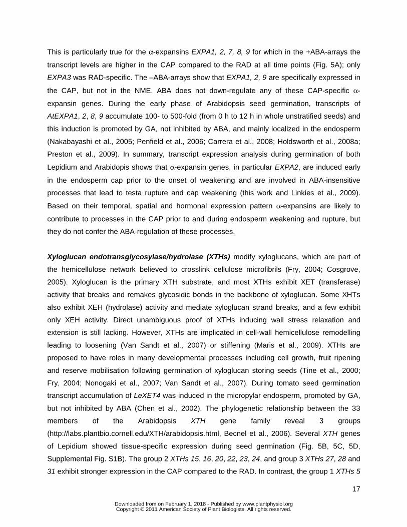