regulation of nkcc2 trafficking by vesicle fusion proteins
TRANSCRIPT
Wayne State University
Wayne State University Dissertations
1-1-2014
Regulation Of Nkcc2 Trafficking By Vesicle FusionProteins Vamp2 And Vamp3 In The ThickAscending LimbPaulo Sebastian Caceres PuzzellaWayne State University,
Follow this and additional works at: http://digitalcommons.wayne.edu/oa_dissertations
Part of the Cell Biology Commons, Molecular Biology Commons, and the Physiology Commons
This Open Access Dissertation is brought to you for free and open access by DigitalCommons@WayneState. It has been accepted for inclusion inWayne State University Dissertations by an authorized administrator of DigitalCommons@WayneState.
Recommended CitationCaceres Puzzella, Paulo Sebastian, "Regulation Of Nkcc2 Trafficking By Vesicle Fusion Proteins Vamp2 And Vamp3 In The ThickAscending Limb" (2014). Wayne State University Dissertations. Paper 874.
REGULATION OF NKCC2 TRAFFICKING BY VESICLE FUSION PROTEINS VAMP2 AND VAMP3 IN THE THICK ASCENDING LIMB
by
PAULO SEBASTIAN CACERES PUZZELLA
DISSERTATION
Submitted to the Graduate School
of Wayne State University,
Detroit, Michigan
in partial fulfillment of the requirements
for the degree of
DOCTOR OF PHILOSOPHY
2014
MAJOR: PHYSIOLOGY
Approved by:
______________________________________ Advisor Date
______________________________________
______________________________________
______________________________________
______________________________________
COPYRIGHT BY
PAULO S. CACERES PUZZELLA
2014
All Rights Reserved
ii
DEDICATION
To my parents Ana and Carlos for their unconditional love and support.
To my family and friends for their constant encouragement.
To the memory of my grandparents, undying source of inspiration.

iii
ACKNOWLEDGMENTS
I would like to thank my advisor and mentor Dr. Pablo Ortiz for his guidance and
encouragement. His valuable advice and his commitment in my formation always pointed
me in the right direction. I couldn’t have been more fortunate to find a role model with such
enthusiasm and passion for science.
I would like to thank the members of my dissertation committee, Dr. Jeff Garvin, Dr.
Douglas Yingst, Dr. Noreen Rossi and Dr. Stanley Terlecky for their constructive feedback.
I am also very grateful to Dr. Mariela Mendez for her technical and intellectual input
and her friendship, to Dr. Mohammed Haque for his assistance with the blood pressure
measurements, to my professors, classmates and colleagues at Wayne State University
and the Henry Ford Hospital, and to Christine Cupps for all her help and good
predisposition with the administrative matters.
I would like to thank Dr. Mark Knepper from the National Heart, Lung and Blood
Institute, NIH, for generously sharing the anti-pSer126 NKCC2 antibody, Dr. Romano
Regazzi from University of Lausanne, Switzerland, for providing the VAMP2-eGFP and
VAMP3-eGFP constructs and Dr. Jeffrey Pessin from the Albert Einstein College of
Medicine, New York, for the gift of the VAMP3 knockout mice.
I would also like to acknowledge the living beings whose lives have been sacrificed
for the advancement of medical research and the pursuit of scientific knowledge. To them,
humankind owes immense gratitude.
Sources of Funding: This research was supported by the Interdisciplinary Biomedical
Science program from the Graduate School and pre-doctoral fellowships 10PRE3710001
and 12PRE12070224 from the American Heart Association to Paulo S. Caceres. Funding

iv
was also provided by the National Institutes of Health from grants R0-1 HL080409 and
1P01HL090550-01A1, by an American Heart Association Grant-in-Aid and by internal
funds from the Henry Ford Health System to Pablo A. Ortiz.

v
TABLE OF CONTENTS
Dedication ii
Acknowledgments iii
List of Tables viii
List of Figures ix
Preface xi
CHAPTER 1: Background and General Hypothesis 1
The kidneys and control of blood pressure 1
NaCl absorption by the thick ascending limb: role of NKCC2 2
Role of NKCC2 in control of blood pressure 3
Role of cAMP in NKCC2 function and NaCl absorption 4
Regulation of NKCC2 by protein trafficking 5
The SNARE family of membrane fusion proteins 6
Role of SNAREs in trafficking of renal transporters 6
Role of VAMP2 and VAMP3 in trafficking of transmembrane transporters 7
Protein-protein interactions as a possible mechanism for VAMP-mediated exocytosis 8
General hypothesis and project aims 9
CHAPTER 2: Role of VAMP2 in cAMP-stimulated NKCC2 trafficking in the thick ascending limb 11
Introduction 11
Aim 1- Hypothesis: VAMP2 mediates cAMP-stimulated trafficking
of NKCC2 in the thick ascending limb 11
Rationale 11
Results 13
vi
Conclusion 20
Aim 2- Hypothesis: cAMP stimulates NKCC2-VAMP2 interaction and
co-localization in thick ascending limbs 21
Rationale 21
Results 22
Conclusion 30
CHAPTER 3: Role of VAMP3 in constitutive NKCC2 trafficking in the
thick ascending limb and renal function ` 32
Introduction 32
Aim 3- Hypothesis: VAMP3 mediates constitutive trafficking of NKCC2
in the thick ascending limb 32
Rationale 32
Results 33
Conclusion 39
Aim 4- Hypothesis: VAMP3 interacts with NKCC2 in the thick ascending
limb and maintains normal renal function and blood pressure 39
Rationale 39
Results 40
Conclusion 45
CHAPTER 4: Concluding remarks 47
Summary of results 47
Proposed model 47
Discussion 48
Strengths and limitations of the study 71
Perspectives 74
vii
CHAPTER 5: General Methods 76
Animals used in the study 76
Antibodies 76
Reagents 77
Medullary thick ascending limb suspensions 77
Primary culture of medullary thick ascending limbs 78
Western blot 78
In vivo gene silencing 79
Biotinylation of steady-state surface NKCC2 in thick ascending limb suspensions 80
Exocytic delivery of surface proteins 80
Co-immunoprecipitation 81
GST pull down in thick ascending limb lysates 82
Immunolabeling of surface NKCC2 , VAMP2 and VAMP3
in thick ascending limb cells 82
Blood pressure measurements 83
Measurements of urine parameters 83
Statistical analysis 84
APPENDIX A: List of acronyms 85
APPENDIX B: IACUC approval 87
References 89
Abstract 124
Autobiographical Statement 126
viii
LIST OF TABLES
Table 1: Urine parameters in wild-type and VAMP3 knockout mice 42

ix
LIST OF FIGURES
Figure 1: Representation of the general kidney anatomy and organization
of the nephron 1
Figure 2: Model for NaCl absorption by the thick ascending limb 2
Figure 3: NKCC2 trafficking to the apical membrane in the thick ascending limb 5
Figure 4: General hypothesis and project aims 9
Figure 5: Expression of VAMPs in the thick ascending limb 13
Figure 6: VAMP2 silencing in vitro 13
Figure 7: In vivo delivery of adenoviruses to the outer medulla of rat kidneys 14
Figure 8: VAMP2 silencing in vivo 14
Figure 9: Surface biotinylation in thick ascending limb suspensions 15
Figure 10: VAMP2 silencing decreased cAMP-stimulated but not
constitutive steady-state surface NKCC2 in TALs 16
Figure 11: Exocytic delivery assay in TAL suspensions 17
Figure 12: VAMP2 silencing blocked cAMP-stimulated but not
constitutive exocytic delivery of NKCC2 in TALs 18
Figure 13: Silencing VAMP2 did not decrease total NKCC2 expression in TALs 19
Figure 14: Silencing VAMP2 did not decrease cAMP-stimulated
NKCC2 phosphorylation at Ser-126 20
Figure 15: NKCC2 and VAMP2 co-immunoprecipitate in TALs 22
Figure 16: VAMP2 interacts with the carboxy-terminus of NKCC2 23
Figure 17: Generation of a TAL primary culture 24
Figure 18: Imaging of apical surface NKCC2 in TAL primary cultures 25
Figure 19: VAMP2-NKCC2 co-localization at the apical membrane of TALs 26
Figure 20: Comparison of different co-localization stringency criteria 27
x
Figure 21: cAMP stimulates VAMP2-NKCC2 co-localization at
the apical surface of TALs 28
Figure 22: cAMP stimulates VAMP2 exocytic delivery in TALs 29
Figure 23: PKA stimulation enhances VAMP2-NKCC2 interaction in TALs 30
Figure 24: Tetanus toxin decreases steady-state surface NKCC2 in TALs 33
Figure 25: VAMP3 silencing in vitro and in vivo 34
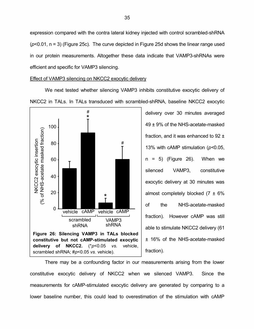
Figure 26: Silencing VAMP3 in TALs blocked constitutive but not cAMP-stimulated exocytic delivery of NKCC2 35
Figure 27: Silencing VAMP3 decreased constitutive but not cAMP-stimulated steady-state surface NKCC2 expression in TALs 36
Figure 28: Silencing VAMP3 decreased total NKCC2 expression in TALs 37
Figure 29: NKCC2 expression is decreased in TALs from VAMP3 knockout mice 38
Figure 30: Decreased steady-state surface NKCC2 expression in VAMP3 knockout mice 38
Figure 31: VAMP3 co-immunoprecipitates with NKCC2 in TALs 40
Figure 32: NKCC2 co-localized with VAMP3 at the apical surface of TALs 41
Figure 33: Enhanced urinary Na, Cl and K excretion in VAMP3 knockout
mice during water restriction 43
Figure 34: Delayed adjustment of urinary Na excretion in VAMP3
knockout mice on a low-sodium diet 44
Figure 35: Decreased systolic blood pressure in VAMP3 knockout mice 45
Figure 36: Proposed model for VAMP2- and VAMP3-mediated NKCC2
trafficking in the thick ascending limb 48
Figure 37: Proposed mechanism for renal phenotype of VAMP3 knockout mice 66
xi
PREFACE
“Come forth into the light of things, let nature be your teacher” – William Wordsworth.
The dissertation that follows is the result of my training as a graduate student at the
laboratory of Dr. Pablo Ortiz at the Hypertension and Vascular Research Division in the
Henry Ford Hospital, Detroit. During this time we tried to understand a fundamental
molecular process that controls sodium absorption by specialized renal cells. The seminal
work that led to this dissertation was a research article published by Dr. Ortiz in 2006 before
I joined his laboratory. In that work he determined that two proteins, VAMP2 and VAMP3,
regulate the presence of the renal co-transporter NKCC2 at the plasma membrane of renal
cells. An important conclusion from that work was that trafficking of NKCC2 to the cell
membrane is a critical mechanism to regulate NaCl absorption by the kidney, a process
with profound consequences in arterial pressure. It became evident that understanding
how NKCC2 reaches the plasma membrane will provide valuable insights into the
mechanisms by which the kidneys maintain fluid and electrolyte balance and blood
pressure. By the time I joined the team, the focus of the research was clearly pointing in
that direction.
In the years preceding the preparation of this dissertation, we published four original
articles all aimed to characterize the mechanisms that control NKCC2 trafficking under
physiological and pathological conditions. That research led to the hypothesis tested in this
dissertation. Here we demonstrate that VAMP2 and VAMP3 mediate two different
pathways in NKCC2 trafficking. Inhibition of these pathways for NKCC2 delivery to the cell
surface alters renal function and decreases blood pressure. The data used in this thesis
are in the process of being published. We have submitted two manuscripts to the Journal of

xii
Biological Chemistry. They are titled: a) “Vesicle-Associated Membrane Protein 2 (VAMP2)
but not VAMP3 Mediates cAMP-Stimulated Trafficking of the Renal Na-K-2Cl Co-
transporter NKCC2 in Thick Ascending Limbs”, and b) “Vesicle-Associated Membrane
Protein 3 (VAMP3) Mediates Constitutive Trafficking of the Renal Na-K-2Cl Co-transporter
NKCC2 in Thick Ascending Limbs: Role in Renal Function and Blood Pressure”.
1
CHAPTER 1
BACKGROUND AND GENERAL HYPOTHESIS
The kidneys and control of blood pressure
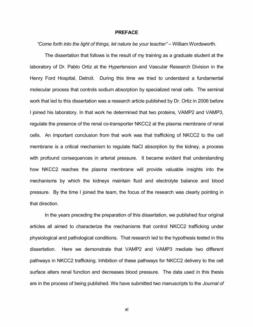
The functional units of the kidneys are tubular structures called nephrons (Figure 1).
In each nephron, blood is filtered in the glomerulus into the tubular space in Bowman’s
capsule. The filtrate is then modified along the nephron to form urine, which is delivered to
the renal pelvis, collected in the bladder and finally excreted. Along the nephron we find
specialized segments with different absorptive and secretory properties. One of these
segments is the loop of Henle, which penetrates deep into the renal medulla. The distal
portion of the ascending loop of Henle is the thick ascending limb (TAL), which is divided in
a medullary and a cortical portion. The main function of the TAL is to absorb ions that
contribute to the creation of a hyperosmotic interstitium. The resulting osmotic gradient in
the medullary interstitium drives water absorption in the collecting duct, concentrating the
urine.
The production of urine by the kidneys finally results in elimination of waste
Figure 1: Representation of the general kidney anatomy and organization of the nephron.
renal artery
renal vein
ureter
pelvis
major calyx
capsule
Bowman’s capsule
glomerulus
afferent arteriole
efferent arteriole
proximal tubule
distal tubule
thin ascending
limb
collecting duct
THICK ASCENDING
LIMB
from another
nephron
to renal pelvis
Human kidney
Nephron
cortex
outer medullapyramid
inner medulla
descending limb
Cortex
Outer medulla
Inner medulla
minor calyx
macula densa
Figure 1: Representation of the general kidney anatomy and organization of the nephron.

2
metabolites and excretion of ions and osmolytes, which regulate blood pH, osmolality and
water content. In addition, the kidneys also secrete vasoactive agents. The combination of
these processes allows tight regulation of fluid volume in extracellular compartments and
short- and long-term control of blood pressure.
NaCl absorption by the thick ascending limb: role of NKCC2
The TAL reabsorbs 25-30% of the NaCl that was filtered from the blood at the
glomerulus. The main function of the TAL is to dilute the forming urine by absorbing Na, Cl
and Ca while water absorption is close to zero. This creates a hyperosmotic interstitium via
a countercurrent multiplier mechanism that provides the osmotic force to absorb water from
the collecting duct. In this way, the TAL influences the ability of the kidney to reabsorb
water and regulate arterial pressure.
The currently accepted model for NaCl absorption by the TAL is depicted in Figure
2. The epithelial cells defining the
TAL walls exhibit apical-basolateral
polarity. In the basolateral
membrane, the Na-K ATPase
provides the driving force by
generating an electrochemical
gradient (1-3). This electrochemical
gradient is used by the apical
Na/K/2Cl co-transporter NKCC2 and
the Na-H exchanger NHE3 (4,5) to
absorb Na, K and Cl from the lumen. Na leaves the cell though the basolateral Na-K
ATPase and Cl exits through Cl channels and K/2Cl co-transporters (3,6,7). Potassium is
K+
Na+
Na+
K+
2Cl-
K+
NKCC2
cAMP
AVP
PTH
β-AR(+)
Figure 2: Model for NaCl absorption by the thick ascending limb
Apical Basolateral
ROMK ClC-K Cl-
Cl-KCC
K+
Na-K
ATPase
NHE3Na+
H+
+
+
+
+
+
+
Tight
junction
Tight
junction
Ca++
Mg++
Na+
+
Figure 2: Model for NaCl absorption by the
thick ascending limb.
3
recycled back to the tubular lumen via the renal outer medullary potassium channel ROMK
(8). The accumulation of positive charges in the luminal membrane due to ROMK creates
a lumen-positive electric potential that drives Na, Ca and Mg absorption through the
paracellular pathway (9,10).
About 80% of the Na and 100% of the Cl reabsorbed by the TAL enter the cell via
NKCC2, which is expressed exclusively in an apical location in the TAL and the macula
densa (11-13). Activation of cAMP signaling in the TAL is a potent stimulus for NKCC2 and
NaCl absorption (14-19). Conversely, NKCC2 inhibitors like the loop diuretic furosemide
(20) and intracellular messengers like nitric oxide (20-22) and cGMP (15,23,24) decrease
NaCl absorption by the TAL. NKCC2-mediated NaCl absorption by the TAL is crucial in
regulation of blood pressure (25-30).
Role of NKCC2 in control of blood pressure
Loss of function mutations in NKCC2 cause Bartter syndrome type I (27,31-36), a
condition characterized by polyuria, inability to concentrate urine, loss of Na, Ca and Mg in
the urine and low blood pressure (37-39), all symptoms associated with impaired TAL
function. Consistent with this, genetic disruption of the NKCC2 gene in mice produces a
Bartter-like phenotype (40). Both Bartter syndrome patients and NKCC2 knockout mice
experience severe dehydration at an early age that can be life-threatening unless fluid
balance is restored. Two independent studies (41,42) have shown that rare NKCC2
mutations in the human population cause reduced NKCC2 activity and decrease blood
pressure.
Opposite to loss-of-function NKCC2 mutations, enhanced NKCC2-mediated NaCl
absorption by the TAL has been linked to salt-sensitive and spontaneous hypertension in
animal models (43-46) and has been associated with salt sensitivity in humans (25,47), in

4
particular the African-American population (48).
NKCC2 is also the target site of the loop diuretics bumetanide, furosemide and
torasemide, which decrease blood pressure (20,49-55). However, they are not routinely
used in the long term treatment of hypertension due to side effects from the profound
volume and electrolyte depletion. This highlights the important contribution of NKCC2 to
maintaining fluid homeostasis.
Role of cAMP in NKCC2 function and NaCl absorption
Hormonal stimulation of cAMP by vasopressin (2,56-61), calcitonin (57,62-64),
parathyroid hormone (PTH) (56,63,65) or β-adrenergic receptors (66-69) enhances
NKCC2-mediated NaCl absorption in the TAL. The stimulatory effect of cAMP on NKCC2-
dependent NaCl absorption is mainly mediated by protein kinase A (PKA) (17,18,70,71).
This kinase directly phosphorylates NKCC2 at Serine 126 in the amino-terminus and Serine
874 in the carboxy-terminus (residues in the NKCC2 rat sequence) (72). Mutagenesis
analysis indicate that elimination of Ser-126 decreases NKCC2 activity in vitro (73).
Two other phosphorylation sites at the amino terminus of NKCC2 become
phosphorylated indirectly after stimulation with vasopressin (74), threonines 96 and 101 (rat
sequence) and are also required for normal NKCC2-mediated ion transport in vitro (75).
However, the signaling pathway that leads to Thr-96 and Thr-101 phosphorylation by
vasopressin is not clear.
Altogether, these pieces of evidence indicate that the cAMP-PKA pathway is an
important stimulus for NKCC2 and TAL activity. In this dissertation we studied the
molecular mechanism that mediates cAMP stimulation of NKCC2 in TALs and how it differs
from the mechanism that controls NKCC2 under basal non-stimulated conditions.
5
Regulation of NKCC2 by protein trafficking
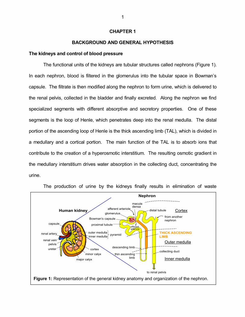
In order to transport ions from the lumen to the intracellular space, NKCC2 has to be
inserted in the apical membrane.
However, most NKCC2 is found in
sub-apical vesicles in the TAL as
observed by electron microscopy
(76). Our laboratory has shown
that under baseline conditions only
3-5% of total NKCC2 is expressed
at a steady-state in the cell surface
(14). Despite this small proportion, a decrease in surface NKCC2 expression lowers NaCl
reabsorption by the TAL to a great extent (14,24). Steady-state surface NKCC2 levels are
the result of exocytic delivery (77) and endocytic retrieval (78,79). We will refer to this
process as protein trafficking (Figure 3). Exocytic delivery can occur from the biosynthetic
pathway and from recycling compartments at the same time. Previous data from our
laboratory indicate that only a small fraction of internalized NKCC2 undergoes recycling
back to the plasma membrane (78).
NKCC2 is present at the cell surface under baseline conditions (14,24,77), indicating
that trafficking occurs constitutively in the absence of stimulation. However, cAMP
stimulates steady-state surface NKCC2. This can be the result of either stimulated exocytic
delivery or decreased endocytic retrieval. We have found that vasopressin stimulates
exocytic delivery of NKCC2 in TALs (74,77,80). Stimulation of β-adrenergic receptors also
augments surface NKCC2 via cAMP (69). Inhibition of PKA blunts cAMP-stimulated
exocytic delivery and cAMP-stimulated steady-state surface NKCC2 expression (77).
cAMP
PKA
Exocytic
delivery
(+)
VAMP
NKCC2
Tetanus
Toxin
Endocytosis
SNARE complex
Apical membrane
Golgi
AVP
PTH
β-AR
Figure 3: NKCC2 trafficking to the apical membrane in the thick
ascending limb.
vesicle
Figure 3: NKCC2 trafficking to the apical
membrane in the thick ascending limb.
6
In addition, we have shown that surface NKCC2 was abnormally high in a rat model
of salt-sensitive hypertension (46). Altogether, these observations illustrate the tight
relationship between NKCC2 presence at the apical surface and NaCl absorption by the
TAL. This also highlights the importance of NKCC2 trafficking as a mechanism to regulate
renal function and blood pressure.
The SNARE family of membrane fusion proteins
In order to reach the apical membrane, NKCC2 has to be delivered from intracellular
vesicles to the plasma membrane. The details of this mechanism are largely unknown. In
other eukaryotic cells, vesicles deliver their cargo by means of membrane fusion. This
process is mediated by the family of proteins known as Soluble NSF Attachment protein
REceptors (SNARE) (81-84). There are three types of SNAREs: Vesicle-Associated
Membrane Proteins (VAMP), syntaxins and Synaptosome-Associated Proteins (SNAP).
VAMPs reside in the vesicle membrane (85,86) and syntaxin and SNAP are associated
with the target membrane (87-89). When the vesicle approaches the target membrane, the
three SNAREs combine with each other and form a helical complex (90,91) that drives
fusion of the vesicle membrane with the target membrane (82,92-96). This is the basis of
the SNARE hypothesis and is accepted to be universal for all eukaryotic cells (83,84).
There are seven VAMP isoforms, at least fifteen syntaxin isoforms and four SNAP
isoforms (97-99). In addition, SNAREs bind to accessory proteins to complete the
membrane fusion process (100-104). The different combinations of SNAREs and
accessory proteins help direct protein trafficking along different cell compartments in a
specific way according to the cell type and under defined physiological conditions.
Role of SNAREs in trafficking of renal transporters
In the kidney, SNAREs are probably best characterized in the collecting duct, where
7
they mediate cAMP-stimulated trafficking of the water channel aquaporin-2, the H-ATPase
and the epithelial Na channel ENaC (105-120). For instance, VAMP8 knockout mice
exhibit impaired translocation of aquaporin-2 in the collecting duct and fail to concentrate
urine (121). In the TAL, expression of SNAP-23 (122), syntaxin 3 and 4 (118,123,124), and
VAMP2 and 3 (14) has been reported. However, their role in NKCC2 trafficking remains
largely unexplored.
Tetanus toxin selectively cleaves VAMP2 and VAMP3. Our laboratory has found
that tetanus toxin prevents the stimulatory effect of cAMP on surface NKCC2 expression
and NaCl absorption (14), indicating that VAMPs mediate cAMP-stimulated NKCC2
trafficking. However, tetanus toxin does not distinguish between VAMP2 and VAMP3. In
addition, the details of the mechanism that mediates NKCC2 trafficking remain unknown. In
this dissertation we addressed the relative contribution of VAMP2 and VAMP3 to the
process of apical NKCC2 trafficking in the TAL.
Role of VAMP2 and VAMP3 in trafficking of transmembrane transporters
In epithelial cells, SNAREs exhibit polarized distribution, mediating distinct
membrane fusion processes at specific cell compartments (123,125-136). Only the VAMP
isoforms 2, 3, 7 and 8 have been reported to participate in exocytic processes in polarized
epithelia (106,113,121,128,137-144). However, VAMP7 (145) and VAMP8 (146)
traditionally mediate homotypic fusion between intracellular vesicles. In renal epithelia
including the TAL and the collecting duct, VAMP2 and VAMP3 localize apically (14,147).
Cleavage of VAMP2 and VAMP3 with tetanus toxin (which does not cleave VAMP7 and
VAMP8) inhibits cAMP-stimulated NKCC2 trafficking in the TAL (14). However, it is not
clear whether VAMP2 and VAMP3 mediate different pathways that control constitutive or
cAMP-stimulated NKCC2 exocytic delivery.
8
In other cells, VAMP3 has been associated with early endosomes and may be
involved in recycling (148-150), whereas VAMP2 has been reported to be excluded from
recycling compartments (151). This suggests distinct compartmentalization of the two
isoforms, at least in some cases. VAMP2 has been shown to mediate cAMP-stimulated
delivery of transmembrane proteins (106,113,116,137,139,152). In the collecting duct,
VAMP2 co-localizes with aquaporin-2 in the same intracellular vesicles (137,152) and is
required for cAMP-stimulated translocation to the apical membrane (106,113). In addition,
in other cells VAMP2 is regulated by phosphorylation by ATM kinase (153) and PKC
(154,155). In this dissertation we proposed that VAMP2 selectively mediates cAMP-
stimulated NKCC2 exocytic delivery whereas VAMP3 mediates constitutive delivery.
Protein-protein interactions as a possible mechanism for VAMP-mediated
exocytosis
There are some examples in the literature of protein-protein interactions between
SNAREs and transmembrane proteins (87,156,157). VAMP2 for instance, interacts with
P/Q-type calcium channels (158) and the potassium channel Kv2.1 (159,160) in neurons.
These interactions may be of physiological relevance since VAMP2- Kv2.1 interaction
facilitates Kv2.1 inactivation (159,160). In the collecting duct, VAMP2 interacts with the
proton pump H-ATPase and tetanus toxin prevents H-ATPase exocytic insertion (116). In
the TAL, VAMP2 co-localizes with NKCC2 (14) and VAMP3 is also located apically.
However whether they interact with NKCC2 is unknown.
It is known that protein-protein interactions regulate NKCC2. The Ste20-related
proline-alanine-rich kinase (SPAK) and oxidative stress response 1 (OSR1) kinase interact
with NKCC2 in vitro (161). These kinases phosphorylate the amino-terminus of NKCC2
(162). The myelin and lymphocytes-associated protein (MAL) also interacts with NKCC2
9
(163) and decreases NKCC2 retrieval from the plasma membrane in cultured cells. Moesin,
a member of the ezrin-radixin-moesin family, interacts with the carboxy-terminus of NKCC2
and is necessary for NKCC2 delivery to the plasma membrane (164). Two additional
NKCC2 interactors include Aldolase B (165) and the secretory carrier membrane protein 2
(SCAMP2) (166). Both of them decrease NKCC2 at the cell surface in heterologous
expression systems. Aldolase B stimulates NKCC2 retrieval from the membrane (165) and
SCAMP2 inhibits exocytic delivery (166). In this dissertation, we tested whether VAMP2
and VAMP3 interact with NKCC2 as a possible mechanism that mediates NKCC2 exocytic
delivery.
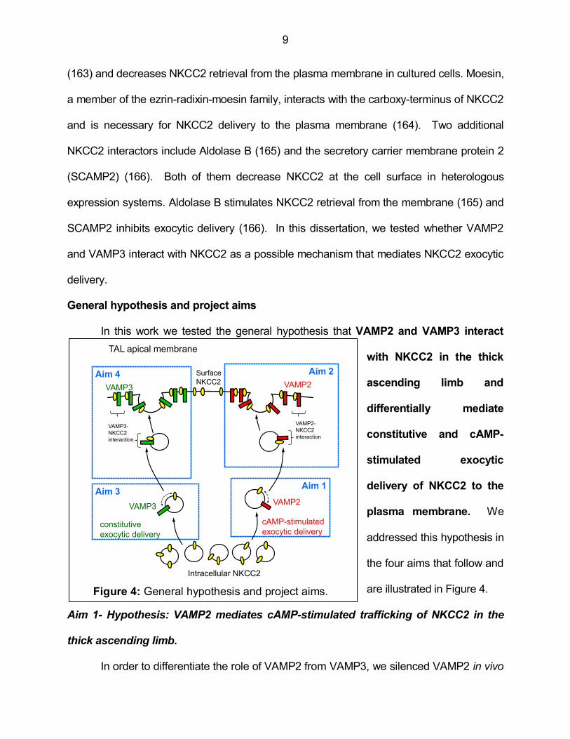
General hypothesis and project aims
In this work we tested the general hypothesis that VAMP2 and VAMP3 interact
with NKCC2 in the thick
ascending limb and
differentially mediate
constitutive and cAMP-
stimulated exocytic
delivery of NKCC2 to the
plasma membrane. We
addressed this hypothesis in
the four aims that follow and
are illustrated in Figure 4.
Aim 1- Hypothesis: VAMP2 mediates cAMP-stimulated trafficking of NKCC2 in the
thick ascending limb.
In order to differentiate the role of VAMP2 from VAMP3, we silenced VAMP2 in vivo
Aim 3
Surface
NKCC2
VAMP2VAMP3
Figure 4: General hypothesis and project aims.
Intracellular NKCC2
TAL apical membrane
VAMP2VAMP3
cAMP-stimulated
exocytic deliveryconstitutive
exocytic delivery
Aim 1
Aim 2Aim 4
VAMP2-
NKCC2
interaction
VAMP3-
NKCC2
interaction
Figure 4: General hypothesis and project aims.
10
with short hairpin RNAs (shRNAs) in rat TALs. Then we tested the effect of silencing
VAMP2 on constitutive and cAMP-stimulated NKCC2 trafficking and NKCC2 expression.
Aim 2- Hypothesis: cAMP stimulates NKCC2-VAMP2 interaction and co-localization
in thick ascending limbs.
We addressed the possibility that VAMP2 may mediate cAMP-stimulated NKCC2
trafficking by protein-protein interactions. We measured VAMP2 co-immunoprecipitation
with NKCC2 in TALs and identified the region of NKCC2 that interacts with VAMP2. We
measured co-localization of NKCC2 and VAMP2 at the apical surface of the TAL. We
tested whether cAMP increases delivery of VAMP2 to the apical membrane of TALs and
whether it enhances NKCC2-VAMP2 interaction.
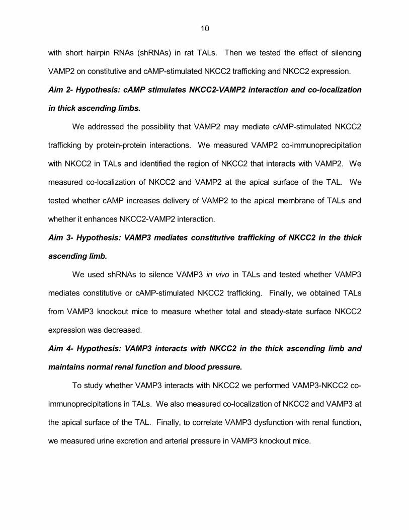
Aim 3- Hypothesis: VAMP3 mediates constitutive trafficking of NKCC2 in the thick
ascending limb.
We used shRNAs to silence VAMP3 in vivo in TALs and tested whether VAMP3
mediates constitutive or cAMP-stimulated NKCC2 trafficking. Finally, we obtained TALs
from VAMP3 knockout mice to measure whether total and steady-state surface NKCC2
expression was decreased.
Aim 4- Hypothesis: VAMP3 interacts with NKCC2 in the thick ascending limb and
maintains normal renal function and blood pressure.
To study whether VAMP3 interacts with NKCC2 we performed VAMP3-NKCC2 co-
immunoprecipitations in TALs. We also measured co-localization of NKCC2 and VAMP3 at
the apical surface of the TAL. Finally, to correlate VAMP3 dysfunction with renal function,
we measured urine excretion and arterial pressure in VAMP3 knockout mice.

11
CHAPTER 2
ROLE OF VAMP2 IN cAMP-STIMULATED NKCC2 TRAFFICKING IN THE THICK
ASCENDING LIMB
Introduction
Biological pathways that stimulate cAMP production, such as hormonal stimulation
with vasopressin or β-adrenergic receptors, enhance NaCl reabsorption by the TAL by
increasing steady-state surface NKCC2 expression (14,74,77,80,167). We have previously
shown that cAMP increases steady-state surface NKCC2 by stimulating exocytic delivery of
the co-transporter to the TAL surface (77). However, the molecular mechanism by which
cAMP stimulates NKCC2 trafficking in TALs remains uncharacterized. In this chapter we
tested whether VAMP2 mediates cAMP-stimulated NKCC2 trafficking, since VAMP2
mediates cAMP-stimulated exocytosis in other cells (106,113,168,169). We also explored
whether the underlying mechanism may involve protein-protein interactions between
NKCC2 and VAMP2. We tested this in two aims, one addressing the effect of silencing
VAMP2 on NKCC2 trafficking and the other one focused on NKCC2-VAMP2 interaction
and how it is affected by cAMP. We will discuss the relevance of NKCC2-VAMP2
interaction to NKCC2 trafficking and activity.
Aim 1- Hypothesis: VAMP2 mediates cAMP-stimulated trafficking of NKCC2 in the
thick ascending limb.
Rationale
Our laboratory has previously shown that VAMP2 and VAMP3 are expressed in the
TAL (14). However, a more complete profile of the VAMP isoforms expressed in the TAL is
still missing. We began by screening other VAMPs previously shown to be expressed in
12
polarized epithelia, i.e. VAMP7 and VAMP8 (106,113,121,128,137-144).
We previously showed that tetanus toxin blocks the stimulation of cAMP on NKCC2-
mediated NaCl absorption (14). Since the toxin does not distinguish between VAMP2 and
VAMP3, in this aim we used silencing shRNAs in vivo to address whether VAMP2
mediates cAMP-stimulated NKCC2 exocytic delivery. In non-renal cells, VAMP2 has
frequently been associated with stimulated exocytosis (151,170-172). Also, VAMP2
mediates cAMP-stimulated exocytosis of synaptic vesicles in neurons (168) and cAMP-
stimulated delivery of the water channel aquaporin-2 in the renal collecting duct (106,113).
In addition, in renal juxtaglomerular cells, VAMP2 mediates cAMP-stimulated secretion of
renin (169). Interestingly, VAMP3 does not mediate cAMP-stimulated renin release.
Although most of this evidence suggests that VAMP2 mediates cAMP-stimulated exocytic
delivery of NKCC2 in the TAL, this has never been directly addressed. In this aim we tested
whether VAMP2 mediates cAMP-stimulated NKCC2 trafficking. In order to increase
intracellular cAMP, in our experiments we used forskolin to stimulate cAMP production by
adenylyl cyclases, and IMBX to inhibit cAMP degradation by phosphodiesterases. We have
used this maneuver in the past to produce a potent stimulation of NKCC2 trafficking in
TALs (14,77).
A reliable biochemical methodology to study NKCC2 trafficking is by means of
surface biotinylation, which will be explained in detail later. We have used this technique in
the past in medullary TAL suspensions in combination with Western blot to specifically
detect NKCC2 at the TAL surface (14,24,77). The specificity of the technique is achieved
by using a specific anti-NKCC2 antibody directed against the amino-terminus of NKCC2
(173). The amino-terminal region is highly divergent from the closely related NKCC1 co-
transporter (174), which is expressed in basolateral membrane of the collecting duct and
13
could be contaminating our TAL suspensions. In this way, if there is minimal contamination
with other nephron segments in our preparations, any confounding effect is minimized by
detecting specifically NKCC2, which is expressed exclusively in the TAL.
Results
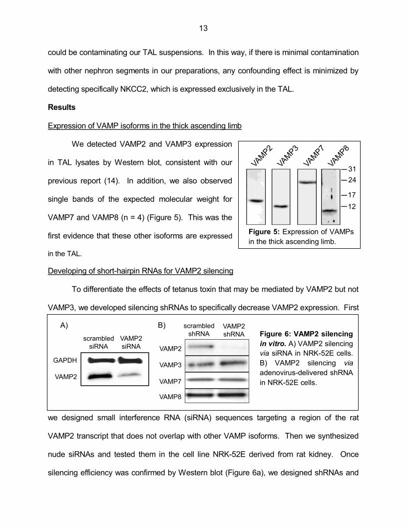
Expression of VAMP isoforms in the thick ascending limb
We detected VAMP2 and VAMP3 expression
in TAL lysates by Western blot, consistent with our
previous report (14). In addition, we also observed
single bands of the expected molecular weight for
VAMP7 and VAMP8 (n = 4) (Figure 5). This was the
first evidence that these other isoforms are expressed
in the TAL.
Developing of short-hairpin RNAs for VAMP2 silencing
To differentiate the effects of tetanus toxin that may be mediated by VAMP2 but not
VAMP3, we developed silencing shRNAs to specifically decrease VAMP2 expression. First
we designed small interference RNA (siRNA) sequences targeting a region of the rat
VAMP2 transcript that does not overlap with other VAMP isoforms. Then we synthesized
nude siRNAs and tested them in the cell line NRK-52E derived from rat kidney. Once
silencing efficiency was confirmed by Western blot (Figure 6a), we designed shRNAs and
Figure 5: Expression of VAMPs
in the thick ascending limb.
B)
VAMP2
VAMP2
shRNA
scrambled
shRNA
VAMP3
VAMP8
VAMP7
A)
GAPDH
VAMP2
VAMP2
siRNA
scrambled
siRNA
Figure 6: VAMP2 silencing
in vitro. A) VAMP2 silencing
via siRNA in NRK-52E cells.
B) VAMP2 silencing via
adenovirus-delivered shRNA
in NRK-52E cells.
Figure 5: Expression of VAMPs
in the thick ascending limb.
31
24
17
12
14
produced adenovirus particles. The efficiency of the adenoviruses was tested again in vitro
by transducing the rat cell line. We observed a high degree of VAMP2 silencing that did not
decrease expression of the other VAMPs (Figure 6b).
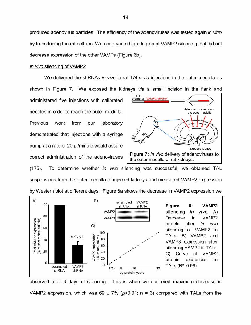
In vivo silencing of VAMP2
We delivered the shRNAs in vivo to rat TALs via injections in the outer medulla as
shown in Figure 7. We exposed the kidneys via a small incision in the flank and
administered five injections with calibrated
needles in order to reach the outer medulla.
Previous work from our laboratory
demonstrated that injections with a syringe
pump at a rate of 20 µl/minute would assure
correct administration of the adenoviruses
(175). To determine whether in vivo silencing was successful, we obtained TAL
suspensions from the outer medulla of injected kidneys and measured VAMP2 expression
by Western blot at different days. Figure 8a shows the decrease in VAMP2 expression we
observed after 3 days of silencing. This is when we observed maximum decrease in
VAMP2 expression, which was 69 ± 7% (p<0.01; n = 3) compared with TALs from the
Figure 7: In vivo delivery of adenoviruses to
the outer medulla of rat kidneys.
B)
VAMP2
VAMP3
scrambled
shRNA
VAMP2
shRNA
A)
p < 0.01
To
tal V
AM
P2
exp
ressio
n
(% o
f scra
mb
led
sh
RN
A)
VAMP2
shRNA
scrambled
shRNA
0
20
40
60
80
100
0
20
40
60
80
100
0 2 4 6 8 10 12 14 16 18 20 22 24 26 28 30 32
C)
VA
MP
2 e
xp
ressio
n
(% o
f m
axim
um
)
µg protein lysate
1 2 4 8 16 32
Figure 8: VAMP2
silencing in vivo. A)
Decrease in VAMP2
protein after in vivo
silencing of VAMP2 in
TALs. B) VAMP2 and
VAMP3 expression after
silencing VAMP2 in TALs.
C) Curve of VAMP2
protein expression in
TALs (R²=0.99).
15
contra lateral kidney injected with control scrambled-shRNA adenoviruses. VAMP2
silencing did not affect VAMP3 expression in vivo (Figure 8b), indicating that the sequence
retained specificity. To assure that we would be able to detect changes in VAMP2
expression, we used 10 µg of protein in the Western blots. This amount of protein falls
within the linear range shown in Figure 8c. Altogether these data indicate that the procedure
for silencing VAMP2 in vivo is efficient and specific.
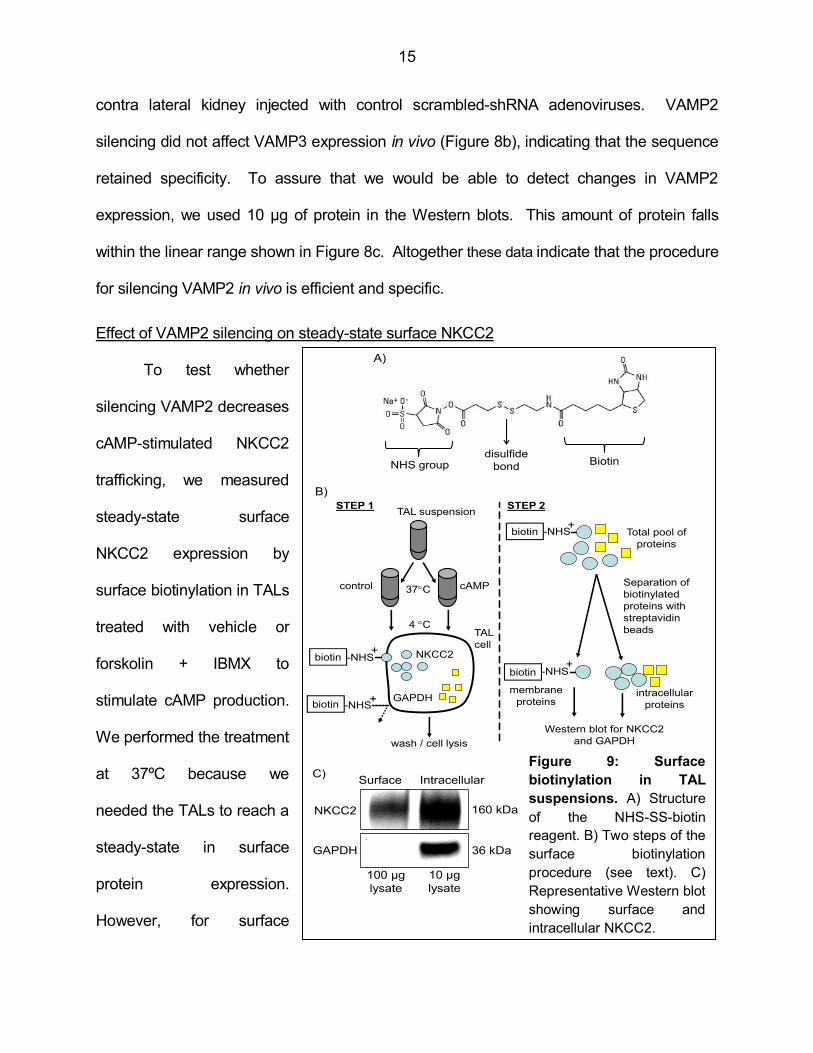
Effect of VAMP2 silencing on steady-state surface NKCC2
To test whether
silencing VAMP2 decreases
cAMP-stimulated NKCC2
trafficking, we measured
steady-state surface
NKCC2 expression by
surface biotinylation in TALs
treated with vehicle or
forskolin + IBMX to
stimulate cAMP production.
We performed the treatment
at 37ºC because we
needed the TALs to reach a
steady-state in surface
protein expression.
However, for surface
Biotindisulfide
bondNHS group
intracellular
proteins
Separation of
biotinylated
proteins with
streptavidin
beads
Western blot for NKCC2
and GAPDH
membrane
proteins
wash / cell lysis
Total pool of
proteins
NKCC2
GAPDH
TAL suspension
control cAMP
STEP 1 STEP 2
37C
4 CTAL
cell
biotin
biotin
biotin
-NHS
-NHS
-NHS
biotin -NHS
A)
B)
+
+
+
+
C)
160 kDa
36 kDaGAPDH
NKCC2
Surface Intracellular
100 µg
lysate
10 µg
lysate
Figure 9: Surface
biotinylation in TAL
suspensions. A) Structure
of the NHS-SS-biotin
reagent. B) Two steps of the
surface biotinylation
procedure (see text). C)
Representative Western blot
showing surface and
intracellular NKCC2.

16
biotinylation we needed to stop protein trafficking rapidly. We achieved this by quickly
cooling the TAL suspensions to 4ºC. Then we proceeded with the surface biotinylation.
We incubated the TALs with a biotinylation reagent consisting on a biotin portion linked via
a disulfide bond to an NHS group (Figure 9a). The NHS group reacts with extracellular
lysines in surface proteins (Figure 9b). Since the biotinylation reagent is charged, it does
not cross the plasma membrane, therefore it does not biotinylate intracellular proteins. In
this way, when lysing the TAL suspensions, we obtained a total pool of proteins consisting
on surface, biotinylated proteins and intracellular, non-biotinylated proteins. We separated
the biotinylated proteins by pull down with streptavidin-conjugated agarose beads and
recovered them by boiling in reducing conditions to break the disulfide bond. NKCC2 was
detected at the surface and in the intracellular fraction by Western blot (Figure 9c). The
intracellular protein GAPDH was used a control since it was not detected at the surface.
To test the effect of VAMP2 silencing, we obtained TALs from VAMP2-shRNA
transduced rat kidneys and measured constitutive and cAMP-stimulated steady-state
surface NKCC2 (Figure 10). We observed that VAMP2 silencing did not affect constitutive
Ste
ady-s
tate
surf
ace N
KC
C2
(% o
f scam
ble
dshR
NA
)
A)
0
50
100
150
200
cAMPvehicle cAMP vehicle
VAMP2-shRNAscrambled-shRNA
*
*
p < 0.05
0
50
100
150
200
0
20
40
60
80
B)
scrambled
shRNA
VAMP2
shRNA
cA
MP
stim
ula
ted
ste
ady-s
tate
surf
ace N
KC
C2
(% s
tim
ula
tion fro
m v
ehic
le)
0
20
40
60
80
p < 0.05
Figure 10: VAMP2 silencing decreased cAMP-stimulated but not constitutive steady-state
surface NKCC2 in TALs. A) Steady-state surface NKCC2 in scrambled- and VAMP2-shRNA
transduced TALs. *p<0.05 vs. vehicle. B) cAMP-stimulated steady-state surface NKCC2 in TALs.

17
steady-state surface NKCC2 expression (scrambled-shRNA: 100% vs. VAMP2-shRNA: 91
± 8%; n = 7). However, cAMP stimulated steady-state surface NKCC2 by 79 ± 7% in
control TALs, but in VAMP2-shRNA transduced TALs stimulation was only 45 ± 6%
(p<0.05 vs. scrambled-shRNA) (Figure 10a). The quantification of the per cent increase
after cAMP stimulation is shown in Figure 10b. Silencing VAMP2 blunted the stimulatory
effect of cAMP on steady-state surface NKCC2 by 43% (p<0.05).
These data indicate that VAMP2 mediates cAMP-stimulated but not constitutive
steady- state surface NKCC2 expression in the TAL.
Effect of VAMP2 silencing on NKCC2 exocytic delivery
The decrease in cAMP-stimulated steady-state surface NKCC2 we observed when
we silenced VAMP2 may be due to reduced NKCC2 delivery to the surface. We next
tested whether VAMP2 mediates cAMP-stimulated exocytic delivery of NKCC2. We
silenced VAMP2 expression in vivo with shRNAs and measured exocytic delivery of
Figure 11: Exocytic delivery assay in TAL suspensions. A) Masking of surface
proteins with NHS-acetate to measure exocytic delivery of NKCC2 in TALs. B) Time
course of NKCC2 appearance at the TAL surface after 15, 30 and 45 minutes at 37ºC
following masking with NHS-acetate.
NKCC2
Treatment
cAMP
Warm to
37 ºC
Lysis
Separation of
biotinylated
proteins with
streptavidin
beads
4 ºC
0 min 15 min 45 min30 min
Masked with NHS-acetateNo NHS-
acetate
Biotinylated NKCC2 160 kDa
A)
B)
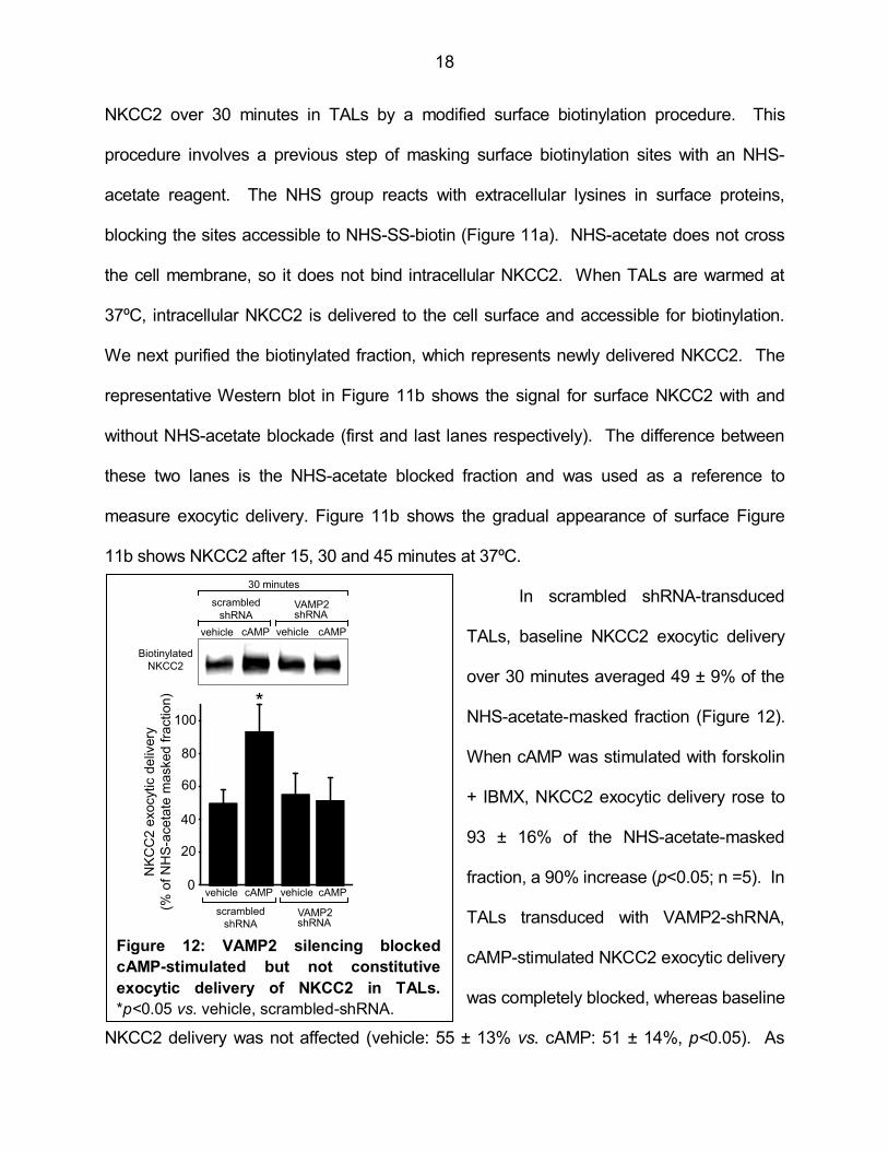
18
NKCC2 over 30 minutes in TALs by a modified surface biotinylation procedure. This
procedure involves a previous step of masking surface biotinylation sites with an NHS-
acetate reagent. The NHS group reacts with extracellular lysines in surface proteins,
blocking the sites accessible to NHS-SS-biotin (Figure 11a). NHS-acetate does not cross
the cell membrane, so it does not bind intracellular NKCC2. When TALs are warmed at
37ºC, intracellular NKCC2 is delivered to the cell surface and accessible for biotinylation.
We next purified the biotinylated fraction, which represents newly delivered NKCC2. The
representative Western blot in Figure 11b shows the signal for surface NKCC2 with and
without NHS-acetate blockade (first and last lanes respectively). The difference between
these two lanes is the NHS-acetate blocked fraction and was used as a reference to
measure exocytic delivery. Figure 11b shows the gradual appearance of surface Figure
11b shows NKCC2 after 15, 30 and 45 minutes at 37ºC.
In scrambled shRNA-transduced
TALs, baseline NKCC2 exocytic delivery
over 30 minutes averaged 49 ± 9% of the
NHS-acetate-masked fraction (Figure 12).
When cAMP was stimulated with forskolin
+ IBMX, NKCC2 exocytic delivery rose to
93 ± 16% of the NHS-acetate-masked
fraction, a 90% increase (p<0.05; n =5). In
TALs transduced with VAMP2-shRNA,
cAMP-stimulated NKCC2 exocytic delivery
was completely blocked, whereas baseline
NKCC2 delivery was not affected (vehicle: 55 ± 13% vs. cAMP: 51 ± 14%, p<0.05). As
Figure 12: VAMP2 silencing blocked
cAMP-stimulated but not constitutive
exocytic delivery of NKCC2 in TALs.
*p<0.05 vs. vehicle, scrambled-shRNA.
vehicle
scrambled
shRNAVAMP2shRNA
vehicle cAMP cAMP0
20
40
60
80
100
NK
CC
2 e
xocytic
deliv
ery
(% o
f N
HS
-aceta
te m
asked fra
ction)
*
vehicle
scrambled
shRNAVAMP2shRNA
vehicle cAMP cAMP
30 minutes
Biotinylated
NKCC2
Figure 7: VAMP2 silencing blocked
cAMP-stimulated but not constitutive
exocytic delivery of NKCC2 in TALs.
*p<0.05 vs. vehicle, scrambled-shRNA .
19
expected, intracellular control GAPDH was not detected in the surface fraction (not shown).
These data indicate that VAMP2 mediates all of the cAMP-stimulated NKCC2 exocytic
delivery without affecting constitutive delivery of NKCC2 to the cell surface.
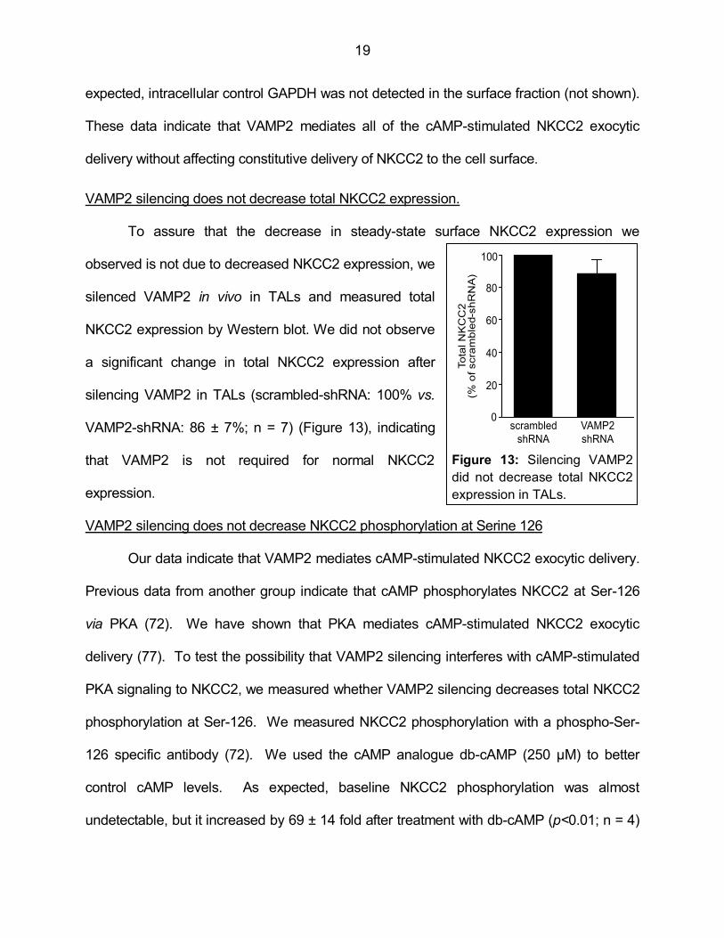
VAMP2 silencing does not decrease total NKCC2 expression.
To assure that the decrease in steady-state surface NKCC2 expression we
observed is not due to decreased NKCC2 expression, we
silenced VAMP2 in vivo in TALs and measured total
NKCC2 expression by Western blot. We did not observe
a significant change in total NKCC2 expression after
silencing VAMP2 in TALs (scrambled-shRNA: 100% vs.
VAMP2-shRNA: 86 ± 7%; n = 7) (Figure 13), indicating
that VAMP2 is not required for normal NKCC2
expression.
VAMP2 silencing does not decrease NKCC2 phosphorylation at Serine 126
Our data indicate that VAMP2 mediates cAMP-stimulated NKCC2 exocytic delivery.
Previous data from another group indicate that cAMP phosphorylates NKCC2 at Ser-126
via PKA (72). We have shown that PKA mediates cAMP-stimulated NKCC2 exocytic
delivery (77). To test the possibility that VAMP2 silencing interferes with cAMP-stimulated
PKA signaling to NKCC2, we measured whether VAMP2 silencing decreases total NKCC2
phosphorylation at Ser-126. We measured NKCC2 phosphorylation with a phospho-Ser-
126 specific antibody (72). We used the cAMP analogue db-cAMP (250 µM) to better
control cAMP levels. As expected, baseline NKCC2 phosphorylation was almost
undetectable, but it increased by 69 ± 14 fold after treatment with db-cAMP (p<0.01; n = 4)
VAMP2
shRNA
scrambled
shRNA
To
tal N
KC
C2
(% o
f scra
mb
led-s
hR
NA
)
0
20
40
60
80
100
Figure 9: Silencing VAMP2 did not decrease
total NKCC2 expression in TALs.
Figure 13: Silencing VAMP2
did not decrease total NKCC2
expression in TALs.

20
in control scrambled-shRNA-transfected
TALs (Figure 14). We observed that after
silencing VAMP2, db-cAMP strongly
enhanced Ser-126 phosphorylation by 73
± 24 fold (p<0.01 vs. vehicle). This
stimulation was not significantly different
between control and VAMP2-shRNA-
transduced TALs. These data indicate
that VAMP2 silencing does not decrease
the ability of cAMP to stimulate PKA and
phosphorylate NKCC2.
Conclusion
We observed that in addition to VAMP2 and VAMP3, also VAMP7 and VAMP8 are
expressed in the TAL. In this dissertation we focused in VAMP2 and VAMP3 since they
are the targets of tetanus toxin. In this aim we studied VAMP2 because it mediates cAMP-
stimulated exocytosis in other cells. We concluded that VAMP2 mediates cAMP-stimulated
steady-state surface NKCC2 expression and cAMP-stimulated exocytic delivery of NKCC2
in medullary TALs. VAMP2 does not mediate constitutive steady-state surface NKCC2
expression, constitutive NKCC2 exocytic delivery or is required for total NKCC2 expression.
We also concluded that the ability of cAMP to stimulate PKA and phosphorylate NKCC2 at
Serine-126 is not affected by VAMP2 silencing. The significance and in depth interpretation
of all these observations will be addressed in the discussion in Chapter 4.
0
2000
4000
6000
8000
10000
scrambled
shRNA
p-S
er
-126 N
KC
C2
(fold
incr
ease
fro
m v
ehic
le,
scra
mble
d s
hR
NA
)
VAMP2
shRNA
vehicle db-
cAMPvehicle
100
80
60
40
20
0
*
*
pSer-126 NKCC2
Total NKCC2
Figure 10: Silencing VAMP2 does not decrease
cAMP-stimulated NKCC2 phosphorylation at
Ser-126. *p<0.01 vs. vehicle.
db-
cAMP
Figure 14: Silencing VAMP2 did not decrease
cAMP-stimulated NKCC2 phosphorylation at
Ser-126. *p<0.01 vs. vehicle.

21
Aim 2- Hypothesis: cAMP stimulates NKCC2-VAMP2 interaction and co-localization
in thick ascending limbs.
Rationale
In aim 1 we showed that VAMP2 mediates cAMP-stimulated NKCC2 trafficking.
However, the mechanism by which this occurs remains unexplored. One possible
mechanism may involve protein-protein interactions, since NKCC2 interacts with proteins
that regulate its trafficking to the plasma membrane (163,165,166). Also, VAMP2
physically interacts with potassium (159,160) and calcium channels (158) in neurons.
These interactions modulate channel activity. In this aim we will test whether NKCC2
interacts with VAMP2 and we will discuss the relevance of this interaction for NKCC2
function.
We have previously shown that cAMP stimulation enhances apical membrane
exocytosis in TALs (77). If VAMP2 is in the same vesicles as NKCC2, it is expected that
cAMP will also promote delivery of VAMP2 to the apical surface. In this aim we also
quantified the rate at which cAMP stimulates delivery of VAMP2 to the TAL surface. We
also studied whether once at the surface, VAMP2 displays a localization pattern that
overlaps with NKCC2’s.
In neurons, phosphorylation is a mechanism to regulate SNARE interaction with
calcium channels and this regulates neurotransmitter release (156,176). However, none of
the kinases described to date are cAMP-dependent. The focus of this aim is to study how
cAMP affects NKCC2-VAMP2 interaction as a possible component of the mechanism that
mediates VAMP2 control over cAMP-stimulated NKCC2 trafficking.

22
Results
Co-immunoprecipitation of NKCC2 and VAMP2 in thick ascending limbs
Previous data from our laboratory
indicated that NKCC2 co-localizes with
VAMP2 in an intracellular sub-apical location
in TALs (14). To test whether NKCC2
interacts with VAMP2, we performed co-
immunoprecipitation assays in fresh TAL
lysates obtained from rat renal outer
medullas. We observed that VAMP2 co-
immunoprecipitated with NKCC2 in TALs
whereas VAMP7 and VAMP8 did not (Figure
15a, n = 4). To confirm these results, we
performed the reciprocal immunoprecipitation
with an anti-VAMP2 antibody and detected
NKCC2 by Western blot (Figure 15b, n= 4).
Controls with non-immune IgG showed absence of NKCC2, VAMP2, VAMP7 or VAMP8.
These data indicate that VAMP2 is a protein binding partner of NKCC2 in TALs. However,
it is not known whether this interaction is direct or via intermediate proteins.
Interaction of VAMP2 with the carboxy-terminus of NKCC2
To further define the region of NKCC2 that interacts with VAMP2, we designed the
two GST-fusion proteins shown in Figure 16a. We fused GST to the amino-terminus of
NKCC2 (GST-N-NKCC2) or a 71 amino acid-long sequence in the carboxy-terminus (GST-
C-NKCC2). This carboxy-terminal region has previously been shown to contain an apical
Figure 15: NKCC2 and VAMP2 co-
immunoprecipitate in TALs. A) VAMP2
but not VAMP7 and VAMP8 co-
immunoprecipitated with NKCC2. B)
Reciprocal immunoprecipitation with anti-
NKCC2 antibody confirmed interaction
with VAMP2 in TAL lysates.
A)
VAMP7
VAMP8
VAMP2
Immunoprecipitation
Imm
unoblo
t
NKCC2
Immunoblot
B) Immunoprecipitation
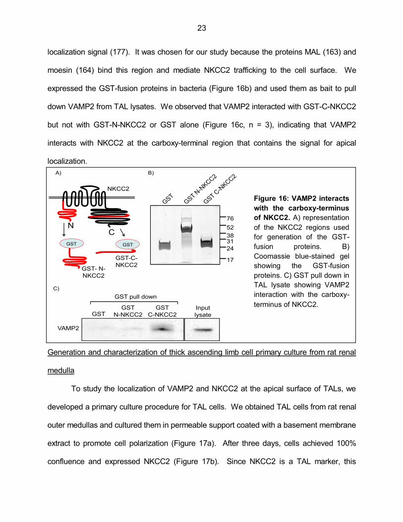
23
localization signal (177). It was chosen for our study because the proteins MAL (163) and
moesin (164) bind this region and mediate NKCC2 trafficking to the cell surface. We
expressed the GST-fusion proteins in bacteria (Figure 16b) and used them as bait to pull
down VAMP2 from TAL lysates. We observed that VAMP2 interacted with GST-C-NKCC2
but not with GST-N-NKCC2 or GST alone (Figure 16c, n = 3), indicating that VAMP2
interacts with NKCC2 at the carboxy-terminal region that contains the signal for apical
localization.
Generation and characterization of thick ascending limb cell primary culture from rat renal
medulla
To study the localization of VAMP2 and NKCC2 at the apical surface of TALs, we
developed a primary culture procedure for TAL cells. We obtained TAL cells from rat renal
outer medullas and cultured them in permeable support coated with a basement membrane
extract to promote cell polarization (Figure 17a). After three days, cells achieved 100%
confluence and expressed NKCC2 (Figure 17b). Since NKCC2 is a TAL marker, this
NC
NKCC2
GST- N-
NKCC2
GST-C-
NKCC2
GST
GSTGST
N-NKCC2
GST
C-NKCC2
Input
lysate
VAMP2
GST pull down
Figure 12: VAMP2 interacts the carboxy-terminus of
NKCC2. A) representation of the NKCC2 regions used for
generation of the GST-fusion proteins. B) Coomassie blue-
stained gel showing the GST-fusion proteins. C) GST pull down
in TAL lysate showing VAMP2 interaction with the carboxy-
terminus of NKCC2.
38
52
31
24
76
17
GST
A)
C)
B)
Figure 16: VAMP2 interacts
with the carboxy-terminus
of NKCC2. A) representation
of the NKCC2 regions used
for generation of the GST-
fusion proteins. B)
Coomassie blue-stained gel
showing the GST-fusion
proteins. C) GST pull down in
TAL lysate showing VAMP2
interaction with the carboxy-
terminus of NKCC2.
24
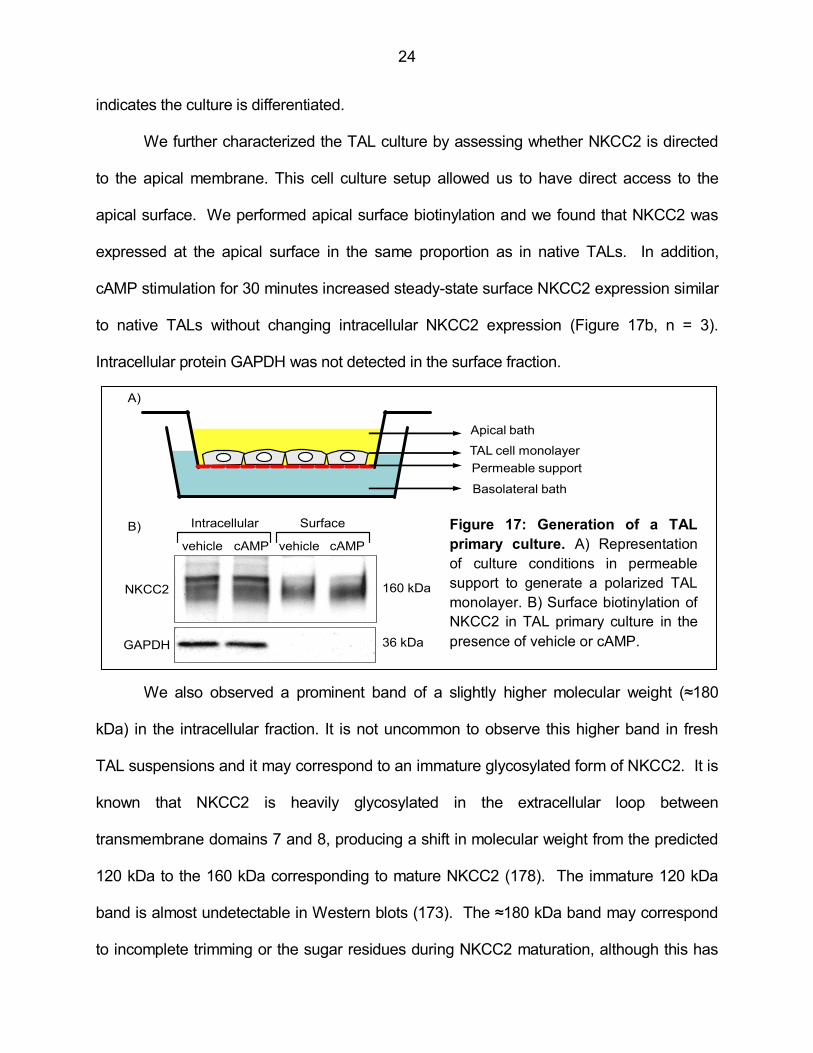
indicates the culture is differentiated.
We further characterized the TAL culture by assessing whether NKCC2 is directed
to the apical membrane. This cell culture setup allowed us to have direct access to the
apical surface. We performed apical surface biotinylation and we found that NKCC2 was
expressed at the apical surface in the same proportion as in native TALs. In addition,
cAMP stimulation for 30 minutes increased steady-state surface NKCC2 expression similar
to native TALs without changing intracellular NKCC2 expression (Figure 17b, n = 3).
Intracellular protein GAPDH was not detected in the surface fraction.
We also observed a prominent band of a slightly higher molecular weight (≈180
kDa) in the intracellular fraction. It is not uncommon to observe this higher band in fresh
TAL suspensions and it may correspond to an immature glycosylated form of NKCC2. It is
known that NKCC2 is heavily glycosylated in the extracellular loop between
transmembrane domains 7 and 8, producing a shift in molecular weight from the predicted
120 kDa to the 160 kDa corresponding to mature NKCC2 (178). The immature 120 kDa
band is almost undetectable in Western blots (173). The ≈180 kDa band may correspond
to incomplete trimming or the sugar residues during NKCC2 maturation, although this has
TAL cell monolayer
A)
vehicle vehiclecAMP cAMP
NKCC2
GAPDH
Intracellular SurfaceB)
160 kDa
36 kDa
Permeable support
Basolateral bath
Apical bath
Figure 17: Generation of a TAL
primary culture. A) Representation
of culture conditions in permeable
support to generate a polarized TAL
monolayer. B) Surface biotinylation of
NKCC2 in TAL primary culture in the
presence of vehicle or cAMP.
25
never been addressed. In native TAL suspensions, the ≈180 kDa is not as strong as the
one we observed in TAL cultures compared to the 160 kDa band. This may indicate
incomplete maturation of NKCC2 in primary cultures. However, the ≈180 kDa band almost
disappeared in the surface fraction, suggesting that only mature NKCC2 reaches the
surface, making the TAL primary cultures a suitable model to study NKCC2 trafficking.
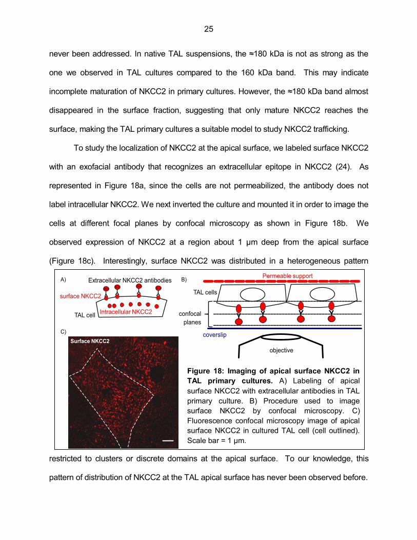
To study the localization of NKCC2 at the apical surface, we labeled surface NKCC2
with an exofacial antibody that recognizes an extracellular epitope in NKCC2 (24). As
represented in Figure 18a, since the cells are not permeabilized, the antibody does not
label intracellular NKCC2. We next inverted the culture and mounted it in order to image the
cells at different focal planes by confocal microscopy as shown in Figure 18b. We
observed expression of NKCC2 at a region about 1 µm deep from the apical surface
(Figure 18c). Interestingly, surface NKCC2 was distributed in a heterogeneous pattern
restricted to clusters or discrete domains at the apical surface. To our knowledge, this
pattern of distribution of NKCC2 at the TAL apical surface has never been observed before.
C)
Surface NKCC2
TAL cell
surface NKCC2
A)
Intracellular NKCC2
568568 568
568
Extracellular NKCC2 antibodies
TAL cells
confocal
planes
Permeable support
coverslip
objective
B)
Figure 18: Imaging of apical surface NKCC2 in
TAL primary cultures. A) Labeling of apical
surface NKCC2 with extracellular antibodies in TAL
primary culture. B) Procedure used to image
surface NKCC2 by confocal microscopy. C)
Fluorescence confocal microscopy image of apical
surface NKCC2 in cultured TAL cell (cell outlined).
Scale bar = 1 µm.
26
Altogether, these data indicate that in TAL primary cultures NKCC2 retains polarity,
the mature form is directed to the apical membrane and responds to cAMP in the same
way as native TALs.
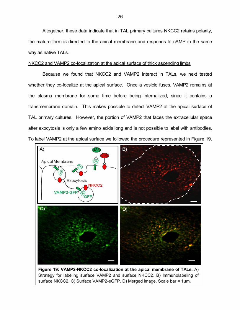
NKCC2 and VAMP2 co-localization at the apical surface of thick ascending limbs
Because we found that NKCC2 and VAMP2 interact in TALs, we next tested
whether they co-localize at the apical surface. Once a vesicle fuses, VAMP2 remains at
the plasma membrane for some time before being internalized, since it contains a
transmembrane domain. This makes possible to detect VAMP2 at the apical surface of
TAL primary cultures. However, the portion of VAMP2 that faces the extracellular space
after exocytosis is only a few amino acids long and is not possible to label with antibodies.
To label VAMP2 at the apical surface we followed the procedure represented in Figure 19.
Figure 19: VAMP2-NKCC2 co-localization at the apical membrane of TALs. A)
Strategy for labeling surface VAMP2 and surface NKCC2. B) Immunolabeling of
surface NKCC2. C) Surface VAMP2-eGFP. D) Merged image. Scale bar = 1µm.

27
We transfected TAL cells in culture with a VAMP2 construct fused to eGFP in the carboxy-
terminus (179). When vesicles carrying VAMP2-eGFP undergo exocytosis, the GFP tag
faces the extracellular space (Figure 19a). Then, cells were cooled to 4°C and incubated
with an extracellular anti-GFP antibody (green) and the exofacial anti-NKCC2 antibody
(red), to label surface VAMP2 and surface NKCC2 respectively. The antibodies do not
label intracellular proteins since cells are not permeabilized. Imaging conditions were
adjusted so intrinsic fluorescence from GFP was not detectable. We observed that surface
NKCC2 (Figure 19b) and surface VAMP2 (Figure 19c) localized at the apical cell surface in
a heterogeneous pattern forming discrete clusters. After image deconvolution, we different
determined the degree of co-localization by measuring the Mander’s overlap coefficient.
Figure 20 shows images of the same cell where co-localizing pixels were obtained at
stringency conditions. A high Mander’s overlap coefficient (closer to 1) is represented in
Figure 20a and is the most stringent condition. However, this criterion would overlook co-
localization between pixels of different intensity, which can vary with the labeling and
image acquisition conditions. On the other hand, a low Mander’s overlap coefficient (0.7)
(Figure 20b) would relax the stringency of the measurement and overestimate the co-
localization. We used a Mander’s overlap coefficient equal or higher than 0.95 to measure
Figure 20: Comparison of different co-localization stringency criteria. Mander’s
overlap coefficients of 0.99 (A), 0.7 (B) and 0.95 (C) were applied to the same picture to
generate the different images of co-localizing pixels.
28
co-localization at the apical surface (Figure 20c). We counted the number of NKCC2 and
co-localizing clusters and determined that 47 ± 8% of NKCC2 clusters at the apical surface
also contained VAMP2 (n = 5). It is important to notice that since labeling is performed in a
single plane at the cell surface, artifacts due to signal originated at different planes do not
occur in our setup. These data indicate that VAMP2 and NKCC2 are located in similar
clusters or micro domains at the apical surface of TAL cells.
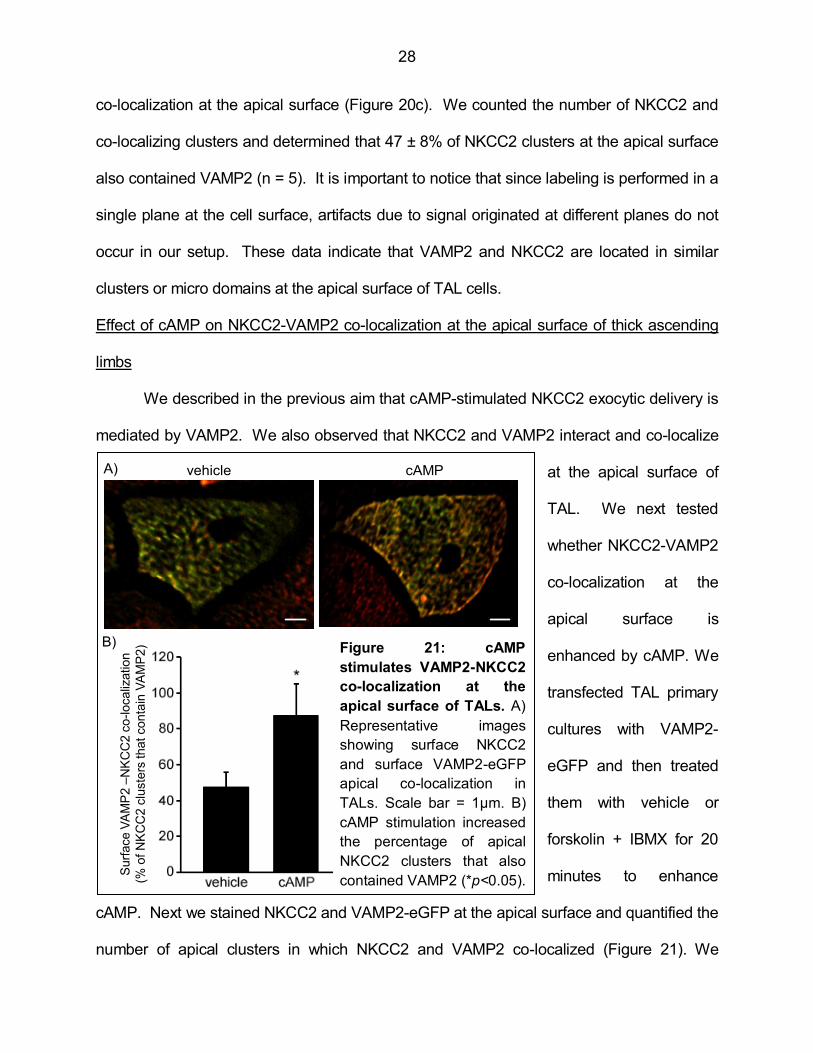
Effect of cAMP on NKCC2-VAMP2 co-localization at the apical surface of thick ascending
limbs
We described in the previous aim that cAMP-stimulated NKCC2 exocytic delivery is
mediated by VAMP2. We also observed that NKCC2 and VAMP2 interact and co-localize
at the apical surface of
TAL. We next tested
whether NKCC2-VAMP2
co-localization at the
apical surface is
enhanced by cAMP. We
transfected TAL primary
cultures with VAMP2-
eGFP and then treated
them with vehicle or
forskolin + IBMX for 20
minutes to enhance
cAMP. Next we stained NKCC2 and VAMP2-eGFP at the apical surface and quantified the
number of apical clusters in which NKCC2 and VAMP2 co-localized (Figure 21). We
vehicle cAMPA)
B)
Surf
ace V
AM
P2 –
NK
CC
2 c
o-localiz
ation
(% o
f N
KC
C2 c
luste
rs that conta
in V
AM
P2) Figure 21: cAMP
stimulates VAMP2-NKCC2
co-localization at the
apical surface of TALs. A)
Representative images
showing surface NKCC2
and surface VAMP2-eGFP
apical co-localization in
TALs. Scale bar = 1µm. B)
cAMP stimulation increased
the percentage of apical
NKCC2 clusters that also
contained VAMP2 (*p<0.05).
29
observed that cAMP stimulation increased the number of NKCC2 clusters that also
contained VAMP2 from 47 ± 8% to 87 ± 18% (p<0.05, n = 8), indicating that cAMP
increased NKCC2-VAMP2 co-localization at the apical surface of TALs.
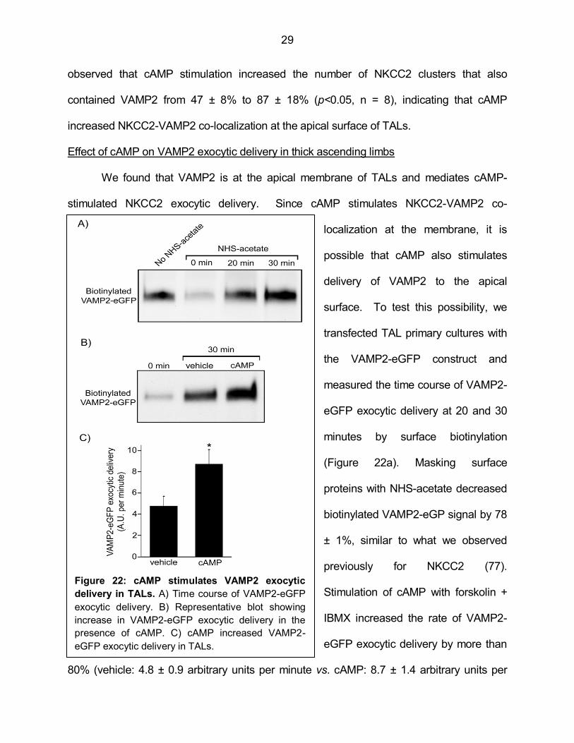
Effect of cAMP on VAMP2 exocytic delivery in thick ascending limbs
We found that VAMP2 is at the apical membrane of TALs and mediates cAMP-
stimulated NKCC2 exocytic delivery. Since cAMP stimulates NKCC2-VAMP2 co-
localization at the membrane, it is
possible that cAMP also stimulates
delivery of VAMP2 to the apical
surface. To test this possibility, we
transfected TAL primary cultures with
the VAMP2-eGFP construct and
measured the time course of VAMP2-
eGFP exocytic delivery at 20 and 30
minutes by surface biotinylation
(Figure 22a). Masking surface
proteins with NHS-acetate decreased
biotinylated VAMP2-eGP signal by 78
± 1%, similar to what we observed
previously for NKCC2 (77).
Stimulation of cAMP with forskolin +
IBMX increased the rate of VAMP2-
eGFP exocytic delivery by more than
80% (vehicle: 4.8 ± 0.9 arbitrary units per minute vs. cAMP: 8.7 ± 1.4 arbitrary units per
Figure 22: cAMP stimulates VAMP2 exocytic
delivery in TALs. A) Time course of VAMP2-eGFP
exocytic delivery. B) Representative blot showing
increase in VAMP2-eGFP exocytic delivery in the
presence of cAMP. C) cAMP increased VAMP2-
eGFP exocytic delivery in TALs.
30
minute, p<0.05, n = 4) (Figures 22b and 22c). These results indicate that cAMP stimulates
VAMP2 exocytic delivery in TALs.
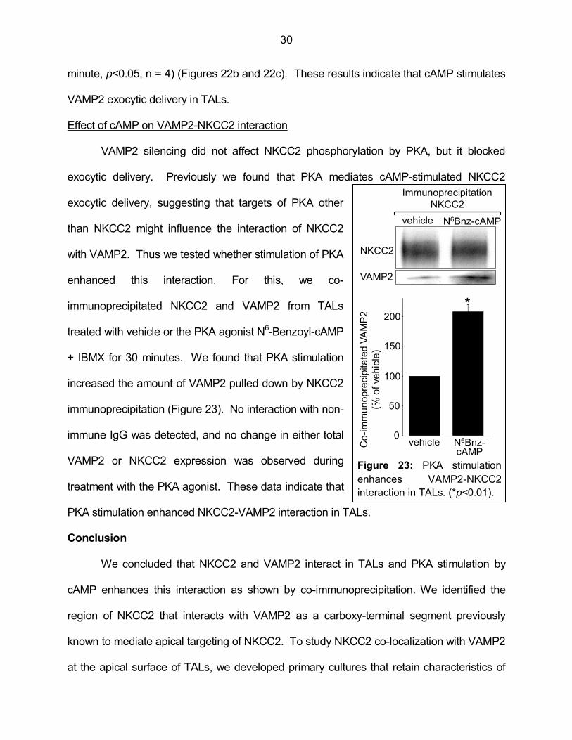
Effect of cAMP on VAMP2-NKCC2 interaction
VAMP2 silencing did not affect NKCC2 phosphorylation by PKA, but it blocked
exocytic delivery. Previously we found that PKA mediates cAMP-stimulated NKCC2
exocytic delivery, suggesting that targets of PKA other
than NKCC2 might influence the interaction of NKCC2
with VAMP2. Thus we tested whether stimulation of PKA
enhanced this interaction. For this, we co-
immunoprecipitated NKCC2 and VAMP2 from TALs
treated with vehicle or the PKA agonist N6-Benzoyl-cAMP
+ IBMX for 30 minutes. We found that PKA stimulation
increased the amount of VAMP2 pulled down by NKCC2
immunoprecipitation (Figure 23). No interaction with non-
immune IgG was detected, and no change in either total
VAMP2 or NKCC2 expression was observed during
treatment with the PKA agonist. These data indicate that
PKA stimulation enhanced NKCC2-VAMP2 interaction in TALs.
Conclusion
We concluded that NKCC2 and VAMP2 interact in TALs and PKA stimulation by
cAMP enhances this interaction as shown by co-immunoprecipitation. We identified the
region of NKCC2 that interacts with VAMP2 as a carboxy-terminal segment previously
known to mediate apical targeting of NKCC2. To study NKCC2 co-localization with VAMP2
at the apical surface of TALs, we developed primary cultures that retain characteristics of
50
0
100
150
200
vehicle N6Bnz-cAMP
vehicle N6Bnz-cAMP
NKCC2
VAMP2
Immunoprecipitation
NKCC2
Co
-im
mu
no
pre
cip
ita
ted
VA
MP
2
(% o
f ve
hic
le)
*
Figure 23: PKA stimulation
enhances VAMP2-NKCC2
interaction in TALs. (*p<0.01).

31
differentiated TALs. We labeled NKCC2 and VAMP2 at the apical surface and measured
co-localization. We observed that NKCC2 and VAMP2 co-localize at the cell surface in
apical clusters or micro domains. This allowed us to determine that NKCC2 and VAMP2
remain at a similar localization after they are exocytosed. Whether they physically interact
at the apical surface is not clear from the co-localization analysis. The co-
immunoprecipitation and the GST pull down experiments indicate that the two proteins
physically interact, either directly or via intermediate proteins. However the cellular
compartment where this interaction occurs was not possible to determine in our study.
Stimulation with cAMP enhanced VAMP2-NKCC2 co-localization at apical surface
clusters. We also observed that cAMP stimulated VAMP2 delivery to the apical TAL
surface. Together with enhanced NKCC2-VAMP2 interaction in the presence of cAMP, our
data prompt us to speculate that protein-protein interactions may be part of the mechanism
by which VAMP2 mediates cAMP-stimulated NKCC2 trafficking.
32
CHAPTER 3
ROLE OF VAMP3 IN CONSTITUTIVE NKCC2 TRAFFICKING IN THE THICK
ASCENDING LIMB AND RENAL FUNCTION
Introduction
We have previously shown that NKCC2 undergoes constitutive exocytic delivery in
the absence of any stimulation (77). Since VAMP2 inhibition did not affect baseline steady-
state surface NKCC2 and exocytic delivery, some other VAMP isoform must mediate
constitutive trafficking of NKCC2. Another observation from the previous chapter is that, in
addition to VAMP2, the TAL also expresses three other VAMP isoforms. Two of them,
VAMP7 and VAMP8, known as endobrevins, are usually involved in intracellular vesicle-
vesicle fusion events (145,146), and are unlikely to mediate delivery of NKCC2 to the
plasma membrane. The fourth isoform expressed in the TAL is VAMP3, which mediates
exocytic events in most cells studied to date (150,180-185). In this chapter we addressed
the role of VAMP3 in constitutive NKCC2 trafficking. We divided this chapter into two
additional aims. In aim 3 we tested whether VAMP3 mediates constitutive trafficking of
NKCC2 in the TAL. In aim 4, following the approach we used for VAMP2, we studied
whether VAMP3 and NKCC2 interact. In addition, we addressed the role of VAMP3 in
mediating normal renal function and blood pressure.
Aim 3- Hypothesis: VAMP3 mediates constitutive trafficking of NKCC2 in the thick
ascending limb.
Rationale
Tetanus toxin exclusively cleaves VAMP2 and VAMP3, since these are the only two
proteins that contain the cleavage site for the toxin. If VAMP3 mediates constitutive
33
NKCC2 trafficking, we should observe a decrease in surface NKCC2 after treatment with
tetanus toxin. Our observations from aim 1 indicate that VAMP2 does not mediate
constitutive trafficking of NKCC2. Therefore, any effect of tetanus toxin on baseline steady-
state surface NKCC2 can be attributed to VAMP3. In this aim we studied whether tetanus
toxin decreases constitutive steady-state surface NKCC2 expression. We tested the
hypothesis that VAMP3 mediates constitutive trafficking of NKCC2 in the thick ascending
limb. To specifically address the role of VAMP3, we used silencing shRNAs and a VAMP3
knockout mouse model to study NKCC2 trafficking.
Results
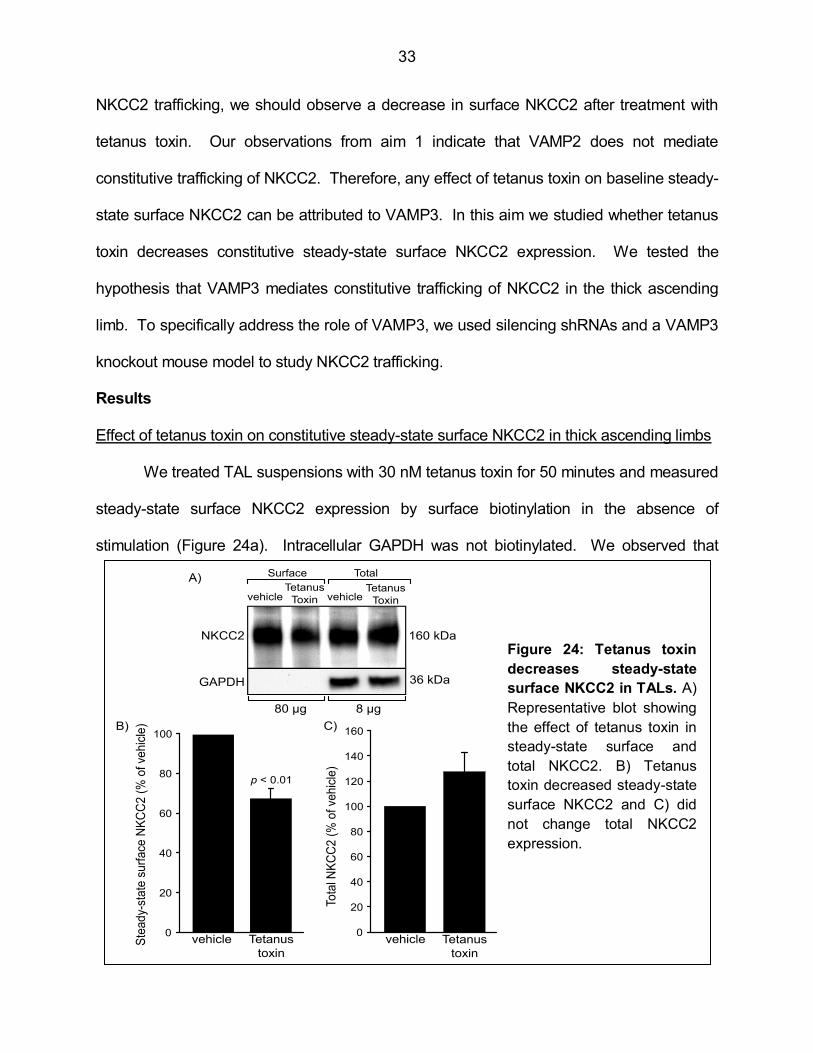
Effect of tetanus toxin on constitutive steady-state surface NKCC2 in thick ascending limbs
We treated TAL suspensions with 30 nM tetanus toxin for 50 minutes and measured
steady-state surface NKCC2 expression by surface biotinylation in the absence of
stimulation (Figure 24a). Intracellular GAPDH was not biotinylated. We observed that
160 kDa
36 kDaGAPDH
NKCC2
Surface Total
vehicle vehicleTetanus
ToxinTetanus
Toxin
80 µg 8 µg
A)
B)
Ste
ad
y-st
ate
su
rface
NK
CC
2 (
% o
f ve
hic
le)
vehicle Tetanus
toxin
p < 0.01
Tota
l NK
CC
2 (
% o
f ve
hic
le)
vehicle Tetanus
toxin
0
20
40
60
80
100
0
20
40
60
80
100
120
140
160
Figure 18: Tetanus toxin decreases steady-state surface
NKCC2 in TALs. A) Representative blot showing the effect of
tetanus toxin in steady-state surface and total NKCC2. B) Tetanus
toxin decreased steady-state surface NKCC2 and C) did not
change total NKCC2 expression.
C)
Figure 24: Tetanus toxin
decreases steady-state
surface NKCC2 in TALs. A)
Representative blot showing
the effect of tetanus toxin in
steady-state surface and
total NKCC2. B) Tetanus
toxin decreased steady-state
surface NKCC2 and C) did
not change total NKCC2
expression.

34
constitutive steady-state surface NKCC2 was decreased by 33 ± 5% compared to vehicle
(Figure 24b, n = 6). Tetanus toxin did not decrease total NKCC2 expression (Figure 24c).
These data indicate that constitutive NKCC2 trafficking to the TAL surface is mediated by a
tetanus toxin-sensitive VAMP. In order to address whether this VAMP isoform is VAMP3,
we silenced VAMP3 expression in TALs.
VAMP3 silencing in vitro and in vivo
To directly address the role of VAMP3 in NKCC2 trafficking, we designed silencing
RNAs. We followed the same protocol we developed in aim 1 for VAMP2. We observed
efficient and specific silencing of VAMP3 in NRK-52E cells (Figure 25a) and in medullary
TAL suspensions (Figure 25b). We did not observe any decrease in VAMP2 expression.
After 72 hours of silencing, we achieved a decrease of 68 ± 10% in VAMP3 protein
A)
GAPDH
VAMP3
VAMP3
siRNA
scrambled
siRNA
C)
0
20
40
60
80
100
Tota
l V
AM
P3 e
xpre
ssio
n
(% o
f scra
mble
d s
hR
NA
)
p < 0.01
VAMP3
shRNA
scrambled
shRNA
Figure 19: VAMP3 silencing in vitro and in vivo. A) VAMP3 silencing via
siRNA in NRK-52E cells. B) VAMP2 and VAMP3 expression after silencing
VAMP3 in TALs. C) Decrease in VAMP3 expression after silencing in TALs.
D) Curve of VAMP3 protein expression in TALs (R² = 0.995).
VAMP2
VAMP3
scrambled
shRNAVAMP3
shRNAB)
0
20
40
60
80
100
0 2 4 6 8 10 12 14 16
D)
VA
MP
3 e
xpre
ssio
n(%
of
maxim
um
)
1 2 4 8 16
µg protein lysate
Figure 25: VAMP3 silencing in vitro and in vivo. A) VAMP3 silencing via siRNA in
NRK-52E cells. B) VAMP2 and VAMP3 expression after silencing VAMP3 in TALs. C)
Decrease in VAMP3 expression after silencing in TALs. D) Curve of VAMP3 protein
expression in TALs (R² = 0.995).
35
expression compared with the contra lateral kidney injected with control scrambled-shRNA
(p<0.01, n = 3) (Figure 25c). The curve depicted in Figure 25d shows the linear range used
in our protein measurements. Altogether these data indicate that VAMP3-shRNAs were
efficient and specific for VAMP3 silencing.
Effect of VAMP3 silencing on NKCC2 exocytic delivery
We next tested whether silencing VAMP3 inhibits constitutive exocytic delivery of
NKCC2 in TALs. In TALs transduced with scrambled-shRNA, baseline NKCC2 exocytic
delivery over 30 minutes averaged
49 ± 9% of the NHS-acetate-masked
fraction, and it was enhanced to 92 ±
13% with cAMP stimulation (p<0.05,
n = 5) (Figure 26). When we
silenced VAMP3, constitutive
exocytic delivery at 30 minutes was
almost completely blocked (7 ± 6%
of the NHS-acetate-masked
fraction). However cAMP was still
able to stimulate NKCC2 delivery (61
± 16% of the NHS-acetate-masked
fraction).
There may be a confounding factor in our measurements arising from the lower
constitutive exocytic delivery of NKCC2 when we silenced VAMP3. Since the
measurements for cAMP-stimulated exocytic delivery are generated by comparing to a
lower baseline number, this could lead to overestimation of the stimulation with cAMP
vehicle
scrambled
shRNAVAMP3shRNA
vehicle cAMP cAMP
NK
CC
2 e
xo
cytic in
se
rtio
n
(% o
f N
HS
-ace
tate
ma
ske
d fra
ctio
n)
0
20
40
60
80
100
*
*
Figure 20: Silencing VAMP3 in TALs blocked
constitutive but not cAMP-stimulated exocytic
delivery of NKCC2 (*p<0.05 vs. vehicle,
scrambled shRNA; #p<0.05 vs. vehicle).
#
#
Figure 26: Silencing VAMP3 in TALs blocked
constitutive but not cAMP-stimulated exocytic
delivery of NKCC2. (*p<0.05 vs. vehicle,
scrambled shRNA; #p<0.05 vs. vehicle).
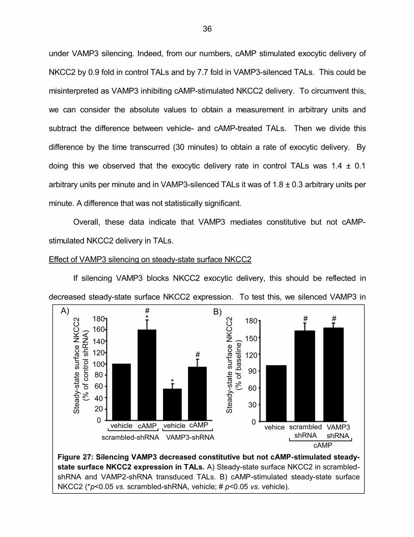
36
under VAMP3 silencing. Indeed, from our numbers, cAMP stimulated exocytic delivery of
NKCC2 by 0.9 fold in control TALs and by 7.7 fold in VAMP3-silenced TALs. This could be
misinterpreted as VAMP3 inhibiting cAMP-stimulated NKCC2 delivery. To circumvent this,
we can consider the absolute values to obtain a measurement in arbitrary units and
subtract the difference between vehicle- and cAMP-treated TALs. Then we divide this
difference by the time transcurred (30 minutes) to obtain a rate of exocytic delivery. By
doing this we observed that the exocytic delivery rate in control TALs was 1.4 ± 0.1
arbitrary units per minute and in VAMP3-silenced TALs it was of 1.8 ± 0.3 arbitrary units per
minute. A difference that was not statistically significant.
Overall, these data indicate that VAMP3 mediates constitutive but not cAMP-
stimulated NKCC2 delivery in TALs.
Effect of VAMP3 silencing on steady-state surface NKCC2
If silencing VAMP3 blocks NKCC2 exocytic delivery, this should be reflected in
decreased steady-state surface NKCC2 expression. To test this, we silenced VAMP3 in
B)
vehice scrambled
shRNAVAMP3
shRNA
cAMP
Ste
ad
y-s
tate
su
rfa
ce
NK
CC
2
(% o
f b
ase
line
)
# #
0
30
60
90
120
150
180
Figure 21: Silencing VAMP3 decreased constitutive but not cAMP-stimulated
steady-state surface NKCC2 expression in TALs. A) Steady-state surface NKCC2 in
scrambled-shRNA and VAMP2-shRNA transduced TALs. B) cAMP-stimulated steady-
state surface NKCC2 (*p<0.05 vs. scrambled-shRNA,vehicle; # p<0.05 vs. vehicle).
A)
cAMPvehicle cAMP
Ste
ad
y-s
tate
su
rfa
ce
NK
CC
2
(% o
f co
ntr
ol sh
RN
A)
vehicle
VAMP3-shRNAscrambled-shRNA
0
20
40
60
80
100
120
140
160
180 *
*
#
#
Figure 27: Silencing VAMP3 decreased constitutive but not cAMP-stimulated steady-
state surface NKCC2 expression in TALs. A) Steady-state surface NKCC2 in scrambled-
shRNA and VAMP2-shRNA transduced TALs. B) cAMP-stimulated steady-state surface
NKCC2 (*p<0.05 vs. scrambled-shRNA, vehicle; # p<0.05 vs. vehicle).
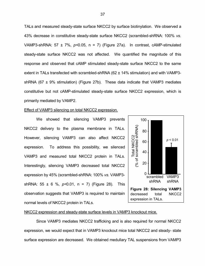
37
TALs and measured steady-state surface NKCC2 by surface biotinylation. We observed a
43% decrease in constitutive steady-state surface NKCC2 (scrambled-shRNA: 100% vs.
VAMP3-shRNA: 57 ± 7%, p<0.05, n = 7) (Figure 27a). In contrast, cAMP-stimulated
steady-state surface NKCC2 was not affected. We quantified the magnitude of this
response and observed that cAMP stimulated steady-state surface NKCC2 to the same
extent in TALs transfected with scrambled-shRNA (62 ± 14% stimulation) and with VAMP3-
shRNA (67 ± 9% stimulation) (Figure 27b). These data indicate that VAMP3 mediates
constitutive but not cAMP-stimulated steady-state surface NKCC2 expression, which is
primarily mediated by VAMP2.
Effect of VAMP3 silencing on total NKCC2 expression.
We showed that silencing VAMP3 prevents
NKCC2 delivery to the plasma membrane in TALs.
However, silencing VAMP3 can also affect NKCC2
expression. To address this possibility, we silenced
VAMP3 and measured total NKCC2 protein in TALs.
Interestingly, silencing VAMP3 decreased total NKCC2
expression by 45% (scrambled-shRNA: 100% vs. VAMP3-
shRNA: 55 ± 6 %, p<0.01, n = 7) (Figure 28). This
observation suggests that VAMP3 is required to maintain
normal levels of NKCC2 protein in TALs.
NKCC2 expression and steady-state surface levels in VAMP3 knockout mice.
Since VAMP3 mediates NKCC2 trafficking and is also required for normal NKCC2
expression, we would expect that in VAMP3 knockout mice total NKCC2 and steady- state
surface expression are decreased. We obtained medullary TAL suspensions from VAMP3
VAMP3
shRNA
scrambled
shRNA
Tota
l N
KC
C2
(% o
f scra
mble
d s
hR
NA
)
0
20
40
60
80
100
p < 0.01
Figure 22: Silencing VAMP3
decreased total NKCC2
expression in TALs.
Figure 28: Silencing VAMP3
decreased total NKCC2
expression in TALs.

38
knockout mice and
measured: a) VAMP3
protein to assure that
it is not expressed, b)
total NKCC2
expression and c)
steady-state surface
NKCC2 expression.
We did not
detect any VAMP3
expression in TALs from VAMP3 knockout mice, and there was no change in expression of
VAMP2, VAMP7 or VAMP8 (Figure 29a, n = 3). Total NKCC2 expression was decreased
by 66 ± 12% (p<0.05, n = 6)
compared to wild-type mice
(Figure 29b) and steady-state
surface NKCC2 was
decreased by 46% (wild-type
= 100% vs. VAMP3 knockout
= 54 ± 4, p<0.05, n = 3)
(Figure 30), consistent with
the VAMP3 silencing results.
Altogether these observations
confirm that VAMP3
mediates steady-state surface NKCC2 expression and normal total NKCC2 expression. Figure 24: Decreased steady-state
surface NKCC2 expression in VAMP3
knockout mice. A) Representative blot
showing total and steady-state surface
NKCC2 in VAMP3 knockout mice. B)
Steady-state surface NKCC2 in TALs
from VAMP3 knockout mice.
NKCC2
GAPDH
Total SurfaceA)
B)
Wild
typeVAMP3 -/-
20
0
40
60
80
100
Ste
ad
y-s
tate
su
rface
NK
CC
2 (
% o
f w
ild t
yp
e)
p<0.05
Figure 30: Decreased
steady-state surface
NKCC2 expression in
VAMP3 knockout
mice. A)
Representative blot
showing total and
steady-state surface
NKCC2 in VAMP3
knockout mice. B)
Steady-state surface
NKCC2 in TALs from
VAMP3 knockout mice.
0
20
40
60
80
100
p < 0.05
Wild
type
Tota
l NK
CC
2 e
xpre
ssio
n
(% o
f w
ild-t
ype)
VAMP3
knockout
NKCC2
GAPDH
wild
type
VAMP3
knockoutB)
GAPDH
VAMP2
VAMP3
wild
typeVAMP3
knockoutA)
VAMP7
VAMP8
Figure 29: NKCC2 expression
is decreased in TALs from
VAMP3 knockout mice. A)
Expression of VAMPs in
VAMP3 knockout mice. B)
Decreased NKCC2 expression
in VAMP3 knockout mice.

39
Conclusion
We concluded that VAMP3 mediates constitutive NKCC2 exocytic delivery and steady-
state surface NKCC2 expression. Stimulation by cAMP was not affected by VAMP3
silencing in TALs. Interestingly, silencing VAMP3 also decreased total NKCC2 protein,
indicating that VAMP3 is required for normal NKCC2 expression. The mechanism by
which VAMP3 mediates normal NKCC2 expression was not directly addressed in this
dissertation. However, it may involve maturation of intracellular compartments or re-routing
of NKCC2 vesicles that cannot reach the plasma membrane when VAMP3 is silenced.
These possibilities will be discussed in detail in Chapter 4.
The previous observations during VAMP3 silencing were confirmed in TALs from
VAMP3 knockout mice, where total NKCC2 and baseline steady-state surface NKCC2
expression were also decreased. This role of VAMP3 explains why tetanus toxin
decreases constitutive steady-state surface NKCC2 expression in TALs. Altogether, out
results indicate that the constitutive pathway for NKCC2 trafficking is controlled
independently from the cAMP-stimulated pathway and is mediated by VAMP3.
Aim 4- Hypothesis: VAMP3 interacts with NKCC2 in the thick ascending limb and
maintains normal renal function and blood pressure.
Rationale
In the previous aim we showed that VAMP3 mediates constitutive NKCC2 trafficking
and is required for normal NKCC2 expression. We also showed in aim 2 that VAMP2
interacts with NKCC2. In this aim we tested whether VAMP3 also interacts with NKCC2
and whether they co-localize at the apical surface of TALs.
Since NKCC2 plays a crucial role in renal physiology, we would expect that renal
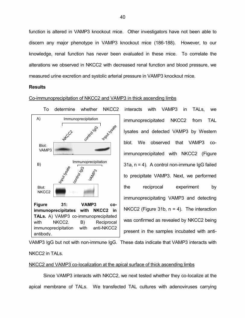
40
function is altered in VAMP3 knockout mice. Other investigators have not been able to
discern any major phenotype in VAMP3 knockout mice (186-188). However, to our
knowledge, renal function has never been evaluated in these mice. To correlate the
alterations we observed in NKCC2 with decreased renal function and blood pressure, we
measured urine excretion and systolic arterial pressure in VAMP3 knockout mice.
Results
Co-immunoprecipitation of NKCC2 and VAMP3 in thick ascending limbs
To determine whether NKCC2 interacts with VAMP3 in TALs, we
immunoprecipitated NKCC2 from TAL
lysates and detected VAMP3 by Western
blot. We observed that VAMP3 co-
immunoprecipitated with NKCC2 (Figure
31a, n = 4). A control non-immune IgG failed
to precipitate VAMP3. Next, we performed
the reciprocal experiment by
immunoprecipitating VAMP3 and detecting
NKCC2 (Figure 31b, n = 4). The interaction
was confirmed as revealed by NKCC2 being
present in the samples incubated with anti-
VAMP3 IgG but not with non-immune IgG. These data indicate that VAMP3 interacts with
NKCC2 in TALs.
NKCC2 and VAMP3 co-localization at the apical surface of thick ascending limbs
Since VAMP3 interacts with NKCC2, we next tested whether they co-localize at the
apical membrane of TALs. We transfected TAL cultures with adenoviruses carrying
A)
B)
Blot:
VAMP3
Figure 25: VAMP3 co-immunoprecipitates
with NKCC2 in TALs. A) VAMP3 co-
immunoprecipitated with NKCC2. B) Reciprocal
immunoprecipitation with anti-NKCC2 antibody.
Immunoprecipitation
Blot:
NKCC2
Immunoprecipitation
Figure 31: VAMP3 co-
immunoprecipitates with NKCC2 in
TALs. A) VAMP3 co-immunoprecipitated
with NKCC2. B) Reciprocal
immunoprecipitation with anti-NKCC2
antibody.
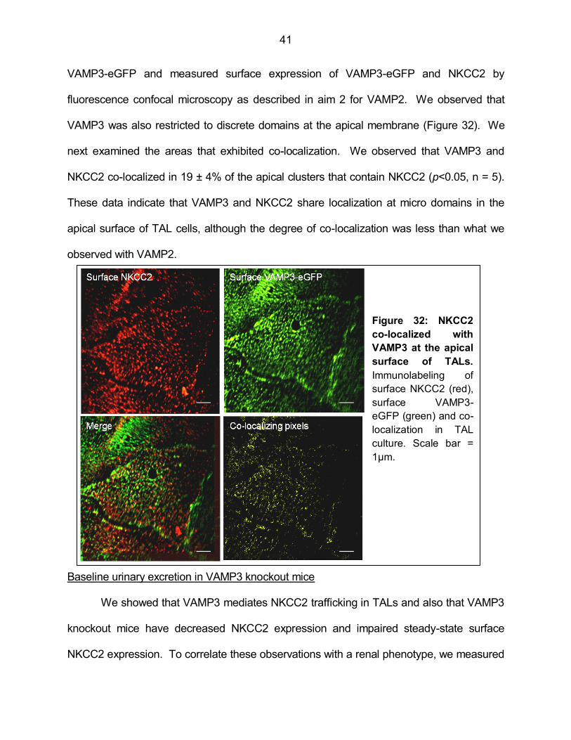
41
VAMP3-eGFP and measured surface expression of VAMP3-eGFP and NKCC2 by
fluorescence confocal microscopy as described in aim 2 for VAMP2. We observed that
VAMP3 was also restricted to discrete domains at the apical membrane (Figure 32). We
next examined the areas that exhibited co-localization. We observed that VAMP3 and
NKCC2 co-localized in 19 ± 4% of the apical clusters that contain NKCC2 (p<0.05, n = 5).
These data indicate that VAMP3 and NKCC2 share localization at micro domains in the
apical surface of TAL cells, although the degree of co-localization was less than what we
observed with VAMP2.
Baseline urinary excretion in VAMP3 knockout mice
We showed that VAMP3 mediates NKCC2 trafficking in TALs and also that VAMP3
knockout mice have decreased NKCC2 expression and impaired steady-state surface
NKCC2 expression. To correlate these observations with a renal phenotype, we measured
Figure 32: NKCC2
co-localized with
VAMP3 at the apical
surface of TALs.
Immunolabeling of
surface NKCC2 (red),
surface VAMP3-
eGFP (green) and co-
localization in TAL
culture. Scale bar =
1µm.

42
urine volume, osmolality and electrolyte excretion in VAMP3 knockout mice. The results
are summarized in Table 1. We observed that compared to wild-type, VAMP3 knockout
mice excreted a slightly higher urine volume, produced more diluted urine and excreted
more Ca (p<0.05, n = 6), consistent with a TAL defect. We did not observe any difference
in urinary excretion of Na, Cl and K. Although this resembles the phenotype of NKCC2
knockout mice (40), we would expect enhanced excretion of these ions if TAL function was
impaired. To test how VAMP3 mice adjust urinary excretion to disturbances of water and
electrolyte homeostasis, we challenged them to water deprivation or low-salt diet in the next
experiments.
Table 1: Urine parameters in wild-type and VAMP3 knockout mice.
* p<0.05
Urinary excretion in VAMP3 knockout mice during 24 hour water deprivation
To further test how renal function in VAMP3 knockout mice responds to a water
restriction challenge, we deprived them of water for 24 hours. In wild type mice, urine
volume decreased from 1.34 ± 0.12 ml to 0.65 ± 0.08 ml (p<0.05) as expected. Urine
volume also decreased in VAMP3 knockout mice from 1.62 ± 0.17 ml to 0.67 ± 0.08 ml
(p<0.05), indicating that VAMP3 knockouts can still prevent fluid loss in the urine when
water supply is limited. Urine osmolality increased in wild type mice from 3433 ± 166
wild-type VAMP3 knockout
water consumed (ml/24 hs) 4.7 0.2 5.1 0.3
urine volume (ul/24 hs) 1337 118 1625 167
urine osmolality (mOsm) 3433 166 2924 131 *UNa (µmol/day) 264.8 18.9 266.3 31.2
UK (µmol/day) 779.8 51.2 781.9 98.2
UCl (µmol/day) 541.4 43.7 611.5 65.8
UCa (µmol/day) 0.60 0.02 0.81 0.05 *UMg (µmol/day) 1.9 0.19 1.73 0.13
UCreat. (mg/day) 0.31 0.02 0.35 0.04
Table 1: Urine parameters in wild-type and VAMP3 knockout mice (*p<0.05)

43
mOsm to 5307 ± 235 mOsm (p<0.05). This response is expected since decreased
excretion of water would produce more concentrated urine. VAMP3 knockout mice,
besides initially producing a more diluted urine of 2924 ± 131 mOsm, they could still
concentrate the urine (p<0.05) to an osmolality comparable with wild types under water
deprivation (5763 ± 456 mOsm). This suggests that VAMP3 do not experience more water
loss than wild-types when they are deprived of drinking water for 24 hours.
Since we did not observe any major change in urinary Na, Cl and K under baseline
conditions, we next tested whether excretion of these ions changed after water deprivation.
In wild type and in knockout mice, urinary Na, Cl and K were increased following water
deprivation (Figure 33). This is a normal response independent of vasopressin (189,190),
probably mediated by atrial natriuretic peptide released from the heart in response to
oxytocin (191-193). We will expand this in the general discussion in Chapter 4. In VAMP3
knockout mice, urinary Na, Cl and K were also increased during water deprivation but to a
greater extent than in wild-type mice (p<0.05, n = 6), indicating that VAMP3 knockouts
excrete excess Na, Cl and K when deprived of water.
0
200
400
600
800
1000
1200
0.9
UC
l (µ
mo
l/d
ay
Wild type
VAMP3 knockout
baseline 24h water
deprivation
B)
0
100
200
300
400
500
600
0.9
UN
a (µ
mo
l/d
ay)
Wild type
VAMP3 knockout
baseline 24h water
deprivation
A) * *
0
200
400
600
800
1000
1200
1400
1600
0.9
UK
(µ
mo
l/d
ay
Wild type
VAMP3 knockout
baseline 24h water
deprivation
C) *
Figure 33: Enhanced urinary Na, Cl and K excretion in VAMP3 knockout mice during
water restriction. A) 24-hour urinary Na excretion, B) 24-hour urinary Cl excretion, C) 24-
hour urinary K excretion during 24-hour water deprivation (*p<0.05 vs. wild-type).
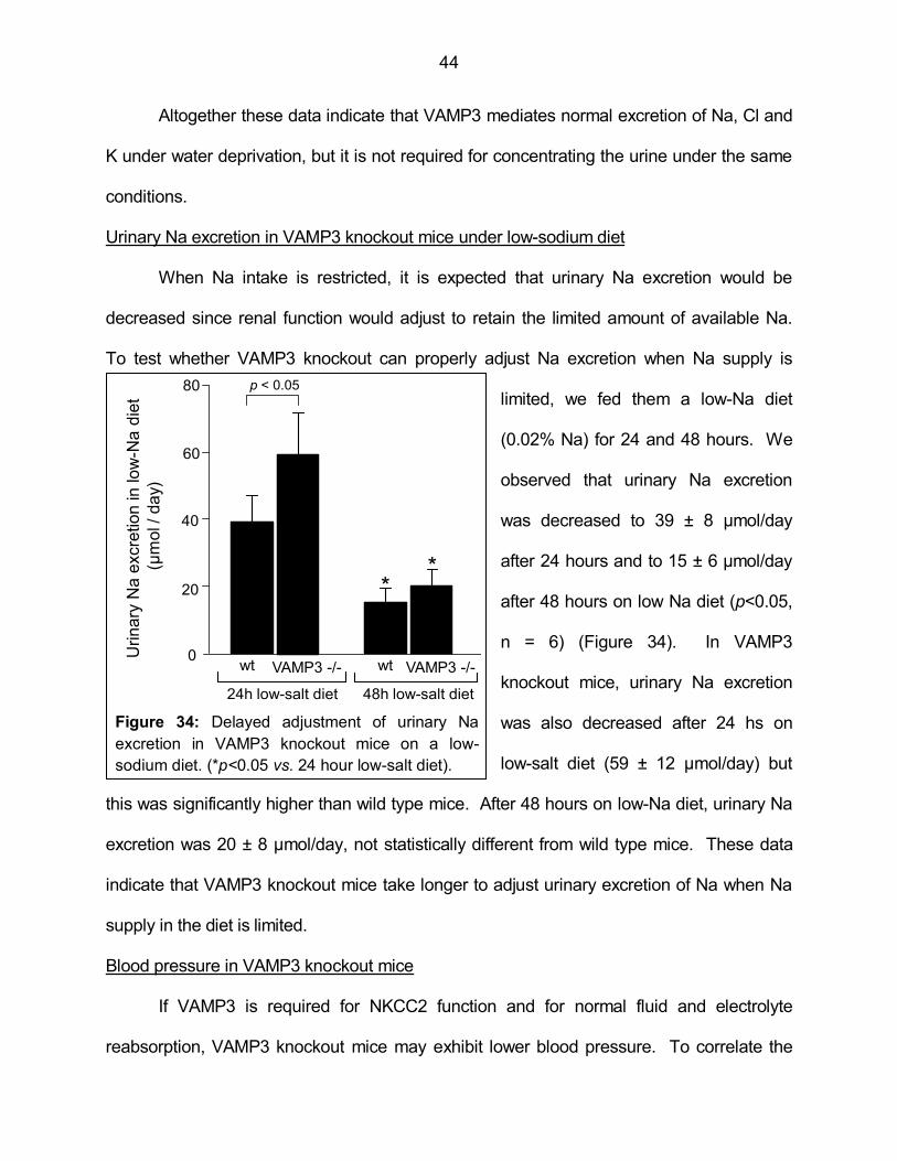
44
Altogether these data indicate that VAMP3 mediates normal excretion of Na, Cl and
K under water deprivation, but it is not required for concentrating the urine under the same
conditions.
Urinary Na excretion in VAMP3 knockout mice under low-sodium diet
When Na intake is restricted, it is expected that urinary Na excretion would be
decreased since renal function would adjust to retain the limited amount of available Na.
To test whether VAMP3 knockout can properly adjust Na excretion when Na supply is
limited, we fed them a low-Na diet
(0.02% Na) for 24 and 48 hours. We
observed that urinary Na excretion
was decreased to 39 ± 8 µmol/day
after 24 hours and to 15 ± 6 µmol/day
after 48 hours on low Na diet (p<0.05,
n = 6) (Figure 34). In VAMP3
knockout mice, urinary Na excretion
was also decreased after 24 hs on
low-salt diet (59 ± 12 µmol/day) but
this was significantly higher than wild type mice. After 48 hours on low-Na diet, urinary Na
excretion was 20 ± 8 µmol/day, not statistically different from wild type mice. These data
indicate that VAMP3 knockout mice take longer to adjust urinary excretion of Na when Na
supply in the diet is limited.
Blood pressure in VAMP3 knockout mice
If VAMP3 is required for NKCC2 function and for normal fluid and electrolyte
reabsorption, VAMP3 knockout mice may exhibit lower blood pressure. To correlate the
wt VAMP3 -/-
20
0
40
60
80
Uri
nary
Na e
xcre
tion in low
-Na d
iet
(µm
ol /
day)
p < 0.05
wt VAMP3 -/-
**
24h low-salt diet 48h low-salt diet
Figure 34: Delayed adjustment of urinary Na
excretion in VAMP3 knockout mice on a low-
sodium diet. (*p<0.05 vs. 24 hour low-salt diet).

45
renal phenotype we observed with differences in
blood pressure, we measured systolic arterial
pressure in VAMP3 knockout mice by tail cuff. We
observed that wild-type mice had an arterial
pressure of 114 ± 2 mmHg, while arterial pressure in
VAMP3 knockouts was 19 mmHg lower (95 ± 2
mmHg, p<0.05, n = 6) (Figure 35). These results
indicate that VAMP3 is required to maintain normal
blood pressure in mice.
Conclusion
We concluded that VAMP3 interacts with NKCC2 as shown by co-
immunoprecipitation in TAL protein lysates. Whether NKCC2-VAMP3 interaction is part of
the mechanism by which VAMP3 mediates NKCC2 trafficking is not clear, but it is a
possibility as discussed for VAMP2. Also, whether NKCC2 and VAMP3 interact at the
surface or at an intracellular localization was not tested. Although we also observed
VAMP3 co-localization with NKCC2 at apical clusters in the TAL surface, this only indicates
close proximity but not necessarily direct physical interaction. However, our data indicate
that NKCC2 and VAMP3 are directed to the same membrane localization, in agreement
with the role of VAMP3 on NKCC2 trafficking we described in aim 3.
We showed for the first time that genetic deletion of VAMP3 causes a renal
phenotype. VAMP3 knockout mice excrete slightly higher urine volume, produce less
concentrated urine and excrete excess Ca under baseline conditions. These data are
consistent with decreased TAL reabsorptive capacity and with the phenotype observed in
NKCC2 knockout mice (40). In addition, when VAMP3 knockout mice were challenged to
0
20
40
60
80
100
120
p < 0.05
Wild
type
VAMP3
knockout
Sys
tolic
blo
od
pre
ssu
re (
mm
Hg)
Figure 28: Decreased systolic blood
pressure in VAMP3 knockout mice.Figure 35: Decreased systolic
blood pressure in VAMP3
knockout mice.
46
water deprivation conditions, they failed to retain Na, Cl and K compared to wild-type mice
in spite of still being able to concentrate the urine. The Na loss in the urine was also
evident after 24 hours in low Na diet, but it recovered to normal levels after 48 hs, indicating
that renal Na excretion took longer to adjust. Finally, VAMP3 knockout mice had lower
systolic blood pressure as expected from the excess fluid and electrolyte excretion we
observed in the urine. Altogether, we conclude that VAMP3 is required for normal renal
function and maintenance of blood pressure. The phenotype of VAMP3 knockout mice will
be discussed in detail and in a broader context later in Chapter 4.

47
CHAPTER 4
CONCLUDING REMARKS
Summary of results
In this study we have shown that:
1. VAMP2, VAMP3, VAMP7 and VAMP8 are expressed in the TAL.
2. VAMP2 mediates cAMP-stimulated exocytic delivery of NKCC2 to the TAL surface
while VAMP3 mediates constitutive delivery of NKCC2 to the apical surface.
3. VAMP2 mediates cAMP-stimulated steady-state surface NKCC2 expression in TALs
while VAMP3 mediates constitutive steady-state surface NKCC2 expression.
4. Tetanus toxin decreases constitutive steady-state surface NKCC2 expression.
5. VAMP2 and VAMP3 co-immunoprecipitate with NKCC2 in TALs.
6. VAMP2 interacts with a region of the carboxy-terminus of NKCC2 previously shown
to determine apical targeting.
7. NKCC2 is distributed in discrete micro domains at the apical surface of TALs.
8. VAMP2 and VAMP3 co-localize with NKCC2 at micro domains in the apical surface
of TALs.
9. cAMP stimulates VAMP2 exocytic delivery in TALs, enhances VAMP2 presence at
the apical surface, and promotes NKCC2-VAMP2 interaction.
10. VAMP3 is required for normal NKCC2 expression, urine concentrating ability, water
and salt reabsorption, and to maintain normal blood pressure.
Proposed model
Based on our data we propose the model depicted in Figure 36. In TALs, NKCC2
may be segregated into two intracellular pools. One pool is associated with VAMP2 and
responds to cAMP stimulation to be delivered into the apical membrane. The other pool
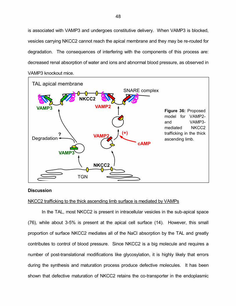
48
is associated with VAMP3 and undergoes constitutive delivery. When VAMP3 is blocked,
vesicles carrying NKCC2 cannot reach the apical membrane and they may be re-routed for
degradation. The consequences of interfering with the components of this process are:
decreased renal absorption of water and ions and abnormal blood pressure, as observed in
VAMP3 knockout mice.
Discussion
NKCC2 trafficking to the thick ascending limb surface is mediated by VAMPs
In the TAL, most NKCC2 is present in intracellular vesicles in the sub-apical space
(76), while about 3-5% is present at the apical cell surface (14). However, this small
proportion of surface NKCC2 mediates all of the NaCl absorption by the TAL and greatly
contributes to control of blood pressure. Since NKCC2 is a big molecule and requires a
number of post-translational modifications like glycosylation, it is highly likely that errors
during the synthesis and maturation process produce defective molecules. It has been
shown that defective maturation of NKCC2 retains the co-transporter in the endoplasmic
NKCC2
(+)VAMP2
SNARE complex
VAMP3
cAMPDegradation
Figure 29: Proposed model for VAMP2- and VAMP3-
mediated NKCC2 trafficking in the thick ascending limb.
TGN
?
NKCC2
TAL apical membrane
VAMP2VAMP3Figure 36: Proposed
model for VAMP2-
and VAMP3-
mediated NKCC2
trafficking in the thick
ascending limb.
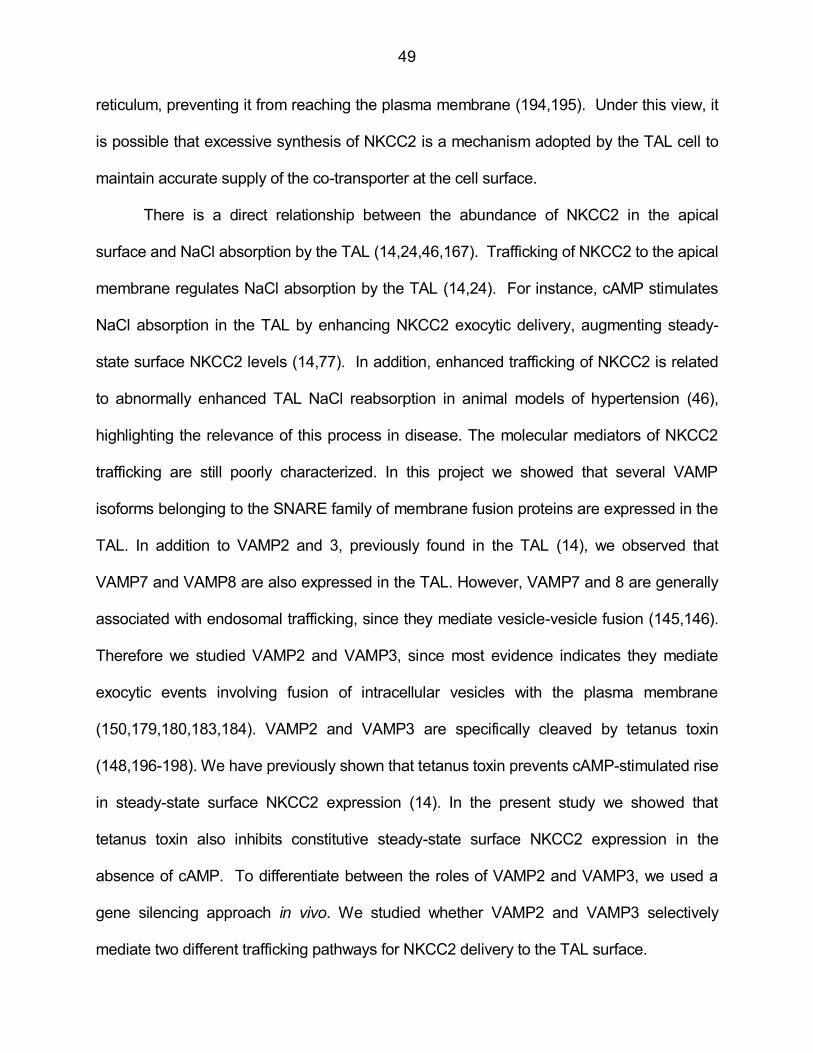
49
reticulum, preventing it from reaching the plasma membrane (194,195). Under this view, it
is possible that excessive synthesis of NKCC2 is a mechanism adopted by the TAL cell to
maintain accurate supply of the co-transporter at the cell surface.
There is a direct relationship between the abundance of NKCC2 in the apical
surface and NaCl absorption by the TAL (14,24,46,167). Trafficking of NKCC2 to the apical
membrane regulates NaCl absorption by the TAL (14,24). For instance, cAMP stimulates
NaCl absorption in the TAL by enhancing NKCC2 exocytic delivery, augmenting steady-
state surface NKCC2 levels (14,77). In addition, enhanced trafficking of NKCC2 is related
to abnormally enhanced TAL NaCl reabsorption in animal models of hypertension (46),
highlighting the relevance of this process in disease. The molecular mediators of NKCC2
trafficking are still poorly characterized. In this project we showed that several VAMP
isoforms belonging to the SNARE family of membrane fusion proteins are expressed in the
TAL. In addition to VAMP2 and 3, previously found in the TAL (14), we observed that
VAMP7 and VAMP8 are also expressed in the TAL. However, VAMP7 and 8 are generally
associated with endosomal trafficking, since they mediate vesicle-vesicle fusion (145,146).
Therefore we studied VAMP2 and VAMP3, since most evidence indicates they mediate
exocytic events involving fusion of intracellular vesicles with the plasma membrane
(150,179,180,183,184). VAMP2 and VAMP3 are specifically cleaved by tetanus toxin
(148,196-198). We have previously shown that tetanus toxin prevents cAMP-stimulated rise
in steady-state surface NKCC2 expression (14). In the present study we showed that
tetanus toxin also inhibits constitutive steady-state surface NKCC2 expression in the
absence of cAMP. To differentiate between the roles of VAMP2 and VAMP3, we used a
gene silencing approach in vivo. We studied whether VAMP2 and VAMP3 selectively
mediate two different trafficking pathways for NKCC2 delivery to the TAL surface.
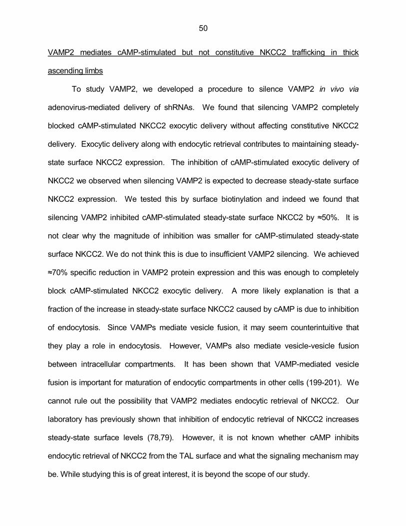
50
VAMP2 mediates cAMP-stimulated but not constitutive NKCC2 trafficking in thick
ascending limbs
To study VAMP2, we developed a procedure to silence VAMP2 in vivo via
adenovirus-mediated delivery of shRNAs. We found that silencing VAMP2 completely
blocked cAMP-stimulated NKCC2 exocytic delivery without affecting constitutive NKCC2
delivery. Exocytic delivery along with endocytic retrieval contributes to maintaining steady-
state surface NKCC2 expression. The inhibition of cAMP-stimulated exocytic delivery of
NKCC2 we observed when silencing VAMP2 is expected to decrease steady-state surface
NKCC2 expression. We tested this by surface biotinylation and indeed we found that
silencing VAMP2 inhibited cAMP-stimulated steady-state surface NKCC2 by ≈50%. It is
not clear why the magnitude of inhibition was smaller for cAMP-stimulated steady-state
surface NKCC2. We do not think this is due to insufficient VAMP2 silencing. We achieved
≈70% specific reduction in VAMP2 protein expression and this was enough to completely
block cAMP-stimulated NKCC2 exocytic delivery. A more likely explanation is that a
fraction of the increase in steady-state surface NKCC2 caused by cAMP is due to inhibition
of endocytosis. Since VAMPs mediate vesicle fusion, it may seem counterintuitive that
they play a role in endocytosis. However, VAMPs also mediate vesicle-vesicle fusion
between intracellular compartments. It has been shown that VAMP-mediated vesicle
fusion is important for maturation of endocytic compartments in other cells (199-201). We
cannot rule out the possibility that VAMP2 mediates endocytic retrieval of NKCC2. Our
laboratory has previously shown that inhibition of endocytic retrieval of NKCC2 increases
steady-state surface levels (78,79). However, it is not known whether cAMP inhibits
endocytic retrieval of NKCC2 from the TAL surface and what the signaling mechanism may
be. While studying this is of great interest, it is beyond the scope of our study.
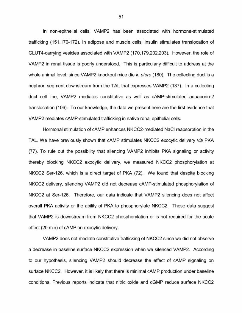
51
In non-epithelial cells, VAMP2 has been associated with hormone-stimulated
trafficking (151,170-172). In adipose and muscle cells, insulin stimulates translocation of
GLUT4-carrying vesicles associated with VAMP2 (170,179,202,203). However, the role of
VAMP2 in renal tissue is poorly understood. This is particularly difficult to address at the
whole animal level, since VAMP2 knockout mice die in utero (180). The collecting duct is a
nephron segment downstream from the TAL that expresses VAMP2 (137). In a collecting
duct cell line, VAMP2 mediates constitutive as well as cAMP-stimulated aquaporin-2
translocation (106). To our knowledge, the data we present here are the first evidence that
VAMP2 mediates cAMP-stimulated trafficking in native renal epithelial cells.
Hormonal stimulation of cAMP enhances NKCC2-mediated NaCl reabsorption in the
TAL. We have previously shown that cAMP stimulates NKCC2 exocytic delivery via PKA
(77). To rule out the possibility that silencing VAMP2 inhibits PKA signaling or activity
thereby blocking NKCC2 exocytic delivery, we measured NKCC2 phosphorylation at
NKCC2 Ser-126, which is a direct target of PKA (72). We found that despite blocking
NKCC2 delivery, silencing VAMP2 did not decrease cAMP-stimulated phosphorylation of
NKCC2 at Ser-126. Therefore, our data indicate that VAMP2 silencing does not affect
overall PKA activity or the ability of PKA to phosphorylate NKCC2. These data suggest
that VAMP2 is downstream from NKCC2 phosphorylation or is not required for the acute
effect (20 min) of cAMP on exocytic delivery.
VAMP2 does not mediate constitutive trafficking of NKCC2 since we did not observe
a decrease in baseline surface NKCC2 expression when we silenced VAMP2. According
to our hypothesis, silencing VAMP2 should decrease the effect of cAMP signaling on
surface NKCC2. However, it is likely that there is minimal cAMP production under baseline
conditions. Previous reports indicate that nitric oxide and cGMP reduce surface NKCC2

52
and NKCC2 activity by decreasing intracellular cAMP concentration below baseline levels
(15,24). This seems to contradict our proposed model since by silencing VAMP2 we would
expect a decrease in NKCC2 trafficking due to reduction of cAMP signaling below baseline
levels. However it is possible that reduction in cAMP and increases in cAMP mediate
different signaling pathways. In fact, the PKA inhibitor H-89 does not decrease baseline
steady-state surface NKCC2 expression in TALs (69,77). It is possible that PKA mediates
cAMP stimulation of NKCC2 trafficking, but it does not maintain constitutive NKCC2
trafficking. The cAMP signaling pathway by which nitric oxide and cGMP decrease surface
NKCC2 is still not clear. We propose that the VAMP2-dependent pathway for NKCC2
trafficking may be mediated by PKA, but it should not influence constitutive NKCC2
trafficking.
In conclusion, we have described for the first time a VAMP2-mediated pathway in
renal epithelial cells that specifically mediates cAMP-stimulated but not constitutive NKCC2
trafficking.
VAMP3 mediates constitutive but not cAMP-stimulated NKCC2 trafficking in thick
ascending limbs
Tetanus toxin cleaves VAMP2 and VAMP3 and we found that VAMP2 does not
mediate constitutive NKCC2 trafficking. Thus, our data suggest that VAMP3 mediates
constitutive NKCC2 trafficking. To directly address this, we silenced VAMP3 in vivo in
TALs via adenoviruses-delivered shRNA. We achieved efficient and specific silencing, with
almost 70% decrease in VAMP3 expression and no change in other VAMP isoforms. We
observed that VAMP3 mediates constitutive exocytic delivery of NKCC2 and steady-state
surface expression. In addition, VAMP3 does not mediate cAMP-stimulated exocytic
delivery or steady-state surface NKCC2 expression, which we showed is mediated by
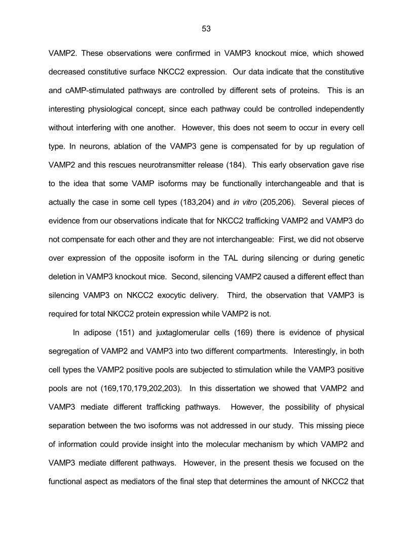
53
VAMP2. These observations were confirmed in VAMP3 knockout mice, which showed
decreased constitutive surface NKCC2 expression. Our data indicate that the constitutive
and cAMP-stimulated pathways are controlled by different sets of proteins. This is an
interesting physiological concept, since each pathway could be controlled independently
without interfering with one another. However, this does not seem to occur in every cell
type. In neurons, ablation of the VAMP3 gene is compensated for by up regulation of
VAMP2 and this rescues neurotransmitter release (184). This early observation gave rise
to the idea that some VAMP isoforms may be functionally interchangeable and that is
actually the case in some cell types (183,204) and in vitro (205,206). Several pieces of
evidence from our observations indicate that for NKCC2 trafficking VAMP2 and VAMP3 do
not compensate for each other and they are not interchangeable: First, we did not observe
over expression of the opposite isoform in the TAL during silencing or during genetic
deletion in VAMP3 knockout mice. Second, silencing VAMP2 caused a different effect than
silencing VAMP3 on NKCC2 exocytic delivery. Third, the observation that VAMP3 is
required for total NKCC2 protein expression while VAMP2 is not.
In adipose (151) and juxtaglomerular cells (169) there is evidence of physical
segregation of VAMP2 and VAMP3 into two different compartments. Interestingly, in both
cell types the VAMP2 positive pools are subjected to stimulation while the VAMP3 positive
pools are not (169,170,179,202,203). In this dissertation we showed that VAMP2 and
VAMP3 mediate different trafficking pathways. However, the possibility of physical
separation between the two isoforms was not addressed in our study. This missing piece
of information could provide insight into the molecular mechanism by which VAMP2 and
VAMP3 mediate different pathways. However, in the present thesis we focused on the
functional aspect as mediators of the final step that determines the amount of NKCC2 that
54
reaches the plasma membrane.
Considerations about the separate roles of VAMP2 and VAMP3 in NKCC2 trafficking
The concept that constitutive and cAMP-stimulated trafficking of NKCC2 is
controlled by distinct proteins mediating two separate pathways can have important
ramifications. The only NKCC2 inhibitors currently used in the clinic are loop diuretics like
furosemide, bumetanide and torasemide. They are among the first choices for conditions
like congestive heart failure and edema, when a dramatic reduction in extracellular volume
is required. However, long term use to treat hypertension is currently limited due to the high
potency of these drugs, which leads to severe side effects associated with the extreme loss
of fluid and electrolytes. The problem with current NKCC2 inhibitors is that they do not
discriminate between the constitutive and stimulated pathways. In this dissertation we took
the first step in identifying the molecular components that mediate constitutive and cAMP-
stimulated NKCC2 trafficking. Further characterization of additional components will lead to
the identification of selective targets in each pathway. Targeted therapeutic intervention in
one pathway could provide the benefits of an effective loop diuretic, while leaving the other
pathway undisturbed could minimize the side effects.
One step closer towards the characterization of the VAMP2 and the VAMP3-
mediated pathways is to determine whether they are associated with two separate NKCC2
pools. In other cells, VAMP2 and VAMP3 are physically segregated in two different vesicle
populations (151,169). This can be of importance since preliminary experiments in our
laboratory indicate that VAMP2 co-localizes with Rab-11 in TALs, a marker of recycling
endosomes (unpublished). It is possible that the identity of the VAMP2 and VAMP3-
positive compartments may be different, associated with the distinct roles in NKCC2
trafficking. However we did not address this further in our study.
55
It may be possible to purify vesicles carrying VAMP2 or VAMP3 from TALs by
immunoprecipitation as performed previously in collecting duct preparations (137,152).
Then it should be possible to determine whether these vesicles also express NKCC2
and the other VAMP isoform. Alternatively, co-localization of VAMP2 and VAMP3 in the
same vesicles could be addressed by electron microscopy. We did not test whether
VAMP2 and VAMP3 are localized in different NKCC2-positive compartments. However,
this does not have to be necessarily the case since the two VAMP isoforms can be in the
same vesicle and one of them can be inactive. For instance, in parotid acinar cells,
VAMP2-mediated exocytosis of amylase granules is constitutively repressed by an
inhibitory protein that binds the SNARE domain of VAMP2 (207). This inhibition is released
after activation of the PKA pathway and amylase granules get exocytosed. Unfortunately, it
was not studied whether amylase granules also contained other VAMP isoforms.
If NKCC2 is differentially associated with compartments of different molecular
identity, this could be due to deliberate sorting of NKCC2 into two or more different
intracellular pools. To our knowledge, there are no studies to this respect in the TAL.
Alternatively, the NKCC2 compartments could change composition with time, in this way
they may acquire a different VAMP isoform at a later stage in the maturation process. The
age of vesicles in different cell systems has been monitored by using a special fluorescent
protein that changes the emission wavelength from green to red as the vesicles age (208).
In astrocytes, as VAMP2 vesicles age, they are sorted into two spatially separated
populations (209). Similarly, secretory granules in chromafin cells are also segregated
according to their age (210). Interestingly, some stimuli selectively produce the release of
younger vesicles, whereas other stimuli preferentially cause the release of older vesicles.
Whether this temporal segregation occurs in NKCC2 vesicles associated with VAMP2 or
56
VAMP3 is an interesting possibility that remains to be studied.
VAMP3 but not VAMP2 is required for normal NKCC2 expression
While silencing VAMP2 did not affect total NKCC2 expression, silencing VAMP3
decreased total NKCC2 by 45%. NKCC2 expression was also decreased by 66% in
VAMP3 knockout mice. We concluded that VAMP3 but not VAMP2 is required for normal
levels of NKCC2 protein expression. However, tetanus toxin did not decrease total NKCC2
expression in TALs. This is likely due to the shorter time required for treatment with tetanus
toxin (50 minutes) compared to the three days required for VAMP3 silencing. We
speculate that VAMP3 is required for maintenance of normal levels of NKCC2 in a process
that takes longer than one hour.
There are at least three possibilities by which VAMP3 could regulate NKCC2
expression. First, VAMP3 could be required for NKCC2 gene expression. However, to our
knowledge, there is no evidence in the literature implicating SNAREs as mediators of gene
transcription and/or translation. Second, VAMP3 could be required for endosome
maturation and when VAMP3 is silenced, NKCC2 maturation is interrupted. Traditionally,
VAMP7 (145) and VAMP8 (146) have been considered the VAMPs involved in endosome
maturation, with early reports indicating that VAMP3 does not play any role in intracellular
endosome fusion events (211). However, increasing amounts of evidence indicate that
VAMP3 mediates several vesicle-vesicle fusion processes in the endosomal pathway (212-
215). Recent reports have found that VAMP3 mediates autophagosome maturation in
cultured cell lines (199,214). We cannot discard this possibility based on our experimental
design. The third possibility is that blockade of VAMP3 function prevents NKCC2 from
reaching the plasma membrane and the molecules retained in the intracellular
compartment are re-routed for degradation. Our data showing that VAMP3 silencing

57
completely prevents NKCC2 exocytic delivery suggest this may be the case. One way of
addressing the role of VAMP3 in maintaining NKCC2 expression is by pulse-chase
experiments that can be performed in TAL cultures. This would allow distinguishing
between NKCC2 synthesis/maturation and degradation. If VAMP3 mediates NKCC2
synthesis or maturation, silencing VAMP3 would produce a decrease in the rate of
appearance of either the mature or immature NKCC2 form in pulse-chase experiments.
Alternatively, if preventing NKCC2 from reaching the cell surface when VAMP3 is silenced
causes re-direction of NKCC2 to degradation, we would expect enhanced disappearance
of labeled NKCC2 in pulse-chase experiments. Although these studies would provide a
more complete picture of the mechanism we propose, they do not contribute to the focused
hypothesis we tested here. We propose the pulse-chase studies as a future direction along
with additional experiments to test a different hypothesis aimed to elucidate the mechanism
for VAMP3-mediated NKCC2 trafficking.
The fact that constitutive NKCC2 exocytic delivery is almost completely inhibited
when silencing VAMP3 also indicates that the decrease in steady-state surface NKCC2 is
not just due to a proportional decrease in total NKCC2 protein expression. If this was the
case, there is no reason to expect an 85% decrease in the rate of exocytic delivery as we
observed.
NKCC2 co-localizes with VAMP2 and VAMP3 at apical surface clusters in thick ascending
limbs
It is known that after vesicle fusion, VAMPs will remain inserted in the plasma
membrane until they are retrieved to be re-used in another round of vesicle fusion (81,216).
This allowed us to detect VAMP2 and VAMP3 at the plasma membrane after exocytosis
has occurred. We used VAMP2-GFP and VAMP3-GFP constructs to study their surface

58
localization in primary cultures and whether they co-localized with surface NKCC2. We
performed our studies in polarized TAL cultures that retain the characteristics of native
tissue. This setup allowed us to easily access the apical membrane. We described for the
first time that NKCC2 exhibits a heterogeneous distribution pattern at the apical membrane,
being restricted to discrete micro domains or clusters. NKCC2 co-localized with VAMP2
and to a lesser extent with VAMP3 in those surface clusters.
The biological significance of this membrane compartmentalization is not clear. In
hypocampal neurons, the Kv2.1 potassium channel is also distributed in clusters composed
of non-conducting channels (217) that serve as platforms for exocytosis of Kv2.1 and Kv1.4
channels (218). Whether NKCC2 clusters conduct ions or serve as platforms for
exocytosis in the TAL is an interesting possibility but it remains to be tested.
We observed that VAMP2 and VAMP3 are also distributed in apical micro domains.
There is at least one previous report showing that in a cultured adrenal gland cell line, the
site of exocytic vesicle fusion is highly dependent on the density of plasma membrane-
resident SNAREs (syntaxin 1) (219). This prompted us to speculate that VAMP2- and
VAMP3-mediated exocytosis and delivery of NKCC2 may occur at defined domains at the
cell surface. An alternative interpretation for the clustered distribution of VAMP2 and
VAMP3 is that once they reach the membrane, they undergo translational movement and
coalesce into high density areas as described for other plasma membrane proteins (220-
225). These high density areas can be sites of enhanced endocytosis (223,226-228).
Translational mobility of transmembrane proteins in the plane of the membrane is
determined by the cytoskeleton (222,229-231) and their interaction with lipids. VAMP2 and
VAMP3 (232), as well as other SNAREs (233-235) have been reported to associate with
the cytoskeleton in other cell types, suggesting they might be anchored or retained in a
59
micro domain after exocytosis.
Our co-localization analysis shows that NKCC2 shares an apical location with
VAMP2 and VAMP3 at surface clusters. Whether VAMP2 and VAMP3 are present in the
same or different clusters was not addressed in our study. Also, the resolution of confocal
microscopy only allowed us to conclude that they share an apical location. Whether they
are close enough at the clusters to physically interact with each other is not known. In
neurons, VAMP2 interacts with potassium channels and this interaction modulates channel
gating at the plasma membrane (236,159). This kind of regulation is not necessarily linked
to trafficking, but it can be direct modulation of the intrinsic activity of the channel. Whether
VAMPs directly regulate intrinsic NKCC2 activity was not addressed in this project.
Studying the dynamics of surface NKCC2, surface VAMP2 and VAMP3 micro
domains is an interesting future direction in understanding the molecular regulation of
NKCC2-mediated TAL function and renal physiology.
NKCC2 interacts with VAMP2 and VAMP3 in the thick ascending limb
We showed here that VAMP2 and VAMP3 mediate NKCC2 trafficking in TALs. To
clarify how this may occur, we explored the possibility of protein-protein interaction between
NKCC2 and these two VAMP isoforms. We showed by co-immunoprecipitation that
VAMP2 and VAMP3 interact with NKCC2 in the TAL. To our knowledge, there are no
reports in the literature of physical interaction between VAMP3 and non-SNARE proteins.
However, in neurons VAMP2 physically interacts with P/Q-type Ca channels (158) and
Kv2.1 potassium channels (159,160). The significance of these interactions is that they
modulate channel activity. VAMP2 interacts with the cytoplasmic amino-terminus of Kv2.1,
interfering with binding of a regulatory carboxy-terminal domain (236) and enhancing
inactivation of channel gating (159). This effect is independent of channel trafficking to the

60
plasma membrane, suggesting that VAMPs may have a direct regulatory role not related
with vesicle fusion. We showed that VAMP2 and VAMP3 mediate NKCC2 trafficking, but
we cannot rule out a possible role as direct regulators of NKCC2 ion transport activity.
Here we also showed that VAMP2 interacts with a cytoplasmic domain in NKCC2.
The functional significance of this was not directly addressed in our studies. However, this
interaction region is a carboxy-terminal domain in NKCC2 previously shown to contain a
signal that directs NKCC2 to the apical membrane (177). Also, this carboxy-terminal region
has been previously reported to interact with MAL (163), a protein that inhibits NKCC2
endocytic retrieval, and with moesin (164), which enhances NKCC2 delivery to the surface.
Both interacting proteins have the effect of increasing surface NKCC2 expression. This
suggests that VAMP2 interaction with a targeting signal in NKCC2 may be important in
regulated NKCC2 trafficking. It is known that incorrect targeting of NKCC2 to the wrong
cellular localization has detrimental consequences for NKCC2-mediated NaCl transport
(42,194). Another carboxy-terminal region, more proximal to the transmembrane portion of
NKCC2, interacts with SCAMP2 (166) and Aldolase B (165). SCAMP2 inhibits NKCC2
exocytic delivery (166) and Aldolase B stimulates NKCC2 endocytosis (165) in cultured
cells, both having the effect of decreasing the abundance of NKCC2 at the plasma
membrane. Preliminary GST pull down experiments from our laboratory indicate that
VAMP2 does not interact with the rest of the carboxy-terminus of NKCC2. However, the
tree-dimensional structure of the whole NKCC2 molecule may have an influence in the
interaction with VAMP2. This information may be overlooked when using fragments for
GST pull downs. NKCC2-VAMP3 interaction presents a similar problem. We were not able
to identify the region of NKCC2 that interacts with VAMP3 in our preliminary GST pull
downs with the different NKCC2 fragments (not shown). There are three possibilities that

61
could explain this: 1) the interacting region falls in between the segments we used, 2)
NKCC2 interaction with VAMP3 requires bigger portions or the whole NKCC2 molecule, 3)
VAMP3 interacts with the transmembrane domains of NKCC2 (the amino- and carboxy-
terminal portions of NKCC2 are cytoplasmic).
We propose that NKCC2 interactions with VAMP2 and VAMP3 may be part of the
mechanism that regulates NKCC2 exocytic delivery to the TAL surface. However, we did
not directly address the significance of these interactions. Although this goes beyond the
scope of our hypothesis, addressing this is important to differentiate the roles of VAMP2
and VAMP3 in NKCC2 trafficking from a possible role in direct regulation of ion transport
activity.
cAMP stimulates VAMP2 exocytosis, VAMP2-NKCC2 interaction and apical surface co-
localization
We showed that VAMP2 mediates cAMP-stimulated NKCC2 trafficking. We also
showed that VAMP2 reaches the apical surface and is located in clusters. Therefore we
next tested whether cAMP enhances delivery of VAMP2 to the apical TAL surface. We
found that cAMP almost doubled the rate of VAMP2 delivery to the TAL surface as tested
by surface biotinylation. We also found by confocal microscopy that cAMP enhanced co-
localization of surface NKCC2 with VAMP2 at apical membrane clusters. Interestingly,
cAMP did not affect the clustered distribution pattern of surface VAMP2 or NKCC2. This
may be an indication that VAMP2 exocytosis occurs preferentially at these clusters. In order
to confirm this directly, real time monitoring of active VAMP2 exocytosis may be required.
In the TAL, cAMP stimulates NKCC2-mediated NaCl absorption via PKA (16,18).
Also, we have previously shown that cAMP stimulates NKCC2 exocytic delivery via PKA
(74,77,80). Here we found that stimulation of PKA with a cAMP analogue enhanced
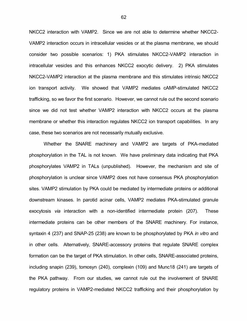
62
NKCC2 interaction with VAMP2. Since we are not able to determine whether NKCC2-
VAMP2 interaction occurs in intracellular vesicles or at the plasma membrane, we should
consider two possible scenarios: 1) PKA stimulates NKCC2-VAMP2 interaction in
intracellular vesicles and this enhances NKCC2 exocytic delivery. 2) PKA stimulates
NKCC2-VAMP2 interaction at the plasma membrane and this stimulates intrinsic NKCC2
ion transport activity. We showed that VAMP2 mediates cAMP-stimulated NKCC2
trafficking, so we favor the first scenario. However, we cannot rule out the second scenario
since we did not test whether VAMP2 interaction with NKCC2 occurs at the plasma
membrane or whether this interaction regulates NKCC2 ion transport capabilities. In any
case, these two scenarios are not necessarily mutually exclusive.
Whether the SNARE machinery and VAMP2 are targets of PKA-mediated
phosphorylation in the TAL is not known. We have preliminary data indicating that PKA
phosphorylates VAMP2 in TALs (unpublished). However, the mechanism and site of
phosphorylation is unclear since VAMP2 does not have consensus PKA phosphorylation
sites. VAMP2 stimulation by PKA could be mediated by intermediate proteins or additional
downstream kinases. In parotid acinar cells, VAMP2 mediates PKA-stimulated granule
exocytosis via interaction with a non-identified intermediate protein (207). These
intermediate proteins can be other members of the SNARE machinery. For instance,
syntaxin 4 (237) and SNAP-25 (238) are known to be phosphorylated by PKA in vitro and
in other cells. Alternatively, SNARE-accessory proteins that regulate SNARE complex
formation can be the target of PKA stimulation. In other cells, SNARE-associated proteins,
including snapin (239), tomosyn (240), complexin (109) and Munc18 (241) are targets of
the PKA pathway. From our studies, we cannot rule out the involvement of SNARE
regulatory proteins in VAMP2-mediated NKCC2 trafficking and their phosphorylation by
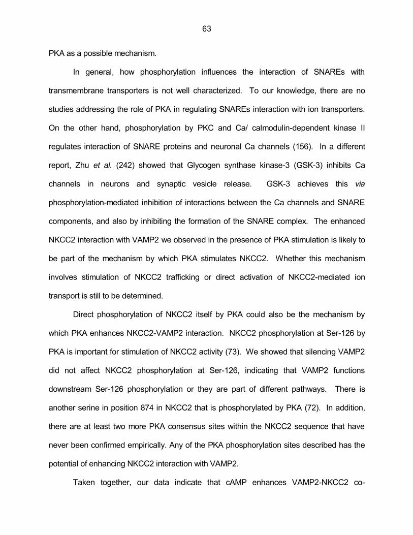
63
PKA as a possible mechanism.
In general, how phosphorylation influences the interaction of SNAREs with
transmembrane transporters is not well characterized. To our knowledge, there are no
studies addressing the role of PKA in regulating SNAREs interaction with ion transporters.
On the other hand, phosphorylation by PKC and Ca/ calmodulin-dependent kinase II
regulates interaction of SNARE proteins and neuronal Ca channels (156). In a different
report, Zhu et al. (242) showed that Glycogen synthase kinase-3 (GSK-3) inhibits Ca
channels in neurons and synaptic vesicle release. GSK-3 achieves this via
phosphorylation-mediated inhibition of interactions between the Ca channels and SNARE
components, and also by inhibiting the formation of the SNARE complex. The enhanced
NKCC2 interaction with VAMP2 we observed in the presence of PKA stimulation is likely to
be part of the mechanism by which PKA stimulates NKCC2. Whether this mechanism
involves stimulation of NKCC2 trafficking or direct activation of NKCC2-mediated ion
transport is still to be determined.
Direct phosphorylation of NKCC2 itself by PKA could also be the mechanism by
which PKA enhances NKCC2-VAMP2 interaction. NKCC2 phosphorylation at Ser-126 by
PKA is important for stimulation of NKCC2 activity (73). We showed that silencing VAMP2
did not affect NKCC2 phosphorylation at Ser-126, indicating that VAMP2 functions
downstream Ser-126 phosphorylation or they are part of different pathways. There is
another serine in position 874 in NKCC2 that is phosphorylated by PKA (72). In addition,
there are at least two more PKA consensus sites within the NKCC2 sequence that have
never been confirmed empirically. Any of the PKA phosphorylation sites described has the
potential of enhancing NKCC2 interaction with VAMP2.
Taken together, our data indicate that cAMP enhances VAMP2-NKCC2 co-
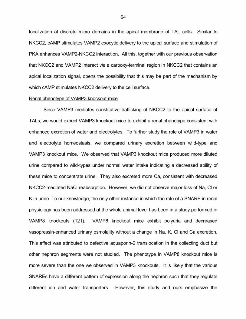
64
localization at discrete micro domains in the apical membrane of TAL cells. Similar to
NKCC2, cAMP stimulates VAMP2 exocytic delivery to the apical surface and stimulation of
PKA enhances VAMP2-NKCC2 interaction. All this, together with our previous observation
that NKCC2 and VAMP2 interact via a carboxy-terminal region in NKCC2 that contains an
apical localization signal, opens the possibility that this may be part of the mechanism by
which cAMP stimulates NKCC2 delivery to the cell surface.
Renal phenotype of VAMP3 knockout mice
Since VAMP3 mediates constitutive trafficking of NKCC2 to the apical surface of
TALs, we would expect VAMP3 knockout mice to exhibit a renal phenotype consistent with
enhanced excretion of water and electrolytes. To further study the role of VAMP3 in water
and electrolyte homeostasis, we compared urinary excretion between wild-type and
VAMP3 knockout mice. We observed that VAMP3 knockout mice produced more diluted
urine compared to wild-types under normal water intake indicating a decreased ability of
these mice to concentrate urine. They also excreted more Ca, consistent with decreased
NKCC2-mediated NaCl reabsorption. However, we did not observe major loss of Na, Cl or
K in urine. To our knowledge, the only other instance in which the role of a SNARE in renal
physiology has been addressed at the whole animal level has been in a study performed in
VAMP8 knockouts (121). VAMP8 knockout mice exhibit polyuria and decreased
vasopressin-enhanced urinary osmolality without a change in Na, K, Cl and Ca excretion.
This effect was attributed to defective aquaporin-2 translocation in the collecting duct but
other nephron segments were not studied. The phenotype in VAMP8 knockout mice is
more severe than the one we observed in VAMP3 knockouts. It is likely that the various
SNAREs have a different pattern of expression along the nephron such that they regulate
different ion and water transporters. However, this study and ours emphasize the

65
importance of VAMPs in the ability of the kidney to reabsorb water and salt.
VAMP3 knockout mice excreted a urine volume that was 21% higher than wild-
types. However, it did not reach statistical significance. Whether this increase is enough to
cause the observed decrease in urine osmolality is not clear. We only observed enhanced
excretion of Ca, whose contribution to urine osmolality is minimal compared to other ions.
We did not observe any change in excretion of Na, Cl and K. However, we did not
measure other osmolytes like urea, which can account for the lower urine osmolality in
VAMP3 knockout mice.
Overall, the renal phenotype exhibited by VAMP3 knockouts is mild. This is not
surprising, since these mice have previously been shown to display normal constitutive and
insulin-stimulated trafficking (186), normal granule content release from platelets (188),
normal phagocytosis in macrophages (187) and their overall health we and others
observed is good. It is possible that other VAMPs compensate for the loss of VAMP3.
There is evidence that VAMP2 compensates for lost VAMP3 function in nerve cells in the
VAMP3 knockout (184). The opposite does not occur during VAMP2 genetic deletion,
which is lethal (180). In the TAL we observed little evidence of compensation by VAMP2.
However, silencing VAMP3 by ≈70% decreased steady-state surface NKCC2 by 43% and
genetically deleting VAMP3 decreased steady-state surface NKCC2 by 46%, suggesting
that another VAMP different than VAMP2 could compensate part of VAMP3 function in
knockout mice.
Fluid and Ca loss stimulate the cAMP pathway in the distal nephron by enhancing
circulating vasopressin and parathyroid hormone (PTH) respectively. Stimulation of cAMP
in the TAL would increase VAMP2-mediated NKCC2 trafficking and activity. We discuss
the possible compensatory mechanisms for water and ion handling separately (Figure 37).
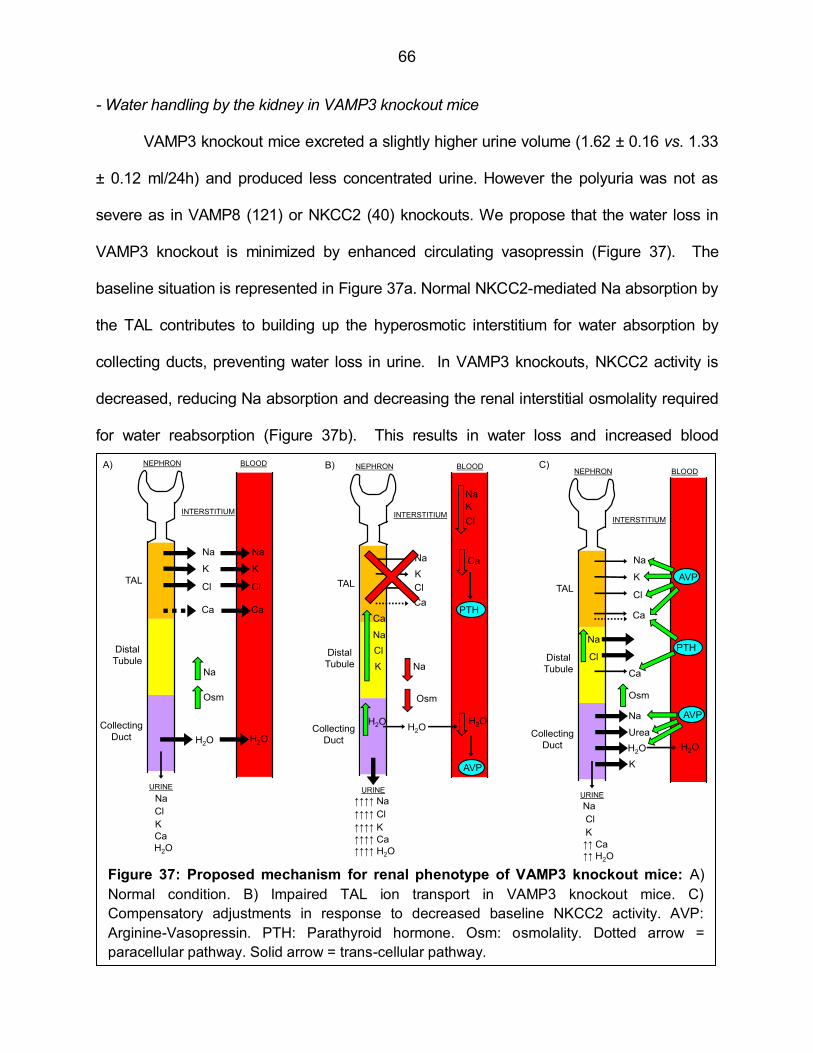
66
- Water handling by the kidney in VAMP3 knockout mice
VAMP3 knockout mice excreted a slightly higher urine volume (1.62 ± 0.16 vs. 1.33
± 0.12 ml/24h) and produced less concentrated urine. However the polyuria was not as
severe as in VAMP8 (121) or NKCC2 (40) knockouts. We propose that the water loss in
VAMP3 knockout is minimized by enhanced circulating vasopressin (Figure 37). The
baseline situation is represented in Figure 37a. Normal NKCC2-mediated Na absorption by
the TAL contributes to building up the hyperosmotic interstitium for water absorption by
collecting ducts, preventing water loss in urine. In VAMP3 knockouts, NKCC2 activity is
decreased, reducing Na absorption and decreasing the renal interstitial osmolality required
for water reabsorption (Figure 37b). This results in water loss and increased blood
Figure 37: Proposed mechanism for renal phenotype of VAMP3 knockout mice: A)
Normal condition. B) Impaired TAL ion transport in VAMP3 knockout mice. C)
Compensatory adjustments in response to decreased baseline NKCC2 activity. AVP:
Arginine-Vasopressin. PTH: Parathyroid hormone. Osm: osmolality. Dotted arrow =
paracellular pathway. Solid arrow = trans-cellular pathway.
Figure 30: Proposed mechanism for renal phenotype of VAMP3 knockout mice: A) Normal condition. B) Impaired TAL
ion transport in VAMP3 knockout mice. C) Compensatory adjustments in response to decreased baseline NKCC2 activity.
AVP: Arginine-Vasopressin. PTH: Parathyroid hormone. Osm: osmolality. Dotted arrow = paracellular pathway. Solid arrow =
trans-cellular pathway.
NEPHRON
INTERSTITIUM
BLOOD
TAL
Distal
Tubule
Collecting
Duct
Na
K
Cl
Ca
Na
K
Cl
Ca
Na
Osm
H2O H2O
URINE
H2O
Na
K
Cl
Ca
NEPHRON
INTERSTITIUM
BLOOD
TAL
Distal
Tubule
Collecting
Duct
Na
K
Cl
Ca
Na
K
Cl
Ca
Na
Osm
H2OH2O
URINE
↑↑↑↑ H2O
↑↑↑↑ Na
↑↑↑↑ K
↑↑↑↑ Cl
↑↑↑↑ Ca
Na
K
Cl
H2O
NEPHRON
INTERSTITIUM
BLOOD
TAL
Distal
Tubule
Collecting
Duct
Na
K
Cl
Ca
H2O H2O
URINE
↑↑ H2O
Na
K
Cl
↑↑ Ca
Na
Cl
Osm
AVP
PTH
AVP
Ca
Na
Urea
K
PTH
AVP
A) B) C)
Ca

67
osmolality which stimulates vasopressin release to the circulation from the pituitary gland.
Vasopressin activates the cAMP pathway in the TAL and stimulates NKCC2 via VAMP2
(Figure 37c). In addition, vasopressin enhances water reabsorption in the collecting duct by
stimulating the water channel aquaporin-2 (243-245) and enhances urea transport (246)
which contributes to the hyperosmotic medulla. All this would tend to minimize water loss
in VAMP3 knockout mice. We did not measure circulating vasopressin in VAMP3 knockout
mice, however we would expect it to be increased. In addition, increased circulating PTH
would also activate NKCC2 via cAMP (56,63,65). We expect circulating PTH to be
increased due to the Ca loss and we will discuss the role of PTH next.
- Urinary Ca handling in VAMP3 knockout mice
We found enhanced urinary Ca excretion in VAMP3 knockouts, which is consistent
with decreased NKCC2 activity since Ca loss is also observed in NKCC2 knockout mice
(40) and also in patients with Bartter syndrome (32,34-36,247-250), a pathology caused by
loss of NKCC2 function (27,31,35). The major site for renal Ca reabsorption is the proximal
tubule. However the fine-tuning of Ca reabsorption occurs in the TAL (Figure 37a). Ca is
reabsorbed in the TAL via the paracellular pathway, in a process driven by the lumen-
positive electric potential created by the joint action of NKCC2 and the potassium channel
ROMK (Figure 2). If VAMP3 knockouts have decreased NKCC2 activity, the driving force
for Ca absorption is decreased, producing Ca loss in the urine (Figure 37b). The ensuing
Ca depletion in the blood stimulates PTH release from the parathyroid glands. PTH
stimulates NKCC2 and ROMK via cAMP, restoring the electrical gradient but also
stimulates the paracellular pathway for Ca absorption in the TAL and Ca reabsorption
across the distal tubule (Figure 37c). We expect all these compensatory mechanisms are
active in VAMP3 knockout mice, thereby minimizing urinary Ca loss.
68
- Urinary Na, Cl and K handling in VAMP3 knockout mice
Normally between 25-30% of the Na and Cl filtered in the glomerulus is reabsorbed
in the TAL. NKCC2 is a major contributor to this process, preventing loss in the urine
(Figure 37a). If VAMP3 knockouts have decreased NKCC2 activity, we would expect
increased Na, Cl and K excretion (Figure 37b). However, VAMP3 knockouts did not
experience any major wasting of Na, K and Cl in the urine under baseline conditions. It is
likely that other nephron segments distal to the TAL enhance Na, K, and Cl transport to
counteract any major defect in the TAL (Figure 37c). It is known that enhanced luminal Na
delivery to the distal tubule enhances NaCl reabsorption via the thiazide-sensitive Na/Cl co-
transporter NCC (251,252). Also, enhanced circulating vasopressin can stimulate Na
reabsorption by NCC and in the collecting duct via the epithelial Na channel ENaC
(253,254). In addition, NKCC2 function could be partially restored via the cAMP-VAMP2
pathway as consequence of the increased circulating vasopressin and PTH mentioned
above. Finally, the collecting duct also contributes to K excretion by balancing secretion
and reabsorption. All these actions could contribute to minimize Na, Cl and K loss in
VAMP3 knockout mice.
Since we did not observe any difference in baseline Na, Cl and K excretion in
VAMP3 knockout mice, we next tested whether they could adjust excretion of these ions
when challenged to water deprivation. It is known that water restriction produces an
increase in ion excretion in many mammal species (255-260). This response is believed to
limit the increase in plasma osmolality caused by the limited fluid supply, which can be life
threatening. A known mediator of this response is the hormone oxytocin (191), which is
released from the neurohypophysis in response to hyperosmolality (261). It has been
proposed that oxytocin mediates its natriuretic and kaliuretic effect by stimulating release of

69
atrial natriuretic peptide (ANP) from the heart (262). There are oxytocin receptors in the
heart (192) and infusion of oxytocin in isolated hearts produce release of ANP (192). ANP
can enhance ion excretion by increasing glomerular filtration rate or by directly inhibiting ion
reabsorption by the nephron. In addition, oxytocin itself is believed to synergize with ANP
to inhibit tubular ion reabsorption via cGMP (263). The concerted actions of ANP and
oxytocin cause natriuresis and kaliuresis independently of vasopressin (189,190), which is
also released to the circulation in response to water deprivation.
When mice were water-deprived for 24 hours Na, K and Cl excretion augmented in
both strains. However, VAMP3 knockouts excreted more Na, Cl and K than wild-types
under water deprivation. This indicates that when challenged to water restriction stress,
renal function does not adjust completely or it takes longer to adjust. This response is
expected according to the model we propose. If there is compensation by other nephron
segments more distal to the TAL, excretion of Na, Cl and K would be normal in VAMP3
knockouts under baseline conditions. When water-deprived, the inhibitory actions of ANP
in ion transport in more distal nephron segments would override the initial compensation.
This would allow us to observe a defect in TAL ion transport as excessive excretion of Na,
Cl and K.
Another way of appreciating a renal defect in VAMP3 knockout mice, is by limiting
the amount of Na in the diet. We achieved this by switching the animals to a low-Na diet.
Under this condition, the kidneys would adjust to excrete less Na in an attempt to retain the
limited Na supply in the diet. We observed that urinary Na excretion was not decreased as
much in VAMP3 knockout mice as in wild-type mice after 24 hours in low-Na diet, but after
48 hours excretion of Na was comparable in both strains. This indicates that in VAMP3
knockouts renal function takes longer to adjust to limit Na excretion in the urine when

70
challenged to a condition like low-salt diet, when Na intake is limited. They eventually are
successful in limiting Na excretion, otherwise this wasting of Na could be life-threatening.
This adjustment most likely occurs via the compensatory mechanisms described before.
Altogether, our data indicate that VAMP3 knockout mice have decreased renal
function, which limits their ability to concentrate the urine and to properly excrete
electrolytes under baseline conditions and after challenges that demand homeostatic
adjustments of water an ion balance. To our knowledge this is the first phenotype ever
described for VAMP3 knockout mice, emphasizing the importance of VAMP3 in renal
function.
Decreased arterial pressure in VAMP3 knockout mice
Although VAMP3 shows mild fluid and electrolyte loss in urine, blood pressure
should be fully compensated by activation of the renin-angiotensin-aldosterone system.
However, we observed a significantly lower systolic blood pressure (-19 mmHg) compared
to wild-type mice. NKCC2 mutations in humans also decrease blood pressure (32-
36,41,42). Our data indicate that VAMP3 plays an important role in maintaining normal
blood pressure. The mechanism by which this occurs is unclear but may involve the role of
VAMP3 in NKCC2-mediated NaCl absorption or a role of VAMP3 in regulation of other
renal transporters. It has been suggested that VAMP3 participates in aquaporin-2
trafficking to the collecting duct membrane (106,147). In addition, VAMP3 can be important
for endocrine control of blood pressure. It is known that VAMP3 does not mediate baseline
or stimulated renin release from juxtaglomerular cells (169). However other components of
the renin-angiotensin-aldosterone system may require VAMP3. For instance,
angiotensinogen release from liver cells or aldosterone production from the adrenal cortex.
Finally, VAMP3 may regulate vascular reactivity or neural control of arterial tone. To our
71
knowledge the role of VAMP3 in cardiovascular function and blood pressure has not been
addressed.
Strengths and limitations of the study
Strengths: We performed all our experiments in native TALs or in primary culture of
TALs which retain the characteristics of the native tissue. We consider that this approach
enhances the significance of our findings. In addition, we described a mechanism that
regulates NKCC2 trafficking at the molecular level and then tested the role of the major
components of this mechanism at the whole animal level. We developed new techniques
to silence genes in vivo and took advantage of a knockout model. All these approaches
increase the translational relevance of the mechanisms we described to human and animal
physiology.
Despite the differences that may exist between species, the underlying basic
principles in cell biology, renal physiology and TAL function are well conserved among
mammals. We believe the knowledge we gathered in this dissertation project is useful as
first evidence of an important mechanism with potential implications in renal physiology and
hypertension.
Limitations: There is a general limitation with any study using animal models as a
representation of human physiology. However, the kind of analysis we conducted here
would have been impossible to perform in human subjects.
Among the specific limitations concerning the current study we could mention:
1. Most of the experiments were performed in medullary TAL suspensions, which is an
enriched sample but not completely pure. However, 90-95 % of the tubules are TALs and
the rest is composed of other nephron segments and blood vessels. Nevertheless, NKCC2
is expressed exclusively in the TAL, making contamination irrelevant in this case since we

72
used a specific NKCC2 antibody. Regarding other proteins, we consider a suspension
which is >90% TALs acceptable as a representative sample to measure protein expression.
2. The studies performed in primary cell culture isolate TAL cells from their native
environment in the renal medulla. Any response we observed here is devoid of the context
found in vivo and may not represent the natural condition. However, this was the only
methodology available to us to gain access to the apical membrane after heterologous
expression of VAMPs. On the other hand, these are more controlled conditions that
eliminate confounding factors found in the complex environment in vivo.
3. We used forskolin + IBMX to maximally stimulate intracellular cAMP, since in our
hands this is the most potent stimulus for NKCC2 trafficking. However, we recognize that
this condition is not physiological and could only represent the convergence of more than
one signaling pathway that uses cAMP as second messenger.
4. The co-immunoprecipitation protocols allowed us to determine that NKCC2 interacts
with VAMP2 and VAMP3. However, we did not determine whether this interaction is direct
or via intermediate proteins, since NKCC2 and VAMPs could precipitate as part of a larger
protein complex.
5. We determined that cAMP stimulates NKCC2-VAMP2 interaction. However, we
recognize that VAMP2 mediates exocytosis by inducing the formation of a SNARE
complex. We have yet not identified the SNARE partners of VAMP2 that mediate NKCC2
trafficking. This goes beyond the scope of the hypothesis tested in this study.
6. VAMP2 and VAMP3 are very likely to be involved in delivery of other plasma
membrane proteins beside NKCC2 in the TAL. This could have an indirect effect on
NKCC2 trafficking. However, we found direct evidence of NKCC2 interaction and co-
localization with VAMP2 and VAMP3 in the TAL, suggesting they are very likely to play a

73
direct role. By no means is this an indication that such a role is exclusive for NKCC2, and
that is not the way we formulated our hypothesis or interpreted our results. It is very likely
that VAMP2 or VAMP3 mediate trafficking of other components of the ion absorption
machinery in the TAL, which include NHE3, ROMK, KCC, Na-K ATPase or Cl channels.
We could not cover all of them in our study. Altering the trafficking of any of these
components will most likely change the intracellular concentration of ions. It is known that
decreased intracellular Cl stimulates NKCC2 activity (264), and this is likely due to
enhanced ion transport instead of enhanced trafficking. We do not think that ion transport
influences NKCC2 trafficking since unpublished experiments from our laboratory indicate
that furosemide, which blocks ion transport by competing for the Cl binding site in NKCC2,
does not affect surface NKCC2 expression.
We consider that testing whether VAMP2 and VAMP3 mediate trafficking of other
membrane transporters in the TAL and how this may influence NKCC2 trafficking is a
separate hypothesis that requires a different experimental design.
7. We measured renal function and blood pressure in global VAMP3 knockout mice. In
order to address whether VAMP3 expressed specifically in the TAL mediates the effects we
observed, a different model with TAL-targeted VAMP3 deletion is necessary. Although we
believe our studies provide the first observation that VAMP3 is important for renal function
and blood pressure, this does not mean VAMP3 in the TAL is exclusively responsible for
the effects we found.
8. We were not able to study the role of VAMP2 deletion in TALs. Global VAMP2
knockouts are not viable. In order to address the role of VAMP2 in renal physiology, tissue-
specific deletion of VAMP2, which is not lethal, would be required.

74
Perspectives
About 30% of Americans are hypertensive (265), costing the nation billions of dollars
in healthcare costs and lost productivity (266). Hypertension is a major risk factor for
cardiovascular disease, the number one cause of death worldwide (267) and in the United
States (268). About 20% of hypertensive patients do not respond to current treatments
(269), highlighting the need to further investigate the mechanisms of hypertension. The
kidneys reduce extracellular fluid volume by enhancing sodium and water excretion in
response to increases in arterial pressure (270), playing a central role in controlling blood
pressure. In this dissertation we have described a molecular mechanism that regulates
NKCC2, a key component of the machinery for NaCl reabsorption by the TAL, a nephron
segment that has major influence on blood pressure. The only known inhibitors of NKCC2
are the loop diuretics furosemide, bumetanide and torasemide, with similar mechanism of
action. However, their use is restricted to cases like congestive heart failure and edema in
which a rapid and dramatic decrease in extracellular fluid volume is required. Despite the
promise of drugs targeting NKCC2 to treat hypertension, the long-term use of loop diuretics
is limited by their side effects, which include severe dehydration, potassium and calcium
loss, cataracts and diminished hearing (271-276).
Here we propose a mechanism that regulates NKCC2 via the vesicle fusion proteins
VAMP2 and VAMP3, whose genes overlap with several quantitative trait loci (QTL) in
hypertensive rat strains. According to the Rat Genome Database, VAMP2 (chromosome
10) is present in 12 blood pressure QTL and VAMP3 (chromosome 5) is present in 5. The
only known agents that target SNAREs are the several serotypes of botulinum toxin which
cleave target membrane-SNAREs and VAMPs, and tetanus toxin which cleaves VAMP2
and VAMP3. More recently, small molecule inhibitors that interfere with the SNARE

75
complex formation have been developed (277). However, since SNAREs are ubiquitous,
the therapeutic use of these drugs will be limited until a way to direct them to specific
tissues is found. In this dissertation we found that different VAMPs control two distinct
pathways in NKCC2 regulation. Further knowledge into the details of these independent
pathways will lead to the eventual development of more precise and effective diuretics to
treat hypertension. Being able to intervene selectively in a particular regulatory pathway
without interfering with other pathways is appealing since the beneficial effects of a loop
diuretic could be achieved with minimal side effects.
76
CHAPTER 5
GENERAL METHODS
Animals used in the study
All animal procedures were approved by the Institutional Animal Care and Use
Committee (IACUC) and were performed according to the committee’s guidelines. We used
male Sprague-Dawley rats weighing 130g, 18-week-old C57BL/6J mice and VAMP3
knockout mice on a C57BL/6J background. Rats were purchased from Charles River
Breeding Laboratories (Wilmington, MA) and wild-type mice from Jackson Laboratory (Bar
Harbor, ME). VAMP3 knockout mice were generated by the laboratory of Dr. Jeffrey Pessin
(186) and generously shared with us. The VAMP3 gene was disrupted by deleting exons 2
and 3. Animals were fed a diet containing 0.22% sodium and 1.1% potassium (Purina,
Richmond, IN) and had free access to water ad libitum.
Antibodies
The anti-NKCC2 antibody was raised in chicken by Lofstrand Labs Limited
(Gaithersburg, MD). It recognizes amino acids 33–55 of the amino terminus of rat NKCC2
as does the L-320 antibody by Kim et al. (173). The mouse anti-GAPDH antibody was
from Chemicon (Temecula, CA). The mouse anti-VAMP2 and rabbit anti-VAMP8
antibodies were purchased from Synaptic Systems (Goettingen, Germany). The rabbit
anti-VAMP3 and mouse anti-VAMP7 antibodies were from Abcam (Cambridge, MA). The
rabbit anti-phospho-serine 126 NKCC2 antibody was a generous gift from Dr. Mark
Knepper (72). The HRP-conjugated secondary anti-mouse and anti-rabbit antibodies were
obtained from GE Healthcare Life Sciences (Piscataway, NJ). The HRP-conjugated
secondary anti-chicken IgG was from Gallus Immunotech (Fergus, ON, Canada). The
rabbit NKCC2 antibody for immunoprecipitation was obtained from EMD Millipore (Billerica,

77
MA) and recognizes the carboxy-terminus of NKCC2. The exofacial NKCC2 antibody was
developed by us in rabbit (24) and recognizes an epitope in the extracellular loop between
transmembrane segments 5 and 6 in rat NKCC2. The Alexa Fluor 488- conjugated anti-
GFP antibody and the Alexa Fluor 568-conjugated secondary antibody were from Life
Technologies (Grand Island, NY). The mouse anti-GFP antibody for Western blot was from
Clontech (Mountain View, CA).
Reagents
Reagents for steady-state surface biotinylation and exocytic delivery, and
proteinG/proteinA-coupled agarose beads were from ThermoFisher Scientific (Waltham,
MA). Forskolin and IBMX were from Sigma-Aldrich (St. Louis, MO). The glutathione-
coupled sepharose beads were from Roche (Basel, Switzerland). N6-Benzoyl-cAMP was
purchased from BioLog (Bremen, Germany). Tetanus toxin was from List Biological
Laboratories (Campbell, CA). The glutathione S-transferase (GST) expression vector was
pGEX-6P-1 from GE Healthcare Life Sciences. The VQ Ad5CMV K-NpA adenovector was
from Viraquest (North Liberty, IA). The VAMP2-eGFP and VAMP3-eGFP constructs were
generously provided by Dr. Regazzi (179).
Medullary thick ascending limb suspensions
Suspensions of outer medullary TALs were obtained from rats according to a
standard protocol used routinely in our laboratory (20). Briefly, rats were anesthetized with
ketamine (100 mg/kg body wt ip) and xylazine (20 mg/kg body wt ip). Kidneys were
perfused through the aorta with 0.1% collagenase in O2-saturated HEPES-buffered
perfusion solution. The outer medulla was dissected, minced and incubated at 37°C for 30
min with 0.1% collagenase. The composition of the HEPES-buffered perfusion solution
was 135 mM NaCl; 10 mM HEPES; 4 mM KCl; 1.5 mM NaH2PO4; 1.2 mM MgSO4; 1 mM
78
Ca-lactate2; 5 mM alanine; 5.5 mM glucose; 1 mM Na3-citrate; pH 7.4.
Primary culture of medullary thick ascending limbs
Individual TAL cells were obtained from rat medullary TAL suspensions after
digesting the tubules with collagenase (0.1%), trypsin (0.25%) and DNAse (0.0021%).
Single TAL cells were isolated by centrifugation at 41,300 x g in a 35% Percoll gradient,
and seeded in Trans-well permeable supports (Corning, Tewksbury, MA) coated with
basement membrane extract (Trevigen, Gaithersburg, MD). Cells were grown in DMEM
medium supplemented with 1% fetal bovine serum and insulin-transferrin-selenium (Life
Technologies) at 37ºC and 5% CO2.
Western blot
TAL suspensions were lysed in buffer containing 150 mM NaCl, 50 mM HEPES (pH
7.8), 5 mM EDTA, 1% Triton X-100, 0.2% SDS and protease inhibitors (benzamidine,
leupeptin, aprotinin, chymostatin and pepstatin-A, from Sigma-Aldrich). When measuring
phosphorylation, the lysis buffer also contained PhosSTOP phosphatase inhibitors (Roche).
Protein samples were run by electrophoresis in 6% SDS-polyacrylamide gels to detect
NKCC2 and in 5% stacking- 12% resolving gels to detect VAMPs. Proteins were
transferred to PVDF membranes overnight and the next day blocked in 5% milk in TBS-T
buffer for one hour at room temperature. Next, membranes were incubated with the
primary antibody for one hour at room temperature at the following dilutions: 1/700
(NKCC2), 1/2000 (p-Ser126-NKCC2), 1/2000 (VAMP2), 1/30,000 (VAMP3), 1/500
(VAMP7), 1/50,000 (GAPDH). Membranes were then incubated with the corresponding
HRP-conjugated secondary antibody at 1/7000 (chicken) or 1/5000 (mouse and rabbit) for
one hour at room temperature and developed by chemiluminescence.
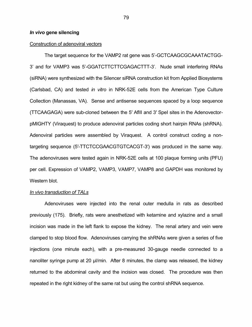
79
In vivo gene silencing
Construction of adenoviral vectors
The target sequence for the VAMP2 rat gene was 5’-GCTCAAGCGCAAATACTGG-
3’ and for VAMP3 was 5’-GGATCTTCTTCGAGACTTT-3’. Nude small interfering RNAs
(siRNA) were synthesized with the Silencer siRNA construction kit from Applied Biosystems
(Carlsbad, CA) and tested in vitro in NRK-52E cells from the American Type Culture
Collection (Manassas, VA). Sense and antisense sequences spaced by a loop sequence
(TTCAAGAGA) were sub-cloned between the 5′ AflII and 3′ SpeI sites in the Adenovector-
pMIGHTY (Viraquest) to produce adenoviral particles coding short hairpin RNAs (shRNA).
Adenoviral particles were assembled by Viraquest. A control construct coding a non-
targeting sequence (5′-TTCTCCGAACGTGTCACGT-3′) was produced in the same way.
The adenoviruses were tested again in NRK-52E cells at 100 plaque forming units (PFU)
per cell. Expression of VAMP2, VAMP3, VAMP7, VAMP8 and GAPDH was monitored by
Western blot.
In vivo transduction of TALs
Adenoviruses were injected into the renal outer medulla in rats as described
previously (175). Briefly, rats were anesthetized with ketamine and xylazine and a small
incision was made in the left flank to expose the kidney. The renal artery and vein were
clamped to stop blood flow. Adenoviruses carrying the shRNAs were given a series of five
injections (one minute each), with a pre-measured 30-gauge needle connected to a
nanoliter syringe pump at 20 μl/min. After 8 minutes, the clamp was released, the kidney
returned to the abdominal cavity and the incision was closed. The procedure was then
repeated in the right kidney of the same rat but using the control shRNA sequence.
80
Biotinylation of steady-state surface NKCC2 in thick ascending limb suspensions
Medullary TAL suspensions were obtained from rats, equilibrated at 37°C for 15 min
and then treated with either vehicle or forskolin (20 µM) plus IBMX (0.5 mM) for 30 min.
TALs were biotinylated at 4°C with membrane-impermeant NHS-SS-biotin (0.9 mg/ml),
washed and lysed in the presence of protease inhibitors. Biotinylated proteins were
separated with streptavidin-coated beads overnight at 4°C and eluted by boiling in Laemmli
loading buffer containing dithiothreitol (DTT) and β-mercaptoethanol. NKCC2 and GAPDH
were detected by Western blot. In every experiment, we monitored total NKCC2
expression and absence of intracellular biotinylation by examination of GAPDH expression
in the surface fraction.
Exocytic delivery of surface proteins
NKCC2 exocytic delivery
TAL suspensions were incubated with membrane-impermeant NHS-acetate (2
mg/ml) at pH 7.8 and 4°C for one hour to mask surface sites accessible to NHS-SS-biotin.
Fresh NHS-acetate was added every 15 min. Forskolin and IBMX were added at 4°C and
samples warmed to 37°C for 30 min. TALs were then cooled and newly inserted NKCC2
biotinylated with NHS-SS-biotin. he efficiency of NHS-acetate masking for NKCC2 was
calculated in every experiment from the difference between a TAL aliquot that was not
masked with NHS-acetate (100% basal surface NKCC2) and an aliquot that was masked
at 4°C but never warmed to 37°C (0 time-point). The difference between these two
samples represents the NHS-acetate-masked surface NKCC2 fraction, which was used to
express exocytic delivery over time.
VAMP2-eGFP exocytic delivery
Primary cultures of TALs were obtained from rats and transfected with VAMP2-

81
eGFP via adenoviruses. Twenty four hours after transfection, cells were treated with
vehicle or forskolin (20 µM) plus IBMX (0.5 mM). The GFP portion of VAMP2-eGFP faces
the extracellular space and is accessible for biotinylation. We measured exocytic delivery
by masking with NHS-acetate and biotinylating newly delivered VAMP2-eGFP in the same
way as described for NKCC2. Surface VAMP2-eGFP was measured by Western blot with
anti-GFP antibody.
Co-immunoprecipitation
We obtained protein samples from medullary TAL suspensions lysed in buffer
containing 150 mM NaCl, 50 mM HEPES (pH 7.5), 5 mM EDTA, 1% Triton X-100, 0.1%
SDS and protease inhibitors. 200 µg of protein from TALs were pre-cleared 30 min at 4ºC
with proteinG/proteinA-coupled agarose beads in immunoprecipitation buffer (100 mM
NaCl, 50 mM HEPES (pH 7.5), 5 mM EDTA, 1% Triton X-100, 1% CHAPS and protease
inhibitors). Beads were precipitated by centrifugation and discarded. Supernatants were
incubated with 5 µg of rabbit anti-NKCC2 IgG overnight at 4ºC. Control tubes were
incubated with a non-immune rabbit IgG. Next day, proteinG/proteinA-coupled agarose
beads were added in two sequential rounds for a total of 4 hrs at 4ºC. At the end of the
incubation period, beads were sequentially washed in immunoprecipitation buffer, 50 mM
HEPES/500 mM NaCl buffer and 50 mM HEPES buffer. Proteins were extracted by
incubation at 37ºC in loading buffer and VAMPs were detected by polyacrylamide gel
electrophoresis and Western blot. For VAMP2 and VAMP3 immunoprecipitation we used
100 µg of TAL protein lysate and 5 µg of anti-VAMP2, anti-VAMP3 or non-immune rabbit
IgG. Beads were eluted as indicated and NKCC2 detected by Western blot.

82
GST pull down in thick ascending limb lysates
Generation of GST constructs
We generated GST-fusion proteins by sub-cloning the amino-terminus of NKCC2
and a portion of the carboxy-terminus of NKCC2 in GST bacterial expression vectors
(named GST-N-NKCC2 and GST-C-NKCC2, respectively). The carboxy-terminal region of
NKCC2 was a 71 amino acid- long sequence known to contain an apical localization signal
(177). We transformed E.coli bacteria with GST, GST-N-NKCC2 or GST-C-NKCC2 and
induced expression of the GST fusion proteins by incubating with 0.2 mM IPTG for 4 hours.
We lysed the bacteria by sonication in lysis buffer containing 20 mM Tris HCl (pH 7.4), 200
mM NaCl and 10 mM EDTA. We purified the GST proteins by incubating with glutathione-
coupled sepharose beads in bacterial lysis buffer overnight at 4ºC.
GST pull down
We lysed TAL suspensions from rats to obtain a protein homogenate as described
above. We pre-cleared 200 µg of protein sample by incubating with sepharose beads
already bound to the GST protein generated before. After one hour of incubation at 4ºC,
we centrifuged the samples, eliminated the beads and incubated the supernatants with
sepharose beads already bound to GST, GST-N-NKCC2 or GST-C-NKCC2. We
incubated overnight at 4ºC. Then we washed with 50 mM HEPES/500 mM NaCl buffer
and 50 mM HEPES buffer. We eluted the beads by incubation at 37ºC in Laemmli loading
buffer. Samples were run by electrophoresis in 12% polyacrylamide gels and VAMP2
detected by Western blot as described above.
Immunolabeling of surface NKCC2, VAMP2 and VAMP3 in thick ascending limb cells
Cultured TAL cells were transfected with adenoviruses carrying VAMP2-eGFP or
VAMP3-eGFP under control of a CMV promoter for 24 hours. The VAMP2- and VAMP3-
83
eGFP constructs were sub-cloned into the VQ Ad5CMV K-NpA adenovector. Viral
particles were produced and purified by Viraquest. Protein trafficking was rapidly stopped
by adding cold medium and incubating the cultures at 4°C for 30 minutes. Cells were
blocked with 5% albumin in physiological solution for 30 minutes at 4°C. Then the primary
rabbit antibody against an exofacial epitope on NKCC2 was added to the apical bath (1/50)
at 4ºC for 2 hours. Cells were washed and incubated with the secondary anti-rabbit
antibody conjugated with Alexa Fluor 568 (1/100) for 1 hour at 4ºC. After washout, surface
VAMP2/ 3-eGFP was labeled with Alexa Fluor 488-conjugated anti-GFP antibody (1/100)
for 1 hour at 4ºC. Finally cells were fixed in 4% paraformaldehyde and mounted. Images
were acquired using a laser scanning confocal microscopy system (Visitech, Sutherland,
England) with a 488 nm diode or Kr/Ar 568 nm gas laser excitation controlled by an
acousto-optic tunable filter. Images were acquired at 100x (1.4 NA) and fluorescence
observed using 525/55-nm Band-pass or 590-nm Low-pass filters, respectively. Image files
(TIFF format) were minimally deconvolved with Autoquant software (Media Cybernetics,
MD) using 2-D blind deconvolution. Images from both channels were aligned and pixel-by-
pixel co-localization measured using a minimum Mander's overlap coefficient of 0.95. An
image for overlapping pixels was generated.
Blood pressure measurements
Systolic arterial pressure was measured by tail cuff in pre-trained mice with MC4000
Multi Channel Blood Pressure Analysis System (Cary, NC).
Measurements of urine parameters
Mice were placed in metabolic cages (Techniplast, Exton, PA) with free access to
food and water. After a period of three days of acclimatization, urine samples were
collected for 24 hours and volume measured. Then we measured urine osmolality by

84
freezing point depression with Advanced Model 3300 Micro-Osmometer (Advanced
Instruments Inc. Norwood, MA). We also measured urinary Na, K, Cl, and Ca with a
StatProfile pHOx Ultra Analyzer (Nova Biomedical, Waltham, MA). Then mice were
restricted from access to water for 24 hours. During that period urine was collected to
measure the parameters again.
Statistical analysis
Results are expressed as mean ± standard error. One-way analysis of variance
(ANOVA) was used to determine differences between means in treatments with more than
two groups. Post hoc analysis was performed when differences between means was found
using the Bonferroni correction for multiple comparisons. For comparisons between two
groups we used Student’s t-test and paired t-test for paired samples. p<0.05 was
considered significant.
85
APPENDIX A
List of acronyms
ANOVA: Analysis of Variance
ANP: Atrial natriuretic peptide
ATM kinase: Ataxia telangiectasia mutated kinase
AVP: Arginine-vasopressin
β-AR: Beta-adrenergic receptor
cAMP: 3'-5'-cyclic adenosine monophosphate
cGMP: 3',5'-cyclic guanosine monophosphate
DTT: Dithiothreitol
E. coli: Escherichia coli
ENaC: Epithelial Na channel
GAPDH: Glyceraldehyde 3-phosphate dehydrogenase
GFP: Green fluorescent protein
GSK-3: Glycogen synthase kinase-3
GST: Glutathione S-transferase
IACUC: Institutional Animal Care and Use Committee
IBMX: 3-Isobutyl-1-methylxanthine
IgG: Immunoglobulin G
IPTG: Isopropyl β-D-1-thiogalactopyranoside
MAL: Myelin and lymphocytes-associated protein
N6-Bnz-cAMP: N6-Benzoyl-cAMP
NHE: Na/H exchanger
NHS: N-Hydroxysulfosuccinimide

86
NIH: National Institutes of Health
NKCC2: Na/K/2Cl co-transporter
OSR1: oxidative stress response 1 kinase
PFU: Plaque-forming unit
PKA: Protein kinase A
PKC: Protein kinase C
PTH: Parathyroid hormone
QTL: Quantitative trait loci
ROMK: Renal outer medullary K channel
SCAMP2: Secretory carrier membrane protein 2
shRNA: Short hairpin RNA
siRNA: Small interfering RNA
SNAP: Synaptosome-Associated Protein
SNARE: Soluble NSF Attachment protein Receptors
SPAK: Ste20-related proline-alanine-rich kinase
TAL: Thick ascending limb
VAMP: Vesicle Associated Membrane Protein
VAMP3 -/- : VAMP3 knockout
87
APPENDIX B
IACUC approval
88
89
REFERENCES
1. Molony,D.A., Reeves,W.B., and Andreoli,T.E. 1989. Na+:K+:2Cl- cotransport and the
thick ascending limb. Kidney Int. 36:418-426.
2. Hebert,S.C., and Andreoli,T.E. 1984. Control of NaCl transport in the thick ascending
limb. Am J Physiol. 246:F745-F756.
3. Greger,R. 1985. Ion transport mechanisms in thick ascending limb of Henle's loop of
mammalian nephron. Physiol Rev. 65:760-797.
4. Good,D.W. 1985. Sodium-dependent bicarbonate absorption by cortical thick ascending
limb of rat kidney. Am J Physiol. Renal Physiol. 248:F821-F829.
5. Good,D.W., and Watts,B.A., III 1996. Functional roles of apical membrane Na+/H+
exchange in rat medullary thick ascending limb. Am J Physiol. Renal Physiol.
270:F691-F699.
6. Greger,R., Bleich,M., and Schlatter,E. 1990. Ion channels in the thick ascending limb of
Henle's loop. Renal Physiology & Biochemistry. 13:37-50.
7. Di Stefano,A., Jounier,S., and Wittner,M. 2001. Evidence supporting a role for KCl
cotransporter in the thick ascending limb of Henle's loop. Kidney Int. 60:1809-1823.
8. Bleich,M., Schlatter,E., and Greger,R. 1990. The luminal K+ channel of the thick
ascending limb of Henle's loop. Pflugers Archiv - European Journal of Physiology.
415:449-460.
9. Greger,R. 1981. Cation selectivity of the isolated perfused cortical thick ascending limb of
Henle's loop of rabbit kidney. Pflugers Archiv - European Journal of Physiology.
390:30-37.
10. Mandon,B., Siga,E., Roinel,N., and de Rouffignac,C. 1993. Ca2+, Mg2+ and K+
transport in the cortical and medullary thick ascending limb of the rat nephron:
90
influence of transepithelial voltage. Pflugers Arch. 424:558-560.
11. Kaplan,M.R., Plotkin,M.D., Lee,W.S., Xu,Z.C., Lytton,J., and Hebert,S.C. 1996. Apical
localization of the Na-K-Cl cotransporter, rBSC1, on rat thick ascending limbs.
Kidney Int. 49:40-47.
12. Mount,D.B., Baekgaard,A., Hall,A.E., Plata,C., Xu,J., Beier,D.R., Gamba,G., and
Hebert,S.C. 1999. Isoforms of the Na-K-2Cl cotransporter in murine TAL I.
Molecular characterization and intrarenal localization. Am J Physiol. 276:F347-F358.
13. Obermuller,N., Kunchaparty,S., Ellison,D.H., and Bachmann,S. 1996. Expression of the
Na-K-2Cl cotransporter by macula densa and thick ascending limb cells of rat and
rabbit nephron. J Clin Invest. 98:635-640.
14. Ortiz,P.A. 2006. cAMP increases surface expression of NKCC2 in rat thick ascending
limbs: role of VAMP. Am J Physiol. Renal Physiol. 290:F608-F616.
15. Ortiz,P.A., and Garvin,J.L. 2001. NO Inhibits NaCl Absorption by Rat Thick Ascending
Limb Through Activation of cGMP-Stimulated Phosphodiesterase. Hypertension.
37:467-471.
16. Aoki,Y., Albrecht,F.E., Bergman,K.R., and Jose,P.A. 1996. Stimulation of Na(+)-K(+)-
2Cl- cotransport in rat medullary thick ascending limb by dopamine. Am J Physiol.
271:R1561-R1567.
17. Ito,O., Kondo,Y., Takahashi,N., Kudo,K., Igarashi,Y., Omata,K., Imai,Y., and Abe,K.
1994. Insulin stimulates NaCl transport in isolated perfused MTAL of Henle's loop of
rabbit kidney. Am J Physiol. 267:F265-F270.
18. Amlal,H., LeGoff,C., Vernimmen,C., Paillard,M., and Bichara,M. 1996. Na(+)-
K+(NH4+)-2Cl- cotransport in medullary thick ascending limb: control by PKA, PKC,
and 20-HETE. Am J Physiol. 271:C455-C463.
91
19. Jesus Ferreira,M.C., and Bailly,C. 1998. Extracellular Ca2+ decreases chloride
reabsorption in rat CTAL by inhibiting cAMP pathway. Am J Physiol. 275:F198-
F203.
20. Ortiz,P.A., Hong,N.J., and Garvin,J.L. 2001. NO decreases thick ascending limb
chloride absorption by reducing Na(+)-K(+)-2Cl(-) cotransporter activity. Am J
Physiol. Renal Physiol. 281:F819-F825.
21. Ortiz,P.A., Hong,N.J., Wang,D., and Garvin,J.L. 2003. Gene transfer of eNOS to the
thick ascending limb of eNOS-KO mice restores the effects of L-arginine on NaCl
absorption. Hypertension. 42:674-679.
22. He,H., Podymow,T., Zimpelmann,J., and Burns,K.D. 2003. NO inhibits Na+-K+-2Cl-
cotransport via a cytochrome P-450-dependent pathway in renal epithelial cells
(MMDD1). Am J Physiol. Renal Physiol. 284:F1235-F1244.
23. Garcia,N.H., Plato,C.F., Stoos,B.A., and Garvin,J.L. 1999. Nitric oxide-induced
inhibition of transport by thick ascending limbs from Dahl salt-sensitive rats.
Hypertension. 34:508-513.
24. Ares,G.R., Caceres,P., Alvarez-Leefmans,F.J., and Ortiz,P.A. 2008. cGMP decreases
surface NKCC2 levels in the thick ascending limb: role of phosphodiesterase 2
(PDE2). Am J Physiol. Renal Physiol. 295:F877-F887.
25. Glorioso,N., Filigheddu,F., Troffa,C., Soro,A., Parpaglia,P.P., Tsikoudakis,A.,
Myers,R.H., Herrera,V.L., and Ruiz-Opazo,N. 2001. Interaction of alpha(1)-Na,K-
ATPase and Na,K,2Cl-cotransporter genes in human essential hypertension.
Hypertension. 38:204-209.
26. Rieg,T., Bundey,R.A., Chen,Y., Deschenes,G., Junger,W., Insel,P.A., and Vallon,V.
2007. Mice lacking P2Y2 receptors have salt-resistant hypertension and facilitated
92
renal Na+ and water reabsorption. FASEB J. 21:3717-3726.
27. Simon,D.B., Karet,F.E., Hamdan,J.M., DiPietro,A., Sanjad,S.A., and Lifton,R.P. 1996.
Bartter's syndrome, hypokalaemic alkalosis with hypercalciuria, is caused by
mutations in the Na-K-2Cl cotransporter NKCC2. Nat Genet. 13:183-188.
28. Ares,G.R., Haque,M.Z., Delpire,E., and Ortiz,P.A. 2012. Hyperphosphorylation of Na-K-
2Cl cotransporter in thick ascending limbs of Dahl salt-sensitive rats. Hypertension.
60:1464-1470.
29. Carmosino,M., Rizzo,F., Ferrari,P., Torielli,L., Ferrandi,M., Bianchi,G., Svelto,M., and
Valenti,G. 2011. NKCC2 is activated in Milan hypertensive rats contributing to the
maintenance of salt-sensitive hypertension. Pflugers Arch. 462(2):281-291.
30. Hong,N.J., and Garvin,J.L. 2012. Angiotensin II type 2 receptor-mediated inhibition of
NaCl absorption is blunted in thick ascending limbs from Dahl salt-sensitive rats.
Hypertension. 60:765-769.
31. Starremans,P.G., Kersten,F.F., Knoers,N.V., van den Heuvel,L.P., and Bindels,R.J.
2003. Mutations in the human Na-K-2Cl cotransporter (NKCC2) identified in Bartter
syndrome type I consistently result in nonfunctional transporters. J Am Soc Nephrol.
14:1419-1426.
32. Bettinelli,A., Ciarmatori,S., Cesareo,L., Tedeschi,S., Ruffa,G., Appiani,A.C., Rosini,A.,
Grumieri,G., Mercuri,B., Sacco,M. et al 2000. Phenotypic variability in Bartter
syndrome type I. Pediatr Nephrol. 14:940-945.
33. Fukuyama,S., Okudaira,S., Yamazato,S., Yamazato,M., and Ohta,T. 2003. Analysis of
renal tubular electrolyte transporter genes in seven patients with hypokalemic
metabolic alkalosis. Kidney Int. 64:808-816.
34. Kurtz,C.L., Karolyi,L., Seyberth,H.W., Koch,M.C., Vargas,R., Feldmann,D., Vollmer,M.,
93
Knoers,N.V., Madrigal,G., and Guay-Woodford,L.M. 1997. A common NKCC2
mutation in Costa Rican Bartter's syndrome patients: evidence for a founder effect. J
Am Soc Nephrol. 8:1706-1711.
35. Vargas-Poussou,R., Feldmann,D., Vollmer,M., Konrad,M., Kelly,L., van den
Heuvel,L.P., Tebourbi,L., Brandis,M., Karolyi,L., Hebert,S.C. et al 1998. Novel
molecular variants of the Na-K-2Cl cotransporter gene are responsible for antenatal
Bartter syndrome. Am J Hum Genet. 62:1332-1340.
36. Adachi,M., Asakura,Y., Sato,Y., Tajima,T., Nakajima,T., Yamamoto,T., and Fujieda,K.
2007. Novel SLC12A1 (NKCC2) mutations in two families with Bartter syndrome
type 1. Endocr J. 54:1003-1007.
37. Ellison,D.H. 2000. Divalent cation transport by the distal nephron: insights from Bartter's
and Gitelman's syndromes. Am J Physiol. Renal Physiol. 279:F616-F625.
38. Bartter,F.C., Pronove,P., Gill,J.R., Jr., and MacCardle,R.C. 1998. Hyperplasia of the
juxtaglomerular complex with hyperaldosteronism and hypokalemic alkalosis. A new
syndrome. 1962. J Am Soc Nephrol. 9:516-528.
39. Shaer,A.J. 2001. Inherited primary renal tubular hypokalemic alkalosis: a review of
Gitelman and Bartter syndromes. Am J Med Sci. 322:316-332.
40. Takahashi,N., Chernavvsky,D.R., Gomez,R.A., Igarashi,P., Gitelman,H.J., and
Smithies,O. 2000. Uncompensated polyuria in a mouse model of Bartter's
syndrome. PNAS. 97:5434-5439.
41. Acuna,R., Martinez-de-la-Maza,L., Ponce-Coria,J., Vazquez,N., Ortal-Vite,P., Pacheco-
Alvarez,D., Bobadilla,N.A., and Gamba,G. 2011. Rare mutations in SLC12A1 and
SLC12A3 protect against hypertension by reducing the activity of renal salt
cotransporters. J Hypertens. 29:475-483.
94
42. Monette,M.Y., Rinehart,J., Lifton,R.P., and Forbush,B. 2011. Rare mutations in the
human Na-K-Cl cotransporter (NKCC2) associated with lower blood pressure exhibit
impaired processing and transport function. Am J Physiol. Renal Physiol. 300:F840-
F847.
43. Kirchner,K.A., Crosby,B.A., Patel,A.R., and Granger,J.P. 1995. Segmental chloride
transport in the Dahl-S rat kidney during L-arginine administration. J Am Soc
Nephrol. 5:1567-1572.
44. Kirchner,K.A. 1990. Greater loop chloride uptake contributes to blunted pressure
natriuresis in Dahl salt sensitive rats. J Am Soc Nephrol. 1:180-186.
45. Kirchner,K.A. 1992. Increased loop chloride uptake precedes hypertension in Dahl salt-
sensitive rats. Am J Physiol. 262:R263-R268.
46. Haque,M.Z., Ares,G.R., Caceres,P.S., and Ortiz,P.A. 2011. High Salt Differentially
Regulates Surface NKCC2 Expression in Thick Ascending Limbs of Dahl Salt
Sensitive and Salt Resistant Rats. Am J Physiol. Renal Physiol. 300:F1096-1104.
47. Jung,J., Basile,D.P., and Pratt,J.H. 2011. Sodium reabsorption in the thick ascending
limb in relation to blood pressure: a clinical perspective. Hypertension. 57(5):873-
879.
48. Chun,T.Y., Bankir,L., Eckert,G.J., Bichet,D.G., Saha,C., Zaidi,S.A., Wagner,M.A., and
Pratt,J.H. 2008. Ethnic differences in renal responses to furosemide. Hypertension.
52:241-248.
49. Kim,K.E., Onesti,G., Moyer,J.H., and Swartz,C. 1971. Ethacrynic acid and furosemide.
Diuretic and hemodynamic effects and clinical uses. Am J Cardiol. 27:407-415.
50. Shankar,S.S., and Brater,D.C. 2003. Loop diuretics: from the Na-K-2Cl transporter to
clinical use. Am J Physiol. Renal Physiol. 284:F11-F21.

95
51. Brater,D.C. 1986. Disposition and response to bumetanide and furosemide. Am J
Cardiol. 57:20A-25A.
52. Feig,P.U. 1986. Cellular mechanism of action of loop diuretics: implications for drug
effectiveness and adverse effects. Am J Cardiol. 57:14A-19A.
53. Baumgart,P. 1993. Torasemide in comparison with thiazides in the treatment of
hypertension. Cardiovasc Drugs Ther. 7 Suppl 1:63-8.:63-68.
54. Roca-Cusachs,A., racil-Vilar,J., Calvo-Gomez,C., Vaquer-Perez,J.V., Laporta-
Crespo,F., Rojas-Serrano,M.J., Guglietta,A., and Gropper,S. 2008. Clinical effects of
torasemide prolonged release in mild-to-moderate hypertension: a randomized
noninferiority trial versus torasemide immediate release. Cardiovasc Ther. 26:91-
100.
55. Wittner,M., Di Stefano,A., Schlatter,E., Delarge,J., and Greger,R. 1986. Torasemide
inhibits NaCl reabsorption in the thick ascending limb of the loop of Henle. Pflugers
Arch. 407:611-614.
56. Borensztein,P., Juvin,P., Vernimmen,C., Poggioli,J., Paillard,M., and Bichara,M. 1993.
cAMP-dependent control of Na+/H+ antiport by AVP, PTH, and PGE2 in rat
medullary thick ascending limb cells. Am J Physiol. 264:F354-F364.
57. Bertuccio,C., Ibarra,F.R., Pignataro,O., Toledo,J., Paz,L., Arrizurieta,E., and Martin,R.S.
1998. Regulation of cell cyclic AMP in medullary thick ascending limb of Henle in a
rat model of chronic renal failure. Acta Physiol Scand. 164:107-114.
58. Molony,D.A., Reeves,W.B., Hebert,S.C., and Andreoli,T.E. 1987. ADH increases apical
Na+, K+, 2Cl- entry in mouse medullary thick ascending limbs of Henle. Am J
Physiol. 252:F177-F187.
59. Hebert,S.C., Culpepper,R.M., and Andreoli,T.E. 1981. NaCl transport in mouse

96
medullary thick ascending limbs. II. ADH enhancement of transcellular NaCl
cotransport; origin of transepithelial voltage. Am J Physiol. 241:F432-F442.
60. Knepper,M.A., Kim,G.H., Fernandez-Llama,P., and Ecelbarger,C.A. 1999. Regulation
of thick ascending limb transport by vasopressin. J Am Soc Nephrol. 10:628-634.
61. Liu,H.J., Wei,Y., Ferreri,N.R., Nasjletti,A., and Wang,W.H. 2000. Vasopressin and
PGE(2) regulate activity of apical 70 pS K(+) channel in thick ascending limb of rat
kidney. Am J Physiol. Cell Physiol. 278(5):C905-C913.
62. Jans,F., Vandenabeele,F., Helbert,M., Lambrichts,I., Ameloot,M., and Steels,P. 2000. A
simple method for obtaining functionally and morphologically intact primary cultures
of the medullary thick ascending limb of Henle's loop (MTAL) from rabbit kidneys.
Pflugers Arch. 440:643-651.
63. de Rouffignac,C., Di Stefano,A., Wittner,M., Roinel,N., and Elalouf,J.M. 1991.
Consequences of differential effects of ADH and other peptide hormones on thick
ascending limb of mammalian kidney. Am J Physiol. 260:R1023-R1035.
64. Vuillemin,T., Teulon,J., Geniteau-Legendre,M., Baudouin,B., Estrade,S.,
Cassingena,R., Ronco,P., and Vandewalle,A. 1992. Regulation by calcitonin of
Na(+)-K(+)-Cl- cotransport in a rabbit thick ascending limb cell line. Am J Physiol.
263:C563-C572.
65. Wittner,M., and Di Stefano,A. 1990. Effects of antidiuretic hormone, parathyroid
hormone and glucagon on transepithelial voltage and resistance of the cortical and
medullary thick ascending limb of Henle's loop of the mouse nephron. Pflugers
Archiv - European Journal of Physiology. 415:707-712.
66. Garg,L.C. 1992. Actions of adrenergic and cholinergic drugs on renal tubular cells.
Pharmacol Rev. 44:81-102.
97
67. Plato,C.F. 2001. Alpha-2 and beta-adrenergic receptors mediate NE's biphasic effects
on rat thick ascending limb chloride flux. Am J Physiol. Regul. Integr. Comp Physiol.
281:R979-R986.
68. Chabardes,D., Imbert-Teboul,M., Montegut,M., Clique,A., and Morel,F. 1975.
Catecholamine sensitive adenylate cyclase activity in different segments of the
rabbit nephron. Pflugers Arch. 19;361:9-15.
69. Haque,M.Z., Caceres,P.S., and Ortiz,P.A. 2012. beta-Adrenergic receptor stimulation
increases surface NKCC2 expression in rat thick ascending limbs in a process
inhibited by phosphodiesterase 4. Am J Physiol. Renal Physiol. 303:F1307-F1314.
70. Liu,H.J., Wei,Y., Ferreri,N.R., Nasjletti,A., and Wang,W.H. 2000. Vasopressin and
PGE(2) regulate activity of apical 70 pS K(+) channel in thick ascending limb of rat
kidney. Am J Physiol. -Cell Physiol. 278:C905-C913.
71. Gapstur,S.M., Homma,S., and Dousa,T.P. 1988. cAMP-binding proteins in medullary
tubules from rat kidney: effect of ADH. Am J Physiol. 255:F292-F300.
72. Gunaratne,R., Braucht,D.W., Rinschen,M.M., Chou,C.L., Hoffert,J.D., Pisitkun,T., and
Knepper,M.A. 2010. Quantitative phosphoproteomic analysis reveals
cAMP/vasopressin-dependent signaling pathways in native renal thick ascending
limb cells. PNAS. 107:15653-15658.
73. Fraser,S.A., Gimenez,I., Cook,N., Jennings,I., Katerelos,M., Katsis,F., Levidiotis,V.,
Kemp,B.E., and Power,D.A. 2007. Regulation of the renal-specific Na+-K+-2Cl- co-
transporter NKCC2 by AMP-activated protein kinase (AMPK). Biochem J. 405(1):85-
93.
74. Gimenez,I., and Forbush,B. 2003. Short-term stimulation of the renal Na-K-Cl
cotransporter (NKCC2) by vasopressin involves phosphorylation and membrane
98
translocation of the protein. J Biol Chem. 278:26946-26951.
75. Gimenez,I., and Forbush,B. 2005. Regulatory phosphorylation sites in the NH2
terminus of the renal Na-K-Cl cotransporter (NKCC2). Am J Physiol. Renal Physiol.
289:F1341-F1345.
76. Nielsen,S., Maunsbach,A.B., Ecelbarger,C.A., and Knepper,M.A. 1998. Ultrastructural
localization of Na-K-2Cl cotransporter in thick ascending limb and macula densa of
rat kidney. Am J Physiol. 275:F885-F893.
77. Caceres,P.S., Ares,G.R., and Ortiz,P.A. 2009. cAMP stimulates apical exocytosis of the
renal Na(+)-K(+)-2Cl(-) cotransporter NKCC2 in the thick ascending limb: role of
protein kinase A. J Biol Chem. 284:24965-24971.
78. Ares,G.R., and Ortiz,P.A. 2010. Constitutive endocytosis and recycling of NKCC2 in rat
Thick Ascending Limbs. Am J Physiol. Renal Physiol. 299:F1193-F1202.
79. Ares,G.R., and Ortiz,P.A. 2012. Dynamin2, Clathrin, and Lipid Rafts Mediate
Endocytosis of the Apical Na/K/2Cl Cotransporter NKCC2 in Thick Ascending
Limbs. J Biol Chem. 287:37824-37834.
80. Welker,P., Bohlick,A., Mutig,K., Salanova,M., Kahl,T., Schluter,H., Blottner,D., Ponce-
Coria,J., Gamba,G., and Bachmann,S. 2008. Renal Na+-K+-Cl- cotransporter
activity and vasopressin-induced trafficking are lipid raft-dependent. Am J Physiol.
Renal Physiol. 295:F789-F802.
81. Sollner,T., Bennett,M.K., Whiteheart,S.W., Scheller,R.H., and Rothman,J.E. 1993. A
protein assembly-disassembly pathway in vitro that may correspond to sequential
steps of synaptic vesicle docking, activation, and fusion. Cell. 75:409-418.
82. Sollner,T., Whiteheart,S.W., Brunner,M., Erdjument-Bromage,H., Geromanos,S.,
Tempst,P., and Rothman,J.E. 1993. SNAP receptors implicated in vesicle targeting
99
and fusion. Nature. 362:318-324.
83. Bennett,M.K., and Scheller,R.H. 1993. The molecular machinery for secretion is
conserved from yeast to neurons. PNAS. 90:2559-2563.
84. Ferro-Novick,S., and Jahn,R. 1994. Vesicle fusion from yeast to man. Nature. 370:191-
193.
85. Trimble,W.S., Cowan,D.M., and Scheller,R.H. 1988. VAMP-1: a synaptic vesicle-
associated integral membrane protein. PNAS. 85:4538-4542.
86. Baumert,M., Maycox,P.R., Navone,F., De Camilli,P., and Jahn,R. 1989. Synaptobrevin:
an integral membrane protein of 18,000 daltons present in small synaptic vesicles of
rat brain. EMBO J. 8:379-384.
87. Bennett,M.K., Calakos,N., and Scheller,R.H. 1992. Syntaxin: a synaptic protein
implicated in docking of synaptic vesicles at presynaptic active zones. Science.
257:255-259.
88. Inoue,A., Obata,K., and Akagawa,K. 1992. Cloning and sequence analysis of cDNA for
a neuronal cell membrane antigen, HPC-1. J Biol Chem. 267:10613-10619.
89. Oyler,G.A., Higgins,G.A., Hart,R.A., Battenberg,E., Billingsley,M., Bloom,F.E., and
Wilson,M.C. 1989. The identification of a novel synaptosomal-associated protein,
SNAP-25, differentially expressed by neuronal subpopulations. J Cell Biol.
109:3039-3052.
90. Wilson,D.W., Whiteheart,S.W., Wiedmann,M., Brunner,M., and Rothman,J.E. 1992. A
multisubunit particle implicated in membrane fusion. J Cell Biol. 117:531-538.
91. Sutton,R.B., Fasshauer,D., Jahn,R., and Brunger,A.T. 1998. Crystal structure of a
SNARE complex involved in synaptic exocytosis at 2.4 A resolution. Nature.
395:347-353.
100
92. Weber,T., Zemelman,B.V., McNew,J.A., Westermann,B., Gmachl,M., Parlati,F.,
Sollner,T.H., and Rothman,J.E. 1998. SNAREpins: minimal machinery for
membrane fusion. Cell. 20;92:759-772.
93. McNew,J.A., Parlati,F., Fukuda,R., Johnston,R.J., Paz,K., Paumet,F., Sollner,T.H., and
Rothman,J.E. 2000. Compartmental specificity of cellular membrane fusion encoded
in SNARE proteins. Nature. 407:153-159.
94. Parlati,F., McNew,J.A., Fukuda,R., Miller,R., Sollner,T.H., and Rothman,J.E. 2000.
Topological restriction of SNARE-dependent membrane fusion. Nature. 407:194-
198.
95. Fukuda,R., McNew,J.A., Weber,T., Parlati,F., Engel,T., Nickel,W., Rothman,J.E., and
Sollner,T.H. 2000. Functional architecture of an intracellular membrane t-SNARE.
Nature. 407:198-202.
96. Abdulreda,M.H., Bhalla,A., Rico,F., Berggren,P.O., Chapman,E.R., and Moy,V.T. 2009.
Pulling force generated by interacting SNAREs facilitates membrane hemifusion.
Integr Biol. 1:301-310.
97. Teng,F.Y., Wang,Y., and Tang,B.L. 2001. The syntaxins. Genome Biol.
2(11):REVIEWS3012.
98. Chen,Y.A., and Scheller,R.H. 2001. SNARE-mediated membrane fusion. Nat Rev Mol
Cell Biol. 2(2):98-106.
99. Jahn,R., and Scheller,R.H. 2006. SNAREs--engines for membrane fusion. Nat Rev Mol
Cell Biol. 7(9):631-643.
100. Hata,Y., Slaughter,C.A., and Sudhof,T.C. 1993. Synaptic vesicle fusion complex
contains unc-18 homologue bound to syntaxin. Nature. 366:347-351.
101. Ilardi,J.M., Mochida,S., and Sheng,Z.H. 1999. Snapin: a SNARE-associated protein
101
implicated in synaptic transmission. Nat Neurosci. 2:119-124.
102. Fujita,Y., Shirataki,H., Sakisaka,T., Asakura,T., Ohya,T., Kotani,H., Yokoyama,S.,
Nishioka,H., Matsuura,Y., Mizoguchi,A. et al 1998. Tomosyn: a syntaxin-1-binding
protein that forms a novel complex in the neurotransmitter release process. Neuron.
20:905-915.
103. McMahon,H.T., Missler,M., Li,C., and Sudhof,T.C. 1995. Complexins: cytosolic
proteins that regulate SNAP receptor function. Cell. 83:111-119.
104. Lao,G., Scheuss,V., Gerwin,C.M., Su,Q., Mochida,S., Rettig,J., and Sheng,Z.H. 2000.
Syntaphilin: a syntaxin-1 clamp that controls SNARE assembly. Neuron. 25:191-
201.
105. Mistry,A.C., Mallick,R., Klein,J.D., Weimbs,T., Sands,J.M., and Frohlich,O. 2009.
Syntaxin specificity of aquaporins in the inner medullary collecting duct. Am J
Physiol. Renal Physiol. 297:F292-F300.
106. Procino,G., Barbieri,C., Tamma,G., De Benedictis,L., Pessin,J.E., Svelto,M., and
Valenti,G. 2008. AQP2 exocytosis in the renal collecting duct -- involvement of
SNARE isoforms and the regulatory role of Munc18b. J Cell Sci. 121:2097-2106.
107. Schwartz,J.H., Li,G., Yang,Q., Suri,V., Ross,J.J., and Alexander,E.A. 2007. Role of
SNAREs and H+-ATPase in the targeting of proton pump-coated vesicles to
collecting duct cell apical membrane. Kidney Int. 72:1310-1315.
108. Quigley,R., Chu,P.Y., and Huang,C.L. 2005. Botulinum toxins inhibit the antidiuretic
hormone (ADH)-stimulated increase in rabbit cortical collecting-tubule water
Permeability. J Membr Biol. 204:109-116.
109. Butterworth,M.B., Frizzell,R.A., Johnson,J.P., Peters,K.W., and Edinger,R.S. 2005.
PKA-dependent ENaC trafficking requires the SNARE-binding protein complexin.
102
Am J Physiol. Renal Physiol. 289:F969-F977.
110. Li,G., Yang,Q., Alexander,E.A., and Schwartz,J.H. 2005. Syntaxin 1A Has A Specific
Binding Site In The H3 Domain That Is Critical For The Targeting Of H+-ATPase To
The Apical Membrane Of Renal Epithelial Cells. Am J Physiol. Cell Physiol .
111. Nicoletta,J.A., Ross,J.J., Li,G., Cheng,Q., Schwartz,J., Alexander,E.A., and
Schwartz,J.H. 2004. Munc-18-2 regulates exocytosis of H(+)-ATPase in rat inner
medullary collecting duct cells. Am J Physiol. Cell Physiol 287:C1366-C1374.
112. Li,G., Alexander,E.A., and Schwartz,J.H. 2003. Syntaxin Isoform Specificity in the
Regulation of Renal H+-ATPase Exocytosis. J Biol Chem. 278:19791-19797.
113. Gouraud,S., Laera,A., Calamita,G., Carmosino,M., Procino,G., Rossetto,O.,
Mannucci,R., Rosenthal,W., Svelto,M., and Valenti,G. 2002. Functional involvement
of VAMP/synaptobrevin-2 in cAMP-stimulated aquaporin 2 translocation in renal
collecting duct cells. J Cell Sci. 115:3667-3674.
114. Banerjee,A., Li,G., Alexander,E.A., and Schwartz,J.H. 2001. Role of SNAP-23 in
trafficking of H+-ATPase in cultured inner medullary collecting duct cells. Am J
Physiol. Cell Physiol. 280:C775-C781.
115. Shukla,A., Hager,H., Corydon,T.J., Bean,A.J., Dahl,R., Vajda,Z., Li,H., Hoffmann,H.J.,
and Nielsen,S. 2001. SNAP-25-associated Hrs-2 protein colocalizes with AQP2 in
rat kidney collecting duct principal cells. Am J Physiol. Renal Physiol. 281:F546-
F556.
116. Banerjee,A., Shih,T., Alexander,E.A., and Schwartz,J.H. 1999. SNARE proteins
regulate H(+)-ATPase redistribution to the apical membrane in rat renal inner
medullary collecting duct cells. J Biol Chem. 274:26518-26522.
117. Alexander,E.A., Shih,T., and Schwartz,J.H. 1997. H+ secretion is inhibited by
103
clostridial toxins in an inner medullary collecting duct cell line. Am J Physiol.
273:F1054-F1057.
118. Mandon,B., Nielsen,S., Kishore,B.K., and Knepper,M.A. 1997. Expression of
syntaxins in rat kidney. Am J Physiol. 273:F718-F730.
119. Jo,I., Harris,H.W., Amendt-Raduege,A.M., Majewski,R.R., and Hammond,T.G. 1995.
Rat kidney papilla contains abundant synaptobrevin protein that participates in the
fusion of antidiuretic hormone-regulated water channel-containing endosomes in
vitro. PNAS. 92:1876-1880.
120. Franki,N., Macaluso,F., Gao,Y., and Hays,R.M. 1995. Vesicle fusion proteins in rat
inner medullary collecting duct and amphibian bladder. Am J Physiol. 268:C792-
C797.
121. Wang,C.C., Ng,C.P., Shi,H., Liew,H.C., Guo,K., Zeng,Q., and Hong,W. 2010. A role
for VAMP8/endobrevin in surface deployment of the water channel aquaporin 2. Mol
Cell Biol. 30:333-343.
122. Inoue,T., Nielsen,S., Mandon,B., Terris,J., Kishore,B.K., and Knepper,M.A. 1998.
SNAP-23 in rat kidney: colocalization with aquaporin-2 in collecting duct vesicles.
Am J Physiol. 275:F752-F760.
123. Li,X., Low,S.H., Miura,M., and Weimbs,T. 2002. SNARE expression and localization in
renal epithelial cells suggest mechanism for variability of trafficking phenotypes. Am
J Physiol. Renal Physiol. 283:F1111-F1122.
124. Breton,S., Inoue,T., Knepper,M.A., and Brown,D. 2002. Antigen retrieval reveals
widespread basolateral expression of syntaxin 3 in renal epithelia. Am J Physiol.
Renal Physiol. 282:F523-F529.
125. Wang,L., Kolachala,V., Walia,B., Balasubramanian,S., Hall,R.A., Merlin,D., and
104
Sitaraman,S.V. 2004. Agonist-induced polarized trafficking and surface expression
of the adenosine 2b receptor in intestinal epithelial cells: role of SNARE proteins.
Am J Physiol. Gastrointest Liver Physiol. 287:G1100-G1107.
126. Low,S.H., Marmorstein,L.Y., Miura,M., Li,X., Kudo,N., Marmorstein,A.D., and
Weimbs,T. 2002. Retinal pigment epithelial cells exhibit unique expression and
localization of plasma membrane syntaxins which may contribute to their trafficking
phenotype. J Cell Sci. 115:4545-4553.
127. Low,S.H., Chapin,S.J., Weimbs,T., Komuves,L.G., Bennett,M.K., and Mostov,K.E.
1996. Differential localization of syntaxin isoforms in polarized Madin-Darby canine
kidney cells. Mol Biol Cell. 7:2007-2018.
128. Galli,T., Zahraoui,A., Vaidyanathan,V.V., Raposo,G., Tian,J.M., Karin,M., Niemann,H.,
and Louvard,D. 1998. A novel tetanus neurotoxin-insensitive vesicle-associated
membrane protein in SNARE complexes of the apical plasma membrane of
epithelial cells. Mol Biol Cell. 9:1437-1448.
129. Low,S.H., Roche,P.A., Anderson,H.A., van Ijzendoorn,S.C., Zhang,M., Mostov,K.E.,
and Weimbs,T. 1998. Targeting of SNAP-23 and SNAP-25 in polarized epithelial
cells. J Biol Chem. 273:3422-3430.
130. Riento,K., Galli,T., Jansson,S., Ehnholm,C., Lehtonen,E., and Olkkonen,V.M. 1998.
Interaction of Munc-18-2 with syntaxin 3 controls the association of apical SNAREs
in epithelial cells. J Cell Sci. 111:2681-2688.
131. Gaisano,H.Y., Ghai,M., Malkus,P.N., Sheu,L., Bouquillon,A., Bennett,M.K., and
Trimble,W.S. 1996. Distinct cellular locations of the syntaxin family of proteins in rat
pancreatic acinar cells. Mol Biol Cell. 7:2019-2027.
132. Peng,X.R., Yao,X., Chow,D.C., Forte,J.G., and Bennett,M.K. 1997. Association of
105
syntaxin 3 and vesicle-associated membrane protein (VAMP) with H+/K(+)-ATPase-
containing tubulovesicles in gastric parietal cells. Mol Biol Cell. 8:399-407.
133. Lehtonen,S., Riento,K., Olkkonen,V.M., and Lehtonen,E. 1999. Syntaxin 3 and Munc-
18-2 in epithelial cells during kidney development. Kidney Int. 56:815-826.
134. Delgrossi,M.H., Breuza,L., Mirre,C., Chavrier,P., and Le Bivic,A. 1997. Human
syntaxin 3 is localized apically in human intestinal cells. J Cell Sci. 110:2207-2214.
135. Fujita,H., Tuma,P.L., Finnegan,C.M., Locco,L., and Hubbard,A.L. 1998. Endogenous
syntaxins 2, 3 and 4 exhibit distinct but overlapping patterns of expression at the
hepatocyte plasma membrane. Biochem J. 329:527-538.
136. Sharma,N., Low,S.H., Misra,S., Pallavi,B., and Weimbs,T. 2006. Apical targeting of
syntaxin 3 is essential for epithelial cell polarity. J Cell Biol. 19;173:937-948.
137. Nielsen,S., Marples,D., Birn,H., Mohtashami,M., Dalby,N.O., Trimble,M., and
Knepper,M. 1995. Expression of VAMP-2-like protein in kidney collecting duct
intracellular vesicles. Colocalization with Aquaporin-2 water channels. J Clin Invest.
96:1834-1844.
138. Advani,R.J., Bae,H.R., Bock,J.B., Chao,D.S., Doung,Y.C., Prekeris,R., Yoo,J.S., and
Scheller,R.H. 1998. Seven novel mammalian SNARE proteins localize to distinct
membrane compartments. J Biol Chem. 273:10317-10324.
139. Karvar,S., Yao,X., Duman,J.G., Hybiske,K., Liu,Y., and Forte,J.G. 2002. Intracellular
distribution and functional importance of vesicle-associated membrane protein 2 in
gastric parietal cells. Gastroenterology. 123:281-290.
140. Pocard,T., Le Bivic,A., Galli,T., and Zurzolo,C. 2007. Distinct v-SNAREs regulate
direct and indirect apical delivery in polarized epithelial cells. J Cell Sci. 120:3309-
3320.
106
141. Oishi,Y., Arakawa,T., Tanimura,A., Itakura,M., Takahashi,M., Tajima,Y., Mizoguchi,I.,
and Takuma,T. 2006. Role of VAMP-2, VAMP-7, and VAMP-8 in constitutive
exocytosis from HSY cells. Histochem Cell Biol. 125:273-281.
142. Cosen-Binker,L.I., Binker,M.G., Wang,C.C., Hong,W., and Gaisano,H.Y. 2008.
VAMP8 is the v-SNARE that mediates basolateral exocytosis in a mouse model of
alcoholic pancreatitis. J Clin Invest. 118:2535-2551.
143. Fields,I.C., Shteyn,E., Pypaert,M., Proux-Gillardeaux,V., Kang,R.S., Galli,T., and
Folsch,H. 2007. v-SNARE cellubrevin is required for basolateral sorting of AP-1B-
dependent cargo in polarized epithelial cells. J Cell Biol. 177:477-488.
144. Weng,N., Thomas,D.D., and Groblewski,G.E. 2007. Pancreatic acinar cells express
vesicle-associated membrane protein 2- and 8-specific populations of zymogen
granules with distinct and overlapping roles in secretion. J Biol Chem. 282:9635-
9645.
145. Advani,R.J., Yang,B., Prekeris,R., Lee,K.C., Klumperman,J., and Scheller,R.H. 1999.
VAMP-7 mediates vesicular transport from endosomes to lysosomes. J Cell Biol.
146:765-776.
146. Antonin,W., Holroyd,C., Tikkanen,R., Honing,S., and Jahn,R. 2000. The R-SNARE
endobrevin/VAMP-8 mediates homotypic fusion of early endosomes and late
endosomes. Mol Biol Cell. 11:3289-3298.
147. Franki,N., Macaluso,F., Schubert,W., Gunther,L., and Hays,R.M. 1995. Water
channel-carrying vesicles in the rat IMCD contain cellubrevin. Am J Physiol.
269:C797-C801.
148. McMahon,H.T., Ushkaryov,Y.A., Edelmann,L., Link,E., Binz,T., Niemann,H., Jahn,R.,
and Sudhof,T.C. 1993. Cellubrevin is a ubiquitous tetanus-toxin substrate
107
homologous to a putative synaptic vesicle fusion protein. Nature. 364:346-349.
149. Daro,E., van Der,S.P., Galli,T., and Mellman,I. 1996. Rab4 and cellubrevin define
different early endosome populations on the pathway of transferrin receptor
recycling. PNAS. 93:9559-9564.
150. Galli,T., Chilcote,T., Mundigl,O., Binz,T., Niemann,H., and De Camilli,P. 1994.
Tetanus toxin-mediated cleavage of cellubrevin impairs exocytosis of transferrin
receptor-containing vesicles in CHO cells. J Cell Biol. 125:1015-1024.
151. Martin,S., Tellam,J., Livingstone,C., Slot,J.W., Gould,G.W., and James,D.E. 1996.
The glucose transporter (GLUT-4) and vesicle-associated membrane protein-2
(VAMP-2) are segregated from recycling endosomes in insulin-sensitive cells. J Cell
Biol. 134:625-635.
152. Barile,M., Pisitkun,T., Yu,M.J., Chou,C.L., Verbalis,M.J., Shen,R.F., and Knepper,M.A.
2005. Large scale protein identification in intracellular aquaporin-2 vesicles from
renal inner medullary collecting duct. Mol Cell Proteomics. 4(8):1095-1106.
153. Li,J., Han,Y.R., Plummer,M.R., and Herrup,K. 2009. Cytoplasmic ATM in neurons
modulates synaptic function. Curr Biol. 19:2091-2096.
154. Braiman,L., Alt,A., Kuroki,T., Ohba,M., Bak,A., Tennenbaum,T., and Sampson,S.R.
2001. Activation of protein kinase C zeta induces serine phosphorylation of VAMP2
in the GLUT4 compartment and increases glucose transport in skeletal muscle. Mol
Cell Biol. 21:7852-7861.
155. Fritzius,T., Frey,A.D., Schweneker,M., Mayer,D., and Moelling,K. 2007. WD-repeat-
propeller-FYVE protein, ProF, binds VAMP2 and protein kinase Czeta. FEBS J.
274:1552-1566.
156. Yokoyama,C.T., Sheng,Z.H., and Catterall,W.A. 1997. Phosphorylation of the synaptic
108
protein interaction site on N-type calcium channels inhibits interactions with SNARE
proteins. J Neurosci. 17:6929-6938.
157. Gorini,G., Ponomareva,O., Shores,K.S., Person,M.D., Harris,R.A., and Mayfield,R.D.
2010. Dynamin-1 co-associates with native mouse brain BKCa channels:
proteomics analysis of synaptic protein complexes. FEBS Lett. 584:845-851.
158. Martin-Moutot,N., Charvin,N., Leveque,C., Sato,K., Nishiki,T., Kozaki,S.,
Takahashi,M., and Seagar,M. 1996. Interaction of SNARE complexes with P/Q-type
calcium channels in rat cerebellar synaptosomes. J Biol Chem. 271:6567-6570.
159. Lvov,A., Chikvashvili,D., Michaelevski,I., and Lotan,I. 2008. VAMP2 interacts directly
with the N terminus of Kv2.1 to enhance channel inactivation. Pflugers Arch.
456:1121-1136.
160. Tsuk,S., Lvov,A., Michaelevski,I., Chikvashvili,D., and Lotan,I. 2008. Formation of the
full SNARE complex eliminates interactions of its individual protein components with
the Kv2.1 channel. Biochemistry. 47:8342-8349.
161. Piechotta,K., Lu,J., and Delpire,E. 2002. Cation chloride cotransporters interact with
the stress-related kinases Ste20-related proline-alanine-rich kinase (SPAK) and
oxidative stress response 1 (OSR1). J Biol Chem. 277:50812-50819.
162. Moriguchi,T., Urushiyama,S., Hisamoto,N., Iemura,S., Uchida,S., Natsume,T.,
Matsumoto,K., and Shibuya,H. 2005. WNK1 regulates phosphorylation of cation-
chloride-coupled cotransporters via the STE20-related kinases, SPAK and OSR1. J
Biol Chem. 280:42685-42693.
163. Carmosino,M., Rizzo,F., Procino,G., Basco,D., Valenti,G., Forbush,B., Schaeren-
Wiemers,N., Caplan,M.J., and Svelto,M. 2010. MAL/VIP17, a new player in the
regulation of NKCC2 in the kidney. Mol Biol Cell. 21:3985-3997.
109
164. Carmosino,M., Rizzo,F., Procino,G., Zolla,L., Timperio,A.M., Basco,D., Barbieri,C.,
Torretta,S., and Svelto,M. 2012. Identification of moesin as NKCC2-interacting
protein and analysis of its functional role in the NKCC2 apical trafficking. Biol Cell.
104:658-676.
165. Benziane,B., Demaretz,S., Defontaine,N., Zaarour,N., Cheval,L., Bourgeois,S.,
Klein,C., Froissart,M., Blanchard,A., Paillard,M. et al 2007. NKCC2 surface
expression in mammalian cells: down-regulation by novel interaction with aldolase
B. J Biol Chem. 282:33817-33830.
166. Zaarour,N., Defontaine,N., Demaretz,S., Azroyan,A., Cheval,L., and Laghmani,K.
2011. Secretory carrier membrane protein 2 regulates exocytic insertion of NKCC2
into the cell membrane. J Biol Chem. 286:9489-9502.
167. Meade,P., Hoover,R.S., Plata,C., Vazquez,N., Bobadilla,N.A., Gamba,G., and
Hebert,S.C. 2003. cAMP-dependent activation of the renal-specific Na+-K+-2Cl-
cotransporter is mediated by regulation of cotransporter trafficking. Am J Physiol.
Renal Physiol. 284:F1145-F1154.
168. Yoshihara,M., Ueda,A., Zhang,D., Deitcher,D.L., Schwarz,T.L., and Kidokoro,Y. 1999.
Selective effects of neuronal-synaptobrevin mutations on transmitter release evoked
by sustained versus transient Ca2+ increases and by cAMP. J Neurosci. 19:2432-
2441.
169. Mendez,M., Gross,K.W., Glenn,S.T., Garvin,J.L., and Carretero,O.A. 2011. Vesicle-
associated Membrane Protein-2 (VAMP2) Mediates cAMP-stimulated Renin
Release in Mouse Juxtaglomerular Cells. J Biol Chem. 286:28608-28618.
170. Schwenk,R.W., Dirkx,E., Coumans,W.A., Bonen,A., Klip,A., Glatz,J.F., and Luiken,J.J.
2010. Requirement for distinct vesicle-associated membrane proteins in insulin- and
110
AMP-activated protein kinase (AMPK)-induced translocation of GLUT4 and CD36 in
cultured cardiomyocytes. Diabetologia. 53:2209-2219.
171. Cheatham,B., Volchuk,A., Kahn,C.R., Wang,L., Rhodes,C.J., and Klip,A. 1996.
Insulin-stimulated translocation of GLUT4 glucose transporters requires SNARE-
complex proteins. PNAS. 93:15169-15173.
172. Martin,L.B., Shewan,A., Millar,C.A., Gould,G.W., and James,D.E. 1998. Vesicle-
associated membrane protein 2 plays a specific role in the insulin-dependent
trafficking of the facilitative glucose transporter GLUT4 in 3T3-L1 adipocytes. J Biol
Chem. 273:1444-1452.
173. Kim,G.H., Ecelbarger,C.A., Mitchell,C., Packer,R.K., Wade,J.B., and Knepper,M.A.
1999. Vasopressin increases Na-K-2Cl cotransporter expression in thick ascending
limb of Henle's loop. Am J Physiol. 276:F96-F103.
174. Payne,J.A., Xu,J.C., Haas,M., Lytle,C.Y., Ward,D., and Forbush,B., III 1995. Primary
structure, functional expression, and chromosomal localization of the bumetanide-
sensitive Na-K-Cl cotransporter in human colon. J Biol Chem. 270:17977-17985.
175. Ortiz,P.A., Hong,N.J., Plato,C.F., Varela,M., and Garvin,J.L. 2003. An in vivo method
for adenovirus-mediated transduction of thick ascending limbs. Kidney Int. 63:1141-
1149.
176. Tomizawa,K., Ohta,J., Matsushita,M., Moriwaki,A., Li,S.T., Takei,K., and Matsui,H.
2002. Cdk5/p35 regulates neurotransmitter release through phosphorylation and
downregulation of P/Q-type voltage-dependent calcium channel activity. J Neurosci.
22:2590-2597.
177. Carmosino,M., Gimenez,I., Caplan,M., and Forbush,B. 2008. Exon loss accounts for
differential sorting of Na-K-Cl cotransporters in polarized epithelial cells. Mol Biol
111
Cell. 19:4341-4351.
178. Paredes,A., Plata,C., Rivera,M., Moreno,E., Vazquez,N., Munoz-Clares,R.,
Hebert,S.C., and Gamba,G. 2006. Activity of the renal Na+-K+-2Cl- cotransporter is
reduced by mutagenesis of N-glycosylation sites: role for protein surface charge in
Cl- transport. Am J Physiol. Renal Physiol. 290:F1094-F1102.
179. Randhawa,V.K., Bilan,P.J., Khayat,Z.A., Daneman,N., Liu,Z., Ramlal,T., Volchuk,A.,
Peng,X.R., Coppola,T., Regazzi,R. et al 2000. VAMP2, but not VAMP3/cellubrevin,
mediates insulin-dependent incorporation of GLUT4 into the plasma membrane of
L6 myoblasts. Mol Biol Cell. 11:2403-2417.
180. Schoch,S., Deak,F., Konigstorfer,A., Mozhayeva,M., Sara,Y., Sudhof,T.C., and
Kavalali,E.T. 2001. SNARE function analyzed in synaptobrevin/VAMP knockout
mice. Science. 294(5544):1117-1122.
181. Regazzi,R., Wollheim,C.B., Lang,J., Theler,J.M., Rossetto,O., Montecucco,C.,
Sadoul,K., Weller,U., Palmer,M., and Thorens,B. 1995. VAMP-2 and cellubrevin are
expressed in pancreatic beta-cells and are essential for Ca(2+)-but not for GTP
gamma S-induced insulin secretion. EMBO J. 14:2723-2730.
182. Regazzi,R., Sadoul,K., Meda,P., Kelly,R.B., Halban,P.A., and Wollheim,C.B. 1996.
Mutational analysis of VAMP domains implicated in Ca2+-induced insulin
exocytosis. EMBO J. 15:6951-6959.
183. Deak,F., Shin,O.H., Kavalali,E.T., and Sudhof,T.C. 2006. Structural determinants of
synaptobrevin 2 function in synaptic vesicle fusion. J Neurosci. 26:6668-6676.
184. Borisovska,M., Zhao,Y., Tsytsyura,Y., Glyvuk,N., Takamori,S., Matti,U., Rettig,J.,
Sudhof,T., and Bruns,D. 2005. v-SNAREs control exocytosis of vesicles from
priming to fusion. EMBO J. 24:2114-2126.
112
185. Breton,S., Nsumu,N.N., Galli,T., Sabolic,I., Smith,P.J., and Brown,D. 2000. Tetanus
toxin-mediated cleavage of cellubrevin inhibits proton secretion in the male
reproductive tract. Am J Physiol. Renal Physiol. 278:F717-F725.
186. Yang,C., Mora,S., Ryder,J.W., Coker,K.J., Hansen,P., Allen,L.A., and Pessin,J.E.
2001. VAMP3 null mice display normal constitutive, insulin- and exercise-regulated
vesicle trafficking. Mol Cell Biol. 21:1573-1580.
187. Allen,L.A., Yang,C., and Pessin,J.E. 2002. Rate and extent of phagocytosis in
macrophages lacking vamp3. J Leukoc Biol. 72:217-221.
188. Schraw,T.D., Rutledge,T.W., Crawford,G.L., Bernstein,A.M., Kalen,A.L., Pessin,J.E.,
and Whiteheart,S.W. 2003. Granule stores from cellubrevin/VAMP-3 null mouse
platelets exhibit normal stimulus-induced release. Blood. 102:1716-1722.
189. Conrad,K.P., Gellai,M., North,W.G., and Valtin,H. 1993. Influence of oxytocin on renal
hemodynamics and sodium excretion. Ann. N Y. Acad Sci. 689:346-362.
190. Edwards,B.R., and LaRochelle,F.T., Jr. 1984. Antidiuretic effect of endogenous
oxytocin in dehydrated Brattleboro homozygous rats. Am J Physiol. 247:F453-F465.
191. Huang,W., Lee,S.L., Arnason,S.S., and Sjoquist,M. 1996. Dehydration natriuresis in
male rats is mediated by oxytocin. Am J Physiol. 270:R427-R433.
192. Gutkowska,J., Jankowski,M., Lambert,C., Mukaddam-Daher,S., Zingg,H.H., and
McCann,S.M. 1997. Oxytocin releases atrial natriuretic peptide by combining with
oxytocin receptors in the heart. PNAS. 94:11704-11709.
193. Favaretto,A.L., Ballejo,G.O., buquerque-Araujo,W.I., Gutkowska,J., ntunes-
Rodrigues,J., and McCann,S.M. 1997. Oxytocin releases atrial natriuretic peptide
from rat atria in vitro that exerts negative inotropic and chronotropic action. Peptides.
18:1377-1381.

113
194. Zaarour,N., Demaretz,S., Defontaine,N., Mordasini,D., and Laghmani,K. 2009. A
highly conserved motif at the COOH terminus dictates endoplasmic reticulum exit
and cell surface expression of NKCC2. J Biol Chem. 284:21752-21764.
195. Zaarour,N., Demaretz,S., Defontaine,N., Zhu,Y., and Laghmani,K. 2012. Multiple
evolutionarily conserved Di-leucine like motifs in the carboxyl terminus control the
anterograde trafficking of NKCC2. J Biol Chem. 287:42642-42653.
196. Schiavo,G., Benfenati,F., Poulain,B., Rossetto,O., Polverino de,L.P., DasGupta,B.R.,
and Montecucco,C. 1992. Tetanus and botulinum-B neurotoxins block
neurotransmitter release by proteolytic cleavage of synaptobrevin. Nature. 359:832-
835.
197. Pellizzari,R., Rossetto,O., Lozzi,L., Giovedi',S., Johnson,E., Shone,C.C., and
Montecucco,C. 1996. Structural determinants of the specificity for synaptic vesicle-
associated membrane protein/synaptobrevin of tetanus and botulinum type B and G
neurotoxins. J Biol Chem. 271:20353-20358.
198. Tonello,F., Morante,S., Rossetto,O., Schiavo,G., and Montecucco,C. 1996. Tetanus
and botulism neurotoxins: a novel group of zinc-endopeptidases. Adv Exp Med Biol.
389:251-60.:251-260.
199. Fader,C.M., Sanchez,D.G., Mestre,M.B., and Colombo,M.I. 2009. TI-VAMP/VAMP7
and VAMP3/cellubrevin: two v-SNARE proteins involved in specific steps of the
autophagy/multivesicular body pathways. Biochim Biophys Acta. 1793:1901-1916.
200. Ward,D.M., Pevsner,J., Scullion,M.A., Vaughn,M., and Kaplan,J. 2000. Syntaxin 7 and
VAMP-7 are soluble N-ethylmaleimide-sensitive factor attachment protein receptors
required for late endosome-lysosome and homotypic lysosome fusion in alveolar
macrophages. Mol Biol Cell. 11:2327-2333.
114
201. Furuta,N., Fujita,N., Noda,T., Yoshimori,T., and Amano,A. 2010. Combinational
soluble N-ethylmaleimide-sensitive factor attachment protein receptor proteins
VAMP8 and Vti1b mediate fusion of antimicrobial and canonical autophagosomes
with lysosomes. Mol Biol Cell. 21:1001-1010.
202. Millar,C.A., Shewan,A., Hickson,G.R., James,D.E., and Gould,G.W. 1999. Differential
regulation of secretory compartments containing the insulin-responsive glucose
transporter 4 in 3T3-L1 adipocytes. Mol Biol Cell. 10:3675-3688.
203. Sevilla,L., Tomas,E., Munoz,P., Guma,A., Fischer,Y., Thomas,J., Ruiz-Montasell,B.,
Testar,X., Palacin,M., Blasi,J. et al 1997. Characterization of two distinct intracellular
GLUT4 membrane populations in muscle fiber. Differential protein composition and
sensitivity to insulin. Endocrinology. 138:3006-3015.
204. Bhattacharya,S., Stewart,B.A., Niemeyer,B.A., Burgess,R.W., McCabe,B.D., Lin,P.,
Boulianne,G., O'Kane,C.J., and Schwarz,T.L. 2002. Members of the
synaptobrevin/vesicle-associated membrane protein (VAMP) family in Drosophila
are functionally interchangeable in vivo for neurotransmitter release and cell viability.
PNAS. 99:13867-13872.
205. Fasshauer,D., Antonin,W., Margittai,M., Pabst,S., and Jahn,R. 1999. Mixed and non-
cognate SNARE complexes. Characterization of assembly and biophysical
properties. J Biol Chem. 274:15440-15446.
206. Yang,B., Gonzalez,L., Jr., Prekeris,R., Steegmaier,M., Advani,R.J., and Scheller,R.H.
1999. SNARE interactions are not selective. Implications for membrane fusion
specificity. J Biol Chem. 274:5649-5653.
207. Fujita-Yoshigaki,J., Dohke,Y., Hara-Yokoyama,M., Furuyama,S., and Sugiya,H. 1999.
Presence of a complex containing vesicle-associated membrane protein 2 in rat
115
parotid acinar cells and its disassembly upon activation of cAMP-dependent protein
kinase. J Biol Chem. 274:23642-23646.
208. Terskikh,A., Fradkov,A., Ermakova,G., Zaraisky,A., Tan,P., Kajava,A.V., Zhao,X.,
Lukyanov,S., Matz,M., Kim,S. et al 2000. "Fluorescent timer": protein that changes
color with time. Science. 290:1585-1588.
209. Loson,O.C., Ha,C.M., and Parpura,V. 2011. Age-dependent spatial segregation of
synaptobrevin 2-containing vesicles in astrocytes. J Neurochem. 116:909-915.
210. Duncan,R.R., Greaves,J., Wiegand,U.K., Matskevich,I., Bodammer,G., Apps,D.K.,
Shipston,M.J., and Chow,R.H. 2003. Functional and spatial segregation of secretory
vesicle pools according to vesicle age. Nature. 422:176-180.
211. Link,E., McMahon,H., Fischer von,M.G., Yamasaki,S., Niemann,H., Sudhof,T.C., and
Jahn,R. 1993. Cleavage of cellubrevin by tetanus toxin does not affect fusion of
early endosomes. J Biol Chem. 268:18423-18426.
212. Mallard,F., Tang,B.L., Galli,T., Tenza,D., Saint-Pol,A., Yue,X., Antony,C., Hong,W.,
Goud,B., and Johannes,L. 2002. Early/recycling endosomes-to-TGN transport
involves two SNARE complexes and a Rab6 isoform. J Cell Biol. 156:653-664.
213. Riggs,K.A., Hasan,N., Humphrey,D., Raleigh,C., Nevitt,C., Corbin,D., and Hu,C. 2012.
Regulation of integrin endocytic recycling and chemotactic cell migration by syntaxin
6 and VAMP3 interaction. J Cell Sci. 125:3827-3839.
214. Puri,C., Renna,M., Bento,C.F., Moreau,K., and Rubinsztein,D.C. 2013. Diverse
autophagosome membrane sources coalesce in recycling endosomes. Cell.
154:1285-1299.
215. Yamazaki,Y., Schonherr,C., Varshney,G.K., Dogru,M., Hallberg,B., and Palmer,R.H.
2013. Goliath family E3 ligases regulate the recycling endosome pathway via
116
VAMP3 ubiquitylation. EMBO J. 20;32:524-537.
216. Gordon,S.L., Leube,R.E., and Cousin,M.A. 2011. Synaptophysin is required for
synaptobrevin retrieval during synaptic vesicle endocytosis. J Neurosci. 31:14032-
14036.
217. O'Connell,K.M., Loftus,R., and Tamkun,M.M. 2010. Localization-dependent activity of
the Kv2.1 delayed-rectifier K+ channel. PNAS. 107:12351-12356.
218. Deutsch,E., Weigel,A.V., Akin,E.J., Fox,P., Hansen,G., Haberkorn,C.J., Loftus,R.,
Krapf,D., and Tamkun,M.M. 2012. Kv2.1 cell surface clusters are insertion platforms
for ion channel delivery to the plasma membrane. Mol Biol Cell. 23:2917-2929.
219. Yang,L., Dun,A.R., Martin,K.J., Qiu,Z., Dunn,A., Lord,G.J., Lu,W., Duncan,R.R., and
Rickman,C. 2012. Secretory Vesicles Are Preferentially Targeted to Areas of Low
Molecular SNARE Density. PLoS One. 7:e49514.
220. Weigel,A.V., Simon,B., Tamkun,M.M., and Krapf,D. 2011. Ergodic and nonergodic
processes coexist in the plasma membrane as observed by single-molecule
tracking. PNAS. 19;108:6438-6443.
221. O'Connell,K.M., Rolig,A.S., Whitesell,J.D., and Tamkun,M.M. 2006. Kv2.1 potassium
channels are retained within dynamic cell surface microdomains that are defined by
a perimeter fence. J Neurosci. 20;26:9609-9618.
222. Tamkun,M.M., O'Connell,K.M., and Rolig,A.S. 2007. A cytoskeletal-based perimeter
fence selectively corrals a sub-population of cell surface Kv2.1 channels. J Cell Sci.
120:2413-2423.
223. Tardin,C., Cognet,L., Bats,C., Lounis,B., and Choquet,D. 2003. Direct imaging of
lateral movements of AMPA receptors inside synapses. EMBO J. 22:4656-4665.
224. Kusumi,A., Sako,Y., and Yamamoto,M. 1993. Confined lateral diffusion of membrane
117
receptors as studied by single particle tracking (nanovid microscopy). Effects of
calcium-induced differentiation in cultured epithelial cells. Biophys J. 65:2021-2040.
225. Peran,M., Hicks,B.W., Peterson,N.L., Hooper,H., and Salas,R. 2001. Lateral mobility
and anchoring of recombinant GABAA receptors depend on subunit composition.
Cell Motil Cytoskeleton. 50:89-100.
226. Mettlen,M., Loerke,D., Yarar,D., Danuser,G., and Schmid,S.L. 2010. Cargo- and
adaptor-specific mechanisms regulate clathrin-mediated endocytosis. J Cell Biol.
188:919-933.
227. Andrews,N.L., Pfeiffer,J.R., Martinez,A.M., Haaland,D.M., Davis,R.W., Kawakami,T.,
Oliver,J.M., Wilson,B.S., and Lidke,D.S. 2009. Small, mobile FcepsilonRI receptor
aggregates are signaling competent. Immunity. 31:469-479.
228. Tse,S.M., Furuya,W., Gold,E., Schreiber,A.D., Sandvig,K., Inman,R.D., and
Grinstein,S. 2003. Differential role of actin, clathrin, and dynamin in Fc gamma
receptor-mediated endocytosis and phagocytosis. J Biol Chem. 278:3331-3338.
229. Sako,Y., and Kusumi,A. 1995. Barriers for lateral diffusion of transferrin receptor in the
plasma membrane as characterized by receptor dragging by laser tweezers: fence
versus tether. J Cell Biol. 129:1559-1574.
230. Tsuji,A., Kawasaki,K., Ohnishi,S., Merkle,H., and Kusumi,A. 1988. Regulation of band
3 mobilities in erythrocyte ghost membranes by protein association and cytoskeletal
meshwork. Biochemistry. 20;27:7447-7452.
231. Treanor,B., Depoil,D., Gonzalez-Granja,A., Barral,P., Weber,M., Dushek,O.,
Bruckbauer,A., and Batista,F.D. 2010. The membrane skeleton controls diffusion
dynamics and signaling through the B cell receptor. Immunity. 32:187-199.
232. Rosado,J.A., Redondo,P.C., Salido,G.M., Sage,S.O., and Pariente,J.A. 2005.
118
Cleavage of SNAP-25 and VAMP-2 impairs store-operated Ca2+ entry in mouse
pancreatic acinar cells. Am J Physiol. Cell Physiol. 288:C214-C221.
233. Woronowicz,K., Dilks,J.R., Rozenvayn,N., Dowal,L., Blair,P.S., Peters,C.G.,
Woronowicz,L., and Flaumenhaft,R. 2010. The platelet actin cytoskeleton
associates with SNAREs and participates in alpha-granule secretion. Biochemistry.
49:4533-4542.
234. Tomasoni,R., Repetto,D., Morini,R., Elia,C., Gardoni,F., Di,L.M., Turco,E., Defilippi,P.,
and Matteoli,M. 2013. SNAP-25 regulates spine formation through postsynaptic
binding to p140Cap. Nat Commun. 4:2136.
235. Torregrosa-Hetland,C.J., Villanueva,J., Garcia-Martinez,V., Exposito-Romero,G.,
Frances,M.M., and Gutierrez,L.M. 2013. Cortical F-actin affects the localization and
dynamics of SNAP-25 membrane clusters in chromaffin cells. Int J Biochem Cell
Biol. 45:583-592.
236. Lvov,A., Greitzer,D., Berlin,S., Chikvashvili,D., Tsuk,S., Lotan,I., and Michaelevski,I.
2009. Rearrangements in the relative orientation of cytoplasmic domains induced by
a membrane-anchored protein mediate modulations in Kv channel gating. J Biol
Chem. 284:28276-28291.
237. Foster,L.J., Yeung,B., Mohtashami,M., Ross,K., Trimble,W.S., and Klip,A. 1998.
Binary interactions of the SNARE proteins syntaxin-4, SNAP23, and VAMP-2 and
their regulation by phosphorylation. Biochemistry (Mosc). 37:11089-11096.
238. Nagy,G., Reim,K., Matti,U., Brose,N., Binz,T., Rettig,J., Neher,E., and Sorensen,J.B.
2004. Regulation of releasable vesicle pool sizes by protein kinase A-dependent
phosphorylation of SNAP-25. Neuron. 41:417-429.
239. Chheda,M.G., Ashery,U., Thakur,P., Rettig,J., and Sheng,Z.H. 2001. Phosphorylation
119
of Snapin by PKA modulates its interaction with the SNARE complex. Nat Cell Biol.
3:331-338.
240. Baba,T., Sakisaka,T., Mochida,S., and Takai,Y. 2005. PKA-catalyzed phosphorylation
of tomosyn and its implication in Ca2+-dependent exocytosis of neurotransmitter. J
Cell Biol. 170:1113-1125.
241. Liu,Y., Ding,X., Wang,D., Deng,H., Feng,M., Wang,M., Yu,X., Jiang,K., Ward,T.,
Aikhionbare,F. et al 2007. A mechanism of Munc18b-syntaxin 3-SNAP25 complex
assembly in regulated epithelial secretion. FEBS Lett. 581:4318-4324.
242. Zhu,L.Q., Liu,D., Hu,J., Cheng,J., Wang,S.H., Wang,Q., Wang,F., Chen,J.G., and
Wang,J.Z. 2010. GSK-3 beta inhibits presynaptic vesicle exocytosis by
phosphorylating P/Q-type calcium channel and interrupting SNARE complex
formation. J Neurosci. 30:3624-3633.
243. Marples,D., Knepper,M.A., Christensen,E.I., and Nielsen,S. 1995. Redistribution of
aquaporin-2 water channels induced by vasopressin in rat kidney inner medullary
collecting duct. Am J Physiol. 269:C655-C664.
244. Nielsen,S., Chou,C.L., Marples,D., Christensen,E.I., Kishore, BK, and Knepper,M.A.
1995. Vasopressin increases water permeability of kidney collecting duct by
inducing translocation of aquaporin-CD water channels to plasma membrane.
PNAS. 92:1013-1017.
245. Nielsen,S., DiGiovanni,S.R., Christensen,E.I., Knepper,M.A., and Harris,H.W. 1993.
Cellular and subcellular immunolocalization of vasopressin-regulated water channel
in rat kidney. PNAS. 90:11663-11667.
246. Knepper,M.A., and Star,R.A. 1990. The vasopressin-regulated urea transporter in
renal inner medullary collecting duct. Am J Physiol. 259:F393-F401.
120
247. Bettinelli,A., Bianchetti,M.G., Girardin,E., Caringella,A., Cecconi,M., Appiani,A.C.,
Pavanello,L., Gastaldi,R., Isimbaldi,C., Lama,G. et al 1992. Use of calcium excretion
values to distinguish two forms of primary renal tubular hypokalemic alkalosis:
Bartter and Gitelman syndromes. J Pediatr. 120:38-43.
248. McCredie,D.A., Rotenberg,E., and Williams,A.L. 1974. Hypercalciuria in potassium-
losing nephropathy: a variant of Bartter's syndrome. Aus Paediatr J. 10:286-295.
249. Matsumoto,J., Han,B.K., Restrepo de,R.C., and Welch,T.R. 1989. Hypercalciuric
Bartter syndrome: resolution of nephrocalcinosis with indomethacin. AJR Am J
Roentgenol. 152:1251-1253.
250. Ohlsson,A., Sieck,U., Cumming,W., Akhtar,M., and Serenius,F. 1984. A variant of
Bartter's syndrome. Bartter's syndrome associated with hydramnios, prematurity,
hypercalciuria and nephrocalcinosis. Acta Paediatr Scand. 73:868-874.
251. Ellison,D.H., Velazquez,H., and Wright,F.S. 1989. Adaptation of the distal convoluted
tubule of the rat. Structural and functional effects of dietary salt intake and chronic
diuretic infusion. J Clin Invest. 83:113-126.
252. Gamba,G., Saltzberg,S.N., Lombardi,M., Miyanoshita,A., Lytton,J., Hediger,M.A.,
Brenner,B.M., and Hebert,S.C. 1993. Primary structure and functional expression of
a cDNA encoding the thiazide-sensitive, electroneutral sodium-chloride
cotransporter. PNAS. 90:2749-2753.
253. Reif,M.C., Troutman,S.L., and Schafer,J.A. 1986. Sodium transport by rat cortical
collecting tubule. Effects of vasopressin and desoxycorticosterone. J Clin Invest.
77:1291-1298.
254. Bugaj,V., Pochynyuk,O., and Stockand,J.D. 2009. Activation of the epithelial Na+
channel in the collecting duct by vasopressin contributes to water reabsorption. Am

121
J Physiol. Renal Physiol. 297:F1411-F1418.
255. Lucke,C., Erbler,H., Herting,T., and Dohler,K.D. 1980. Secretion of arginin-
vasopressin, aldosterone and corticosterone and plasma-renin activity in water-
deprived rats. Contrib Nephrol. 19:63-70.
256. McKenna,T.M., and Haines,H. 1981. Sodium metabolism during acclimation to water
restriction by wild mice, Mus musculus. Am J Physiol. 240:R319-R326.
257. McKinley,M.J., Denton,D.A., Nelson,J.F., and Weisinger,R.S. 1983. Dehydration
induces sodium depletion in rats, rabbits, and sheep. Am J Physiol. 245:R287-R292.
258. Merrill,D.C., Skelton,M.M., and Cowley,A.W., Jr. 1986. Humoral control of water and
electrolyte excretion during water restriction. Kidney Int. 29:1152-1161.
259. Thrasher,T.N., Wade,C.E., Keil,L.C., and Ramsay,D.J. 1984. Sodium balance and
aldosterone during dehydration and rehydration in the dog. Am J Physiol. 247:R76-
R83.
260. Zucker,A., Gleason,S.D., and Schneider,E.G. 1982. Renal and endocrine response to
water deprivation in dog. Am J Physiol. 242:R296-R302.
261. Verbalis,J.G., and Dohanics,J. 1991. Vasopressin and oxytocin secretion in
chronically hyposmolar rats. Am J Physiol. 261:R1028-R1038.
262. Gutkowska,J., Jankowski,M., Mukaddam-Daher,S., and McCann,S.M. 2000. Oxytocin
is a cardiovascular hormone. Braz. J Med Biol Res. 33:625-633.
263. Soares,T.J., Coimbra,T.M., Martins,A.R., Pereira,A.G., Carnio,E.C., Branco,L.G.,
buquerque-Araujo,W.I., de,N.G., Favaretto,A.L., Gutkowska,J. et al 1999. Atrial
natriuretic peptide and oxytocin induce natriuresis by release of cGMP. PNAS.
96:278-283.
264. Ponce-Coria,J., San Cristobal,P., Kahle,K.T., Vazquez,N., Pacheco-Alvarez,D., de
122
Los,H.P., Juarez,P., Munoz,E., Michel,G., Bobadilla,N.A. et al 2008. Regulation of
NKCC2 by a chloride-sensing mechanism involving the WNK3 and SPAK kinases.
PNAS. 105:8458-8463.
265. Yoon,S.S., Burt,V., Louis,T., and Carroll,M.D. Hypertension among adults in the
United States, 2009-2010. 1-8.
266. Heidenreich,P.A., Trogdon,J.G., Khavjou,O.A., Butler,J., Dracup,K., Ezekowitz,M.D.,
Finkelstein,E.A., Hong,Y., Johnston,S.C., Khera,A. et al 2011. Forecasting the future
of cardiovascular disease in the United States: a policy statement from the American
Heart Association. Circulation. 123:933-944.
267. World Health Organization 2011. Global status report on noncommunicable diseases
2010. 1-176.
268. Miniño,A.M., and Murphy,S.L. 2012. Death in the United States, 2010. 1-8.
269. Pimenta,E., and Calhoun,D.A. 2012. Resistant hypertension: incidence, prevalence,
and prognosis. Circulation. 125:1594-1596.
270. Guyton,A.C., Coleman,T.G., Cowley,A.V., Jr., Scheel,K.W., Manning,R.D., Jr., and
Norman,R.A., Jr. 1972. Arterial pressure regulation. Overriding dominance of the
kidneys in long-term regulation and in hypertension. Am J Med. 52:584-594.
271. Marks,R.G., Hale,W.E., Perkins,L.L., May,F.E., and Stewart,R.B. 1988. Cataracts in
Dunedin Program participants: an evaluation of risk factors. J Cataract Refract.
Surg. 14:58-63.
272. Schacht,J. 1986. Molecular mechanisms of drug-induced hearing loss. Hear Res.
22:297-304.:297-304.
273. Greger,R., and Wangemann,P. 1987. Loop diuretics. Ren Physiol. 10:174-183.
274. Ryan,M.P. 1986. Magnesium and potassium-sparing diuretics. Magnesium. 5:282-
123
292.
275. Gallagher,K.L., and Jones,J.K. 1979. Furosemide-induced ototoxicity. Ann Intern Med.
91:744-745.
276. Dormans,T.P., van Meyel,J.J., Gerlag,P.G., Tan,Y., Russel,F.G., and Smits,P. 1996.
Diuretic efficacy of high dose furosemide in severe heart failure: bolus injection
versus continuous infusion. J Am Coll Cardiol. 28:376-382.
277. Yang,Y., Shin,J.Y., Oh,J.M., Jung,C.H., Hwang,Y., Kim,S., Kim,J.S., Yoon,K.J.,
Ryu,J.Y., Shin,J. et al 2010. Dissection of SNARE-driven membrane fusion and
neuroexocytosis by wedging small hydrophobic molecules into the SNARE zipper.
PNAS. 107:22145-22150.
124
ABSTRACT
REGULATION OF NKCC2 TRAFFICKING BY VESICLE FUSION PROTEINS VAMP2
AND VAMP3 IN THE THICK ASCENDING LIMB
by
PAULO SEBASTIAN CACERES PUZZELLA
May 2014
Advisor: Dr. Pablo A. Ortiz
Major: Physiology
Degree: Doctor of Philosophy
The thick ascending limb (TAL) in the kidney regulates extracellular fluid volume and
blood pressure. The Na/K/2Cl cotransporter NKCC2 plays a central role in NaCl absorption
by the TAL and blood pressure. NKCC2 trafficking to the apical membrane is a major
mechanism to control NKCC2 activity. However, little is known about the proteins that
mediate NKCC2 trafficking. Inhibition of the vesicle fusion proteins VAMP2 and VAMP3
blunts the increase in surface NKCC2 expression and NaCl absorption in response to
stimulation by cAMP. In other cells, VAMPs mediate fusion of exocytic vesicles with the
plasma membrane. Whether VAMP2 and VAMP3 mediate different pathways for NKCC2
exocytic delivery to the plasma membrane is not known. We hypothesized that VAMP2
and VAMP3 interact with NKCC2 in the TAL and differentially mediate constitutive and
cAMP-stimulated exocytic delivery of NKCC2 to the plasma membrane. We silenced
VAMP2 and VAMP3 in vivo via shRNAs in rat TALs and measured NKCC2 exocytic
delivery and steady-state surface NKCC2 by surface biotinylation before and after cAMP
stimulation. We observed that silencing VAMP2 decreased cAMP-stimulated exocytic
delivery of NKCC2 and steady-state surface NKCC2 expression, whereas it did not affect
125
constitutive NKCC2 trafficking in the absence of cAMP stimulation. Silencing VAMP3
decreased total NKCC2 expression and blunted constitutive NKCC2 exocytic delivery and
steady-state surface NKCC2 expression, but had no effect on cAMP-stimulated NKCC2
trafficking. We also found by co-immunoprecipitation that VAMP2 and VAMP3 interact with
NKCC2 and observed NKCC2 co-localization with VAMP2 and VAMP3 at the apical
surface of TAL primary cultures. Interestingly, they co-localized at discrete micro domains
at the apical surface. cAMP enhanced NKCC2-VAMP2 interaction, VAMP2 exocytic
delivery and co-localization with NKCC2. Finally, to correlate NKCC2 trafficking with a
renal phenotype, we studied renal function and blood pressure in VAMP3 knockout mice
(VAMP2 knockouts die in utero). Urine volume was increased, urine osmolality decreased
and Ca excretion was enhanced in VAMP3 knockout mice. This phenotype is similar to
NKCC2 knockouts as described previously. When challenged to 24-hours water
deprivation, VAMP3 knockouts experienced enhanced Na, Cl and K loss in the urine.
When fed a low-salt diet, VAMP3 knockouts excreted excess Na after 24 hours, but then
adjusted to normal excretion after 48 hours. Consistent with this renal phenotype, blood
pressure was decreased in VAMP3 knockouts. Altogether, this phenotype is consistent
with decreased TAL function and is the first phenotype described in VAMP3 knockout mice.
We conclude that VAMP2 and VAMP3 interact and co-localize with NKCC2 in the TAL.
VAMP2 mediates cAMP-stimulated NKCC2 trafficking while VAMP3 mediates constitutive
trafficking. Also, VAMP3 is required for normal NKCC2 expression, renal function and
blood pressure.
126
AUTOBIOGRAPHICAL STATEMENT
PAULO SEBASTIAN CACERES PUZZELLA
Education 2014: Ph.D. in Physiology: Wayne State University, School of Medicine, Physiology Department,
MI, USA. 2005: M.S. in Biology: National University of Córdoba. Córdoba, Argentina.
Experience Graduate Research Assistant: Wayne State University, Physiology Dept, Detroit, MI.2008-present Research Fellow: Hypertension and Vascular Research, Henry Ford Hosp., Detroit, MI,2006-2008. Research Assistant: Medical Research Institute MyM Ferreyra. Córdoba, Argentina. 2004-2006.
Scholarships and Fellowships
1. Predoctoral Fellowship, granted by the American Heart Association, Midwest Affiliate 12PRE12070224. Period July 2012 - June 2013.
2. Predoctoral Fellowship, granted by the American Heart Association, Midwest Affiliate 10PRE3710001. Period July 2010 - June 2012.
3. Interdisciplinary Biomedical Sciences (IBS) Fellowship, granted by the Graduate School, School of Medicine, Wayne State University. Period August 2008 - June 2010.
Awards (selected)
1. 2013 Centennial Award. Department of Physiology, Wayne State University, 06/2013. 2. Research Recognition Award. Epithelial Transport Group, American Physiological Society, 2013. 3. Professional Development Seminar travel award. American Society of Nephrology, 11/2012. 4. Peter K Lauf Student Travel Award. Ohio Physiological Society, 10/2010. 5. Poster competition winner. WSU–U of M. Joint Physiology Symposium, 08/2010. 6. Predoctoral Excellence in Renal Research. Renal Section, American Physiological Society, 2010. 7. Minority Travel Award. NIDDK, American Physiological Society, 04/2010. 8. Oral presentation,first place. XIII Graduate Student Research Day.Wayne State University. 2009. 9. Golden Key International Honour Society. Member by invitation since 2009. 10. Caroline tum Suden/Francis Hellebrandt Professional Opportunity Award, APS, 04/2008.
Peer-reviewed publications 1. Caceres PS, Mendez M, Haque MZ, Ortiz PA. VAMP3 mediates trafficking of the co-transporter
NKCC2 in thick ascending limbs: Role in renal function and blood pressure. J Biol Chem. 2013 (submitted).
2. Caceres PS, Mendez M, Ortiz PA. VAMP2 but not VAMP3 mediates cAMP-stimulated trafficking of the renal co-transporter NKCC2 in thick ascending limbs. J Biol Chem. 2013 (In revision).
3. Haque MZ, Caceres PS, Ortiz PA. β-adrenergic receptor stimulation increases surface NKCC2 expression in rat thick ascending limbs in a process inhibited by phosphodiesterase 4. Am J Physiol.- Renal Physiol. 2012, 303(9):F1307-14.
4. Ares GR, Caceres PS, Ortiz PA. Molecular regulation of NKCC2 in the thick ascending limb. Am J Physiol.- Renal Physiol. 2011, 301(6):F1143-59.
5. Haque MZ, Ares GR, Caceres PS, Ortiz PA. High salt differentially regulates surface NKCC2 expression in TALs of Dahl SS and SR rats. AJP-Renal Physiol. 2011, 300(5):F1096-104.
6. Caceres PS, Ares GR, Ortiz PA. cAMP stimulates apical exocytosis of the renal Na-K-2Cl co-transporter NKCC2 in the thick ascending limb: Role of PKA.J Biol Chem. 2009, 284(37):24965-71.
7. Ares GR, Caceres P, Alvarez-Leefmans FJ, Ortiz PA. cGMP decreases surface NKCC2 levels in the thick ascending limb: role of PDE2. AJP- Renal Physiol. 2008, 295(4):F877-87.