regulation of lipid metabolism by obeticholic acid in ... · regulation of lipid metabolism by...
TRANSCRIPT
1
Regulation of lipid metabolism by obeticholic acid in hyperlipidemic hamsters
Bin Dong1, Mark Young
2, Xueqing Liu
2, Amar Bahadur Singh
1, and Jingwen Liu
1,*
1 Veterans Affairs Palo Alto Health Care System, Palo Alto, CA 94304
2 Intercept Pharmaceuticals, Inc., San Diego, CA 92121
Short title: Regulation of lipid metabolism by OCA in vivo
*Address correspondence to: Jingwen Liu, Ph.D., VA Palo Alto Health Care System
3801 Miranda Avenue, Palo Alto, CA 94304
Tel. 650 493-5000, ext. 64411
Fax. 650 496-2505
Email: [email protected]
Abbreviations: BA, bile acid; FXR, farnesoid X receptor; HFHCD, high fat and high cholesterol
diet; LDL-C, LDL-cholesterol; LDLR, LDL receptor; OCA, obeticholic acid; NAFLD, non-
alcoholic fatty liver disease; NASH, non-alcoholic steatohepatitis; NCD, normal chow diet; SR-
BI, scavenger receptor class B type I; PCSK9, proprotein convertase subtilisin/kexin type 9;
SRE, sterol-regulatory element; SREBP, SRE binding protein
by guest, on June 7, 2018w
ww
.jlr.orgD
ownloaded from
2
Abstract
The farnesoid X receptor (FXR) plays critical roles in plasma cholesterol metabolism, in
particular HDL-C homeostasis. Obeticholic acid (OCA) is a FXR agonist being developed for
treating various chronic liver diseases. Previous studies reported inconsistent effects of OCA on
regulating plasma cholesterol levels in different animal models and in different patient
populations. The mechanisms underlying its divergent effects yet have not been thoroughly
investigated. The scavenger receptor class B type I (SR-BI) is a FXR modulated gene and the
major receptor for HDL-C. We investigated effects of OCA on hepatic SR-BI expression and
correlated such effects with plasma HDL-C levels and hepatic cholesterol efflux in
hyperlipidemic hamsters. We demonstrated that OCA induced a time-dependent reduction in
serum HDL-C levels after 14 days treatment, which was accompanied by a significant reduction
of liver cholesterol content and increases in fecal cholesterol in OCA treated hamsters.
Importantly, hepatic SR-BI mRNA and protein levels in hamsters were increased to 1.9- and 1.8-
fold of control by OCA treatment. Further investigations in normolipidemic hamsters did not
reveal OCA-induced changes in serum HDL-C levels or hepatic SR-BI expression. We conclude
that OCA reduces plasma HDL-C levels and promotes transhepatic cholesterol efflux in
hyperlipidemic hamsters via a mechanism involving upregulation of hepatic SR-BI.
Keywords
FXR, Obeticholic acid, SR-BI, HDL-C, HNF4α, LDLR, SREBP2
by guest, on June 7, 2018w
ww
.jlr.orgD
ownloaded from

3
Introduction
Bile acids (BAs) are the major metabolites of cholesterol and are predominantly produced in
the liver and secreted into the small intestine. Secretion of biliary BAs and cholesterol into the
intestine is the major route by which cholesterol is excreted from the body. BAs function both as
detergents that facilitate lipid absorption and as endogenous ligands that regulate metabolic
pathways through activation of the farnesoid X receptor (FXR).
FXR is expressed mainly in the liver, intestine, kidney and adrenal glands. FXR forms a
heterodimer with RXR to modulate expression of target genes by binding to DNA sequences
referred as FXR response elements (FXRREs) that are typically composed of two inverted
repeats separated by one nucleotide (IR1) (1-3). In addition to inducing gene expression directly,
FXR mediates the repression of a number of genes involved in BAs synthesis indirectly through
the upregulation of small heterodimer partner (SHP) and MAFG (V-Maf Avian
Musculoaponeurotic Fibrosarcoma Oncogene Homolog G) that are FXR-induced transcriptional
repressors (4). Activation of hepatic FXR modulates the expression of numerous hepatic genes
involved in lipid homeostasis, including CYP7A1, CYP8B1, BSEP and scavenger receptor class
B type I (SR-BI).
SR-BI is a physiologically relevant HDL receptor and plays distinct roles in plasma HDL
metabolism and transhepatic cholesterol efflux in preclinical animal models and in humans (5-9).
SR-BI mediates selective uptake of cholesterol ester from HDL particles into cells. Consistent
with this role, SR-BI expression is highest in the liver and steroidogenic tissues. Increased
hepatic SR-BI expression by adenoviral mediated overexpression in mice was associated with a
reduction in plasma HDL-C (10,11). Conversely, targeted gene ablation of SR-BI in mice
resulted in elevation of plasma HDL-C and reduced hepatic cholesterol excretion into feces (6,
by guest, on June 7, 2018w
ww
.jlr.orgD
ownloaded from

4
12). Furthermore, additional studies in mice have established an inverse relationship between
SR-BI expression and atherosclerosis primarily via the mechanism of promoting reverse
cholesterol transport (12,13). The impacts of SR-BI on human HDL metabolism were
demonstrated by the identification of a loss of function SR-BI variant that is associated with
extremely high plasma HDL-C level (9) and identification of human SR-BI variants (14,15).
Obeticholic acid (OCA) is a first-in-class FXR agonist being developed for primary biliary
cholangitis (PBC), non-alcoholic steatohepatitis (NASH) and non-alcoholic fatty liver disease
(NAFLD) (16). In adult patients with NASH, OCA treatment significantly improved the
biochemical and histological features of NASH and also affected plasma lipoprotein profiles in
those patients with elevated total cholesterol (TC), LDL-C and reduced HDL-C levels (17). The
treatment related elevations of serum TC and LDL-C were also observed in another study of
NAFLD patients treated with OCA (18) and in healthy subjects (19). Interestingly, in a study of
PBC patients, OCA was shown to lower serum TC and HDL-C effectively without any impact
on serum LDL-C (20). Currently, how OCA treatment differentially modulates plasma
cholesterol metabolism in patients with various liver diseases is largely unknown.
In preclinical animal models, OCA treatment produced variable effects on plasma lipoprotein
profiles. In Zucker (fa/fa) obese rat fed a normal chow diet OCA effectively lowered plasma
HDL-C and triglycerides (TG) levels which were accompanied by reduced expressions of
hepatic lipogenic genes (21). In LDLR-/-
mice, OCA treatment did not alter plasma TC levels or
plasma lipoprotein profile (22). In another study conducted in wild type C57BL/6J mice fed a
high-fat diet (HFD), OCA showed no effect on plasma TC level or LDL-C levels but caused a
small increase in HDL-C (23). In contrast to the lack of effect in reduction of plasma HDL-C in
mice, administration of OCA to golden Syrian hamsters fed a HFD led to a reduction of HDL-C
by guest, on June 7, 2018w
ww
.jlr.orgD
ownloaded from

5
and a small increase in VLDL-C while total plasma cholesterol levels were unchanged (22,24).
Interestingly, despite the inconsistent changes on plasma HDL-C metabolism in these animal
models after OCA treatment, OCA-modulated expression of typical FXR regulated genes
involved in the BA synthetic pathway including CYP71 and SHP were consistently
demonstrated in all those studies. Since SR-BI plays a key role in HDL-C uptake and the
induction of hepatic SR-BI expression by OCA has not been thoroughly examined, in this
current study we investigated the effects of OCA on hepatic SR-BI expression and correlated
such effects with plasma HDL-C levels and hepatic cholesterol efflux in dyslipidemic and
normolipidemic hamsters. Our results demonstrate that OCA treatment effectively increased
hepatic SR-BI mRNA and protein levels which were associated with reduced plasma HDL-C
levels and increased transhepatic cholesterol excretion into feces in hamsters fed a high fat and
high cholesterol diet (HFHCD) but not in hamsters fed a normal chow diet (NCD).
Materials and Methods
Animals, diet and drug treatment
All animal experiments were performed according to procedures approved by the VA Palo
Alto Health Care System Institutional Animal Care and Use Committee (IACUC). Six-week old
male golden Syrian hamsters were purchased from Harlan Sprague Dawley. Hamsters were
housed (2 animals/cage) under controlled temperature (22°C) and lighting (12 h light/dark
cycle). Animals had free access to autoclaved water and food. After an acclimatization period of
7 days, hamsters were fed a HFHCD containing 40% calories from fat and 0.5% cholesterol
(#D12107C, Research Diets, Inc., New Brunswick, NJ) for two weeks. OCA was suspended in
0.5% carboxyl-methyl cellulose (vehicle) at a concentration of 3 mg/ml and sonicated at 4°C in a
by guest, on June 7, 2018w
ww
.jlr.orgD
ownloaded from

6
Bioruptor 300 instrument (Diagenode, Inc.) for 4-6 cycles of 30sec ON: 30sec OFF at a
"medium" setting with intermittent vortexing. Continuous on the HFHCD, hamsters were then
divided into two groups (n = 8 per group) and were given a daily dose of OCA at 10 mg/kg by
oral gavage. The control group received vehicle. The drug treatment lasted 14 days. Serum
samples were collected after 16 h fasting before and during the drug treatment.
In another in vivo study, male hamsters fed a NCD were treated with OCA (10 mg/kg, n = 6)
or vehicle (n = 6) for 14 days and fasting serum samples were collected before and during the
drug treatment. In the third in vivo study, male hamsters fed a NCD were gavaged with OCA at
doses of 10 mg/kg, 20 mg/kg or 30 mg/kg for three days (n = 5 per group). The control animals
received vehicle (n = 5) for 3 days. Overnight fasting serum samples were collected before and
after the drug treatment. In addition to fasting serum collection, fed serum samples were
collected on day 13 of the drug treatment in first and second studies. Health parameters including
body weight and food intake were monitored and recorded throughout the experimental duration.
After the last dosing, all animals were sacrificed for collection of fasting serum and liver tissues.
Livers were immediately removed, weighed, cut into small pieces, and stored at –80ºC for RNA
and protein isolations and lipid measurement. Fecal samples were collected over a 24 h period
before the treatment and after 12 day of treatment from 14-day treatment studies.
Measurement of serum lipids
Standard enzymatic methods were used to determine TC, HDL-C and TG with kits purchased
from Stanbio Laboratory.
Measurement of serum total bilirubin and alanine transaminase (ALT)
by guest, on June 7, 2018w
ww
.jlr.orgD
ownloaded from
7
Serum total bilirubin concentration was measured using the bilirubin assay kit (Sigma-
Aldrich, catalog #: MAK126) following the instructions. Serum ALT activity was measured
using the ALT/SGPT Liqui-UV kit (Stanbio, catlog #: 2930-430) following the instructions.
HPLC separation of serum lipoprotein cholesterols and triglycerides
For the hyperlipidemic hamster study, after 14 days of treatment, individual fasting serum
samples from OCA-treated group and vehicle control group were analyzed for cholesterol and
triglycerides levels of each of the major lipoprotein classes including chylomicron (CM, >80
nm), VLDL (30-80 nm), LDL (16-30 nm), and HDL (8-16 nm) with a dual detection HPLC
system consisting of two tandem connected TSK gel Lipopropak XL columns (300 X 7.8-mm;
Tosoh, Japan) at Skylight Biotech, Inc. (Tokyo, Japan) as we previously described (25). In
addition, 50 μl of serum sample from two animals of the same treatment group of day 0, day 7
and day 13 were pooled together and were analyzed for cholesterol and TG levels in different
lipoprotein fractions after HPLC separation at Skylight Biotech, Inc.
Detection of hamster PCSK9 in serum
Levels of hamster serum PCSK9 were measured using the Mouse PCSK9 Quantikine ELISA
Kit (R&D Systems) (26). Briefly, serum samples were diluted 1:10 in Calibrator diluent and
allowed to bind for 2 h onto microplate wells that were precoated with the capture antibody.
Samples were then sequentially incubated with PCSK9 conjugate followed by the PCSK9
substrate solution with extensive intermittent washes between each step. The amount of PCSK9
in serum was estimated colorimetrically using a standard microplate reader (MDS Analytical
technologies).
Measurement of hepatic and fecal lipids
by guest, on June 7, 2018w
ww
.jlr.orgD
ownloaded from

8
Fifty mg of frozen liver tissue or 20 mg of dried feces were homogenized in 1 ml
chloroform/methanol (2:1). After homogenization, lipids were further extracted by rocking
samples overnight at room temperature, followed by centrifugation at 5000 rpm for 10 min.
Supernatant was transferred to a new tube and mixed with 0.2 mL 0.9% saline. The mixture was
then centrifuged at 2000 rpm for 5 min and the lower phase containing the lipids was transferred
into a new tube. The lipid phase was dried overnight and dissolved in 0.25 ml isopropanol
containing 10% triton X-100. Total cholesterol and triglycerides were measured using kits from
Stanbio Laboratory.
Measurement of fecal total bile acids
Twenty mg of dried feces were homogenized and extracted in 1 ml of 75% ethanol at 50°C
for 2 h (27). The extract was centrifuged and the supernatant was used to measure total bile acids
using a kit from Diazyme, Poway, CA.
RNA isolation and real time quantitative RT-PCR (qRT-PCR)
Total RNA was extracted from liver tissue using the Quick RNA mini Prep kit (Zymo
Research) and was reverse-transcribed into cDNA. Real-time qRT-PCR was performed with
50 ng of cDNA template and specific primers using a SYBR Green PCR Kit (power SYBR®
Green PCR Master Mix) and an ABI Prism 7700 system (Applied Biosystems® Life
Technologies) according to the manufacturer's protocols. qRT-PCR primers for each gene are
listed in Table 1. Target mRNA expression in each sample was normalized to the housekeeping
gene actin. The 2-ΔΔCt
method was used to calculate relative mRNA expression levels.
Western blot analysis
Approximately 50 mg of frozen liver tissue was homogenized in 0.3 ml RIPA buffer
containing 1 mM PMSF and protease inhibitor cocktail (Roche). After protein quantitation using
by guest, on June 7, 2018w
ww
.jlr.orgD
ownloaded from
9
BCA protein assay reagent (Pierce), 50 μg of homogenate proteins from individual liver samples
were resolved by SDS-PAGE and transferred to nitrocellulose membranes. Anti-SR-BI antibody
was purchased from Abcam (Cambridge, MA). Anti-LDLR antibody was obtained from
BioVision (Mountain View, CA). Anti-HNF4anti-FASN and anti-SCD1 antibodies were
purchased from Santa Cruz Biotechnology (Santa Cruz, CA). Anti-hamster PCSK9 antibody that
recognizes the C-terminal end of hamster PCSK9 (CRNRPSAKASWVHQ) was developed in
our laboratory and previously reported (28). Rabbit anti-SREBP2 antibodies were generously
provided by Dr. Sahng Wook Park (Yonsei University College of Medicine, Seoul, Korea) and
were used as previously described (29). Anti--actin antibody was purchased from Sigma-
Aldrich. All primary antibodies were used at 1:1000 dilution and the secondary antibody dilution
was 1:10000. Immunoreactive bands of predicted molecular mass were visualized using
SuperSignal West Substrate (Thermo Scientific) and quantified with the Alpha View Software
with normalization by signals of β-actin.
Statistical analysis
Values are presented as mean ± SEM. Significant differences between control and treatment
groups were assessed by One-way ANOVA with Dunnett's Multiple Comparison posttest or
Student two-tailed t-test. Statistical significance is displayed as p < 0.05 (one asterisk), p < 0.01
(two asterisks) or p < 0.001 (three asterisks).
by guest, on June 7, 2018w
ww
.jlr.orgD
ownloaded from

10
Results
Time-dependent reduction of serum HDL-C levels by OCA treatment
To determine the effects of OCA on plasma cholesterol metabolism, male hamsters fed a
HFHCD for two weeks were orally treated with OCA (10 mg/kg/day) or vehicle as control for 14
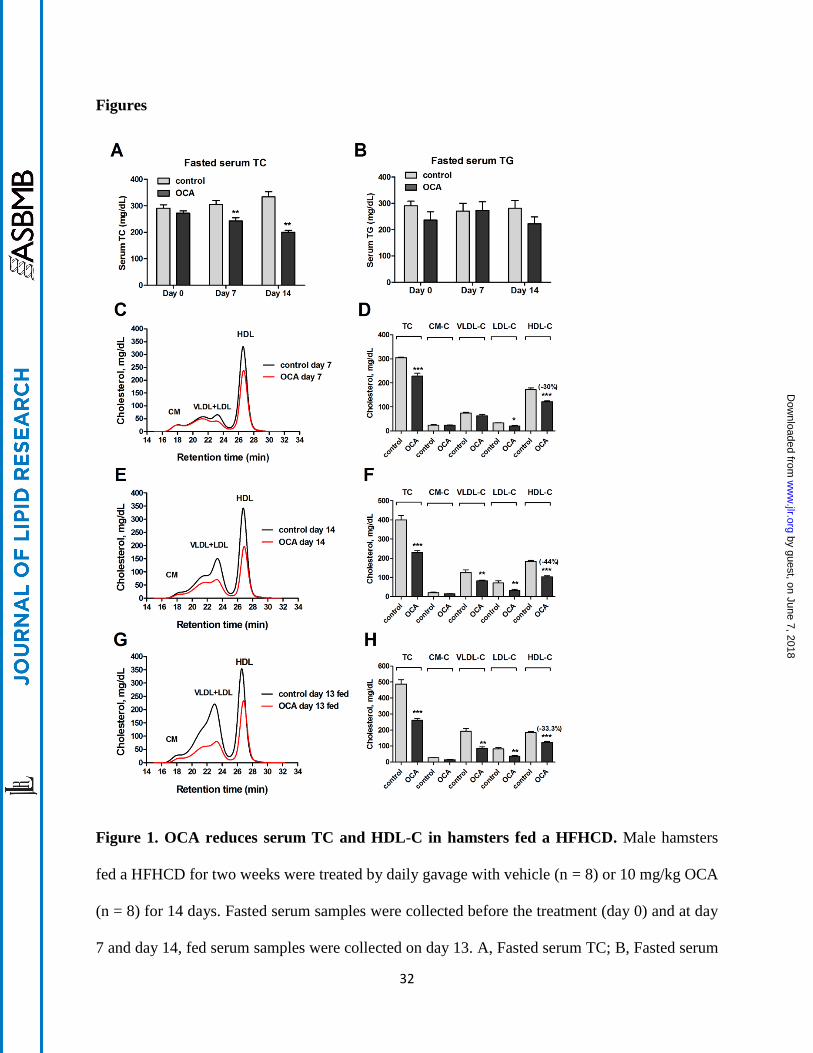
days. Measurements of fasting serum TC and TG levels showed that OCA administration
reduced TC levels to 80% of vehicle control (p < 0.01) after 7-days of treatment and TC levels
further declined to 60% of control after 14 days in OCA treated hamsters (p < 0.01) (Fig. 1A)
while OCA did not significantly affect serum TG levels (Fig. 1B). We also examined the effects
of OCA on serum lipids under fed conditions. Although there was a trend toward a reduction in
non-fasting serum TG levels with OCA (vehicle, 350 ± 28 mg/dL, OCA, 272 ± 31 mg/dL), it did
not reach a statistical significance, whereas non-fasting TC levels were markedly decreased in
OCA-treated hamsters (vehicle, 475 ± 36 mg/dL, OCA, 261 ± 7.2 mg/dL; p < 0.001), consistent
with the effects of OCA under the fasted state. OCA treatment for 14 days slightly reduced body
weight gain (8% versus 12.4% in the control group, p < 0.05) (Supplemental Figure S1A),
which was likely caused by lower food intake in OCA-treated animals (Supplemental Figure
S1B). The liver weights were slightly lower in OCA-treated hamsters while liver index was not
affected (Supplemental Figure S1C, D). Thus, the cholesterol lowering effects of OCA in these
hyperlipidemic hamsters were time-dependent and were prominent.
To gain a better understanding of the cholesterol lowering effect of OCA and its impact on
plasma lipoprotein cholesterol profiles, we performed HPLC separation of all serum samples
individually (day 14 group) or pooled samples that combined two serum samples together from
the same group (day 0, day 7 and day 13). Results showed that before the drug treatment, the
control and OCA groups have identical lipoprotein fractions and cholesterol is largely carried in
by guest, on June 7, 2018w
ww
.jlr.orgD
ownloaded from

11
the HDL fraction in hamsters fed the HFHC diet for two weeks (Supplemental Figure S2A, B).
After 7-days of OCA treatment, the reduction of TC was driven nearly by the sole reduction of
HDL-C as cholesterol levels in chylomicron and VLDL fractions were unchanged and only a
small decrease in LDL fraction was observed (Fig. 1C, D). At the end of 14 days treatment,
cholesterol levels in HDL fraction were further decreased by OCA treatment to 56% of control (-
80.3 mg/dL, p < 0.001) (Fig. 1E, F). In addition, a 35% reduction in VLDL-C (-44 mg/dL) and a
54.5% reduction in LDL-C (-39 mg/dL) were also observed. Furthermore, we observed a
consistent reduction in HDL-C and other lipoprotein cholesterol concentrations by OCA under
the fed state (Fig. 1G, H). HPLC analysis of lipoprotein-TG fractions (Supplemental Figure
S2C-G) revealed only a small reduction of VLDL-TG after 14 days of OCA treatment, which
was consistent with overall insignificant effects of OCA on serum TG levels in hyperlipidemic
hamsters. In addition, we measured serum ALT levels and total bilirubin levels which were in
normal ranges of hamster values and were not significantly elevated by OCA treatment
(Supplemental Figure S3), indicating that liver functions were not disturbed under the diet or
treatment conditions. Altogether, these data demonstrate that FXR activation by OCA led to a
strong cholesterol lowering effect driven largely by reducing serum HDL-C levels in this
hyperlipidemic animal model under both fasted and non-fasted conditions.
OCA treatment reduces hepatic cholesterol and increases fecal cholesterol contents
Next we investigated whether the plasma HDL-C lowering effect of OCA was associated
with changes in hepatic and fecal cholesterol contents. Measurement of hepatic lipids showed
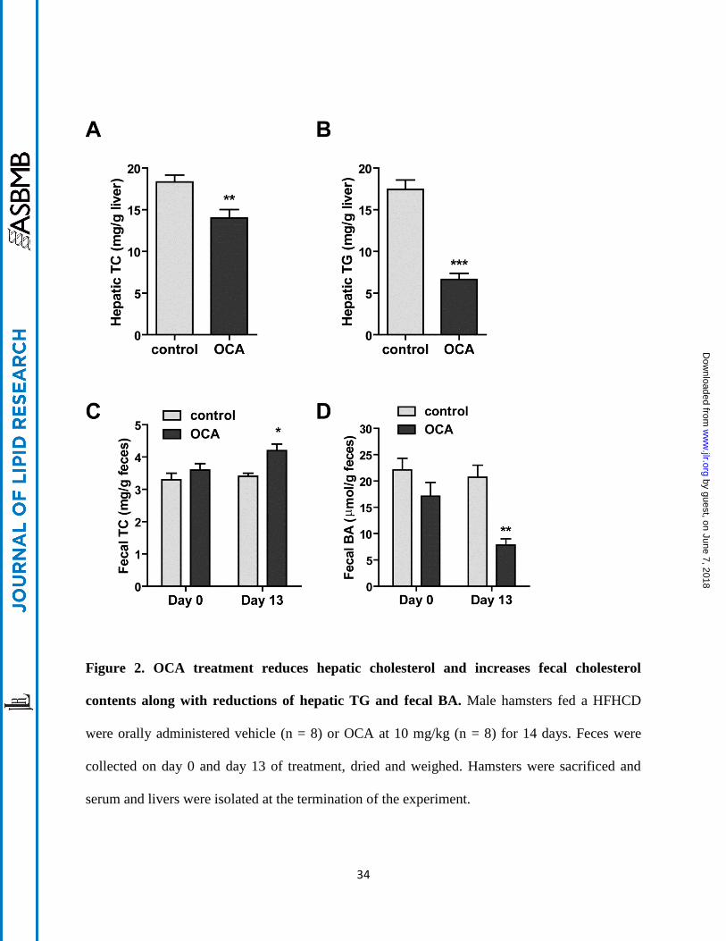
that OCA treatment of 14 days significantly reduced hepatic cholesterol contents by
approximately 24% compared to vehicle control (p < 0.01) (Fig. 2A). OCA also reduced hepatic
by guest, on June 7, 2018w
ww
.jlr.orgD
ownloaded from

12
TG contents (Fig. 2B), which was consistent with reported effects of other FXR agonists on
lowering hepatic TG (23, 30, 31). The cholesterol contents of fecal samples collected on day 0
and day 13 of control group were nearly identical, but cholesterol levels were significantly
increased in fecal samples after 13 days of OCA treatment (Fig. 2C). We also detected a
substantial reduction in fecal BAs levels after OCA treatment (Fig. 2D). Collectively, these data
demonstrated that the removal of HDL-C from circulation by OCA was accompanied by an
increase in transhepatic cholesterol movement into feces.
OCA upregulates hepatic SR-BI mRNA and protein levels
To gain a mechanistic insight into the OCA-mediated increases of fecal cholesterol levels
and reductions of BA synthesis, we investigated the influence of FXR activation by OCA on
expressions of hepatic genes that are involved in BA synthesis and transhepatic cholesterol
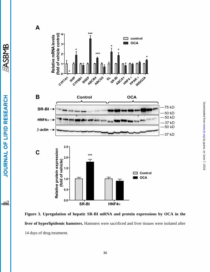
efflux (Fig. 3A). Hepatic gene expression analysis by qRT-PCR showed that the mRNA
expression of classical FXR-controlled genes in BA synthetic pathway (CPY7A1, SHP, BSEP)
was strongly modulated by OCA. The substantial reduction of CYP7A1 resulted in the decreased
fecal BA content via inhibition of BA synthesis in liver tissue. Interestingly, among the four
genes involved in transhepatic cholesterol efflux, with the exception of ATP-binding cassette
(ABC) G5, mRNA levels of ABCB4, endothelial lipase (EL) and SR-BI were all elevated in
OCA-treated hamsters. In addition, we measured mRNA levels of ABCA1, ApoA1, PPARα and
HNF4α. ABCA1 is involved in hepatic efflux of cholesterol and phospholipid to lipid-poor HDL
particles (32) and we detected a small reduction (< 20%) in ABCA1 mRNA levels by OCA
treatment whereas ApoA1, HNF4α and PPARmRNA levels were unchanged. Interestingly, a
previous study has demonstrated a FXR-dependent upregulation of mouse Insig2a gene
by guest, on June 7, 2018w
ww
.jlr.orgD
ownloaded from
13
transcription (33). Using qPCR and specific hamster primers, we detected a nearly 40% increase
(p < 0.05) in the mRNA levels of Insig2a in OCA treated hamster livers, confirming the original
findings made in mice.
The function of SR-BI in reversal cholesterol transport is well characterized and the
increased expression of SR-BI protein in liver is linked to enhanced HDL-C uptake from plasma
in mice (34). Western blotting of liver homogenates of all liver samples demonstrated a 1.8-fold
increase (p < 0.001) in SR-BI protein levels in OCA-treated animals compared to control (Fig.
3B, C). Increased expression of HNF4α by FXR agonist GW4064 was reported as a causal factor
for FXR-mediated elevation of SR-BI expression in mice (35). However, in our study, HNF4α
protein levels were unchanged in these samples which were in line with the negative results of
qRT-PCR. It was shown that activation of Janus N-terminal kinase (p-JNK) by GW4064 was
responsible for increased HNF4α expression and subsequent SR-BI upregulation by this FXR
ligand in mice (35). Thus, we examined p-JNK and total JNK protein levels in hamster livers and
we did not detect differences between OCA-treated and control groups (Supplemental Figure
S4). Thus, our data suggest that upregulation of SR-BI by OCA does not involve changes in
hepatic HNF4α abundance. Altogether, these results of mRNA and protein analysis demonstrated
that OCA upregulates hepatic SR-BI expression which may account for the reduction of plasma
HDL-C and the increase in fecal cholesterol in these dyslipidemic hamsters treated with OCA.
Effects of OCA treatment on SREBP pathway
The plasma cholesterol metabolism is also critically influenced by the expression level of
hepatic LDL receptor (LDLR) and its negative regulator PCSK9, both of which are
transcriptionally activated by the mature form of sterol-regulatory element binding protein 2 (m-
by guest, on June 7, 2018w
ww
.jlr.orgD
ownloaded from

14
SREBP2) via the SRE-1 sites in their gene regulatory region (36,37). Using total liver
homogenates, we analyzed hepatic LDLR, PCSK9 and SREBP2 precursor and mature protein
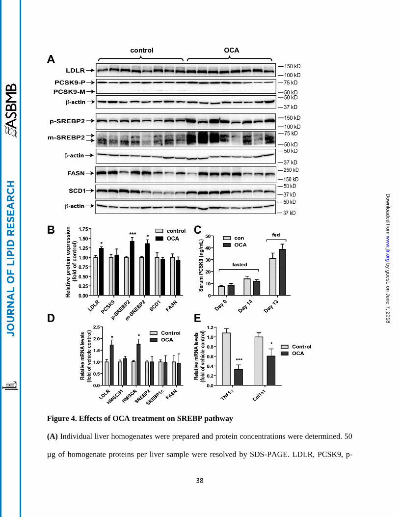
levels by Western blotting (Fig. 4A, B) and serum PCSK9 levels by ELISA (Fig. 4C). OCA
treatment produced small but significant increases in LDLR protein level to 24% over control
and p-SREBP2 and m-SREBP2 protein levels to approximately 40% over control whereas serum
and hepatic PCSK9 protein levels were not changed by OCA treatment. In addition, we
examined the protein levels of fatty acid synthetase (FASN) and stearoyl-CoA desaturase-1
(SCD1) and no differences in their expressions between the two groups were observed. The
results of Western blotting were further corroborated by qRT-PCR analysis of hepatic gene
expression (Fig. 4D). OCA treatment increased mRNA levels of LDLR by 72% compared to
control. The mRNA level of HMG CoA-reductase (HMGCR), another SREBP2-modulated gene
was also elevated in OCA-treated liver tissues. Combined with the observation that hepatic
cholesterol levels were reduced in liver tissues of OCA treated hamsters, these data together
suggest that a modest increase in hepatic LDLR abundance as the result of activated SREBP
pathway may contribute to the reduction of serum LDL-C levels at the latter time point of OCA
treatment. In addition, measurement of mRNA levels of TNF1α and collagen type I (Col1a1)
(Fig. 4E) demonstrated significant mRNA reductions of these inflammatory and fibrosis-related
marker genes, which were in line with anti-inflammatory effects of this FXR agonist (16).
OCA treatment did not affect serum cholesterol and hepatic SR-BI expression levels in
normolipidemic hamsters
We were interested in learning whether the effects of OCA on plasma cholesterol metabolism
and SR-BI expression are affected by hepatic cholesterol levels. We treated hamsters fed a NCD
by guest, on June 7, 2018w
ww
.jlr.orgD
ownloaded from

15
with OCA at 10 mg/kg for two weeks. No obvious differences in food intake, body weight or
liver weight were observed between OCA and the control groups (Supplemental Figure S5A-
D). Measurement of serum lipids showed that administration of OCA to normolipidemic
hamsters had no effects on serum TC and HDL-C levels (Fig. 5A, B) while serum TG levels
were modestly increased owing to an increase in the VLDL-TG fraction (Supplemental Figure
S5E, F). Hepatic TG content was significantly reduced while hepatic cholesterol content was
increased in OCA-treated animals by 38% compared to the control group (Fig. 5C, D).
Measurement of fecal cholesterol and BAs demonstrated a reduction in fecal BAs but no changes
in fecal cholesterol contents after OCA treatment (Fig. 5E, F).
Hepatic gene expression analysis by qRT-PCR demonstrated that among FXR-modulated
genes, CYP7A1, SHP, BSEP, INSIG2A and ABCB4 were modulated by OCA to levels
comparable to its effects seen in hamsters fed the HFHCD. However, the induction on EL and
SR-BI gene expression by OCA were not observed in the normolipidemic hamsters (Fig. 6A).
Furthermore, hepatic SR-BI protein levels were not increased by OCA treatment (Fig. 6B, C),
which corroborated the results of qRT-PCR. Despite a small increase in hepatic cholesterol
content, we did not detect differences in hepatic expressions of a panel of SREBP target genes
including LDLR mRNA and PCSK9 (Fig. 6D) as well as their proteins (Fig. 6B, C).
To further examine dose dependent effects of OCA on hepatic SR-BI and other FXR
modulated genes in normolipidemic hamsters, another cohort of hamsters fed a NCD were orally
dosed with OCA for three days at 10 mg/kg, 20 mg/kg or 30 mg/kg while the control animals
received the vehicle. Treatment of animals with OCA at these doses did not affect serum levels
of TC and HDL-C (Supplemental Figure S6A, B). Health parameters including serum bilirubin
levels, food intake and body weights of hamsters were not significantly different among the
by guest, on June 7, 2018w
ww
.jlr.orgD
ownloaded from
16
groups (Supplemental Figure S6C-E). Measurements of liver weight showed a tendency of
dose-dependent increase in liver weight, which was further manifested in comparisons of liver
index (Supplemental Figure S6F, G). The liver index of OCA 30 mg/kg group was 19% higher
than the control group (p < 0.001), suggesting a mild adaptive changes in the liver.
Hepatic gene expression analysis of a panel of FXR-modulated genes (Fig. 7A) demonstrated
dose-dependent effects of OCA on mRNA levels of SHP, CYP7A1, CYP8B1, BSEP and
INSIG2A and showed that OCA at a dose of 20 mg/kg had a greater effect than 30 mg/kg. The
relative lower effect of 30 mg/kg OCA treatment on modulating FXR target genes including
SHP, BSEP and INSIG2A could reflect a mild liver toxicity at this higher dose. Importantly, we
did not observe a significant upregulation of EL or SR-BI mRNA expressions by OCA under
these doses, which was further confirmed by Western blot analysis of liver SR-BI protein
abundances (Fig. 7B). Overall, results of the 3-day treatment study with OCA at doses up to 30
mg/kg largely confirmed our results obtained from 14-day OCA treatment of 10 mg/kg.
Collectively, these results suggest that under normolipidemic conditions, FXR activation by
OCA inhibited BA synthesis without inducing SR-BI mediated transhepatic cholesterol
movement.
LXR activation alone did not induce hepatic expression of SR-BI in hamsters
One substantial difference between NCD and HCHFD fed hamsters is the hepatic cholesterol
content. Higher hepatic cholesterol amount in HFHCD fed hamsters might have a stimulating
effect on SR-BI expression through LXR activation as it was reported that SR-BI expression in
human macrophages was induced by LXR agonists (38). To learn whether SR-BI expression in
hamsters could be directly induced by LXR activation, we examined SR-BI protein levels in
by guest, on June 7, 2018w
ww
.jlr.orgD
ownloaded from

17
liver tissues of NCD fed hamsters that were previously treated with a specific LXR agonist
GW3965 (30 mg/kg) or vehicle for 7 days (39). As shown in Supplemental Figure S7, hepatic
SR-BI protein levels did not differ between the two groups. We further confirmed the lack of
inducing effect of GW3965 treatment on hepatic SR-BI expression in hamsters fed a high fat diet
or a dyslipidemic fructose diet (data not shown).
Discussion
We set out this study to understand how OCA, a therapeutic FXR agonist, regulates plasma
cholesterol metabolism under dyslipidemic conditions by utilizing hyperlipidemic hamsters, a
model that has been used with increasing frequency in recent years to study lipoprotein
metabolism, atherosclerosis and to evaluate effects of hypolipidemic agents including PPAR
activators and LXR agonists (39-42). The important new findings of our current study are that
activation of hepatic FXR by OCA increases SR-BI expression and accelerates the removal of
circulating HDL-C with increased cholesterol fecal excretion in hyperlipidemic hamsters.
Previous animal studies of OCA conducted in rat, wild type mice, LDLR deficient mice and
CETP transgenic mice produced inconsistent effects on plasma cholesterol levels, in particular
HDL-C levels. For example, Hambruch et al. reported that in C57BL/6J mice fed a HFD, OCA
treatment of four weeks at a dose of 30 mg/kg caused a small increase in HDL-C without
significant effect on total plasma cholesterol (23); however, in the same study, it was reported
that after 12 weeks of treatment, OCA at 10 mg/kg dose reduced plasma total cholesterol
significantly and showed a trend in HDL-C lowering in CETPtg-LDLR(-/-) mice.
In this study, we demonstrated a specific and a time-dependent reduction of HDL-C levels by
treating hyperlipidemic hamsters with 10 mg/kg OCA. The facts that reduction of HDL-C
by guest, on June 7, 2018w
ww
.jlr.orgD
ownloaded from
18
preceded the decrease in LDL-C and VLDL-C levels and the majority of plasma cholesterol was
carried in HDL particles suggest that enhanced HDL-C removal by OCA is likely the primary
driving force for plasma cholesterol lowering observed in this animal model. It was previously
reported that in a cohort of a HFD fed hamsters, OCA treatment of 11 days at a daily dose of 30
mg/kg reduced cumulative food intake by 13%, lowered HDL-C and increased VLDL-C,
resulting in unchanged total plasma cholesterol levels (22, 24). In our study, we observed a
similar amount of reduction in cumulative food intake (~14%), but we observed significant
HDL-C reduction without any elevation of VLDL-C or LDL-C. It is not clear what factors
contributed to the different effects of OCA on VLDL-C fraction in these different hamster
studies. The conflicting results might be due to different OCA doses, different diet compositions
or different OCA preparations.
In the previous report of HFD-fed hamsters, OCA reduced liver BA pool size and fecal BAs
(22), which was in line with our current study of HFHCD fed hamsters in which OCA treatment
of 14 days reduced fecal BAs by 62%. Importantly, in addition to reduction of fecal BAs, the
current study demonstrated a reduction of hepatic cholesterol contents and an increase in fecal
cholesterol levels in OCA-treated hamsters. Among FXR modulated genes that are involved in
BA metabolism, the upregulations of SR-BI, ABCB4, ABCG5 and EL by FXR agonists FXR-
450 and PX20606 in mice were linked to the clearance of cholesteryl esters from HDL and their
excretion into feces via the bile (23). In the same study, it was reported that OCA did not
increase hepatic SR-BI, ABCB4, EL or ABCG5 mRNA levels in mice, which was correlated
with a lack of effect of OCA on cholesterol efflux in mice (23). However, we detected significant
inductions of SR-BI, ABCB4 and EL mRNA levels in hamster livers after OCA treatment. We
further demonstrated a 1.8-fold increase in hepatic SR-BI protein levels by OCA treatment. It has
by guest, on June 7, 2018w
ww
.jlr.orgD
ownloaded from

19
been suggested that the function of SR-BI in biliary cholesterol secretion is mediated through
ABCG5/ABCG8 dependent as well as independent mechanisms (43,44). While ABCB4 is
mainly involved in phospholipid secretion (45) and is shown not to be dependent on biliary sterol
secretion (46), a model emerges in hyperlipidemic hamsters in which the increased hepatic
uptake of cholesterol esters from HDL via SR-BI and enhanced HDL metabolism by endothelial
lipase, coupled with increased cholesterol excretion via ABCB4 lead to stimulated transhepatic
cholesterol efflux in hyperlipidemic hamsters by OCA treatment. Further investigations using
radioisotope labeled cholesterol to demonstrate a direct effect of OCA on upregulation of HDL-
C uptake in SR-BI wild-type and deficient animals will be required to validate this working
model.
The results from our chow fed hamster studies showed that OCA treatment of 14 days
inhibited BAs hepatic synthesis which was evidenced by strong effects on hepatic CPY7A1 and
SHP gene expression and reduction of fecal BAs. However, these OCA effects occurred in the
absence of changes in serum HDL-C, fecal cholesterol and hepatic expression of EL and SR-BI,
indicating that the transhepatic cholesterol efflux was not induced by OCA in these
normolipidemic hamsters, which might account for the small increase in hepatic cholesterol
content. The lack of OCA effect on hepatic SR-BI expression under normolipidemic conditions
was also consistently observed in chow fed hamsters treated with OCA at daily doses up to 30
mg/kg.
It has been shown that the regulation of SR-BI expression by FXR involves different
mechanisms. One study reported that FXR activation by synthetic agonist GW4064 increased
transcription factor HNF4 protein levels that led to the transcriptional activation of SR-BI gene
through HNF4 binding sequences embedded in promoter region and intronic sequences of
by guest, on June 7, 2018w
ww
.jlr.orgD
ownloaded from
20
murine SR-BI gene (35). Another study identified multiple functional FXR binding sites (IR1) in
the first intron of the murine SR-BI gene (47). Furthermore, it was reported that treating HepG2
cells with GW4064 increased SR-BI mRNA levels. This effect was linked to the binding of FXR
to a putative FXRRE site (DR8) in the promoter region of human SR-BI gene (48). In this
current study we observed increased SR-BI mRNA and protein expression to similar extent by
OCA treatment in the absence of changes in hepatic HNF4α abundance of hamsters fed a
HFHCD. Thus, our data suggest that SR-BI gene transcription is directly induced by FXR
activation in hamster species under hyperlipidemic conditions but not under normolipidemic
state. One major difference between NCD and HCHFD fed hamsters is the hepatic cholesterol
content. Higher hepatic cholesterol in HFHCD fed hamsters might have a stimulating effect on
SR-BI expression through LXR activation as SR-BI expression in human macrophages was
induced by LXR agonists (38). However, our examination of SR-BI protein abundances in livers
of normolipidemic hamsters treated with LXR agonist GW3965 or vehicle failed to detect any
differences in SR-BI expression levels (Supplemental Figure S7), suggesting that in hamster
species, SR-BI is not directly regulated by LXR alone. Thus, currently, it is unclear how
different levels of hepatic cholesterol or cholesterol metabolites could impact the effect of OCA
on SR-BI gene expression in hamster species. Since activation of SR-BI transcription is
associated with a favorable lipoprotein cholesterol profile, our findings warrant further
investigations to better understand the influence of dietary cholesterol on the inducibility of SR-
BI expression by FXR agonists including OCA at the gene transcriptional level in different
animal models and in humans.
In our hyperlipidemic hamster study, in addition to HDL-C reduction, OCA treatment
lowered serum LDL-C and VLDL-C fractions at the later treatment time points. Previous in vitro
by guest, on June 7, 2018w
ww
.jlr.orgD
ownloaded from

21
studies reported that FXR activation in hepatic cells led to LDLR mRNA stabilization (49) or
inhibition of PCSK9 transcription (50). Either of these effects could result in decreases in plasma
LDL-C levels owing to increased hepatic LDLR abundance. As investigated here, while we did
not observe changes in serum and hepatic PCSK9 protein levels, we did detect small but
significant increases in hepatic LDLR mRNA and protein levels which were accompanied by
increased mature form of SREBP2 in the liver of OCA treated hamsters fed a HFHCD (Fig. 4).
Combined with the observation of reduced liver cholesterol content, our results suggest that in
hamsters fed a cholesterol enriched diet, hepatic SREBP pathway was repressed; alleviation of
this repression through SR-BI facilitated transhepatic cholesterol excretion into feces probably
generated a positive yet modest impact on the intracellular proteolytic process that converts the
inactive SREBP2 to the active form to enter the nucleus and turn on the LDLR gene transcription
along with a subset of SREBP2-target genes. In the absence of changes in PCSK9 hepatic and
serum levels, the increase in hepatic LDLR expression may contribute, at least in part, to the
LDL-C reduction. Furthermore, previous in vitro and in vivo studies have suggested that SR-BI
mediates selective uptake of cholesterol esters from LDL particles in additional to HDL particles
(8, 51). Thus, the OCA mediated reduction of serum LDL-C and VLDL-C could result from its
combined activities in increasing SR-BI and LDLR abundances in liver tissue.
In summary, OCA treatment of hyperlipidemic hamsters elicited reductions of serum HDL-
cholesterol levels with concomitant upregulation of hepatic SR-BI expression and increased
cholesterol excretion into feces. Our findings in this hamster model suggest that induction of
hepatic SR-BI expression may account for the hypocholesterolemic effect of OCA under
hyperlipidemic states.
by guest, on June 7, 2018w
ww
.jlr.orgD
ownloaded from
22
Acknowledgments
This study was supported by the Department of Veterans Affairs (Office of Research and
Development, Medical Research Service), by a grant (1R01AT006336-01A1) from National
Center for Complementary & Integrative Health and by a collaborative research grant from
Intercept Pharmaceuticals.
References
1. Calkin, A. C., and P. Tontonoz. 2012. Transcriptional integration of metabolism by the
nuclear sterol-activated receptors LXR and FXR. Nat Rev Mol Cell Biol 13: 213-224.
2. Thomas, A. M., S. N. Hart, B. Kong, J. Fang, X. B. Zhong, and G. L. Guo. 2010. Genome-
wide tissue-specific farnesoid X receptor binding in mouse liver and intestine.
Hepatology 51: 1410-1419.
3. Zhan, L., H. X. Liu, Y. Fang, B. Kong, Y. He, X. B. Zhong, J. Fang, Y. J. Wan, and G. L.
Guo. 2014. Genome-wide binding and transcriptome analysis of human farnesoid X
receptor in primary human hepatocytes. PLoS One 9: e105930.
4. de Agular Vallim T. Q., E. J. Tarling, H. Ahn, L. R. Hagey, C. E. Romanoski, R. G. Lee,
M. J. Graham, H. Motohashi, M. Yamamoto, and P. A. Edwards. 2015. MAFG is a
transcriptional repressor of bile acid synthesis and metabolism. Cell Metab 21: 298-310.
5. Rigotti, A., B. Trigatti, J. Babitt, M. Penman, S. Xu, and M. Krieger. 1997. Scavenger
receptor BI-a cell surface receptor for high density lipoprotein. Curr Opin Lipidol 8: 181-
188.
by guest, on June 7, 2018w
ww
.jlr.orgD
ownloaded from

23
6. Rigotti, A., B. L. Trigatti, M. Penman, H. Rayburn, J. Herz, and M. Krieger. 1997. A
targeted mutation in the murine gene encoding the high density lipoprotein (HDL)
receptor scavenger receptor class B type I reveals its key role in HDL metabolism
8. Proc Natl Acad Sci USA 94: 12610-12615.
7. Shen, W. J., J. Hu, Z. Hu, F. B. Kraemer, and S. Azhar. 2014. Scavenger receptor class B
type I (SR-BI): a versatile receptor with multiple functions and actions. Metabolism 63:
875-886.
8. Brodeur, M. R., V. Luangrath, G. Bourret, L. Falstrault, and L. Brissette. 2005.
Physiological importance of SR-BI in the in vivo metabolism of human HDL and LDL in
male and female mice. J Lipid Res 46: 687-696.
9. Zanoni, P., S. A. Khetarpal, D. B. Larach, W. F. Hancock-Cerutti, J. S. Millar, M. Cuchel,
S. DerOhannessian, A. Kontush, P. Surendran, P. et al. 2016. Rare variant in scavenger
receptor BI raises HDL cholesterol and increases risk of coronary heart disease. Science
351: 1166-1171.
10. Kozarsky, K. F., M. H. Donahee, J. M. Glick, M. Krieger, and D. J. Rader. 2000. Gene
transfer and hepatic overexpression of the HDL receptor SR-BI reduces atherosclerosis in
the cholesterol-fed LDL receptor-deficient mouse. Arterioscler Thromb Vasc Biol 20:
721-727.
11. Kozarsky, K. F., Donahee, M. H., Rigotti, A., Iqbal, S. N., Edelman, E. R. & Krieger, M.
(1997) Overexpression of the HDL receptor SR-BI alters plasma HDL and bile
cholesterol levels. Nature 387: 414-417.
by guest, on June 7, 2018w
ww
.jlr.orgD
ownloaded from

24
12. van. E. M. Twisk, J., M. Hoekstra, B. T. Van Rij, C. A. Van der Lans, I. S. Bos, J. K.
Kruijt, F. Kuipers, and T. J. Van Berkel. 2003. Differential effects of scavenger receptor
BI deficiency on lipid metabolism in cells of the arterial wall and in the liver. J Biol
Chem 278: 23699-23705.
13. Kozarsky, K. F., M. H. Donahee, J. M. Glick, M. Krieger, and D. J. Rader. 2000. Gene
transfer and hepatic overexpression of the HDL receptor SR-BI reduces atherosclerosis in
the cholesterol-fed LDL receptor-deficient mouse. Arterioscler Thromb Vasc Biol 20:
721-727.
14. Vergeer, M., Korporaal, S.J., Franssen, R., Meurs, I., Out, R., Hovingh, G.K., Hoekstra,
M., Sierts, J.A., Dallinga-Thie, G.M., Motazacker, M.M., Holleboom, A.G., Van Berkel,
T.J., Kastelein, J.J., Van Eck, M., Kuivenhoven, J.A. 2011. Genetic variant of the
scavenger receptor BI in humans. N Engl J Med. 364:136-45.
15. Brunham, L. R., Tietjen, I., Bochem, A. E., Singaraja, R. R., Franchini, P. L., Radomski, C.,
Mattice, M., Legendre, A., Hovingh, G. K. et al. 2011. Novel mutations in scavenger receptor BI
associated with high HDL cholesterol in humans. Clin Genet 79: 575-581.
16. Adorini, L., M. Pruzanski, and D. Shapiro. 2012. Farnesoid X receptor targeting to treat
nonalcoholic steatohepatitis. Drug Discov Today 17: 988-997.
17. Neuschwander-Tetri, B. A., R. Loomba, A. J. Sanyal, J. E. Lavine, M. L. Van Natta, M. F.
Abdelmalek, N. Chalasani, S. Dasarathy, A. M. Diehl, et al. 2015. Farnesoid X nuclear
receptor ligand obeticholic acid for non-cirrhotic, non-alcoholic steatohepatitis (FLINT):
a multicentre, randomised, placebo-controlled trial. Lancet 385: 956-965.
by guest, on June 7, 2018w
ww
.jlr.orgD
ownloaded from

25
18. Mudaliar, S., R. R. Henry, A. J. Sanyal, L. Morrow, H. U. Marschall, M. Kipnes, L.
Adorini, C. I. Sciacca, P. Clopton, P. et al. 2013. Efficacy and safety of the farnesoid X
receptor agonist obeticholic acid in patients with type 2 diabetes and nonalcoholic fatty
liver disease. Gastroenterology 145: 574-582.
19. Pencek, R., T. Marmon, J. D. Roth, A. Liberman, R. Hooshmand-Rad, and M. Young.
2016. Effects of Obeticholic Acid on Lipoprotein Metabolism in Healthy Volunteers.
Diabetes Obes Metab Apr 25. doi: 10.1111/dom.12681.
20. Hirschfield, G. M., A. Mason, V. Luketic, K. Lindor, S. C. Gordon, M. Mayo, K. V.
Kowdley, C. Vincent, H. C. Bodhenheimer, et al. 2015. Efficacy of obeticholic acid in
patients with primary biliary cirrhosis and inadequate response to ursodeoxycholic acid.
Gastroenterology 148: 751-761.
21. Cipriani, S., A.Mencarelli, G. Palladino, and S. Fiorucci. 2010. FXR activation reverses
insulin resistance and lipid abnormalities and protects against liver steatosis in Zucker
(fa/fa) obese rats. J Lipid Res 51: 771-784.
22. Gardes, C., E. Chaput, A. Staempfli, D. Blum, H. Richter, and G. M. Benson. 2013.
Differential regulation of bile acid and cholesterol metabolism by the farnesoid X
receptor in Ldlr -/- mice versus hamsters. J Lipid Res 54: 1283-1299.
23. Hambruch, E., S. Miyazaki-Anzai, U. Hahn, S. Matysik, A. Boettcher, S. Perovic-Ottstadt,
T. Schluter, O. Kinzel, H. D. Krol, et al. 2012. Synthetic farnesoid X receptor agonists
induce high-density lipoprotein-mediated transhepatic cholesterol efflux in mice and
by guest, on June 7, 2018w
ww
.jlr.orgD
ownloaded from
26
monkeys and prevent atherosclerosis in cholesteryl ester transfer protein transgenic low-
density lipoprotein receptor (-/-) mice. J Pharmacol Exp Ther 343: 556-567.
24. Gardes, C., D. Blum, K. Bleicher, E. Chaput, M. Ebeling, P. Hartman, C. Handschin, H.
Richter, and G. M. Benson. 2011. Studies in mice, hamsters, and rats demonstrate that
repression of hepatic apoA-I expression by taurocholic acid in mice is not mediated by
the farnesoid-X-receptor. J Lipid Res 52: 1188-1199.
25. Dong, B., A. B. Singh, C. F. K. Kan, and J. Liu. 2014. CETP inhibitors downregulate
hepatic LDL receptor and PCSK9 expression in vitro and in vivo through a SREBP2
dependent mechanism. Atherosclerosis 235: 449-462.
26. Dong, B., H. Li, A. B. Singh, A. Cao, and J. Liu. 2015. Inhibition of PCSK9 transcription
by berberine involves down-regulation of hepatic HNF1alpha protein expression through
the ubiquitin-proteasome degradation pathway. J Biol Chem 290: 4047-4058.
27. Yu, C., F. Wang, M. Kan, C. Jin, R. B. Jones, M. Weinstein, C. X. Deng, and W. L.
McKeehan. 2000. Elevated cholesterol metabolism and bile acid synthesis in mice
lacking membrane tyrosine kinase receptor FGFR4. J Biol Chem 275:15482-9.
28. Cao, A., M. Wu, H. Li, and J. Liu. 2011. Janus kinase activation by cytokine oncostatin M
decreases PCSK9 expression in liver cells. J Lipid Res 52: 518-530.
29. Li, H., Dong, B., Park, S. W., Lee, H. S., Chen, W. & Liu, J. 2009. Hepatocyte nuclear
factor 1alpha plays a critical role in PCSK9 gene transcription and regulation by the natural
hypocholesterolemic compound berberine. J Biol Chem 284: 28885-28895.
by guest, on June 7, 2018w
ww
.jlr.orgD
ownloaded from

27
30. Evans, M. J., P.E. Mahaney, L. Borges-Marcucci, K. Lai, S. Wang, J. A. Krueger, S. J.
Gardell, C. Huard, R. Martinez, R. et al. 2009. A synthetic farnesoid X receptor (FXR)
agonist promotes cholesterol lowering in models of dyslipidemia. Am J Physiol
Gastrointest Liver Physiol 296: G543-G552.
31. Genin, M. J., A. B. Bueno, F. J. Agejas, P. R. Manninen, W. P. Bocchinfuso, C. Montrose-
Rafizadeh, E. A. Cannady, T. M. Jones, J. R. Stille, et al. 2015. Discovery of 6-(4-{[5-
Cyclopropyl-3-(2,6-dichlorophenyl)isoxazol-4-yl]methoxy}piperidin-1-yl)- 1-methyl-
1H-indole-3-carboxylic Acid: A Novel FXR Agonist for the Treatment of Dyslipidemia.
J Med Chem 58: 9768-9772.
32. Rosenson, R. S., H. B., Jr. Brewer, B. J. Ansell, P. Barter, M. J. Chapman, J. W. Heinecke,
A. Kontush, A. Tall, and N. R. Webb. 2016. Dysfunctional HDL and atherosclerotic
cardiovascular disease. Nat Rev Cardiol 13: 48-60.
33. Hubbert, M. L., Zhang, Y., Lee, F. Y. & Edwards, P. A. 2007. Regulation of hepatic Insig-
2 by the farnesoid X receptor. Mol. Endocrinol 21: 1359-1369.
34. Trigatti, B., A. Rigotti, and M. Krieger. 2000. The role of the high-density lipoprotein
receptor SR-BI in cholesterol metabolism. Curr Opin Lipidol 11: 123-131.
35. Zhang, Y., L. Yin, J. Anderson, H. Ma, F. J. Gonzalez, T. M. Willson, and P.A. Edwards.
2010. Identification of novel pathways that control farnesoid X receptor-mediated
hypocholesterolemia. J Biol Chem 285: 3035-3043.
36. Horton, J. D., N. A. Shah, J. A Warrington, N. N. Anderson, S. W. Park, M. S. Brown, and
J. L. Goldstein. 2003. Combined analysis of oligonucleotide microarray data from
by guest, on June 7, 2018w
ww
.jlr.orgD
ownloaded from
28
transgenic and knockout mice identifies direct SREBP target genes. Proc Natl Acad Sci
USA 100: 12027-12032.
37. Horton, J. D., J. C. Cohen, and H. H. Hobbs. 2006. Molecular biology of PCSK9: its role
in LDL metabolism. Trends Biochem Sci 32: 71-77.
38. Ma, A. Z., Song, Z. Y. & Zhang, Q. (2014) Cholesterol efflux is LXR alpha isoform-
dependent in human macrophages. BMC. Cardiovasc. Disord. 14: 80.
39. Dong, B., Kan, C. F., Singh, A. B. & Liu, J. 2013. High-fructose diet downregulates long-
chain acyl-CoA synthetase 3 expression in liver of hamsters via impairing LXR/RXR
signaling pathway. J Lipid Res 54: 1241-1254.
40. Srivastava, R. A. and S. He. 2010. Anti-hyperlipidemic and insulin sensitizing activities of
fenofibrate reduces aortic lipid deposition in hyperlipidemic Golden Syrian hamster. Mol
Cell Biochem 345: 197-206.
41. Srivastava, R. A. 2011. Evaluation of anti-atherosclerotic activities of PPAR-alpha, PPAR-
gamma, and LXR agonists in hyperlipidemic atherosclerosis-susceptible F(1)B hamsters.
Atherosclerosis 214: 86-93.
42. Dong, B., M. Wu, A. Cao, H. Li, and J. Liu, J. 2011. Suppression of Idol expression is an
additional mechanism underlying statin-induced up-regulation of hepatic LDL receptor
expression. Int J Mol Med 27: 103-110.
by guest, on June 7, 2018w
ww
.jlr.orgD
ownloaded from

29
43. Wiersma, H., A. Gatti, N. Nijstad, R. P. Oude Elferink, F. Kuipers, and U. J. Tietge. 2009.
Scavenger receptor class B type I mediates biliary cholesterol secretion independent of
ATP-binding cassette transporter g5/g8 in mice. Hepatology 50: 1263-1272.
44. Meyer, J. M., G. A. Graf, and D. R. Van der Westhuyzen. 2013. New developments in
selective cholesteryl ester uptake. Curr Opin Lipidol 24: 386-392.
45. Castro-Torres, I. G., de Jesús R. Cárdenas-Vázquez, C. Velazquez-Gonzalez, De, O. A. R.
Ventura-Martinez, E. B. Naranjo-Rodriguez, and M. Martinez-Vazquez. 2015. Future
therapeutic targets for the treatment and prevention of cholesterol gallstones. Eur J
Pharmacol 765: 366-374.
46. Nijstad, N., T. Gautier, F. Briand, D. J. Rader, and U. J. Tietge. 2011. Biliary sterol
secretion is required for functional in vivo reverse cholesterol transport in mice.
Gastroenterology 140: 1043-1051.
47. Li, G., A. M. Thomas, J. A. Williams, B. Kong, J. Liu, Y. Inaba, W. Xie, and G. L. Guo.
2012. Farnesoid X receptor induces murine scavenger receptor Class B type I via intron
binding. PLoS One 7: e35895.
48. Chao, F., W. Gong, Y. Zheng, Y. Li, G. Huang, M. Gao, J. Li, R. Kuruba, X. Gao, et al.
2010. Upregulation of scavenger receptor class B type I expression by activation of FXR
in hepatocyte. Atherosclerosis 213: 443-448.
49. Yashiro, T., Y. Yokoi, M. Shimizu, J. Inoue, and R. Sato. 2011. Chenodeoxycholic acid
stabilization of LDL receptor mRNA depends on 3'-untranslated region and AU-rich
element-binding protein. Biochem Biophys Res Commun 409: 155-159.
by guest, on June 7, 2018w
ww
.jlr.orgD
ownloaded from
30
50. Langhi, C., M. C. Le, S. Kourimate, S. Caron, B. Staels, M. Krempf, P. Costet, and B.
Cariou. 2008. Activation of the farnesoid X receptor represses PCSK9 expression in
human hepatocytes. FEBS Lett 582: 949-955.
51. Ueda, Y., L. Royer, E. Gong, J. Zhang, P. N. Cooper, O. Francone, and E. M. Rubin. 1999.
Lower plasma levels and accelerated clearance of high density lipoprotein (HDL) and
non-HDL cholesterol in scavenger receptor class B type I transgenic mice. J Biol Chem
274: 7165-7171.
by guest, on June 7, 2018w
ww
.jlr.orgD
ownloaded from

31
Table 1. Primers used in qRT-PCR.
Forward Reverse
ABCA1 AACAGTTTGTGGCCCTTTTG AGTTCCAGGCTGGGGTACTT
ABCB4 TCCTATGCACTGGCCTTCTG GCCCCGATGAGGATTGAGAA
ABCG5 ACTGGACTGCATGACTGCAA AGTCAGGATGGCAATTTTGTCG
APOA1 TGGCTGTGCTCTTCCTGACC CTCTGCCGCTGTCTTTCACC
BSEP AGGGCTCTCAACTCTCTCG ATACAGGTCCGACCCTCTCTG
COL1A1 GCTCCTCTTAGGGGCCACT CCACGTCTCACCATTGGGG
CYP7A1 TTCCTGCAACCTTCTGGAGC GCCTCCTTGATGATGCTATCTAGT
CYP8B1 GATGGCACCCGGAAAGTGGA TAGTGGTGGATCTTCTTGGC
EL ACGCTGGCAACTTTGTGAAA AGGTATGCAGGACATCCACA
FASN AGTCCTTGTCCAGGTTCGTG CCACCTAAGCCACCAGTGAT
HMGCR GACGGTGACACTTACCATCTGT GATGCACCGTGTTATGGTGA
HMGCS1 TTTGATGCAGCTGTTTGAGG CCACCTGTAGGTCTGGCATT
HNF1α GAGGTGGCTCAGCAATTCAC CACTCCTCCACCAAGGTCTC
HNF4α CGAGTGGGCCAAGTACATCC CCGAGGGACGATGTAGTCATT
INSIG2A TTCTCAGTTAGCTTGCGCCT GTACCACATCTTGGCTGAACG
LDLR TTGGGTTGATTCCAAACTCC GATTGGCACTGAAAATGGCT
PCSK9 TGCTCCAGAGGTCATCACAG GTCCCACTCTGTGACATGAAG
PPARα CCTGTCTGTTGGGATGTCAC AGGTAGGCCTCGTGGATTCT
SHP AGGGAGGCCTTGGATGTC AGAAGGACGGCAGGTTCC
SR-BI GCGTGGACCCTATGTCTACAG GTCAGGCTGGAAATGGAGGC
SREBP1c GCACTTTTTGACACGTTTCTTC CTGTACAGGCTCTCCTGTGG
SREBP2 GAGAGCTGTGAATTTTCCAGTG CTACAGATGATATCCGGACCAA
TNF1α ACTGAACTTCGGGGTGATCG CTTGGTGGTTTGCTACGACG
by guest, on June 7, 2018w
ww
.jlr.orgD
ownloaded from
32
Figures
Figure 1. OCA reduces serum TC and HDL-C in hamsters fed a HFHCD. Male hamsters
fed a HFHCD for two weeks were treated by daily gavage with vehicle (n = 8) or 10 mg/kg OCA
(n = 8) for 14 days. Fasted serum samples were collected before the treatment (day 0) and at day
7 and day 14, fed serum samples were collected on day 13. A, Fasted serum TC; B, Fasted serum
by guest, on June 7, 2018w
ww
.jlr.orgD
ownloaded from

33
TG; C-H, Cholesterol distribution in HPLC-separated lipoprotein factions from hamsters on a
HFHCD treated with vehicle or OCA (C and D, treatment day 7 fasted serum samples; E and F,
treatment day 14 fasted serum samples; G and H, treatment day 13 fed serum samples). All
values are expressed as mean ± SEM. Significance is indicated as *p < 0.05; **, p < 0.01 and
***, p < 0.001 as compared to vehicle control group.
by guest, on June 7, 2018w
ww
.jlr.orgD
ownloaded from
34
Figure 2. OCA treatment reduces hepatic cholesterol and increases fecal cholesterol
contents along with reductions of hepatic TG and fecal BA. Male hamsters fed a HFHCD
were orally administered vehicle (n = 8) or OCA at 10 mg/kg (n = 8) for 14 days. Feces were
collected on day 0 and day 13 of treatment, dried and weighed. Hamsters were sacrificed and
serum and livers were isolated at the termination of the experiment.
by guest, on June 7, 2018w
ww
.jlr.orgD
ownloaded from

35
(A-B) Lipids were extracted from individual liver samples and TC, TG were measured. Values
are mean ± SEM of 8 hamsters per group. **p < 0.01 and ***p < 0.001 as compared to the
vehicle control group.
(C-D) Lipids were also extracted from dried feces and TC and BA were measured. Values are
mean ± SEM of 4 fecal samples per group. *p < 0.05, **p < 0.01 as compared to the control
group.
by guest, on June 7, 2018w
ww
.jlr.orgD
ownloaded from
36
Figure 3. Upregulation of hepatic SR-BI mRNA and protein expressions by OCA in the
liver of hyperlipidemic hamsters. Hamsters were sacrificed and liver tissues were isolated after
14 days of drug treatment.
by guest, on June 7, 2018w
ww
.jlr.orgD
ownloaded from

37
(A) Total RNA was isolated from individual liver and relative mRNA abundances of indicated
genes were determined by conducting qRT-PCR and normalized to Actin. Values are mean ±
SEM of 8 hamsters per group.
(B) Individual liver homogenates were prepared and protein concentrations were determined. 50
µg of homogenate proteins per liver sample were resolved by SDS-PAGE. SR-BI and HNF4α
proteins were detected by immunoblotting using anti-SR-BI and anti-HNF4α antibodies. The
membrane was reprobed with anti-β-actin antibody.
(C) The protein abundance of SR-BI and HNF4α was quantified with normalization by signals of
-actin using the Alpha View Software. Values are the mean ± SEM of 8 samples per group.
***p < 0.001 as compared to the vehicle control group.
by guest, on June 7, 2018w
ww
.jlr.orgD
ownloaded from
38
Figure 4. Effects of OCA treatment on SREBP pathway
(A) Individual liver homogenates were prepared and protein concentrations were determined. 50
µg of homogenate proteins per liver sample were resolved by SDS-PAGE. LDLR, PCSK9, p-
by guest, on June 7, 2018w
ww
.jlr.orgD
ownloaded from

39
SREBP2, m-SREBP2, SCD1, FASN and β-actin proteins were detected individually by
immunoblotting using specific antibodies.
(B) The abundance of indicated proteins was quantified with normalization by signals of -actin
using the Alpha View Software. Values are the mean ± SEM of 7-8 samples per group.
(C) Individual hamster serum PCSK9 levels were quantified by a mouse PCSK9 ELISA kit.
Values are the mean ± SEM of 8 samples per group.
(D, E) Total RNA was isolated from individual liver and relative mRNA abundances of
indicated genes were determined by conducting qRT-PCR and normalized to actin. Values are
mean ± SEM of 8 hamsters per group. *p < 0.05 and ***p < 0.001 as compared to the vehicle
control group.
by guest, on June 7, 2018w
ww
.jlr.orgD
ownloaded from

40
Figure 5. OCA treatment did not affect serum cholesterol levels and fecal cholesterol
content in hamsters fed a NCD. Male hamsters fed a NCD were treated by daily gavage with
vehicle (n = 6) or 10 mg/kg OCA (n = 6) for 14 days. Fasting serum samples were collected
by guest, on June 7, 2018w
ww
.jlr.orgD
ownloaded from
41
before the treatment (day 0) and at day 7 and day 14. Feces were collected on day 0 and day 13
of treatment, dried and weighed. Hamsters were sacrificed and serum and livers were isolated at
the termination of the experiment.
(A-B) TC and HDL-C were measured from all serum samples.
(C-D) Lipids were extracted from individual liver samples, and TC and TG and were measured.
Values are mean ± SEM of 6 hamsters per group. **p < 0.01 and ***p < 0.001 as compared to
the vehicle control group.
(E-F) Lipids were also extracted from dried feces and TC and BAs were measured. Values are
mean ± SEM of 3 fecal samples per group. **p < 0.01 as compared to the control group.
by guest, on June 7, 2018w
ww
.jlr.orgD
ownloaded from

42
Figure 6. OCA modulated the expression of genes involved in BA synthetic pathway
without inducing the expression of SR-BI and EL in liver tissue of normolipidemic
hamsters.
(A, D) qRT-PCR analysis of hepatic gene expression in FXR pathway and SREBP pathway. *p
< 0.05 and **p < 0.01 as compared to the vehicle control group.
(B, C) Western blot analysis of hepatic protein expressions. Values are the mean ± SEM of 6
samples per group.
by guest, on June 7, 2018w
ww
.jlr.orgD
ownloaded from
43
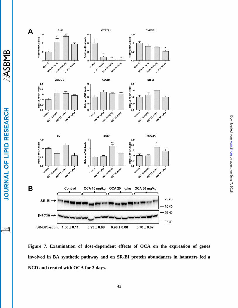
Figure 7. Examination of dose-dependent effects of OCA on the expression of genes
involved in BA synthetic pathway and on SR-BI protein abundances in hamsters fed a
NCD and treated with OCA for 3 days.
by guest, on June 7, 2018w
ww
.jlr.orgD
ownloaded from

44
(A) qRT-PCR analysis of hepatic gene expression in FXR pathway. Values are the mean ± SEM
of 5 liver samples per group. *p < 0.05, **p < 0.01 and ***p < 0.001 as compared to the vehicle
control group.
(B) Western blot analysis of hepatic SR-BI protein expression. Values are the mean ± SEM of 4
randomly chosen liver samples per group.
by guest, on June 7, 2018w
ww
.jlr.orgD
ownloaded from