regulation of a novel gene cluster involved in …jb.asm.org/content/192/19/4973.full.pdfclustered...
TRANSCRIPT

JOURNAL OF BACTERIOLOGY, Oct. 2010, p. 4973–4982 Vol. 192, No. 190021-9193/10/$12.00 doi:10.1128/JB.00681-10Copyright © 2010, American Society for Microbiology. All Rights Reserved.
Regulation of a Novel Gene Cluster Involved in Secondary MetaboliteProduction in Streptomyces coelicolor�†
Hindra, Patricia Pak, and Marie A. Elliot*Department of Biology and Institute for Infectious Disease Research, McMaster University,
1280 Main Street West, Hamilton, Ontario, L8S 4K1, Canada
Received 13 June 2010/Accepted 15 July 2010
Antibiotic biosynthesis in the streptomycetes is a complex and highly regulated process. Here, we provideevidence for the contribution of a novel genetic locus to antibiotic production in Streptomyces coelicolor. Theoverexpression of a gene cluster comprising four protein-encoding genes (abeABCD) and an antisense RNA-encoding gene (�-abeA) stimulated the production of the blue-pigmented metabolite actinorhodin on solidmedium. Actinorhodin production also was enhanced by the overexpression of an adjacent gene (abeR)encoding a predicted Streptomyces antibiotic regulatory protein (SARP), while the deletion of this gene im-paired actinorhodin production. We found the abe genes to be differentially regulated and controlled atmultiple levels. Upstream of abeA was a promoter that directed the transcription of abeABCD at a low butconstitutive level. The expression of abeBCD was, however, significantly upregulated at a time that coincidedwith the initiation of aerial development and the onset of secondary metabolism; this expression was activatedby the binding of AbeR to four heptameric repeats upstream of a promoter within abeA. Expressed divergentlyto the abeBCD promoter was �-abeA, whose expression mirrored that of abeBCD but did not require activationby AbeR. Instead, �-abeA transcript levels were subject to negative control by the double-strand-specific RNase,RNase III.
The streptomycetes are filamentous, soil-dwelling bacteriawith a complex developmental life cycle. They also have pro-digious secondary metabolite production capabilities and syn-thesize the majority of antibiotics currently in clinical use.Secondary metabolism can be correlated with changes in mor-phological development, as a shift from vegetative growth tothe formation of reproductive aerial structures coincides withthe initiation of antibiotic production. This is coordinated at agenetic level, with mutations in many characterized develop-mental regulators not only affecting aerial development butalso reducing or eliminating antibiotic production (10).
Streptomyces coelicolor is the best-studied streptomycete,and it serves as a model system for investigating the regulationof development and secondary metabolism (3). Conveniently,it produces two pigmented antibiotics: the red, cell-associatedundecylprodigiosin and the blue, secreted actinorhodin. Thesepigmented metabolites have greatly facilitated genetic studiesinto antibiotic regulation, as changes in antibiotic levels can bereadily detected by visually screening colonies on a plate (4).Antibiotic production is a tightly controlled process with manyregulatory inputs, such as metabolic and nutritional status (37),small signaling molecule (�-butyrolactone) concentrations(47), and the proposed coupling of antibiotic synthesis andresistance (27, 46). It also is subject to multiple levels of geneticregulation, including the pleiotropic regulators that affect both
development and antibiotic production, global antibiotic reg-ulators that influence the production of multiple antibiotics butdo not affect development, and pathway-specific regulatorsthat control the synthesis of a single antibiotic and often areclustered together with their target biosynthetic genes (4, 29).
Pathway-specific regulators fall into two broad classes (5):the LAL (for large ATP-binding regulators of the LuxR fam-ily) (40, 55) and the SARP (for Streptomyces antibiotic regu-latory protein) family regulators. SARPs have an OmpR-likeDNA-binding domain (49, 54), and in S. coelicolor they controlthe production of both actinorhodin (ActII-ORF4) (2) andundecylprodigiosin (RedD) (48). While all characterizedSARPs appear to have roles in modulating antibiotic produc-tion, they are not limited to functioning solely within specificpathways. In fact, one of the best understood SARPs is AfsR,which is encoded at a genetic locus distinct from any secondarymetabolic gene cluster, and it affects the production of at leasttwo antibiotics in S. coelicolor (15). The DNA binding of AfsRhas been well studied: it interacts specifically with direct, hep-tameric repeats (49), activating the expression of the neighbor-ing afsS gene, which encodes a small, sigma-factor-like proteinthat affects antibiotic production via an unknown mechanism(53), and repressing the expression of genes involved in phos-phate control (phoRP and pstS) (39).
In addition to transcription factor control, antibiotic produc-tion also is affected by the activity of the double-stranded RNAnuclease, RNase III. The mechanism underlying this effect isnot yet understood; however, point mutations in the RNase IIIcoding sequence, and the deletion of the gene itself (knownboth as rnc and as absB), abrogate antibiotic production (16,35, 42), at least in part through the reduced expression of thepathway-specific regulators ActII-ORF4 and RedD (1).
Here, we identify a novel gene cluster that specifically affects
* Corresponding author. Mailing address: Department of Biology,McMaster University, 1280 Main Street West, Hamilton, Ontario, L8S4K1, Canada. Phone: (905) 525-9140, ext. 24225. Fax: (905) 522-6066.E-mail: [email protected].
† Supplemental material for this article may be found at http://jb.asm.org/.
� Published ahead of print on 30 July 2010.
4973
on July 3, 2018 by guesthttp://jb.asm
.org/D
ownloaded from

actinorhodin production when overexpressed. We show thegenes within this cluster to be differentially regulated, display-ing differential expression profiles that are affected by a previ-ously uncharacterized SARP and by RNase III.
MATERIALS AND METHODS
Bacterial strains and plasmids. S. coelicolor strains, Escherichia coli strains,and all plasmids/cosmids used in this study are summarized in Table 1. Strepto-myces strains were grown at 30°C on solid R2YE (rich), MS (soy flour-mannitol),or SMMS (supplemented minimal) agar medium, or in liquid culture in a 1:1tryptone soya broth (TSB)-yeast extract-malt extract (YEME) mixture, as de-scribed by Kieser et al. (24). For phenotypic comparisons of different S. coelicolorstrains, 105 to 106 spores were streaked for single colonies on the different solidmedia. E. coli strains were grown at 37°C on solid LB medium and in liquid LBor SOB medium (38), except for E. coli BW25113, which was grown at 30°C whenit contained the temperature-sensitive plasmid pIJ790 (19).
DNA introduction into S. coelicolor. DNA was introduced into S. coelicolorthrough conjugation when plasmids/cosmids carried an origin of transfer (oriT)or through protoplast transformation, as outlined in Kieser et al. (24).
Construction and complementation of null mutant strains. Null mutantstrains were constructed using ReDirect technology (19). The knockout of theSCO3287-SCO3290 (abeABCD) gene cluster, as well as SCO3291 (abeR), wasaccomplished by gene/cluster replacement with an apramycin resistance cassette(apr) in cosmid StE15, followed by the introduction of the knockout cosmid intowild-type S. coelicolor M600 and selection for a double-crossover recombinationevent, as described previously (7, 14). The same process was used to knock outadpA in an rnc (also called absB) mutant strain, only in this case, adpA wasreplaced with a viomycin resistance cassette (vph) in cosmid 2StC13 (Table 1).Oligonucleotides used to generate the gene-specific disruption cassettes and tocheck the corresponding gene replacements are listed in Table S1 in the supple-mental material. Disruption mutations were confirmed using both PCR andSouthern blot analyses (data not shown). To complement the abeR mutant
phenotype, we isolated wild-type abeR from pMC118 by digesting it with KpnIand HindIII (see below for details on pMC118 construction) and cloned theresulting fragment into pMS82, an integrating plasmid vector, to generatepMC122 (Table 1). This plasmid then was introduced into E. coli ET12567/pUZ8002 before being conjugated into the abeR mutant strain, S. coelicolor E304(Table 1).
Construction of high-copy-number vector overexpression strains. Overexpres-sion strains used in this study were constructed by cloning the gene(s) of interestinto pWHM3 (a high-copy-number shuttle plasmid) (Table 1) and introducingthe resulting recombinant plasmid into S. coelicolor M600 through protoplasttransformation (24). The SCO3287-SCO3291 (abeABCD and abeR) gene clusterwas excised from cosmid E15 (Table 1) using KpnI, and the desired fragment wasgel purified and cloned into pIJ2925 (Table 1) using a rapid DNA ligation kit(Roche) prior to transformation into subcloning-efficiency DH5� E. coli cells(Invitrogen). The recombinant plasmid (pMC123) was isolated and digested withBglII before the resulting SCO3287-SCO3291-containing fragment was sub-cloned into the BamHI site of pWHM3, generating pMC120. pMC120 waspassaged through the nonmethylating E. coli strain ET12567/pUZ8002 (Table 1)prior to introduction into S. coelicolor. To overexpress SCO3287-SCO3290(abeABCD), this gene cluster was excised from pMC123 using BamHI, and theresulting �4.5-kb fragment was subcloned into the BamHI site of pWHM3. Theresulting construct was designated pMC116. For �-3287 (�-abeA) and SCO3291(abeR) overexpression, the corresponding genes were PCR amplified with PfuDNA polymerase (Stratagene) using primers OE�3287–1/OE�3287–2 andOE3291-1/OE3291-2, respectively (see Table S1 in the supplemental material).The PCR products were phosphorylated before ligation into the SmaI site ofpIJ2925; the resulting clones were verified by sequencing. �-3287 (�-abeA) wasexcised from pIJ2925 using BamHI and EcoRI, while SCO3291 (abeR) wasremoved using BglII and EcoRI. Both fragments subsequently were cloned intoBamHI and EcoRI sites of pWHM3 to give pMC117 and pMC118 as �-abeA andabeR overexpression plasmids, respectively.
Secondary metabolite assays. For the actinorhodin and undecylprodigiosinquantification of abe-overexpressing strains (relative to levels for empty-plasmid-
TABLE 1. Bacterial strains and plasmids used in this study
Strain or plasmid Genotype, description, or use Reference or source
S. coelicolorM600 SCP1� SCP2� 8E301 M600 �SCO3287-SCO3290 (abeABCD) This workE304 M600 �SCO3291 (abeR) This workJ3410 M600 rnc::apr 42E305 M600 rnc::apr adpA::vph This work
E. coliDH5� Plasmid construction and general subcloning InvitrogenET12567/pUZ8002 Generation of methylation-free plasmid DNA 28BW25113 Construction of cosmid-based knockouts 19Rosetta 2(DE3)pLysS Protein overexpression Novagen
Plasmids/cosmidspIJ790 Temperature-sensitive plasmid carrying �-RED genes 19pIJ2925 General cloning vector 22pWHM3 High-copy-number E. coli/Streptomyces plasmid 52pMS82 Complementation of mutant strains 17pET15b Overexpression of 6� His-tagged proteins NovagenpMU1 Transcriptional reporter (luminescence) 12pMC116 pWHM3 � abeABCD (SCO3287-SCO3290) This workpMC117 pWHM3 � �-abeA (�-3287) This workpMC118 pWHM3 � abeR (SCO3291) This workpMC119 pET15b � abeR (SCO3291) This workpMC120 pWHM3 � abeABCD, abeR (SCO3287-SCO3291) This workpMC122 pMS82 � abeR (SCO3291) This workpMC123 pIJ2925 � abeABCD, abeR (SCO3287-SCO3291) This workpMC124 pIJ2925 � abeR (SCO3291) This workpMC125 pIJ2925 � abeB (SCO3288) This workpMU1 � pactII-4 Reporter plasmid for actII-orf4 expression Y. Xu and J. NodwellpMU1 � pactIII Reporter plasmid for actIII expression Xu and NodwellStE15 Cosmid for abe gene knockouts and PCR amplification 362StC13 Cosmid for adpA knockout 36
4974 HINDRA ET AL. J. BACTERIOL.
on July 3, 2018 by guesthttp://jb.asm
.org/D
ownloaded from

containing control strains), pregerminated spores were inoculated into R5 liquidmedium (containing thiostrepton) to an optical density at 450 nm (OD450) of0.04 to 0.08. Cultures were shaken at 30°C, and samples were removed after 40,64, 72, and 96 h. Actinorhodin and undecylprodigiosin levels then were quanti-fied as described by Kang et al. (23). To assess the production of the calcium-dependent antibiotic (CDA), minor modifications were made to the methoddescribed by Kieser et al. (24). S. coelicolor spores were spread on nutrient agarplates, and these were incubated at 30°C for 24 or 48 h. Agar plugs then wereexcised from regions of confluent growth and were transferred from these platesinto empty plugs of an equivalent size on fresh nutrient agar plates, both with andwithout 12 mM CaCl2. These plates then were overlaid with overnight cultures ofS. aureus ATCC 29213 diluted 1/100 in soft nutrient agar. The plates wereincubated overnight at 37°C, and zones of growth inhibition were determined forstrains grown on agar plates left unsupplemented or supplemented with calcium.
RNA isolation and transcript analysis. RNA isolation and Northern blotanalyses were carried out as outlined by Swiercz et al. (45). Primers used forNorthern blot hybridization are shown in Table S1 in the supplemental material.S1 nuclease mapping was conducted as described by Elliot et al. (14), usingprimers M13F and 3288R (Table S1) together with pMC125 (Table 1) as thetemplate, to generate the probe used for mapping. For semiquantitative reversetranscription-PCR (RT-PCR), RT reactions were conducted using the Super-Script III reverse transcriptase kit (Invitrogen) according to the manufacturer’sinstructions, with minor modifications. Briefly, 5 g of total RNA was mixed withRNase-free water to a final volume of 8 l. Deoxynucleoside triphosphates(dNTPs) and gene-specific oligonucleotides were added, and the resulting mix-ture was incubated at 95°C for 10 min before being chilled on ice. The remainingreaction components (RNase inhibitor, reverse transcriptase, buffer, and dithio-threitol [DTT]) then were added, and reverse transcription was performed at42°C for 50 min. The reaction was terminated by being heated at 70°C for 15 min.The resulting RT products were used as the template for subsequent PCRamplification using the following program: initial denaturation (95°C, 7 min); 15to 28 cycles of 95°C for 45 s, 57 to 67°C for 30 s (the temperature was dependenton oligonucleotide composition, while the number of cycles was optimized toensure amplification in the linear range of the reaction), and 72°C for 30 to 45 s(extension time was dependent on expected product sizes); and a final extensioncycle of 5 min at 72°C. Negative controls (using RNA as the template) andpositive controls (using chromosomal DNA or appropriate plasmid DNA as thetemplate) were included for each PCR. PCR products were separated on a 2%agarose gel for transcript profile analyses.
lux reporter assays for measuring actII-orf4 and actIII expression. Streptomy-ces strains (105 to 106 spores) were grown on 0.2 ml R2YE agar plugs in 96-wellflat-bottomed plates (Microfluor white plate; Thermo Scientific). Followinggrowth for 16, 24, 32, 40, 48, 56, and 72 h, cultures were exposure to 1% decanal(luciferase substrate), and luminescence levels were determined. For each timepoint, 12 readings were taken (corresponding to 12 different wells/samples), andthe results of three independent time courses were analyzed using the nonpara-metric module (Mann-Whitney test) of the SPSS v13.0 statistical analysis pack-age to test whether luminescence levels (representing actII-orf4 or actIII tran-script levels) were significantly different for overexpression strains relative tothose of empty-plasmid control strains.
His6-AbeR overexpression, purification, and DNA binding analyses. To ana-lyze the DNA binding of AbeR, a His-tagged fusion protein was overexpressedand purified from E. coli. The abeR coding sequence was PCR amplified (TableS1), and the resulting product was cloned into the SmaI site of pIJ2925. Thisrecombinant plasmid was digested with NdeI and BglII to liberate abeR, whichthen was cloned into the overexpression vector pET15b (Novagen) digested withNdeI and BamHI. The integrity of the resulting construct (pMC119) and theassociated T7 promoter was confirmed by sequencing. pMC119 then was intro-duced into E. coli strain Rosetta 2 (Novagen). Protein overexpression wasachieved by growing the plasmid-containing E. coli strains in 100 ml of LBmedium supplemented with ampicillin and chloramphenicol overnight at roomtemperature (without isopropyl--D-thiogalactopyranoside induction). Cellswere collected by centrifugation, and the resulting cell pellet was resuspended inlysis buffer (20 mM Tris-HCl, pH 7.5, 250 mM NaCl, 10 mM imidazole), followedby the addition of lysozyme (750 g/ml) and protease inhibitor (Roche) per themanufacturer’s instructions. The cell suspension was mechanically disrupted bysonication, after which RNase A (80 g/ml) and DNase I (40 U; Roche) wereadded to the cell lysate, and the soluble fraction was separated by centrifugation.This fraction was applied to Ni-nitrilotriacetic acid agarose resin (Qiagen) beforebeing washed three times with wash buffer (20 mM Tris-Cl, pH 7.5, 250 mMNaCl) containing increasing concentrations of imidazole (50 to 125 mM). Puri-fied protein was eluted in wash buffer containing 250 mM imidazole and wasconcentrated and desalted using a Nanosep centrifugal device (Pall) with a
30-kDa cutoff. Protein concentrations were determined using a Bradford assay(Bio-Rad) (6).
Electrophoretic mobility shift assays (EMSAs) were conducted using native5% polyacrylamide gel electrophoresis. The probe was generated by PCR am-plification using primers 3287–2 and 3287–3 (see Table S1 in the supplementalmaterial), with the resulting PCR product being 5� end labeled with[�-32P]dATP. Twenty-microliter reaction mixtures contained 0.5 nM labeledprobe together with 0 to 200 nM purified protein in a buffer consisting of 10 mMTris-HCl (pH 7.5), 150 mM NaCl, 1 mM DTT, 1 g poly(dI-dC), and 10%glycerol. Samples were incubated at room temperature for 25 to 30 min prior toloading. Electrophoresis was performed in 1� Tris-borate-EDTA buffer at 100 Vfor 50 to 60 min. Gels were exposed to Kodak Biomax XAR film at roomtemperature for 1 h.
DNase I footprinting assays were conducted using the same binding conditionsas those for the EMSA experiments. Probes were prepared by PCR amplificationusing 3287–2 and 3287MSAF primers (Table S1), with either 3287MSAF or3287–2 being end labeled with [�-32P]dATP, to create probes corresponding tothe coding (nontemplate) or template strands, respectively. Purified AbeR (0 to1.6 M) was mixed with �6 nM probe before adding 2.5 U DNase I (Roche) for1 min, after which PN buffer (Qiagen) was added to stop the DNase I reaction.The resulting cleavage products were purified using Qiagen MinElute columnsand were eluted in 30 l distilled H2O. The elution products were vacuum driedfor 30 to 45 min and resuspended in loading dye (80% [vol/vol] formamide; 1mM EDTA, pH 8.0; 10 mM NaOH; 0.1% [wt/vol] bromophenol blue; 0.1%[wt/vol] xylene cyanol FF). Sequencing ladders were generated using the Seque-nase v2.0 DNA sequencing kit (USB Corporation) per the manufacturer’s in-structions, with minor modifications. Briefly, alkaline-denatured pMC123 (Table1) was mixed together with 5 M the appropriate primer (3287MSAF/3287–2;see Table S1 in the supplemental material) in a total volume of 10 l. Thesemixtures then were boiled for 3 min and placed immediately on ice for 5 minbefore being centrifuged for 30 s. To each tube, buffer, DTT, [�-33P]dATP, andSequenase were added per the manufacturer’s instructions, and the resultingreaction mixtures were mixed with the dGTP labeling mix and the ddG/A/T/Ctermination mix, which previously had been warmed to 37°C. These mixtureswere incubated at 37°C for 2 min before stop solution was added (USB Corpo-ration). All samples (reaction and sequencing) were heated at 90°C for 3 to 5 minprior to being loaded onto a 6% denaturing polyacrylamide gel, which was run at45 W for either 45 (template strand) or 75 min (coding/nontemplate strand).
RESULTS
Bioinformatic analysis of the SCO3287-SCO3291 genetic re-gion. Our interest in this genetic region originally was stimu-lated by the presence of an antisense RNA, encoded by �-3287on the strand opposite SCO3287 (45). SCO3287 is the firstgene of an apparent four-gene operon that is, to date, uniqueto S. coelicolor and its very close relative, S. lividans (Fig. 1).SCO3287 encodes a highly charged (arginine-rich) cytoplasmicprotein of 172 amino acids (aa). It is devoid of obvious func-tional motifs and bears no sequence similarity to any knownprotein. It is followed by two predicted membrane protein-encoding genes, SCO3288 and SCO3289, whose products are243 and 536 aa, respectively. Neither of these genes is similarto any characterized gene, although SCO3289 encodes a prod-uct with homologues in other Streptomyces species, notably S.avermitilis, where it is cotranscribed with a smaller downstream
FIG. 1. Genetic organization of SCO3287-SCO3291 (the abe genecluster) and the associated antisense RNA-encoding gene (�-3287/�-abeA). Transcription start sites are indicated by vertical lines, with theline widths (and associated arrows) approximating relative transcriptabundance.
VOL. 192, 2010 REGULATION OF A NOVEL GENE CLUSTER IN S. COELICOLOR 4975
on July 3, 2018 by guesthttp://jb.asm
.org/D
ownloaded from

gene, and S. griseus, where it exists as a single genetic unit. InS. coelicolor, SCO3289 appears to be translationally coupledwith SCO3290, which encodes a 249-aa cytoplasmic proteinwith a predicted nucleotide binding domain similar to theToll/interleukin-1 receptor (TIR-like) domain, suggesting apossible role in signal transduction (30) or protein-proteininteractions (44).
Downstream of this four-gene cluster, oriented in the samedirection, is a predicted transcription factor-encoding gene. Itsproduct, SCO3291, is 477 aa, with a multidomain architecture.The C terminus of SCO3291 shares similarity with conservedhypothetical proteins from a wide variety of bacteria, althoughthese are largely single-domain proteins. The function of thisC-terminal region is unknown; it is worth noting that proteinswith this domain usually are encoded adjacent to genes whoseproducts have TIR-like domains, such as that found inSCO3290. The N terminus of SCO3291 contains an OmpR-like DNA binding domain (31) and a so-called BTAD, orbacterial transcription activator domain (58). These two do-mains are common to all SARP family regulators (54, 58) andsuggest that SCO3291 has DNA binding properties. Whilemany SARPs are encoded within antibiotic biosynthetic geneclusters and act as pathway-specific antibiotic regulators,the target genes for SCO3291 are not immediately obvious, asthe nearest secondary metabolic gene cluster is 38 kb away (thecalcium-dependent antibiotic cluster).
Actinorhodin production increases upon gene/gene clusteroverexpression. To address the biological role of the SCO3287-SCO3290 gene products and the function of SCO3291, we firstreplaced the four-gene cluster with an antibiotic resistancegene and compared its phenotype to that of wild-type S. coeli-color. Both colony development and antibiotic production bythe mutant strain closely resembled that of its wild-type parent
when grown on a variety of solid culture media (data notshown). In contrast, the overexpression of SCO3287-SCO3290(on a multicopy plasmid) significantly increased actinorhodinproduction during growth on solid rich (R2YE) (Fig. 2A) andsupplemented minimal (SMMS) media (data not shown);CDA production was unaffected (data not shown). Increasedantibiotic production could be due to the overexpression of thefour-gene cluster itself or could result from the increased ex-pression of the antisense RNA, �-3287, encoded on the strandopposite SCO3287 (Fig. 1). To differentiate between these twopossibilities, we created an �-3287 overexpression strain byintroducing �-3287 on a multicopy plasmid vector into wild-type S. coelicolor, and we found that this strain produced an-tibiotics at a level equivalent to that of an empty-plasmidcontrol strain (data not shown). This suggested that the en-hanced actinorhodin production seen for the SCO3287-SCO3290 overexpression strain was due to the overexpressionof one or more of the four protein-encoding genes and not�-3287. Given the ability of these gene products to modulateantibiotic levels, we propose that SCO3287-SCO3290 be re-named abeABCD for antibiotic enhancement upon overexpres-sion, and that �-3287 be renamed �-abeA.
We used a similar approach to investigate the role ofSCO3291 in S. coelicolor development and metabolism, creat-ing both SCO3291 deletion and overexpression strains. Thereplacement of SCO3291 with an apramycin resistance generesulted in decreased actinorhodin production relative to thatof its wild-type parent (Fig. 2B); this defect could be reversedupon complementation with a wild-type copy of SCO3291 onthe integrating plasmid vector pMS82 (Fig. 2B). In contrast,overexpressing SCO3291 either on its own or with abeABCDresulted in increased actinorhodin production compared tothat of empty-plasmid-containing control strains (Fig. 2A). We
FIG. 2. Phenotypic effects of abe gene overexpression and abeR deletion during growth on rich R2YE agar medium. (A) Wild-type S. coelicolor(M600) containing the high-copy-number plasmid pWHM3 alone or carrying abeABCD (SCO3287-SCO3290), abeR (SCO3291), or both abeABCDand abeR, grown for 2 to 4 days. (B) Comparison of wild-type (M600) and abeR deletion strains carrying the empty integrating (single-copy)plasmid vector pMS82, with the abeR mutant strain carrying a wild-type copy of abeR in pMS82. To best visualize antibiotic production, all pictureswere taken from the underside of the plates, and to ensure reproducibility, experiments were repeated at least four times using a minimum of twoindependent spore stocks as the inoculum.
4976 HINDRA ET AL. J. BACTERIOL.
on July 3, 2018 by guesthttp://jb.asm
.org/D
ownloaded from
therefore propose that SCO3291 be renamed abeR, given theeffect of its overexpression on antibiotic production.
We also monitored the actinorhodin and undecylprodigiosinlevels for abeABCD, abeR, and abeABCD and abeR overex-pression strains relative to those for a control plasmid-contain-ing strain during liquid culture growth. We observed no differ-ence in the level of undecylprodigiosin produced by theoverexpression and control strains (data not shown), and whilewe did observe increased actinorhodin production for the over-expression strains on some occasions, this effect was not con-sistently reproducible (data not shown), unlike the increasedactinorhodin production that was observed consistently foroverexpression strains grown on solid culture (Fig. 2).
Enhanced actinorhodin production by overexpressionstrains may be due to the extended expression of actII-orf4 butnot to an overall increase in expression. Many characterizedantibiotic regulators mediate their effects through the tran-scriptional control of pathway-specific activators (1, 33, 37). Asactinorhodin levels were significantly increased in bothabeABCD and abeR overexpression strains during growth onsolid media, we sought to probe the effect of this overexpres-sion on the transcription of the actinorhodin pathway-specificregulator actII-orf4, and the actinorhodin biosynthetic geneactIII, using luciferase transcriptional reporters (12). We in-troduced the reporter constructs and corresponding controlplasmid into the abeABCD and abeR overexpression strainsand monitored luminescence levels over time. We did notdetect any consistent differences in actIII expression during the3-day time course (data not shown). For actII-orf4, however,expression from 24 to 32 h actually was lower in the twooverexpression strains than in the control strain, but by 72 h weobserved a statistically significant increase in promoter activityin both overexpression strains compared to that of the wildtype (Fig. 3), suggesting that the extended expression of actII-orf4 contributes to enhanced actinorhodin production.
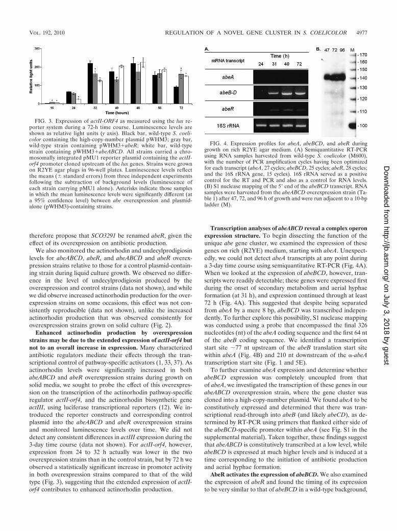
Transcription analyses of abeABCD reveal a complex operonexpression structure. To begin dissecting the function of theunique abe gene cluster, we examined the expression of thesegenes on rich (R2YE) medium, starting with abeA. Unexpect-edly, we could not detect abeA transcripts at any point duringa 3-day time course using semiquantitative RT-PCR (Fig. 4A).When we looked at the expression of abeBCD, however, tran-scripts were readily detectable; these genes were expressed firstduring the onset of secondary metabolism and aerial hyphaeformation (at 31 h), and expression continued through at least72 h (Fig. 4A). This suggested that despite being separatedfrom abeA by a mere 8 bp, abeBCD was transcribed indepen-dently. To further explore this possibility, S1 nuclease mappingwas conducted using a probe that encompassed the final 326nucleotides (nt) of the abeA coding sequence and the first 64 ntof the abeB coding sequence. We identified a transcriptionstart site �77 nt upstream of the abeB translation start sitewithin abeA (Fig. 4B) and 210 nt downstream of the �-abeAtranscription start site (Fig. 1 and 5E).
To further examine abeA expression and determine whetherabeBCD expression was completely uncoupled from thatof abeA, we investigated the transcription of these genes in ourabeABCD overexpression strain, where the gene cluster wascloned into a high-copy-number plasmid. We found abeA to beconstitutively expressed and determined that there was tran-scriptional read-through into abeB (and likely abeCD), as de-termined by RT-PCR using primers that flanked either side ofthe abeBCD-specific promoter within abeA (see Fig. S1 in thesupplemental material). Taken together, these findings suggestthat abeABCD is constitutively transcribed at a low level, whileabeBCD is expressed at much higher levels and is induced at atime corresponding to the initiation of antibiotic productionand aerial hyphae formation.
AbeR activates the expression of abeBCD. We also examinedthe expression of abeR and found the timing of its expressionto be very similar to that of abeBCD in a wild-type background,
FIG. 3. Expression of actII-ORF4 as measured using the lux re-porter system during a 72-h time course. Luminescence levels areshown as relative light units (y axis). Black bar, wild-type S. coeli-color containing the high-copy-number plasmid pWHM3; gray bar,wild-type strain containing pWHM3�abeR; white bar, wild-typestrain containing pWHM3�abeABCD. All strains carried a chro-mosomally integrated pMU1 reporter plasmid containing the actII-orf4 promoter cloned upstream of the lux genes. Strains were grownon R2YE agar plugs in 96-well plates. Luminescence levels reflectthe means (� standard errors) from three independent experimentsfollowing the subtraction of background levels (luminescence ofeach strain carrying pMU1 alone). Asterisks indicate those samplesin which the mean luminescence levels were significantly different (ata 95% confidence level) between abe overexpression and plasmid-alone (pWHM3)-containing strains.
FIG. 4. Expression profiles for abeA, abeBCD, and abeR duringgrowth on rich R2YE agar medium. (A) Semiquantitative RT-PCRusing RNA samples harvested from wild-type S. coelicolor (M600),with the number of PCR amplification cycles having been optimizedfor each transcript (abeA, 27 cycles; abeBCD, 25 cycles; abeR, 28 cycles;and the 16S rRNA gene, 15 cycles). 16S rRNA served as a positivecontrol for the RT and PCR and also as a control for RNA levels.(B) S1 nuclease mapping of the 5� end of the abeBCD transcript. RNAsamples were harvested from the abeABCD overexpression strain (Ta-ble 1) after 47, 72, and 96 h of growth and were run adjacent to a 10-bpladder (M).
VOL. 192, 2010 REGULATION OF A NOVEL GENE CLUSTER IN S. COELICOLOR 4977
on July 3, 2018 by guesthttp://jb.asm
.org/D
ownloaded from

with transcripts detected from 31 to 72 h (Fig. 4A). Given thisfinding and considering that the overexpression of abeR hadphenotypic consequences similar to those of the overexpres-sion of abeABCD, we wondered whether there might be aregulatory connection between AbeR and abeBCD. To probethis possibility, we examined the transcript levels of abeBCD inthe abeR deletion and overexpression strains described above.We found that abeBCD expression was barely detectable in the
abeR deletion strain (Fig. 5A), while in the overexpressionstrain, transcript levels were increased considerably comparedto those of an empty-plasmid-carrying control strain (Fig. 5B).We also tested whether abeA transcripts were detectable in theabeR overexpression strain, but as was the case for the wildtype, no expression could be observed (data not shown).
To determine whether AbeR was directly involved in con-trolling abeBCD expression, we overexpressed AbeR as an
FIG. 5. Regulation of abeBCD transcription by AbeR. Shown are transcription profiles of abeBCD in S. coelicolor wild-type and abeR deletionstrains (A) and in abeR overexpression and plasmid (pWHM3)-alone control strains (B), as determined using semiquantitative RT-PCR. RNAsamples used in the RT-PCR were harvested at time points indicated above each panel; the number of PCR amplification cycles was optimizedfor each experiment: 25 cycles (A) and 27 cycles (B) (note that strains carrying the high-copy-number plasmid pWHM3 appear to have reducedoverall transcript levels compared to those of non-plasmid-containing strains [unpublished data]). For the no-RT control PCR, RNA (not subjectedto reverse transcriptase) served as the template to ensure no DNA contamination. The 16S rRNA gene was amplified (15 cycles) as a control forRNA levels and RNA integrity. (C) EMSA of [�-32P]dATP-radiolabeled probe (214 bp) containing the abeBCD promoter region together withHis6-AbeR. AbeR binding specificity was tested using specific (cold probe) and nonspecific competitor DNA (chpD coding sequence). (D) DNaseI footprinting assay of His6-AbeR binding to the abeBCD promoter region. Protected regions are indicated by the vertical lines to the right of eachfootprint. Increasing concentrations of purified AbeR (0 to 1.67 M, with increasing concentrations of protein indicated by the black triangle) wereincubated together with 6 nM singly end-labeled probe. Sequencing ladders are shown to the left of each footprint. (E) Coding sequence of abeAencompassing the approximate �10 and �35 promoter sequences of �-abeA and abeBCD. Mapped transcriptional start sites for both transcriptsare designated by boldface letters. DNA sequences protected by AbeR are indicated by horizontal lines, and the protected four direct heptamericrepeats are boxed. Sites of DNase I hypersensitivity are indicated by vertical arrows, while a potential AdpA binding site is shaded in gray.
4978 HINDRA ET AL. J. BACTERIOL.
on July 3, 2018 by guesthttp://jb.asm
.org/D
ownloaded from

N-terminally His-tagged protein in E. coli for use in EMSAs,and we purified this protein using Ni affinity chromatography.EMSAs were performed using a DNA fragment that encom-passed both the �-abeA (antisense gene) and abeBCD pro-moter regions as the probe. We found this fragment to beeffectively bound by the AbeR fusion protein at a concentra-tion of 93 nM. Competition experiments confirmed the speci-ficity of this interaction, with cold probe effectively competingwith the labeled probe for binding, but an unrelated DNAfragment (the S. coelicolor chpD coding sequence, where ChpDis associated with aerial development but not antibiotic pro-duction [14]) failed to compete (Fig. 5C). The analysis of thesequence contained within the specifically bound DNA frag-ment revealed there to be four tandemly arranged direct re-peats of 7 nt, with the first three repeats each separated by 4 ntand the fourth located a further 15 nt downstream. DNase Ifootprinting experiments were conducted to determinewhether these repeated regions were protected by AbeR bind-ing. As seen in Fig. 5D and E, protection by AbeR covered therepeated regions, with sites of DNase I hypersensitivity seen onboth strands within the 15-bp region separating the third andfourth repeats.
AbeR does not control �-abeA expression. As the bindingsite of AbeR was located between the promoter regions forabeBCD and �-abeA, we wanted to determine whether AbeRalso affected the expression of �-abeA. Using Northern blot-ting, we examined �-abeA transcript levels in the abeR over-expression and deletion strains and found its expression to beunaffected in each instance (data not shown), suggesting thatthe effect of AbeR binding was limited to the activation ofabeBCD expression.
RNase III contributes to the destabilization of �-abeA tran-scripts. Having established that AbeR played an importantrole in activating the expression of abeBCD but did not impact�-abeA expression, we wanted to identify factors that affected
�-abeA expression, as its expression profile mirrored that ofabeBCD and abeR (Fig. 6A) (45). The positioning of �-abeAsuggested that the most likely interaction partner for �-abeAwould be abeA-containing transcripts, and the resulting dou-ble-stranded RNA complexes would be reasonable targets forRNase III. We therefore examined the expression of �-abeA inwild-type and RNase III (rnc [absB]) mutant strains carryingthe abeABCD overexpression construct. We found that �-abeAtranscripts were present in far greater abundance during laterstages of growth (3 to 4 days) in the rnc (absB) mutant strainthan in the wild type (Fig. 6A), suggesting that �-abeA may betargeted for degradation by RNase III late in development,possibly in conjunction with its predicted target, abeA. How-ever, when we examined abeA expression, expecting to seesimilarly increased levels in the rnc (absB) mutant, we foundthis was not the case. After normalizing abeA transcript levelsusing the 16S rRNA transcript control, and accounting forplasmid copy number using transcript levels of the plasmid-carried tsr (thiostrepton resistance) gene, we found there to beless abeA expression at all time points in the rnc (absB) mutantthan with the wild-type strain (Fig. 6B). Similar effects wereobserved for abeBCD transcript levels (Fig. 6B). No obviouscorrelations could be drawn between �-abeA and abeA expres-sion levels, suggesting that despite being perfectly complemen-tary, �-abeA had little effect on abeA transcript stability.
The RNase III-dependent destabilization of �-abeA is inde-pendent of AdpA activity. The change in �-abeA transcriptlevels in the rnc (absB) mutant compared to that of its corre-sponding wild-type parent was reminiscent of that observed forsti1 (SCO0762) and ramR, whose expression also was upregu-lated later in development in an rnc (absB) mutant (21). Re-cent work has shown this upregulation to be indirect, resultingnot from the reduced cleavage of these mRNAs by RNase IIIbut from increased AdpA-dependent activation (56). AdpA isa pleiotropic regulator (25, 32), and its transcripts are targeted
FIG. 6. Expression of �-abeA, abeA, and abeBCD in S. coelicolor wild-type and mutant strains lacking RNase III. (A) Northern blot analysisof �-abeA expression in wild-type and rnc (absB) mutant strains carrying the abeABCD overexpression plasmid (pMC116; Table 1). 5S rRNA wasexamined as a control for RNA integrity and loading. (B) Levels of �-abeA, abeA, and abeBCD in the rnc (absB) mutant strain relative to itswild-type parent S. coelicolor M600. Transcript levels were normalized relative to 5S rRNA (for �-abeA) and 16S rRNA genes (for abeA andabeBCD) to account for differences in input template RNA and tsr levels to account for differences in plasmid copy number levels for wild-typeand mutant strains. Transcript abundance was determined using the ImageJ analysis of Northern blotting (�-abeA) and RT-PCR (abeA, abeBCD,16S rRNA gene, tsr) results. The data presented are the averages (� standard deviations) from four experiments (two experimental replicates oftwo independent RNA time courses).
VOL. 192, 2010 REGULATION OF A NOVEL GENE CLUSTER IN S. COELICOLOR 4979
on July 3, 2018 by guesthttp://jb.asm
.org/D
ownloaded from

for degradation by RNase III; the loss of RNase III leads toincreased AdpA levels and, correspondingly, increased tran-scription of AdpA-activated genes (56). We examined the se-quence upstream of �-abeA and found a reasonable match tothe AdpA-binding motif (TGGCCGGCCC versus TGGCSNGWWY, where S is G/C, W is A/T, and Y is T/C [57])located 134 nt upstream of the �-abeA transcription start site(Fig. 5E). To determine whether the increased expression of�-abeA in the rnc (absB) mutant was mediated through AdpA,we created an rnc adpA double mutant and examined �-abeAexpression. We found that �-abeA levels remained high in thedouble mutant strain (data not shown), suggesting that in-creased �-abeA transcription in the rnc mutant was not due toincreased activation by AdpA but was instead mediatedthrough RNase III by another, yet-to-be-determined mecha-nism.
DISCUSSION
Here, we elucidate the regulation of a novel gene clusterhaving a role in antibiotic production in S. coelicolor. On solidmedia, we showed that the overexpression of abeABCD and/orabeR resulted in the increased production of the blue-pig-mented antibiotic actinorhodin, while the deletion of abeRresulted in decreased actinorhodin production. Unlike manygenes that affect antibiotic production in S. coelicolor whenoverexpressed (e.g., metK [33] and afsR [15]), abe gene/clusteroverexpression resulted in the maintenance of actII-orf4 ex-pression at elevated levels later in development rather thanstimulating expression at all stages of culture growth.
Antibiotic production is subject to multiple levels of regula-tory control and is impacted by physiological factors, like met-abolic precursor concentrations, and by environmental condi-tions, like nutrient availability and signaling moleculeabundance. Given the putative membrane localization ofAbeB and AbeC and the cytoplasmic positioning of the TIR-like domain-containing AbeD, we considered the possibilitythat these proteins may have a role in sensing and respondingto environmental cues. S. coelicolor produces at least threechromosomally encoded �-butyrolactone signaling molecules,SCB1, SCB2, and SCB3 (20, 47), along with the plasmid-en-coded methylenomycin furans (MMFs) (11, 34). However,these molecules all are freely diffusible across cytoplasmicmembranes and bind dedicated cytoplasmic receptor proteins,making it unlikely that the abe proteins have an intermediaryrole in sensing or transducing signals in response to thesemolecules, although we cannot exclude the possibility that theyrecognize a currently unknown signaling molecule(s). We alsoconsidered the possibility that the abe gene cluster responds tochanges in the nutritional status of the colony and/or influ-ences the switch from primary to secondary metabolism, giventhat abeBCD and abeR expression initiated at a time consistentwith this physiological transition. We also observed increasedantibiotic production only on media where glucose was in-cluded as the primary carbon source (SMMS and R2YE),suggesting that the abe gene effect is subject to cataboliterepression. Recent work has begun to illuminate the regulatoryconnections linking primary and secondary metabolism, withthe regulators DasR and AtrA having central but antagonisticroles (37, 51). DasR negatively regulates actinorhodin and
undecylprodigiosin production through the repression of actII-orf4 and redZ (encoding a regulator of RedD expression);repression is relieved in the presence of N-acetylglucosamine(GlcNAc), although only during growth on poor carbonsources (37). AtrA directly activates the expression of actII-orf4 and stimulates actinorhodin production but does not affectthe production of undecylprodigiosin (51). It also activates theexpression of nagE2, which encodes the permease specific forthe import of GlcNAc into the cell (31a). There is no obviousconnection between the abe genes and either DasR or AtrA:there are no binding sites for either protein upstream of abeR,the effect of abe gene overexpression does not require a poorcarbon source, and actII-orf4 expression during abe overex-pression is not significantly altered, instead appearing to beextended during culture growth.
Both abeR and abeABCD overexpression enhanced actinor-hodin production, but interestingly, the deletion of each didnot have the same phenotypic consequences: the deletion ofabeR significantly reduced actinorhodin production, while theloss of abeABCD had little effect on antibiotic levels. Thissuggested that AbeR has additional regulatory targets in thecell, or that AbeR has other functions in the cell that areindependent of its transcriptional activator role (possibly me-diated through its uncharacterized C-terminal domain). Wedefined the AbeR binding site through EMSA and DNase Ifootprinting experiments and found that, like other SARPs,AbeR bound heptameric direct repeats separated by 4 or 15 ntpositioned on the same face of the DNA helix upstream ofabeBCD. A search of the genome using the CGGAA(G/C)C(n4/15)CGGAA(G/C)C sequence as a query failed to identifyother candidate binding sites. Thus, the only known AbeRbinding sites are within the abeA coding sequence. It is inter-esting that most SARPs control the expression of genes thatare located in close proximity on the chromosome.
The differential expression of genes within the abeABCDoperon is not unprecedented in S. coelicolor (9), but it also isnot considered to be the norm. Microarray studies of S. coeli-color gene expression have shown that the first gene of anoperon is typically the most highly expressed, with expressionlevels then decreasing throughout the length of the operon(26). This is obviously not the case for abeA, which was ex-pressed at far lower levels than any of the other abe genes.Comprehensive transcriptome analyses of E. coli (13, 41),Helicobacter pylori (43), Listeria monocytogenes (50), and My-coplasma pneumonia (18) now are beginning to shed light onthe transcriptional complexity that exists in bacteria. Recentstudies of H. pylori and M. pneumoniae have revealed extensiveintraoperon expression dynamics, with different genes withinan operon having distinct induction/repression characteristicsrelative to other genes within the same operon. Our findingshere are consistent with there being flexible operon expressionin S. coelicolor. Additional complexity at the antisense tran-script level also is appearing to be widespread, with antisensetranscripts being detected throughout the E. coli, H. pylori, andM. pneumonia genomes (13, 18, 41, 43). What role these anti-sense transcripts have in regulating gene expression or proteinactivity remains to be seen.
Unexpectedly, we found that �-abeA did not appear to havea role in modulating the transcript stability of its sense coun-terpart, abeA, as increased levels of �-abeA in an rnc (absB)
4980 HINDRA ET AL. J. BACTERIOL.
on July 3, 2018 by guesthttp://jb.asm
.org/D
ownloaded from

mutant could not be correlated with a subsequent change inabeA transcript abundance. While we cannot exclude the pos-sibility that �-abeA controls abeA expression via an RNaseIII-independent mechanism (e.g., abeA translation), we didobserve changes in the overall transcript abundance of bothabeA and �-abeA when RNase III was absent. abeA transcripts,as well as abeBCD transcripts, were consistently less abundantin an rnc (absB) mutant than in its wild-type parent. This isreminiscent of the reduced expression seen for the antibioticbiosynthetic clusters of actinorhodin, undecylprodigiosin,CDA, and a cryptic polyketide in an absB mutant (21). Incontrast, �-abeA levels were increased in the rnc (absB) knock-out strain. This effect was not mediated through AdpA, whichis negatively regulated by RNase III, and may instead reflecteither the direct targeting of �-abeA by RNase III during latergrowth stages or control by some other RNase III-dependentfactor.
ACKNOWLEDGMENTS
We thank David Capstick, Henry Haiser, and Justin Nodwell for thecritical reading of the manuscript and for helpful comments and sug-gestions and Ye Xu and Justin Nodwell for the use of their reporterplasmids.
This work was funded by grants from the Natural Sciences andEngineering Research Council of Canada (NSERC Discovery Grantno. 312495 and NSERC Collaborative Research and DevelopmentGrant no. 335144), the Canada Research Chairs (CRC) program, andgraduate student awards from the Province of Ontario (to H.) andNSERC (to P.P.).
REFERENCES
1. Aceti, D. J., and W. C. Champness. 1998. Transcriptional regulation ofStreptomyces coelicolor pathway-specific antibiotic regulators by the absA andabsB loci. J. Bacteriol. 180:3100–3106.
2. Arias, P., M. A. Fernandez-Moreno, and F. Malpartida. 1999. Characteriza-tion of the pathway-specific positive transcriptional regulator for actinor-hodin biosynthesis in Streptomyces coelicolor A3(2) as a DNA-binding pro-tein. J. Bacteriol. 181:6958–6968.
3. Bentley, S. D., K. F. Chater, A. M. Cerdeno-Tarraga, G. L. Challis, N. R.Thomson, K. D. James, D. E. Harris, M. A. Quail, H. Kieser, D. Harper, A.Bateman, S. Brown, G. Chandra, C. W. Chen, M. Collins, A. Cronin, A.Fraser, A. Goble, J. Hidalgo, T. Hornsby, S. Howarth, C. H. Huang, T.Kieser, L. Larke, L. Murphy, K. Oliver, S. O’Neil, E. Rabbinowitsch, M. A.Rajandream, K. Rutherford, S. Rutter, K. Seeger, D. Saunders, S. Sharp, R.Squares, S. Squares, K. Taylor, T. Warren, A. Wietzorrek, J. Woodward,B. G. Barrell, J. Parkhill, and D. A. Hopwood. 2002. Complete genomesequence of the model actinomycete Streptomyces coelicolor A3(2). Nature417:141–147.
4. Bibb, M. J. 1996. The regulation of antibiotic production in Streptomycescoelicolor A3(2). Microbiology 142:1335–1344.
5. Bibb, M. J. 2005. Regulation of secondary metabolism in streptomycetes.Curr. Opin. Microbiol. 8:208–215.
6. Bradford, M. M. 1976. A rapid and sensitive method for the quantitation ofmicrogram quantities of protein utilizing the principle of protein-dye bind-ing. Anal. Biochem. 72:248–254.
7. Capstick, D. S., J. M. Willey, M. J. Buttner, and M. A. Elliot. 2007. SapB andthe chaplins: connections between morphogenetic proteins in Streptomycescoelicolor. Mol. Microbiol. 64:602–613.
8. Chakraburtty, R., and M. Bibb. 1997. The ppGpp synthetase gene (relA) ofStreptomyces coelicolor A3(2) plays a conditional role in antibiotic produc-tion and morphological differentiation. J. Bacteriol. 179:5854–5861.
9. Charaniya, S., S. Mehra, W. Lian, K. P. Jayapal, G. Karypis, and W. S. Hu.2007. Transcriptome dynamics-based operon prediction and verification inStreptomyces coelicolor. Nucleic Acids Res. 35:7222–7236.
10. Chater, K. F. 2006. Streptomyces inside-out: a new perspective on the bac-teria that provide us with antibiotics. Philos. Trans. R. Soc. B Biol. Sci.361:761–768.
11. Corre, C., L. Song, S. O’Rourke, K. F. Chater, and G. L. Challis. 2008.2-Alkyl-4-hydroxymethylfuran-3-carboxylic acids, antibiotic production in-ducers discovered by Streptomyces coelicolor genome mining. Proc. Natl.Acad. Sci. U. S. A. 105:17510–17515.
12. Craney, A., T. Hohenauer, Y. Xu, N. K. Navani, Y. Li, and J. Nodwell. 2007.A synthetic luxCDABE gene cluster optimized for expression in high-GCbacteria. Nucleic Acids Res. 35:e46.
13. Dornenburg, J. E., A. M. DeVita, M. J. Palumbo, and J. T. Wade. 2010.Widespread antisense transcription in Escherichia coli. mBio 1:e00024–10.
14. Elliot, M. A., N. Karoonuthaisiri, J. Huang, M. J. Bibb, S. N. Cohen, C. M.Kao, and M. J. Buttner. 2003. The chaplins: a family of hydrophobic cell-surface proteins involved in aerial mycelium formation in Streptomyces coeli-color. Genes Dev. 17:1727–1740.
15. Floriano, B., and M. Bibb. 1996. afsR is a pleiotropic but conditionallyrequired regulatory gene for antibiotic production in Streptomyces coelicolorA3(2). Mol. Microbiol. 21:385–396.
16. Gravenbeek, M. L., and G. H. Jones. 2008. The endonuclease activity ofRNase III is required for the regulation of antibiotic production by Strepto-myces coelicolor. Microbiology 154:3547–3555.
17. Gregory, M. A., R. Till, and M. C. M. Smith. 2003. Integration site forStreptomyces phage phiBT1 and development of site-specific integrating vec-tors. J. Bacteriol. 185:5320–5323.
18. Guell, M., V. van Noort, E. Yus, W. H. Chen, J. Leigh-Bell, K. Michalodimi-trakis, T. Yamada, M. Arumugam, T. Doerks, S. Kuhner, M. Rode, M.Suyama, S. Schmidt, A. C. Gavin, P. Bork, and L. Serrano. 2009. Transcrip-tome complexity in a genome-reduced bacterium. Science 326:1268–1271.
19. Gust, B., G. L. Challis, K. Fowler, T. Kieser, and K. F. Chater. 2003.PCR-targeted Streptomyces gene replacement identifies a protein domainneeded for biosynthesis of the sesquiterpene soil odor geosmin. Proc. Natl.Acad. Sci. U. S. A. 100:1541–1546.
20. Hsiao, N. H., S. Nakayama, M. E. Merlo, M. de Vries, R. Bunet, S. Kitani,T. Nihira, and E. Takano. 2009. Analysis of two additional signaling mole-cules in Streptomyces coelicolor and the development of a butyrolactone-specific reporter system. Chem. Biol. 16:951–960.
21. Huang, J., J. Shi, V. Molle, B. Sohlberg, D. Weaver, M. J. Bibb, N. Karoo-nuthaisiri, C.-J. Lih, C. M. Kao, M. J. Buttner, and S. N. Cohen. 2005.Cross-regulation among disparate antibiotic biosynthetic pathways of Strep-tomyces coelicolor. Mol. Microbiol. 58:1276–1287.
22. Janssen, G. R., and M. J. Bibb. 1993. Derivatives of pUC18 that have BglIIsites flanking a modified multiple cloning site and that retain the ability toidentify recombinant clones by visual screening of Escherichia coli colonies.Gene 124:133–134.
23. Kang, S. G., W. Jin, M. J. Bibb, and K. J. Lee. 1998. Actinorhodin andundecylprodigiosin production in wild-type and relA mutant strains of Strep-tomyces coelicolor A3(2) grown in continuous culture. FEMS Microbiol. Lett.168:221–226.
24. Kieser, T., M. J. Bibb, M. J. Buttner, K. F. Chater, and D. A. Hopwood. 2000.Practical Streptomyces genetics. John Innes Foundation, Norwich, UnitedKingdom.
25. Kim, D. W., K. Chater, K. J. Lee, and A. Hesketh. 2005. Changes in theextracellular proteome caused by the absence of the bldA gene product, adevelopmentally significant tRNA, reveal a new target for the pleiotropicregulator AdpA in Streptomyces coelicolor. J. Bacteriol. 187:2957–2966.
26. Laing, E., V. Mersinias, C. Smith, and S. Hubbard. 2006. Analysis of geneexpression in operons of Streptomyces coelicolor. Genome Biol. 7:R46.
27. Le, T. B. K., H. P. Fiedler, C. D. den Hengst, S. K. Ahn, A. Maxwell, andM. J. Buttner. 2009. Coupling of the biosynthesis and export of the DNAgyrase inhibitor simocyclinone in Streptomyces antibioticus. Mol. Microbiol.72:1462–1474.
28. MacNeil, D. J., K. M. Gewain, C. L. Ruby, G. Dezeny, P. H. Gibbons, and T.MacNeil. 1992. Analysis of Streptomyces avermitilis genes required for aver-mectin biosynthesis utilizing a novel integration vector. Gene 111:61–68.
29. Malpartida, F., and D. A. Hopwood. 1986. Physical and genetic characteri-sation of the gene cluster for the antibiotic actinorhodin in Streptomycescoelicolor A3(2). Mol. Gen. Genet. 205:66–73.
30. Martin, M. U., and H. Wesche. 2002. Summary and comparison of thesignaling mechanisms of the Toll/interleukin-1 receptor family. Biochim.Biophys. Acta Mol. Cell Res. 1592:265–280.
31. Martínez-Hackert, E., and A. M. Stock. 1997. The DNA-binding domain ofOmpR: crystal structures of a winged helix transcription factor. Structure5:109–124.
31a.Nothaft, H., S. Rigali, B. Boomsma, M. Swiatek, K. J. McDowall, G. P. vanWezel, and F. Titgemeyer. 2010. The permease gene nagE2 is the key toN-acetylglucosamine sensing and utilization in Streptomyces coelicolor and issubject to multi-level control. Mol. Microbiol. 75:1133–1144.
32. Ohnishi, Y., S. Kameyama, H. Onaka, and S. Horinouchi. 1999. The A-fac-tor regulatory cascade leading to streptomycin biosynthesis in Streptomycesgriseus: identification of a target gene of the A-factor receptor. Mol. Micro-biol. 34:102–111.
33. Okamoto, S., A. Lezhava, T. Hosaka, Y. Okamoto-Hosoya, and K. Ochi.2003. Enhanced expression of S-adenosylmethionine synthetase causes over-production of actinorhodin in Streptomyces coelicolor A3(2). J. Bacteriol.185:601–609.
34. O’Rourke, S., A. Wietzorrek, K. Fowler, C. Corre, G. L. Challis, and K. F.Chater. 2009. Extracellular signalling, translational control, two repressorsand an activator all contribute to the regulation of methylenomycin produc-tion in Streptomyces coelicolor. Mol. Microbiol. 71:763–778.
35. Price, B., T. Adamidis, R. Kong, and W. Champness. 1999. A Streptomyces
VOL. 192, 2010 REGULATION OF A NOVEL GENE CLUSTER IN S. COELICOLOR 4981
on July 3, 2018 by guesthttp://jb.asm
.org/D
ownloaded from

coelicolor antibiotic regulatory gene, absB, encodes an RNase III homolog. J.Bacteriol. 181:6142–6151.
36. Redenbach, M., H. M. Kieser, D. Denapaite, A. Eichner, J. Cullum, H.Kinashi, and D. A. Hopwood. 1996. A set of ordered cosmids and a detailedgenetic and physical map for the 8 Mb Streptomyces coelicolor A3(2) chro-mosome. Mol. Microbiol. 21:76–96.
37. Rigali, S., F. Titgemeyer, S. Barends, S. Mulder, A. W. Thomae, D. A.Hopwood, and G. P. van Wezel. 2008. Feast or famine: the global regulatorDasR links nutrient stress to antibiotic production by Streptomyces. EMBORep. 9:670–675.
38. Sambrook, J., and D. W. Russell. 2001. Molecular cloning: a laboratorymanual, 3rd ed. Cold Spring Harbor Laboratory Press, Cold Spring Har-bor, NY.
39. Santos-Beneit, F., A. Rodríguez-Garcia, A. Sola-Landa, and J. F. Martín.2009. Cross-talk between two global regulators in Streptomyces: PhoP andAfsR interact in the control of afsS, pstS and phoRP transcription. Mol.Microbiol. 72:53–68.
40. Sekurova, O. N., T. Brautaset, H. Sletta, S. E. F. Borgos, Ø. M. Jakobsen,T. E. Ellingsen, A. R. Strøm, S. Valla, and S. B. Zotchev. 2004. In vivoanalysis of the regulatory genes in the nystatin biosynthetic gene cluster ofStreptomyces noursei ATCC 11455 reveals their differential control overantibiotic biosynthesis. J. Bacteriol. 186:1345–1354.
41. Selinger, D. W., K. J. Cheung, R. Mei, E. M. Johansson, C. S. Richmond,F. R. Blattner, D. J. Lockhart, and G. M. Church. 2000. RNA expressionanalysis using a 30 base pair resolution Escherichia coli genome array. Nat.Biotechol. 18:1262–1268.
42. Sello, J. K., and M. J. Buttner. 2008. The gene encoding RNase III inStreptomyces coelicolor is transcribed during exponential phase and is re-quired for antibiotic production and for proper sporulation. J. Bacteriol.190:4079–4083.
43. Sharma, C. M., S. Hoffmann, F. Darfeuille, J. Reignier, S. Findeisz, A.Sittka, S. Chabas, K. Reiche, J. Hackermuller, R. Reinhardt, P. F. Stadler,and J. Vogel. 2010. The primary transcriptome of the major human pathogenHelicobacter pylori. Nature 464:250–255.
44. Spear, A. M., N. J. Loman, H. S. Atkins, and M. J. Pallen. 2009. MicrobialTIR domains: not necessarily agents of subversion? Trends Microbiol. 17:393–398.
45. Swiercz, J., P. Hindra, J. Bobek, H. J. Haiser, C. Di Berardo, B. Tjaden, andM. A. Elliot. 2008. Small noncoding RNAs in Streptomyces coelicolor. NucleicAcids Res. 36:7240–7251.
46. Tahlan, K., S. K. Ahn, A. Sing, T. D. Bodnaruk, A. R. Willems, A. R.Davidson, and J. R. Nodwell. 2007. Initiation of actinorhodin export inStreptomyces coelicolor. Mol. Microbiol. 64:951–961.
47. Takano, E. 2006. �-Butyrolactones: Streptomyces signalling molecules regu-lating antibiotic production and differentiation. Curr. Opin. Microbiol.9:287–294.
48. Takano, E., H. C. Gramajo, E. Strauch, N. Andres, J. White, and M. J. Bibb.1992. Transcriptional regulation of the redD transcriptional activator geneaccounts for growth-phase-dependent production of the antibiotic undecyl-prodigiosin in Streptomyces coelicolor A3(2). Mol. Microbiol. 6:2797–2804.
49. Tanaka, A., Y. Takano, Y. Ohnishi, and S. Horinouchi. 2007. AfsR recruitsRNA polymerase to the afsS promoter: a model for transcriptional activationby SARPs. J. Mol. Biol. 369:322–333.
50. Toledo-Arana, A., O. Dussurget, G. Nikitas, N. Sesto, H. Guet-Revillet, D.Balestrino, E. Loh, J. Gripenland, T. Tiensuu, K. Vaitkevicius, M. Bar-thelemy, M. Vergassola, M. A. Nahori, G. Soubigou, B. Regnault, J. Y.Coppee, M. Lecuit, J. Johansson, and P. Cossart. 2009. The Listeria tran-scriptional landscape from saprophytism to virulence. Nature 459:950–956.
51. Uguru, G. C., K. E. Stephens, J. A. Stead, J. E. Towle, S. Baumberg, and K. J.McDowall. 2005. Transcriptional activation of the pathway-specific regulatorof the actinorhodin biosynthetic genes in Streptomyces coelicolor. Mol. Mi-crobiol. 58:131–150.
52. Vara, J., M. Lewandowska-Skarbek, Y. G. Wang, S. Donadio, and C. R.Hutchinson. 1989. Cloning of genes governing the deoxysugar portion of theerythromycin biosynthesis pathway in Saccharopolyspora erythraea (Strepto-myces erythreus). J. Bacteriol. 171:5872–5881.
53. Vogtli, M., P. C. Chang, and S. N. Cohen. 1994. afsR2: a previously unde-tected gene encoding a 63-amino-acid protein that stimulates antibiotic pro-duction in Streptomyces lividans. Mol. Microbiol. 14:643–653.
54. Wietzorrek, A., and M. Bibb. 1997. A novel family of proteins that regulatesantibiotic production in streptomycetes appears to contain an OmpR-likeDNA-binding fold. Mol. Microbiol. 25:1181–1184.
55. Wilson, D. J., Y. Xue, K. A. Reynolds, and D. H. Sherman. 2001. Charac-terization and analysis of the PikD regulatory factor in the pikromycinbiosynthetic pathway of Streptomyces venezuelae. J. Bacteriol. 183:3468–3475.
56. Xu, W., J. Huang, R. Lin, J. Shi, and S. N. Cohen. 2010. Regulation ofmorphological differentiation in S. coelicolor by RNase III (AbsB) cleavageof mRNA encoding the AdpA transcription factor. Mol. Microbiol. 75:781–791.
57. Yamazaki, H., A. Tomono, Y. Ohnishi, and S. Horinouchi. 2004. DNA-binding specificity of AdpA, a transcriptional activator in the A-factor reg-ulatory cascade in Streptomyces griseus. Mol. Microbiol. 53:555–572.
58. Yeats, C., S. Bentley, and A. Bateman. 2003. New knowledge from old: insilico discovery of novel protein domains in Streptomyces coelicolor. BMCMicrobiol. doi:10.1186/1471-2180-3-3.
4982 HINDRA ET AL. J. BACTERIOL.
on July 3, 2018 by guesthttp://jb.asm
.org/D
ownloaded from