regulating toxin-antitoxin expression: controlled detonation of
TRANSCRIPT

Toxins 2014, 6, 337-358; doi:10.3390/toxins6010337
toxinsISSN 2072-6651
www.mdpi.com/journal/toxins
Review
Regulating Toxin-Antitoxin Expression: Controlled Detonation of Intracellular Molecular Timebombs
Finbarr Hayes 1,* and Barbara Kędzierska 2,*
1 Faculty of Life Sciences and Manchester Institute of Biotechnology, The University of Manchester,
131 Princess Street, Manchester M1 7DN, UK 2 Department of Molecular Biology, University of Gdańsk, Wita Stwosza 59, Gdańsk 80-308, Poland
* Authors to whom correspondence should be addressed;
E-Mails: [email protected] (F.H.); [email protected] (B.K.);
Tel.: +44-0-161-3068934 (F.H.); +48-58-5236036 (B.K.); Fax: +44-0-161-3065201 (F.H.);
+48-58-5236025 (B.K.).
Received: 6 December 2013; in revised form: 20 December 2013 / Accepted: 8 January 2014 /
Published: 15 January 2014
Abstract: Genes for toxin-antitoxin (TA) complexes are widely disseminated in bacteria,
including in pathogenic and antibiotic resistant species. The toxins are liberated from
association with the cognate antitoxins by certain physiological triggers to impair vital
cellular functions. TAs also are implicated in antibiotic persistence, biofilm formation, and
bacteriophage resistance. Among the ever increasing number of TA modules that have
been identified, the most numerous are complexes in which both toxin and antitoxin are
proteins. Transcriptional autoregulation of the operons encoding these complexes is key to
ensuring balanced TA production and to prevent inadvertent toxin release. Control
typically is exerted by binding of the antitoxin to regulatory sequences upstream of the
operons. The toxin protein commonly works as a transcriptional corepressor that remodels
and stabilizes the antitoxin. However, there are notable exceptions to this paradigm.
Moreover, it is becoming clear that TA complexes often form one strand in an
interconnected web of stress responses suggesting that their transcriptional regulation may
prove to be more intricate than currently understood. Furthermore, interference with TA
gene transcriptional autoregulation holds considerable promise as a novel antibacterial
strategy: artificial release of the toxin factor using designer drugs is a potential approach to
induce bacterial suicide from within.
OPEN ACCESS
Toxins 2014, 6
338
Keywords: bacteria; toxin-antitoxin; transcription; regulation; antibacterial
1. Introduction: A Brief Overview of Toxin-Antitoxin Complexes
Toxin-antitoxin (TA) systems are compact modules, usually comprising a pair of genes coding for a
toxin and a cognate antidote. TA complexes are abundant on plasmids and chromosomes of many
bacterial and archaeal species [1–6]. The toxic components of TA systems can be regarded as
intracellular molecular bombs whose release from a complex with their cognate antitoxins triggers
bacterial cell death or stasis. In this review we provide current insights into the mechanisms by which
expression and activation of these modules are controlled at the transcriptional level which is crucial to
understand TA functioning and their possible practical exploitation as emerging targets for novel
antibacterial agents.
Plasmid encoded TA systems act in postsegregational killing of bacterial cells that have failed to
inherit a plasmid during cell division [7]. In progeny deprived of a plasmid, proteolytic degradation of
the more labile antitoxin and the lack of its de novo synthesis lead to the release of the stable
toxin which interacts with its intracellular target, causing cell death or inhibition of metabolic
processes. Thus, bacteria become “addicted” to TA modules located on plasmids, as daughter cells die
when the plasmid is lost. Chromosomal TAs instead are involved in response to various stress
conditions, ensure genomic stability, function as anti-addiction modules, or may act only as selfish
genetic entities [3,8–10]. TAs also are implicated in antibiotic persistence, biofilm formation, and
bacteriophage resistance [11–13].
TA cassettes currently are classified into five types, based on the characteristics of the
antitoxins and the mechanisms by which they counteract the cognate toxins [14,15]. In a typical type I
TA system the toxin is a small hydrophobic protein whereas the antitoxin acts as an antisense RNA
which pairs with the toxin mRNA [16,17]. Inhibition of toxin translation occurs via degradation of
RNA duplexes or by masking of the ribosome binding site [18]. In type II modules both toxin and
antitoxin are small proteins which form a stable complex. The antitoxin blocks activity of the toxin by
hiding the region responsible for toxicity [3]. In type III complexes an antitoxin RNA interacts
directly with the toxin protein and in this way abolishes its toxicity [19]. Type IV consists of a protein
toxin and a protein antitoxin, the latter preventing toxin to access its target [20,21]. Finally, type V
TAs comprise a protein antitoxin which acts as a ribonuclease that specifically cleaves the
toxin mRNA and disables its synthesis [22]. TA systems belonging to the first two types are the most
abundant in the prokaryotic world, whereas only single representatives are known to date for the
other three classes.
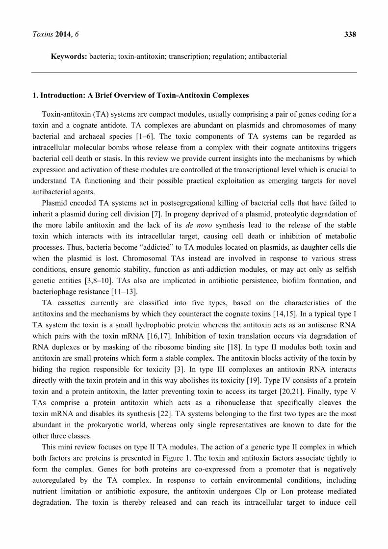
This mini review focuses on type II TA modules. The action of a generic type II complex in which
both factors are proteins is presented in Figure 1. The toxin and antitoxin factors associate tightly to
form the complex. Genes for both proteins are co-expressed from a promoter that is negatively
autoregulated by the TA complex. In response to certain environmental conditions, including
nutrient limitation or antibiotic exposure, the antitoxin undergoes Clp or Lon protease mediated
degradation. The toxin is thereby released and can reach its intracellular target to induce cell

Toxins 2014, 6
339
dormancy, stasis or death [3]. Toxins hinder cellular activities by targeting various structures and key
molecular processes [6,14]. Most class II toxins examined to date regulate the translation process by
acting as endoribonucleases [23,24]. Some of these endoribonucleases cleave free mRNA in a
sequence dependent manner, whereas others target mRNA associated with the ribosomal A site.
Certain type II toxins instead inhibit the translation machinery by cleaving initiator tRNA, by
phosphorylation of the elongation factor EF-Tu, or by binding to ribosomal subunits [6,25–27]. In
contrast with factors that target the translation process, certain type II toxins affect DNA replication by
direct inhibition of gyrase activity [28–30] or by interfering with the β sliding clamp [31]. There are
also known examples of cell wall synthesis inhibitors that act by phosphorylating a peptidoglycan
precursor [32].
Figure 1. Action of a generic type II toxin-antitoxin (TA) complex in which both factors
are proteins. The antitoxin (A) and toxin (T) genes are expressed in an operon. A poorly
structured domain within the antitoxin protein is remodelled by the toxin to produce a
stable complex that autoregulates operon expression at the transcriptional level (1); In
response to certain environmental conditions, e.g., nutrient limitation or antibiotic
exposure, the antitoxin is proteolytically degraded (2); The toxin is thereby released to act
on its intracellular target to induce cell dormancy or stasis (3). P, promoter; O, operator.
2. Transcriptional Autoregulation Is a Characteristic Feature of Type II TA Cassettes
As summarized above, type II complexes comprise a toxin and antitoxin that both are proteins [7].
Toxicity is neutralized by physical association of the two factors. When released from the complex, the
toxin targets a specific intracellular component to trigger temporary stasis or cell death. In view of its
Toxins 2014, 6
340
latent potential to interfere with vital cellular processes, inadvertent release of the toxin is catastrophic
for cell viability. To circumvent this possibility, transcriptional autoregulation is a characteristic
feature of TA cassettes that ensures balanced toxin and antitoxin production.
Transcriptional autoregulation of type II modules typically is mediated by the antitoxin that
partially represses expression by binding to an operator site which overlaps the promoter motifs
recognized by RNA polymerase. However, full repression is achieved only by binding of the TA
complex to the site. Type II antitoxins usually comprise a well-structured N-terminal domain that
mediates DNA binding and a partly-structured C-terminal region. The toxin interacts with and
restructures the latter thereby stabilizing the antitoxin for more effective operator binding (Figure 1).
The following sections outline salient features of transcriptional autoregulation in selected type II TA
complexes that illustrate the variations on a theme that have developed in TA gene expression control.
3. CcdA-CcdB: Transcriptional Repression of a DNA Gyrase Poison
The CcdA-CcdB TA complex first was identified as a maintenance system encoded by the F
plasmid in Escherichia coli [33]. Genes for homologous complexes are distributed widely on both
bacterial plasmids and chromosomes, although are less common than certain other TA loci [34]. DNA
gyrase is an essential type II topoisomerase that introduces negative supercoils into bacterial DNA.
The CcdB toxin poisons covalent gyrase-DNA complexes both by entrapping the gyrase cleavage
complex and by inhibiting the enzyme’s catalytic reactions [28,35]. The CcdA antitoxin sequesters
CcdB thereby blocking interference with gyrase [36].
The ccdA-ccdB genes form an operon that is autoregulated at the transcriptional level. Although
CcdA alone binds the ccdA-ccdB promoter-operator region in vitro, repression in vivo is mediated by
the CcdA-CcdB complex [37–40]. The two-protein complex also binds DNA more avidly and with
higher specificity in vitro than does CcdA alone [41,42]. CcdB does not bind DNA [37]. The
N-terminal region of CcdA adopts a dimeric, ribbon-helix-helix (RHH) fold that is characteristic of a
broad family of prokaryotic transcriptional repressors that regulate a variety of cellular processes
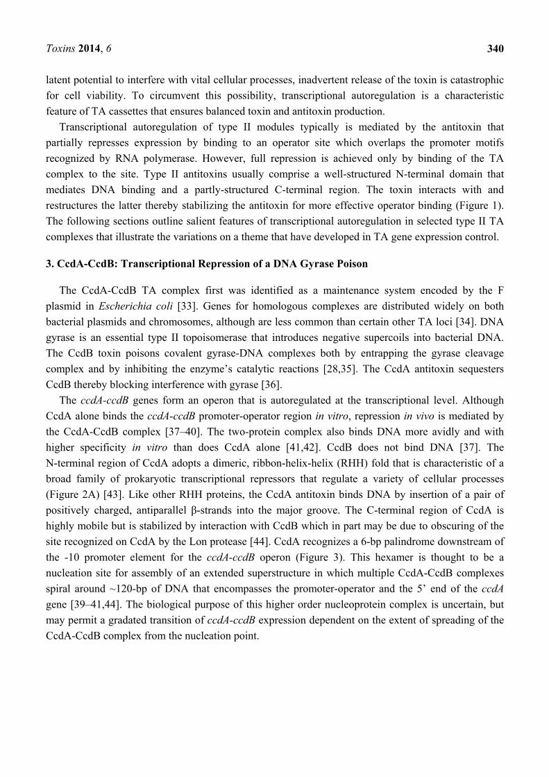
(Figure 2A) [43]. Like other RHH proteins, the CcdA antitoxin binds DNA by insertion of a pair of
positively charged, antiparallel β-strands into the major groove. The C-terminal region of CcdA is
highly mobile but is stabilized by interaction with CcdB which in part may be due to obscuring of the
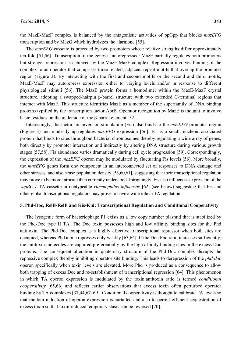
site recognized on CcdA by the Lon protease [44]. CcdA recognizes a 6-bp palindrome downstream of
the -10 promoter element for the ccdA-ccdB operon (Figure 3). This hexamer is thought to be a
nucleation site for assembly of an extended superstructure in which multiple CcdA-CcdB complexes
spiral around ~120-bp of DNA that encompasses the promoter-operator and the 5’ end of the ccdA
gene [39–41,44]. The biological purpose of this higher order nucleoprotein complex is uncertain, but
may permit a gradated transition of ccdA-ccdB expression dependent on the extent of spreading of the
CcdA-CcdB complex from the nucleation point.

Toxins 2014, 6
341
Figure 2. Tertiary structures of selected antitoxins and TA complexes. (A) solution
structure of CcdA antitoxin [44]. The structure comprises a dimeric, N-terminal
ribbon-helix-helix (RHH) fold (red and blue) that binds DNA connected to a pair of
C-terminal extensions (orange) that become structured upon interaction with the CcdB
toxin [44]. The antiparallel β-strands in the RHH fold that insert into the DNA major
groove are highlighted in blue. (B) crystal structure of the YefM-YoeB complex [45]. The
YefM antitoxin dimer and YoeB toxin monomer are coloured in blue and magenta,
respectively. Conserved arginine residues in the N-terminal segment of YefM that are
involved in operator recognition [46] are highlighted in yellow. (C) crystal structure of the
FitA-FitB-DNA complex [47]. FitA and FitB are shown in magenta and green,
respectively. The structure comprises four FitA-FitB heterodimers assembled on a 36-bp
DNA segment (orange) from the regulatory region usptream of fitA-fitB. (D) crystal
structure of the MsqA antitoxin [48]. Monomers within the dimeric structure are coloured
green and blue. Zinc ions involved in maintaining MsqA structural stability are shown as
red spheres. (E) crystal structure of the VapB2-VapC2 complex of Rickettsia felis [49].
The structure consists of four VapC toxin molecules (green) and four antitoxin monomers
(magenta) which form a tetramer of heterodimers assembled on a 26-bp DNA segment.
Images constructed using the Research Collaboratory for Structural Bioinformatics Protein
Data Bank and the Molecular Biology Toolkit [50].
A B C
D E

Toxins 2014, 6
342
Figure 3. Organization of the promoter-operator regions upstream of the ccdA-ccdB,
mazE-mazF, relB-relE, kis-kid, and yefM-yoeB TA genes (top to bottom). −10 and −35
promoter motifs are coloured red and yellow, respectively. Repeat sequences are denoted
by blue arrows; the sequences of these repeats differ among the different systems. The FIS
binding site in the mazE-mazF promoter region is shown as a hatched box.
4. MazE-MazF: Autoregulation of a Chromosomally Encoded Type II Paradigm
The MazF toxin encoded by E. coli is the archetype of a broad family of sequence-specific
endoribonucleases that cleave either free mRNA or ribosome-associated transcripts resulting in
translation inhibition [23,24]. The activity of MazF is counteracted directly by the MazE
antitoxin [51,52]. Conflicting evidence indicates that the MazE-MazF complex either is a primitive
programmed cell death mechanism in bacteria, or that MazF toxicity instead induces reversible
bacteriostasis from which cells can recover by subsequent production of MazE antitoxin as discussed
elsewhere [3,9,53,54].
Antibiotics that inhibit transcription and/or translation prevent replenishment of the MazE antitoxin.
As a consequence MazF, which is less proteolytically labile than MazE, is released to cause toxicity.
Stressful conditions such as extreme amino acid starvation also inhibit mazEF transcription. When
uncharged tRNA species occupy the ribosomal A-site during amino acid starvation, the RelA protein is
activated and catalyzes synthesis of the alarmone guanosine tetraphosphate (ppGpp). Overproduction
of ppGpp represses the mazEF cassette by interfering with RNA polymerase function. The consequent
decrease in MazE levels liberates the MazF toxin to act intracellularly [51]. Interestingly, the mazG
gene that is cotranscribed with mazEF provides another level of control to MazF action. MazG is a
nucleoside triphosphate pyrophosphohydrolase that hydrolyzes dNTPs, thereby depleting ppGpp
concentration. MazG activity is counteracted by the MazE-MazF complex. However, when amino acid
starvation induces elevated ppGpp concentration with a concomitant reduction in expression of the
mazEFG module, MazG function is no longer blocked and ppGpp level decreases. Thus, the action of
-35 -10 ccdA
-35 -10
relB
-35 -10
kis
-35 -10
yefM
-35 -35 -10 -10
mazE
10-bp
Toxins 2014, 6
343
the MazE-MazF complex is balanced by the antagonistic activities of ppGpp that blocks mazEFG
transcription and by MazG which hydrolyzes the alarmone [55].
The mazEFG cassette is preceded by two promoters whose relative strengths differ approximately
ten-fold [51,56]. Transcription of the genes is autorepressed: MazE partially regulates both promoters
but stronger repression is achieved by the MazE-MazF complex. Repression involves binding of the
complex to an operator that comprises three related, adjacent repeat motifs that overlap the promoter
region (Figure 3). By interacting with the first and second motifs or the second and third motifs,
MazE-MazF may autorepress expression either to varying levels and/or in response to different
physiological stimuli [56]. The MazE protein forms a homodimer within the MazE-MazF crystal
structure, adopting a swapped-hairpin β-barrel structure with two extended C-terminal regions that
interact with MazF. This structure identifies MazE as a member of the superfamily of DNA binding
proteins typified by the transcription factor AbrB. Operator recognition by MazE is thought to involve
basic residues on the underside of the β-barrel element [52].
Interestingly, the factor for inversion stimulation (Fis) also binds to the mazEFG promoter region
(Figure 3) and modestly up-regulates mazEFG expression [56]. Fis is a small, nucleoid-associated
protein that binds to sites throughout bacterial chromosomes thereby regulating a wide array of genes,
both directly by promoter interaction and indirectly by altering DNA structure during various growth
stages [57,58]. Fis abundance varies dramatically during cell cycle progression [59]. Correspondingly,
the expression of the mazEFG operon may be modulated by fluctuating Fis levels [56]. More broadly,
the mazEFG genes form one component in an interconnected set of responses to DNA damage and
other stresses, and also sense population density [53,60,61], suggesting that their transcriptional regulation
may prove to be more intricate than currently understood. Intriguingly, Fis also influences expression of the
vapBC-1 TA cassette in nontypeable Haemophilus influenzae [62] (see below) suggesting that Fis and
other global transcriptional regulators may prove to have a wide role in TA regulation.
5. Phd-Doc, RelB-RelE and Kis-Kid: Transcriptional Regulation and Conditional Cooperativity
The lysogenic form of bacteriophage P1 exists as a low copy number plasmid that is stabilized by
the Phd-Doc type II TA. The Doc toxin possesses high and low affinity binding sites for the Phd
antitoxin. The Phd-Doc complex is a highly effective transcriptional repressor when both sites are
occupied, whereas Phd alone represses only weakly [63,64]. If the Doc:Phd ratio increases sufficiently,
the antitoxin molecules are captured preferentially by the high affinity binding sites in the excess Doc
proteins. The consequent alteration in quaternary structure of the Phd-Doc complex disrupts the
repressive complex thereby inhibiting operator site binding. This leads to derepression of the phd-doc
operon specifically when toxin levels are elevated. More Phd is produced as a consequence to allow
both trapping of excess Doc and re-establishment of transcriptional repression [64]. This phenomenon
in which TA operon expression is modulated by the toxin:antitoxin ratio is termed conditional
cooperativity [65,66] and reflects earlier observations that excess toxin often perturbed operator
binding by TA complexes [37,44,67–69]. Conditional cooperativity is thought to calibrate TA levels so
that random induction of operon expression is curtailed and also to permit efficient sequestration of
excess toxin so that toxin-induced temporary stasis can be reversed [70].

Toxins 2014, 6
344
RelB-RelE is one of the most prevalent and well-studied TA complexes [71,72]. Like MazF, the
RelE toxin is an endoribonuclease, albeit with a different cleavage specificity on mRNA [73–75].
Moreover, unlike MazF, transcript cleavage by RelE is translation-dependent [74]. The N-terminus of
the RelB antitoxin adopts a dimeric RHH fold connected to a pair of C-terminal extensions that capture
two RelE monomers, forming an open V-shaped heterotetrameric stucture in which RelB is positioned
at the vertex [65,76,77]. This configuration blocks access by the toxin to the ribosomal A site and
induces structural rearrangements that perturb the endoribonuclease pocket [65,78].
Transcription of the relB-relE module is autoregulated in E. coli. In common with numerous TA
complexes, the RelB antitoxin represses transcription weakly whereas the RelE toxin is a co-repressor [79].
The operator site recognized by RelB-RelE comprises a pair of tandem 12-bp inverted repeats one of
which overlaps the −10 promoter motif for the relBE module (Figure 3) [76,77]. The RelB-RelE
complex exhibits conditional cooperativity: RelE promotes operator binding by RelB at subequimolar
ratios but ablates binding at higher concentrations [66]. In agreement, modelling of the complex onto
DNA suggests that a RelB dimer associated with a single RelE monomer binds one inverted repeat
within the operator. The second inverted repeat is occupied by another heterotrimeric complex that
interacts cooperatively with the adjacent complex. In contrast, steric clashes between RelE molecules
are predicted to block operator binding by a complex with an equimolar RelB:RelE ratio [65]. The
outcome of conditional cooperativity in the RelB-RelE system parallels that of the Phd-Doc complex
described above [64], i.e., differential regulation of the cognate TA genes by toxin:antitoxin ratios. As
with Phd-Doc, RelE possesses high and low affinity binding sites for RelB that likely influence the
RelB:RelE ratio intracellularly. The low affinity site for the RelB-RelE interaction could play a role in
the interaction of excess of toxin with the TA repressor bound to the operator and in the release of the
repressor under these circumstances. However, unlike with Phd-Doc, these sites are not invoked in
conditional cooperativity. Instead, interactions between RelB dimers positioned on the inverted repeats
and stabilized by subequimolar binding of RelE are sufficient for cooperative operator recognition [65].
The Kis-Kid type II TA complex is encoded by the R1 plasmid of E. coli. Kid is a sequence-specific,
translation-independent endoribonuclease whose activation inhibits the growth of plasmid-free
cells [80]. The protein is a member of a broad group of structurally-related toxins that includes MazF
and CcdB [1], whereas the Kis antitoxin is homologous to MazE [81]. The kis-kid operon is
transcriptionally autoregulated. As is characteristic of type II complexes, the antitoxin partially
represses expression but the Kis-Kid complex represses more effectively [82]. The operator site
recognized by Kis-Kid comprises a pair of imperfect 18-bp inverted repeats that are separated by a
33-bp spacer in the promoter region for the kis-kid genes (Figure 3). The repeat that overlaps the
−10 promoter box is bound more strongly by the complex suggesting that the interaction of Kis-Kid
with RNA polymerase modulates kis-kid expression [69]. Kis and Kid assemble into numerous
complexes in vitro [81]. As is characteristic of conditional cooperativity, complexes that contain
equimolar or subequimolar Kis:Kid concentrations bind the operator site most effectively. In contrast,
less stable complexes with DNA are formed when the Kid toxin is in excess of Kis antitoxin and
transcriptional repression may be limited. Consequent kis-kid expression will rebalance the Kis:Kid
ratio away from toxin excess [69]. Two transcripts are produced from the kis-kid operon in equimolar
concentrations and with similar half-lives. The longer of these transcripts spans both genes and is also
processed by limited degradation to a shorter species that terminates within kid. Moreover, translation

Toxins 2014, 6
345
of the Kid toxin and Kis antitoxin are coupled which further minimizes the synthesis of Kid in the
event that insufficient Kis has been translated [83]. Transcriptional activity of the kis-kid operon is also
dependent on plasmid copy number [84]. Thus, conditional cooperativity, RNA processing and
translational coupling, act in concert to ensure balanced production of the Kis antitoxin relative to the
Kid toxin. However, conditional cooperativity is not a universal control mechanism among type II TA
systems [85] and is predicted to be one of a variety of emerging regulatory strategies that have evolved
to fine-tune TA gene expression.
6. YefM-YoeB and Axe-Txe: Diverse Transcriptional Control of Homologous TA Complexes
The YefM-YoeB system of E. coli is a hybrid type II TA complex: the YoeB toxin and YefM
antitoxin are related evolutionarily to RelE and Phd, respectively, whereas the usual associations are
RelB-RelE and Phd-Doc [86–88]. YoeB binding to the 50S ribosomal subunit prevents the formation
of the initiation complex and induces mRNA cleavage three bases downstream of the initiation
codon [89]. However, the toxin’s catalytic fold is concealed when it is bound to YefM thereby
blocking its enzymatic activity. This interaction, which also stabilizes YefM, is crucial for ensuring
that free YoeB is not available erroneously to degrade mRNA [45].
The yefM-yoeB genes are transcriptionally autoregulated. YefM is the principal repressor, whereas
YoeB is a repression enhancer [90]. YefM lacks a canonical DNA binding motif [45], but dual
conserved arginine residues in the N-terminal segment of the protein are involved in operator
recognition (Figure 2B) [46]. Free YefM sequentially recognizes adjacent long and short DNA
palindromes during transcriptional repression of the yefM-yoeB operon. The palindromes share
core hexamer 5’-TGTACA-3’ motifs, possess a centre-to-centre distance of 12-bp and overlap the
yefM-yoeB promoter (Figure 3). The YefM-YoeB complex binds the palindromes more avidly than
free antitoxin via cooperative interactions [46,90]. Changing the inter-palindrome spacing perturbs
cooperative binding to the repeats: YefM-YoeB interaction with the long repeat is retained but binding
to the short repeat is disrupted [46]. Paired hexamer motifs are frequent in yefM-yoeB regulatory
regions in diverse genomes suggesting that interaction of YefM-YoeB with these motifs is a conserved
mode of operon transcriptional autoregulation [90].
The Axe-Txe complex encoded widely by enterococcal plasmids is homologous to YefM-YoeB [87].
Analogously, the Axe antitoxin represses the axe-txe promoter weakly whereas Axe-Txe represses
more strongly [91]. However, an internal promoter within axe also directs expression of the
downstream txe toxin gene. This internal promoter is not regulated by Axe-Txe and is essential for the
function of the complex as a plasmid maintenance system suggesting that it plays a vital role in setting
the Axe:Txe ratio. A cryptic transcript that originates within txe and a putative transcription terminator
in the region 3’ of the operon may be additional regulatory elements that contribute to transcriptional
control of axe-txe [91]. The yefM-yoeB operon of Streptococcus pneumoniae provides another example
of multilayered transcriptional control. In this case, a pair of promoters, one of which is constitutive
and one of which is autoregulated by YefM-YoeB, are located upstream of the genes [92].
The interactions between the homologous YefM-YoeB and Axe-Txe complexes are species-specific.
Accordingly, the complexes repress expression of the cognate promoters, but not of the non-cognate
promoters, even though both operator regions are composed of a pair of inverted repeats with the same
Toxins 2014, 6
346
hexameric core [88]. However, a single substitution near the C-terminus which converts a Txe-specific
residue to a YoeB-specific amino acid permitted neutralization of Txe by YefM in vivo. Moreover, the
complex of wild-type YefM and mutated Txe partially corepressed the yefM-yoeB promoter [88].
These data illustrate that subtle amino acid sequence changes can impose interaction specificity,
including transcriptional regulation specificity, on homologous TA complexes, and provide insight into
the mechanisms by which expression of multiple, homologous type II TA complexes in a single host
may be regulated differentially under disparate physiological conditions [93].
7. Diverging from the Canonical Pattern of Type II TA Transcriptional Control: Tripartite
Protein Complexes and a Toxin that Directly Binds DNA
Certain exceptions to the general pattern of type II TA transcriptional regulation have been
described. Three component TA modules include the pasABC complex encoded by plasmid pTF-FC2,
paaA-parE-paaR of E. coli O157:H7, and the ε-ζ-ω system specified by streptococcal plasmid
pSM19035.
The pasA gene codes for an antitoxin and pasB encodes a toxin [94]. The pasABC promoter
undergoes full autorepression by the PasAB complex, and the PasC protein has little effect on
promoter expression [95]. Instead, PasC enhances the neutralizing effect of the antidote [94].
In the case of the paaR-paaA-parE operon, the PaaA antitoxin and ParE toxin form a complex that
autorepresses the main promoter only partially. The transcriptional regulator encoded by an upstream
gene within the same operon, PaaR, is required for full down-regulation of transcription. These
different repressor complexes probably act independently [96]. The PaaR repressor also is essential in
maintaining an appropriate PaaA:ParE ratio [96].
Another tripartite type II TA system is ε-ζ-ω which was discovered initially on plasmid pSM19035
of Streptococcus pyogenes. In this case the ζ toxin and ε antitoxin have no roles in transcriptional
control. Instead, their expression is inhibited solely by the ω protein encoded by the first gene in the
operon. In addition to being a repressor of its own promoter, ω is encoded widely by plasmids
belonging to the Inc18 family and acts as a global negative regulator that also controls transcription of
genes required for plasmid copy number control and stable inheritance, thereby promoting accurate
plasmid segregation [97–100]. Like certain other type II TA transcriptional regulators, including CcdA
(Figure 2A) and RelB, ω is a RHH protein [101]. However, the binding site recognized by ω is
distinctive, comprising palindromic and non-palindromic DNA heptad repeats (5’-NATCACN-3’) in
the cognate operator sites [98].
The VapC subfamily of PIN domain proteins forms the toxin component of a widespread type II
TA complex. PIN domain proteins cleave single-stranded RNA and are characterized by a cluster of
strictly conserved acidic amino acids within the active site. VapC is counteracted by the VapB
antitoxin [102]. Expression of the vapB-vapC genes is autoregulated negatively by the VapB-VapC
complex which specifically binds inverted repeat sequences within the vapBC-1 operator region [103,104].
However, the vapBC-1 cassette of nontypeable Haemophilus influenzae presents notable differences
to the typical pattern. First, contrary to other TA systems described to date, the VapC-1 toxin possesses
DNA binding activity whereas the VapB-1 antitoxin does not directly interact with DNA.
Nevertheless, VapB-1 increases VapC-1 interaction specificity with the operator region [62]. Second,

Toxins 2014, 6
347
the Fis protein upregulates vapBC-1 expression during nutrient upshift in H. influenzae. The influence
of Fis on vapBC-1 expression is thought to occur indirectly by altering DNA structure in the
promoter-operator region [62].
8. Type II TAs that Regulate Other Genes or Do Not Display Autoregulation
The mqsRA module encoded by E. coli displays many features that differ from canonical type II TA
systems. First, the gene for the mqsR toxin precedes the mqsA antitoxin gene, whereas the standard
organization comprises an antitoxin gene followed by a toxin gene (Figure 1). This unusual genetic
organization has been observed only rarely in other TA systems characterized to date, including the
higBA [105] and hicAB modules [106]. Second, the MqsA antitoxin is well-ordered throughout its
entire length and requires zinc ions to maintain its structural stability, properties that are unique among
known antitoxins (Figure 2D) [48].
Autoregulation of the mqsRA module also is distinctive. The antitoxin repressor, MqsA, undergoes
extensive domain rearrangements upon DNA binding and is the only antitoxin known to interact with
DNA via its C-terminal domain [48,107]. However, the MqsA N-terminal domain, which binds the
MqsR toxin, also makes direct interactions with DNA. It twists and collapses over the DNA and this
rotation clamps the DNA thereby enhancing binding [107]. Interestingly, the MqsR toxin does not
function as a transcriptional corepressor as in many other TA systems. Instead, MqsR destabilizes the
MqsA-DNA complex. This reflects that the binding sites of DNA and MqsR on MqsA partially
overlap rendering simultaneous binding of both by MqsA impossible. Thus, MqsR is a transcriptional
activator of mqsRA expression, not a transcriptional repressor [85].
Distinct from other type II TA systems, the MqsR-MqsA complex also regulates other genomic
promoter regions. Specifically, the MqsA antitoxin and the MqsR-MqsA complex regulate the promoters
of genes that are important for E. coli metabolism, including the mcbR, spy and cspD loci [48,108]. The
mcbR gene encodes a colanic acid regulator, Spy is a periplasmic chaperone of proteins, and CspD is a
stress-induced cold shock protein that is a DNA replication inhibitor. MqsA also directly binds to an
MqsRA-like palindrome located within the promoter of the rpoS gene and thereby represses
transcription of a major regulator of stress, σS [109]. This lowers the concentration of the internal
messenger 3,5-cyclic diguanylic acid (c-di-GMP) which in turn causes increased motility and reduction
of biofilm formation, as well as decreased oxidative stress resistance through catalase activity [107].
MsqA also modulates biofilm formation by acting as a transcriptional regulator of the gene that
encodes CsgD, a master controller of biofilm formation [110]. The endoribonuclease toxin MqsR is
also a global regulator [108,111]. It cleaves specific mRNA mainly at 5’-GCU-3’ sites [112,113]
which significantly increases the presence of mRNAs coding for CstA, CspD, RpoS, Dps and HokD
proteins that are known to be associated with stress response [108]. Thus, the MqsR-MqsA system
controls cell physiology both by its own toxicity as well as through the regulation of other genes.
The chromosomal mazE-mazF operon of Staphylococcus aureus is transcriptionally linked to the
downstream, polycistronic sigB operon and is transcribed both as part of the operon and as a shorter
transcript. Unlike the homologous genes in E. coli, staphylococcal mazE-mazF is not subject to
autoregulation. Instead, the activity of the mazE-mazF promoter is inhibited by the product of the sigB
gene that codes for the alternative sigma factor, σB. Moreover, the mazE-mazF promoter is required for
Toxins 2014, 6
348
full σB activity due both to its transcriptional coupling with the sigB operon via readthrough of a weak
downstream rho-independent terminator and to the ability to respond to multiple stresses [114]. On the
other hand, the mazE-mazF promoter is positively and directly regulated by SarA, a winged-helix,
global transcriptional regulator of virulence gene expression in S. aureus [115]. This probably occurs
by binding of SarA to a region that overlaps the −35 box in the mazE-mazF promoter and which
exhibits high similarity to the consensus SarA binding site [114]. The intricate transcriptional control
of the mazE-mazF genes in S. aureus highlights an emerging theme in understanding TA activity and
function: TA systems frequently form parts of complex gene regulation networks that respond to
diverse environmental and physiological signals.
9. Information from Toxin-Antitoxin-DNA Costructures
Although a limited number of type II toxin-antitoxin-DNA costructures are available currently,
these structures provide valuable insights into the mechanism of DNA binding by antitoxins and how
toxins coregulate this activity. For the FitA-FitB complex, a heterotetramer is formed by the binding of
two FitB PIN domain toxin monomers to a pair of FitA antitoxin dimers. The FitA antitoxin dimers are
thereby tethered to operator DNA. FitA binds DNA via a RHH fold. The toxin monomers do not interact
directly with DNA, but promote binding by stabilizing the FitA-FitB heterotetramer (Figure 2C) [47].
The structure of the VapB2-VapC2 TA complex of Rickettsia felis with DNA has been
determined [49]. As with the FitA-FitB complex, the VapB2-VapC2 structure comprises a tetramer of
heterodimers in which four toxin subunits assemble together with four antitoxin subunits (Figure 2E).
The VapC2 toxin harbours a PIN domain that forms homodimers homologous to those of FitB. However,
unlike the RHH structure of FitA antitoxin, the VapB2 antitoxin dimers contain a swapped-hairpin β-barrel
fold, similar to MazE described above. The interaction of VapB2 with DNA is mediated by the
β-hairpins. Moreover, the DNA structure is distorted when bound by the TA complex with the VapB2
homodimers interacting with the concave face of the curved DNA [49]. The VapB2-VapC2 complex
provides an interesting illustration of how extensive evolutionary shuffling of toxin and antitoxin genes
has occurred resulting in considerable diversity in TA combinations [3,71,72].
The HipA toxin possesses a eucaryotic serine/threonine kinase-like fold that structurally is most
related to human CDK2/cyclin A kinase [26]. HipB antitoxin cooperatively binds four operator sites
(consensus sequence 5’-TATCCN8GGATA-3’) located in the hipBA promoter region thereby
repressing hipBA expression. HipA is a co-repressor [116]. HipB is dimeric each subunit comprising a
single β strand and four α helices. Two of the helices form a canonical helix-turn-helix motif that
mediates DNA major groove contacts. The HipB structure is related to the Cro repressor family of
proteins. The protein induces a 70° bend in its operator DNA that assists cooperative binding by
aligning the recognition helices for specific binding to successive major grooves [26]. The HipA-HipB
structure assembled on DNA consists of dimeric HipB flanked on either side by one HipA monomer.
The N-terminal domains of HipA interact with one of the HipB subunits, whereas the HipA C-terminal
domains mainly contact the second HipB subunit. These and other interactions trap HipA in an
inactive, non-toxic conformation when bound with HipB on DNA. Interestingly, although HipA alone
does not bind DNA, a pair of residues in each monomer within the HipA-HipB-DNA structure make
phosphate backbone contacts with operator DNA [26].
Toxins 2014, 6
349
10. New Antibiotics that Interfere with TA Transcriptional Autoregulation to Detonate
Toxins Artificially
Bacteria evolve rapidly. Shuttling of mobile genetic elements - plasmids, transposons, bacteriophage
and integrons - between bacteria is especially important in promoting genome plasticity and
diversification [117,118]. By acquiring antibiotic resistance genes that are frequently located on these
elements, bacteria adapt, survive and proliferate in antibiotic containing niches in which sensitive
strains perish. The spread of resistance genes on mobile elements in bacteria has been compounded
significantly by antibiotic misprescription, by use of antibiotics as growth promoting and prophylactic
agents in animals, and by indiscriminate release of antibacterials into the biosphere [119]. Thus, the
global surge in antibiotic usage inexorably has selected for bacterial isolates that are resistant not only
to single compounds, but in many cases to multiple drugs [120]. As a consequence, certain infections
that previously were treatable now have few, if any, therapeutic options [121]. For example, one-third
of the world’s population is infected with Mycobacterium tuberculosis, the causative agent of
tuberculosis, which causes two million deaths globally per annum. The emergence of multidrug
resistant strains, then extensively drug resistant isolates, and more recently totally drug resistant strains
of M. tuberculosis threatens even further devastation in developing countries, as well as the deadly
re-emergence of an ancient scourge in the Western world [122]. Similarly, multiresistant strains of
Gram-negative Pseudomonas aeruginosa, Acinetobacter spp., and Enterobacteriaceae that produce
extended-spectrum beta-lactamases are serious hospital-acquired pathogens that are becoming evermore
difficult to treat with existing antibiotics, as are Gram-positive species including methicillin-resistant
Staphylcocccus aureus and vancomycin-resistant enterococci [119,120,123]. Despite the impending crisis,
only one new class of antibiotic has come to market in the past 50 years. Moreover, pharmaceutical
companies are withdrawing from antimicrobial development in pursuit of more lucrative therapeutics:
a perilous gap has opened in the race for novel antibacterials [124].
Ectopic expression of toxin genes induces severe bacterial growth defects, including cell death.
Moreover, genes for TA complexes have no known human homologues and are abundant on chromosomes
and plasmids of most bacteria, including pathogenic species, which contributes to their attractiveness
as potential targets for novel antibacterial agents. There is considerable interest in identifying natural
and artificial molecules that destabilize TA complexes to promote toxin activation [125]. A variety of
different strategies may be implemented to exploit TA systems to this end. For example, the disruption
of the toxin-antitoxin interaction may free the former to induce cell death [126]. Similarly, interference
with antitoxin oligomerization will destabilize the TA complex thereby liberating the toxin. In both
cases, perturbation of TA complex assembly or organization also is predicted to interfere with correct
transcriptional autoregulation. Moreover, compounds that bind the TA promoter region will directly
inhibit transcription of TA genes. Replenishment of the labile antitoxin will be prevented thereby
releasing the more stable toxin to induce bacterial cell suicide [90]. Similarly, ligands that block
antitoxin interaction with the operator site may derepress TA gene expression, imbalance the TA ratio
and, again, cause inappropriate toxin release. Persister cells comprise a rare, antibiotic tolerant fraction
of bacterial populations. TA loci are implicated in bacterial persistence as the toxin factors reduce
metabolism and render cells insenstive to many antibiotics [11]. This phenomenon needs to be
considered carefully when assessing the effect of artificial toxin release on bacterial survival.

Toxins 2014, 6
350
Studies on disruption of the streptococcal ε-ζ interaction and inhibition of TA complex formation in
the anthrax agent of Bacillus anthracis suggest that designer peptides may be useful in modulating TA
system interactions [127–129]. Peptides based on the structure of the ParE toxin have been used as
inhibitors of bacterial topoisomerases [130]. Analogously, the Extracellular Death Factor is an
endogenous pentapeptide activator of the MazF toxin [60] which indicates the existence of natural
ligands that may be exploited to moderate TA complex function. Although these initial studies suggest
that peptidomimetics may be potentially powerful in disrupting TA function, designing effective
molecules is not straightforward [131]. Developing molecules that alter TA transcriptional regulation
directly is an alternative approach for controlled toxin release. Sequence-specific DNA binders already
function, for example, in anticancer therapies [132–134]. Potential antibacterial drugs targeting TA
systems could alter interactions of antitoxins with DNA regulatory sites by a variety of different
mechanisms. Drugs that target DNA or change its architecture may compete for antitoxin DNA
binding sequences. Synthetic oligonucleotides or other ligands may compete for DNA recognition
motifs on the antitoxin. Ligand binding to other regions of the antitoxin may change its ability or
sensitivity to interact with regulatory DNA. New weapons are urgently required in the war against
infectious disease and antibiotic resistance. The toxin factors in TA complexes have evolved to induce
intracellular damage, albeit in a controlled way. By devising strategies that artificially unleash the
toxin, including by disrupting proper transcriptional regulation, it may be possible to develop novel
antibacterial strategies based on bacterial suicide from within.
Acknowledgments
Work in the laboratory of BK was supported by the Polish Ministry of Science and Higher
Education (project grant no. N N301 251936).
Conflicts of Interest
The authors declare no conflict of interest.
References
1. Blower, T.R.; Salmond, G.P.; Luisi, B.F. Balancing at survival’s edge: The structure and adaptive
benefits of prokaryotic toxin-antitoxin partners. Curr. Opin. Struct. Biol. 2011, 21, 109–118.
2. Fozo, E.M.; Makarova, K.S.; Shabalina, S.A.; Yutin, N.; Koonin, E.V.; Storz, G. Abundance of
type I toxin-antitoxin systems in bacteria: Searches for new candidates and discovery of novel
families. Nucleic Acids Res. 2010, 38, 3743–3759.
3. Hayes, F.; van Melderen, L. Toxins-antitoxins: Diversity, evolution and function. Crit. Rev.
Biochem. Mol. Biol. 2011, 46, 386–408.
4. Makarova, K.S.; Wolf, Y.I.; Koonin, E.V. Comprehensive comparative-genomic analysis of type
2 toxin-antitoxin systems and related mobile stress response systems in prokaryotes. Biol. Direct
2009, 4, 19.
5. Pandey, D.P.; Gerdes, K. Toxin-antitoxin loci are highly abundant in freeliving but lost from
host-associated prokaryotes. Nucleic Acids Res. 2005, 33, 966–976.
Toxins 2014, 6
351
6. Yamaguchi, Y.; Inouye, M. Regulation of growth and death in Escherichia coli by
toxin-antitoxin systems. Nat. Rev. Microbiol. 2011, 9, 779–790.
7. Hayes, F. Toxins-antitoxins: Plasmid maintenance, programmed cell death, and cell cycle arrest.
Science 2003, 301, 1496–1499.
8. Saavedra De Bast, M.; Mine, N.; van Melderen, L. Chromosomal toxin-antitoxin systems may
act as anti-addiction modules. J. Bacteriol. 2008, 190, 4603–4609.
9. Van Melderen, L. Toxin-antitoxin systems: Why so many, what for? Curr. Opin. Microbiol.
2010, 13, 781–785.
10. Van Melderen, L.; Saavedra De Bast, M. Bacterial toxin-antitoxin systems: More than selfish
entities? PLoS Genet. 2009, 5, e000437.
11. Gerdes, K.; Maisonneuve, E. Bacterial persistence and toxin-antitoxin loci. Annu. Rev.
Microbiol. 2012, 66, 103–123.
12. Magnuson, R.D. Hypothetical functions of toxin-antitoxin systems. J. Bacteriol. 2007, 189,
6089–6092.
13. Wang, X.; Wood, T.K. Toxin-antitoxin systems influence biofilm and persister cell formation
and the general stress response. Appl. Environ. Microbiol. 2011, 77, 5577–5583.
14. Schuster, C.F.; Bertram, R. Toxin-antitoxin systems are ubiquitous and versatile modulators of
prokaryotic cell fate. FEMS Microbiol. Lett. 2013, 340, 73–85.
15. Mruk, I.; Kobayashi, I. To be or not to be: Regulation of restriction-modification systems and
other toxin-antitoxin systems. Nucleic Acids Res. 2014, 42, 70–86.
16. Fozo, E.M.; Hemm, M.R.; Storz, G. Small toxic proteins and the antisense RNAs that repress
them. Microbiol. Mol. Biol. Rev. 2008, 72, 579–589.
17. Gerdes, K.; Wagner, E. RNA antitoxins. Curr. Opin. Microbiol. 2007, 10, 117–124.
18. Darfeuille, F.; Unoson, C.; Vogel, J.; Wagner, E.G. An antisense RNA inhibits translation by
competing with standby ribosomes. Mol. Cell 2007, 26, 381–392.
19. Blower, T.R.; Short, F.L.; Rao, F.; Mizuguchi, K.; Pei, X.Y.; Fineran, P.C.; Luisi, B.F.;
Salmond, G.P. Identification and classification of bacterial type III toxin-antitoxin systems
encoded in chromosomal and plasmid genomes. Nucleic Acids Res. 2012, 40, 6158–6173.
20. Masuda, H.; Tan, Q.; Awano, N.; Wu, K.P.; Inouye, M. YeeU enhances the bundling of
cytoskeletal polymers of MreB and FtsZ, antagonizing the CbtA (YeeV) toxicity in Escherichia
coli. Mol. Microbiol. 2012, 84, 979–989.
21. Tan, Q.; Awano, N.; Inouye, M. YeeV is an Escherichia coli toxin that inhibits cell division by
targeting the cytoskeleton proteins, FtsZ and MreB. Mol. Microbiol. 2011, 79, 109–118.
22. Wang, X.; Lord, D.M.; Cheng, H.Y.; Osbourne, D.O.; Hong, S.H.; Sanchez-Torres, V.;
Quiroga, C.; Zheng, K.; Herrmann, T.; Peti, W.; et al. A new type V toxin-antitoxin system
where mRNA for toxin GhoT is cleaved by antitoxin GhoS. Nat. Chem. Biol. 2012, 8, 855–861.
23. Cook, G.M.; Robson, J.R.; Frampton, R.A.; McKenzie, J.; Przybilski, R.; Fineran, P.C.;
Arcus, V.L. Ribonucleases in bacterial toxin-antitoxin systems. Biochim. Biophys. Acta 2013,
1829, 523–531.
24. Yamaguchi, Y.; Inouye, M. mRNA interferases, sequence-specific endoribonucleases from the
toxin-antitoxin systems. Prog. Mol. Biol. Transl. Sci. 2009, 85, 467–500.

Toxins 2014, 6
352
25. Liu, M.; Zhang, Y.; Inouye, M.; Woychik, N.A. Bacterial addiction module toxin Doc inhibits
translation elongation through its association with the 30S ribosomal subunit. Proc. Natl. Acad.
Sci. USA 2008, 105, 5885–5890.
26. Schumacher, M.A.; Piro, K.M.; Xu, W.; Hansen, S.; Lewis, K.; Brennan, R.G. Molecular
mechanisms of HipA-mediated multidrug tolerance and its neutralization by HipB. Science 2009,
323, 396–401.
27. Winther, K.S.; Gerdes, K. Enteric virulence associated protein VapC inhibits translation by
cleavage of initiator tRNA. Proc. Natl. Acad. Sci. USA 2011, 108, 7403–7407.
28. Bernard, P.; Couturier, M. Cell killing by the F plasmid CcdB protein involves poisoning of
DNA-topoisomerase II complexes. J. Mol. Biol. 1992, 226, 735–745.
29. Jiang, Y.; Pogliano, J.; Helinski, D.R.; Konieczny, I. ParE toxin encoded by the broad-host-range
plasmid RK2 is an inhibitor of Escherichia coli gyrase. Mol. Microbiol. 2002, 44, 971–979.
30. Yuan, J.; Sterckx, Y.; Mitchenall, L.A.; Maxwell, A.; Loris, R.; Waldor, M.K. Vibrio cholerae
ParE2 poisons DNA gyrase via a mechanism distinct from other gyrase inhibitors. J. Biol. Chem.
2010, 285, 40397–40408.
31. Aakre, C.D.; Phung, T.N.; Huang, D.; Laub, M.T. A bacterial toxin inhibits DNA replication
elongation through a direct interaction with the β sliding clamp. Mol. Cell 2013, 52, 617–628.
32. Mutschler, H.; Gebhardt, M.; Shoeman, R.L.; Meinhart, A. A novel mechanism of programmed
cell death in bacteria by toxin-antitoxin systems corrupts peptidoglycan synthesis. PLoS Biol.
2011, 9, e1001033.
33. Ogura, T.; Hiraga, S. Mini-F plasmid genes that couple host cell division to plasmid
proliferation. Proc. Natl. Acad. Sci. USA 1983, 80, 4784–4788.
34. Guérout, A.M.; Iqbal, N.; Mine, N.; Ducos-Galand, M.; van Melderen, L.; Mazel, D.
Characterization of the phd-doc and ccd toxin-antitoxin cassettes from Vibrio superintegrons.
J. Bacteriol. 2013, 195, 2270–2283.
35. Smith, A.B.; Maxwell, A. A strand-passage conformation of DNA gyrase is required to allow the
bacterial toxin, CcdB, to access its binding site. Nucleic Acids Res. 2006, 34, 4667–4676.
36. De Jonge, N.; Garcia-Pino, A.; Buts, L.; Haesaerts, S.; Charlier, D.; Zangger, K.; Wyns, L.;
de Greve, H.; Loris, R. Rejuvenation of CcdB-poisoned gyrase by an intrinsically disordered
protein domain. Mol. Cell 2009, 35, 154–163.
37. Afif, H.; Allali, N.; Couturier, M.; van Melderen, L. The ratio between CcdA and CcdB
modulates the transcriptional repression of the ccd poison-antidote system. Mol. Microbiol. 2001,
41, 73–82.
38. De Feyter, R.; Wallace, C.; Lane, D. Autoregulation of the ccd operon in the F plasmid.
Mol. Gen. Genet. 1989, 218, 481–486.
39. Tam, J.E.; Kline, B.C. Control of the ccd operon in plasmid F. J. Bacteriol. 1989, 171,
2353–2360.
40. Tam, J.E.; Kline, B.C. The F plasmid ccd autorepressor is a complex of CcdA and CcdB
proteins. Mol. Gen. Genet. 1989, 219, 26–32.
41. Dao-Thi, M.H.; Charlier, D.; Loris, R.; Maes, D.; Messens, J.; Wyns, L.; Backmann, J. Intricate
interactions within the ccd plasmid addiction system. J. Biol. Chem. 2002, 277, 3733–3742.

Toxins 2014, 6
353
42. Salmon, M.A.; van Melderen, L.; Bernard, P.; Couturier, M. The antidote and autoregulatory
functions of the F plasmid CcdA protein: A genetic and biochemical survey. Mol. Gen. Genet.
1994, 244, 530–538.
43. Schreiter, E.R.; Drennan, C.L. Ribbon-helix-helix transcription factors: Variations on a theme.
Nat. Rev. Microbiol. 2007, 5, 710–720.
44. Madl, T.; van Melderen, L.; Mine, N.; Respondek, M.; Oberer, M.; Keller, W.; Khatai, L.;
Zangger, K. Structural basis for nucleic acid and toxin recognition of the bacterial antitoxin
CcdA. J. Mol. Biol. 2006, 364, 170–185.
45. Kamada, K.; Hanaoka, F. Conformational change in the catalytic site of the ribonuclease YoeB
toxin by YefM antitoxin. Mol. Cell 2005, 19, 497–509.
46. Bailey, S.E.S.; Hayes, F. Influence of operator site geometry on transcriptional control by the
YefM-YoeB toxin-antitoxin complex. J. Bacteriol. 2009, 191, 762–772.
47. Mattison, K.; Wilbur, J.S.; So, M.; Brennan, R.G. Structure of FitAB from Neisseria
gonorrhoeae bound to DNA reveals a tetramer of toxin-antitoxin heterodimers containing pin
domains and ribbon-helix-helix motifs. J. Biol. Chem. 2006, 281, 37942–37951.
48. Brown, B.L.; Grigoriu, S.; Kim, Y.; Arruda, J.M.; Davenport, A.; Wood, T.K.; Peti, W.; Page, R.
Three dimensional structure of the MqsR:MqsA complex: A novel TA pair comprised of a toxin
homologous to RelE and an antitoxin with unique properties. PLoS Pathog. 2009, 5, e1000706.
49. Maté, M.J.; Vincentelli, R.; Foos, N.; Raoult, D.; Cambillau, C.; Ortiz-Lombardía, M.
Crystal structure of the DNA-bound VapBC2 antitoxin/toxin pair from Rickettsia felis.
Nucleic Acids Res. 2012, 40, 3245–3258
50. Moreland, J.L.; Gramada, A.; Buzko, O.V.; Zhang, Q.; Bourne, P.E. The Molecular Biology
Toolkit (MBT): A modular platform for developing molecular visualization applications. BMC
Bioinformatics 2005, 6, 21.
51. Aizenman, E.; Engelberg-Kulka, H.; Glaser, G. An Escherichia coli chromosomal “addiction
module” regulated by guanosine 3',5'-bispyrophosphate: A model for programmed bacterial cell
death. Proc. Natl. Acad. Sci. USA 1996, 93, 6059–6063.
52. Kamada, K.; Hanaoka, F.; Burley, S.K. Crystal structure of the MazE/MazF complex: Molecular
bases of antidote-toxin recognition. Mol. Cell 2003, 11, 875–884.
53. Erental, A.; Sharon, I.; Engelberg-Kulka, H. Two programmed cell death systems in Escherichia
coli: An apoptotic-like death is inhibited by the mazEF-mediated death pathway. PLoS Biol.
2012, 10, e1001281.
54. Tsilibaris, V.; Maenhaut-Michel, G.; Mine, N.; van Melderen, L. What is the benefit to
Escherichia coli of having multiple toxin-antitoxin systems in its genome? J. Bacteriol. 2007,
189, 6101–6108.
55. Gross, M.; Marianovsky, I.; Glaser, G. MazG-a regulator of programmed cell death in
Escherichia coli. Mol. Microbiol. 2006, 59, 590–601.
56. Marianovsky, I.; Aizenman, E.; Engelberg-Kulka, H.; Glaser, G. The regulation of the
Escherichia coli mazEF promoter involves an unusual alternating palindrome. J. Biol. Chem.
2001, 276, 5975–5984.
Toxins 2014, 6
354
57. Schneider, R.; Lurz, R.; Lüder, G.; Tolksdorf, C.; Travers, A.; Muskhelishvili, G. An
architectural role of the Escherichia coli chromatin protein FIS in organising DNA.
Nucleic Acids Res. 2001, 29, 5107–5114.
58. Skoko, D.; Yoo, D.; Bai, H.; Schnurr, B.; Yan, J.; McLeod, S.M.; Marko, J.F.; Johnson, R.C.
Mechanism of chromosome compaction and looping by the Escherichia coli nucleoid protein
Fis. J. Mol. Biol. 2006, 364, 777–798.
59. Dorman, C.J. Nucleoid-associated proteins and bacterial physiology. Adv. Appl. Microbiol. 2009,
67, 47–64.
60. Belitsky, M.; Avshalom, H.; Erental, A.; Yelin, I.; Kumar, S.; London, N.; Sperber, M.;
Schueler-Furman, O.; Engelberg-Kulka, H. The Escherichia coli extracellular death factor EDF
induces the endoribonucleolytic activities of the toxins MazF and ChpBK. Mol. Cell 2011, 41,
625–635.
61. Dorsey-Oresto, A.; Lu, T.; Mosel, M.; Wang, X.; Salz, T.; Drlica, K.; Zhao, X. YihE kinase is a
central regulator of programmed cell death in bacteria. Cell Rep. 2013, 3, 528–337.
62. Cline, S.D.; Saleem, S.; Daines, D.A. Regulation of the vapBC-1 toxin-antitoxin locus in
nontypeable Haemophilus influenzae. PLoS One 2012, 7, e32199.
63. Magnuson, R.; Lehnherr, H.; Mukhopadhyay, G.; Yarmolinsky, M.B. Autoregulation of the
plasmid addiction operon of bacteriophage P1. J. Biol. Chem. 1996, 271, 18705–18710.
64. Garcia-Pino, A.; Balasubramanian, S.; Wyns, L.; Gazit, E.; de Greve, H.; Magnuson, R.D.;
Charlier, D.; van Nuland, N.A.; Loris, R. Allostery and intrinsic disorder mediate transcription
regulation by conditional cooperativity. Cell 2010, 142, 101–111.
65. Bøggild, A.; Sofos, N.; Andersen, K.R.; Feddersen, A.; Easter, A.D.; Passmore, L.A.;
Brodersen, D.E. The crystal structure of the intact E. coli RelBE toxin-antitoxin complex
provides the structural basis for conditional cooperativity. Structure 2012, 20, 1641–1648.
66. Overgaard, M.; Borch, J.; Jørgensen, M.G.; Gerdes, K. Messenger RNA interferase RelE controls
relBE transcription by conditional cooperativity. Mol. Microbiol. 2008, 69, 841–857.
67. Johnson, E.P.; Strom, A.R.; Helinski, D.R. Plasmid RK2 toxin protein ParE: Purification and
interaction with the ParD antitoxin protein. J. Bacteriol. 1996 178, 1420–1429.
68. Magnuson, R.; Yarmolinsky, M.B. Corepression of the P1 addiction operon by Phd and Doc.
J. Bacteriol. 1998, 180, 6342–6351.
69. Monti, M.C.; Hernandez-Arriaga, A.M.; Kamphuis, M.B.; Lopez-Villarejo, J.; Heck, A.J.R.;
Boelens, R.; Díaz-Orejas, R.; van den Heuvel, R.H. Interactions of Kid-Kis toxin-antitoxin
complexes with the parD operator-promoter region of plasmid R1 are piloted by the Kis antitoxin
and tuned by the stoichiometry of Kid-Kis oligomers. Nucleic Acids Res. 2007, 35, 1737–1749.
70. Cataudella, I.; Trusina, A.; Sneppen, K.; Gerdes, K.; Mitarai, N. Conditional cooperativity in
toxin-antitoxin regulation prevents random toxin activation and promotes fast translational
recovery. Nucleic Acids Res. 2012, 40, 6424–6434.
71. Anantharaman, V.; Aravind, L. New connections in the prokaryotic toxin-antitoxin network:
Relationship with the eukaryotic nonsense-mediated RNA decay system. Genome Biol. 2003, 4, R81.
72. Leplae, R.; Geeraerts, D.; Hallez, R.; Guglielmini, J.; Dreze, P.; van Melderen, L. Diversity of
bacterial type II toxin-antitoxin systems: A comprehensive search and functional analysis of
novel families. Nucleic Acids Res. 2011, 39, 5513–5525.

Toxins 2014, 6
355
73. Goeders, N.; Drèze, P.L.; van Melderen, L. Relaxed cleavage specificity within the RelE toxin
family. J. Bacteriol. 2013, 195, 2541–2549.
74. Neubauer, C.; Gao, Y.G.; Andersen, K.R.; Dunham, C.M.; Kelley, A.C.; Hentschel, J.;
Gerdes, K.; Ramakrishnan, V.; Brodersen, D.E. The structural basis for mRNA recognition and
cleavage by the ribosome-dependent endonuclease RelE. Cell 2009, 139, 1084–1095.
75. Pedersen, K.; Zavialov, A.V.; Pavlov, M.Y.; Elf, J.; Gerdes, K.; Ehrenberg, M. The bacterial
toxin RelE displays codon-specific cleavage of mRNAs in the ribosomal A site. Cell 2003, 112,
131–140.
76. Li, G.Y.; Zhang, Y.; Inouye, M.; Ikura, M. Structural mechanism of transcriptional
autorepression of the Escherichia coli RelB/RelE antitoxin/toxin module. J. Mol. Biol. 2008,
380, 107–119.
77. Overgaard, M.; Borch, J.; Gerdes, K. RelB and RelE of Escherichia coli form a tight complex
that represses transcription via the ribbon-helix-helix motif in RelB. J. Mol. Biol. 2009, 394,
183–196.
78. Li, G.Y.; Zhang, Y.; Inouye, M.; Ikura, M. Inhibitory mechanism of Escherichia coli RelE-RelB
toxin-antitoxin module involves a helix displacement near an mRNA interferase active site.
J. Biol. Chem. 2009, 284, 14628–14636.
79. Gotfredsen, M.; Gerdes, K. The Escherichia coli relBE genes belong to a new toxin-antitoxin
gene family. Mol. Microbiol. 1998, 29, 1065–1076.
80. Diago-Navarro, E.; Hernandez-Arriaga, A.M.; López-Villarejo, J.; Muñoz-Gómez, A.J.;
Kamphuis, M.B.; Boelens, R.; Lemonnier, M.; Díaz-Orejas, R. parD toxin-antitoxin system
of plasmid R1-basic contributions, biotechnological applications and relationships with
closely-related toxin-antitoxin systems. FEBS J. 2010, 277, 3097–3117.
81. Kamphuis, M.B.; Monti, M.C.; van den Heuvel, R.H.; Santos-Sierra, S.; Folkers, G.E.;
Lemonnier, M.; Díaz-Orejas, R.; Heck, A.J.; Boelens, R. Interactions between the toxin Kid of
the bacterial parD system and the antitoxins Kis and MazE. Proteins 2007, 67, 219–231.
82. Ruiz-Echevarria, M.J.; Berzal-Herranz, A.; Gerdes, K.; Diaz-Orejas, R. The kis and kid genes of
the parD maintenance system of plasmid R1 form an operon that is autoregulated at the level of
transcription by the co-ordinated action of the Kis and Kid proteins. Mol. Microbiol. 1991, 5,
2685–2693.
83. Ruiz-Echevarría, M.J.; de la Cueva, G.; Díaz-Orejas, R. Translational coupling and limited
degradation of a polycistronic messenger modulate differential gene expression in the parD
stability system of plasmid R1. Mol. Gen. Genet. 1995, 248, 599–609.
84. López-Villarejo, J.; Diago-Navarro, E.; Hernández-Arriaga, A.M.; Díaz-Orejas, R. Kis antitoxin
couples plasmid R1 replication and parD (kis,kid) maintenance modules. Plasmid 2012, 67,
118–127.
85. Brown, B.L.; Lord, D.M.; Grigoriu, S.; Peti, W.; Page, R. The Escherichia coli toxin MqsR
destabilizes the transcriptional repression complex formed between the antitoxin MqsA and the
mqsRA operon promoter. J. Biol. Chem. 2013, 288, 1286–1294.
86. Pomerantsev, A.P.; Golovliov, I.R.; Ohara, Y.; Mokrievich, A.N.; Obuchi, M.; Norqvist, A.;
Kuoppa, K.; Pavlov, V.M. Genetic organization of the Francisella plasmid pFNL10. Plasmid
2001, 46, 210–222.
Toxins 2014, 6
356
87. Grady, R.; Hayes, F. Axe-Txe, a broad spectrum proteic toxin-antitoxin system specified
by a multidrug-resistant, clinical isolate of Enterococcus faecium. Mol. Microbiol. 2003, 47,
1419–1432.
88. Połom, D.; Boss, L.; Węgrzyn, G.; Hayes, F.; Kędzierska, B. Amino acids crucial for specificity
of toxin-antitoxin interactions in the homologous Axe-Txe and YefM-YoeB complexes. FEBS J.
2013, 280, 5906–5918.
89. Zhang, Y.; Inouye, M. The inhibitory mechanism of protein synthesis by YoeB, an Escherichia
coli toxin. J. Biol. Chem. 2009, 284, 6627–6638.
90. Kędzierska, B.; Lian, L.Y.; Hayes, F. Toxin-antitoxin regulation: Bimodal interaction of
YefM-YoeB with paired DNA palindromes exerts transcriptional autorepression. Nucleic Acids
Res. 2007, 35, 325–339.
91. Boss, L.; Labudda, Ł.; Węgrzyn, G.; Hayes, F.; Kędzierska, B. The Axe-Txe complex of
Enterococcus faecium presents a multilayered mode of toxin-antitoxin gene expression
regulation. PLoS One 2013, 8, e73569.
92. Chan, W.T.; Nieto, C.; Harikrishna, J.A.; Khoo, S.K.; Othman, R.Y.; Espinosa, M.; Yeo, C.C.
Genetic regulation of the yefM-yoeB toxin-antitoxin locus of Streptococcus pneumoniae.
J. Bacteriol. 2011, 193, 4612–4625.
93. Fiebig, A.; Castro Rojas, C.M.; Siegal-Gaskins, D.; Crosson, S. Interaction specificity, toxicity
and regulation of a paralogous set of ParE/RelE-family toxin-antitoxin systems. Mol. Microbiol.
2010, 77, 236–251.
94. Smith, A.S.G.; Rawlings, D.E. The poison antidote stability system of the broad-host range
Thiobacillus ferrooxidans plasmid pTF-FC2. Mol. Microbiol. 1997, 26, 261–270.
95. Smith, A.S.G.; Rawlings, D.E. Autoregulation of the pTF-FC2 proteic poison-antidote plasmid
addiction system (pas) is essential for plasmid stabilization. J. Bacteriol. 1998, 180, 5463–5465.
96. Hallez, R.; Geeraerts, D.; Sterckx, Y.; Mine, N.; Loris, R.; van Melderen, L. New toxins
homologous to ParE belonging to three-component toxin-antitoxin systems in Escherichia coli
O157:H7. Mol. Microbiol. 2010, 76, 719–732.
97. De la Hoz, A.B.; Ayora, S.; Sitkiewicz, I.; Fernandez, S.; Pankiewicz, R.; Alonso, J.C.;
Cegłowski, P. Plasmid copy-number control and better-than-random segregation genes of
pSM19035 share a common regulator. Proc. Natl. Acad. Sci. USA 2000, 97, 728–733.
98. De la Hoz, A.B.; Pratto, F.; Misselwitz, R.; Speck, C.; Weihofen, W.; Welfle, K.; Saenger, W.;
Welfle, H.; Alonso, J.C. Recognition of DNA by ω protein from the broad-host range
Streptococcus pyogenes plasmid pSM19035: Analysis of binding to operator DNA with one to
four heptad repeats. Nucleic Acids Res. 2004, 32, 3136–3147.
99. Camacho, A.G.; Misselwitz, R.; Behlke, J.; Ayora, S.; Welfle, K.; Meinhart, A.; Lara, B.;
Saenger, W.; Welfle, H.; Alonso, J.C. In vitro and in vivo stability of the ε2ζ2 protein complex of
the broad host-range Streptococcus pyogenes pSM19035 addiction system. Biol. Chem. 2002,
383, 1701–1713.
100. Meinhart, A.; Alonso, J.C.; Strater, N.; Saenger, W. Crystal structure of the plasmid maintenance
system ε/ζ: Functional mechanism of toxin ζ and inactivation by ε2ζ2 complex formation.
Proc. Natl. Acad. Sci. USA 2003, 100, 1661–1666.
Toxins 2014, 6
357
101. Murayama, K.; Orth, P.; de la Hoz, A.B.; Alonso, J.C.; Saenger, W. Crystal structure of ω
transcriptional repressor encoded by Streptococcus pyogenes plasmid pSM19035 at 1.5 Å
resolution. J. Mol. Biol. 2001, 314, 789–796.
102. Arcus, V.L.; McKenzie, J.L.; Robson, J.; Cook, G.M. The PIN-domain ribonucleases and the
prokaryotic VapBC toxin-antitoxin array. Protein Eng. Des. Sel. 2011, 24, 33–40.
103. Robson, J.; McKenzie, J.L.; Cursons, R.; Cook, G.M.; Arcus, V.L. The vapBC operon from
Mycobacterium smegmatis is an autoregulated toxin-antitoxin module that controls growth via
inhibition of translation. J. Mol. Biol. 2009, 390, 353–367.
104. Winther, K.S.; Gerdes, K. Regulation of enteric vapBC transcription: Induction by VapC toxin
dimer-breaking. Nucleic Acids Res. 2012, 40, 4347–4357.
105. Tian, Q.B.; Hayashi, T.; Murata, T.; Terawaki, Y. Gene product identification and promoter
analysis of hig locus of plasmid Rts1. Biochem. Biophys. Res. Commun. 1996, 225, 679–684.
106. Jorgensen, M.G.; Pandey, D.P.; Jaskolska, M.; Gerdes, K. HicA of Escherichia coli defines a
novel family of translation-independent mRNA interferases in bacteria and archaea. J. Bacteriol.
2009, 191, 1191–1199.
107. Brown, B.L.; Wood, T.K.; Peti, W.; Page, R. Structure of the Escherichia coli antitoxin MqsA
(YgiT/b3021) bound to its gene promoter reveals extensive domain rearrangements and the
specificity of transcriptional regulation. J. Biol. Chem. 2011, 286, 2285–2296.
108. Kim, Y.; Wang, X.; Zhang, X.S.; Grigoriu, S.; Page, R.; Peti, W.; Wood, T.K. Escherichia coli
toxin/antitoxin pair MqsR/MqsA regulate toxin Csp. Environ. Microbiol. 2010, 12, 1105–1121.
109. Wang, X.; Kim, Y.; Hong, S.H.; Ma, Q.; Brown, B.L.; Pu, M.; Tarone, A.M.; Benedik, M.J.;
Peti, W.; Page, R.; et al. Antitoxin MqsA helps mediate the bacterial general stress response.
Nat. Chem. Biol. 2011, 7, 359–366.
110. Soo, V.W.; Wood, T.K. Antitoxin MqsA represses curli formation through the master biofilm
regulator CsgD. Sci. Rep. 2013, 3, 3186.
111. González Barrios, A.F.; Zuo, R.; Hashimoto, Y.; Yang, L.; Bentley, W.E.; Wood, T.K.
Autoinducer 2 controls biofilm formation in Escherichia coli through a novel motility
quorum-sensing regulator (MqsR, B3022). J. Bacteriol. 2006, 188, 305–316.
112. Christensen-Dalsgaard, M.; Jørgensen, M.G.; Gerdes, K. Three new RelE-homologous mRNA
interferases of Escherichia coli differentially induced by environmental stresses. Mol. Microbiol.
2010, 75, 333–348.
113. Yamaguchi, Y.; Park, J.H.; Inouye, M. MqsR, a crucial regulator for quorum sensing and biofilm
formation, is a GCU-specific mRNA interferase in Escherichia coli. J. Biol. Chem. 2009, 284,
28746–28753.
114. Donegan, N.P.; Cheung, A.L. Regulation of the mazEF toxin-antitoxin module in
Staphylococcus aureus and its impact on sigB expression. J. Bacteriol. 2009, 191, 2795–2805.
115. Cheung, A.L.; Nishina, K.A.; Trotonda, M.P.; Tamber, S. The SarA protein family of
Staphylococcus aureus. Int. J. Biochem. Cell Biol. 2008, 40, 355–361.
116. Black, D.S.; Irwin, B.; Moyed, H.S. Autoregulation of hip, an operon that affects lethality due to
inhibition of peptidoglycan or DNA synthesis. J. Bacteriol. 1994, 176, 4081–4091.
117. Alekshun, M.N.; Levy, S.B. Molecular mechanisms of antibacterial multidrug resistance. Cell
2007, 128, 1037–1050.

Toxins 2014, 6
358
118. Wright, G.D. The antibiotic resistome: The nexus of chemical and genetic diversity. Nat. Rev.
Microbiol. 2007, 5, 175–186.
119. Davies, J.; Davies, D. Origins and evolution of antibiotic resistance. Microbiol. Mol. Biol. Rev.
2010, 74, 417–433.
120. Nikaido, H. Multidrug resistance in bacteria. Annu. Rev. Biochem. 2009, 78, 119–146.
121. Fauci, A.S.; Touchette, N.A.; Folkers, G.K. Emerging infectious diseases: A 10-year perspective
from the National Institute of Allergy and Infectious Diseases. Emerg. Infect. Dis. 2005, 11,
519–525.
122. Goldberg, D.E.; Siliciano, R.F.; Jacobs, W.R., Jr. Outwitting evolution: Fighting drug-resistant
TB, malaria, and HIV. Cell 2012, 148, 1271–1283.
123. Spellberg, B.; Guidos, R.; Gilbert, D.; Bradley, J.; Boucher, H.W.; Scheld, W.M.; Bartlett, J.G.;
Edwards, J., Jr. The epidemic of antibiotic-resistant infections: A call to action for the medical
community from the Infectious Diseases Society of America. Clin. Infect. Dis. 2008, 46, 155–164.
124. Fischbach, M.A.; Walsh, C.T. Antibiotics for emerging pathogens. Science 2009, 325, 1089–1093.
125. Engelberg-Kulka, H.; Sat, B.; Reches, M.; Amitai, S.; Hazan, R. Bacterial programmed cell death
systems as targets for antibiotics. Trends Microbiol. 2004, 12, 66–71.
126. Williams J.J.; Hergenrother, P.J. Artificial activation of toxin-antitoxin systems as an
antibacterial strategy. Trends Microbiol. 2012, 20, 291–298.
127. Agarwal, S.; Mishra, N.K.; Bhatnagar, S.; Bhatnagar, R. PemK toxin of Bacillus anthracis is a
ribonuclease: An insight into its active site, structure, and function. J. Biol. Chem. 2010, 285,
7254–7270.
128. Chopra, N.; Agarwal, S.; Verma, S.; Bhatnagar, S.; Bhatnagar, R. Modeling of the structure and
interactions of the B. anthracis antitoxin, MoxX: Deletion mutant studies highlight its modular
structure and repressor function. J. Comput. Aided Mol. Des. 2011, 25, 275–291.
129. Lioy, V.S.; Rey, O.; Balsa, D.; Pellicer, T.; Alonso, J.C. A toxin-antitoxin module as a target for
antimicrobial development. Plasmid 2010, 63, 31–39.
130. Barbosa, L.C.; Garrido, S.S.; Garcia, A.; Delfino, D.B.; Santos Ldo, N.; Marchetto, R. Design
and synthesis of peptides from bacterial ParE toxin as inhibitors of topoisomerases. Eur. J. Med.
Chem. 2012, 54, 591–596.
131. Liskamp, R.M.; Rijkers, D.T.; Kruijtzer, J.A.; Kemmink, J. Peptides and proteins as a continuing
exciting source of inspiration for peptidomimetics. Chembiochem 2011, 12, 1626–1653.
132. Gniazdowski, M.; Denny, W.A.; Nelson, S.M.; Czyz, M. Transcription factors as targets for
DNA-interacting drugs. Curr. Med. Chem. 2003, 10, 909–924.
133. Gniazdowski, M.; Denny, W.A.; Nelson, S.M.; Czyz, M. Effects of anticancer drugs on
transcription factor-DNA interactions. Expert Opin. Ther. Targets 2005, 9, 471–489.
134. Raskatov, J.A.; Meier, J.L.; Puckett, J.W.; Yang, F.; Ramakrishnan, P.; Dervan, P.B. Modulation
of NF-kB-dependent gene transcription using programmable DNA minor groove binders.
Proc. Natl. Acad. Sci. USA 2012, 109, 1023–1028.
© 2014 by the authors; licensee MDPI, Basel, Switzerland. This article is an open access article
distributed under the terms and conditions of the Creative Commons Attribution license
(http://creativecommons.org/licenses/by/3.0/).