regulacion del metabolismo en celula cancerígena
TRANSCRIPT

8202019 Regulacion Del Metabolismo en Celula Canceriacutegena
httpslidepdfcomreaderfullregulacion-del-metabolismo-en-celula-cancerigena 111
Over the past 25 years the oncogene revolution has stim-ulated research revealing that the crucial phenotypes thatare characteristic of tumour cells result from a host ofmutational events that combine to alter multiple signalling
pathways Moreover high-throughput sequencing datasuggest that the mutations leading to tumorigenesis areeven more numerous and heterogeneous than previouslythought12 It is now clear that there are thousands of pointmutations translocations amplifications and deletionsthat may contribute to cancer development and that themutational range can differ even among histopathologi-cally identical tumours Detailed bioinformatic analyseshave suggested that cancer-related driver mutations affecta dozen or more core signalling pathways and processesresponsible for tumorigenesis3 These findings have ledto questions about the usefulness of targeting individualsignalling molecules as a practical therapeutic strategy
However it is becoming clear that many key oncogenicsignalling pathways converge to adapt tumour cell metab-olism in order to support their growth and survivalFurthermore some of these metabolic alterations seemto be absolutely required for malignant transformationIn view of these fundamental discoveries we propose thatalterations to cellular metabolism should be considered acrucial hallmark of cancer
Multiple molecular mechanisms both intrinsic andextrinsic converge to alter core cellular metabolismand provide support for the three basic needs of dividingcells rapid ATP generation to maintain energy statusincreased biosynthesis of macromolecules and tightenedmaintenance of appropriate cellular redox status (FIG 1) To
meet these needs cancer cells acquire alterations to themetabolism of all four major classes of macromolecules
carbohydrates proteins lipids and nucleic acids Manysimilar alterations are also observed in rapidly prolifer-ating normal cells in which they represent appropriateresponses to physiological growth signals as opposed to
constitutive cell autonomous adaptations45 In the caseof cancer cells these adaptations must be implementedin the stressful and dynamic microenvironment of thesolid tumour where concentrations of crucial nutrientssuch as glucose glutamine and oxygen are spatially andtemporally heterogeneous6 The nature and importanceof metabolic restriction in cancer has often been maskedowing to the use of tissue culture conditions in whichboth oxygen and nutrients are always in excess
The link between cancer and altered metabolism isnot new as many observations made during the earlyperiod of cancer biology research identified metabolicchanges as a common feature of cancerous tissues (such
as the Warburg effect discussed below)
7
As much of thework in the field to date has focused on rapidly prolif-erating tumour models and cells in vitro it is unclear towhat extent these metabolic changes are important in low-grade slow growing tumours in which metabolic demandsare not as extreme Future clinical data describing themetabolic profiles of human tumours will be required todetermine which metabolic alterations are most preva-lent in specific tumour types However despite the lackof comprehensive clinical data there has been substantialrecent progress in understanding the molecular eventsthat regulate some of these metabolic phenotypes
The Warburg effect
In addition to the ATP that is required to maintain nor-mal cellular processes proliferating tumour cells must
The Campbell Family Cancer
Research Institute
610 University Avenue
Toronto ON M56 2M9
Canada
These authors contributed
equally to this work
Correspondence to TWM
tmakuhnresutorontoca
doi101038nrc2981
Redox status
Balance of the reduced state
versus the oxidized state of a
biochemical system This
balance is influenced by thelevel of reactive oxygen and
nitrogen species (ROS and
RNS) relative to the capacity of
antioxidant systems to
eliminate ROS and RNS
Regulation of cancer cell metabolismRob A Cairns Isaac S Harris and Tak W Mak
Abstract | Interest in the topic of tumour metabolism has waxed and waned over the past
century of cancer research The early observations of Warburg and his contemporaries
established that there are fundamental differences in the central metabolic pathways
operating in malignant tissue However the initial hypotheses that were based on these
observations proved inadequate to explain tumorigenesis and the oncogene revolution
pushed tumour metabolism to the margins of cancer research In recent years interest has
been renewed as it has become clear that many of the signalling pathways that are affected
by genetic mutations and the tumour microenvironment have a profound effect on core
metabolism making this topic once again one of the most intense areas of research in
cancer biology
REVIEWS
NATURE REVIEWS | CANCER VOLUME 11 | FEBRUARY 2011 | 85
copy 2011 Macmillan Publishers Limited All rights reserved
8202019 Regulacion Del Metabolismo en Celula Canceriacutegena
httpslidepdfcomreaderfullregulacion-del-metabolismo-en-celula-cancerigena 211
Oxidative phosphorylation
Oxygen-dependent process
coupling the oxidation of
macromolecules and the
electron transport chain with
ATP synthesis In eukaryotic
cells it occurs within the
mitochondria and is a source of
ROS production
Glycolysis
Oxygen-independent
metabolism of glucose and
other sugars into pyruvate to
produce energy in the form of
ATP and intermediate
substrates for other metabolic
pathways
also generate the energy that is required to support rapidcell division Furthermore tumour cells must evade thecheckpoint controls that would normally block prolif-eration under the stressful metabolic conditions that arecharacteristic of the abnormal tumour microenviron-ment Tumour cells reprogramme their metabolic path-ways to meet these needs during the process of tumourprogression The best characterized metabolic phenotypeobserved in tumour cells is the Warburg effect (FIG 2)which is a shift from ATP generation through oxidative
phosphorylation to ATP generation through glycolysis even
under normal oxygen concentrations
7
As a result unlikemost normal cells many transformed cells derive a sub-stantial amount of their energy from aerobic glycolysisconverting most incoming glucose to lactate rather thanmetabolizing it in the mitochondria through oxidativephosphorylation78 Although ATP production by glyco-lysis can be more rapid than by oxidative phosphorylationit is far less efficient in terms of ATP generated per unitof glucose consumed This shift therefore demands thattumour cells implement an abnormally high rate of glu-cose uptake to meet their increased energy biosynthesisand redox needs
There is some debate about the most important selec-tive advantage that glycolytic metabolism provides to
proliferating tumour cells Initial work focused on the con-cept that tumour cells develop defects in mitochondrial
function and that aerobic glycolysis is therefore a necessaryadaptation to cope with a lack of ATP generation by oxi-dative phosphorylation However it was later appreci-ated that mitochondrial defects are rare9 and that mosttumours retain the capacity for oxidative phosphorylation
and consume oxygen at rates similar to those observed innormal tissues10 In fact mitochondrial function is crucialfor transformation in some systems11ndash13 Other explana-tions include the concept that glycolysis has the capacity togenerate ATP at a higher rate than oxidative phosphory-lation and so would be advantageous as long as glucosesupplies are not limited Alternatively it has been pro-posed that glycolytic metabolism arises as an adaptationto hypoxic conditions during the early avascular phase oftumour development as it allows for ATP production inthe absence of oxygen Adaptation to the resulting acidicmicroenvironment that is caused by excess lactate pro-duction may further drive the evolution of the glycolytic
phenotype
1415
Finally most recently it has been proposedthat aerobic glycolysis provides a biosynthetic advantagefor tumour cells and that a high flux of substrate throughglycolysis allows for effective shunting of carbon to keysubsidiary biosynthetic pathways45
The reliance of cancer cells on increased glucoseuptake has proved useful for tumour detection andmonitoring with this phenotype serving as the basis forclinical [18F]fluorodeoxyglucose positron emission tom-ography (FDGndashPET) imaging FDGndashPET uses a radio-active glucose analogue to detect regions of high glucoseuptake and has proved highly effective for the identifica-tion and monitoring of many tumour types Accordinglythere is now a substantial body of useful clinical data
regarding the importance of glucose as a fuel for malig-nancies16ndash19 Although there have been attempts to blockaerobic glycolysis in tumour cells using compounds suchas 2-deoxyglucose effective therapeutic strategies havenot yet been devised Several new therapeutic approachestargeting numerous points in the glycolytic process arecurrently under evaluation including the inhibitionof lactate dehydrogenase and the inactivation of themonocarboxylate transporters that are responsible forconveying lactate across the plasma membrane2021
The PI3K pathway The PI3K pathway is one of the mostcommonly altered signalling pathways in human can-
cers This pathway is activated by mutations in tumoursuppressor genes such as PTEN mutations in the com-ponents of the PI3K complex itself or by aberrant signal-ling from receptor tyrosine kinases22 Once activated thePI3K pathway not only provides strong growth and sur-
vival signals to tumour cells but also has profound effectson their metabolism Indeed it seems that the integra-tion of growth and proliferation signals with alterationsto central metabolism is crucial for the oncogenic effectsof this signalling pathway 23
The best-studied effector downstream of PI3K isAKT1 (also known as PKB) AKT1 is an importantdriver of the tumour glycolytic phenotype and stimulatesATP generation through multiple mechanisms ensur-
ing that cells have the bioenergetic capacity required torespond to growth signals2425 AKT1 stimulates glycolysis
At a glance
bullMultiple molecular mechanisms both intrinsic and extrinsic converge to alter core
cellular metabolism and provide support for the three basic needs of dividing cells
rapid ATP generation to maintain energy status increased biosynthesis of
macromolecules and tightened maintenance of appropriate cellular redox statusMetabolic changes are a common feature of cancerous tissues although it is unclear
to what extent these metabolic changes are important in low-grade slow
growing tumours
bullThe best characterized metabolic phenotype observed in tumour cells is the Warburg
effect which is a shift from ATP generation through oxidative phosphorylation to ATP
generation through glycolysis even under normal oxygen concentrations This effect
is regulated by the PI3K hypoxia-indicible factor (HIF) p53 MYC and AMP-activated
protein kinase (AMPK)ndashliver kinase B1 (LKB1) pathways
bullMetabolic adaptation in tumours extends beyond the Warburg effect It is becoming
clear that alterations to metabolism balance the need of the cell for energy with its
equally important need for macromolecular building blocks and maintenance of
redox balance To this end a key molecule produced as a result of altered cancer
metabolism is reduced nicotinamide adenine dinucleotide phosphate (NADPH)
which functions as a cofactor and provides reducing power in many enzymatic
reactions that are crucial for macromolecular biosynthesis NADPH is also an
antioxidant and forms part of the defence against reactive oxygen species (ROS)
that are produced during rapid proliferation
bullHigh levels of ROS can cause damage to macromolecules which can induce
senescence and apoptosis Cells counteract the detrimental effects of ROS by
producing antioxidant molecules such as reduced glutathione (GSH) and thioredoxin
(TRX) Several of these antioxidant systems including GSH and TRX rely on the
reducing power of NADPH to maintain their activities
bull In addition to the genetic changes that alter tumour cell metabolism the abnormal
tumour microenvironment mdash such as hypoxia pH and low glucose concentrations mdash
have a major role in determining the metabolic phenotype of tumour cells
bullMutations in oncogenes and tumour suppressor genes cause alterations to multiple
intracellular signalling pathways that affect tumour cell metabolism and re-engineer
it to allow enhanced survival and growth
R E V I E W S
86 | FEBRUARY 2011 | VOLUME 11 wwwnaturecomreviewscancer
copy 2011 Macmillan Publishers Limited All rights reserved
8202019 Regulacion Del Metabolismo en Celula Canceriacutegena
httpslidepdfcomreaderfullregulacion-del-metabolismo-en-celula-cancerigena 311
Genetic alterations(aff ecting p53 MYCAMPK PI3K and HIF1)
Tumour microenvironment(hypoxia pH nutrientsand autophagy)
Abnormalmetabolicphenotype
Bioenergetics Biosynthesis Redox
by increasing the expression and membrane transloca-tion of glucose transporters and by phosphorylating keyglycolytic enzymes such as hexokinase and phospho-fructokinase 2 (also known as PFKFB3)2426 (FIG 2) Theincreased and prolonged AKT1 signalling that is asso-
ciated with transformation inhibits forkhead box sub-family O (FOXO) transcription factors resulting in a hostof complex transcriptional changes that increase glyco-lytic capacity 27 AKT1 also activates ectonucleoside tri-phosphate diphosphohydrolase 5 (ENTPD5) an enzymethat supports increased protein glycosylation in theendoplasmic reticulum and indirectly increases glyco-lysis by creating an ATP hydrolysis cycle28 Finally AKT1strongly stimulates signalling through the kinase mTOR by phosphorylating and inhibiting its negative regulatortuberous sclerosis 2 (TSC2 also known as tuberin)26mTOR functions as a key metabolic integration pointcoupling growth signals to nutrient availability Activated
mTOR stimulates protein and lipid biosynthesis and cellgrowth in response to sufficient nutrient and energyconditions and is often constitutively activated duringtumorigenesis29 At the molecular level mTOR directlystimulates mRNA translation and ribosome biogenesisand indirectly causes other metabolic changes by acti-
vating transcription factors such as hypoxia-induciblefactor 1 (HIF1) even under normoxic conditionsThe subsequent HIF1-dependent metabolic changesare a major determinant of the glycolytic phenotypedownstream of PI3K AKT1 and mTOR (FIG 2)
HIF1 and MYC The HIF1 and HIF2 complexes are themajor transcription factors that are responsible for gene
expression changes during the cellular response to lowoxygen conditions They are heterodimers that are com-posed of the constitutively expressed HIF1β (also knownas ARNT) subunit and either the HIF1α or the HIF2α (also known as EPAS1) subunits which are rapidlystabilized on exposure to hypoxia30 Under normoxic
conditions the HIFα subunits undergo oxygen-dependenthydroxylation by prolyl hydroxylase enzymes whichresults in their recognition by von HippelndashLindautumour suppressor (VHL) an E3 ubiquitin ligaseand subsequent degradation HIF1α is ubiquitously
expressed whereas the expression of HIF2α is restrictedto a more limited subset of cell types30 Although thesetwo transcription factors transactivate an overlapping setof genes the effects on central metabolism have been bet-ter characterized for HIF1 and therefore our discussionis limited to HIF1 specifically
In addition to its stabilization under hypoxic con-ditions HIF1 can also be activated under normoxicconditions by oncogenic signalling pathways includingPI3K2331 and by mutations in tumour suppressor pro-teins such as VHL3233 succinate dehydrogenase (SDH)34 and fumarate hydratase (FH)35 Once activated HIF1amplifies the transcription of genes encoding glucose
transporters and most glycolytic enzymes increasingthe capacity of the cell to carry out glycolysis 36 In addi-tion HIF1 activates the pyruvate dehydrogenase kinases(PDKs) which inactivate the mitochondrial pyruvatedehydrogenase complex and thereby reduce the flowof glucose-derived pyruvate into the tricarboxylic acid(TCA) cycle37ndash39 (FIG 2) This reduction in pyruvate fluxinto the TCA cycle decreases the rate of oxidative phos-phorylation and oxygen consumption reinforcing theglycolytic phenotype and sparing oxygen under hypoxicconditions
Inhibitors of HIF1 or the PDKs could potentiallyreverse some of the metabolic effects of tumorigenic HIF1signalling and several such candidates including the PDK
inhibitor dichloroacetic acid (DCA) are currently underevaluation for their therapeutic utility 40ndash43
In addition to its well-described role in controllingcell growth and proliferation the oncogenic transcrip-tion factor MYC also has several important effects on cellmetabolism44 With respect to glycolysis highly expressedoncogenic MYC has been shown to collaborate with HIFin the activation of several glucose transporters andglycolytic enzymes as well as lactate dehydrogenase A(LDHA) and PDK1 (REFS 4546) However MYC alsoactivates the transcription of targets that increase mito-chondrial biogenesis and mitochondrial function espe-cially the metabolism of glutamine which is discussed
in further detail below
47
AMP-activated protein kinase AMP-activated proteinkinase (AMPK) is a crucial sensor of energy status andhas an important pleiotropic role in cellular responsesto metabolic stress The AMPK pathway couples energystatus to growth signals biochemically AMPK opposesthe effects of AKT1 and functions as a potent inhibitorof mTOR (FIG 2) The AMPK complex thus functions asa metabolic checkpoint regulating the cellular responseto energy availability During periods of energetic stressAMPK becomes activated in response to an increasedAMPATP ratio and is responsible for shifting cells to anoxidative metabolic phenotype and inhibiting cell prolif-
eration48ndash50 Tumour cells must overcome this checkpointin order to proliferate in response to activated growth
Figure 1 | Determinants of the tumour metabolic phenotype The metabolic
phenotype of tumour cells is controlled by intrinsic genetic mutations and external
responses to the tumour microenvironment Oncogenic signalling pathways controlling
growth and survival are often activated by the loss of tumour suppressors (such as p53) or
the activation of oncoproteins (such as PI3K) The resulting altered signalling modifies
cellular metabolism to match the requirements of cell division Abnormal
microenvironmental conditions such as hypoxia low pH andor nutrient deprivation
elicit responses from tumour cells including autophagy which further affect metabolic
activity These adaptations optimize tumour cell metabolism for proliferation by
providing appropriate levels of energy in the form of ATP biosynthetic capacity and the
maintenance of balanced redox status AMPK AMP-activated protein kinase HIF1hypoxia-inducible factor 1
R E V I E W S
NATURE REVIEWS | CANCER VOLUME 11 | FEBRUARY 2011 | 87
copy 2011 Macmillan Publishers Limited All rights reserved
8202019 Regulacion Del Metabolismo en Celula Canceriacutegena
httpslidepdfcomreaderfullregulacion-del-metabolismo-en-celula-cancerigena 411
Quiescent normal cell
Proliferating tumour cell
a
b
Glucose
Glucose
Lactate
Lactate
Glycolysis
Pyruvate
PDH PDK
Acetyl-CoA
TIGAR
p53
SCO2TCA
PI3K p53
PTEN
AKT
mTOR
MYC
MYC
HIFOCT1
LKB1
AMPK
GLUTMCT
Glucose
Glucose
Lactate
Lactate
Glycolysis
Pyruvate
PDH PDK
Acetyl-CoA
TIGAR
p53
SCO2TCA
PI3K
AKT
mTOR
HIFOCT1
LKB1
AMPK
GLUTMCT
PKM2
p53
PTEN
signalling pathways even in a less than ideal microen- vironment49 Several oncogenic mutations and signal-ling pathways can suppress AMPK signalling 49 whichuncouples fuel signals from growth signals allowingtumour cells to divide under abnormal nutrient condi-
tions This uncoupling permits tumour cells to respondto inappropriate growth signalling pathways that are
activated by oncogenes and the loss of tumour sup-pressors Accordingly many cancer cells exhibit a lossof appropriate AMPK signalling an event that may alsocontribute to their glycolytic phenotype
Given the role of AMPK it is not surprising that
STK11 which encodes liver kinase B1 (LKB1) 991252 theupstream kinase necessary for AMPK activation 991252 has been identified as a tumour suppressor gene and ismutated in PeutzndashJeghers syndrome51 This syndromeis characterized by the development of benign gastro-intestinal and oral lesions and an increased risk ofdeveloping a broad range of malignancies LKB1 is alsofrequently mutated in sporadic cases of non-small-celllung cancer52 and cervical carcinoma53 Recent evidencesuggests that LKB1 mutations are tumorigenic owing tothe resulting decrease in AMPK signalling and loss ofmTOR inhibition49 The loss of AMPK signalling allowsthe activation of mTOR and HIF1 and therefore might
also support the shift towards glycolytic metabolismClinically there is currently considerable interest in eval-uating whether AMPK agonists can be used to re-couplefuel and growth signals in tumour cells and to shut downcell growth Two such agonists are the commonly usedantidiabetic drugs metformin and phenformin4954ndash56 Itremains to be seen whether these agents represent a usefulclass of metabolic modifiers with antitumour activity
p53 and OCT1 Although the transcription factor andtumour suppressor p53 is best known for its functionsin the DNA damage response (DDR) and apoptosis it isbecoming clear that p53 is also an important regulator ofmetabolism57 p53 activates the expression of hexokinase 2
(HK2) which converts glucose to glucose-6-phosphate(G6P)58 G6P then either enters glycolysis to produceATP or enters the pentose phosphate pathway (PPP)which supports macromolecular biosynthesis by produc-ing reducing potential in the form of reduced nicotina-
mide adenine dinucleotide phosphate (NADPH) andorribose the building blocks for nucleotide synthesisHowever p53 inhibits the glycolytic pathway by upreg-ulating the expression of TP53-induced glycolysis andapoptosis regulator (TIGAR) an enzyme that decreasesthe levels of the glycolytic activator fructose-26-bisphosphate59 (FIG 2) Wild-type p53 also supports theexpression of PTEN which inhibits the PI3K pathway
thereby suppressing glycolysis (as discussed above)
60
Furthermore p53 promotes oxidative phosphorylationby activating the expression of SCO2 which is requiredfor the assembly of the cytochrome c oxidase complexof the electron transport chain61 Thus the loss of p53might also be a major force behind the acquisition of theglycolytic phenotype
OCT1 (also known as POU2F1) is a transcriptionfactor the expression of which is increased in severalhuman cancers and it may cooperate with p53 in regu-lating the balance between oxidative and glycolyticmetabolism62ndash64 The transcriptional programme that isinitiated by OCT1 supports resistance to oxidative stressand this may cooperate with the loss of p53 during trans-
formation64 Data from studies of knockout mice andhuman cancer cell lines show that OCT1 regulates a set
Figure 2 | Molecular mechanisms driving the Warburg effect Relative to normal cells
(parta) the shift to aerobic glycolysis in tumour cells (partb) is driven by multiple
oncogenic signalling pathways PI3K activates AKT which stimulates glycolysis by directly
regulating glycolytic enzymes and by activating mTOR The liver kinase B1 (LKB1) tumour
suppressor through AMP-activated protein kinase (AMPK) activation opposes theglycolytic phenotype by inhibiting mTOR mTOR alters metabolism in a variety of ways
but it has an effect on the glycolytic phenotype by enhancing hypoxia-inducible factor 1
(HIF1) activity which engages a hypoxia-adaptive transcriptional programme HIF1
increases the expression of glucose transporters (GLUT) glycolytic enzymes and pyruvate
dehydrogenase kinase isozyme 1 (PDK1) which blocks the entry of pyruvate into the
tricarboxylic acid (TCA) cycle MYC cooperates with HIF in activating several genes that
encode glycolytic proteins but also increases mitochondrial metabolism The tumour
suppressor p53 opposes the glycolytic phenotype by suppressing glycolysis through
TP53-induced glycolysis and apoptosis regulator (TIGAR) increasing mitochondrial
metabolism via SCO2 and supporting expression of PTEN OCT1 (also known as POU2F1)
acts in an opposing manner to activate the transcription of genes that drive glycolysis and
suppress oxidative phosphorylation The switch to the pyruvate kinase M2 (PKM2) isoform
affects glycolysis by slowing the pyruvate kinase reaction and diverting substrates into
alternative biosynthetic and reduced nicotinamide adenine dinucleotide phosphate
(NADPH)-generating pathways MCT monocarboxylate transporter PDH pyruvatedehydrogenase The dashed lines indicate loss of p53 function
R E V I E W S
88 | FEBRUARY 2011 | VOLUME 11 wwwnaturecomreviewscancer
copy 2011 Macmillan Publishers Limited All rights reserved
8202019 Regulacion Del Metabolismo en Celula Canceriacutegena
httpslidepdfcomreaderfullregulacion-del-metabolismo-en-celula-cancerigena 511
G6P
PEP
Pyruvate
ATP PKM2
Glycolysis
PPP
Macromolecules
NADPH
Glucose
Pentose phosphate pathway
PPP Biochemical pathway
converting glucose into
substrates for nucleotide
biosynthesis and redox control
such as ribose and NADPH
Owing to multiple connections
to the glycolytic pathway the
PPP can operate in various
modes to allow the production
of NADPH andor ribose as
required
Macromolecular
biosynthesis
Biochemical synthesis of the
carbohydrates nucleotides
proteins and lipids that make
up cells and tissues These
pathways require energy
reducing power and
appropriate substrates
Reduced nicotinamide
adenine dinucleotide
phosphate
NADPH Cofactor that drives
anabolic biochemical reactions
and provides reducing capacity
to combat oxidative stress
of genes that increase glucose metabolism and reducemitochondrial respiration One of these genes encodesan isoform of PDK (PDK4) that has the same functionas the PDK enzymes that are activated by HIF1 (REF 64) (FIG 2) Although the mechanisms by which OCT1 is
upregulated in tumour cells are poorly understoodits downstream effectors may be potential targets fortherapeutic intervention
Beyond the Warburg effect
Metabolic adaptation in tumours extends beyond theWarburg effect It is becoming clear that alterations tometabolism balance the need of the cell for energy withits equally important need for macromolecular buildingblocks and maintenance of redox balance
Pyruvate kinase (PK) As previously discussed the gen-eration of energy in the form of ATP through aerobic
glycolysis is required for unrestricted cancer cell pro-liferation7 However studies of the M2 isoform of PK(PKM2) have shown that ATP generation by aerobicglycolysis is not the sole metabolic requirement of acancer cell and that alterations to metabolism not onlybolster ATP resources but also stimulate macromolecularbiosynthesis and redox control
PK catalyses the rate-limiting ATP-generating step ofglycolysis in which phosphoenolpyruvate (PEP) is con-
verted to pyruvate65 Multiple isoenzymes of PK exist inmammals type L which is found in the liver and kid-neys type R which is expressed in erythrocytes typeM1 which is found in tissues such as muscle and brainand type M2 which is present in self-renewing cells such
as embryonic and adult stem cells65 Intriguingly PKM2is also expressed by many tumour cells Furthermoreit was discovered that although PKM1 could efficientlypromote glycolysis and rapid energy generation PKM2is characteristically found in an inactive state and isineffective at promoting glycolysis66ndash68
This observation was ignored by the scientific com-munity for several years owing to its shear counterin-tuitive nature a tumour-specific glycolytic enzyme thatinhibits ATP generation and antagonizes the Warburgeffect Only on closer examination of the full metabolicrequirements of a cancer cell was the advantage of PKM2expression revealed A cancer cell like any normal cell
must obtain the building blocks that are required for thesynthesis of lipids nucleotides and amino acids Withoutsufficient precursors available for this purpose rapid cellproliferation will halt no matter how vast a supply ofATP is present PKM2 provides an advantage to cancercells because by slowing glycolysis this isozyme allowscarbohydrate metabolites to enter other subsidiarypathways including the hexosamine pathway uridinediphosphate (UDP)ndashglucose synthesis glycerol syn-thesis and the PPP which generate macromoleculeprecursors that are necessary to support cell prolif-eration and reducing equivalents such as NADPH42869 (FIG 3) Subsequent studies have confirmed that PKM2expression by lung cancer cells confers a tumorigenic
advantage over cells expressing the PKM1 isoform70Interestingly the classical oncoprotein MYC has been
found to promote preferential expression of PKM2 overPKM1 by modulating exon splicing The inclusion ofexon 9 in the PK mRNA leads to translation of the PKM1isoform whereas inclusion of exon 10 produces PKM2(REF 71) MYC upregulates the expression of heteroge-neous nuclear ribonucleoproteins (hnRNPs) that bindto exon 9 of the PK mRNA and lead to the preferen-tial inclusion of exon 10 and thus to the predominantproduction of PKM2 By promoting PKM2 expressionMYC promotes the production of NADPH in order tomatch the increased ATP production and to satisfy the
auxiliary needs required for increased proliferationAt the clinical level increased PKM2 expression hasbeen documented in patient samples of various cancertypes leading to the proposal that PKM2 might be a use-ful biomarker for the early detection of tumours6572ndash74However further study of the prevalence of PKM2 incancers and the effect of PKM2 on tumorigenesis is stillrequired
NADPH A key molecule produced as a result of thepromotion of the oxidative PPP by PKM2 is NADPH(FIG 4) NADPH functions as a cofactor and providesreducing power in many enzymatic reactions that arecrucial for macromolecular biosynthesis Although
other metabolites are produced as a result of increasedPPP activity including ribose which can be converted
Figure 3 | PKM2 and its effect on glycolysis and the
pentose phosphate pathway Pyruvate kinase isoform
M2 (PKM2) is present in very few types of proliferating
normal cells but is present at high levels in cancer cells
PKM2 catalyses the rate-limiting step of glycolysis
controlling the conversion of phosphoenolpyruvate (PEP)
to pyruvate and thus ATP generation Althoughcounterintuitive PKM2 opposes the Warburg effect by
inhibiting glycolysis and the generation of ATP in tumours
Although such an effect might at first seem to be
detrimental to tumour growth the opposite is true By
slowing the passage of metabolites through glycolysis
PKM2 promotes the shuttling of these substrates through
the pentose phosphate pathway (PPP) and other
alternative pathways so that large quantities of reduced
nicotinamide adenine dinucleotide phosphate (NADPH)
and other macromolecules are produced These molecules
are required for macromolecule biosynthesis and the
maintenance of redox balance that is needed to support
the rapid cell division that occurs within a tumour G6P
glucose-6-phosphate
R E V I E W S
NATURE REVIEWS | CANCER VOLUME 11 | FEBRUARY 2011 | 89
copy 2011 Macmillan Publishers Limited All rights reserved
8202019 Regulacion Del Metabolismo en Celula Canceriacutegena
httpslidepdfcomreaderfullregulacion-del-metabolismo-en-celula-cancerigena 611
Glucose
G6P
NADPH GSH
Glutamine
Glutamate
Isocitrate
KG
IDH1 orIDH2
MYC
MYC
PKM2
ME1
PPPGlutaminolysis
Redox control
Malate
Pyruvate
2-hydroxyglutarate
2-HG A dicarboxylic acid
metabolite produced from KG
by the NADPH-dependent
reaction of the mutated forms
of IDH1 and IDH2 It is also
produced at low levels by other
enzymes
into nucleotides the supply of these building blocksmay not be as important as the production of NADPHNot only does NADPH fuel macromolecular biosyn-
thesis but it is also a crucial antioxidant quenching thereactive oxygen species (ROS) produced during rapidcell proliferation In particular NADPH provides thereducing power for both the glutathione (GSH) andthioredoxin (TRX) systems that scavenge ROSand repair ROS-induced damage75 The double-prongedimportance of NADPH in cancer cell metabolism hasprompted proposals of clinical intervention by inhibit-ing NADPH production Attenuation of the PPP wouldtheoretically dampen NADPH production in cancer cellsslowing macromolecular biosynthesis and rendering thetransformed cells vulnerable to free radical-mediateddamage In this way the advantage conferred by PKM2
expression would be eliminated In preclinical studiesdrugs such as 6-amino-nicotinamide (6-AN) whichinhibits G6P dehydrogenase (G6PD the enzyme thatinitiates the PPP) have demonstrated anti-tumorigeniceffects in leukaemia glioblastoma and lung cancer celllines76 However additional basic research and completeclinical trials will be required to properly assess theirtherapeutic potential
The discovery and subsequent investigation of theeffects of PKM2 expression has shown that we mustconstruct a post-Warburg model of cancer metabo-lism in which ATP generation is not the sole metabolicrequirement of tumour cells This turning point has ledto the realization that the metabolic alterations present
in cancer cells promote not only ATP resources but alsomacromolecular biosynthesis and redox control (FIG 1)
Isocitrate dehydrogenases Another mechanism bywhich NADPH is produced in mammalian cells is thereaction converting isocitrate to α-ketoglutarate (αKG)which is catalysed by NADP-dependent isocitrate dehy-drogenase 1 (IDH1) and IDH2 IDH1 and IDH2 are
homodimeric enzymes that act in the cytoplasm andmitochondria respectively to produce NADPH by thisreaction IDH1 and IDH2 are highly homologous andstructurally and functionally distinct from the NAD-dependent enzyme IDH3 which functions in the TCAcycle to produce the NADH that is required for oxidativephosphorylation
It has recently been found that specific mutationsin IDH1 and IDH2 are linked to tumorigenesis Twoindependent cancer genome sequencing projects iden-tified driver mutations in IDH1 in glioblastoma andacute myeloid leukaemia (AML)377 Subsequent stud-ies revealed that IDH1 or IDH2 is mutated in approxi-
mately 80 of adult grade II and grade III gliomas andsecondary glioblastomas and in approximately 30 ofcytogenetically normal cases of AML78ndash80 The IDH1 andIDH2 mutations associated with the development ofglioma and AML are restricted to crucial arginine resi-dues required for isocitrate binding in the active site ofthe protein R132 in IDH1 and R172 and R140 in IDH2(REFS 3777980) Affected patients are heterozygous forthese mutations suggesting that these alterations maycause an oncogenic gain-of-function The range of muta-tions differs in the two diseases with the IDH1 R132Hmutation predominating in gliomas (gt90) whereas amore diverse collection of mutations in both IDH1 andIDH2 are found in AML478ndash80
It was initially proposed that these mutations mightact through dominant-negative inhibition of IDH1and IDH2 activity which could lead to a reduction incytoplasmic αKG concentration inhibition of prolylhydroxylase activity and stabilization of HIF1 (REF 81)However it has recently been shown that these muta-tions cause IDH1 and IDH2 to acquire a novel enzy-matic activity that converts αKG to 2-hydroxyglutarate (2-HG) in a NADPH-dependent manner798082 (FIG 5)In fact this change causes the mutated IDH1 and IDH2enzymes to switch from NADPH production to NADPHconsumption with potentially important consequencesfor the cellular redox balance The product of the novel
reaction 2-HG is a poorly understood metabolite 2-HGis present at low concentrations in normal cells andtissues However in patients with somatic IDH1 or IDH2
mutations 2-HG builds up to high levels in glioma tis-sues and in the leukaemic cells and sera of patients withAML798082 It remains to be determined whether thesehigh concentrations of 2-HG are mechanistically respon-sible for the ability of IDH1 and IDH2 mutations to drivetumorigenesis Importantly levels of αKG isocitrate andseveral other TCA metabolites are not altered in celllines or tissues expressing IDH1 mutations suggestingthat other metabolic pathways can adjust and maintainnormal levels of these essential metabolites7982
Studies of IDH1 and IDH2 have established a new
paradigm in oncogenesis a driver mutation that con-fers a new metabolic enzymatic activity that produces a
Figure 4 | Mechanisms of redox control and their alterations in cancer The
production of two of the most abundant antioxidants reduced nicotinamide adenine
dinucleotide phosphate (NADPH) and glutathione (GSH) has been shown to be
modulated in cancers Pyruvate kinase isoform M2 (PKM2) which is overexpressed in
many cancer cells can divert metabolic precursors away from glycolysis and into the
pentose phosphate pathway (PPP) to produce NADPH NADP-dependent isocitrate
dehydrogenase 1 (IDH1) IDH2 and malic enzyme 1 (ME1) also contribute to NADPH
production MYC increases glutamine uptake and glutaminolysis driving the de novo
synthesis of GSH Additionally MYC contributes to NADPH production by promoting the
expression of PKM2 Together NADPH and GSH control increased levels of reactive
oxygen species (ROS) driven by increased cancer cell proliferationαKGα-ketoglutarate
G6P glucose-6-phosphate
R E V I E W S
90 | FEBRUARY 2011 | VOLUME 11 wwwnaturecomreviewscancer
copy 2011 Macmillan Publishers Limited All rights reserved
8202019 Regulacion Del Metabolismo en Celula Canceriacutegena
httpslidepdfcomreaderfullregulacion-del-metabolismo-en-celula-cancerigena 711
Glucose
PyruvateLactate
Acetyl-CoA
Acetyl-CoA
TCA
Citrate
Fatty acids
Isocitrate
IDH1 orIDH2
NADP
NADP
2-HG
IDH1 mutant orIDH2 mutantGlutamine or glutamate
NADPH
KG
potential oncometabolite The molecular mechanisms bywhich IDH1 and IDH2 mutations contribute to tumori-genesis are still under investigation as is the possibil-ity that these mutant enzymes may be useful targets fortherapy Curiously although IDH1 and IDH2 mutations
are clearly powerful drivers of glioma and AML theyseem to be rare or absent in other tumour types 788384This observation highlights the importance of thespecific cellular context in understanding metabolicperturbations in cancer cells
Metabolic alterations supporting redox status
ROS are a diverse class of radical species that are pro-duced in all cells as a normal byproduct of metabolicprocesses ROS are heterogeneous in their propertiesand have a plethora of downstream effects dependingon the concentrations at which they are present
At low levels ROS increase cell proliferation and
survival through the post-translational modification ofkinases and phosphatases85ndash87 The production of thislow level of ROS can be driven by NADPH and NADPHoxidase (NOX) and is required for homeostatic signal-ling events At moderate levels ROS induce the expres-sion of stress-responsive genes such as HIF1 Α which inturn trigger the expression of proteins providing pro-survival signals such as the glucose transporter GLUT1983080also known as SLC2A1983081 and vascular endothelialgrowth factor (VEGF)8889 However at high levels ROScan cause damage to macromolecules including DNAinduce the activation of protein kinase Cδ (PKCδ) trig-gering senescence9091 andor cause permeabilization ofthe mitochondria leading to the release of cytochrome c
and apoptosis9293 Cells counteract the detrimentaleffects of ROS by producing antioxidant moleculessuch as reduced GSH and TRX These molecules reduceexcessive levels of ROS to prevent irreversible cellulardamage94 Importantly several of these antioxidant sys-tems including GSH and TRX rely on the reducingpower of NADPH to maintain their activities In highly
proliferative cancer cells ROS regulation is crucial owingto the presence of oncogenic mutations that promoteaberrant metabolism and protein translation result-ing in increased rates of ROS production Transformedcells counteract this accumulation of ROS by further
upregulating antioxidant systems seemingly creating aparadox of high ROS production in the presence of highantioxidant levels95ndash98 (FIG 6)
RB PTEN and p53 There is currently a scientific con-sensus that cancer cells alter their metabolic pathwaysand regulatory mechanisms so that ROS and antioxi-dants are tightly controlled and maintained at higherlevels than in normal cells However during the processof tumorigenesis loss of tumour suppressors may causecells to become overloaded with the products of aberrantmetabolism and lose control of redox balance For exam-ple when the tumour suppressor TSC2 is deleted mTOR
becomes hyperactivated
99
Hyperactivated mTOR leadsto an upregulation of translation and increased ROSproduction100 In a cancer cell that has additionally lostfunction of the tumour suppressor retinoblastoma (RB983081which normally participates in the antioxidant responsethe increased ROS production is not countered and thecell will undergo apoptosis99 Similar results have beenseen with loss of PTEN and hyperactivation of AKT1leads to FOXO inactivation and increased oxidativestress101
A comparable theory can be proposed for p53 p53may promote oxidative stress while inducing apopto-sis102ndash104 but it also has an important role in reducing oxi-dative stress as a defence mechanism105106 Glutaminase 2
(GLS2) is upregulated by p53 and drives de novo synthesisof GSH107 Furthermore through the p53 target genecyclin-dependent kinase inhibitor 1A (CDKN1A whichencodes p21) p53 promotes the stabilization of the trans-cription factor NRF2 (also known as NFE2L2)108 NRF2is the master antioxidant transcription factor and upreg-ulates the expression of several antioxidant and detoxify-ing molecules108 When ROS levels are low NRF2 bindsto kelch-like ECH-associated protein 1 (KEAP1) which
triggers NRF2 degradation Under oxidative stress p53 isactivated and stimulates expression of p21 p21 preventsthe KEAP1ndashNRF2 interaction and preserves NRF2driving antioxidant countermeasures108 Loss of p53 in
a cancer cell inactivates this redox maintenance mecha-nism because p21 is not activated NRF2 continues to bedegraded antioxidant proteins are not expressed and theredox balance is lost From a clinical point of view it maybe possible to exploit loss-of-function p53 mutations orother tumour suppressor genes by applying additionaloxidative stress In the absence of the redox maintenancepathway that is supported by these tumour suppressorsmalignant cells might be selectively killed109ndash111
DJ1 Much of the research involving ROS and oxidativestress has emerged from work in the field of neurode-generative diseases Only recently has it been realizedthat similar mechanisms maintain appropriate redox
status in both normal neurons and cancer cells Oneprotein involved in preventing neurodegeneration that
Figure 5 | IDH1 and IDH2 mutations cause an oncometabolic gain of function
Certain somatic mutations at crucial arginine residues in isocitrate dehydrogenase 1
(IDH1 which is cytoplasmic) and IDH2 (which is mitochondrial) are common early driver
mutations in glioma and acute myeloid leukaemia (AML) These mutations are unusual
because they cause the gain of a novel enzymatic activity Instead of isocitrate being
converted to α-ketoglutarate (αKG) with the production of reduced nicotinamide
adenine dinucleotide phosphate (NADPH)αKG is converted to 2-hydroxyglutarate
(2-HG) with the consumption of NADPH 2-HG builds up to high levels in tumour cells
and tissues of affected patients and supports tumour progression by a mechanism that isyet to be determined TCA tricarboxylic acid cycle
R E V I E W S
NATURE REVIEWS | CANCER VOLUME 11 | FEBRUARY 2011 | 91
copy 2011 Macmillan Publishers Limited All rights reserved
8202019 Regulacion Del Metabolismo en Celula Canceriacutegena
httpslidepdfcomreaderfullregulacion-del-metabolismo-en-celula-cancerigena 811
Antioxidants
Cancer cell
R O S l e v e l
Metabolism Protein translation
Proliferation Cell survival
Adaptive genes Mutagenesis
Senescence Cell death
Parkinsonrsquos disease
A neurodegenerative disorder
affecting the CNS which is
characterized by muscle
rigidity and the onset of
tremors
Amyotrophic lateral
sclerosis
ALS Also known as Lou
Gehrigrsquos disease it occurs
owing to the degeneration of
the CNS and leads to the
inability to control muscles andeventual muscle atrophy
Glutaminolysis
The catabolic metabolism of
glutamine which yields
substrates that replenish the
TCA cycle produce GSH and
supply building blocks for
amino acid and nucleotide
synthesis
Anapleurosis
Category of reactions that
serve to replenish the
intermediate substrates of an
anabolic biochemical pathway
especially important in the TCA
cycle
has also been investigated in the context of cancer is DJ1(also known as PARK7) Similar to p21 DJ1 stabilizesNRF2 and thereby promotes antioxidant responses112DJ1 is mutated and inactive in several neurodegenera-tive disorders most notably Parkinsonrsquos disease113 Inthese disorders it is believed that loss of DJ1 func-
tion leads to elevated oxidative stress in the brain andincreased neuronal cell death114 In the context of cancerPARK7 has been described as an oncogene115 In patientswith lung ovarian and oesophageal cancers high DJ1expression in the tumour predicts a poor outcome115ndash117At a mechanistic level DJ1 stimulates AKT1 activityboth in vitro and in vivo by regulating the function ofthe tumour suppressor PTEN115 Although this func-tion seems to be a logical candidate for the mechanismunderlying the tumorigenic role of DJ1 high DJ1 expres-sion may also promote tumorigenesis by reducing theoxidative stress caused by aberrant cell proliferation andthereby prevent ROS-induced cell death
Several other proteins that are inactivated in neuro-degenerative disorders have antioxidant propertiesincluding the enzyme superoxide dismutase 1 (SOD1)Mutations in SOD1 are responsible for 20 of familialcases of amyotrophic lateral sclerosis (ALS)118 However itis still unknown whether SOD1 or other key antioxidantenzymes are hyperactivated in cancer cells and whetherthey have important roles in tumorigenesis Supportingthe notion that loss of DJ1 prevents appropriate redoxcontrol in cancers an inverse correlation has beenreported between cancer risk and Parkinsonrsquos disease Arecent meta-analysis of patients with Parkinsonrsquos diseasedetermined that they have an approximately 30 lowerrisk of developing cancers compared with controls119
This lower risk was associated with several differentcancer types including lung prostate and colorectal
cancers Additional investigation of the cancer risk ofpatients with other neurodegenerative disorders such asALS may provide key insights into potential therapeuticexploitation of the heightened need to maintain redoxbalance in a cancer cell
Glutamine and MYC It has long been known that cellculture medium must be supplemented with high con-centrations of glutamine to support robust cell prolif-eration120ndash122 However it has recently been shown thattransformation stimulates glutaminolysis and that manytumour cells are critically dependent on this aminoacid123124 After glutamine enters the cell glutaminaseenzymes convert it to glutamate which has several fatesGlutamate can be converted directly into GSH by theenzyme glutathione cysteine ligase (GCL) (FIG 4) ReducedGSH is one of the most abundant antioxidants found inmammalian cells and is vital to controlling the redox state
of all subcellular compartments
97
Glutamate can also beconverted to αKG and enter the TCA cycle This pro-cess of anapleurosis supplies the carbon input requiredfor the TCA cycle to function as a biosynthetic lsquohubrsquo andpermits the production of other amino acids and fattyacids There is also recent evidence that some glutamine-derived carbon can exit the TCA cycle as malate andserve as a substrate for malic enzyme 1 (ME1) whichproduces NADPH125 The precise mechanisms regulatingthe fate of glutamine in tumour cells are not completelyunderstood and it is likely that genetic background andmicroenvironmental factors have a role
One factor that is known to have a major role in regu-lating glutaminolysis is MYC further supporting the con-
cept that MYC promotes not only proliferation but alsothe production of accompanying macromolecules andantioxidants that are required for growth MYC increasesglutamine uptake by directly inducing the expression of theglutamine transporters SLC5A1 and SLC7A1 (also knownas CAT1)124 Furthermore MYC indirectly increases thelevel of glutaminase 1 (GLS1) the first enzyme of glutami-nolysis by repressing the expression of microRNA-23A and microRNA-23B which inhibit GLS1 (REF 124) ThusMYC may support antioxidant capacity by driving PPP-based NADPH production through promoting the expres-sion of the PKM2 isoform as described above and also byincreasing the synthesis of GSH through glutaminolysis(FIG 4)
A comprehensive and quantitative investigationof glutamine metabolism in patient samples has not yetbeen reported However new techniques for measuringglutamine and its metabolites have been developed andshould soon permit the detailed examination of glutaminemetabolism and MYC expression in patient tumours126Furthermore work is underway to determine whetherother oncoproteins such as PI3K and SRC have a role inpromoting glutaminolysis Supporting this theory it hasbeen shown that cells with a hyperactive Ras oncogenerequire a stable flow of glutamine and GSH generation inorder to balance redox demands13111 It is also interestingto speculate that part of the mechanism responsible for theclinical efficacy of 983148-asparaginase in treating certain leu-
kaemias may be related to this phenomenon as 983148-aspara-ginase therapy reduces serum levels of both asparagine and
Figure 6 | Relationship between the levels of ROS and cancer The effect of
reactive oxygen species (ROS) on cell fate depends on the level at which ROS are
present Low levels of ROS (yellow) provide a beneficial effect supporting cell
proliferation and survival pathways However once levels of ROS become excessively
high (purple) they cause detrimental oxidative stress that can lead to cell death To
counter such oxidative stress a cell uses antioxidants that prevent ROS from
accumulating at high levels In a cancer cell aberrant metabolism and protein
translation generate abnormally high levels of ROS Through additional mutations and
adaptations a cancer cell exerts tight regulation of ROS and antioxidants in such a way
that the cell survives and the levels of ROS are reduced to moderate levels (blue) Thisextraordinary control of ROS and the mechanisms designed to counter it allow the
cancer cell to avoid the detrimental effects of high levels of ROS but also increase the
chance that the cell will experience additional ROS-mediated mutagenic events and
stress responses that promote tumorigenesis Figure inspired by discussions with
Navdeep Chandel Northwestern University Chicago USA
R E V I E W S
92 | FEBRUARY 2011 | VOLUME 11 wwwnaturecomreviewscancer
copy 2011 Macmillan Publishers Limited All rights reserved
8202019 Regulacion Del Metabolismo en Celula Canceriacutegena
httpslidepdfcomreaderfullregulacion-del-metabolismo-en-celula-cancerigena 911
glutamine127128 Nevertheless several questions regardingthe role of glutamine in tumorigenesis remain to beanswered
Metabolic adaptation to the microenvironment
In addition to the genetic changes that alter tumour cellmetabolism the abnormal tumour microenvironment hasa major role in determining the metabolic phenotype oftumour cells Tumour vasculature is structurally and func-tionally abnormal and combined with intrinsically alteredtumour cell metabolism creates spatial and temporal het-erogeneity in oxygenation pH and the concentrations ofglucose and many other metabolites These extreme con-ditions induce a collection of cellular stress responses thatfurther contribute to the distorted metabolic phenotype oftumour cells and influence tumour progression129
Response to hypoxia The response to hypoxia is the best
studied of tumour cell stress responses owing to the well-known effects of hypoxia on tumour radioresistance andmetastasis Consequently tumour hypoxia is a poor prog-nostic factor in a number of malignancies6129ndash131 Severalmolecular pathways that influence cellular metabolismare altered under hypoxia As described above hypoxiaalters transcription through the stabilization of HIFwhich increases glycolytic capacity and decreases mito-chondrial respiration132 In addition and independentlyof HIF hypoxia inhibits signalling through mTOR whichis a major regulator of multiple mechanisms contribut-ing to the altered metabolic phenotype133134 Specificallythe induction of autophagy may be of crucial impor-tance135 Although mTOR inhibition would usually be
considered tumour suppressive there is evidence thatin advanced malignancies such a response can increasethe tolerance to hypoxia and promote tumour cell sur-
vival during metabolic stress This finding supports theconcept that in certain microenvironmental or geneticcontexts as in the case of RB inactivation tumour cellsmay benefit from retaining the ability to moderatemTOR signalling99 Finally extreme hypoxia (lt002 O
2)
causes endoplasmic reticulum stress and activates theunfolded protein response which provides a furtheradaptive mechanism that allows tumour cells to surviveunder adverse metabolic conditions134136ndash138
Other metabolic stress conditions such as low pH
and low glucose are also prevalent in solid tumours andare likely to be major determinants of the metabolicphenotype The molecular pathways that are involvedin responding to these conditions are currently underinvestigation which will undoubtedly enhance ourknowledge of the mechanistic determinants of tumourcell metabolism Since it has been well established thatmicroenvironmental factors affect sensitivity to radia-tion traditional chemotherapy and targeted therapiesa better understanding of the diverse avenues of meta-bolic regulation in cancer cells may offer new oppor-tunities to modify the tumour microenvironment fortherapeutic gain139
It should be noted that the relationship between the
tumour microenvironment and cancer cell metabo-lism is not one of simple cause and effect in which
biochemical conditions in the tumour influencecellular metabolism Because metabolite concentra-tions are governed by both supply by the vasculatureand demand by the tissue changes in metabolism ofboth the tumour and normal stromal cells also have
a profound effect on microenvironmental condi-tions (FIG 1) The complex and dynamic relationshipbetween tumour metabolism and the microenviron-ment emphasizes the importance of studying metabolicregulation in vivo using appropriate model systems aswell as the need for more sophisticated measurementsof cell metabolism and relevant microenvironmentalconditions in human tumours
Metabolic flexibility Although aerobic glycolysis (theWarburg effect) is the best documented metabolic phe-notype of tumour cells it is not a universal feature of allhuman cancers140 Moreover even in glycolytic tumours
oxidative phosphorylation is not completely shut downIt is clear from both clinical FDGndashPET data as well asin vitro and in vivo experimental studies that tumourcells are capable of using alternative fuel sources In factup to 30 of tumours are considered FDGndashPET-negativedepending on the tumour type1617 Amino acids fattyacids and even lactate have been shown to function asfuels for tumour cells in certain genetic and microen-
vironmental contexts125141142 The carnitine palmitoyl-transferase enzymes that regulate the β-oxidation offatty acids may have a key role in determining someof these phenotypes Furthermore owing to the dynamicnature of the tumour microenvironment it is likely thatthe metabolic phenotype of tumour cells changes to
adapt to the prevailing local conditions The regulationof this metabolic flexibility is poorly understood and willrequire a much greater degree of understanding if effec-tive therapeutic strategies targeting metabolism are to bedeveloped and effectively deployed
Conclusion
Mutations in oncogenes and tumour suppressor genescause alterations to multiple intracellular signallingpathways that affect tumour cell metabolism and re-engineer it to allow enhanced survival and growth Infact it is likely that metabolic alterations are requiredfor tumour cells to be able to respond to the prolifera-
tive signals that are delivered by oncogenic signallingpathways In addition the unique biochemical microen- vironment further influences the metabolic phenotypeof tumour cells and thus affects tumour progres-sion response to therapy and patient outcome Thesemetabolic adaptations must balance the three crucialrequirements of tumour cells increased energy produc-tion sufficient macromolecular biosynthesis and main-tenance of redox balance Only by thoroughly dissectingthese processes will we discover the Achilles heels oftumour metabolic pathways and be able to translate thisknowledge to the development and implementation ofnovel classes of therapeutics The ultimate goal is todesign treatment strategies that slow tumour progres-
sion improve the response to therapy and result in apositive clinical outcome
R E V I E W S
NATURE REVIEWS | CANCER VOLUME 11 | FEBRUARY 2011 | 93
copy 2011 Macmillan Publishers Limited All rights reserved
8202019 Regulacion Del Metabolismo en Celula Canceriacutegena
httpslidepdfcomreaderfullregulacion-del-metabolismo-en-celula-cancerigena 1011
1 Stratton M R Campbell P J amp Futreal P A The
cancer genome Nature 458 719ndash724 (2009)
2 The International Cancer Genome Consortium
International network of cancer genome projects
Nature 464 993ndash998 (2010)
3 Parsons D W et al An integrated genomic analysis of
human glioblastoma multiforme Science 321
1807ndash1812 (2008)
Sequencing of the glioblastoma genome in which
mutation of IDH1 was identified as a driver
mutation
4 Vander Heiden M G Cantley L C amp Thompson
C B Understanding the Warburg effect the metabolic
requirements of cell proliferation Science 324
1029ndash1033 (2009)
Provocative review advancing the concept that
glycolytic metabolism supports biosynthetic
pathways
5 Newsholme E A Crabtree B amp Ardawi M S The
role of high rates of glycolysis and glutamine utilization
in rapidly dividing cells Biosci Rep 5 393ndash400
(1985)
6 Tatum J L et al Hypoxia importance in tumor
biology noninvasive measurement by imaging and
value of its measurement in the management of
cancer therapy Int J Radiat Biol 82 699ndash757
(2006)7 Warburg O On the origin of cancer cel ls Science
123 309ndash314 (1956)8 Semenza G L et al lsquoThe metabolism of tumoursrsquo
70 years later Novartis Found Symp 240 251ndash260
discussion 260ndash254 (2001)
9 Frezza C amp Gottlieb E Mitochondria in cancer not
just innocent bystandersSemin Cancer Biol 19
4ndash11 (2009)
10 Weinhouse S The Warburg hypothesis fifty years
later Z Krebsforsch Klin Onkol Cancer Res Clin
Oncol 87 115ndash126 (1976)
11 Funes J M et al Transformation of human
mesenchymal stem cells increases their dependency
on oxidative phosphorylation for energy production
Proc Natl Acad Sci USA 104 6223ndash6228 (2007)
12 Fogal V et al Mitochondrial p32 protein is a critical
regulator of tumor metabolism via maintenance of
oxidative phosphorylation Mol Cell Biol 30
1303ndash1318 (2010)
13 Weinberg F et al Mitochondrial metabolism and ROS
generation are essential for Kras-mediated
tumorigenicity Proc Natl Acad Sci USA 1078788ndash8793 (2010)
14 Gatenby R A amp Gillies R J Why do cancers have
high aerobic glycolysis Nature Rev Cancer 4
891ndash899 (2004)
15 Gillies R J Robey I amp Gatenby R A Causes and
consequences of increased glucose metabolism of
cancers J Nucl Med 49 (Suppl 2) 24S-42S (2008)
16 Gambhir S S Molecular imaging of cancer with
positron emission tomography Nature Rev Cancer 2
683ndash693 (2002)
17 Gambhir S S et al A tabulated summary of the FDG
PET literature J Nucl Med 42 1Sndash93S (2001)18 Jadvar H Alavi A amp Gambhir S S 18F-FDG uptake
in lung breast and colon cancers molecular biology
correlates and disease characterization J Nucl Med
50 1820ndash1827 (2009)
19 Czernin J amp Phelps M E Positron emission
tomography scanning current and future applications
Annu Rev Med 53 89ndash112 (2002)
20 Le A et al Inhibition of lactate dehydrogenase Ainduces oxidative stress and inhibits tumor
progression Proc Natl Acad Sci USA 107
2037ndash2042 (2010)
21 Fantin V R St-Pierre J amp Leder P Attenuation of
LDH-A expression uncovers a link between glycolysis
mitochondrial physiology and tumor maintenance
Cancer Cell 9 425ndash434 (2006)
22 Wong K K Engelman J A amp Cantley L C Targeting
the PI3K signaling pathway in cancer Curr Opin
Genet Dev 20 87ndash90 (2010)
23 Plas D R amp Thompson C B Akt-dependent
transformation there is more to growth than just
surviving Oncogene 24 7435ndash7442 (2005)
24 Elstrom R L et al Akt stimulates aerobic glycolysis in
cancer cells Cancer Res 64 3892ndash3899 (2004)
25 Fan Y Dickman K G amp Zong W X Akt and c-Myc
differentially activate cellular metabolic programs and
prime cells to bioenergetic inhibition J Biol Chem
285 7324ndash7333 (2010)
26 Robey R B amp Hay N Is Akt the ldquoWarburgkinaserdquo-Akt-energy metabolism interactions and
oncogenesis Semin Cancer Biol 19 25ndash31 (2009)
27 Khatri S Yepiskoposyan H Gallo C A Tandon P
amp Plas D R FOXO3a regulates glycolysis via
transcriptional control of tumor suppressor TSC1
J Biol Chem 285 15960ndash15965 (2010)
28 Fang M et al The ER UDPase ENTPD5 promotes
protein N-glycosylation the Warburg effect and
proliferation in the PTEN pathway Cell 143 711ndash724
(2010)
29 Guertin D A amp Sabatini D M Defining the role of
mTOR in cancer Cancer Cell 12 9ndash22 (2007)
30 Bertout J A Patel S A amp Simon M C The impact
of O2 availability on human cancer Nature Rev
Cancer 8 967ndash975 (2008)
31 Inoki K Corradetti M N amp Guan K L
Dysregulation of the TSC-mTOR pathway in human
disease Nature Genet 37 19ndash24 (2005)
32 Kapitsinou P P amp Haase V H The VHL tumor
suppressor and HIF insights from genetic studies in
mice Cell Death Differ 15 650ndash659 (2008)
33 Kaelin W G The von Hippel-Lindau tumour
suppressor protein O2 sensing and cancer Nature
Rev Cancer 8 865ndash873 (2008)
34 Selak M A et al Succinate links TCA cycle
dysfunction to oncogenesis by inhibiting HIF-α prolyl
hydroxylase Cancer Cell 7 77ndash85 (2005)
35 King A Selak M A amp Gottlieb E Succinate
dehydrogenase and fumarate hydratase linking
mitochondrial dysfunction and cancer Oncogene 25
4675ndash4682 (2006)36 Semenza G L HIF-1 upstream and downstream of
cancer metabolism Curr Opin Genet Dev 20
51ndash56 (2010)
37 Papandreou I Cairns R A Fontana L Lim A L amp
Denko N C HIF-1 mediates adaptation to hypoxia by
actively downregulating mitochondrial oxygen
consumption Cell Metab 3 187ndash197 (2006)
38 Kim J W Tchernyshyov I Semenza G L amp
Dang C V HIF-1-mediated expression of pyruvate
dehydrogenase kinase a metabolic switch required for
cellular adaptation to hypoxia Cell Metab 3
177ndash185 (2006)
References 37 and 38 showed that HIF1 induces
expression of PDK1 which limits the flow of
pyruvate into the TCA cycle and decreases oxidative
phosphorylation
39 Lu C W Lin S C Chen K F Lai Y Y amp Tsai S J
Induction of pyruvate dehydrogenase kinase-3 by
hypoxia-inducible factor-1 promotes metabolic switch
and drug resistance J Biol Chem 28328106ndash28114 (2008)
40 Cairns R A et al Pharmacologically increased tumor
hypoxia can be measured by 18F-Fluoroazomycin
arabinoside positron emission tomography and
enhances tumor response to hypoxic cytotoxin
PR-104 Clin Cancer Res 15 7170ndash7174 (2009)
41 Michelakis E D Webster L amp Mackey J R
Dichloroacetate (DCA) as a potential metabolic-
targeting therapy for cancer Br J Cancer 99
989ndash994 (2008)
42 Semenza G L Defining the role of hypoxia-inducible
factor 1 in cancer biology and therapeutics Oncogene
29 625ndash634 (2010)
43 Onnis B Rapisarda A amp Melillo G Development of
HIF-1 inhibitors for cancer therapy J Cell Mol Med
13 2780ndash2786 (2009)
44 Dang C V Le A amp Gao P MYC-induced cancer cell
energy metabolism and therapeutic opportunities
Clin Cancer Res 15 6479ndash6483 (2009)
45 Kim J W Gao P Liu Y C Semenza G L amp DangC V Hypoxia-inducible factor 1 and dysregulated
c-Myc cooperatively induce vascular endothelial
growth factor and metabolic switches hexokinase 2
and pyruvate dehydrogenase kinase 1 Mol Cell Biol
27 7381ndash7393 (2007)
46 Dang C V Kim J W Gao P amp Yustein J The
interplay between MYC and HIF in cancer Nature Rev
Cancer 8 51ndash56 (2008)
47 Li F et al Myc stimulates nuclearly encoded
mitochondrial genes and mitochondrial biogenesis
Mol Cell Biol 25 6225ndash6234 (2005)
48 Kuhajda F P AMP-activated protein kinase and
human cancer cancer metabolism revisited Int
J Obes 32 (Suppl 4) S36ndashS41 (2008)
49 Shackelford D B amp Shaw R J The LKB1-AMPK
pathway metabolism and growth control in tumour
suppression Nature Rev Cancer 9 563ndash575 (2009)
A comprehensive review of AMPK and LKB1 in
cancer metabolism
50 Jones R G et al AMP-activated protein kinaseinduces a p53-dependent metabolic checkpoint Mol
Cell 18 283ndash293 (2005)
51 Jenne D E et al Peutz-Jeghers syndrome is caused
by mutations in a novel serine threonine kinase
Nature Genet 18 38ndash43 (1998)
52 Ji H et al LKB1 modulates lung cancer
differentiation and metastasis Nature 448 807ndash810
(2007)53 Wingo S N et al Somatic LKB1 mutations promote
cervical cancer progression PLoS ONE 4 e5137
(2009)
54 Wang W amp Guan K L AMP-activated protein kinase
and cancer Acta Physiol196 55ndash63 (2009)
55 Libby G et al New users of metformin are at low risk
of incident cancer a cohort study among people with
type 2 diabetes Diabetes Care 32 1620ndash1625
(2009)
56 Anisimov V N et al Effect of metformin on life span
and on the development of spontaneous mammary
tumors in HER-2neu transgenic mice Exp Gerontol
40 685ndash693 (2005)
57 Vousden K H amp Ryan K M p53 and metabolism
Nature Rev Cancer 9 691ndash700 (2009)
58 Mathupala S P Heese C amp Pedersen P L Glucose
catabolism in cancer cells The type II hexokinase
promoter contains functionally active response
elements for the tumor suppressor p53 J Biol Chem
272 22776ndash22780 (1997)
59 Bensaad K et al TIGAR a p53-inducible regulator of
glycolysis and apoptosis Cell 126 107ndash120 (2006)
60 Stambolic V et al Regulation of PTEN transcriptionby p53 Mol Cell 8 317ndash325 (2001)
61 Matoba S et al p53 regulates mitochondrial
respiration Science 312 1650ndash1653 (2006)
62 Almeida R et al OCT-1 is over-expressed in intestinal
metaplasia and intestinal gastric carcinomas and
binds to but does not transactivate CDX2 in gastric
cells J Pathol 207 396ndash401 (2005)
63 Jin T et al Examination of POU homeobox gene
expression in human breast cancer cells Int J Cancer
81 104ndash112 (1999)
64 Shakya A et al Oct1 loss of function induces a
coordinate metabolic shift that opposes
tumorigenicity Nature Cell Biol 11 320ndash327 (2009)
65 Mazurek S Boschek C B Hugo F amp Eigenbrodt E
Pyruvate kinase type M2 and its role in tumor growth
and spreading Semin Cancer Biol 15 300ndash308
(2005)
66 Mazurek S Zwerschke W Jansen-Durr P amp
Eigenbrodt E Metabolic cooperation between
different oncogenes during cell transformationinteraction between activated ras and HPV-16 E7
Oncogene 20 6891ndash6898 (2001)
67 Zwerschke W et al Modulation of type M2 pyruvate
kinase activity by the human papillomavirus type 16
E7 oncoprotein Proc Natl Acad Sci USA 96
1291ndash1296 (1999)
68 Christofk H R Vander Heiden M G Wu N
Asara J M amp Cantley L C Pyruvate kinase M2 is a
phosphotyrosine-binding protein Nature 452
181ndash186 (2008)
69 Marshall S Bacote V amp Traxinger R R Discovery of
a metabolic pathway mediating glucose-induced
desensitization of the glucose transport system Role
of hexosamine biosynthesis in the induction of insulin
resistance J Biol Chem 266 4706ndash4712 (1991)
70 Christofk H R et al The M2 splice isoform of
pyruvate kinase is important for cancer metabolism
and tumour growth Nature 452 230ndash233 (2008)
The first mechanistic investigation of PKM2 using
experimental cancer models confirming thehypothesis that PKM2 expression provides an
advantage for tumour growth
71 David C J Chen M Assanah M Canoll P amp
Manley J L HnRNP proteins controlled by c-Myc
deregulate pyruvate kinase mRNA splicing in cancer
Nature 463 364ndash368 (2009)
Discovery and explanation of the connection
between the oncoprotein MYC and PKM2
expression
72 Schneider J et al Tumor M2-pyruvate kinase in lung
cancer patients immunohistochemical detection and
disease monitoring Anticancer Res 22 311ndash318
(2002)73 Cerwenka H et al TUM2-PK (pyruvate kinase type
tumor M2) CA19ndash19 and CEA in patients with
benign malignant and metastasizing pancreatic
lesions Anticancer Res 19 849ndash851 (1999)
74 Luftner D et al Tumor type M2 pyruvate kinase
expression in advanced breast cancer Anticancer Res
20 5077ndash5082 (2000)75 Nathan C amp Ding A SnapShot reactive oxygen
intermediates (ROI) Cell 140 951 (2010)
R E V I E W S
94 | FEBRUARY 2011 | VOLUME 11 wwwnaturecomreviewscancer
copy 2011 Macmillan Publishers Limited All rights reserved
8202019 Regulacion Del Metabolismo en Celula Canceriacutegena
httpslidepdfcomreaderfullregulacion-del-metabolismo-en-celula-cancerigena 1111
76 Budihardjo I I et al 6-Aminonicotinamide sensitizes
human tumor cell lines to cisplatin Clin Cancer Res
4 117ndash130 (1998)
77 Mardis E R et al Recurring mutations found by
sequencing an acute myeloid leukemia genome
N Engl J Med 361 1058ndash1066 (2009)78 Yan H et al IDH1 and IDH2 mutations in gliomas
N Engl J Med 360 765ndash773 (2009)
79 Gross S et al Cancer-associated metabolite
2-hydroxyglutarate accumulates in acute myelogenous
leukemia with isocitrate dehydrogenase 1 and 2
mutations J Exp Med 207 339ndash344 (2010)
80 Ward P S et al The common feature of leukemia-
associated IDH1 and IDH2 mutations is a neomorphic
enzyme activity converting α-ketoglutarate to
2-hydroxyglutarate Cancer Cell 17 225ndash234 (2010)
81 Zhao S et al Glioma-derived mutations in IDH1
dominantly inhibit IDH1 catalytic activity and induce
HIF-1α Science 324 261ndash265 (2009)
82 Dang L et al Cancer-associated IDH1 mutations
produce 2-hydroxyglutarate Nature 462 739ndash744
(2009)
Discovery that driver mutations in IDH1 cause the
acquisition of a novel enzymatic activity and
production of 2-HG
83 Bleeker F E et al IDH1 mutations at residue pR132
(IDH1R132) occur frequently in high-grade gliomas but
not in other solid tumors Hum Mutat 30 7ndash11
(2009)84 Kang M R et al Mutational analysis of IDH1 codon
132 in glioblastomas and other common cancers Int
J Cancer 125 353ndash355 (2009)
85 Giannoni E Buricchi F Raugei G Ramponi G amp
Chiarugi P Intracellular reactive oxygen species
activate Src tyrosine kinase during cell adhesion and
anchorage-dependent cell growth Mol Cell Biol 25
6391ndash6403 (2005)86 Lee S R et al Reversible inactivation of the tumor
suppressor PTEN by H2O2 J Biol Chem 277
20336ndash20342 (2002)
87 Cao J et al Prdx1 inhibits tumorigenesis via
regulating PTENAKT activity EMBO J 28
1505ndash1517 (2009)
88 Gao P et al HIF-dependent antitumorigenic effect of
antioxidants in vivo Cancer Cell 12 230ndash238
(2007)
89 Bell E L Emerling B M amp Chandel N S
Mitochondrial regulation of oxygen sensing
Mitochondrion 5 322ndash332 (2005)90 Ramsey M R amp Sharpless N E ROS as a tumour
suppressor Nature Cell Biol 8 1213ndash1215
(2006)
91 Takahashi A et al Mitogenic signalling and the
p16INK4a-Rb pathway cooperate to enforce
irreversible cellular senescence Nature Cell Biol 8
1291ndash1297 (2006)
92 Garrido C et al Mechanisms of cytochrome c release
from mitochondria Cell Death Differ 13 1423ndash1433
(2006)
93 Han D Antunes F Canali R Rettori D amp
Cadenas E Voltage-dependent anion channels
control the release of the superoxide anion from
mitochondria to cytosol J Biol Chem 278
5557ndash5563 (2003)
94 Fruehauf J P amp Meyskens F L Reactive oxygen
species a breath of life or death Clin Cancer Res
13 789ndash794 (2007)
95 Bae Y S et al Epidermal growth factor (EGF)-induced
generation of hydrogen peroxide Role in EGFreceptor-mediated tyrosine phosphorylation J Biol
Chem 272 217ndash221 (1997)
96 Sundaresan M Yu Z X Ferrans V J Irani K amp
Finkel T Requirement for generation of H2O2 for
platelet-derived growth factor signal transduction
Science 270 296ndash299 (1995)
97 Vaughn A E amp Deshmukh M Glucose metabolism
inhibits apoptosis in neurons and cancer cells by redox
inactivation of cytochrome c Nature Cell Biol 10
1477ndash1483 (2008)
Evidence that redox control by the GSH system is
important in neurons and cancer cells and that
reduction of cytochrome c prevents apoptosis98 Schafer Z T et al Antioxidant and oncogene rescue
of metabolic defects caused by loss of matrix
attachment Nature 461 109ndash113 (2009)
99 Li B Gordon G M Du C H Xu J amp Du W
Specific killing of Rb mutant cancer cells by
inactivating TSC2 Cancer Cell 17 469ndash480 (2010)
Evidence that inappropriate activation of growthand proliferation pathways can lead to excessive
stress and cell death
100 Ozcan U et al Loss of the tuberous sclerosis complex
tumor suppressors triggers the unfolded protein
response to regulate insulin signaling and apoptosis
Mol Cell 29 541ndash551 (2008)
101 Nogueira V et al Akt determines replicative
senescence and oxidative or oncogenic premature
senescence and sensitizes cells to oxidative apoptosis
Cancer Cell 14 458ndash470 (2008)
102 Liu Y et al MnSOD inhibits proline oxidase-induced
apoptosis in colorectal cancer cells Carcinogenesis
26 1335ndash1342 (2005)
103 Liu Y Borchert G L Surazynski A amp Phang J M
Proline oxidase a p53-induced gene targets COX-2
PGE2 signaling to induce apoptosis and inhibit tumor
growth in colorectal cancers Oncogene 27
6729ndash6737 (2008)
104 Liu Y et al Proline oxidase functions as a mitochondrial
tumor suppressor in human cancers Cancer Res 69
6414ndash6422 (2009)
105 Budanov A V Sablina A A Feinstein E
Koonin E V amp Chumakov P M Regeneration of
peroxiredoxins by p53-regulated sestrins homologs of
bacterial AhpD Science 304 596ndash600 (2004)106 Yoon K A Nakamura Y amp Arakawa H Identification
of ALDH4 as a p53-inducible gene and its protective
role in cellular stresses J Hum Genet 49 134ndash140
(2004)
107 Suzuki S et al Phosphate-activated glutaminase
(GLS2) a p53-inducible regulator of glutaminemetabolism and reactive oxygen species Proc Natl
Acad Sci USA 107 7461ndash7466 (2010)
108 Chen W et al Direct interaction between Nrf2 and
p21Cip1WAF1 upregulates the Nrf2-mediated antioxidant
response Mol Cell 34 663ndash673 (2009)
109 Trachootham D Alexandre J amp Huang P Targeting
cancer cells by ROS-mediated mechanisms a radical
therapeutic approach Nature Rev Drug Discov 8
579ndash591 (2009)
110 Trachootham D et al Effective elimination of
fludarabine-resistant CLL cells by PEITC through a redox-
mediated mechanism Blood 112 1912ndash1922 (2008)
111 Trachootham D et al Selective killing of oncogenically
transformed cells through a ROS-mediated
mechanism by β-phenylethyl isothiocyanate Cancer
Cell 10 241ndash252 (2006)
112 Clements C M McNal ly R S Conti B J Mak T W
amp Ting J P DJ-1 a cancer- and Parkinsonrsquos disease-
associated protein stabilizes the antioxidant
transcriptional master regulator Nrf2 Proc Natl Acad Sci USA 103 15091ndash15096 (2006)
113 Gasser T et al Genetic complexity and Parkinsonrsquos
disease Science 277 388ndash389 (1997)
114 Kim R H et al Hypersensitivity of DJ-1-deficient mice
to 1-methyl-4-phenyl-1236-tetrahydropyrindine
(MPTP) and oxidative stress Proc Natl Acad Sci USA
102 5215ndash5220 (2005)
115 Kim R H et al DJ-1 a novel regulator of the tumor
suppressor PTEN Cancer Cell 7 263ndash273 (2005)
Discovery that PARK7 a gene mutated in
Parkinsonrsquos disease is an oncogene
116 Davidson B et al Expression and clinical role of DJ-1
a negative regulator of PTEN in ovarian carcinoma
Hum Pathol 39 87ndash95 (2008)
117 Yuen H F et al DJ-1 could predict worse prognosis
in esophageal squamous cell carcinoma Cancer
Epidemiol Biomarkers Prev 17 3593ndash3602 (2008)
118 Lin M T amp Beal M F Mitochondrial dysfunction and
oxidative stress in neurodegenerative diseases Nature
443 787ndash795 (2006)119 Bajaj A Driver J A amp Schernhammer E S
Parkinsonrsquos disease and cancer risk a systematic
review and meta-analysis Cancer Causes Control 21
697ndash707 (2010)
120 Coles N W amp Johnstone R M Glutamine
metabolism in Ehrlich ascites-carcinoma cells
Biochem J 83 284ndash291 (1962)
121 Reitzer L J Wice B M amp Kennell D Evidence that
glutamine not sugar is the major energy source for
cultured HeLa cells J Biol Chem 254 2669ndash2676
(1979)
122 Eagle H Nutrition needs of mammalian cells in tissue
culture Science 122 501ndash514 (1955)
123 Wise D R et al Myc regulates a transcriptional
program that stimulates mitochondrial glutaminolysis
and leads to glutamine addiction Proc Natl Acad Sci
USA 105 18782ndash18787 (2008)
124 Gao P et al c-Myc suppression of miR-23ab enhances
mitochondrial glutaminase expression and glutamine
metabolism Nature 458 762ndash765 (2009)Strong mechanistic evidence that MYC participates
in promoting mitochondrial glutaminase activity
125 DeBerardinis R J et al Beyond aerobic glycolysis
transformed cells can engage in glutamine metabolism
that exceeds the requirement for protein and
nucleotide synthesis Proc Natl Acad Sci USA 104
19345ndash19350 (2007)
126 Gallagher F A Kettunen M I Day S E
Lerche M amp Brindle K M 13C MR spectroscopy
measurements of glutaminase activity in human
hepatocellular carcinoma cells using hyperpolarized
13C-labeled glutamine Magn Reson Med 60
253ndash257 (2008)
127 Richards N G amp Kilberg M S Asparagine
synthetase chemotherapy Annu Rev Biochem 75
629ndash654 (2006)
128 Reinert R B et al Role of glutamine depletion in
directing tissue-specific nutrient stress responses to
L-asparaginase J Biol Chem 281 31222ndash31233
(2006)
129 Lunt S J Chaudary N amp Hill R P The tumor
microenvironment and metastatic disease Clin Exp
Metastasis 26 19ndash34 (2009)
130 Bristow R G amp Hill R P Hypoxia and metabolism
Hypoxia DNA repair and genetic instability Nature
Rev Cancer 8 180ndash192 (2008)
131 Semenza G L Regulation of cancer cell metabolism
by hypoxia-inducible factor 1 Semin Cancer Biol 19
12ndash16 (2009)
132 Denko N C Hypoxia HIF1 and glucose metabolism
in the solid tumour Nature Rev Cancer 8 705ndash713(2008)
133 Wouters B G amp Koritzinsky M Hypoxia
signalling through mTOR and the unfolded protein
response in cancer Nature Rev Cancer 8 851ndash864
(2008)
134 Koritzinsky M et al Gene expression during acute
and prolonged hypoxia is regulated by distinct
mechanisms of translational control EMBO J 25
1114ndash1125 (2006)
135 Rouschop K M amp Wouters B G Regulation of
autophagy through multiple independent hypoxic
signaling pathways Curr Mol Med 9 417ndash424
(2009)
136 Koumenis C et al Regulation of protein synthesis by
hypoxia via activation of the endoplasmic reticulum
kinase PERK and phosphorylation of the translation
initiation factor eIF2α Mol Cell Biol 22 7405ndash7416
(2002)137 Bi M et al ER stress-regulated translation
increases tolerance to extreme hypoxia andpromotes tumor growth EMBO J 24 3470ndash3481
(2005)
138 Romero-Ramirez L et al XBP1 is essential for
survival under hypoxic conditions and is required
for tumor growth Cancer Res 64 5943ndash5947
(2004)
139 Cairns R Papandreou I amp Denko N Overcoming
physiologic barriers to cancer treatment by
molecularly targeting the tumor microenvironment
Mol Cancer Res 4 61ndash70 (2006)
140 Moreno-Sanchez R Rodriguez-Enriquez S
Marin-Hernandez A amp Saavedra E Energy
metabolism in tumor cells FEBS J 274
1393ndash1418 (2007)
141 Yang C et al Glioblastoma cells require glutamate
dehydrogenase to survive impairments of glucose
metabolism or Akt signaling Cancer Res 69
7986ndash7993 (2009)
142 Sonveaux P et al Targeting lactate-fueled respiration
selectively kills hypoxic tumor cells in mice J ClinInvest 118 3930ndash3942 (2008)
AcknowledgementsThe authors would like to thank M Saunders for scientific
editing of the Review
Competing interests statementThe authors declare no competing financial interests
DATABASESNational Cancer Institute Drug Dictionary
httpwwwcancergovdrugdictionary
l-asparaginase
Pathway Interaction Database httppidncinihgov
AMPK | HIF1 | HIF2 | LKB1 | mTOR | MYC | p53 | PI3K
FURTHER INFORMATIONTak W Makrsquos homepage
httpmedbioutorontocafacultymakhtml
ALL LINKS ARE ACTIVE IN THE ONLINE PDF
R E V I E W S
NATURE REVIEWS | CANCER VOLUME 11 | FEBRUARY 2011 | 95
copy 2011 Macmillan Publishers Limited All rights reserved

8202019 Regulacion Del Metabolismo en Celula Canceriacutegena
httpslidepdfcomreaderfullregulacion-del-metabolismo-en-celula-cancerigena 711
Glucose
PyruvateLactate
Acetyl-CoA
Acetyl-CoA
TCA
Citrate
Fatty acids
Isocitrate
IDH1 orIDH2
NADP
NADP
2-HG
IDH1 mutant orIDH2 mutantGlutamine or glutamate
NADPH
KG
potential oncometabolite The molecular mechanisms bywhich IDH1 and IDH2 mutations contribute to tumori-genesis are still under investigation as is the possibil-ity that these mutant enzymes may be useful targets fortherapy Curiously although IDH1 and IDH2 mutations
are clearly powerful drivers of glioma and AML theyseem to be rare or absent in other tumour types 788384This observation highlights the importance of thespecific cellular context in understanding metabolicperturbations in cancer cells
Metabolic alterations supporting redox status
ROS are a diverse class of radical species that are pro-duced in all cells as a normal byproduct of metabolicprocesses ROS are heterogeneous in their propertiesand have a plethora of downstream effects dependingon the concentrations at which they are present
At low levels ROS increase cell proliferation and
survival through the post-translational modification ofkinases and phosphatases85ndash87 The production of thislow level of ROS can be driven by NADPH and NADPHoxidase (NOX) and is required for homeostatic signal-ling events At moderate levels ROS induce the expres-sion of stress-responsive genes such as HIF1 Α which inturn trigger the expression of proteins providing pro-survival signals such as the glucose transporter GLUT1983080also known as SLC2A1983081 and vascular endothelialgrowth factor (VEGF)8889 However at high levels ROScan cause damage to macromolecules including DNAinduce the activation of protein kinase Cδ (PKCδ) trig-gering senescence9091 andor cause permeabilization ofthe mitochondria leading to the release of cytochrome c
and apoptosis9293 Cells counteract the detrimentaleffects of ROS by producing antioxidant moleculessuch as reduced GSH and TRX These molecules reduceexcessive levels of ROS to prevent irreversible cellulardamage94 Importantly several of these antioxidant sys-tems including GSH and TRX rely on the reducingpower of NADPH to maintain their activities In highly
proliferative cancer cells ROS regulation is crucial owingto the presence of oncogenic mutations that promoteaberrant metabolism and protein translation result-ing in increased rates of ROS production Transformedcells counteract this accumulation of ROS by further
upregulating antioxidant systems seemingly creating aparadox of high ROS production in the presence of highantioxidant levels95ndash98 (FIG 6)
RB PTEN and p53 There is currently a scientific con-sensus that cancer cells alter their metabolic pathwaysand regulatory mechanisms so that ROS and antioxi-dants are tightly controlled and maintained at higherlevels than in normal cells However during the processof tumorigenesis loss of tumour suppressors may causecells to become overloaded with the products of aberrantmetabolism and lose control of redox balance For exam-ple when the tumour suppressor TSC2 is deleted mTOR
becomes hyperactivated
99
Hyperactivated mTOR leadsto an upregulation of translation and increased ROSproduction100 In a cancer cell that has additionally lostfunction of the tumour suppressor retinoblastoma (RB983081which normally participates in the antioxidant responsethe increased ROS production is not countered and thecell will undergo apoptosis99 Similar results have beenseen with loss of PTEN and hyperactivation of AKT1leads to FOXO inactivation and increased oxidativestress101
A comparable theory can be proposed for p53 p53may promote oxidative stress while inducing apopto-sis102ndash104 but it also has an important role in reducing oxi-dative stress as a defence mechanism105106 Glutaminase 2
(GLS2) is upregulated by p53 and drives de novo synthesisof GSH107 Furthermore through the p53 target genecyclin-dependent kinase inhibitor 1A (CDKN1A whichencodes p21) p53 promotes the stabilization of the trans-cription factor NRF2 (also known as NFE2L2)108 NRF2is the master antioxidant transcription factor and upreg-ulates the expression of several antioxidant and detoxify-ing molecules108 When ROS levels are low NRF2 bindsto kelch-like ECH-associated protein 1 (KEAP1) which
triggers NRF2 degradation Under oxidative stress p53 isactivated and stimulates expression of p21 p21 preventsthe KEAP1ndashNRF2 interaction and preserves NRF2driving antioxidant countermeasures108 Loss of p53 in
a cancer cell inactivates this redox maintenance mecha-nism because p21 is not activated NRF2 continues to bedegraded antioxidant proteins are not expressed and theredox balance is lost From a clinical point of view it maybe possible to exploit loss-of-function p53 mutations orother tumour suppressor genes by applying additionaloxidative stress In the absence of the redox maintenancepathway that is supported by these tumour suppressorsmalignant cells might be selectively killed109ndash111
DJ1 Much of the research involving ROS and oxidativestress has emerged from work in the field of neurode-generative diseases Only recently has it been realizedthat similar mechanisms maintain appropriate redox
status in both normal neurons and cancer cells Oneprotein involved in preventing neurodegeneration that
Figure 5 | IDH1 and IDH2 mutations cause an oncometabolic gain of function
Certain somatic mutations at crucial arginine residues in isocitrate dehydrogenase 1
(IDH1 which is cytoplasmic) and IDH2 (which is mitochondrial) are common early driver
mutations in glioma and acute myeloid leukaemia (AML) These mutations are unusual
because they cause the gain of a novel enzymatic activity Instead of isocitrate being
converted to α-ketoglutarate (αKG) with the production of reduced nicotinamide
adenine dinucleotide phosphate (NADPH)αKG is converted to 2-hydroxyglutarate
(2-HG) with the consumption of NADPH 2-HG builds up to high levels in tumour cells
and tissues of affected patients and supports tumour progression by a mechanism that isyet to be determined TCA tricarboxylic acid cycle
R E V I E W S
NATURE REVIEWS | CANCER VOLUME 11 | FEBRUARY 2011 | 91
copy 2011 Macmillan Publishers Limited All rights reserved
8202019 Regulacion Del Metabolismo en Celula Canceriacutegena
httpslidepdfcomreaderfullregulacion-del-metabolismo-en-celula-cancerigena 811
Antioxidants
Cancer cell
R O S l e v e l
Metabolism Protein translation
Proliferation Cell survival
Adaptive genes Mutagenesis
Senescence Cell death
Parkinsonrsquos disease
A neurodegenerative disorder
affecting the CNS which is
characterized by muscle
rigidity and the onset of
tremors
Amyotrophic lateral
sclerosis
ALS Also known as Lou
Gehrigrsquos disease it occurs
owing to the degeneration of
the CNS and leads to the
inability to control muscles andeventual muscle atrophy
Glutaminolysis
The catabolic metabolism of
glutamine which yields
substrates that replenish the
TCA cycle produce GSH and
supply building blocks for
amino acid and nucleotide
synthesis
Anapleurosis
Category of reactions that
serve to replenish the
intermediate substrates of an
anabolic biochemical pathway
especially important in the TCA
cycle
has also been investigated in the context of cancer is DJ1(also known as PARK7) Similar to p21 DJ1 stabilizesNRF2 and thereby promotes antioxidant responses112DJ1 is mutated and inactive in several neurodegenera-tive disorders most notably Parkinsonrsquos disease113 Inthese disorders it is believed that loss of DJ1 func-
tion leads to elevated oxidative stress in the brain andincreased neuronal cell death114 In the context of cancerPARK7 has been described as an oncogene115 In patientswith lung ovarian and oesophageal cancers high DJ1expression in the tumour predicts a poor outcome115ndash117At a mechanistic level DJ1 stimulates AKT1 activityboth in vitro and in vivo by regulating the function ofthe tumour suppressor PTEN115 Although this func-tion seems to be a logical candidate for the mechanismunderlying the tumorigenic role of DJ1 high DJ1 expres-sion may also promote tumorigenesis by reducing theoxidative stress caused by aberrant cell proliferation andthereby prevent ROS-induced cell death
Several other proteins that are inactivated in neuro-degenerative disorders have antioxidant propertiesincluding the enzyme superoxide dismutase 1 (SOD1)Mutations in SOD1 are responsible for 20 of familialcases of amyotrophic lateral sclerosis (ALS)118 However itis still unknown whether SOD1 or other key antioxidantenzymes are hyperactivated in cancer cells and whetherthey have important roles in tumorigenesis Supportingthe notion that loss of DJ1 prevents appropriate redoxcontrol in cancers an inverse correlation has beenreported between cancer risk and Parkinsonrsquos disease Arecent meta-analysis of patients with Parkinsonrsquos diseasedetermined that they have an approximately 30 lowerrisk of developing cancers compared with controls119
This lower risk was associated with several differentcancer types including lung prostate and colorectal
cancers Additional investigation of the cancer risk ofpatients with other neurodegenerative disorders such asALS may provide key insights into potential therapeuticexploitation of the heightened need to maintain redoxbalance in a cancer cell
Glutamine and MYC It has long been known that cellculture medium must be supplemented with high con-centrations of glutamine to support robust cell prolif-eration120ndash122 However it has recently been shown thattransformation stimulates glutaminolysis and that manytumour cells are critically dependent on this aminoacid123124 After glutamine enters the cell glutaminaseenzymes convert it to glutamate which has several fatesGlutamate can be converted directly into GSH by theenzyme glutathione cysteine ligase (GCL) (FIG 4) ReducedGSH is one of the most abundant antioxidants found inmammalian cells and is vital to controlling the redox state
of all subcellular compartments
97
Glutamate can also beconverted to αKG and enter the TCA cycle This pro-cess of anapleurosis supplies the carbon input requiredfor the TCA cycle to function as a biosynthetic lsquohubrsquo andpermits the production of other amino acids and fattyacids There is also recent evidence that some glutamine-derived carbon can exit the TCA cycle as malate andserve as a substrate for malic enzyme 1 (ME1) whichproduces NADPH125 The precise mechanisms regulatingthe fate of glutamine in tumour cells are not completelyunderstood and it is likely that genetic background andmicroenvironmental factors have a role
One factor that is known to have a major role in regu-lating glutaminolysis is MYC further supporting the con-
cept that MYC promotes not only proliferation but alsothe production of accompanying macromolecules andantioxidants that are required for growth MYC increasesglutamine uptake by directly inducing the expression of theglutamine transporters SLC5A1 and SLC7A1 (also knownas CAT1)124 Furthermore MYC indirectly increases thelevel of glutaminase 1 (GLS1) the first enzyme of glutami-nolysis by repressing the expression of microRNA-23A and microRNA-23B which inhibit GLS1 (REF 124) ThusMYC may support antioxidant capacity by driving PPP-based NADPH production through promoting the expres-sion of the PKM2 isoform as described above and also byincreasing the synthesis of GSH through glutaminolysis(FIG 4)
A comprehensive and quantitative investigationof glutamine metabolism in patient samples has not yetbeen reported However new techniques for measuringglutamine and its metabolites have been developed andshould soon permit the detailed examination of glutaminemetabolism and MYC expression in patient tumours126Furthermore work is underway to determine whetherother oncoproteins such as PI3K and SRC have a role inpromoting glutaminolysis Supporting this theory it hasbeen shown that cells with a hyperactive Ras oncogenerequire a stable flow of glutamine and GSH generation inorder to balance redox demands13111 It is also interestingto speculate that part of the mechanism responsible for theclinical efficacy of 983148-asparaginase in treating certain leu-
kaemias may be related to this phenomenon as 983148-aspara-ginase therapy reduces serum levels of both asparagine and
Figure 6 | Relationship between the levels of ROS and cancer The effect of
reactive oxygen species (ROS) on cell fate depends on the level at which ROS are
present Low levels of ROS (yellow) provide a beneficial effect supporting cell
proliferation and survival pathways However once levels of ROS become excessively
high (purple) they cause detrimental oxidative stress that can lead to cell death To
counter such oxidative stress a cell uses antioxidants that prevent ROS from
accumulating at high levels In a cancer cell aberrant metabolism and protein
translation generate abnormally high levels of ROS Through additional mutations and
adaptations a cancer cell exerts tight regulation of ROS and antioxidants in such a way
that the cell survives and the levels of ROS are reduced to moderate levels (blue) Thisextraordinary control of ROS and the mechanisms designed to counter it allow the
cancer cell to avoid the detrimental effects of high levels of ROS but also increase the
chance that the cell will experience additional ROS-mediated mutagenic events and
stress responses that promote tumorigenesis Figure inspired by discussions with
Navdeep Chandel Northwestern University Chicago USA
R E V I E W S
92 | FEBRUARY 2011 | VOLUME 11 wwwnaturecomreviewscancer
copy 2011 Macmillan Publishers Limited All rights reserved
8202019 Regulacion Del Metabolismo en Celula Canceriacutegena
httpslidepdfcomreaderfullregulacion-del-metabolismo-en-celula-cancerigena 911
glutamine127128 Nevertheless several questions regardingthe role of glutamine in tumorigenesis remain to beanswered
Metabolic adaptation to the microenvironment
In addition to the genetic changes that alter tumour cellmetabolism the abnormal tumour microenvironment hasa major role in determining the metabolic phenotype oftumour cells Tumour vasculature is structurally and func-tionally abnormal and combined with intrinsically alteredtumour cell metabolism creates spatial and temporal het-erogeneity in oxygenation pH and the concentrations ofglucose and many other metabolites These extreme con-ditions induce a collection of cellular stress responses thatfurther contribute to the distorted metabolic phenotype oftumour cells and influence tumour progression129
Response to hypoxia The response to hypoxia is the best
studied of tumour cell stress responses owing to the well-known effects of hypoxia on tumour radioresistance andmetastasis Consequently tumour hypoxia is a poor prog-nostic factor in a number of malignancies6129ndash131 Severalmolecular pathways that influence cellular metabolismare altered under hypoxia As described above hypoxiaalters transcription through the stabilization of HIFwhich increases glycolytic capacity and decreases mito-chondrial respiration132 In addition and independentlyof HIF hypoxia inhibits signalling through mTOR whichis a major regulator of multiple mechanisms contribut-ing to the altered metabolic phenotype133134 Specificallythe induction of autophagy may be of crucial impor-tance135 Although mTOR inhibition would usually be
considered tumour suppressive there is evidence thatin advanced malignancies such a response can increasethe tolerance to hypoxia and promote tumour cell sur-
vival during metabolic stress This finding supports theconcept that in certain microenvironmental or geneticcontexts as in the case of RB inactivation tumour cellsmay benefit from retaining the ability to moderatemTOR signalling99 Finally extreme hypoxia (lt002 O
2)
causes endoplasmic reticulum stress and activates theunfolded protein response which provides a furtheradaptive mechanism that allows tumour cells to surviveunder adverse metabolic conditions134136ndash138
Other metabolic stress conditions such as low pH
and low glucose are also prevalent in solid tumours andare likely to be major determinants of the metabolicphenotype The molecular pathways that are involvedin responding to these conditions are currently underinvestigation which will undoubtedly enhance ourknowledge of the mechanistic determinants of tumourcell metabolism Since it has been well established thatmicroenvironmental factors affect sensitivity to radia-tion traditional chemotherapy and targeted therapiesa better understanding of the diverse avenues of meta-bolic regulation in cancer cells may offer new oppor-tunities to modify the tumour microenvironment fortherapeutic gain139
It should be noted that the relationship between the
tumour microenvironment and cancer cell metabo-lism is not one of simple cause and effect in which
biochemical conditions in the tumour influencecellular metabolism Because metabolite concentra-tions are governed by both supply by the vasculatureand demand by the tissue changes in metabolism ofboth the tumour and normal stromal cells also have
a profound effect on microenvironmental condi-tions (FIG 1) The complex and dynamic relationshipbetween tumour metabolism and the microenviron-ment emphasizes the importance of studying metabolicregulation in vivo using appropriate model systems aswell as the need for more sophisticated measurementsof cell metabolism and relevant microenvironmentalconditions in human tumours
Metabolic flexibility Although aerobic glycolysis (theWarburg effect) is the best documented metabolic phe-notype of tumour cells it is not a universal feature of allhuman cancers140 Moreover even in glycolytic tumours
oxidative phosphorylation is not completely shut downIt is clear from both clinical FDGndashPET data as well asin vitro and in vivo experimental studies that tumourcells are capable of using alternative fuel sources In factup to 30 of tumours are considered FDGndashPET-negativedepending on the tumour type1617 Amino acids fattyacids and even lactate have been shown to function asfuels for tumour cells in certain genetic and microen-
vironmental contexts125141142 The carnitine palmitoyl-transferase enzymes that regulate the β-oxidation offatty acids may have a key role in determining someof these phenotypes Furthermore owing to the dynamicnature of the tumour microenvironment it is likely thatthe metabolic phenotype of tumour cells changes to
adapt to the prevailing local conditions The regulationof this metabolic flexibility is poorly understood and willrequire a much greater degree of understanding if effec-tive therapeutic strategies targeting metabolism are to bedeveloped and effectively deployed
Conclusion
Mutations in oncogenes and tumour suppressor genescause alterations to multiple intracellular signallingpathways that affect tumour cell metabolism and re-engineer it to allow enhanced survival and growth Infact it is likely that metabolic alterations are requiredfor tumour cells to be able to respond to the prolifera-
tive signals that are delivered by oncogenic signallingpathways In addition the unique biochemical microen- vironment further influences the metabolic phenotypeof tumour cells and thus affects tumour progres-sion response to therapy and patient outcome Thesemetabolic adaptations must balance the three crucialrequirements of tumour cells increased energy produc-tion sufficient macromolecular biosynthesis and main-tenance of redox balance Only by thoroughly dissectingthese processes will we discover the Achilles heels oftumour metabolic pathways and be able to translate thisknowledge to the development and implementation ofnovel classes of therapeutics The ultimate goal is todesign treatment strategies that slow tumour progres-
sion improve the response to therapy and result in apositive clinical outcome
R E V I E W S
NATURE REVIEWS | CANCER VOLUME 11 | FEBRUARY 2011 | 93
copy 2011 Macmillan Publishers Limited All rights reserved
8202019 Regulacion Del Metabolismo en Celula Canceriacutegena
httpslidepdfcomreaderfullregulacion-del-metabolismo-en-celula-cancerigena 1011
1 Stratton M R Campbell P J amp Futreal P A The
cancer genome Nature 458 719ndash724 (2009)
2 The International Cancer Genome Consortium
International network of cancer genome projects
Nature 464 993ndash998 (2010)
3 Parsons D W et al An integrated genomic analysis of
human glioblastoma multiforme Science 321
1807ndash1812 (2008)
Sequencing of the glioblastoma genome in which
mutation of IDH1 was identified as a driver
mutation
4 Vander Heiden M G Cantley L C amp Thompson
C B Understanding the Warburg effect the metabolic
requirements of cell proliferation Science 324
1029ndash1033 (2009)
Provocative review advancing the concept that
glycolytic metabolism supports biosynthetic
pathways
5 Newsholme E A Crabtree B amp Ardawi M S The
role of high rates of glycolysis and glutamine utilization
in rapidly dividing cells Biosci Rep 5 393ndash400
(1985)
6 Tatum J L et al Hypoxia importance in tumor
biology noninvasive measurement by imaging and
value of its measurement in the management of
cancer therapy Int J Radiat Biol 82 699ndash757
(2006)7 Warburg O On the origin of cancer cel ls Science
123 309ndash314 (1956)8 Semenza G L et al lsquoThe metabolism of tumoursrsquo
70 years later Novartis Found Symp 240 251ndash260
discussion 260ndash254 (2001)
9 Frezza C amp Gottlieb E Mitochondria in cancer not
just innocent bystandersSemin Cancer Biol 19
4ndash11 (2009)
10 Weinhouse S The Warburg hypothesis fifty years
later Z Krebsforsch Klin Onkol Cancer Res Clin
Oncol 87 115ndash126 (1976)
11 Funes J M et al Transformation of human
mesenchymal stem cells increases their dependency
on oxidative phosphorylation for energy production
Proc Natl Acad Sci USA 104 6223ndash6228 (2007)
12 Fogal V et al Mitochondrial p32 protein is a critical
regulator of tumor metabolism via maintenance of
oxidative phosphorylation Mol Cell Biol 30
1303ndash1318 (2010)
13 Weinberg F et al Mitochondrial metabolism and ROS
generation are essential for Kras-mediated
tumorigenicity Proc Natl Acad Sci USA 1078788ndash8793 (2010)
14 Gatenby R A amp Gillies R J Why do cancers have
high aerobic glycolysis Nature Rev Cancer 4
891ndash899 (2004)
15 Gillies R J Robey I amp Gatenby R A Causes and
consequences of increased glucose metabolism of
cancers J Nucl Med 49 (Suppl 2) 24S-42S (2008)
16 Gambhir S S Molecular imaging of cancer with
positron emission tomography Nature Rev Cancer 2
683ndash693 (2002)
17 Gambhir S S et al A tabulated summary of the FDG
PET literature J Nucl Med 42 1Sndash93S (2001)18 Jadvar H Alavi A amp Gambhir S S 18F-FDG uptake
in lung breast and colon cancers molecular biology
correlates and disease characterization J Nucl Med
50 1820ndash1827 (2009)
19 Czernin J amp Phelps M E Positron emission
tomography scanning current and future applications
Annu Rev Med 53 89ndash112 (2002)
20 Le A et al Inhibition of lactate dehydrogenase Ainduces oxidative stress and inhibits tumor
progression Proc Natl Acad Sci USA 107
2037ndash2042 (2010)
21 Fantin V R St-Pierre J amp Leder P Attenuation of
LDH-A expression uncovers a link between glycolysis
mitochondrial physiology and tumor maintenance
Cancer Cell 9 425ndash434 (2006)
22 Wong K K Engelman J A amp Cantley L C Targeting
the PI3K signaling pathway in cancer Curr Opin
Genet Dev 20 87ndash90 (2010)
23 Plas D R amp Thompson C B Akt-dependent
transformation there is more to growth than just
surviving Oncogene 24 7435ndash7442 (2005)
24 Elstrom R L et al Akt stimulates aerobic glycolysis in
cancer cells Cancer Res 64 3892ndash3899 (2004)
25 Fan Y Dickman K G amp Zong W X Akt and c-Myc
differentially activate cellular metabolic programs and
prime cells to bioenergetic inhibition J Biol Chem
285 7324ndash7333 (2010)
26 Robey R B amp Hay N Is Akt the ldquoWarburgkinaserdquo-Akt-energy metabolism interactions and
oncogenesis Semin Cancer Biol 19 25ndash31 (2009)
27 Khatri S Yepiskoposyan H Gallo C A Tandon P
amp Plas D R FOXO3a regulates glycolysis via
transcriptional control of tumor suppressor TSC1
J Biol Chem 285 15960ndash15965 (2010)
28 Fang M et al The ER UDPase ENTPD5 promotes
protein N-glycosylation the Warburg effect and
proliferation in the PTEN pathway Cell 143 711ndash724
(2010)
29 Guertin D A amp Sabatini D M Defining the role of
mTOR in cancer Cancer Cell 12 9ndash22 (2007)
30 Bertout J A Patel S A amp Simon M C The impact
of O2 availability on human cancer Nature Rev
Cancer 8 967ndash975 (2008)
31 Inoki K Corradetti M N amp Guan K L
Dysregulation of the TSC-mTOR pathway in human
disease Nature Genet 37 19ndash24 (2005)
32 Kapitsinou P P amp Haase V H The VHL tumor
suppressor and HIF insights from genetic studies in
mice Cell Death Differ 15 650ndash659 (2008)
33 Kaelin W G The von Hippel-Lindau tumour
suppressor protein O2 sensing and cancer Nature
Rev Cancer 8 865ndash873 (2008)
34 Selak M A et al Succinate links TCA cycle
dysfunction to oncogenesis by inhibiting HIF-α prolyl
hydroxylase Cancer Cell 7 77ndash85 (2005)
35 King A Selak M A amp Gottlieb E Succinate
dehydrogenase and fumarate hydratase linking
mitochondrial dysfunction and cancer Oncogene 25
4675ndash4682 (2006)36 Semenza G L HIF-1 upstream and downstream of
cancer metabolism Curr Opin Genet Dev 20
51ndash56 (2010)
37 Papandreou I Cairns R A Fontana L Lim A L amp
Denko N C HIF-1 mediates adaptation to hypoxia by
actively downregulating mitochondrial oxygen
consumption Cell Metab 3 187ndash197 (2006)
38 Kim J W Tchernyshyov I Semenza G L amp
Dang C V HIF-1-mediated expression of pyruvate
dehydrogenase kinase a metabolic switch required for
cellular adaptation to hypoxia Cell Metab 3
177ndash185 (2006)
References 37 and 38 showed that HIF1 induces
expression of PDK1 which limits the flow of
pyruvate into the TCA cycle and decreases oxidative
phosphorylation
39 Lu C W Lin S C Chen K F Lai Y Y amp Tsai S J
Induction of pyruvate dehydrogenase kinase-3 by
hypoxia-inducible factor-1 promotes metabolic switch
and drug resistance J Biol Chem 28328106ndash28114 (2008)
40 Cairns R A et al Pharmacologically increased tumor
hypoxia can be measured by 18F-Fluoroazomycin
arabinoside positron emission tomography and
enhances tumor response to hypoxic cytotoxin
PR-104 Clin Cancer Res 15 7170ndash7174 (2009)
41 Michelakis E D Webster L amp Mackey J R
Dichloroacetate (DCA) as a potential metabolic-
targeting therapy for cancer Br J Cancer 99
989ndash994 (2008)
42 Semenza G L Defining the role of hypoxia-inducible
factor 1 in cancer biology and therapeutics Oncogene
29 625ndash634 (2010)
43 Onnis B Rapisarda A amp Melillo G Development of
HIF-1 inhibitors for cancer therapy J Cell Mol Med
13 2780ndash2786 (2009)
44 Dang C V Le A amp Gao P MYC-induced cancer cell
energy metabolism and therapeutic opportunities
Clin Cancer Res 15 6479ndash6483 (2009)
45 Kim J W Gao P Liu Y C Semenza G L amp DangC V Hypoxia-inducible factor 1 and dysregulated
c-Myc cooperatively induce vascular endothelial
growth factor and metabolic switches hexokinase 2
and pyruvate dehydrogenase kinase 1 Mol Cell Biol
27 7381ndash7393 (2007)
46 Dang C V Kim J W Gao P amp Yustein J The
interplay between MYC and HIF in cancer Nature Rev
Cancer 8 51ndash56 (2008)
47 Li F et al Myc stimulates nuclearly encoded
mitochondrial genes and mitochondrial biogenesis
Mol Cell Biol 25 6225ndash6234 (2005)
48 Kuhajda F P AMP-activated protein kinase and
human cancer cancer metabolism revisited Int
J Obes 32 (Suppl 4) S36ndashS41 (2008)
49 Shackelford D B amp Shaw R J The LKB1-AMPK
pathway metabolism and growth control in tumour
suppression Nature Rev Cancer 9 563ndash575 (2009)
A comprehensive review of AMPK and LKB1 in
cancer metabolism
50 Jones R G et al AMP-activated protein kinaseinduces a p53-dependent metabolic checkpoint Mol
Cell 18 283ndash293 (2005)
51 Jenne D E et al Peutz-Jeghers syndrome is caused
by mutations in a novel serine threonine kinase
Nature Genet 18 38ndash43 (1998)
52 Ji H et al LKB1 modulates lung cancer
differentiation and metastasis Nature 448 807ndash810
(2007)53 Wingo S N et al Somatic LKB1 mutations promote
cervical cancer progression PLoS ONE 4 e5137
(2009)
54 Wang W amp Guan K L AMP-activated protein kinase
and cancer Acta Physiol196 55ndash63 (2009)
55 Libby G et al New users of metformin are at low risk
of incident cancer a cohort study among people with
type 2 diabetes Diabetes Care 32 1620ndash1625
(2009)
56 Anisimov V N et al Effect of metformin on life span
and on the development of spontaneous mammary
tumors in HER-2neu transgenic mice Exp Gerontol
40 685ndash693 (2005)
57 Vousden K H amp Ryan K M p53 and metabolism
Nature Rev Cancer 9 691ndash700 (2009)
58 Mathupala S P Heese C amp Pedersen P L Glucose
catabolism in cancer cells The type II hexokinase
promoter contains functionally active response
elements for the tumor suppressor p53 J Biol Chem
272 22776ndash22780 (1997)
59 Bensaad K et al TIGAR a p53-inducible regulator of
glycolysis and apoptosis Cell 126 107ndash120 (2006)
60 Stambolic V et al Regulation of PTEN transcriptionby p53 Mol Cell 8 317ndash325 (2001)
61 Matoba S et al p53 regulates mitochondrial
respiration Science 312 1650ndash1653 (2006)
62 Almeida R et al OCT-1 is over-expressed in intestinal
metaplasia and intestinal gastric carcinomas and
binds to but does not transactivate CDX2 in gastric
cells J Pathol 207 396ndash401 (2005)
63 Jin T et al Examination of POU homeobox gene
expression in human breast cancer cells Int J Cancer
81 104ndash112 (1999)
64 Shakya A et al Oct1 loss of function induces a
coordinate metabolic shift that opposes
tumorigenicity Nature Cell Biol 11 320ndash327 (2009)
65 Mazurek S Boschek C B Hugo F amp Eigenbrodt E
Pyruvate kinase type M2 and its role in tumor growth
and spreading Semin Cancer Biol 15 300ndash308
(2005)
66 Mazurek S Zwerschke W Jansen-Durr P amp
Eigenbrodt E Metabolic cooperation between
different oncogenes during cell transformationinteraction between activated ras and HPV-16 E7
Oncogene 20 6891ndash6898 (2001)
67 Zwerschke W et al Modulation of type M2 pyruvate
kinase activity by the human papillomavirus type 16
E7 oncoprotein Proc Natl Acad Sci USA 96
1291ndash1296 (1999)
68 Christofk H R Vander Heiden M G Wu N
Asara J M amp Cantley L C Pyruvate kinase M2 is a
phosphotyrosine-binding protein Nature 452
181ndash186 (2008)
69 Marshall S Bacote V amp Traxinger R R Discovery of
a metabolic pathway mediating glucose-induced
desensitization of the glucose transport system Role
of hexosamine biosynthesis in the induction of insulin
resistance J Biol Chem 266 4706ndash4712 (1991)
70 Christofk H R et al The M2 splice isoform of
pyruvate kinase is important for cancer metabolism
and tumour growth Nature 452 230ndash233 (2008)
The first mechanistic investigation of PKM2 using
experimental cancer models confirming thehypothesis that PKM2 expression provides an
advantage for tumour growth
71 David C J Chen M Assanah M Canoll P amp
Manley J L HnRNP proteins controlled by c-Myc
deregulate pyruvate kinase mRNA splicing in cancer
Nature 463 364ndash368 (2009)
Discovery and explanation of the connection
between the oncoprotein MYC and PKM2
expression
72 Schneider J et al Tumor M2-pyruvate kinase in lung
cancer patients immunohistochemical detection and
disease monitoring Anticancer Res 22 311ndash318
(2002)73 Cerwenka H et al TUM2-PK (pyruvate kinase type
tumor M2) CA19ndash19 and CEA in patients with
benign malignant and metastasizing pancreatic
lesions Anticancer Res 19 849ndash851 (1999)
74 Luftner D et al Tumor type M2 pyruvate kinase
expression in advanced breast cancer Anticancer Res
20 5077ndash5082 (2000)75 Nathan C amp Ding A SnapShot reactive oxygen
intermediates (ROI) Cell 140 951 (2010)
R E V I E W S
94 | FEBRUARY 2011 | VOLUME 11 wwwnaturecomreviewscancer
copy 2011 Macmillan Publishers Limited All rights reserved
8202019 Regulacion Del Metabolismo en Celula Canceriacutegena
httpslidepdfcomreaderfullregulacion-del-metabolismo-en-celula-cancerigena 1111
76 Budihardjo I I et al 6-Aminonicotinamide sensitizes
human tumor cell lines to cisplatin Clin Cancer Res
4 117ndash130 (1998)
77 Mardis E R et al Recurring mutations found by
sequencing an acute myeloid leukemia genome
N Engl J Med 361 1058ndash1066 (2009)78 Yan H et al IDH1 and IDH2 mutations in gliomas
N Engl J Med 360 765ndash773 (2009)
79 Gross S et al Cancer-associated metabolite
2-hydroxyglutarate accumulates in acute myelogenous
leukemia with isocitrate dehydrogenase 1 and 2
mutations J Exp Med 207 339ndash344 (2010)
80 Ward P S et al The common feature of leukemia-
associated IDH1 and IDH2 mutations is a neomorphic
enzyme activity converting α-ketoglutarate to
2-hydroxyglutarate Cancer Cell 17 225ndash234 (2010)
81 Zhao S et al Glioma-derived mutations in IDH1
dominantly inhibit IDH1 catalytic activity and induce
HIF-1α Science 324 261ndash265 (2009)
82 Dang L et al Cancer-associated IDH1 mutations
produce 2-hydroxyglutarate Nature 462 739ndash744
(2009)
Discovery that driver mutations in IDH1 cause the
acquisition of a novel enzymatic activity and
production of 2-HG
83 Bleeker F E et al IDH1 mutations at residue pR132
(IDH1R132) occur frequently in high-grade gliomas but
not in other solid tumors Hum Mutat 30 7ndash11
(2009)84 Kang M R et al Mutational analysis of IDH1 codon
132 in glioblastomas and other common cancers Int
J Cancer 125 353ndash355 (2009)
85 Giannoni E Buricchi F Raugei G Ramponi G amp
Chiarugi P Intracellular reactive oxygen species
activate Src tyrosine kinase during cell adhesion and
anchorage-dependent cell growth Mol Cell Biol 25
6391ndash6403 (2005)86 Lee S R et al Reversible inactivation of the tumor
suppressor PTEN by H2O2 J Biol Chem 277
20336ndash20342 (2002)
87 Cao J et al Prdx1 inhibits tumorigenesis via
regulating PTENAKT activity EMBO J 28
1505ndash1517 (2009)
88 Gao P et al HIF-dependent antitumorigenic effect of
antioxidants in vivo Cancer Cell 12 230ndash238
(2007)
89 Bell E L Emerling B M amp Chandel N S
Mitochondrial regulation of oxygen sensing
Mitochondrion 5 322ndash332 (2005)90 Ramsey M R amp Sharpless N E ROS as a tumour
suppressor Nature Cell Biol 8 1213ndash1215
(2006)
91 Takahashi A et al Mitogenic signalling and the
p16INK4a-Rb pathway cooperate to enforce
irreversible cellular senescence Nature Cell Biol 8
1291ndash1297 (2006)
92 Garrido C et al Mechanisms of cytochrome c release
from mitochondria Cell Death Differ 13 1423ndash1433
(2006)
93 Han D Antunes F Canali R Rettori D amp
Cadenas E Voltage-dependent anion channels
control the release of the superoxide anion from
mitochondria to cytosol J Biol Chem 278
5557ndash5563 (2003)
94 Fruehauf J P amp Meyskens F L Reactive oxygen
species a breath of life or death Clin Cancer Res
13 789ndash794 (2007)
95 Bae Y S et al Epidermal growth factor (EGF)-induced
generation of hydrogen peroxide Role in EGFreceptor-mediated tyrosine phosphorylation J Biol
Chem 272 217ndash221 (1997)
96 Sundaresan M Yu Z X Ferrans V J Irani K amp
Finkel T Requirement for generation of H2O2 for
platelet-derived growth factor signal transduction
Science 270 296ndash299 (1995)
97 Vaughn A E amp Deshmukh M Glucose metabolism
inhibits apoptosis in neurons and cancer cells by redox
inactivation of cytochrome c Nature Cell Biol 10
1477ndash1483 (2008)
Evidence that redox control by the GSH system is
important in neurons and cancer cells and that
reduction of cytochrome c prevents apoptosis98 Schafer Z T et al Antioxidant and oncogene rescue
of metabolic defects caused by loss of matrix
attachment Nature 461 109ndash113 (2009)
99 Li B Gordon G M Du C H Xu J amp Du W
Specific killing of Rb mutant cancer cells by
inactivating TSC2 Cancer Cell 17 469ndash480 (2010)
Evidence that inappropriate activation of growthand proliferation pathways can lead to excessive
stress and cell death
100 Ozcan U et al Loss of the tuberous sclerosis complex
tumor suppressors triggers the unfolded protein
response to regulate insulin signaling and apoptosis
Mol Cell 29 541ndash551 (2008)
101 Nogueira V et al Akt determines replicative
senescence and oxidative or oncogenic premature
senescence and sensitizes cells to oxidative apoptosis
Cancer Cell 14 458ndash470 (2008)
102 Liu Y et al MnSOD inhibits proline oxidase-induced
apoptosis in colorectal cancer cells Carcinogenesis
26 1335ndash1342 (2005)
103 Liu Y Borchert G L Surazynski A amp Phang J M
Proline oxidase a p53-induced gene targets COX-2
PGE2 signaling to induce apoptosis and inhibit tumor
growth in colorectal cancers Oncogene 27
6729ndash6737 (2008)
104 Liu Y et al Proline oxidase functions as a mitochondrial
tumor suppressor in human cancers Cancer Res 69
6414ndash6422 (2009)
105 Budanov A V Sablina A A Feinstein E
Koonin E V amp Chumakov P M Regeneration of
peroxiredoxins by p53-regulated sestrins homologs of
bacterial AhpD Science 304 596ndash600 (2004)106 Yoon K A Nakamura Y amp Arakawa H Identification
of ALDH4 as a p53-inducible gene and its protective
role in cellular stresses J Hum Genet 49 134ndash140
(2004)
107 Suzuki S et al Phosphate-activated glutaminase
(GLS2) a p53-inducible regulator of glutaminemetabolism and reactive oxygen species Proc Natl
Acad Sci USA 107 7461ndash7466 (2010)
108 Chen W et al Direct interaction between Nrf2 and
p21Cip1WAF1 upregulates the Nrf2-mediated antioxidant
response Mol Cell 34 663ndash673 (2009)
109 Trachootham D Alexandre J amp Huang P Targeting
cancer cells by ROS-mediated mechanisms a radical
therapeutic approach Nature Rev Drug Discov 8
579ndash591 (2009)
110 Trachootham D et al Effective elimination of
fludarabine-resistant CLL cells by PEITC through a redox-
mediated mechanism Blood 112 1912ndash1922 (2008)
111 Trachootham D et al Selective killing of oncogenically
transformed cells through a ROS-mediated
mechanism by β-phenylethyl isothiocyanate Cancer
Cell 10 241ndash252 (2006)
112 Clements C M McNal ly R S Conti B J Mak T W
amp Ting J P DJ-1 a cancer- and Parkinsonrsquos disease-
associated protein stabilizes the antioxidant
transcriptional master regulator Nrf2 Proc Natl Acad Sci USA 103 15091ndash15096 (2006)
113 Gasser T et al Genetic complexity and Parkinsonrsquos
disease Science 277 388ndash389 (1997)
114 Kim R H et al Hypersensitivity of DJ-1-deficient mice
to 1-methyl-4-phenyl-1236-tetrahydropyrindine
(MPTP) and oxidative stress Proc Natl Acad Sci USA
102 5215ndash5220 (2005)
115 Kim R H et al DJ-1 a novel regulator of the tumor
suppressor PTEN Cancer Cell 7 263ndash273 (2005)
Discovery that PARK7 a gene mutated in
Parkinsonrsquos disease is an oncogene
116 Davidson B et al Expression and clinical role of DJ-1
a negative regulator of PTEN in ovarian carcinoma
Hum Pathol 39 87ndash95 (2008)
117 Yuen H F et al DJ-1 could predict worse prognosis
in esophageal squamous cell carcinoma Cancer
Epidemiol Biomarkers Prev 17 3593ndash3602 (2008)
118 Lin M T amp Beal M F Mitochondrial dysfunction and
oxidative stress in neurodegenerative diseases Nature
443 787ndash795 (2006)119 Bajaj A Driver J A amp Schernhammer E S
Parkinsonrsquos disease and cancer risk a systematic
review and meta-analysis Cancer Causes Control 21
697ndash707 (2010)
120 Coles N W amp Johnstone R M Glutamine
metabolism in Ehrlich ascites-carcinoma cells
Biochem J 83 284ndash291 (1962)
121 Reitzer L J Wice B M amp Kennell D Evidence that
glutamine not sugar is the major energy source for
cultured HeLa cells J Biol Chem 254 2669ndash2676
(1979)
122 Eagle H Nutrition needs of mammalian cells in tissue
culture Science 122 501ndash514 (1955)
123 Wise D R et al Myc regulates a transcriptional
program that stimulates mitochondrial glutaminolysis
and leads to glutamine addiction Proc Natl Acad Sci
USA 105 18782ndash18787 (2008)
124 Gao P et al c-Myc suppression of miR-23ab enhances
mitochondrial glutaminase expression and glutamine
metabolism Nature 458 762ndash765 (2009)Strong mechanistic evidence that MYC participates
in promoting mitochondrial glutaminase activity
125 DeBerardinis R J et al Beyond aerobic glycolysis
transformed cells can engage in glutamine metabolism
that exceeds the requirement for protein and
nucleotide synthesis Proc Natl Acad Sci USA 104
19345ndash19350 (2007)
126 Gallagher F A Kettunen M I Day S E
Lerche M amp Brindle K M 13C MR spectroscopy
measurements of glutaminase activity in human
hepatocellular carcinoma cells using hyperpolarized
13C-labeled glutamine Magn Reson Med 60
253ndash257 (2008)
127 Richards N G amp Kilberg M S Asparagine
synthetase chemotherapy Annu Rev Biochem 75
629ndash654 (2006)
128 Reinert R B et al Role of glutamine depletion in
directing tissue-specific nutrient stress responses to
L-asparaginase J Biol Chem 281 31222ndash31233
(2006)
129 Lunt S J Chaudary N amp Hill R P The tumor
microenvironment and metastatic disease Clin Exp
Metastasis 26 19ndash34 (2009)
130 Bristow R G amp Hill R P Hypoxia and metabolism
Hypoxia DNA repair and genetic instability Nature
Rev Cancer 8 180ndash192 (2008)
131 Semenza G L Regulation of cancer cell metabolism
by hypoxia-inducible factor 1 Semin Cancer Biol 19
12ndash16 (2009)
132 Denko N C Hypoxia HIF1 and glucose metabolism
in the solid tumour Nature Rev Cancer 8 705ndash713(2008)
133 Wouters B G amp Koritzinsky M Hypoxia
signalling through mTOR and the unfolded protein
response in cancer Nature Rev Cancer 8 851ndash864
(2008)
134 Koritzinsky M et al Gene expression during acute
and prolonged hypoxia is regulated by distinct
mechanisms of translational control EMBO J 25
1114ndash1125 (2006)
135 Rouschop K M amp Wouters B G Regulation of
autophagy through multiple independent hypoxic
signaling pathways Curr Mol Med 9 417ndash424
(2009)
136 Koumenis C et al Regulation of protein synthesis by
hypoxia via activation of the endoplasmic reticulum
kinase PERK and phosphorylation of the translation
initiation factor eIF2α Mol Cell Biol 22 7405ndash7416
(2002)137 Bi M et al ER stress-regulated translation
increases tolerance to extreme hypoxia andpromotes tumor growth EMBO J 24 3470ndash3481
(2005)
138 Romero-Ramirez L et al XBP1 is essential for
survival under hypoxic conditions and is required
for tumor growth Cancer Res 64 5943ndash5947
(2004)
139 Cairns R Papandreou I amp Denko N Overcoming
physiologic barriers to cancer treatment by
molecularly targeting the tumor microenvironment
Mol Cancer Res 4 61ndash70 (2006)
140 Moreno-Sanchez R Rodriguez-Enriquez S
Marin-Hernandez A amp Saavedra E Energy
metabolism in tumor cells FEBS J 274
1393ndash1418 (2007)
141 Yang C et al Glioblastoma cells require glutamate
dehydrogenase to survive impairments of glucose
metabolism or Akt signaling Cancer Res 69
7986ndash7993 (2009)
142 Sonveaux P et al Targeting lactate-fueled respiration
selectively kills hypoxic tumor cells in mice J ClinInvest 118 3930ndash3942 (2008)
AcknowledgementsThe authors would like to thank M Saunders for scientific
editing of the Review
Competing interests statementThe authors declare no competing financial interests
DATABASESNational Cancer Institute Drug Dictionary
httpwwwcancergovdrugdictionary
l-asparaginase
Pathway Interaction Database httppidncinihgov
AMPK | HIF1 | HIF2 | LKB1 | mTOR | MYC | p53 | PI3K
FURTHER INFORMATIONTak W Makrsquos homepage
httpmedbioutorontocafacultymakhtml
ALL LINKS ARE ACTIVE IN THE ONLINE PDF
R E V I E W S
NATURE REVIEWS | CANCER VOLUME 11 | FEBRUARY 2011 | 95
copy 2011 Macmillan Publishers Limited All rights reserved
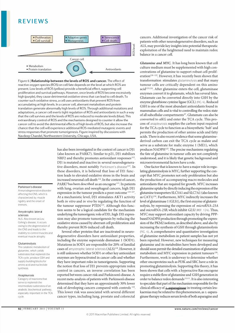
8202019 Regulacion Del Metabolismo en Celula Canceriacutegena
httpslidepdfcomreaderfullregulacion-del-metabolismo-en-celula-cancerigena 1111
76 Budihardjo I I et al 6-Aminonicotinamide sensitizes
human tumor cell lines to cisplatin Clin Cancer Res
4 117ndash130 (1998)
77 Mardis E R et al Recurring mutations found by
sequencing an acute myeloid leukemia genome
N Engl J Med 361 1058ndash1066 (2009)78 Yan H et al IDH1 and IDH2 mutations in gliomas
N Engl J Med 360 765ndash773 (2009)
79 Gross S et al Cancer-associated metabolite
2-hydroxyglutarate accumulates in acute myelogenous
leukemia with isocitrate dehydrogenase 1 and 2
mutations J Exp Med 207 339ndash344 (2010)
80 Ward P S et al The common feature of leukemia-
associated IDH1 and IDH2 mutations is a neomorphic
enzyme activity converting α-ketoglutarate to
2-hydroxyglutarate Cancer Cell 17 225ndash234 (2010)
81 Zhao S et al Glioma-derived mutations in IDH1
dominantly inhibit IDH1 catalytic activity and induce
HIF-1α Science 324 261ndash265 (2009)
82 Dang L et al Cancer-associated IDH1 mutations
produce 2-hydroxyglutarate Nature 462 739ndash744
(2009)
Discovery that driver mutations in IDH1 cause the
acquisition of a novel enzymatic activity and
production of 2-HG
83 Bleeker F E et al IDH1 mutations at residue pR132
(IDH1R132) occur frequently in high-grade gliomas but
not in other solid tumors Hum Mutat 30 7ndash11
(2009)84 Kang M R et al Mutational analysis of IDH1 codon
132 in glioblastomas and other common cancers Int
J Cancer 125 353ndash355 (2009)
85 Giannoni E Buricchi F Raugei G Ramponi G amp
Chiarugi P Intracellular reactive oxygen species
activate Src tyrosine kinase during cell adhesion and
anchorage-dependent cell growth Mol Cell Biol 25
6391ndash6403 (2005)86 Lee S R et al Reversible inactivation of the tumor
suppressor PTEN by H2O2 J Biol Chem 277
20336ndash20342 (2002)
87 Cao J et al Prdx1 inhibits tumorigenesis via
regulating PTENAKT activity EMBO J 28
1505ndash1517 (2009)
88 Gao P et al HIF-dependent antitumorigenic effect of
antioxidants in vivo Cancer Cell 12 230ndash238
(2007)
89 Bell E L Emerling B M amp Chandel N S
Mitochondrial regulation of oxygen sensing
Mitochondrion 5 322ndash332 (2005)90 Ramsey M R amp Sharpless N E ROS as a tumour
suppressor Nature Cell Biol 8 1213ndash1215
(2006)
91 Takahashi A et al Mitogenic signalling and the
p16INK4a-Rb pathway cooperate to enforce
irreversible cellular senescence Nature Cell Biol 8
1291ndash1297 (2006)
92 Garrido C et al Mechanisms of cytochrome c release
from mitochondria Cell Death Differ 13 1423ndash1433
(2006)
93 Han D Antunes F Canali R Rettori D amp
Cadenas E Voltage-dependent anion channels
control the release of the superoxide anion from
mitochondria to cytosol J Biol Chem 278
5557ndash5563 (2003)
94 Fruehauf J P amp Meyskens F L Reactive oxygen
species a breath of life or death Clin Cancer Res
13 789ndash794 (2007)
95 Bae Y S et al Epidermal growth factor (EGF)-induced
generation of hydrogen peroxide Role in EGFreceptor-mediated tyrosine phosphorylation J Biol
Chem 272 217ndash221 (1997)
96 Sundaresan M Yu Z X Ferrans V J Irani K amp
Finkel T Requirement for generation of H2O2 for
platelet-derived growth factor signal transduction
Science 270 296ndash299 (1995)
97 Vaughn A E amp Deshmukh M Glucose metabolism
inhibits apoptosis in neurons and cancer cells by redox
inactivation of cytochrome c Nature Cell Biol 10
1477ndash1483 (2008)
Evidence that redox control by the GSH system is
important in neurons and cancer cells and that
reduction of cytochrome c prevents apoptosis98 Schafer Z T et al Antioxidant and oncogene rescue
of metabolic defects caused by loss of matrix
attachment Nature 461 109ndash113 (2009)
99 Li B Gordon G M Du C H Xu J amp Du W
Specific killing of Rb mutant cancer cells by
inactivating TSC2 Cancer Cell 17 469ndash480 (2010)
Evidence that inappropriate activation of growthand proliferation pathways can lead to excessive
stress and cell death
100 Ozcan U et al Loss of the tuberous sclerosis complex
tumor suppressors triggers the unfolded protein
response to regulate insulin signaling and apoptosis
Mol Cell 29 541ndash551 (2008)
101 Nogueira V et al Akt determines replicative
senescence and oxidative or oncogenic premature
senescence and sensitizes cells to oxidative apoptosis
Cancer Cell 14 458ndash470 (2008)
102 Liu Y et al MnSOD inhibits proline oxidase-induced
apoptosis in colorectal cancer cells Carcinogenesis
26 1335ndash1342 (2005)
103 Liu Y Borchert G L Surazynski A amp Phang J M
Proline oxidase a p53-induced gene targets COX-2
PGE2 signaling to induce apoptosis and inhibit tumor
growth in colorectal cancers Oncogene 27
6729ndash6737 (2008)
104 Liu Y et al Proline oxidase functions as a mitochondrial
tumor suppressor in human cancers Cancer Res 69
6414ndash6422 (2009)
105 Budanov A V Sablina A A Feinstein E
Koonin E V amp Chumakov P M Regeneration of
peroxiredoxins by p53-regulated sestrins homologs of
bacterial AhpD Science 304 596ndash600 (2004)106 Yoon K A Nakamura Y amp Arakawa H Identification
of ALDH4 as a p53-inducible gene and its protective
role in cellular stresses J Hum Genet 49 134ndash140
(2004)
107 Suzuki S et al Phosphate-activated glutaminase
(GLS2) a p53-inducible regulator of glutaminemetabolism and reactive oxygen species Proc Natl
Acad Sci USA 107 7461ndash7466 (2010)
108 Chen W et al Direct interaction between Nrf2 and
p21Cip1WAF1 upregulates the Nrf2-mediated antioxidant
response Mol Cell 34 663ndash673 (2009)
109 Trachootham D Alexandre J amp Huang P Targeting
cancer cells by ROS-mediated mechanisms a radical
therapeutic approach Nature Rev Drug Discov 8
579ndash591 (2009)
110 Trachootham D et al Effective elimination of
fludarabine-resistant CLL cells by PEITC through a redox-
mediated mechanism Blood 112 1912ndash1922 (2008)
111 Trachootham D et al Selective killing of oncogenically
transformed cells through a ROS-mediated
mechanism by β-phenylethyl isothiocyanate Cancer
Cell 10 241ndash252 (2006)
112 Clements C M McNal ly R S Conti B J Mak T W
amp Ting J P DJ-1 a cancer- and Parkinsonrsquos disease-
associated protein stabilizes the antioxidant
transcriptional master regulator Nrf2 Proc Natl Acad Sci USA 103 15091ndash15096 (2006)
113 Gasser T et al Genetic complexity and Parkinsonrsquos
disease Science 277 388ndash389 (1997)
114 Kim R H et al Hypersensitivity of DJ-1-deficient mice
to 1-methyl-4-phenyl-1236-tetrahydropyrindine
(MPTP) and oxidative stress Proc Natl Acad Sci USA
102 5215ndash5220 (2005)
115 Kim R H et al DJ-1 a novel regulator of the tumor
suppressor PTEN Cancer Cell 7 263ndash273 (2005)
Discovery that PARK7 a gene mutated in
Parkinsonrsquos disease is an oncogene
116 Davidson B et al Expression and clinical role of DJ-1
a negative regulator of PTEN in ovarian carcinoma
Hum Pathol 39 87ndash95 (2008)
117 Yuen H F et al DJ-1 could predict worse prognosis
in esophageal squamous cell carcinoma Cancer
Epidemiol Biomarkers Prev 17 3593ndash3602 (2008)
118 Lin M T amp Beal M F Mitochondrial dysfunction and
oxidative stress in neurodegenerative diseases Nature
443 787ndash795 (2006)119 Bajaj A Driver J A amp Schernhammer E S
Parkinsonrsquos disease and cancer risk a systematic
review and meta-analysis Cancer Causes Control 21
697ndash707 (2010)
120 Coles N W amp Johnstone R M Glutamine
metabolism in Ehrlich ascites-carcinoma cells
Biochem J 83 284ndash291 (1962)
121 Reitzer L J Wice B M amp Kennell D Evidence that
glutamine not sugar is the major energy source for
cultured HeLa cells J Biol Chem 254 2669ndash2676
(1979)
122 Eagle H Nutrition needs of mammalian cells in tissue
culture Science 122 501ndash514 (1955)
123 Wise D R et al Myc regulates a transcriptional
program that stimulates mitochondrial glutaminolysis
and leads to glutamine addiction Proc Natl Acad Sci
USA 105 18782ndash18787 (2008)
124 Gao P et al c-Myc suppression of miR-23ab enhances
mitochondrial glutaminase expression and glutamine
metabolism Nature 458 762ndash765 (2009)Strong mechanistic evidence that MYC participates
in promoting mitochondrial glutaminase activity
125 DeBerardinis R J et al Beyond aerobic glycolysis
transformed cells can engage in glutamine metabolism
that exceeds the requirement for protein and
nucleotide synthesis Proc Natl Acad Sci USA 104
19345ndash19350 (2007)
126 Gallagher F A Kettunen M I Day S E
Lerche M amp Brindle K M 13C MR spectroscopy
measurements of glutaminase activity in human
hepatocellular carcinoma cells using hyperpolarized
13C-labeled glutamine Magn Reson Med 60
253ndash257 (2008)
127 Richards N G amp Kilberg M S Asparagine
synthetase chemotherapy Annu Rev Biochem 75
629ndash654 (2006)
128 Reinert R B et al Role of glutamine depletion in
directing tissue-specific nutrient stress responses to
L-asparaginase J Biol Chem 281 31222ndash31233
(2006)
129 Lunt S J Chaudary N amp Hill R P The tumor
microenvironment and metastatic disease Clin Exp
Metastasis 26 19ndash34 (2009)
130 Bristow R G amp Hill R P Hypoxia and metabolism
Hypoxia DNA repair and genetic instability Nature
Rev Cancer 8 180ndash192 (2008)
131 Semenza G L Regulation of cancer cell metabolism
by hypoxia-inducible factor 1 Semin Cancer Biol 19
12ndash16 (2009)
132 Denko N C Hypoxia HIF1 and glucose metabolism
in the solid tumour Nature Rev Cancer 8 705ndash713(2008)
133 Wouters B G amp Koritzinsky M Hypoxia
signalling through mTOR and the unfolded protein
response in cancer Nature Rev Cancer 8 851ndash864
(2008)
134 Koritzinsky M et al Gene expression during acute
and prolonged hypoxia is regulated by distinct
mechanisms of translational control EMBO J 25
1114ndash1125 (2006)
135 Rouschop K M amp Wouters B G Regulation of
autophagy through multiple independent hypoxic
signaling pathways Curr Mol Med 9 417ndash424
(2009)
136 Koumenis C et al Regulation of protein synthesis by
hypoxia via activation of the endoplasmic reticulum
kinase PERK and phosphorylation of the translation
initiation factor eIF2α Mol Cell Biol 22 7405ndash7416
(2002)137 Bi M et al ER stress-regulated translation
increases tolerance to extreme hypoxia andpromotes tumor growth EMBO J 24 3470ndash3481
(2005)
138 Romero-Ramirez L et al XBP1 is essential for
survival under hypoxic conditions and is required
for tumor growth Cancer Res 64 5943ndash5947
(2004)
139 Cairns R Papandreou I amp Denko N Overcoming
physiologic barriers to cancer treatment by
molecularly targeting the tumor microenvironment
Mol Cancer Res 4 61ndash70 (2006)
140 Moreno-Sanchez R Rodriguez-Enriquez S
Marin-Hernandez A amp Saavedra E Energy
metabolism in tumor cells FEBS J 274
1393ndash1418 (2007)
141 Yang C et al Glioblastoma cells require glutamate
dehydrogenase to survive impairments of glucose
metabolism or Akt signaling Cancer Res 69
7986ndash7993 (2009)
142 Sonveaux P et al Targeting lactate-fueled respiration
selectively kills hypoxic tumor cells in mice J ClinInvest 118 3930ndash3942 (2008)
AcknowledgementsThe authors would like to thank M Saunders for scientific
editing of the Review
Competing interests statementThe authors declare no competing financial interests
DATABASESNational Cancer Institute Drug Dictionary
httpwwwcancergovdrugdictionary
l-asparaginase
Pathway Interaction Database httppidncinihgov
AMPK | HIF1 | HIF2 | LKB1 | mTOR | MYC | p53 | PI3K
FURTHER INFORMATIONTak W Makrsquos homepage
httpmedbioutorontocafacultymakhtml
ALL LINKS ARE ACTIVE IN THE ONLINE PDF
R E V I E W S
NATURE REVIEWS | CANCER VOLUME 11 | FEBRUARY 2011 | 95
copy 2011 Macmillan Publishers Limited All rights reserved