regional changes in collagen fiber alignment may identify ... · regional changes in collagen fiber...
TRANSCRIPT
INJURY BIOMECHANICS RESEARCH Proceedings of the Thirty-Sixth International Workshop
Regional Changes in Collagen Fiber Alignment May Identify the Onset of Damage in the Facet Capsular Ligament
K. P. Quinn and B. A. Winkelstein
This paper has not been screened for accuracy nor refereed by any body of scientific peers and should not be referenced in the open literature.
ABSTRACT Detecting the initiation of mechanical injury of soft tissue, and not only its ultimate failure, is critical to enable a sensitive and specific characterization of tissue tolerance, to develop quantitative relationships between macro- and micro-structural tissue responses, and to appropriately interpret physiological responses to loading. We have developed a novel methodological approach to detect the onset and spatial location of structural damage in collagenous soft tissue before its visible rupture, via the identification of atypical regional collagen fiber kinematics that were produced during loading. Our methods utilize high-speed quantitative polarized light imaging and identify the onset of tissue damage in ligament regions where the mean collagen fiber rotation significantly deviates from its behavior during previous noninjurious loading. This technique was validated by its ability to correctly predict the location of visible rupture (p<0.001). The proposed fiber rotation-based metric identifies potential facet capsular ligament damage beginning well-before rupture, at 51±12% of the displacement required to produce tissue failure. While traditional macro-scale strain metrics fail to identify the location of microstructural damage, detection determined by altered fiber rotation is significantly correlated (R=0.757; p=0.049) with the occurrence of tissue yield, supporting the capabilities of this method. Damaged regions also exhibit higher variance in fiber direction compared to undamaged regions (p=0.041). These methods enable the characterization of a previously undefined class of mechanical injuries to the facet capsule, and have potential utility to define more refined injury tolerances and to provide region-specific and fiber kinematic data for finite element and tissue-level models.
INTRODUCTION issue injury resulting from mechanical trauma has traditionally been defined by gross measures of mechanical failure and/or evidence of a visible tissue rupture (Kliewer et al., 1993; Siegmund et al.,
2001). However, these conventional indicators of mechanical injury may not actually identify the tolerance of a particular tissue to its injury. Subfailure loads can produce a variety of altered mechanical phenomena in ligament and tendon, including increased laxity (Iatridis et al., 2005; Panjabi et al., 1996; Pollock et al., 2000; Provenzano et al., 2002), decreased stiffness (Panjabi et al., 1999; Provenzano et al., 2002; Quinn et al., 2007), and altered viscoelastic responses (Panjabi et al., 1999). These mechanical responses are also coupled
T

Regional Changes in Collagen Fiber Alignment May Identify the Onset of Damage in the Facet Capsular Ligament
2
with the onset of pathophysiological conditions, such as collagen disorganization (Gimbel et al., 2004; Quinn et al., 2007), fibroblast necrosis (Provenzano et al., 2002), and nociceptor activation (Lu et al., 2005). Although these studies collectively identify a host of mechanical and physiological changes in soft tissue for particular subfailure loading cases, they cannot directly identify the initiation of local microstructural tissue damage. This inability to directly detect or localize injury could result in the mischaracterization of injury thresholds, and/or focus efforts for preventing or treating tissue pathology in the wrong anatomical regions of tissues.
Owing to the inability to visualize and identify subfailure damage as it occurs, macro-scale strain metrics such as maximum principal strain are commonly used to establish injury criteria and tolerances, and to locate the site of tissue injury (Bain and Meaney, 2000; Gefen et al., 2008; Lee et al., 2004; Lu et al., 2005; Siegmund et al., 2001; Winkelstein et al., 2000). However, macroscopic strain fields in ligaments and tendons may lack the sensitivity to localize either microstructural damage or even gross rupture, due to collagen fiber movement or high spatial variability in the strain field (DeFrate et al., 2006; Phatak et al., 2007; Quinn et al., 2007; Screen et al., 2004). Therefore, detecting the initiation of soft tissue damage during mechanical loading, rather than estimating strains or the resultant structural effects of injurious loading, can enable the sensitive and specific characterization of tissue injury tolerance and the development of integrative mechanistic relationships between tissue loading, fiber-level mechanics, physiologic responses, and the interactions among these.
A variety of non-invasive imaging techniques have quantified tissue microstructure or fiber alignment, including optical coherence tomography, electron microscopy, X-ray diffraction, diffusion tensor magnetic resonance imaging, and nonlinear optical microscopy (Hadian et al., 2007; Hansen et al., 2002; Hurschler et al., 2003; Matcher et al., 2004; Sasaki et al., 1999; Wedeen et al., 2001; Williams et al., 2005; Yahia et al., 1990). While these techniques allow acquisition and quantification of a variety of microstructural components at different loading conditions, few of them can facilitate a continuous assessment of fiber alignment during loading. Polarized light has been used to determine fiber organization, kinematics, and crimp patterns in soft tissues such as tendon, ligament, and heart valve by exploiting the natural birefringence of collagen (Dickey et al., 1998; Thomopoulos et al., 2003, Tower et al., 2002; Tower and Tranquillo, 2001a, b; Whittaker and Canham, 1991). Tower et al. (2002) developed a quantitative polarized light imaging (QPLI) system capable of creating collagen fiber alignment maps for soft tissue during continuous loading. While that work demonstrated complex fiber realignment during tissue loading up to its rupture, no study has used QPLI or any other polarized light technique to specifically detect and/or quantify the initial occurrence of microstructural damage during ligament loading.
QPLI provides the ideal approach to identify microstructural damage by evaluating entire regions of the ligament surface area during loading, via the acquisition of continuous fiber information with the potential for pixel-level resolution. This technique is ideally suited to determine the preferred fiber direction in any relatively planar tissue through which light can be transmitted and for which linear birefringence dominates the optical response. Given that mechanical injury to the capsular ligament of the facet joint has been proposed to occur during whiplash and other subfailure loading scenarios for the spine (Cavanaugh et al., 1996; Lee et al., 2006; Lu et al., 2005; Pearson et al. 2004; Winkelstein et al., 2000; Yang and King, 2003), this ligament is an ideal tissue to study the use of QPLI to localize subfailure microstructural tissue damage. Despite many anatomic, histological, and biomechanical studies characterizing the mechanical and anatomical responses of the cervical facet capsule (Cavanaugh et al., 1996; Deng et al., 2000; Lu et al., 2005; Pearson et al. 2004; Quinn et al., 2007; Siegmund et al., 2001; Winkelstein et al., 2000; Yang and King, 2003), there are currently no reports detailing the fiber orientation of the human facet capsular ligament. Capsuloligamentous material, due to its functional demands, generally demonstrates greater spatial variability in fiber direction compared to more organized cable-like ligaments; the planar geometry of the human facet capsule makes it more amenable to the QPLI technique than other more organized ligaments. Our overall hypothesis is that damage to facet capsule tissue can be detected before visible gross rupture of the ligament occurs, by quantifying the regional fiber kinematics of the collagen matrix during loading. As such, the primary goal of this study was to develop optical injury detection methods and metrics using QPLI that are capable of identifying structural damage in the human facet capsular ligament. Specifically, fiber alignment was quantified and summarized in different regions covering the lateral face of the ligament during its tensile loading. Damage was identified by significant increases in the rotation of the regional mean fiber
Injury Biomechanics Research
directions, and validation of this method of damage detection was also performed through a direct comparison to the location of visible tissue rupture. The loading conditions and location at which damage was first detected were also compared with more conventional mechanical measures of injury, including tissue yield and macro-scale strain fields.
METHODS Isolated right and left facet joints (n=8) were removed en bloc from the C4/C5 spinal motion
segments of five fresh, unembalmed human cadavers (mean age: 59±12.8 years). Through fine dissection, all musculature and tendon insertions on the surface of the facet capsules were removed. The posterior side of the lateral aspect of the facet capsule was isolated for testing and all other regions of the capsule were transected. Portions of the superior and inferior articular processes of the joint were removed to facilitate the transmission of polarized light through the ligament tissue. In addition, an array of 15-24 fiduciary markers was applied to the ligament using a 0.2mm diameter felt-tip pen to track tissue deformation and estimate strain fields during loading. The bony ends of the specimens were cast in testing cups using dental stone, and mounted to an Instron 5385 testing machine (Instron Corporation, Norwood, MA). Prior to mechanical testing, sample width and thickness were measured with digital calipers, and the average cross sectional area was calculated assuming a rectangular shape for the unloaded tissue. To provide a consistent initial position for all samples, a 5 kPa preload was applied to establish the initial joint displacement for each specimen. Specimens were preconditioned with 30 cycles of tensile loading between 0 and 0.5 mm (producing loads less than 5% of the failure load for the facet capsule). After preconditioning, specimens were distracted in tension at 0.5 mm/s until gross failure of the ligament was produced. Load and displacement data were collected at 1 kHz.
Integrated QPLI mechanical testing system A QPLI system, enabling the simultaneous acquisition of mechanical, fiber alignment, and strain
data, was assembled based on the design reported by Glazer et al. (1996) and Tower et al. (2002). It was modified to operate and interface with the Instron testing machine. For the system in this study, a NEMA 17 stepper motor, driver and controller system (Lin Engineering, Santa Clara, CA) rotated a 20 cm cast acrylic disk equipped with a linear polarizing laminated film (Edmund Optics Inc., Barrington, NJ). A fiber optic illuminator with focusing lens (Edmund Optics Inc., Barrington, NJ) provided a light source behind the rotating polarizer to transmit light through the ligament. A circular analyzer was constructed using a Mica quarter-wave plate (Optosigma Corp., Santa Ana, CA) and linear polarizing film to analyze the polarized light transmitted through the ligament. The circular analyzer was aligned and mounted to a 6X macro zoom lens and a high-speed CCD camera (Vision Research Inc., Wayne, NJ).
A two camera system (Phantom v5.1 & Phantom v4.3; Vision Research, Inc., Wayne, NJ) was used
to collect polarized light data and was synchronized with the acquisition of mechanical data. One CCD camera imaged the ligament deformation and acquired polarized light data, while the second CCD camera monitored the position of the rotating polarizer. Both CCD cameras collected images with a field of view of 17.5 mm by 36 mm and 11 pixel/mm resolution; light intensity was stored with 8-bit resolution. All images were acquired at 500 fps while the linear polarizer rotated at 750 rpm, producing a set of 20 images as the polarizer rotated through 180˚ every 40 milliseconds. Each set of 20 images was used to create a single map of fiber alignment corresponding to every 0.02 mm of distraction.
Mechanical data analysis Gross failure was defined to occur at the maximum force recorded, and the corresponding
displacement and energy to failure were also measured at this point for each specimen. To provide the most conservative detection of a loss of microstructural integrity using the force-displacement data, the first occurrence of ligament yielding was defined based on a decrease in stiffness (Quinn and Winkelstein, 2007; Yoganandan et al., 1989). Tangent stiffness was calculated at each data point using a centered finite difference approximation (Quinn and Winkelstein, 2007):

Regional Changes in Collagen Fiber Alignment May Identify the Onset of Damage in the Facet Capsular Ligament
4
11
11
−+
−+
−−
=ii
iii
FFkδδ
,
where the stiffness (ki) at a given point i, was calculated from the difference in force (F) and displacement (δ) between the previous (i–1) and following (i+1) data points. Ligament yield was defined as the point when the tangent stiffness first began decreasing by at least 10% of its peak value during distraction (Quinn and Winkelstein, 2007). The force and displacement at yield, and the energy to yield, were also recorded for comparison to the collagen fiber data.
Planar tissue deformation during distraction was quantified using the fiduciary markers on the
capsule surface in the QPLI images. Marker locations were digitized and tracked during tissue loading, and the capsule surface was divided into regions by constructing four-node elements from the fiduciary marker locations (Figure 1). Through isoparametric mapping, Lagrangian strain was computed for each element for every 0.02 mm of distraction. The element(s) with the maximum principal strain and maximum shear strain were noted in each image.
Figure 1: Image of a typical specimen (C390-C45L) showing element definitions and corresponding principal strain field at the initial detection of damage. Based on fiduciary markers (shown in the image on the left), principal strain was computed within four-node elements. Maximum principal strain at the initial detection of damage in this specimen was 38% and located at element 7 (labeled 7 on the right).
Collagen fiber analysis & damage detection To analyze the fiber alignment in the ligament tissue, harmonic analysis was employed to generate
alignment maps (Tower and Tranquillo, 2001b) over the full range of the ligament’s distraction. The intensity of each pixel in any set of consecutive images was described by the harmonic relationship:
( ) ( )iii CBAI θθθ 2sin2cos)( ++= ,
where θi indicates the polarizer rotation angle with respect to the horizontal at each interval, i. A represents the mean intensity, and B and C are the signed harmonic amplitudes. The Fourier coefficients, A, B, and C, were determined using a summation approximation and scaled by pixel intensity (Tower et al., 2002; Tower and Tranquillo, 2001b). Coefficients B and C were used to calculate the retardation (δ) of light, a measure of

Injury Biomechanics Research
the strength of fiber alignment through the tissue thickness, and the fiber alignment direction (α) at each pixel using the following equations (Geday et al., 2000; Glazer et al., 1996; Tower et al., 2002):
( )221 1cos CB −−= −δ ,
( )CB −= − /tan21 1α .
To assess the accuracy of the fiber alignment direction (α) measurements, ligament tissue with a
known visible collagen fiber direction was compared to the QPLI analysis. Specifically, a 60-micron thick section of caprine anterior cruciate ligament tissue was imaged by the QPLI system. Using the normal bright-field images for that specimen, any visible fiber direction on the tissue was digitized (Figure 2). Pixels within the bright-field image that were assigned a digitized fiber direction were then compared to the fiber direction measured by the QPLI system, and the mean error in fiber direction was computed (Figure 2). The mean error in fiber direction (α) is related to the magnitude of the signed harmonic coefficients (B and C); pixels with insufficient light intensity amplitude will produce a highly variable, inaccurate fiber direction. To remove the effect of these low-amplitude pixels on the remainder of the directional analyses, the intensity amplitude corresponding to a mean error in direction exceeding 9˚ was computed, and any pixels determined to have amplitudes lower than this value were not included in the subsequent fiber direction-based analysis of microstructural damage.
Figure 2: Error analysis of the QPLI fiber direction measurements. (a) Image showing where the visible fiber direction of a caprine anterior cruciate ligament tissue section was digitized (green lines). (b) Overlay of those digitized fiber directions and the direction of the collagen fibers derived from the QPLI system (magenta lines) at those pixels.
Regional increases in the mean fiber rotation were used to identify the occurrence and location of
structural damage in the ligament. Ligament regions for collagen fiber analysis were defined by the same four-node elements used in the strain analysis (Figure 1). Within each element, the mean fiber direction, variance in direction, and mean retardation were each computed based on circular statistics for every 0.02 mm increment during the distraction of each specimen. The mean fiber direction of each element was filtered using a 41st order generalized Butterworth filter with a 2.5 Hz cutoff frequency, and then differentiated with respect to displacement using a centered finite difference approach to quantify the rotation of the mean fiber direction within each element during loading.
Structural damage was defined to occur in an element when the fiber rotation in that element
exceeded its previous mean response by four standard deviations. For each element at a given displacement, the measurement of standard deviation was based on the distribution of all rotation data from the start of loading to that displacement. These region- and displacement-specific measures of standard deviation were calculated based on the assumption that the mean fiber rotation was not biased towards clockwise or counter-
(a) (b)

Regional Changes in Collagen Fiber Alignment May Identify the Onset of Damage in the Facet Capsular Ligament
6
clockwise rotation, and so, had a normal distribution about a mean of zero. This assumption was validated through a z-test using rotation data from all elements and assured that only an increase in the magnitude of rotation would be detected as damage. Damage was defined to occur when the mean fiber rotation of any element first exceeded four standard deviations (p<0.001), which minimized the detection of false-positives as the number of data points increases with increasing distraction. The displacement, force, and energy at the initial detection of damage were recorded, and each element of the ligament in which damage was detected was noted. The standard deviation measures for each region at the point of initial damage detection were used to detect additional damage during further loading up to and including gross failure.
Statistical analysis The displacements at which initial damage, yield, and failure occurred were compared to each other
using a one-way ANOVA and post-hoc Bonferroni corrections to determine whether each of these events occur at different distractions. To validate the ability of our rotation-based damage metric to localize damage, the elements identified as damaged at gross failure were compared with the elements where rupture was first visible from the video data, using a Fisher’s exact test. To further determine the effectiveness of this damage metric, the strength of association between the detection of damage at failure and evidence of visible rupture was estimated by computing the odds ratio of successfully classifying an element as damaged or undamaged. At the displacement where initial damage was detected for each specimen, fiber alignment and strain measurements were compared between damaged and undamaged elements to evaluate other metrics as having the potential to identify damage. The absolute value of fiber rotation, variance in fiber direction, mean retardation, the offset of the mean fiber direction from specimen loading direction, principal strain, and maximum shear strain were compared. For each of these outcomes, significant differences between damaged and undamaged elements, and between specimens, were determined through an ANOVA with elements nested within specimens. Significance for each ANOVA was defined by p<0.05; all tests were performed using JMP 7 (SAS Institute Inc., Cary, NC).
RESULTS The average width and thickness of the eight capsular ligament specimens were 7.46±1.38 mm and
0.43±0.92 mm, respectively, corresponding to a mean cross-sectional area of 3.2±0.9 mm2. At the reference displacement, an average of 11.0±2.1 elements covered a surface area of 26.3±12.1mm2 in each ligament midsubstance. Gross structural failure of the ligament was detected at 21.62±8.96 N and 3.67±0.49 mm, and the energy required for failure was 28.99±14.10 mJ (Table 1; Figure 3). Ligament yield was first detected at 1.81±0.65 mm of distraction, and was significantly lower (p<0.001) than the displacement at failure. The displacement at yield corresponded to a force of 8.31±6.70 N, and a mean energy of 4.57±4.22 mJ (Table 1). In four specimens, rupture was visible after tissue failure within the elements covering the ligament midsubstance where collagen fiber alignment was being measured using the QPLI system. However, two other specimens (C846-C45R & C500-C45R) ruptured in the midsubstance just superior or inferior to the element regions measured by QPLI, and another specimen (C947-C45R) ruptured near the ligament insertion to the C5 bone. For an additional specimen (C457-C45L), the ligament remained intact over the entire imposed loading, but structural failure occurred as a fracture in the C5 articular bone. No ruptures were visible from the video data at yield for any specimen.
The mean error in collagen fiber direction measurements was -0.59±6.12˚ for the QPLI system.
However, this mean error exceeded 9˚ when the peak-to-peak amplitude of light intensity at a pixel was less than six (out of 255) in our system. Pixels with amplitudes less than six were not assigned a fiber direction and were excluded from analysis. Excluded pixels generally corresponded to regions of poor light transmission (Figure 4); however, the QPLI system was able to assess an average of 89.5±3.9% of the midsubstance area for all specimens.

Injury Biomechanics Research
Table 1. Summary of tissue mechanics at failure, yield, and the initial detection of damage. Force, displacement, and energy were determined for each event.
Failure d Yield d Detected Damage _
Specimen Force
(N) Disp. (mm)
Energy (mJ)
Force (N)
Disp. (mm)
Energy (mJ)
Force (N)
Disp. (mm)
Energy (mJ)
C390-C45L 25.43 4.13 34.87 13.84 2.92 9.93 9.03 2.51 5.33 C457-C45R 34.69 3.96 59.03 12.73 1.70 5.47 15.52 1.87 7.85 C846-C45L 27.04 3.23 26.62 19.54 2.61 11.51 11.15 2.18 4.95 C846-C45R 16.84 4.31 31.44 3.33 1.41 1.36 4.16 1.62 2.13 C947-C45R 11.60 3.59 15.93 1.49 1.18 0.57 4.96 2.12 3.39 C500-C45L 14.19 3.74 23.45 6.16 1.91 3.86 4.97 1.69 2.65 C500-C45R 12.37 3.55 13.57 0.44 1.10 0.15 0.67 1.30 0.26 C457-C45L* 30.76 2.81 27.04 8.91 1.64 3.75 N/A N/A N/A
Mean 21.62 3.67 28.99 8.31 1.81 4.57 7.21 1.90 3.80 SD 8.96 0.49 14.10 6.70 0.65 4.22 5.00 0.41 2.48
* bone fractured at failure, no ligament rupture noted.
Figure 3: Structural response of specimen C390-C45L during tensile distraction to failure. The maximum force value on the force-displacement trace (blue line) defined gross failure of the specimen. The stiffness-displacement curve (red line) was used to determine when yield occurred. The initial loss of stiffness at yield (2.92 mm) is ultimately recovered with increasing distraction. Initial damage was detected by fiber rotation at 2.51 mm in this specimen is indicated here by the black dashed line.
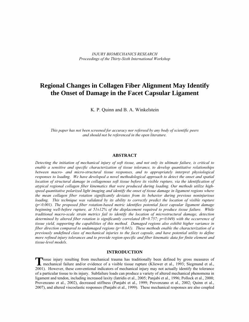
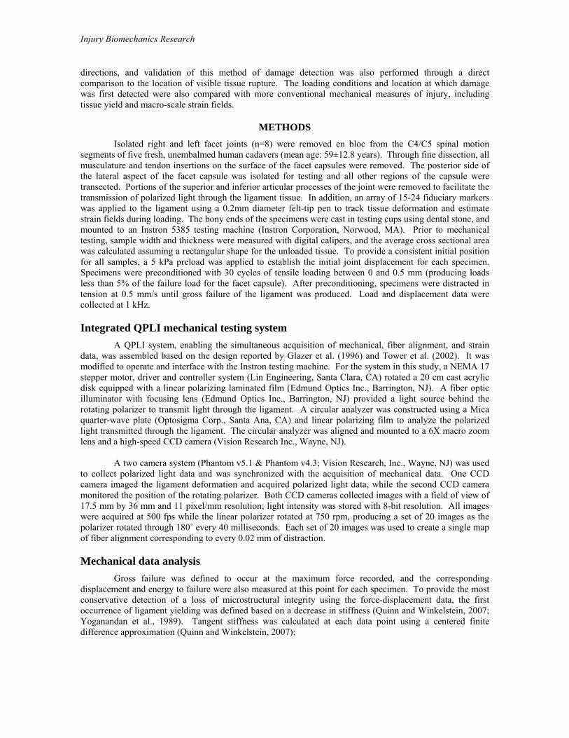
The use of increased fiber rotation as a metric of damage was validated based on its ability to predict the location of visible tissue rupture noted after structural failure. For all four specimens in which rupture initiated within an element being analyzed by QPLI, the fiber rotation-based metric also detected damage in that element at tissue failure (Figure 4). Of the four other specimens in which rupture occurred outside of the elements measured by the QPLI system, damage was detected in an element only once at failure. The damage detection data from all of the elements monitored from all specimens at failure were placed in a contingency table, and the detected location of damage at failure was found to be significantly associated with the location of visible tissue rupture (p<0.001). The odds of an element being correctly identified by
Damage
Yield
Failure
Regional Changes in Collagen Fiber Alignment May Identify the Onset of Damage in the Facet Capsular Ligament
8
our damage metric as one where rupture would or would not occur were 25.7:1, suggesting a strong association between those two measures.
The rotation-based metric identified initial damage at a mean displacement of 1.90±0.41 mm for 7
of the 8 specimens. No ligament damage was detected at any point during loading in one specimen (C457-C45L), which is consistent with that specimen not having ligament rupture at failure. Initial damage was detected to occur significantly before gross failure (p<0.001), but not before yield. The displacements at which damage and yield were detected were significantly correlated (R=0.757; p=0.049). The mean values for force at initial damage (7.21±5.00 N) and energy required to produce damage (3.80±2.48 mJ) were lower than those for yield.
Figure 4: Fiber alignment maps of specimen C457-C45R at failure (a) and 0.4 mm after its failure (b). (a) At the point of failure (3.96 mm), three elements were identified as damaged (indicated by arrows), based on the rotation of their mean fiber direction. (b) Following additional ligament distraction, rupture was clearly visible as a large hole in one of those elements damaged at failure in (a). Rupture was not visible in the two other elements identified in (a), but the development of the indicated hole in (b) may have prevented the microstructural damage detected in these other elements at failure from propagating into visible ruptures.
The elements detected as damaged by significant mean fiber rotation also exhibited fiber alignment
properties that were different from the undamaged elements. Damaged elements exhibited significantly larger (p=0.003) fiber rotation values (102.0±66.3˚/mm) compared to their undamaged counterparts (29.4±46.5˚/mm) (Table 2). These damaged elements also demonstrated a partial reorganization of fiber alignment that persisted during further loading (Figure 5). For each specimen at the first detection of damage, the variance in fiber direction (Table 2) was significantly greater (p=0.041) in the damaged element (mean variance of 0.827±0.074) relative to the other elements that were deemed undamaged (mean variance of 0.472±0.238). While the variance was higher in the damaged elements, the spatial distribution of fiber directions was not random, but rather due to different fiber populations being oriented in nearly orthogonal directions (Figure 5).
While fiber alignment in the damaged and undamaged elements was different, retardation and the
macro-scale strain metrics were not different between these two groups of elements. Specifically, mean retardation was not significantly different between elements initially identified as damaged (13.8±4.2˚) and those not damaged (12.5±6.1˚) (Table 2). The mean values of principal strain (18.2±11.1%) and maximum shear strain (14.0±6.1%) in the damaged elements were not significantly different from those of the non-damaged elements (22.9±17.0% and 15.8±7.8%, respectively). Furthermore, the location of neither the maximum principal strain nor the maximum shear strain corresponded to elements with initial damage for
Damage detected at failure
(a) 3.96 mm
Hole in tissue (i.e. rupture)
(b) 4.36 mm

Injury Biomechanics Research
any specimen (Figures 1 & 5). At failure, the locations of maximum principal and shear strains matched where visible rupture was evident in only two of the eight specimens.
Table 2. Summary of strain and fiber measurements from damaged and undamaged elements at initial detection of damage.
Outcome Damaged Elements Undamaged Elements
Number of elements 7 73
Fiber rotation * 102.0±66.3˚/mm 29.4±46.5˚/mm
Variance in direction * 0.827±0.074 0.472±0.238
Offset of mean direction from specimen loading 42.9±34.5˚ 43.1±25.1˚
Mean retardation 13.8±4.2˚ 12.5±6.1˚
Principal strain 18.2±11.1% 22.9±17.0%
Maximum shear strain 14.0±6.1% 15.8±7.8%
* denotes significant difference
Figure 5: Fiber alignment maps of specimen C390-C45L during (a) and after (b) the initial detection of damage. (a) Collagen fiber alignment exhibited great spatial variability upon the initial detection of damage. The damaged element is indicated by arrow and the sub-region where the majority of fiber realignment occurred is magnified (2.5x) in the inset. (b) After an additional 0.4 mm of distraction, the element where damage was initially detected in (a) had undergone substantial realignment that persisted until failure. The same sub-region as in (a) is blown up in the inset showing a shift in the alignment of fibers towards the

Regional Changes in Collagen Fiber Alignment May Identify the Onset of Damage in the Facet Capsular Ligament
10
horizontal. At 2.91 mm, a second element (highlighted in yellow) is also detected as damaged (top left of (b)); yield was detected at 2.92 mm of distraction for this specimen (see Table 1).
CONCLUSIONS This study uses quantitative polarized light imaging techniques to localize damage in ligament tissue
that has been otherwise undetectable, and this damage occurs during loading well-below tissue failure (Figure 3, Table 1). The initial detection of damage is significantly correlated (R=0.757) with the occurrence of yield (defined by a decrease in stiffness), which supports that this damage detection metric is sensitive to a loss of structural integrity even at loading magnitudes well-below failure or the onset of visible rupture of the tissue. The demonstration of significantly altered fiber kinematics near or before the yielding of the ligament (Figure 3, Table 1) also suggests that identifying the occurrence of yield may provide more appropriate estimates of structural tolerance than that provided by gross failure. While there appears to be a relationship between regional fiber kinematics and damage, the element where initial damage was identified did differ from the site of maximum principal strain (Figures 1 & 5). Although the onset of damage was correlated to the macro-scale mechanical response of yield, the discrepancy between strain and damage detection in this study suggests that traditional macro-scale strain measurements may not be suitable to localize subfailure damage or be appropriate as a tolerance criterion for structural injury in this ligament.
Damage to the collagen extracellular matrix and the ensuing effects of such damage on both the
mechanical and cellular responses of that tissue have been reported for subfailure loading of ligament and tendon. Provenzano et al. (2002, 2005) reported that non-recoverable laxity in rat medial collateral ligaments was initiated at slightly less than half the strain required to produce tissue failure, and that this type of loading also corresponded to a decrease in cell viability and an initiation of fibroblast-mediated remodeling. Laxity has also been noted in other soft tissues as a result of some subfailure loading conditions (Iatridis et al., 2005; Panjabi et al., 1996; Pollock et al., 2000), and changes in ligament stiffness or tangential modulus have also been reported (Panjabi et al., 1996, 1999; Provenzano et al., 2002). The detection of damage via changes in collagen fiber rotation at 51±12% of the displacement for gross failure (Table 1) in our study further supports the hypothesis that collagen fiber damage may produce non-recoverable laxity, joint instability, and the initiation of collagen matrix remodeling. Although mechanistic studies are required to understand the putative nociceptive and inflammatory responses that follow the initiation of damage in the facet capsule, the percentage (12.65±6.69%) of the failure energy required for damage in the current study is sufficient to also produce yield, sustained modifications in the collagen fiber alignment of the rat facet capsule, and persistent pain symptoms (Quinn et al., 2007; Quinn and Winkelstein, 2007). Together, our previous reports and these current findings imply that the increases in the rotation of collagen fibers observed during loading in the current study may be sufficient to produce non-recoverable damage and sustained physiological consequences, such as persistent pain. This study provides a novel method to determine the loading conditions, location, and potentially fiber-based mechanisms that can produce collagen fiber injury, which ultimately may enable a more refined interpretation of the ensuing physiological consequences associated with structural damage in ligament.
The current fiber kinematic data suggest a number of potential mechanisms that can lead to
structural damage, ligament yield, and ultimately to potential permanent physiologic dysfunction. The significant correlation between yield and evidence of altered fiber kinematics during initial damage suggests these two metrics may be different measurements of the same injury and evidence of microstructural damage manifesting itself in the macro-scale stiffness response observed as yield. Since altered fiber kinematics were initially identified prior to yield in three specimens (Table 1), our fiber rotation-based damage metric may be more sensitive for detecting damage than yield in loading conditions where continued fiber recruitment can offset the putative micro-failures that have been hypothesized to produce yield (Tower et al., 2002; Yoganandan et al., 1989). A previous report demonstrating the realignment of collagen fibers in engineered constructs at the beginning of yielding (Tower et al., 2002) further supports this assertion. The regional increases in the rotation of collagen fiber direction during the detection of damage found in our study (Figure 5) may be evidence of the formation of micro-tears in localized regions of the collagen matrix. Increases in tissue stiffness were also noted after the initial detection of damage and yield (Figure 3), which suggests such initial micro-tears could result from the breaking of fiber cross-links before the ultimate failure of the load-bearing collagen fibers. While the mean fiber direction in some regions of our samples did not
Injury Biomechanics Research
rotate towards the direction of loading even after damage (Figure 5), non-affine fiber kinematics have also been demonstrated following loading to high strains in other planar soft tissues (Billiar and Sacks, 1997). Non-affine fiber network models have previously been implemented to describe the mechanical response of collagen constructs (Chandran and Barocas, 2006; Thomopoulos et al., 2007) and may help to further define the complex relationship between fiber kinematics and tissue loading for this capsular ligament. The present data, nonetheless, demonstrate a complex fiber kinematic response to joint distraction that will be important in understanding how potentially injurious loading to the collagen network may impose abnormal forces on the fibroblasts and/or afferent pain fibers in this ligament.
This study is the first to our knowledge to demonstrate the integrated acquisition of continuous fiber
alignment information during loading across an entire ligament midsubstance. In order to rapidly acquire microstructural data spanning a surface with substantial cross-sectional area, fiber alignment was defined at individual pixels. As a result, the orientation and position of individual collagen fibers could not be imaged, but regions of the tissue were used instead for analysis. Accordingly, for this study, damage and strain were localized to elements based on an array of fiduciary markers (Figure 1). An increase in the spatial density of those markers or advanced image registration techniques could help to further evaluate and refine the accuracy and precision in defining the location of damage using this fiber rotation-based detection technique. While the acquisition of fiber alignment data was limited to two dimensions in this study, the retardation data suggest that the extent of fiber alignment through the thickness of this tissue may not be different between damaged and undamaged regions of the tissue (Table 2). The incorporation of retardation data into the damage metric in the future may help to refine its accuracy in detection, but sample thickness variation will need to be accounted for due to its effect on the retardation measurements. Complementary electron microscopy studies capturing evidence of modified organization in the capsular ligament microstructure are needed to determine the severity and the underlying mechanisms of the microstructural failure that is detected here. While this study reports results from a small sample size, the damage detected at gross ligament failure propagated into obvious tissue tears in all four specimens that failed within the elements covering the midsubstance of the ligament (Figure 4). Including the 88 total elements from all specimens, the high odds ratio for our fiber rotation-based metric to correctly identify the location of rupture within the analyzed element regions demonstrates a strong association between that metric and gross tissue damage, and may actually underestimate that association given that damage was detected in multiple regions at failure for some specimens (Figure 4). The propagation of micro-tears into visible rupture likely depends on the severity of the initial damage and the alignment of the collagen matrix surrounding that damage. Given the spatial variability of the collagen alignment in this tissue (Figure 5), the simultaneous development of visible ruptures from all regions detected as being damaged at failure would not be expected. By utilizing QPLI data in the development of a collagen fiber rotation-based metric for damage, this study helps lay a framework to define mechanistic relationships between tissue microstructure, deformation, and structural damage, and suggests strain thresholds for injury may need to be modified to reflect regional-dependence or functionalized to incorporate measures of the underlying microstructure.
Strain is a common metric used to localize injury and to define tissue tolerances (Bain and Meaney,
2000; Cater et al., 2006; Deng et al., 2000; Gefen et al., 2008; Lee et al., 2004; Lu et al., 2005; Pearson et al., 2004), and is often used to serve as criteria in finite element (Gardiner and Weiss, 2003; Mao et al., 2006) and computational (Lanir, 1983; Meaney, 2003; Vande Geest et al., 2006) models to predict injury. However, the use of strain for these purposes may not always be appropriate for biological tissues in which macro-scale measurements do not translate to similar micro-scale strain values. For example, strain at the micro-scale level is highly variable and does not correspond to macro-scale deformations in tendon, annulus fibrosis, and meniscus tissue (Bruehlmann et al., 2004a, b; DeFrate et al., 2006; Screen et al., 2004; Upton et al., 2008). In fact, differences in strain measurements across length scales of the same tissue have been specifically attributed to the collagen fiber kinematics and organization (Bruehlmann et al., 2004b; Chandran and Barocas, 2006; Screen et al., 2004). The discrepancy found between the locations of maximum principal strain (Figure 1) and damage (Figure 5) in this study may be due either to the macro-scale strain measurements inadequately representing the more local strains of the failing microstructural constituents or to the inherent composition and organization of the facet capsule tissue. The current work, together with reports in the literature (Bruehlmann et al., 2004a; Meaney, 2003; Phatak et al., 2007; Quinn et al., 2007; Screen et al., 2004; Upton et al., 2008), suggests that attempts to define strain thresholds for structural

Regional Changes in Collagen Fiber Alignment May Identify the Onset of Damage in the Facet Capsular Ligament
12
damage or physiologic responses in ligament and other soft tissues may depend on the resolution of the strain measurements and/or the appropriate incorporation of fiber-level data.
Certainly, techniques such as QPLI offer promise for specifically defining relevant subfailure tissue
responses; however, they still require very specific testing protocols and specimen preparation to enable light transmission. Unless techniques that utilize backscattering to determine fiber alignment are integrated with the fiber-based damage detection approach presented here, QPLI and other polarized light methods will have limited utility in certain applications where non-invasive strain measurements can enable detailed characterization of tissue responses. However, integrative experimental approaches and studies at the tissue level which implement coordinated structure-, strain-, and fiber-based metrics will provide more complete and also a more detailed understanding of the physical relationships between all of these outcomes. The apparent disconnect between strain fields and potential collagen matrix damage in the current study highlights the utility of these methods in directly defining the tolerance of collagenous soft tissues to structural damage and in establishing injury thresholds that may be more relevant to pathophysiologic outcomes.
ACKNOWLEDGEMENTS This work was funded by grant support from: the Southern Consortium for Injury
Biomechanics/NHTSA, the Catharine D. Sharpe Foundation, and the Association for the Advancement of Automotive Medicine. In addition, this material is based upon work supported by the National Science Foundation under Grant No. 0547451.
REFERENCES BAIN, A. C. and MEANEY, D. F. (2000). Tissue-level thresholds for axonal damage in an experimental
model of central nervous system white matter injury. J Biomech Eng, 122, 615-622.
BILLIAR, K. L. and SACKS, M. S. (1997). A method to quantify the fiber kinematics of planar tissues under biaxial stretch. J Biomech, 30, 753-756.
BRUEHLMANN, S. B., HULME, P. A., and DUNCAN, N. A. (2004a). In situ intercellular mechanics of the bovine outer annulus fibrosus subjected to biaxial strains. J Biomech, 37, 223-231.
BRUEHLMANN, S.B., MATYAS, J.R., and DUNCAN, N.A. (2004b). ISSLS prize winner: Collagen fibril sliding governs cell mechanics in the anulus fibrosus - An in situ confocal microscopy study of bovine discs. Spine, 29, 2612-2620.
CATER, H.L., SUNDSTROM, L.E., and MORRISON, B. (2006). Temporal development of hippocampal cell death is dependent on tissue strain but not strain rate. J Biomech, 39, 2810-2818.
CAVANAUGH, J. M., OZAKTAY, A. C., YAMASHITA, H. T., and KING, A. I. (1996). Lumbar facet pain: biomechanics, neuroanatomy and neurophysiology. J Biomech, 29, 1117-1129.
CHANDRAN, P. L. and BAROCAS, V.H. (2006). Affine versus non-affine fibril kinematics in collagen networks: Theoretical studies of network behavior. J Biomech Eng, 128, 259-270.
DEFRATE, L. E., VAN DER VEN, A., BOYER, P. J., GILL, T. J., and LI, G. A. (2006). The measurement of the variation in the surface strains of Achilles tendon grafts using imaging techniques. J Biomech, 39, 399-405.
DENG, B., BEGEMAN, P. C., YANG, K. H., TASHMAN, S., and KING, A. I. (2000). Kinematics of human cadaver cervical spine during low speed rear-end impacts. Stapp Car Crash J, 44, 171-188.
DICKEY, J. P., HEWLETT, B. R., DUMAS, G. A., and BEDNAR, D. A. (1998). Measuring collagen fiber orientation: a two-dimensional quantitative macroscopic technique. J Biomech Eng, 120, 537-540.
GARDINER, J. C. and WEISS, J. A. (2003). Subject-specific finite element analysis of the human medial collateral ligament during valgus knee loading. J Orthop Res, 21, 1098-1106.
GEDAY, M. A., KAMINSKY, W., LEWIS, J. G., and GLAZER, A. M. (2000). Images of absolute retardance L.Deltan, using the rotating polariser method. J Microsc, 198 (Pt 1), 1-9.

Injury Biomechanics Research
GEFEN, A., VAN NIEROP, B., BADER, D. L., and OOMENS, C. W. (2008). Strain-time cell-death threshold for skeletal muscle in a tissue-engineered model system for deep tissue injury. J Biomech, 41, 2003-2012.
GIMBEL, J. A., VAN KLEUNEN, J. P., MEHTA, S., PERRY, S. M., WILLIAMS, G. R., and SOSLOWSKY, L. J. (2004). Supraspinatus tendon organizational and mechanical properties in a chronic rotator cuff tear animal model. J Biomech, 37, 739-749.
GLAZER, A. M., LEWIS, J. G., and KAMINSKY, W. (1996). An Automatic Optical Imaging System for Birefringent Media. Proc R Soc Lond A, 452, 2751-2765.
HADIAN, M., CORCORAN, B. M., HAN, R. I., GROSSMANN, J. G., and BRADSHAW, J. P. (2007). Collagen organization in canine myxomatous mitral valve disease: an x-ray diffraction study. Biophys J, 93, 2472-2476.
HANSEN, K. A., WEISS, J. A., and BARTON, J. K. (2002). Recruitment of tendon crimp with applied tensile strain. J Biomech Eng, 124, 72-77.
HURSHLER, C., PROVENZANO, P. P., and VANDERBY JR., R. (2003). Scanning electron microscopic characterization of healing and normal rat ligament microstructure under slack and loaded conditions. Connect Tissue Res, 44, 59-68.
IATRIDIS, J. C., MACLEAN, J. J., and RYAN, D. A. (2005). Mechanical damage to the initervertebral disc annulus fibrosus subjected to tensile loading. J Biomech, 38, 557-565.
KLIEWER, M. A., GRAY, L., PAVER, J., RICHARDSON, W. D., VOGLER, J. B., MCELHANEY, J. H., and MYERS, B. S. (1993). Acute spinal ligament disruption- MR imaging with anatomic correlation. J Mag Reson Imaging, 3, 855-861.
LANIR, Y. (1983). Constitutive equations for fibrous connective tissues. J Biomech, 16, 1-12.
LEE, K. E., FRANKLIN, A. N., DAVIS, M. B., and WINKELSTEIN, B. A. (2006). Tensile cervical facet capsule ligament mechanics: failure and subfailure responses in the rat. J Biomech, 39, 1256-1264.
LEE, K. E., THINNES, J. H., GOKHIN, D. S., and WINKELSTEIN, B. A. (2004). A novel rodent neck pain model of facet-mediated behavioral hypersensitivity: implications for persistent pain and whiplash injury. J Neurosci Meth, 137, 151-159.
LU, Y., CHEN, C., KALLAKURI, S., PATWARDHAN, A., and CAVANAUGH, J. M. (2005). Neurophysiological and biomechanical characterization of goat cervical facet joint capsules. J Orthop Res, 23, 779-787.
MAO, H., ZHANG, L., YANG, K. H., and KING, A. I. (2006). Application of a finite element model of the brain to study traumatic brain injury mechanisms in the rat. Stapp Car Crash J, 50, 583-600.
MATCHER, S. J., WINLOVE, C. P., and GANGUS, S. V. (2004). The collagen structure of bovine intervertebral disc studied using polarization-sensitive optical coherence tomography. Phys Med Biol, 49, 1295-1306.
MEANEY, D. F. (2003). Relationship between structural modeling and hyperelastic material behavior: application to CNS white matter. Biomech Model Mechanobiol,1, 279-293.
PANJABI, M. M., MOY, P., OXLAND, T. R., and CHOLEWICKI, J. (1999). Subfailure injury affects the relaxation behavior of rabbit ACL. Clin Biomech, 14, 24-31.
PANJABI, M. M., YOLDAS, E., OXLAND, T. R., and CRISCO, J. J. (1996). Subfailure injury of the rabbit anterior cruciate ligament. J Orthop Res, 14, 216-222.
PEARSON, A. M., IVANCIC, P. C., ITO, S., and PANJABI, M. M. (2004). Facet joint kinematics and injury mechanisms during simulated whiplash. Spine, 29,390-397.
PHATAK, N. S., SUN, Q., KIM, S. E., PARKER, D. L., SANDERS, R. K., VERESS, A. I., ELLIS, B. J., and WEISS, J. A. (2007). Noninvasive determination of ligament strain with deformable image registration. Ann Biomed Eng, 35, 1175-1187.
Regional Changes in Collagen Fiber Alignment May Identify the Onset of Damage in the Facet Capsular Ligament
14
POLLOCK, R. G., WANG, V. M., BUCCHIERI, J. S., COHEN, N. P., HUANG, C. Y., PAWLUK, R. J., FLATOW, E. L., BIGLIANI, L. U., and MOW, V. C. (2000). Effects of repetitive subfailure strains on the mechanical behavior of the inferior glenohumeral ligament. J Shoulder Elbow Surg, 9, 427-435.
PROVENZANO, P. P., ALEJANDRO-OSORIO, A. L., VALHMU, W. B., JENSEN, K. T., and VANDERBY JR., R. (2005). Intrinsic fibroblast-mediated remodeling of damaged collagenous matrices in vivo. Matrix Biol, 23, 543-555.
PROVENZANO, P. P., HEISEY, D., HAYASHI, K., LAKES, R., and VANDERBY JR., R. (2002). Subfailure damage in ligament: a structural and cellular evaluation. J Appl Physiol, 92, 362-371.
QUINN, K. P., LEE, K. E., AHAGHOTU, C. C., and WINKELSTEIN, B. A. (2007). Structural changes in the cervical facet capsular ligament: potential contributions to pain following subfailure loading. Stapp Car Crash J, 51, 169-187.
QUINN, K. P. and WINKELSTEIN, B. A. (2007). Cervical facet capsular ligament yield defines the threshold for injury and persistent joint-mediated neck pain. J Biomech, 40, 2299-2306.
SASAKI, N., SHUKUNAMI, N., MATSUSHIMA, N., and IZUMI, Y. (1999). Time-resolved X-ray diffraction from tendon collagen during creep using synchrotron radiation. J Biomech, 32, 285-292.
SCREEN, H. R. C., LEE, D. A., BADER, D. L., and SHELTON, J. C. (2004). An investigation into the effects of the hierarchical structure of tendon fascicles on micromechanical properties. Proc Instit Mech Eng Part H- J Eng Med, 218, 109-119.
SIEGMUND, G. P., MYERS, B. S., DAVIS, M. B., BOHNET, H. F., and WINKELSTEIN, B. A. (2001). Mechanical evidence of cervical facet capsule injury during whiplash: a cadaveric study using combined shear, compression, and extension loading. Spine, 26, 2095-2101.
THOMOPOULOS, S., FOMOVSKY, G. M., CHANDRAN, P. L., and HOLMES, J. W. (2007). Collagen fiber alignment does not explain mechanical anisotropy in fibrolast populated collagen gels. J Biomech Eng, 129, 642-650.
THOMOPOULOS, S., WILLIAMS, G. R., GIMBEL, J. A., FAVATA, M., and SOSLOWSKY, L. J. (2003). Variation of biomechanical, structural, and compositional properties along the tendon to bone insertion site. J Orthop Res, 21, 413-419.
TOWER, T. T., NEIDERT, M. R., and TRANQUILLO, R. T. (2002). Fiber alignment imaging during mechanical testing of soft tissues. Ann Biomed Eng, 30, 1221-1233.
TOWER, T. T. and TRANQUILLO, R. T. (2001a). Alignment maps of tissues: I. Microscopic elliptical polarimetry. Biophys J, 81, 2954-2963.
TOWER, T. T. and TRANQUILLO, R. T. (2001b). Alignment maps of tissues: II. Fast harmonic analysis for imaging. Biophys J, 81, 2964-2971.
UPTON, M. L., GILCHRIST, C. L., GUILAK, F., and SETTON, L. A. (2008). Transfer of macro-scale tissue strain to micro-scale cell regions in the deformed meniscus. Biophys J, 95, 2116-24.
VANDE GEEST, J. P., SACKS, M. S., and VORP, D. A. (2006). A planar biaxial constitutive relation for the luminal layer of intra-luminal thrombus in abdominal aortic aneurysms. J Biomech, 39, 2347-2354.
WEDEEN, V. J., REESE, T. G., NAPADOW, V. J., and GILBERT, R. J. (2001). Demonstration of primary and secondary muscle fiber architecture of the bovine tongue by diffusion tensor magnetic resonance imaging. Biophys J, 80, 1024-1028.
WHITTAKER, P. and CANHAM, P. B. (1991). Demonstration of quantitative fabric analysis of tendon collagen using two-dimensional polarized light microscopy. Matrix, 11, 56-62.
WILLIAMS, R. M., ZIPFEL, W. R., and WEBB, W. W. (2005). Interpreting second-harmonic generation images of collagen I fibrils. Biophys J, 88, 1377-1386.
Injury Biomechanics Research
WINKELSTEIN, B. A., NIGHTINGALE, R. W., RICHARDSON, W. J., and MYERS, B. S. (2000). The cervical facet capsule and its role in whiplash injury: a biomechanical investigation. Spine, 25, 1238-1246.
YAHIA, L., BRUNET, J., LABELLE, S., and RIVARD, C. H. (1990). A scanning electron microscopic study of rabbit ligaments under strain. Matrix, 10, 58-64.
YANG, K. H., and KING, A. I. (2003). Neck kinematics in rear-end impacts. Pain Res Manag, 8, 79-85.
YOGANANDAN, N., RAY, G., PINTAR, F. A., MYKELBUST, J. B., and SANCES, A. (1989). Stiffness and strain energy criteria to evaluate the threshold of injury to an intervertebral joint. J Biomech, 22, 135-142.
Regional Changes in Collagen Fiber Alignment May Identify the Onset of Damage in the Facet Capsular Ligament
16
DISCUSSION
PAPER: Regional Changes in Collagen Fiber Alignment May Identify the Onset of Damage in the Facet Capsular Ligament
PRESENTER: Kyle Quinn, University of Pennsylvania
QUESTION: Costin Untaroiu, University of Virginia This is more of a comment. You measure surface strain, right?
ANSWER: Right.
Q: But actually, the failure appeared in the interior. Did you try somehow to use some computational model to see how actually—What is the difference between surface strain and interior strain?
A: No, we didn’t check that out. Now certainly, this is going to be a plan or deformation. We tried to select the flattest portion of the facet for this loading. I don’t know that there would be a whole lot of difference between the surface and the more interior strains, but that’s something we could definitely look at in the future.
Q: Yes, because maybe it will help you to detect the failure location better. Thank you.
Q: Scott Gayzik, Wake Forest University I think it’s a very nice study. I just have a question about it. If I understand correctly, you’re getting
stresses and strains from the elements that you drew from through the fiducial markers. Is that right?
A: Right.
Q: I’m just curious if you looked, at all, at the sensitivity of those values based on the quality or the warpage of those elements in any way? The quality of the elements, the warpage or if you looked at any sort of elemental quality checks of those, like Jacobean or warpage angle or anything like that? Just out of curiosity.
A: No. We didn’t investigate that. Basically, we used pretty much the same strain measurement techniques and computational techniques that some of the previous facet capsule papers have used. So we could check that out and that may be some of the reason that strain is not matching up with what we’re detecting.
Q: Just a thought. Thanks.
Q: Robert Levine, Wayne State University I think you sort of jumped into medicine when you used the term “injury” and there from, I’d like to ask
you a question because you did mention pain. How come when I see patients who have torn ankle ligaments, torn knee ligaments, torn whatever ligaments—you know, Grade 1, Grade 2, Grade 3 as you graded them, these get better and don’t have pain? How come you see these little so-called injuries in the neck, mere medical injuries, do they continue to have pain and not get better? You mentioned pain earlier so that’s why I feel it’s a fair question.
A: Sure. I don’t think we’re totally sure; but when you look at the location of this ligament, it’s sitting right next to these nerve roots. And so, you could have some inflammatory responses within the facet capsule and this may affect what’s going on in those nerve roots. And perhaps, that’s kind of the reason that we can see some of these chronic pain conditions.
Q: Guy Nusholtz, Daimler Chrysler It looked like you were indicating damage. Yet on your force deflection, you didn’t even have a blip or
any sort of change in the properties; and, you’re also not seeing strain or stress in your models where the damage is occurring. Something should occur, if it’s a physically real event. And if you just see it
Injury Biomechanics Research
on your scheme and you don’t see it anywhere else, then somehow you need to have a lot more validation that that’s what you’re seeing.
A: Right. Our damage detection metric was correlated with the yielding event: the decrease in stiffness. So as far as one may be more out of the phase than the other—I mean when you look at failure—maximum point of load, we don’t see any rupture until a little bit after. So as far as when these events are occurring, I’m not sure why they would potentially be out of phase with each other. As far as the yielding, we could have a decrease in stiffness, a loss of one collagen fiber-bearing load. And at the same time, we could gain another fiber—another fiber could straighten out. And so, maybe that’s why we don’t pick up on any blips in the mechanical response.
Q: That’s a complex explanation. Okay. Thank you.
Q: Peter Letarte, NHTSA Without belaboring the pain question, one of the current theories on whiplash is not radicular pain; it’s
not proximity to the roots that will cause the pain, but it’s the micro nerves, the small nerves within the capsule itself that create the pain and injury to those nerves could be explained by what you’re proposing. So it’s not the root, but it’s the small nerves in the facet.
A: Right. Absolutely. I mean as soon as we see some sort of realignment of the collagen fiber response, this certainly could change the loading on the nociceptive fibers within the capsule as well.
Q: Erik Takhounts, NHTSA One of the reasons, probably, that you don’t see correlation with strains is that you cannot use
Lagrangian strains when you have a directional dependence of any sort because Lagrangian strains within the element approximate as homogeneous and isotropic distribution of the strain within that element.
A: Right.
Q: So you have to take into account the directionality of that, so that’s why you probably have to use some kind of a directional dependence.
A: Definitely what we’re looking at doing in the future when we do our collaboration with Minnesota.