refractometry of living cells
TRANSCRIPT

Refractometry of Living Cells
Part II. The Immersion Medium
By R. BARER AND S. JOSEPH
(From the Department of Human Anatomy, Oxford)
' With one plate (fig. 3)
SUMMARY
This paper continues the discussion of an immersion method of refractometry ofliving cells, the basic principles of which were given in Part I (Barer and Joseph, 1954).A suitable immersion medium should be non-toxic, must not penetrate the cell, andmust be in osmotic equilibrium with it so that no change in volume (and hence inconcentration) occurs. These requirements are best met by solutions of substances ofhigTf molecular weight. The effects observed when cells become freely permeable tosuch substances are described. An account of tests with various immersion media isgiven. The main substances tried have been peptone, proteose, protein hydrolysate,dextran, polyvinyl alcohol, polyvinylpyrrolidone, acacia gum, egg albumin, bovinegamma globulins, carboxyhaemoglobin, and bovine plasma albumin. Of these, bovineplasma albumin has proved to be most generally useful though acacia gum may be agood inexpensive substitute for some cells, particularly fungi.
The osmotic properties of the immersion medium are most important and mustbe carefully controlled if true determinations of solid concentration are to be made.Some of the many difficulties in denning an isotonic physiological medium are re-viewed. It is suggested that the best practical definition is that such a medium shouldbe innocuous and should not change the cell volume. A method of adjusting the saltcontent of the medium, based on this definition, is described, and the possibility thatsome cells may exhibit a degree of osmotic regulation is discussed. Finally, evidenceis presented to demonstrate the harmlessness of the isotonic protein medium to manytypes of cells.-Photomicrographs of cell division over a period of 48 hours in a proteinmedium are shown.
CONTENTSPAGE
B A S I C R E Q U I R E M E N T S Z
T H E P E R M E A B I L I T Y O F C E L L S T O T H E I M M E R S I O N M E D I U M . . . . . 3
I M M E R S I O N M E D I A O T H E R T H A N B O V I N E P L A S M A A L B U M I N . . . . . . 6
P e p t o n e . . . . . . . . . . . . . 6P r o t e o s e . . . . . . . . . . . . . 7P r o t e i n h y d r o l y s a t e . . . . . . . . . . . 8D e x t r a n . . . . . . . . . . . . . 8
P o l y v i n y l a l c o h o l . . . . . . . . . . . . 8P o l y v i n y l p y r r o l i d o n e . . . . . . . . . . . 8A c a c i a g u m ( g u m - a r a b i c ) . . . . . . . . . .E g g a l b u m i n . . . . . . . . . . . .B o v i n e g a m m a g l o b u l i n s . . . . . . . . . .C a r b o x y h a e m o g l o b i n . . . . . . . . . . .
O t h e r s u b s t a n c e s . . . . . . . . . . .
B O V I N E P L A S M A A L B U M I N . . . . . . . . . . .
[Quarterly Journal of Microscopical Science, Vol. 96, part 1, pp. 1-26, March 1955.]2421.1 B

2 Barer and Joseph—Refractometry of Living Cells
O S M O T I C P R O P E R T I E S O F T H E I M M E R S I O N M E D I U M 1 3
B l o o d a n d t i s s u e fluids . . . . . . . . . . . 1 5T h e o s m o t i c b e h a v i o u r o f c e l l s . . . . . . . . . 1 6W h a t i s a n i s o t o n i c s o l u t i o n ? . . . . . . . . . . 1 7A d j u s t m e n t o f t o n i c i t y o f i m m e r s i o n m e d i a . . . . . . . 1 8A r e i n d i v i d u a l c e l l s c a p a b l e o f o s m o t i c r e g u l a t i o n ? . . . . . . 2 0
E V I D E N C E F O R N O N - T O X I C I T Y O F T H E M E D I U M . . . . . . . 2 3
P r o t o z o a . . . . . . . . . . . . . 2 3C i l i a t e d e p i t h e l i a l c e l l s . . . . . . . . . . . 2 4S p e r m a t o z o a . . . . . . . . . . . . 2 4A m o e b o c y t e s . . . . . . . . . . . . 2 4M a m m a l i a n l e u c o c y t e s . . . . . . . . . . . 2 5G r o w t h o f f u n g a l s p o r e s . . . . . . . . . . 2 5C e l l d i v i s i o n . . . . . . . . . . . . . 2 5
BASIC REQUIREMENTS
AS already indicated the present method depends on matching the re-/ J L fractive index of the cytoplasm of a living cell with that of the immersionmedium. The properties of the latter are very important and it is possible tolay down certain conditions which should be fulfilled by the ideal immersionfluid.
(1) Since the purpose of the investigation is to carry out measurements onnormal living cells, the immersion medium must be non-toxic and must notaffect the structure or function of these cells.
(2) The medium must not penetrate the cell. Should penetration occureven to a limited extent, the refractive index of the cell interior would bechanged and an accurate measurement against the external medium wouldbecome impossible. The effects obtained when cells become freely permeableto the medium are further discussed below.
(3) The medium must be in osmotic equilibrium with the cell so that thelatter remains unaltered in volume. Any swelling or shrinkage would in-evitably change the refractive index.
(4) The refractive index of the immersion medium must be variable insmall steps or preferably continuously and the range of refractive indexcovered must include that of the cells to be measured.
Certain conclusions can be drawn from these requirements. In the firstplace most of the oils and inorganic solvents which are commonly used forthe refractometry of crystals and other inert objects are unsuitable for livingcells. Even in those few cases where the oil is non-toxic or does not damagethe cell (e.g. liquid paraffin) trouble may be caused by a thin film of waterwhich remains in contact with it. The desirability of being able to change therefractive index continuously suggests the use of a solution of variable con-centration. The only suitable solvent appears to be water; we have been unableto think of any water-soluble substance of low molecular weight which couldbe used. Inorganic salts (particularly iodides, which have a high refractiveindex in solution), urea, sugars, and glycerol were all ruled out because ofeither toxicity, penetration, or lack of osmotic balance. The most obvioustype of substance to use is one of high molecular weight. Such substances exert

Part II. The Immersion Medium 3
very low osmotic pressures so that they can be dissolved in a suitable salinesolution in order to make the final solution isotonic with the cells studied.Another advantage of materials of high molecular weight is that their largesize and low diffusibility hinder penetration into the cell interior. On theother hand, a rather concentrated solution may be needed to match therefractive index of certain cell structures. This requirement rules out manysubstances which are insufficiently soluble; the great viscosity of concentratedsolutions of some substances of high molecular weight also makes them un-suitable for mechanical reasons. From the practical point of view, therefore,we have attempted to find inert non-toxic substances of molecular weightexceeding 10,000, freely soluble in water, and whose solutions do not exhibitexcessive viscosity. Other practical considerations were that the chosensubstance should, if possible, be readily available commercially and reasonablyinexpensive.
THE PERMEABILITY OF CELLS TO THE IMMERSION MEDIUM
During the early stages of this work some experiments were carried outby Barer and Ross in an attempt to measure the refractive indices of fixed cells.For this purpose hanging drop preparations of spermatocytes of variousspecies were subjected for a short time (generally 1-5 minutes) to the actionof osmium tetroxide vapour or formaldehyde vapour. On immersing suchcells in a suitable protein medium, it was found to be quite impossible tobring about reversal of contrast, no matter how high the concentration ofprotein used. Further investigations of a similar nature were then under-taken by the authors, using a variety of cells treated with different physicaland chemical agents. As a result of this work it became clear that when cellsare treated with certain agents they become freely permeable to bovine plasmaalbumin so that no matter what the concentration of the latter, the refractiveindex of the external medium can never exceed that of the cell. What pre-sumably happens in these circumstances is that the aqueous medium withinthe cell is replaced by a protein solution, so that the total concentration ofsolids is always slightly higher than that in the external medium. This theoryexplains the observed fact that when viewed by positive phase-contrast suchcells remain dark when immersed in protein, but become progressively paler(though never disappear) as the concentration of protein is increased.
In the case of agents such as precipitant fixatives most of the cell proteinsare presumably deposited as a network of rather dense, more or less dehy-drated strands, which would be expected to have a refractive index of about1-54, corresponding to that of many dry protein films and fibres. It is not, ofcourse, possible to immerse cells in a protein medium of such a high refractiveindex, but the refractive index of fixed and dehydrated cells can be measuredvery easily by phase-contrast microscopy with non-aqueous immersion media.The use of such media for varying the contrast of unstained tissue sections isa familiar method in phase-contrast microscopy (see Bennett and others,

4 Barer andjoseph—Refractometry of Living Cells
1951), and Crossmon (1949) employed it to measure the refractive index offixed sections, obtaining values in the region of 1-536. Unfortunately, a recentpaper by Davies and others (1954) gives the impression that the refractiveindex of a fixed cell cannot be determined by phase-contrast and Crossmon'swork is not mentioned; a very much more laborious and less accurate methodusing interference microscopy is described.
It was first thought that the loss of the permeability barrier against proteinswas an inevitable accompaniment of cellular death, but further work hasindicated that this is probably not the case. The action of many chemicals,particularly the common fixatives, fat solvents such as ether, alcohol, andchloroform, and many acids and alkalis in sufficient concentration, almostinvariably abolishes the permeability barrier even though the gross structureof the cell may be comparatively little affected. Many cells are peculiarlysusceptible to slight drying. If, for example, a thin film of mammalian bloodis deposited on a slide, a wave of drying may often be seen to pass along thefilm. If a drop of concentrated protein solution is placed over the boundaryregion between the moist and drying parts of the film, it is found that most ofthe cells in the moist region can be reversed in contrast, whereas most ofthose in the drying region are dark and irreversible. This damage to the per-meability properties of the cell by drying can therefore occur with extremerapidity. The effect is not due to heat as all drying was carried out at roomtemperature and normally took place within a few seconds of forming a film.Nor did it appear to be related to the surface action of the glass slide, becausea high proportion of cells taken from the surface of a large blood-drop ex-posed to the air also showed loss of the permeability barrier. Surface actionat an air-cell interface cannot be ruled out. These observations cast con-siderable doubt on the validity of a method proposed by Mellors, Kupfer,and Hollender (1953) for measuring the mass and thickness of living cells bymultiple-beam interferometry. In this method an air-bubble is manoeuvredto surround a cell and the resulting fringe system is photographed. Experi-ments carried out here by Dr. D. A. T. Dick with a special irrigation chamber(Dick, 1955) have shown that even the momentary passage of an air-bubbleacross living chick fibroblasts or snail amoebocytes leaves a track of swollenand damaged cells, some of which are already permeable to protein, and therest soon become so. The effect is drastic and dramatic and has been describedas being 'like the passage of a steam-roller'. It seems unlikely, therefore, thatcells observed in an air-bubble could be regarded as viable or even normal inshape.
In contrast to these results autolytic changes were not always accompaniedby rapid loss of the permeability barrier. Cells such as spermatocytes, varioustypes of white blood corpuscles, and mouse ascites tumour cells, when keptfor several days in protein solutions usually underwent considerable swelling,but still appeared to be impermeable to protein. If the original concentrationof protein was sufficiently high to make these cells appear with reversedcontrast, they became even more brightly reversed as time went on because
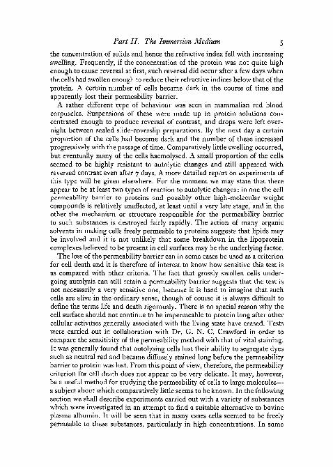
Part II. The Immersion Medium 5
the concentration of solids and hence the refractive index fell with increasingswelling. Frequently, if the concentration of the protein was not quite highenough to cause reversal at first, such reversal did occur after a few days whenthe cells had swollen enough to reduce their refractive indices below that of theprotein. A certain number of cells became dark in the course of time andapparently lost their permeability barrier.
A rather different type of behaviour was seen in mammalian red bloodcorpuscles. Suspensions of these were made up in protein solutions con-centrated enough to produce reversal of contrast, and drops were left over-night between sealed slide-coverslip preparations. By the next day a certainproportion of the cells had become dark and the number of these increasedprogressively with the passage of time. Comparatively little swelling occurred,but eventually many of the cells haemolysed. A small proportion of the cellsseemed to be highly resistant to autolytic changes and still appeared withreversed contrast even after 7 days. A more detailed report on experiments ofthis type will be given elsewhere. For the moment we may state that thereappear to be at least two types of reaction to autolytic changes: in one the cellpermeability barrier to proteins and possibly other high-molecular weightcompounds is relatively unaffected, at least until a very late stage, and in theother the mechanism or structure responsible for the permeability barrierto such substances is destroyed fairly rapidly. The action of many organicsolvents in making cells freely permeable to proteins suggests that lipids maybe involved and it is not unlikely that some breakdown in the lipoproteincomplexes believed to be present in cell surfaces may be the underlying factor.
The loss of the permeability barrier can in some cases be used as a criterionfor cell death and it is therefore of interest to know how sensitive this test isas compared with other criteria. The fact that grossly swollen cells under-going autolysis can still retain a permeability barrier suggests that the test isnot necessarily a very sensitive one, because it is hard to imagine that suchcells are alive in the ordinary sense, though of course it is always difficult todefine the terms life and death rigorously. There is no special reason why thecell surface should not continue to be impermeable to protein long after othercellular activities generally associated with the living state have ceased. Testswere carried out in collaboration with Dr. G. N. C. Crawford in order tocompare the sensitivity of the permeability method with that of vital staining.It was generally found that autolysing cells lost their ability to segregate dyessuch as neutral red and became diffusely stained long before the permeabilitybarrier to protein was lost. From this point of view, therefore, the permeabilitycriterion for cell death does not appear to be very delicate. It may, however,be a useful method for studying the permeability of cells to large molecules—a subject about which comparatively little seems to be known. In the followingsection we shall describe experiments carried out with a variety of substanceswhich were investigated in an attempt to find a suitable alternative to bovineplasma albumin. It will be seen that in many cases cells seemed to be freelypermeable to these substances, particularly in high concentrations. In some

6 Barer and Joseph—Refractometry of Living Cells
cases the presence of diffusible compounds of lower molecular weight mayhave been responsible for a toxic action accompanied by destruction of thepermeability barrier. On the other hand, some of these substances stillappeared to penetrate the cells even after they had been subjected to pro-longed dialysis, so that the presence of impurities of low molecular weightbecame unlikely. The mechanism of penetration of such compounds is quiteunknown; it is being investigated. It was also found that there were strikingdifferences between the behaviour of the same substances with respect todifferent types of cells. Gum acacia, for example, did not appear to penetratethe spermatocytes and fibroblasts of several species, whereas it did penetratethe majority of bacteria tested. If a range of different non-penetrating com-pounds of varying molecular weight were available, it might be possible toassess varying degrees of damage to the permeability barrier. Many of thesubstances discussed below were investigated from this point of view, but oursearch has not been completely successful. As regards convenience and generalapplicability no substitute has been found for bovine plasma albumin. Thelist of compounds tested is not exhaustive, and no attempt has been made todescribe properties of unsuitable substances in detail. Brief mention has beenmade of some of these, however, in order to assist others who might wish toinvestigate the properties of possible protein substitutes.
IMMERSION MEDIA OTHER THAN BOVINE PLASMA ALBUMIN
The concentrations of most of the substances discussed here were measuredby refractometry, and for convenience they are regarded as having the samerefraction increment as protein, so that concentrations are expressed in termsof the equivalent concentration of protein solution having the same refractiveindex.
PeptoneMany peptone preparations are available commercially. In general they are
made from heart or muscle by acid or enzymic digestion, and differ in theircontent of insoluble material and in the molecular size-range of their com-ponents. Thus some preparations were found to contain a high proportionof substances incapable of passing through a collodion membrane, whereasothers were almost completely dialysable.
The materials we have mostly used are Armour's numbers i and 2, andEvans's bacteriological peptone. The first two gave dark brown, cloudysolutions which had to be clarified by filtration or centrifuging. Solutions ofhigh refractive index corresponding to that of a 50 per cent, protein solutioncould be prepared without difficulty and were much less viscous. Evans'sbacteriological peptone was completely soluble up to at least 50 per cent, andcontained virtually no insoluble residue. It appeared to be composed mainlyof dialysable substances. The pH of all these solutions lay between pH 4and pH 5.
Part II. The Immersion Medium 7
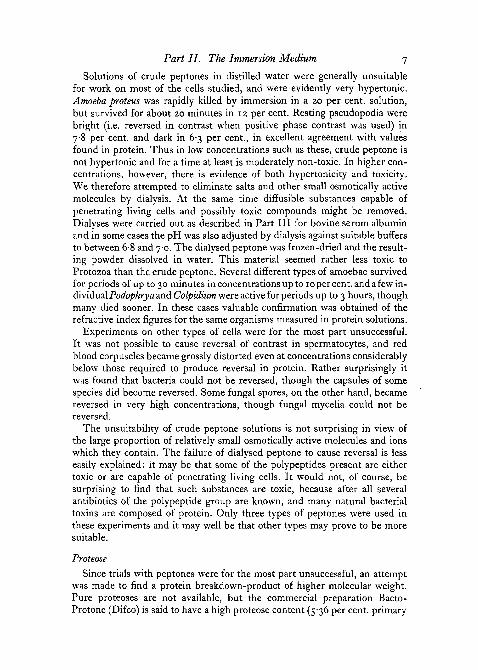
Solutions of crude peptones in distilled water were generally unsuitablefor work on most of the cells studied, and were evidently very hypertonic.Amoeba proteus was rapidly killed by immersion in a 20 per cent, solution,but survived for about 20 minutes in 12 per cent. Resting pseudopodia werebright (i.e. reversed in contrast when positive phase contrast was used) in78 per cent, and dark in 6-3 per cent., in excellent agreement with valuesfound in protein. Thus in low concentrations such as these, crude peptone isnot hypertonic and for a time at least is moderately non-toxic. In higher con-centrations, however, there is evidence of both hypertonicity and toxicity.We therefore attempted to eliminate salts and other small osmotically activemolecules by dialysis. At the same time diffusible substances capable ofpenetrating living cells and possibly toxic compounds might be removed.Dialyses were carried out as described in Part III for bovine serum albuminand in some cases the pH was also adjusted by dialysis against suitable buffersto between 6-8 and 7-0. The dialysed peptone was frozen-dried and the result-ing powder dissolved in water. This material seemed rather less toxic toProtozoa than the crude peptone. Several different types of amoebae survivedfor periods of up to 30 minutes in concentrations up to 10 per cent, and a few in-dividual Podophrya and Colpidium were active for periods up to 3 hours, thoughmany died sooner. In these cases valuable confirmation was obtained of therefractive index figures for the same organisms measured in protein solutions.
Experiments on other types of cells were for the most part unsuccessful.It was not possible to cause reversal of contrast in spermatocytes, and redblood corpuscles became grossly distorted even at concentrations considerablybelow those required to produce reversal in protein. Rather surprisingly itwas found that bacteria could not be reversed, though the capsules of somespecies did become reversed. Some fungal spores, on the other hand, becamereversed in very high concentrations, though fungal mycelia could not bereversed.
The unsuitability of crude peptone solutions is not surprising in view ofthe large proportion of relatively small osmotically active molecules and ionswhich they contain. The failure of dialysed peptone to cause reversal is lesseasily explained; it may be that some of the polypeptides present are eithertoxic or are capable of penetrating living cells. It would not, of course, besurprising to find that such substances are toxic, because after all severalantibiotics of the polypeptide group are known, and many natural bacterialtoxins are composed of protein. Only three types of peptones were used inthese experiments and it may well be that other types may prove to be moresuitable.
Proteose
Since trials with peptones were for the most part unsuccessful, an attemptwas made to find a protein breakdown-product of higher molecular weight.Pure proteoses are not available, but the commercial preparation Bacto-Protone (Difco) is said to have a high proteose content (5-36 per cent, primary
8 Barer and Joseph—Refractometry of Living Cells
proteose nitrogen and 7-60 per cent, secondary proteose nitrogen accordingto the manufacturers). This material contained much insoluble debris whichhad to be removed by centrifuging. The pH of the supernatant liquid wasapproximately 6-5. Preliminary tests on several different types of cells wereuniformly unsuccessful and further detailed tests were not carried out.
Protein hydrolysate
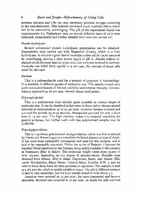
Several commercial protein hydrolysate preparations can be obtained.Experiments were carried out with Hepamino (Evans), which is a liverhydrolysate. It contains a good deal of insoluble matter which can be removedby centrifuging, leaving a clear brown liquid of pH 6. Amoeba radiosa re-mained motile for some time in 12 per cent., but were not reversed in contrast.Vorticella was killed fairly rapidly in 9 per cent. No reversal of tissue cellscould be obtained.
Dextran
This is a polysaccharide used for a number of purposes in haematology.It is available in different grades of molecular size. The samples tested werequite unsuitable because of limited solubility and extreme viscosity. Concen-trations approaching 20 per cent, formed almost solid pastes.
Polyvinyl alcohol
This is a polymerized vinyl alcohol, again available in various ranges ofmolecular size. It can be dissolved in hot water to form rather viscous neutralsolutions at concentrations up to 25 per cent. Amoebae became reversed andsurvived for periods up to 30 minutes. Paramecium survived for only a shorttime in 14 per cent. The high viscosity makes this material unsuitable forgeneral purposes, but further work with less polymerized samples may beprofitable.
Polyvinylpyrrolidone
This is a synthetic polymerized vinylpyrrolidone which was first employedby Hecht and Weese (1943) as a substitute for blood-plasma in cases of shock.It has since been extensively investigated and used for this purpose, and issaid to be reasonably non-toxic. Under the name of Periston it became thestandard blood substitute in the German Army and is available in this countryas Plasmosan (May & Baker). The molecular weight varies from 30,000 toover 100,000, depending on the degree of polymerization. Samples wereobtained from Messrs. May & Baker, Dagenham, Essex, and Messrs. Mil-wards Merchandise, Dacre House, Victoria Street, London, S.W. r, and wewish to thank these firms for their generous co-operation. The material is soldas a dry powder which is readily soluble in water. The pH of different samplesis said to vary somewhat, but we have usually found it to be about 5-5.
Amoebae were reversed in 15 per cent., but soon contracted and becameimmobile. Reversal also occurred in 10 per cent., in which the cells survived

Part II. The Immersion Medium 9
for about 30 minutes. Values for the refractive index obtained in polyvinyl-pyrrolidone agreed with those obtained in dialysed peptone and protein. Itwas not possible to cause reversal of contrast in bacteria and fungal spores,and red blood corpuscles became grossly distorted in high concentrations. Acurious effect, first pointed out by Mr. K. F. A. Ross, was observed in snailspermatocytes. Both the cytoplasm and nucleoplasm of these cells werereversed in contrast by immersion in 20 per cent, solutions. Within a fewminutes, however, the cytoplasm became progressively fainter and then dark,indicating that the permeability barrier had been damaged and that polyvinyl-pyrrolidone had leaked into the cytoplasm. The nucleoplasm, on the otherhand, remained reversed in contrast for at least 30 minutes, suggesting thatthe nucleus possessed a permeability barrier of its own, and was still capableof keeping out polyvinylpyrrolidone. We have found similar effects in sperma-tocytes of other species, though the time relationships were different.
Attempts to purify the material by dialysis were usually unsuccessfulbecause very great pressure was developed in the membrane. However, onedialysed sample was prepared by using several thicknesses of protectivestocking (see Part III) in order to withstand this pressure. The properties ofthis sample did not, however, appear to differ greatly from those of un-dialysed solutions. Nor were better results obtained when the pH of the solu-tion was adjusted to neutrality.
This material is thus disappointing as an immersion medium, and its un-suitability is all the more surprising in view of the fact that it has been sowidely used clinically. It is only fair to point out that the concentrationsemployed in clinical practice, namely, about 3-5 per cent, are very muchlower than we have used for immersion refractometry, so that gross celldamage is not necessarily likely to occur. Our results do, however, suggestthat polyvinylpyrrolidone is capable of leaking into cells and a number ofinvestigations have been reported in which histological changes have beenfound in various organs after the injection of massive doses of this substanceinto animals (Ammon and Miiller, 1949). Weese (1951) made the interestingobservation that after a first injection only about half the amount injectedcould be accounted for in the blood and very little was excreted. Examinationof various organs revealed the presence of polyvinylpyrrolidone in amountssufficient to account for the quantity injected. He made the significant sug-gestion that the substance became bound to plasma globulins and cell-wallglobulins.
Thrower and Campbell (1951), on the other hand, found evidence of onlya little interaction between polyvinylpyrrolidone and some protein films,though they did not investigate globulins. They did, however, observe thatthe material was slightly surface-active and lowered surface tension. Anyinteraction between polyvinylpyrrolidone and the proteins of the cell surfacemight very well account for the effects already described. The low concentra-tions used for clinical work may be insufficient to produce permanent damage,but in the presence of high concentrations, such as are necessary for immersion

io Barer and Joseph—Refractometry of Living Cells
refractometry, considerable disruption of the cell surface may occur on thesubmicroscopic scale. It may be that the proteins at the surface of the nuclearmembrane differ in character and react much more slowly with polyvinyl-pyrrolidone. These suggestions are highly speculative, however, and muchmore detailed knowledge is required concerning the possible interaction ofpolyvinylpyrrolidone (and other substances of high molecular weight) withproteins.
Acacia gum [gum-arabic)
This substance is of considerable historical importance as it was one of theearliest to be employed as a blood substitute (Bayliss, 1917). It is also ofspecial interest in that it was used for immersion refractometry by Faure-Fremiet (1929), who carried out measurements on the amoebocytes ofLumbricus. He did not, however, make any further use of the method, nor didhe control the tonicity of the medium.
Acacia gum is a natural product whose structure has been extensivelyinvestigated (see Hirst, 1942). It is an acid polysaccharide with a molecularweight in the region of 200,000 (Oakley, 1935). It is available commerciallyeither in granular form or as a powder; the latter is more convenient formaking solutions. When added to water, the gum swells at first and then goesinto solution. Concentrations up to 50 per cent, can be reached, though withrather more difficulty than in the case of bovine plasma albumin. The viscosityof concentrated solutions, though high, is not excessive for refractometry.The pH is usually about 4-0, but can easily be adjusted to neutrality by theaddition of alkali.
Amoebae survived quite well in concentrations below 15 per cent., butwere killed in 3-5 minutes in 30 per cent. Paramecium remained active formore than 1 hour in 18 per cent., but became immobile and discharged theirtrichocysts in 30 per cent., though their cilia continued to beat. Reversal ofcontrast was obtained in spermatocytes and other types of tissue-cells andunlike what was observed with polyvinylpyrrolidone such reversal appearedto be permanent and the cells remained, so far as could be judged, in goodcondition for some time. Spermatozoa continued to move actively in con-centrations up to about 20 per cent. The movements were slower in higherconcentrations, possibly because of the greater viscosity. Excellent resultswere obtained with fungal spores and mycelia even in concentrations up to50 per cent., and identical values were found for the refractive indices ofsuch material measured both in acacia and in bovine albumin. Germination offungal spores was observed in acacia solutions without any added nutrientsubstances. The mycelia from such spores did not, however, attain a greatlength and no reproductive stages could be seen. This suggests that acacia isnon-toxic to fungi, but is incapable of supplying their nutritional require-ments. In contrast to this, acacia appeared to be quite unsuitable for use withbacteria, which never became reversed at any concentration. Mammalianred blood-cells too became grossly distorted.

Part II. The Immersion Medium 11
Further purification was attempted by the process of dialysis followed byfreezing-drying. The purified material behaved in a very similar manner tothe crude gum, though the viscosity seemed to be somewhat less. Sperma-tozoa appeared to be rather more active and survived longer, but no betterresults were obtained with bacteria or red blood-cells.
It is thus evident that gum acacia is a promising material for use as animmersion medium for some types of cells. We have in fact used it extensivelyfor work on fungi, for which it does not appear to be inferior to bovinealbumin. It is also suitable for many types of tissue-cells which only requirecomparatively low concentrations not exceeding 25 per cent., but it causesdistortion of blood corpuscles and other cells in higher concentrations. Sofar as can be judged at present, it is not a complete substitute for protein, butit may be adequate for certain purposes. Its great advantage over bovinealbumin is its very low cost. It is in fact by far the cheapest of all the sub-stances capable of being used as immersion media. The failure of acacia gumto cause reversal of contrast in bacteria is interesting and suggests that the bac-terial cell wall is very different in constitution from that of fungi and tissue cells.
Egg albumin
A small quantity of fairly pure egg albumin was obtained from Dr. A. G.Ogston. This material was found to behave rather similarly to bovine albumin,though extended trials were not possible. Commercial pure egg albumin isprohibitively expensive and has not been investigated. Crude egg albumin is,however, available commercially at a very low price in the form of driedflakes. It is commonly used in histology for attaching sections to slides.Although it seemed likely that such albumin would be grossly denatured, wenevertheless investigated its properties. The flakes go into solution in waterfairly readily and concentrations up to 50 per cent, can be attained. Theviscosity is lower than that of bovine albumin, but the pH (5-0) is approxi-mately the same. The solutions have a very disagreeable odour. The crudematerial appears to be hypertonic and toxic. Spermatozoa were killed ratherrapidly and spermatocytes became shrunk and distorted. Paramecium waskilled almost at once in 32 per cent., but some survived for up to 30 minutesin 20 per cent.
The crude egg albumin was purified by dialysis followed by freezing-drying. The resulting material could not be dissolved in distilled water, as aslow precipitation occurred. Satisfactory clear solutions could, however, bemade in sodium chloride solutions exceeding about 0-4 per cent, in concentra-tion. Such solutions were much less yellowish than the solutions of crudematerial, and all trace of the disagreeable odour disappeared. Since thepurified egg albumin has to be dissolved in salt solutions, it is not very suitablefor work on fresh water protozoa, but it has given good results with tissue-cells, spermatoza, fungi, and bacteria. It is also one of the very few mediawhich has given results with red blood-cells which are in any way satisfactory.We have not employed purified crude egg albumin very extensively because

12 Barer and Joseph—Refractometry of Living Cells
much more work is needed in order to determine how far the properties ofdifferent samples are constant. Like crude acacia gum it may prove to be avaluable alternative to bovine albumin, though again not quite so generallyuseful.
Bovine gamma globulinsA commercial preparation of plasma globulins (fraction II) is available
from Armour Laboratories. This material has to be dissolved in dilute saltsolution and appears to be highly soluble (up to at least 40 per cent.). It hasgiven good results with tissue-cells and blood-cells. It is slightly more ex-pensive than bovine albumin and its only advantage may be that the pH ofsolutions is a little on the alkaline side of neutrality. Extensive tests have notbeen carried out.
CarboxyhaemoglobinAs the preliminary observations which led to the development of this
technique were carried out on cells immersed in haemoglobin solutions, itseems natural the latter should be used as an immersion medium. Unfor-tunately oxyhaemoglobin is not very stable and becomes converted fairlyrapidly to methaemoglobin. Carboxyhaemoglobin is, however, more stablethough less soluble. A sample of frozen-dried carboxyhaemoglobin (of sheep)was kindly supplied by Dr. A. G. Ogston. This went into solution readily, butwhen examined under the microscope was found to contain numerous minuteparticles. It is possible that the material may have been partly denatured.The presence of the particles did not greatly interfere with its use for immer-sion refractometry. Good results were obtained with tissue cells, amoebae,various Protozoa, and particularly with spermatozoa, which survived ex-tremely well for long periods. An important advantage of haemoglobin overbovine albumin may be that its isoelectric point is close to pH 7; on the otherhand, its deep red colour is a disadvantage and may make the determinationof refractive index difficult when high concentrations are used. The mainreason for not using it more extensively, however, is that no suitable prepara-tion seems to be available commercially. Crude technical haemoglobin isavailable, but this was found to be quite unsatisfactory as it contained muchinsoluble matter.
Other substancesSeveral other materials were investigated, but were found to be unsuitable
for various reasons. Dextrin and 'soluble' starch were insufficiently soluble.Preparations of pectin, soluble alginates, and methyl cellulose gave excessivelyviscous solutions. Technical grade blood albumin contained much insolublematter. None of these materials were examined in detail.
BOVINE PLASMA ALBUMIN
Almost the first substance used as an immersion medium was a preparationof bovine plasma albumin fraction V, manufactured in the form of a powder

Part II. The Immersion Medium 13
by Armour & Co. (Barer and Ross, 1952). This material has been used morethan any other and has been found suitable for a very wide range of cells.Indeed no type of cell has yet been found in which it has been impossible toproduce reversal of contrast in at least some regions.
The Armour product is prepared according to the method of Cohn andothers (1946), which involves successive precipitation of plasma at lowtemperatures by ethyl alcohol in the presence of acetic acid and sodiumacetate. The fifth precipitate contains almost all the plasma albumin. Thisfraction is further purified and re-precipitated by alcohol at pH 5-2, in orderto remove most of the acetic acid. The final material is an amorphous powderwhich according to the makers' brochure contains 3-5 per cent, globulins,less than 2 per cent, ash, and less than 6 per cent, moisture. More accuratefigures were obtained from the Research Division of Armour & Co., whostated that the ash content is usually less than 1 per cent, and the moisture2-3 per cent. There are usually less than 50 parts per million of heavy metalspresent. The ash consists mainly of sodium acetate and sodium chloride.Every attempt is made to keep the extraction procedure constant and onlyminor variations are to be expected between different batches. We have usedmany batches of fraction V over the last 3 years and with the exception oftwo batches which gave anomalous results with red blood-cells (thoughnormal results with other types of cells) we have found its properties to beremarkably constant, at least as far as the present technique is concerned.
A crystalline preparation of bovine plasma albumin is also available fromArmour & Co. This is almost free from globulins and contains less ash. It isvery much more expensive than fraction V and seems to offer no specialadvantages for refractometry. We have also used human plasma albumin withexcellent results.
Fraction V can be dissolved in water or salt solution to give a clear, faintlyyellow solution. The pale yellow colour is due to a component with anabsorption band at 405 m/x, possibly a navine or flavoprotein present in verysmall amounts. The pH of the solution is approximately 5-2-5-5, which is alittle higher than the isoelectric point of plasma albumin (5-0). Full details ofthe method used for making up solutions for different purposes are given inthe section on technical methods in Part III. At this stage, however, it isnecessary to discuss in some detail the osmotic properties of the immersionmedium with particular reference to fraction V.
OSMOTIC PROPERTIES OF THE IMMERSION MEDIUM
If accurate quantitative measurements of concentration are to be made, itis essential that the cell volume should be identical in the immersion mediumand in life. This is a condition which applies to all techniques for the deter-mination of concentration of any constituent of living cells, and is not peculiarto refractometry alone. There is no absolute certainty that the cell volumeremains unchanged when cells are removed from the body and examined

14 Barer and Joseph—Refractometry of Living Cells
under the microscope or otherwise manipulated, and so far as can be seenthere is no method available for measuring such changes if they occur. Allone can do is to take the volume occupied by the cell in some accepted'physiological' medium as a standard. This sounds simple, but in fact itraises a host of important questions which have received little considerationin the past. In the first place, what is an accepted 'physiological' medium?Many such media have been suggested, varying considerably in salt com-position, pH, and osmotic properties. A valuable summary of twelve basicsolutions used for mammalian tissue-culture work has been compiled recentlyby Stewart and Kirk (1954). It is remarkable how these media differ. Thesodium chloride content, for example, ranges from o-68 per cent, to 1-5 percent. Stewart and Kirk have calculated the total particle concentrations ofthese media, i.e. the concentrations of ions and molecules after assessingthe degree of dissociation of various constituents. This quantity should beproportional to the osmotic effect of the medium. The figure 0-307 M wasobtained for human plasma, but although a few of the media approximated tothis, the range extended from 0-294 M to 0-534 M. Even solutions advocatedby the same workers showed wide variations in concentration. Thus Lewis'smedia extend from 0-354 M to 0-534 M. The calcium Ringer of Vogelaar andErlichman (quoted by Stewart and Kirk) has a concentration of 0-455 Mwhereas that of the magnesium Ringer of the same authors is only 0-294 M.A study of the classical paper of M. R. and W. H. Lewis (1911) reveals evenmore dramatically what wide variations in salt content are compatible withcell growth. They cultured chick embryo tissues (liver, intestine, kidney,heart, and spleen) in hanging drop preparations, using only salt solutions ofknown composition. In the majority of these experiments the concentrationsof calcium chloride (0-025 per cent.), potassium chloride (0-042 per cent.),and sodium bicarbonate (0-02 per cent.) were kept constant, while the sodiumchloride content was varied over a wide range. At one extreme 'good' growthwas obtained with 0-45 per cent, sodium chloride, at the other 'extensive'growth was obtained with 1-585 per cent, sodium chloride and 'slight' growtheven occurred in 1 -6975 per cent, sodium chloride. As Lewis and Lewis them-selves point out, 'it is quite remarkable that cells will grow in such widelydifferent solutions where the osmotic pressure must vary considerably'. Itwill be seen from these figures that chick embryo cells are capable of activegrowth over about a threefold range of osmotic pressure. It would be interest-ing to know whether cells grown in such diverse media are similar in volumeand general form, but Lewis and Lewis do not discuss this. We are attemptingto repeat these experiments in order to see if the solid content of the cyto-plasm of such cells is affected by the osmotic pressure of the culture medium.
A study of various 'physiological' media suggested for invertebrates showseven greater variations than in mammalian media. Extensive compilations ofchemical analysis of the bloods of many species have been made by Prosserand others (1950), Heilbrunn (1952), and Buck (1953). Such tables showconsiderable differences both in the relative proportions of constituents and

Part II. The Immersion Medium 15
in the total osmotic concentration. For example, the depression of the freezing-point of the blood of fresh-water molluscs is generally between o-i° and 0-2° C ,whereas that for some fresh-water Crustacea exceeds 1° C. Very large differencesoccur between various species of insects (Buck, 1953), and the salt solutionssuggested do not always approximate in composition to that of the blood.Probably few investigators go to the length of making up special media foreach individual species; there is a tendency to use so-called 'amphibian'Ringer or 'insect' Ringer, &c, but there is strictly no such thing as an averagemedium identical in composition with the blood of a wide range of species.The fact that cells survive and even thrive in such media probably onlyshows that they are capable of adjusting themselves to quite gross changesin composition and osmotic pressure.
Blood and tissue fluids
It seems pedantic to question whether blood is a truly physiologicalmedium, but it must be remembered that the majority of cells are not sur-rounded by blood, but by tissue fluid, the composition of which is largelyunknown. It is generally assumed that tissue fluid resembles lymph (Drinkerand Yoffey, 1941), though clear evidence is lacking because of the difficultyin collecting tissue fluid even in large animals.
Lymph and certain other body fluids are said to be basically plasma 'ultra-filtrates' and are generally assumed to have very nearly the same compositionas plasma itself, but with less protein. Even if such fluids were true ultra-filtrates, however, their constitution would inevitably differ to some extentfrom that of plasma because the presence of non-diffusible protein ions wouldlead to a Donnan equilibrium resulting in a greater concentration of negativeions (Ch and HCO3~) and a lower concentration of positive ions (Na+, K + )in the ultra-filtrate than in the plasma itself.
The analysis of lymph is more or less that to be expected of an ultra-filtrate. The protein content of lymph for different parts of the body variesslightly and corresponding differences occur in the ionic composition. Thechloride content of the thoracic duct lymph of the dog is given by Drinkerand Yoffey (1941) as 396 mg. per 100 c.c. as compared with 369 mg. per100 c.c. for serum. Much larger differences are found in the cerebrospinalfluid. In man the chloride content is 440 mg. per 100 c.c. as compared with360 mg. per 100 c.c. in plasma. The sodium content is 324 mg. per 100 c.c.as compared with 316 mg. per 100 c.c. for plasma. The calcium content ofcerebrospinal fluid, however, is only half that of plasma (Merritt and Fre-mont-Smith, 1937). It is generally believed that cerebrospinal fluid is notsimply ultra-filtrate but in part at least a secretion. A similar opinion is nowheld concerning the aqueous humour (Duke-Elder and Goldsmith, 1951).The osmotic pressure of aqueous humour is actually greater than that of theblood. Until more exact figures are available for the composition of tissuefluid, the belief that blood constitutes a true physiological medium for tissue-cells must remain an approximation and it is a matter for conjecture whether

16 Barer and Joseph—Refractometry of Living Cells
the size and behaviour of tissue-cells will necessarily be the same in blood asin life. However, for the present at any rate, blood probably remains thenearest practical approach to a true physiological medium.
The osmotic behaviour of cells
There is an extensive literature which deals with the volume changes under-gone by certain types of cells in solutions of different ionic strengths. Un-fortunately, nearly all such work has been carried out on only two types ofcell, namely, egg-cells (usually echinoderm) and mammalian red blood-cells.Even in the case of this limited material much essential information is lacking.The mammalian red cell is very atypical both as regards its structure and itsosmotic behaviour. It appears to be far more sensitive to osmotic changes inthe medium than are most tissue-cells or white blood-cells. Echinoderm eggsmay be more typical in these respects, but an examination of the literatureshows that almost no experiments have been carried out on their volumechanges in media which depart only slightly from isotonicity. As a rulevolume changes have only been measured in rather grossly hypotonic orhypertonic media with the object of determining the laws connecting osmoticpressure and volume. For some cells the relationship P {V—b) = constanthas been found to hold. P is the osmotic pressure of the solution. V is the cellvolume, and b is a characteristic constant which can be regarded as the volumeoccupied by the osmotically inactive constituents of the cell, b is generallyabout 20-30 per cent, of the resting cell volume. By using this equation it ispossible to extrapolate for osmotic pressures in the region of isotonicity;because of the factor b it will be seen that a given change in P produces arelatively smaller change in Vy so that, for example, halving P would notdouble the cell volume. It is not at all certain, however, in view of the lackof experimental evidence, that this formula can be used for approximatelyisotonic media. We shall discuss the possibility below that individual cellsmay be capable of some degree of osmotic regulation in approximatelyisotonic media.
Shapiro and Parpart (1937) have measured the diameters of human andrabbit leucocytes in media of different osmotic pressures. The volume inRinger-Locke solution was taken as unity. The results plotted in figure r oftheir paper are said to show a more or less linear relationship between Vi/P. On closer inspection, however, it will be seen that V scarcely changesbetween ijP = 1 and ijP = 1-5, in other words, over a range in which theosmotic pressure exceeds about two-thirds of the standard pressure. Theseresults are in fact compatible with the hypothesis that regulation of volumetakes place over a fairly narrow range of concentrations, but that such regula-tion breaks down beyond certain limits. Investigations of this sort are not toodifficult in the case of spherical cells, the diameters of which can be measured,but there are obvious difficulties in working with irregular cells. Brues andMasters (1936) estimated the volume of fibroblasts and sarcoma cells in tissueculture by regarding them as symmetrical spindles and measuring the

Part II. The Immersion Medium 17
diameter at different points along the length of the cells. The behaviour ofthese cells was found to be rather variable when they were transferred fromplasma to 0-9 per cent, sodium chloride. As a rule they swelled a little, butoccasionally showed slight shrinkage. The mean change in volume in fibro-blasts was + 1 3 7 per cent., in sarcoma cells +4-3 per cent. The relationshipP (V—b) = constant was found to hold reasonably well with b = 22 per cent,for fibroblasts, and b = 26 per cent, for sarcoma cells. The change in volumeon transferring to a so-called isotonic salt solution is of some significance, andillustrates another of the difficulties inherent in the task of defining a 'physio-logical' medium. Shear and Fogg (1934) investigated the behaviour of tumourcells and normal cells from various organs when placed in saline media.They found that swelling occurred in so-called isotonic sodium chloride and'physiological' salines. Swelling even took place in hypertonic solutions!Variation of pH between 5 and 10 did not appear to affect this swelling verymuch, though it was a little more marked in alkaline media. The addition ofpotassium, calcium, magnesium, and other salts also made no difference.The only significant finding was that swelling occurred more slowly in serum.Following up this clue, Shear (1935) found that swelling was retarded by eggalbumin, gelatin, casein, serum proteins, and acacia. Some of the proteinsused were even capable of causing reversal of swelling with return towardsnormal volumes.
All these facts point to the great difficulty of virtual impossibility of deter-mining the true volume of the cell in life. One can attempt to measure the cellvolume in blood or some other natural fluid or one can measure it in somearbitrarily selected salt solution or 'physiological' saline, always rememberingthat the volume in the latter may not be the same as in blood or in the body.
What is an isotonic solution?
There is much confusion in the literature and particularly in textbooks asto the definition of the term 'isotonic'. It is quite clear that what is often calledan isotonic solution is really isosmotic, i.e. one having the same osmotic pressureor freezing-point depression as some standard medium such as plasma.Osmotic pressure is the physical property which can be measured withoutreference to the behaviour of living cells. Tonicity, on the other hand, is abiological concept which can only be referred to the properties of living cells.It would, for example, be ridiculous to call a solution of mercuric chloride orpotassium cyanide 'isotonic' simply because it had the same osmotic pressureas plasma. The first essential point in discussing tonicity therefore is that themedium must not be injurious to living cells. Even in such cases, however,the special permeability properties of the cell must be taken into account.Thus, to quote a simple example given by Heilbrunn (1952, p. 127), a0-53 M solution of sodium chloride and a 0-37 M solution of calcium chlorideare both isosmotic with sea-water. Despite this, sea-urchin eggs do not changein volume when immersed in the sodium chloride solution, but shrink in thecalcium chloride solution. The volume is unaltered in 0-30 M calcium

18 Barer and Joseph—Refractometry of Living Cells
chloride. The latter solution is thus isotonic (but not isosmotic) for thes.eparticular cells, though not necessarily for others. Considerations of this sortlead to a simple comprehensive definition of isotonicity. Two solutions are saidto be isotonic for a given type of cell if (a) they are compatible with life and (b) thecell volume is the same in each. This definition is independent of variables suchas differences in composition, relative proportions of ions, and pH, and it doesnot depend on the total osmotic pressure of the solutions. This is the onlysatisfactory practical definition for work on immersion refractometry in whichthe really important factor is the constancy of cell volume. Osmotic pressureseither calculated theoretically or determined by freezing-point measurementsmay not be entirely satisfactory and we have usually adopted the empiricalmethod of adjusting the salt content of our media until the mean cell volumeis the same as in a standard medium. Results obtained in this way have, asa matter of fact, agreed rather well with experimental freezing-point deter-minations.
Adjustment of tonicity of immersion media
The method used is essentially that developed by Ross (1953) in his studyof the volume changes of cells during fixation. We are indebted to Mr. Rossfor most of the results quoted in this paragraph. The basis of the method is todetermine frequency-polygons for cell size in different media. Cell diameterswere determined by means of an eyepiece micrometer. As a rule measure-ments were carried out on not less than 50 cells in each medium. The first cellstested were the primary spermatocytes of Helix aspersa. Attempts to measurethe distribution of cell diameter in snail-blood gave variable results, probablybecause of contamination with mucus and digestive fluids. Hedon-Fleig'ssolution was then used and gave virtually identical results with those obtainedin a simpler medium containing only 0-7 per cent, sodium chloride and 0-02per cent, calcium chloride. Histograms were then derived for cells in a rangeof sodium chloride concentrations varying from 0-3 per cent, to i-o per cent.A remarkable finding was that the general shape and modal value of thehistogram was virtually unaltered over a surprisingly wide range of saltconcentration; in fact, between 0-5 per cent, and o-8 per cent. Significantswelling occurred in 0-4 per cent, and shrinkage in 0-9 per cent. Nucleardiameters were also measured with very similar results. The next stage was tomake up a 20 per cent, protein solution of bovine plasma albumin fraction Vin distilled water and to determine frequency distribution curves of the sametype of cells in this medium. The cells were found to be considerably swollenwith a modal value of 24/x for the diameter as compared with 19/x in Hedon-Fleig or 07 per cent, saline. This frequency distribution corresponded quitewell to those obtained in 0-2 per cent, and 0-15 per cent, sodium chloride(fig. 1). On this evidence, therefore, it appeared that a 10 per cent, fraction Vsolution in distilled water would be equivalent in tonicity to a sodiumchloride concentration of between 0-075 Pe r cent- and o-i per cent. Takingthe latter value as a convenient round figure, a 20 per cent, protein solution

Part II. The Immersion Medium 19
would require the addition of 0-5 per cent, of sodium chloride in order tomake it isotonic with 07 per cent, salt solution. Histograms for spermato-cytes in 20 per cent, protein in 0-5 per cent, sodium chloride and also in20 per cent, protein in o-6 per cent, sodium chloride are shown in fig. 1.
__ NoCI solutions Bovine plasma solutions.
, 0 : 03 %
o-
• 20% Protein in
oi°/o
20% Protein inNaCI
r medium a)
.1 2O% Protein inO 7 % I.L, O6% NaCI
(= medium to
0 7% hi medium a.diluted
An —10% Proteinnedium a.dilu.._•with 07&NaCI
10% ProteinIII mediumb.diluted
iwithO7%NaCI
/ 10 2O 30 4O IO 2O 30 4OFIG. 1. Histograms showing diameters of snail spermatocytes in various media. For
explanation see text. Ordinates, number of cells; abscissae, cell diameters.
These are very similar to those obtained in 07 per cent, protein-free sodiumchloride. Each of these media was then diluted with an equal volume of07 per cent, sodium chloride, giving 10 per cent, protein solutions. Thehistograms obtained in these media were again closely similar to those in0 7 per cent, sodium chloride. Thus within the limits of accuracy of the methodthe simple rule that a 10 per cent, protein concentration is approximately

20 Barer and Joseph—Refractometry of Living Cells
equivalent to a 01 per cent, sodium chloride concentration appears to bejustified. A 20 per cent, protein solution is sufficiently concentrated to causereversal of contrast in the cytoplasm of most tissue-cells. In special cases itmay be necessary to use much more concentrated solutions, and here it ispossible that slightly greater errors may be introduced. If, for example, a10 per cent, solution of protein were equivalent in tonicity to 0-08 per cent,sodium chloride instead of o-i per cent, the error in working with a 20 percent, protein solution dissolved in 0-5 per cent, sodium chloride would onlybe 004 per cent, of sodium chloride, assuming 0 7 per cent, sodium chlorideto be isotonic. If, on the other hand, a 40 per cent, protein solution were used,dissolved in 0-3 per cent, sodium chloride, the error would be equivalent to0-08 per cent, sodium chloride. In view of the fact that snail spermatocytesappear to maintain the same size over quite a wide range of salt concentra-tions, neither of these errors is likely to be of much importance with theseparticular cells. It may be, however, that other cells are more sensitive to saltconcentration, and if in addition they require the use of concentrated proteinsolutions, the errors may be significant. This may be the case with mammalianred blood-cells, which are very sensitive to small changes in salt concentrationand also have high refractive indices, requiring the use of protein solutionsof up to 40 per cent, concentration. It is generally stated that 0-9 per cent,sodium chloride is isotonic for mammalian cells, though Ponder (1948) takes1 per cent, as isotonic for red cells. According to the simple rule given abovea solution of 40 per cent, bovine plasma albumin in 0-5 per cent, sodiumchloride should be isotonic with o-g per cent, sodium chloride, but the valuesfor mean corpuscular haemoglobin concentration obtained by cell refracto-metry with such a solution are 10-20 per cent, lower than the accepted clinicalvalues. If a 40 per cent, protein solution dissolved in o-6 per cent, sodiumchloride is used, the results agree with the lower normal clinical values, whileif 07 per cent, sodium chloride is used instead of O'6 per cent., the results arein excellent agreement with clinical values. Recent freezing-point determina-tions by Dr. D. A. T. Dick have shown that a 10 per cent, bovine plasmaalbumin solution in distilled water is actually equivalent to 0-08 per cent,sodium chloride. On this basis therefore a 40 per cent, protein solutionshould be made up in o-6 per cent, sodium chloride, if 0-9 per cent, sodiumchloride is taken as isotonic; or in 07 per cent, sodium chloride, if Ponder iscorrect in taking 1 per cent, sodium chloride as isotonic. With the majorityof cells, apart from red blood-cells, these differences are unimportant and thesimple rule given above can be used.
Are individual cells capable of osmotic regulation?
Much work has been done on the osmotic regulation and water balance ofwhole organisms, both vertebrate and invertebrate. On the other hand,almost nothing seems to be known about the osmotic regulation of individualtissue-cells. The possibility that such cells may be capable of regulating theirvolume and water content to some extent at least cannot be ignored. If the

Part II. The Immersion Medium 21
concept of the constancy of the internal environment has any general validity,it might be expected that it would be advantageous for the cell to maintaina more or less constant volume and water content, thus ensuring that theconcentrations of enzymes, nucleic acids, and other substances essential fornormal metabolism, growth, and cell-division should be kept within fairlywell defined limits. It may be argued that in mammals at least the externalenvironment of the cell is kept fairly constant by various mechanisms (e.g.respiratory exchanges and renal excretion), which regulate the compositionof the blood. Some of these mechanisms, such as changes in renal excretion,only act slowly, however, and cells may very well be exposed to quite largetemporary fluctuations in the composition of tissue fluid. Thus Hill, Long,and Lupton (1924) showed that enormous quantities of lactic acid may beliberated in severe exercise, and blood concentrations of over 100 mg. per100 c.c. may be attained. The local changes in the immediate vicinity of thetissues may perhaps be much greater. That too much faith should not beplaced in the constancy of blood composition in an individual has been shownby Schreider (1953). He carried out a statistical investigation on the reliabilityof a number of physiological and biochemical characteristics and showed thatsome of these were so variable that in order to determine a modal value,15-20 measurements on different days might be needed for one individual.Schreidtr reached the disturbing conclusion that 'the postulate of fixity mustbe rejected on biometrical grounds'. This does not necessarily mean that theinternal composition of cells varies greatly or that there is any immediatecorrelation between intracellular conditions and fluctuations in blood com-position. Schreider himself has pointed out that living organisms containmany internal environments separated by barriers which may serve to dampout fluctuations. In terms of the present problem such barriers might consti-tute a regulating mechanism whereby the cell volume is maintained relativelyconstant in the face of quite large variations in composition of the externalmedium. It must be admitted that direct evidence for osmotic regulation insingle cells is very scanty. The observations of Ross (1953) on snail spermato-cytes and of Shapiro and Parpart (1937) on mammalian leucocytes suggestthat some degree of regulation does occur over a surprisingly large range ofsalt concentrations. It must be remembered, however, that these workersestimated cell volumes by measuring the cell diameters with a micrometereyepiece. It is doubtful if such measurements of diameter can be made withan accuracy greater than about 2*5 per cent., corresponding to ±0-5^ ina diameter of 20/x. The error in cell volume is therefore three times thisamount or 7-5 per cent. Many more measurements of this type on a varietyof cells would be desirable. Much evidence which may have an importantbearing on this question has been assembled in a valuable review by Robinson(1953) on the active transport of water in living cells. Robinson propoundsthe view that the cell is in a state of dynamic equilibrium and that metabolicprocesses involving energy changes are necessary for maintaining a constantcell volume. The cell contents appear to have a higher osmotic pressure than

22 Barer and Joseph—Refractometry of Living Cells
plasma and are not in thermodynamic equilibrium with it. There is animpressive body of facts to show the correlation between cell volume andrespiratory processes; if the latter are depressed by the action of poisons,cellular swelling occurs in individual cells, and in whole animals the extra-cellular fluids become more concentrated. Many effects of this type arereversible. Further evidence in support of these views has been provided by
n.1-400
1-398
1-396
1-394
1392
1390
1388
1386
13 84
/38^
1380
1-378
1376
I374OS 0-6 0 7 08 0-9 10 II
cFie. 2. Relationship between mean refractive index (n) of human red blood-cells and equiva,lent salt concentration of protein medium expressed in terms of grams per cent, of sodium
chloride (C).
the work of Krebs and his school (for references see Bartley, Davies, andKrebs, 1954). This concept certainly fits in with observations made duringthe course of the present work, for example the swelling of cells found in thefirst stages of autolysis in sealed slide-coverslip preparations. From the pointof view of immersion refractometry the existence of some degree of osmoticregulation would be extremely useful, as it would allow for some margin oferror in making the immersion exactly isotonic. The presence of protein inthe medium may itself be a stabilizing factor in view of Shear's (1935) findingthat swelling in salt media was retarded or reversed by several proteins.
The refractometric method can itself be used to investigate this problem.As a result of swelling or shrinkage, the concentration of solids and hence

Part II. The Immersion Medium • 23
the refractive index will change; the reaction of the cell to changes in saltconcentration can therefore be studied by measuring its refractive index inbovine plasma albumin solutions to which different amounts of salt have beenadded. Fig. 2 shows some preliminary results obtained by Miss F. M. Gaffneyon human red blood-cells. The ordinates are the mean refractive indices ofcells of one individual, the abscissae being the salt content of the mediumexpressed in terms of equivalent sodium chloride concentration. The rela-tionship is virtually linear between 0-5 and i-o per cent, of salt, but there isconsiderable deviation from linearity above i-o per cent. This is only to beexpected, because the cell contents can obviously only be packed down to alimited extent. Thus over the range studied there appears to be no indicationof any special regulatory ability on the part of the cell. This is in keeping withour knowledge of the behaviour of red blood-cells. We have, however, carriedout a few experiments on lymphocytes and have found evidence of a plateauin the region between salt concentration o-8 per cent, and i-o per cent. Thismay suggest some degree of regulation. Further work on the problem is inprogress.
EVIDENCE FOR NON-TOXICITY OF THE MEDIUM
It is extremely difficult to provide clear evidence that a medium is com-pletely harmless to all types of cells. In the case of cells which are non-motileand which do not ordinarily undergo division, the evidence must rest mainlyon the general appearance of the cell before and after immersion and on thereproducibility of refractive index measurements. Provided that mechanicaldamage and compression which often lead to swelling of the cell are avoided,repeated estimations of refractive index should give very reproducible results.Recently Dick (1955), working in this laboratory, has developed a simpleperfusion chamber which enables cells to be irrigated with many changes offluids of different composition. Even after more than ten changes of proteinor gum acacia, cultures of chick fibroblasts or snail amoebocytes often re-mained in excellent condition and gave reproducible values for refractiveindices.
The assessment of damage is perhaps easier in the case of motile or dividingcells. The evidence may be summarized as follows:
Protozoa
Most motile protozoa appear to tolerate bovine plasma albumin dissolvedin distilled water quite well, particularly at concentrations below about20 per cent. At higher concentrations motility decreases or may be abolishedaltogether. It is not always possible to decide whether this is due to a toxicaction or simply to the resistance to motion offered by the viscous medium.In some cases normal motility is recovered when the organisms are trans-ferred from a very concentrated solution to water. One cannot, however,exclude the possibility that concentrated protein solutions may exert some sortof surface action or may hinder the diffusion of gases or nutrient materials

24 Barer and Joseph—Refractometry of Living Cells
into or out of the cell. Occasionally one comes across organisms which do notappear to tolerate the acidity (pH 5-2-5-5) of ordinary albumin solutions.Better results may be obtained in such cases by using dialysed pH-adjustedalbumin.
In general, amoebae appear to be more sensitive to protein than most otherProtozoa studied, but even so they remain in good condition in concentrationsbelow about 15 per cent, for about 30 minutes. Since most amoebae are re-versed in contrast in such concentrations, it is not usually necessary to usehigher ones. Slightly longer survival can be obtained with pH-adjustedprotein. Euglena, Stylonychia, and Rhabdostyla all appeared to tolerate con-centrations up to about 30 per cent, of ordinary albumin and 40 per cent, ormore of pH-adjusted albumin. Chylodon and Podophrya appeared in goodcondition in up to 40 per cent, of ordinary albumin. The parasitic formsTrichomonas muris and Giardia were strongly reversed and in good conditionin 30 per cent, ordinary albumin. Vorticella, on the other hand, though ittolerated fairly low concentrations, usually underwent a strong contractionin high concentrations of even pH-adjusted albumin.
Ciliated epithelial cells
Scrapings from the frog respiratory passages contain ciliated cells whichcontinued to exhibit vigorous ciliary movement for long periods in concen-trations of ordinary albumin which bring about strong reversal of the cyto-plasm.
Spermatozoa
The spermatozoa of a very large number of species ranging widely over theanimal kingdom have been examined. In concentrations of ordinary albuminnot exceeding about 10 per cent, there is scarcely any noticeable effect onsperm motility, and cells survive in apparently good condition for long periods.Human spermatozoa, for example, will survive up to about 3 days in a5 per cent, albumin solution. In higher concentrations motility is reduced,probably because of increasing viscosity. The structural components of mostsperm-heads are very dense and require high concentrations (generallybetween 35 and 55 per cent.) of albumin in order to bring about reversal. Forthis reason sperms which are reversed in contrast are usually immotile or veryfeebly motile. Of the various media tested, only bovine plasma albumin anddialysed egg albumin gave satisfactory results with spermatozoa.
Amoebocytes. The amoebocytes and blood-cells of several species of invertebrates have
been transferred to the protein medium either directly from the blood or bodyfluids or from tissue cultures. Photomicrographs of an earthworm cell under-going active movements in a protein solution sufficiently concentrated tocause strong reversal of contrast were shown in fig. 1 of the first paper of this

Part II. The Immersion Medium 25
series (Barer and Joseph, 1954). In general, such cells appear to tolerate bothordinary and pH-adjusted albumin quite well and promising results haverecently been obtained with acacia gum.
Mammalian leucocytes
Good motility has been observed in mammalian white cells kept for quitelong periods in albumin solutions. A remarkable example of their hardinesswas found by chance in a preparation of rat leucocytes which had beenexamined for several hours in a protein concentration sufficient to bring aboutreversal of contrast. The sealed slide-coverslip preparation was left overnighton the microscope stage at room temperature. The next morning, on glancingcasually down the microscope we were astonished to find many leucocytesstill undergoing active movement across the field of view.
Growth of fungal spores
Excellent germination and development of the spores of many differenttypes of fungi has been observed even in high concentrations of albumin.The complete life-cycle from germination through the sexual and asexualphases to the formation of new spores has been followed in slide-coverslippreparations (Barer and Joseph, 1955). No nutrient substances were addedto the protein medium. The ability to support the growth of fungi is in somerespects a disadvantage because stock protein solutions frequently becomecontaminated with yeasts or moulds after about 5 days even if kept in arefrigerator. Spore germination has also been observed in acacia gum solu-tions, but further growth and development are slower and do not usuallyreach so complete a stage as in protein.
Cell division
Perhaps the strongest evidence which suggests the comparative harmless-ness of bovine plasma albumin is the fact that normal cell division can beobserved in it. Occasional divisions have been seen in the germ cells of thelocust (Ross, 1954) and we have also observed the complete cycle in theprotozoon Cothurnia. Such divisions are, however, comparatively infrequentand sporadic. Much more successful results have been obtained with grass-hopper spermatocytes, and one can be reasonably certain of finding activedivision in several cells of every single preparation. Typical photographs ofthis process are shown in fig. 3. In some cases divisions have been followed inthe same preparation for as long as 3 days. All our experiments have beencarried out so far with ordinary albumin at pH 5-5 and without the addition ofnutrient substances. Experiments to determine whether the rate of divisioncan be influenced by such substances are in progress.
Regarded as a whole, the evidence quoted above, though not as completeas one could wish, nevertheless suggests that at least for a very large numberof different types of cells the albumin medium and in some cases acacia gum,are comparatively innocuous.

26 Barer and Joseph—Refractometry of Living Cells
We again wish to thank the Rockefeller Foundation, Royal Society, andMedical Research Council for making this work possible. We also wish toacknowledge the help of many colleagues, particularly Dr. D. A. T. Dick,Miss F. M. Gaffney, and Mr. K. F. A. Ross. Dr. A. G. Ogston has kindlysupplied several protein samples and Dr. W. E. van Heyningen and Dr. E.Bidwell provided facilities for freezing-drying.
REFERENCESAMMON, R., and MULLER, W., 1949. Dtsch. med. Wschr., 74, 465.BARER, R., and JOSEPH, S., 1954. Quart. J. micr. Sci., 95, 399.
1955- In course of publication.and Ross, K. F. A., 1952. J. Physiol., 118, 38P.
BARTLEY, W., DAVIES, R. E., and KREBS, H. A., 1954. Proc. Roy. Soc. B, 142, 187.BAYLISS, W. M., 1917. Ibid., 89, 380.BENNETT, A. H., JUPNIK, H., OSTERBERG, H., and RICHARDS, O. W., 1951. Phase microscopy.
New York (John Wiley).BRUES, A. M., and MASTERS, C. M. 1936. Amer. J. Cancer, 28, 314.BUCK, J. B., 1953. In Insect physiology. Ed. Roeder, K. D. New York (Wiley).COHN, E. J., STRONG, L. E., HUGHES, W. L., MULFORD, D. J., ASHWORTH, J. N., MELIN, M.,
and TAYLOR, H. L., 1946. J. Amer. Chem. Soc, 68, 459.CROSSMON, J. C, 1949. Stain Techn., 24, 244.DAVIES, H. G., WILKINS, M. H. F., CHAYEN, J., and LA COUR, L. F., 1954. Quart. J. micr.
Sci., 95, 271.DICK, D. A. T., 1955. Quart. J. micr. Sci. (in the press).
FIG. 3 (plate). Photomicrographs showing first meiotic division of primary spermatocytesof the grasshopper Chortippus parallelus. Division is taking place in a 12 per cent, solutionof bovine plasma albumin (equivalent tonicity I-I per cent, sodium chloride). A, diakinesis(late stage). B, diakinesis, shortly before disappearance of nuclear membrane. Note brightnucleoplasm (25 min.). c, early disruption of nuclear membrane (1 hour 25 min.). D, com-plete disruption of nuclear membrane. Chromosomes have moved to periphery of cell(1 hour 27 min.). E, prometaphase. Chromosomes beginning to line up on equatorial plate.Some faint spindle-fibres can be seen (2 hours 27 min.). F, prometaphase. Two dark centro-somes are visible (2 hours 40 min.). G, late prometaphase. Thick spindle-fibres visible(4 hours 30 min.). H, metaphase. Distinct spindle-fibres. Chromosomes and spindle aredisplaced by a crescentic mass of mitochondria (7 hours 6 min.). 1, early anaphase. Chromo-somes beginning to separate. Mitochondria are now clustered on each side of the spindle.Note large bright zone around chromosomes. The outline of the cell shows irregular bulging.(25 hours 47 min.). j , anaphase. The centrosome and spindle-fibres can be seen. Note alsoprominent polar bulging and central bright zone (26 hours 5 min.). K, advanced anaphase.Mitochondria are beginning to form 'sheaves'. Vigorous polar bulging with elongation ofcell (26 hours 15 min.). L, telophase. Incomplete cleavage. Note mitochondrial sheaves,from the ends of which granules appear to be forming. Bulging in equatorial and polar axes(26 hours 40 min.). M, cleavage almost complete. Cells still linked by mitochondrial sheavesand remains of spindle. Granules at ends of sheaves more distinct (26 hours 51 min.).N, separation of daughter-cells which are still linked by a mitochondrial bridge (28 hours13 min.). o, division complete. Daughter-cells transformed into secondary spermatocytes(48 hours 35 min.).
All photographs were taken with a Zeiss Winkel phase-contrast microscope with 4 mm.fluorite objective of N.A. 075. Length of scale, 25 p.
Times from the beginning of observations are given in brackets. It should be stressed thatthese cells are spherical and in no way compressed. With the microscope used, similar cellsin a saline medium give very poor images in which almost no internal detail can be made out.A discussion of this effect and further examples will be found in Barer, R., Naturwiss., 41,206 (1954).

FIG. 3R. BARER and S. JOSEPH

Part II. The Immersion Medium 27DHINKER, C. K., and YOFFEY, J. M., 1941. Lymphatics, lymph and lymphoid tissue. (Harvard
University Press).DUKE-ELDER, S., and GOLDSMITH, A. J. B., 1951. Recent advances in ophthalmology. 4th ed.
London (Churchill).FAURE-FREMIET, E., 1929. Protoplasma, 6, 521.HECHT, G., and WEESE, H., 1943. Miinch. med. Wschr., 90, 11.HEILBRUNN, L. W., 1952. Outline of general physiology. Philadelphia (Saunders).HILL, A. V., LONG, C. N. H., and LUPTON, H., 1924. Proc. Roy. Soc. B, 96, 438.HIRST, E. L., 1942. J. Chem. Soc, 70.LEWIS, M. R., and LEWIS, W. H., 191 I. Anat. Rec. 5, 277.MELLORS, R. C, KUPFER, A., and HOLLENDER, A., 1953. Cancer, 6, 372.MERRITT, H. H., and FREMONT-SMITH, F., 1937. The cerebrospinal fluid. Philadelphia
(Saunders).OAKLEY, H. B., 1935. Trans. Faraday Soc, 31, 136.PONDER, E., 1948. Hemolysis and related phenomena. London (Churchill).PROSSER, C. L., BISHOP, D. W., BROWN, F. A., JAHN, T. L., WULFF, V. G., 1950. Comparative
animal physiology. Philadelphia (Saunders).ROBINSON, J. R., 1953. Biol. Revs., 28, 158.Ross, K. F. A., 1953. Quart. J. micr. Sci., 94, 125.
1954. Ibid., 95, 425.SCHREIDER, E., 1953. Nature, Lond., 171, 339.SHAPIRO, H., and PARPART, A. K., 1937. J. cell. comp. Physiol., io, 147.SHEAR, M. J., 1935. Amer. J. Cancer, 23, 771.
and FOGG, L. C, 1934. U.S. Public Health Reps., 49, 225.STEWART, D. C, and KIRK, P. L., 1954. Biol. Revs., 29, 119.THROWER, W. R., and CAMPBELL, H., 1951. Lancet, 1, 1096.WEESE, H., 195I. Wien. med. Wschr., 101, 263.