redox-linked proton transfer by cytochrome c - helda -
TRANSCRIPT

Redox-linked Proton Transfer by Cytochrome c Oxidase
Camilla Ribacka (née Backgren)
Institute of Biotechnologyand
Division of BiochemistryDepartment of Biological and Environmental Sciences
Faculty of Biosciencesand
Viikki Graduate School in Biosciences
University of Helsinki
ACADEMIC DISSERTATION
To be presented with the permission of the Faculty of Biosciences of the University ofHelsinki for public examination in the auditorium 2402 of Biocenter 3, Viikinkaari 1,
Helsinki, on May 4th, at 9 o’clock.
HELSINKI 2007

Supervisor Docent Anne PuustinenFinnish Institute of Occupational HealthHelsinki, Finland
Reviewers Professor Ilmo HassinenDepartment of Medical Biochemistry and Molecular BiologyFaculty of MedicineUniversity of Oulu, Finland
Docent Moshe FinelDrug Discovery and Development Technology CenterFaculty of PharmacyUniversity of Helsinki, Finland
Opponent Professor Peter BrzezinskiDepartment of Biochemistry and BiophysicsArrhenius Laboratories for Natural SciencesStockholm University, Sweden
Kustos Professor Carl G. GahmbergDepartment of Biological and Environmental SciencesDivision of BiochemistryUniversity of Helsinki, Finland
ISBN 978-952-10-3838-9 (paperback)ISBN 978-952-10-3839-6 (PDF, http://ethesis.helsinki.fi)
ISSN 1795-7079
Yliopistopaino, Helsinki University Printing HouseHelsinki, 2007

To my parents

TABLE OF CONTENTS
List of original publications…………………………………………………………………i
Abbreviations and symbols…………………………………………………………………..ii
Abstract……………………………………………………………………………………...iii
1. INTRODUCTION………………………………………………………………………1
1.1 The respiratory chain………………………………………………………..1
1.2 The family of heme-copper oxidases……………………………………….3
1.3 The terminal oxidases of Paracoccus denitrificans………………………4
2. STRUCTURE AND FUNCTION OF CYTOCROME C OXIDASE…………………5
2.1 Overall structure of cytochrome c oxidase…………………………………5
2.2. Electron pathways and kinetics of electron transfer………………………….8
2.3 Proton input pathways………………………………………………….…...10
2.3.1 Proton migration through hydrogen-bonded networks………..……10
2.3.2 The K-pathway………………………………….……………….….12
2.3.3 The D-pathway………………………………….……………….….13
2.3.4 The H-pathway………………………………………………….…..14
2.4 Exit paths for protons and water molecules………………………………...14
2.5 Oxygen channels.…………………………………………………..……….15
2.6 The catalytic cycle…………………………………………..………………16
2.6.1 The reductive phase…………………………………………………16
2.6.2 The oxidative phase….…………………………………………….18
2.7 Proton transfer across the membrane………………………………………20
2.7.1 Requirements of a redox-linked proton pump…………………….20
2.7.2 Mechanisms for proton translocation by cytochrome c oxidase……21
3. AIMS OF THE STUDY…..………………………………………………………….…24

4. METHODOLOGY…………………...…………………………………………………25
4.1 Isolation of cytochrome c oxidase……………………………………….….25
4.2 Multi-turnover proton pumping measurements……………………………..25
4.3 Flash-photolysis measurements…………….……….………………………26
4.4 Time-resolved optical and electrometrical flow-flash measurements……..27
4.5 The reaction of cytochrome c oxidase with H2O2…………………………28
4.6 Transmittance and attenuated total reflection FT-IR spectroscopy………28
4.7 Molecular dynamics simulations..……………………………………..……29
5. RESULTS AND DISCUSSION……...………..……………………………….………30
5.1 The role of Glu278 in water-mediated proton transfer....………..……….…30
5.1.1 Heme-copper oxidases can have alternative proton-conducting
pathways…………………………………………………………….30
5.1.2 Glu278 is not required for proton translocation by cytochrome c
oxidase………………………………………………………………32
5.2 Arg473 and the -propionate of heme a3 regulate the access of pumped
protons to the P-side of the membrane……………..……………………….36
5.3 The influence of water molecules on the open and closed state of the
Arg473/heme a3 -propionate gate…….….………………………………..39
5.4 The reprotonation rate of Glu278 is affected by the W164F mutation..……41
5.5 The reaction of oxidized cytochrome c oxidase with H2O2……………….43
5.6. Loading of the pump site in the A PR transition……………………45
5.7 A possible mechanism for redox-linked proton transfer………………..49
5.7.1 The presented mechanism in relation to existing experimental data.51
5.7.2 Is the proton pumping mechanism common to all heme–copper
oxidases?…..………………………………………………………..53
6. SUMMARY………………………………….…..………………………………………55
7. ACKNOWLEDGEMENTS…......………………………...……………………………56
8. REFERENCES………..…………………………...……………………………………58

i
LIST OF ORIGINAL PUBLICATIONS
This thesis is based on the following published articles, referred to in the text by their
Roman numerals (I-IV):
I Backgren C., Hummer G., Wikström M., and Puustinen A. (2000) Proton
translocation by cytochrome c oxidase can take place without the conserved
glutamic acid in subunit I, Biochemistry 39, 7863-7867
II Gomes C. M., Backgren C., Teixeira M., Puustinen A., Verkhovskaya M.
L., Wikström M., and Verkhovsky M. I. (2001) Heme-copper oxidases with
modified D- and K-pathways are yet efficient proton pumps, FEBS Lett. 497,
159-164
III Wikström M., Ribacka C., Molin M., Laakkonen L., Verkhovsky M. and
Puustinen A. (2005) Gating of proton and water transfer in the respiratory
enzyme cytochrome c oxidase, Proc. Natl. Acad. Sci U.S.A 102, 10478-
10481
IV Ribacka C., Verkhovsky M. I., Belevich I., Bloch D. A., Puustinen A. and
Wikström M. (2005) An elementary reaction step of the proton pump is
revealed by mutation of tryptophan-164 to phenylalanine in cytochrome c
oxidase from Paracoccus denitrificans, Biochemistry 44, 16502-16512

ii
ABBREVIATIONS AND SYMBOLS
A ferrous-oxy intermediate of the binuclear centerATR FT-IR attenuated total reflection Fourier transform infrared spectroscopyCHES 2-(N-cyclohexylamino)ethanesulfonic acid∆ψ electric membrane potential∆µH+ electrochemical proton gradient across the membraneDM n-dodecyl-β-D-maltosideE the one-electron reduced state of the binuclear centerEPR electron paramagnetic resonanceeT electron transferF ferryl-oxy intermediate of the binuclear centerF´ state of the binuclear center after the oxidized enzyme has reacted with H2O2FT-IR Fourier transform infrared spectroscopyMES 2-(N-morpholino)ethanesulfonic acidMV mixed-valence state of the binuclear centerN-side negatively charged side of the (inner mitochondrial or bacterial) membraneO the fully oxidized form of the binuclear centerPM the stable compound formed when MV enzyme reacts with oxygenPR state of the binuclear center after fully reduced enzyme reacts with O2P-side positively charged side of the (inner mitochondrial or bacterial) membraneR the unliganded fully reduced state of the enzymeτ time constant, 1/kTMPD N,N,N’,N’-tetramethyl-1,4-phenylenediamineQ ubiquinoneQH2 ubiquinolWT wild-type enzyme
The redox-active metal centers of cytochrome c oxidaseCuA the dinuclear copper center in subunit IICuB the copper center in subunit IHeme a the low-spin heme in subunit IHeme a3 the oxygen-binding, high-spin heme in subunit IFea the iron ion of heme aFea3 the iron ion of heme a3
The amino acid numbering is based on the amino acid sequence of Paracoccusdenitrificans, if not otherwise mentioned.

iii
ABSTRACT
The respiratory chain is found in the inner mitochondrial membrane of higher organisms
and in the plasma membrane of many bacteria. It consists of several membrane-spanning
enzymes, which conserve the energy that is liberated from the degradation of food
molecules as an electrochemical proton gradient across the membrane. The proton gradient
can later be utilized by the cell for different energy requiring processes, e.g. ATP
production, cellular motion or active transport of ions.
The difference in proton concentration between the two sides of the membrane is a
result of the translocation of protons by the enzymes of the respiratory chain, from the
negatively charged (N-side) to the positively charged side (P-side) of the lipid bilayer,
against the proton concentration gradient. The endergonic proton transfer is driven by the
flow of electrons through the enzymes of the respiratory chain, from low redox-potential
electron donors to acceptors of higher potential, and ultimately to O2.
Cytochrome c oxidase is the last enzyme in the respiratory chain and catalyzes the
reduction of dioxygen to water. The redox reaction is coupled to proton transport across the
membrane by a yet unresolved mechanism. Cytochrome c oxidase has two proton-
conducting pathways through which protons are taken up to the interior part of the enzyme
from the N-side of the membrane. The K-pathway transfers merely substrate protons, which
are consumed in the process of water formation at the catalytic site. The D-pathway
transfers both substrate protons and protons that are pumped to the P-side of the membrane.
This thesis focuses on the role of two conserved amino acids in proton translocation
by cytochrome c oxidase, glutamate 278 and tryptophan 164. Glu278 is located at the end of
the D-pathway and is thought to constitute the branching point for substrate and pumped
protons. In this work, it was shown that although Glu278 has an important role in the proton
transfer mechanism, its presence is not an obligatory requirement. Alternative structural
solutions in the area around Glu278, much like the ones present in some distantly related
heme-copper oxidases, could in the absence of Glu278 support the formation of a long
hydrogen-bonded water chain through which proton transfer from the D-pathway to the
catalytic site is possible. The other studied amino acid, Trp164, is hydrogen bonded to the
-propionate of heme a3 of the catalytic site. Mutation of this amino acid showed that it may
be involved in regulation of proton access to a proton acceptor, a pump site, from which the

iv
proton later is expelled to the P-side of the membrane. The ion pair that is formed by the -
propionate of heme a3 and arginine 473 is likely to form a gate-like structure, which
regulates proton mobility to the P-side of the membrane. The same gate may also be part of
an exit path through which water molecules produced at the catalytically active site are
removed towards the external side of the membrane.
Time-resolved optical and electrometrical experiments with the Trp164 to
phenylalanine mutant revealed a so far undetected step in the proton pumping mechanism.
During the A to PR transition of the catalytic cycle, a proton is transferred from Glu278 to
the pump site, located somewhere in the vicinity of the -propionate of heme a3. A
mechanism for proton pumping by cytochrome c oxidase is proposed on the basis of the
presented results and the mechanism is discussed in relation to some relevant experimental
data. A common proton pumping mechanism for all members of the heme-copper oxidase
family is moreover considered.


1
1. INTRODUCTION
All living organism on Earth require energy in order to stay alive. Plants, algae and
cyanobacteria obtain their metabolic energy from photosynthesis, whereas animals acquire
their life supporting energy from cellular respiration. Photosynthesizing organisms trap the
energy of sunlight using chlorophyll and utilize it for the production of energy-rich
carbohydrates from CO2 and water, with the simultaneous release of oxygen as a by-
product. Photosynthetic cyanobacteria were among the first organisms to evolve on Earth,
some 3.4 billion years ago. At that time, oxygen was a sparse constituent of the primordial
Earth’s atmosphere. Photosynthetic reactions eventually increased the atmospheric levels of
oxygen. As a consequence, new respiring organisms evolved, which use oxygen as a
terminal electron acceptor during their metabolism.
In cellular respiration, different food molecules, such as glucose and fatty acids, are
oxidized into CO2 and water. The released energy is used to synthesize ATP, which serves
as a molecular storage of energy. The organism can later extract energy from ATP to drive
biosynthesis, motion or active transport of molecules across the membrane. Together
photosynthesis and cellular respiration form a steady-state system that is maintained by the
energy of the sun.
1.1 The respiratory chain
The respiratory chain (also known as the electron transfer chain) catalyzes the final steps of
cellular respiration. It is located in the inner membrane of mitochondria in higher organisms,
or in the plasma membrane of archaea and bacteria. The mitochondrial respiratory chain
consists of a series of membrane-bound protein complexes and two mobile electron carriers,
which transfer electrons from NADH, produced in glycolysis, fatty acid metabolism and the
citric acid cycle, to molecular dioxygen (Figure 1). The transport of electrons through the
respiratory chain towards electron acceptors of higher potential will release energy. The
liberated energy is stored as a proton gradient across the membrane, which can be harnessed
for the generation of ATP. The process is known as oxidative phosphorylation.

2
The first protein complex of the respiratory chain is NADH dehydrogenase, which catalyzes
the oxidation of NADH to NAD+ with the subsequent reduction of ubiquinone (Q) to
ubiquinol (QH2) (reviewed in [1]). This redox reaction is coupled by NADH dehydrogenase
to transfer of protons across the membrane by a yet unknown mechanism. The lipid-soluble
Q-pool present in the membrane may also receive electrons from membrane bound
dehydrogenases, e.g. succinate dehydrogenase (Complex II). Succinate dehydrogenase, a
FAD-containing flavoenzyme, oxidizes succinate to fumarate and transfers the electrons to
Q (reviewed in [2]). The subsequent enzyme in the respiratory chain, the cytochrome bc1
complex (Complex III), extracts two electrons from QH2 in a step-wise manner and
transfers them to the water-soluble one-electron carrier cytochrome c. The electron transfer
reaction is coupled to translocation of two protons across the membrane by the Q-cycle
mechanism (see [3] for review).
Figure 1. The electron transfer chain and ATP synthase of the inner mitochondrial membrane. Electrons aredonated to the chain from NADH or succinate and are thereafter transferred via the Q-pool in the membrane tothe bc1-complex, and from there via cytochrome c and cytochrome c oxidase to the final electron acceptor, O2.The flow of electrons through the chain is associated with proton transfer across the membrane, resulting in anelectrochemical proton gradient. The flow of protons down their concentration gradient through Complex V:ATP synthase drives the production of ATP from ADP and Pi.
Complex I:NADH dehydrogenase
Complex III:bc1 complex
Complex IV:Cytochrome c
oxidase
Complex V:ATP synthaseH+ H+
H+
Inner mitochondrialspace (P-side)
Matrix (N-side)
O2 + 4H+
H+
ADP + PiNAD+
ATP
e-
Complex II:Succinate dehydrogenase
2 H2O
succinate fumarate
QH2
Q
Cyt c
e-e-
e-
H+NADH + H+
e-

3
Cytochrome c carries electrons to the last enzyme in the respiratory chain, cytochrome c
oxidase (Complex IV), which reduces the ultimate electron acceptor, dioxygen, to water.
The redox reaction is coupled by cytochrome c oxidase to translocation of protons across
the membrane. The redox-linked proton transfer by complexes I, III and IV of the
respiratory chain results in a pH (∆pH) and charge (∆ ) difference, called the
electrochemical proton gradient (∆µH+), between the two sides of the lipid membrane. The
∆µH+ is a form of potential energy that can be utilized by different energy-demanding
processes in the cell, such as ATP production by ATP synthase, through a controlled flow of
protons down their concentration gradient (for review see [4]). The concept of energy
conservation through a proton gradient across the membrane was described by Peter
Mitchell in 1961 in the chemiosmotic theory [5].
1.2 The family of heme-copper oxidases
Cytochrome c oxidase belongs to the vast family of terminal heme-copper oxidases, which
reduce molecular dioxygen to water. All heme-copper oxidases contain a low-spin heme and
an oxygen-reducing binuclear center, constituted of a high-spin heme and a copper ion. The
redox-active metal centers are found in subunit I, where they are ligated by six strictly
conserved histidine residues (reviewed in [6]).
The members of the superfamily of heme-copper oxidases can be divided into three
main groups (Type A, B and C) based on common structural features in subunit I [7]. Type
A oxidases include the mitochondrial cytochrome c oxidase and heme-copper oxidases of
high similarity. The group is divided further into two subgroups based on the presence (type
A1) or absence (type A2) of a conserved glutamate in transmembrane helix VI. Examples of
heme-copper oxidases that belong to subgroup A1 are the mitochondrial cytochrome c
oxidase and the closely related enzymes from bacteria such as Paracoccus denitrificans and
Rhodobacter sphaeroides, as well as the ubiquinol oxidase from Escherichia coli. Subgroup
A2, which lack the glutamate in helix VI, can be exemplified by the cytochrome c oxidases
of caa3-type from Rhodothermus marinus and Thermus thermophilus. Heme-copper
oxidases that are classified as type B are found in bacteria and archaea. Type B oxidases are
a heterogeneous group, which have low sequence homology with the mitochondrial
cytochrome c oxidase. A characteristic heme-copper oxidase of type B is the ba3 oxidase

4
from T. thermophilus. The third group, Type C, differs substantially from both other types
of heme-copper oxidases with respect to the amino acid composition. The group consists
only of cbb3 oxidases.
The heme-copper oxidases demonstrate a large flexibility in terms of their electron
donors and their heme composition. The prokaryotic oxidases can alternatively contain
heme A, B or O or derivatives of heme A and O [8]. In addition, the number of subunits
forming the holoenzyme can vary in the prokaryotic oxidases from three to five, whereas the
number of subunits in the eukaryotic oxidases is normally higher. Despite large variations in
structure, members of all three groups of heme-copper oxidases have been reported to
function as proton pumps.
1.3 The terminal oxidases of Paracoccus denitrificans
Paracoccus denitrificans is a Gram-negative facultative anaerobic bacterium encountered in
soil, sludge and sewage. It belongs to the α subdivision of purple bacteria [9], which are
thought to be the precursors of today’s mitochondria [10]. The respiratory chain of P.
denitrificans is very similar to the one found in the mitochondrion [11]. However, on the
contrary to the linear respiratory chain of mitochondria (Figure 1), the counterpart in P.
denitrificans is branched, forming a complex respiratory network [12,13]. The versatility
enables P. denitrificans to grow on a large variety of different carbon sources aerobically,
using oxygen as the terminal electron acceptor or anaerobically, using nitrate, nitrite or
nitrogen oxide as the final electron acceptor. Moreover, the flexibility of its respiratory
network enables P. denitrificans to adapt itself to changes in the oxygen tension of the
environment.
P. denitrificans expresses three different oxygen reducing terminal oxidases. One of
these is a ba3-type quinol oxidase [14,15] whereas the other two are cytochrome c oxidases.
The dominating cytochrome c oxidase in P. denitrificans is of aa3-type and is a close
structural and functional relative to the mitochondrial cytochrome c oxidase [16]. The other
cytochrome c oxidase is of cbb3-type, and is expressed only at low oxygen concentrations
[17]. All terminal oxidase present in P. denitrificans have been shown to pump protons
across the plasma membrane [14,18,19].

5
2. THE STRUCTURE AND FUNCTION OF CYTOCHROME C OXIDASE
Cytochrome c oxidase is the terminal enzyme in the electron transfer chain of mitochondria
and many aerobic bacteria. The enzyme catalyzes the reduction of molecular oxygen to
water. At the same time, cytochrome c oxidase exploits the energy liberated by dioxygen
reduction for translocation of protons across the membrane, against the proton concentration
gradient [20]. A total of four protons are pumped per reduced dioxygen molecule, from the
matrix or, in case of bacteria, the cytoplasm (the negative or N-side of the membrane) to the
intermembrane space or the bacterial periplasm (the positive or P-side of the membrane).
The catalytic reaction in itself contributes moreover to the electrochemical proton gradient,
since electrons and protons used for water formation are taken up from opposite sides of the
membrane. The overall reaction catalyzed by cytochrome c oxidase can be described as:
4 Cyt c2+(P-side) + O2 + 8 H+
(N-side) → 4 Cyt c3+(P-side) + 2 H2O + 4 H+
(P-side)
2.1 Overall structure of cytochrome c oxidase
The crystal structure of cytochrome c oxidase from P. denitrificans was unraveled in 1995
[21]. Simultaneously, the x-ray structure of the 13-subunit cytochrome c oxidase from
bovine heart mitochondria was solved [22]. Although much different in total size, the core
part of the two enzymes are structurally much alike. The mammalian oxidase is a large
multi-subunit complex with a size of approximately 220 kDa. The core of the enzyme is
encoded by the mitochondrial DNA and consists of subunits I-III. The remaining ten
subunits are all small globular or transmembrane polypeptides that are encoded by the
nucleus. The function of the additional subunits is not completely known, but they may
stabilize and protect the catalytically active part of the enzyme [23]. In addition, several
binding sites for adenine nucleotides have been found in these subunits, which may imply a
regulatory role [24].
6
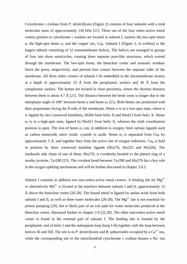
Cytochrome c oxidase from P. denitrificans (Figure 2) consists of four subunits with a total
molecular mass of approximately 130 kDa [21]. Three out of the four redox-active metal
centers present in cytochrome c oxidase are located in subunit I, namely the low-spin heme
a, the high-spin heme a3 and the copper ion, CuB. Subunit I (Figure 2, in iceblue) is the
largest subunit consisting of 12 transmembrane helices. The helices are arranged in groups
of four into three semicircles, creating three separate pore-like structures, which extend
through the membrane. The low-spin heme, the binuclear center and aromatic residues
block the pores, respectively, and prevent free contact between the separate sides of the
membrane. All three redox centers of subunit I lie embedded in the intramembrane moiety
at a depth of approximately 15 Å from the periplasmic surface and 30 Å from the
cytoplasmic surface. The hemes are located in close proximity, where the shortest distance
between them is about 4.7 Å [21]. The distance between the heme irons is longer due to the
interplanar angle of 108° between heme a and heme a3 [21]. Both hemes are positioned with
their propionates facing the P-side of the membrane. Heme a is in a low-spin state, where it
is ligated by two conserved histidines, His94 from helix II and His413 from helix X. Heme
a3 is in a high-spin state, ligated by His411 from helix X, whereas the sixth coordination
position is open. The iron of heme a3 can, in addition to oxygen, bind various ligands such
as carbon monoxide, nitric oxide, cyanide or azide. Heme a3 is separated from CuB by
approximately 5 Å, and together they form the active site of oxygen reduction. CuB is held
in position by three conserved histidine ligands (His276, His325 and His326). The
imidazole side chain of one of these, His276, is covalently bonded to the phenol ring of a
nearby tyrosine, Tyr280 [25]. The covalent bond between Tyr280 and His276 has a key role
in the oxygen splitting mechanism and will be further discussed in chapter 2.6.2.
Subunit I contains in addition two non-redox-active metal centers. A binding site for Mg2+
or alternatively Mn2+ is located at the interface between subunit I and II, approximately 12
Å above the binuclear center [26-28]. The bound metal is ligated by amino acids from both
subunit I and II, as well as three water molecules [26-28]. The Mg2+ site is not essential for
proton pumping [29], but is likely part of an exit path for water molecules produced at the
binuclear center, discussed further in chapter 2.4 [22,30]. The other non-redox-active metal
center is found in the external part of subunit I. The binding site is formed by the
periplasmic end of helix I and the subsequent loop (loop I-II) together with the loop between
helices XI and XII. The site is in P. denitrificans and R. sphaeroides occupied by a Ca2+ ion,
while the corresponding site in the mitochondrial cytochrome c oxidase houses a Na+ ion

7
[26-28]. The physiological role of the bound metal is unclear, but may be related to enzyme
stability and regulation [31]. Mutations targeting the Ca2+ ligands have revealed that the
metal is not necessary for catalytic activity and proton pumping [32,33].
Figure 2. The four-subunit structure of cytochrome c oxidase from P. denitrificans (Protein Data Bank entry1QLE, [34]). The subunits are color-coded as follows; subunit I in iceblue, subunit II in red, subunit III in limeand subunit IV in mauve. The redox-active heme groups (yellow) and CuB (red sphere) are locatedapproximately 15 Å from the P-side and 30 Å from the N-side of the membrane. The redox-active CuA site insubunit II is shown in blue, whereas the non-redox-active Ca2+ and Mg2+ sites are shown in cyan and pink,respectively. The picture were prepared using the Visual Molecular dynamics software (VMD) [35].
Subunit II (Figure 2, in red) consists of two transmembrane helices and a ten-stranded C-
terminal beta barrel, which is located on the P-side of the membrane on top of subunit I. The
large periplasmic domain contains one redox-active metal center, a dinuclear copper site
called CuA, which functions as the primary electron acceptor of cytochrome c oxidase. The
two copper ions lie at an interatomic distance of ~2.5 Å [26] and are bridged by two
cysteins (Cys 216 and Cys 220), and ligated by two histidines (His181 and His 224), one
methionine (Met 227) and the carbonyl oxygen of a glutamate (Glu 218). When oxidized,
CuA is in a mixed-valence [Cu(1.5)-Cu(1.5)] state, and upon reduction the two copper atoms
P-side
N-side
Cytochrome cbinding site
Subunit III
Subunit II
Subunit IV
Subunit I

8
will equally share the electron [36,37]. The docking site for the electron donor, cytochrome
c, is located at the interface between subunit II and the external surface of subunit I [38].
Subunit III (Figure 2, in lime) comprises seven transmembrane helices without any redox-
active centers. Helix I and II of subunit III are separated from the remaining five helices
through a V-shaped cleft, which is occupied by lipid(s) in both cytochrome c oxidase from
P. denitrificans and bovine heart [21,22]. The reason for this structural arrangement is not
obvious. It is plausible that the intramembrane lipids facilitate oxygen entry to the binuclear
center [39]. Subunit III is not involved in the redox events of cytochrome c oxidase, but is
important for the stability of the enzyme [40,41]. In the absence of subunit III, increasing
amounts of inactive enzyme will appear during catalytic turnover [40,42]. Subunit III may
in addition be important for efficient proton uptake via the D-pathway [43,44].
Subunit IV (Figure 2, in mauve) is composed of only one single transmembrane helix. Its
function remains a mystery, especially since it bears no significant homology with any other
known peptide or protein [21,45]. Deletion of the gene encoding subunit IV has no effect on
enzyme expression and activity [45].
2.2 Electron pathways and kinetics of electron transfer
Cytochrome c oxidase is reduced by four consecutive one-electron transfer events from
cytochrome c. The rate-limiting step in the reduction is the formation and dissipation of a
complex between the electron donor and acceptor [46]. The interaction between the two is
of an electrostatic nature and will strongly depend on the ionic strength of the media [47].
When a stable complex is formed an electron is rapidly transferred from cytochrome c to
CuA of cytochrome c oxidase (time constant ~15 µs) [48,49]. The reduction of CuA is a pure
electron transfer reaction and is not linked to protonation of the enzyme from the aqueous
phase [50].
The distance between CuA and the iron of heme a (Fea) is 19.5 Å, whereas the
distance between CuA and the iron of heme a3 (Fea3) is 22.2 Å [26]. In spite of the almost
equal distance from CuA to heme a and heme a3, electrons are solely transferred from CuA
to heme a, and from thereon to the binuclear center. The rates of electron transfer (eT)

9
between the redox centers of cytochrome c oxidase can be studied by several techniques
(see e.g. chapter 4.3). The physiological rate of eT can be studied with photoactivatable one-
electron donors that bind to the surface of cytochrome c oxidase and promptly release an
electron upon the flash of a laser [51]. An electron that is injected this way into the oxidized
cytochrome c oxidase will reach heme a, via CuA, within ~10 µs [52]. The flow of electrons
in the reverse direction, away from the binuclear center, can be examined by the electron
backflow measurement (see chapter 4.3). Two optically distinct phases of eT from the
binuclear center to heme a and CuA appear after dissociation of CO from the mixed-valence
enzyme [53,54]. The first phase, which has a time constant of about 3 µs, is accompanied by
optical changes at 445 and 605 nm and has thus be ascribed to eT between heme a3 and
heme a [54,55]. The second phase, which appears within ~50 µs, has been spectrally
assigned to reduction of CuA by heme a and heme a3 [54,55].
The three factors that control electron transfer between two different redox-active sites are
described by the Marcus theory (reviewed by [56]). These are; 1) the driving force, i.e. the
difference in redox potential between the two sites, 2) the distance between them, and 3) the
re-organization energy, i.e. the energy needed to alter the structure in response to the change
in charge. At the moment, there are at least two different models, which describe how
electrons are transferred in proteins. One of these models favor a structural control of the
rate of eT, which would accordingly occur through specific electron transfer pathways [57].
The other model postulates that the rate of eT between two redox centers is determined
solely by their edge-to-edge distance [58,59]. An extensive study of enzymes, which contain
multi-redox centers, supports mainly the latter model [59]. One of the quoted exceptions to
the model has been the 3 µs heme a3 heme a eT in cytochrome c oxidase, which is
approximately 1000 times slower than what would be expected based on the edge-to-edge
distance between the hemes [59]. Recently, Pilet et al. was able to detect an ultrafast eT
between heme a3 and heme a of cytochrome c oxidase using femtosecond absorption
spectroscopy [60]. The partial eT between the hemes appears within approximately 1.4 ns
after CO-photolysis from the mixed-valence cytochrome c oxidase [60]. This is close to the
calculated eT rate based on the distance between the two hemes and supports the model
where eT is purely limited by distance. The major electron transfer event between the hemes
takes nevertheless place within the slower 3 µs phase. The complete oxidation of heme a3 is
probably regulated by diffusion of CO out of the enzyme from CuB, where it transiently

10
binds upon dissociation from heme a3 [60,61]. The rapid nanosecond electron equilibration
between the hemes precedes by far the eT that occur during the catalytic cycle (see chapter
2.6), which is limited by internal proton transfer reactions [62].
2.3 Proton input pathways
2.3.1 Proton migration through hydrogen-bonded networks
The conductivity of protons in water is extremely high. The phenomenon originates from
the great ability of the water molecule to form hydrogen-bonded networks. In ice, each
water molecule is coordinated by hydrogen bonds to four neighboring water molecules,
whereas the liquid water molecule participates in three to four hydrogen bonds with its
closest neighbors. The structure of bulk water is constantly changing. The fluctuation is due
to the reorientation of each water molecule on average every picosecond. As a consequence,
the hydrogen bonds between adjacent water molecules are continually broken up and
reformed. The result is a rapid proton migration between adjoining water molecules. The
mechanistic basis for the proton transfer is explained by a modern version of the Grotthuss
mechanism, which was originally described in 1806 [63,64]. A schematic presentation of
the mechanism is seen in Figure 3 and it can be briefly described the following way.
An additional proton is at first present at one end of a water chain in the form of a
hydronium ion (H3O+). Rearrangement of hydrogen bonds moves a proton from this H3O+ to
an adjacent water molecule, which thus forms a new H3O+. Subsequent reshuffling of the
hydrogen bonds then transfers another proton from the recently formed H3O+ to one of its
neighboring water molecule. In this way, protons hop along a hydrogen-bonded chain of
water molecules from a proton donor to an acceptor. The proton migration is unidirectional,
and a specific proton is not moved per se, but due to the reshuffling of hydrogen bonds a
proton will ultimately be transferred from one place to another. Before another proton can
move in the same direction as the previous one, all water molecules in the chain must
reorient themselves to their original position.

11
Figure 3. A schematic presentation of proton transfer through a chain of water molecules via a Grotthuss-typeof mechanism. An additional proton is present in the form of a H3O+ at one end of the water chain. The protonis seemingly transferred from one water molecule to its neighbor, through breakage and formation of newhydrogen bonds, until it finally reaches the proton acceptor (A-) at the other end of the water chain. In orderfor another proton to be transferred in the same direction the water molecules will have to revert to theiroriginal conformation. In the figure, the formation of new hydrogen bonds is marked with white and greyarrows, whereas the reorientation of the water molecules is marked with thin black arrows.
A mechanism similar to the above described Grotthuss mechanism can most likely be
applied to intraprotein proton conduction. The hydrophobic nature of the protein interior
makes it unsuitable for proton transfer. Instead, protons may be transferred through proton-
conducting pathways, which can consist of a single file of hydrogen-bonded water
molecules stabilized by protonatable and polar amino acids. This type of proton wire has the
potential to transfer protons extremely fast using a Grotthuss-like mechanism [65],
especially when the hydrogen-bonded chain is constrained to a narrow hydrophobic space
[66].
A-
O
H
HO
H
HO
H
HO
H
HO
H
HHO
H
H+
H
HO H
O
H
OH
H
O
H
H OH
H
OH
H
H+A-
H
HO H
O
H
OH
H
O
H
H OH
H
OH
H
HA
AH
O
H
HO
H
HO
H
HO
H
HO
H
HO
H
H
. . .
. . ..... . .
. . .
. . .. . .
. . .. . .
. . .. . . . . .
. . . . . .
... ......
...
...
...
... ...

12
2.3.2 The K-pathway
The K-pathway is one of the two structurally resolved proton-conducting channels in
subunit I of cytochrome c oxidase from P. denitrificans (Figure 4). It is named after a highly
conserved lysine (Lys354), which is essential for the function of cytochrome c oxidase [67-
69]. The K-pathway leads from the surface of the membrane on the N-side to the vicinity of
the binuclear center. Protons enter the channel, presumably, via an invariant glutamate
(Glu78) in subunit II and continue via Lys354, a threonine (Thr351) and the
hydroxyethylfarnesyl group of heme a3 up to a tyrosine (Tyr280), located in close contact
with the binuclear center. The pathway involves two structurally resolved water molecules.
Yet, a hydrogen-bonded network extending all the way from the surface to the active site of
cytochrome c oxidase is not present without transient structural changes of residues in the
channel or movement of internal water molecules [70-72].
The role of Glu78 in the K-pathway is still under debate. Electrostatic calculations
have shown that Glu78 alters its protonation state upon reduction of the binuclear center
[73]. Mutation of the glutamate in the ubiquinol oxidase from E. coli and the cytochrome c
oxidase from R. sphaeroides confirmed a role in proton conduction through the K-pathway
[74-76], but the results were contradicted by proton translocation experiments and FT-IR
spectroscopy with Glu78 mutants from P. denitrificans [77]. Recent electrostatic
calculations have confirmed a functional role of Glu78 through its strong electrostatic
interactions with Lys354 [78].
The K-pathway is important during the initial reduction of the binuclear center,
which precedes binding of dioxygen. Unless accompanied by proton uptake through the K-
pathway, electrons will not be transferred to the oxidized binuclear center [79,80]. The K-
pathway is not essential for the oxidative part of the catalytic cycle (see chapter 2.6.2)
[79,81], but movement of Lys354 may compensate for the additional negative charge at the
binuclear center present in the PR intermediate [72]. According to recent electrostatic
calculations, Lys354 is protonated at neutral pH [78]. Moreover, the pKa of the residue is
raised upon uncompensated reduction of the binuclear center. This implies that the K-
pathway is not merely a proton-conducting pathway, but also functions as a “dielectric
well”, which can stabilize uncompensated electron transfer to the binuclear center [69].

13
Figure 4. The redox-active centers of cytochrome c oxidase together with key residues lining the K- and D-pathways of proton transfer (PDB entry 1M56, [28]). Structurally resolved water molecules in the channels aredepicted as red spheres. The picture were prepared using VMD [35].
2.3.3 The D-pathway
The entrance of the D-pathway (Figure 4) is located on the N-side of the membrane in the
loop between helices II and III. The loop is rich in charged residues and contains the highly
conserved and functionally essential aspartate, Asp124, from which the channel received its
name. Mutation of Asp124 to an asparagine abolishes proton translocation and lowers the
enzymatic activity dramatically [82-84]. The D-pathway continues from Asp124 via a
cluster of conserved asparagines (Asn113, Asn131 and Asn199), a tyrosine (Tyr35) and
several serines (Ser 189, Ser192 and Ser193) up to a highly conserved and mechanistically
very important glutamate (Glu278). The D-pathway is filled with water molecules, all the
way from the protein surface to Glu278 [28], which can facilitate fast proton transfer
through a Grotthuss-type of mechanism.
Glu278 is located approximately 10 Å from the active site in a hydrophobic cavity
predicted to transiently contain several water molecules [85-87]. The residue is essential for
both oxygen reduction and proton translocation [88,89]. The D-pathway is intriguing from a
mechanistical point of view, since it conveys two out of the four protons that are consumed
14
in the formation of water, as well as all four protons that are translocated across the
membrane during a single turnover [80,88,90]. Glu278 is believed to be the branching point
from where protons are either transferred to the catalytic site (chemical protons) or towards
the heme propionates and eventually across the membrane (pumped protons) [21,70,91,92].
Proton transfer via Glu278 is likely to involve movement of its side chain from the
downward position in the static crystal structure to an upward position towards the hemes
[70,85,92].
2.3.4 The H-pathway
An additional proton-conducting pathway has been proposed based on structural analysis of
the mitochondrial cytochrome c oxidase from bovine heart [22,27,93]. The so-called H-
pathway consists of residues in helices XI and XII of subunit I, and is named after a
partially conserved histidine (His448) located close to the channel entrance. The proton
route through the H-pathway involves an aspartate (Asp51, bovine numbering), which has
no counterpart in the bacterial oxidases. Mutational studies with cytochrome c oxidase of
bacterial origin do not support a functional role of the H-pathway in the prokaryotic
oxidases [94,95].
2.4 Exit paths for protons and water molecules
The area above the heme propionates is rich in structurally resolved water molecules and
contains in addition to the previously described Mg2+ site (chapter 2.1) two highly
conserved arginines, Arg473 and Arg474, which are thought to stabilize the propionates in
their deprotonated state [22,26,73]. Mutations of the arginine residues have implicated the
-propionate of heme a3 as the beginning of an exit path for pumped protons [91].
Alternative exit paths for protons that involve the -propionate of heme a [96] and
propionate A of heme a3 [97-99] have also been proposed. The hydrophilic region above the
hemes is presumably in rapid equilibrium with the P-side of the membrane. Several possible
proton exit points may exist at the interface between the membrane and the external part of
the enzyme, identified by continuum electrostatic calculations [100]. Of these, the one that

15
involves Lys171 and Asp173 in subunit II of the mitochondrial cytochrome c oxidase is
supported by a recent study in which the exposure of backbone hydrogens to the bulk phase
in different parts of cytochrome c oxidase was investigated by amide H/D exchange
detected by mass spectrophotometry [101].
Rapid freeze-quench EPR experiments have revealed that water molecules produced at the
binuclear center propagate via the Mg2+ site on their way to the P-side of the membrane
[30,102]. The exact exit path of the water molecules is not resolved, but it is presumably
located at the interface between subunit I and II.
2.5 Oxygen channels
Structural and computational analysis of cytochrome c oxidase have identified putative
channels through which molecular oxygen can access the active site [22,28,39,70]. For the
mitochondrial cytochrome c oxidase three different oxygen pathways were proposed based
on the structure [22]. One of the channels propagates to the active site via the
hydroxyethylfarnesyl group of heme a3 and another channel approaches via CuB. The third
proposed oxygen channel of the bovine cytochrome c oxidase starts at the V-shaped cleft in
subunit III and enters subunit I between helices IV and V at the level of the hemes. This
channel coincides with the one suggested based on the structure of cytochrome c oxidase
from P. denitrificans [39]. An alternative starting point for the channel was however
proposed for the oxidase from R. sphaeroides [28]. The channel is lined by hydrophobic
aromatic residues and is hence highly suitable for oxygen diffusion. At its most narrow
point, the channel passes between Phe274 and Trp164 [70]. Mutation of a conserved valine
(Val279 to Ile mutation) located in this oxygen path dramatically increased the KM of
oxygen compared to wild-type (WT) enzyme, while the Vmax of the oxygen reaction was
essentially the same [39]. In the same V279I mutant, the rate of formation of the oxygen
bound intermediate A was decreased substantially compared to WT, as well as the
subsequent steps of O2 reduction [103]. The result was interpreted as a partial blockage of
oxygen diffusion to the binuclear center by the isoleucine side chain and perturbation of
water structure located close to the catalytic site, and supports O2 delivery to the catalytic
site through a specific route.

16
2.6 The catalytic cycle
The steady-state turnover rate of cytochrome c oxidase is very rapid. Cytochrome c oxidase
consumes on average 300 molecules of oxygen per second and one single turnover of the
enzyme is normally completed within five milliseconds. The fully reduced state of the
enzyme is unlikely to exist at physiological conditions. Instead, the oxygen reaction is
presumably initiated immediately upon oxygen binding to the two-electron reduced enzyme.
The full reduction of molecular dioxygen involves four consecutive one-electron transfer
steps from the electron donor cytochrome c on the P-side of membrane to the active site
located in the membrane domain. Each electron addition to the active site is charge
compensated by the uptake of a proton from the N-side of the membrane according to the
principle of electroneutrality [104,105]. Consequently, one charge is separated across the
membrane per electron transferred to oxygen. In addition, the coupled proton pumping by
cytochrome c oxidase adds to the charge separation, which therefore amount to two charges
transferred across the membrane per electron delivered to dioxygen.
The catalytic cycle of cytochrome c oxidase is shown in Figure 5. For simplicity,
only the redox state of the catalytically active site is shown, which in addition to the
binuclear heme a3-CuB center also include Tyr280. The catalytic cycle is usually divided
into two parts. The reductive phase (O R), where the active site of cytochrome c oxidase
receives electrons, and an oxidative phase (R O), where oxygen binds and is reduced to
water, with the concomitant oxidation of the enzyme. The cycle involves several
intermediate states of the active site, which are commonly denoted by one-letter codes. The
intermediate states will be separately described on the following pages.
2.6.1 The reductive phase
The introduction of the catalytic cycle starts with the oxidized and resting state of
cytochrome c oxidase referred to as intermediate O. At this stage, the binuclear center is in
a ferric-cupric state and Tyr280 is presumably protonated. The oxidized state of the enzyme
exists in different isoforms, which are recognized by their specific absorption spectrum in
the Soret region and their diverging EPR signals (reviewed by [106]). The isoforms differ in
their rate of reduction as well as their reactivity towards external ligands e.g. cyanide,

17
hydrogen peroxide and carbon monoxide [107-110]. The as-isolated cytochrome c oxidase
is sometimes referred to as the slow (or resting) form of the enzyme (O) (but see e.g. [111]).
The slow isoform can be converted to a fast (or activated) form by full reduction followed
by complete oxidation of the enzyme. The formed OH intermediate is presumably a
metastable state of high energy [112,113]. The energy stored in the OH state can be released
upon immediate re-reduction of the active site, and will then drive proton pumping across
the membrane [112,113]. If the enzyme is not re-reduced within a reasonable time the
energy will be lost as the OH state decays into the O state. The active site in the O and OH
states are thought to differ in their redox properties and bound ligands [112]. Presumably, a
water molecule ligates Fea3 in the O intermediate, whereas Fea3 in the recently oxidized OH
intermediate is ligated by a hydroxide anion.
Figure 5. The intermediate states of the catalytic cycle of cytochrome c oxidase. The structure of the activesite, consisting of heme a3, CuB and Tyr280, at each state of the reaction cycle is shown in boxes. If the two-electron reduced enzyme is allowed to react with O2, the reaction will follow the outer circle, going from the
PM F intermediate. If the fully reduced (four-electron reduced) enzyme is incubated with O2 thereaction will proceed from the A state via PR to the F state.

18
Intermediate E is formed upon arrival of the first electron into the active site. The rate by
which an electron arrives from heme a is limited by the uptake of a proton from the N-side
of the membrane to the active site through the K-pathway [52,62]. The incoming electron
reduces CuB with simultaneous protonation of its bound hydroxide ion, which is
concomitantly released as a water molecule [114]. The transition between the oxidized and
one-electron reduced state of the catalytic cycle is associated with proton translocation
across the membrane only when the enzyme is reduced shortly after its oxidation (the
OH EH transition) [112,113].
Reduction of the active site by a second electron yields the ferrous-cuprous R
intermediate. The reduction of heme a3 is accompanied by protonation of the hydroxide ion
bound to Fea3, which is then released as a water molecule. Formation of the R intermediate
is coupled to proton translocation across the membrane only when the acceptor of the
second electron is the EH state of the binuclear center, formed from the recently oxidized
OH state [113,115].
2.6.2 The oxidative phase
The oxidative phase of the catalytic cycle is initiated by the binding of dioxygen to the
reduced binuclear center. The formed ferrous-oxy adduct of the active site is called
intermediate A. In an O2 saturated environment (~1.2 mM O2), intermediate A is detected
by a characteristic absorption peak at 595 nm that appears within ~10 µs after mixing with
the fully reduced cytochrome c oxidase [116,117].
In the subsequent reaction step, the O-O bond of the bound dioxygen molecule is
broken and two incompletely protonated water molecules are formed in a concurrent four-
electron reduction step. The produced state of the binuclear center is called the P
intermediate. The name originates from the previous notion that heme a3 in this
intermediate was in a peroxy state (Fea33+-O=O2-) with an intact dioxygen bond [118,119].
However, it is now well established by various experimental studies that the active site in
the P intermediate is an oxo-ferryl state (Fea34+=O2-) with an hydroxide ion bound to CuB
2+
[120-123]. Since formation of the P intermediate is not associated with proton uptake from
the bulk solution the proton required for hydroxide formation must be extracted within
cytochrome c oxidase [124-127]. Two out of the four electrons that are needed for the
scission of the dioxygen bond are derived from heme a3 by its oxidation from ferrous to

19
ferryl state (heme a32+ → heme a3
4+). One is derived from CuB by its oxidation from
cuprous to cupric state (CuB1+ → CuB
2+). The origin of the fourth electron can be either
heme a or Tyr280 depending on the initial redox state of the enzyme (see below).
A mixed-valence state of cytochrome c oxidase, where the binuclear center is reduced and
heme a and CuA are oxidized, is produced by incubation of the oxidized enzyme with
carbon monoxide. Laser flash mediated dissociation of the CO bound to the reduced
binuclear center allows O2 to bind, whereafter an intermediate state called PM spontaneous
appears (time constant ~150 µs) [128,129]. The fourth electron required for the splitting of
the dioxygen bond upon formation of PM, as well as the proton needed for hydroxide
formation at CuB, are presumably extracted from Tyr280, which turns into a neutral tyrosyl
radical [130-132]. The radical formation is favored by the unusual crosslink between
Tyr280 and His276, one of the ligands of CuB [25-27].
If the fully reduced cytochrome c oxidase is allowed to react with O2 formation of
intermediate A is followed by a fast (time constant ~20 µs) transition in which an electron is
transferred from heme a to the binuclear center with the simultaneous scission of the
dioxygen bond [129]. The formed state of the binuclear center is called PR, where R refers
to the fully reduced nature of the enzyme at the start of the reaction [128]. The proton
consumed upon formation of the CuB-bound hydroxide ion is presumably extracted from
Tyr280.
The PR and PM intermediates have the same absorption spectra, which is
characterized by an absorption peak at 607 nm in the alpha region [128,133]. Yet, the PM
intermediate has, in comparison to the PR intermediate, one less electron at its catalytically
active site. The PM intermediate is long-lived and will exist until cytochrome c oxidase
receives additional electrons [129]. The PR intermediate is on the contrary unstable and will
quickly decay into the subsequent F intermediate without additional electron transfer to the
binuclear center.
Intermediate F appears (time constant ~50 µs) upon protonation of the PR state by a proton
arriving from Glu278 in the end of the D-pathway [88]. The proton acceptor at the active
site is most likely the tyrosinate (Y280-) formed in the preceding step of the catalytic cycle
[132,134]. Proton transfer to the active site is accompanied by the partial transfer of the last
electron from CuA to heme a [135] and reprotonation of Glu278 via the D-pathway [136].

20
The appearance of intermediate F can be detected optically by a broad, absorption peak at
580 nm in the alpha region [118]. The transition from the PR state to F is associated with
pumping of one proton across the membrane [137,138].
The PM intermediate, formed from the mixed-valance enzyme, will turn into the F
state when cytochrome c oxidase receives an additional electron that is transferred into the
active site. The electron will presumably reduce the neutral tyrosyl radical present in the PM
state, which is simultaneously protonated from the D-pathway via Glu278 [129,139]. The
transition from PM to F is accompanied by translocation of a proton across the membrane.
The final step in the catalytic cycle, F OH transition (time constant ~1.5 ms),
occurs with the transfer of the fourth electron into the active site and the simultaneous
uptake of a proton via the D-pathway [81,140]. This transition is coupled to proton pumping
across the membrane [137,138].
2.7 Proton transfer across the membrane
2.7.1 Requirements of a redox-linked proton pump
There are two ways to couple exergonic electron transfer to active proton translocation
across a lipid membrane. One way is by a redox loop mechanism, where the redox center
that is reduced by an electron simultaneously accepts a proton. The proton is then co-
transported with the electron across the membrane, and is released on the other side upon
the oxidation of the redox center. The other way is by a proton pump mechanism, where the
redox center that accepts an electron not necessarily also accepts a proton. The minimum
requirement of a redox-linked proton pump is, in addition to a redox center, a protonatable
group that can bind a proton from one side (input side) and release to the other side of the
membrane (output side). The protonatable group, hereafter referred to as the pump site, has
to control or gate proton access to the different sides of the membrane, since uncontrolled
diffusion of protons would short-circuit the electrochemical proton gradient [105,141,142].
Proton translocation by a proton pump can be divided into four elementary steps; 1)
uptake of a proton to the pump site from the N-side of the membrane, 2) establishment of a
protonic connection between the pump site and the P-side of the membrane, 3) release of the
proton to the P-side of the membrane, and 4) re-establishment of a protonic connection

21
between the pump site and the N-side of the membrane. The uptake and release of protons
by the pump site can be regulated by changes in its proton affinity (pKa) or in its
conformation. In a redox-linked proton pump, the proton translocation across the membrane
is linked to the redox reaction at the redox center. The coupling can be thermodynamic,
where protonation of the pump site is coupled to the reduction of the redox center [143,144].
This means that the pKa of the pump site is modulated by the redox state of the redox center,
and reciprocally, the midpoint potential of the redox center is modulated by the protonation
state of the pump site. The other alternative is a kinetic coupling, where the pKa of the pump
site is not necessarily regulated by the redox state of the redox center. A state where the
redox center is reduced and the pump site is protonated may in this scenario be present in
only a fraction of the enzyme population. The transition from the proton input to the output
state may still be kinetically favored by a rapid rate constant [145].
2.7.2 Mechanisms for proton translocation by cytochrome c oxidase
The exact mechanism by which cytochrome c oxidase couples O2 reduction to translocation
of protons across the membrane remains ambiguous despite almost 30 years of extensive
studies. During the years, several different coupling mechanisms have been proposed and
later discarded. Only a few of the recent theories will be mentioned below.
Throughout the 1990s, cytochrome c oxidase was believed to pump protons only during the
oxidative part of the catalytic cycle [146]. The assumption was based on the fact that only
the P F and F O transitions are associated with a large enough energy release to drive
protons across the membrane [147]. The P F and F O steps were believed to pump two
protons each based on the equal amount of charge moved relative to the membrane in each
transition in electrometrical measurements [148]. In 1999, Michel questioned this idea and
suggested that one proton is pumped during the reductive phase of the catalytic cycle [149].
Concomitantly, Verkhovsky and co-workers showed by time-resolved measurements of
membrane potential ( ) generation that protons are translocated both in the reductive and
oxidative phase of the catalytic cycle [112]. The reductive phase is however only coupled to
proton pumping if it occurs directly upon enzyme oxidation. A high-energy oxidized state of
the binuclear center, called O~ and later OH, was proposed that would preserve the energy

22
from O2 reduction in the oxidative phase to be used for proton pumping in the subsequent
reductive phase [112].
The proton pumping steps of cytochrome c oxidase is outlined in Figure 6. Each electron
addition from cytochrome c, via heme a to the binuclear center is accompanied by the
uptake of one substrate proton to the active site and the translocation of another proton
across the membrane (the OH EH, EH R, PM/R F and F O transitions) [112,113,115].
The mechanism of vectorial proton transfer is presumably the same in each proton-pumping
step, and will consequently be repeated four times during each enzymatic turnover. Proton
pumping coupled to the PR F transition has gained particular interest when possible
mechanisms for proton translocation are discussed, since formation of the F state is not
linked to electron transfer to the binuclear center, which occurs already in the preceding
A PR transition.
Figure 6. The proton pumping steps in the catalytic cycle of cytochrome c oxidase during continuousenzymatic turnover. The OH EH, EH R, PM/R F and F O transitions are each coupled to translocation ofone proton across the membrane.
All recently proposed proton-pumping mechanisms are in compliance with the principle of
electroneutrality, according to which introduction of an electron into the low dielectric
membrane environment is energetically costly, and must be charge compensated by the
uptake of a proton [105]. Michel and Papa et al. suggest mechanisms where electron arrival
to heme a is coupled to uptake of a proton to the pump site [149,150]. Yoshikawa et al.

23
propose a mechanism where the pump site is protonated upon oxidation of heme a [93].
Several groups argue that proton pumping is coupled to reduction of the binuclear center
instead of heme a. Based on electrostatic calculations, Stuchebrukhov and co-workers
introduce a proton pumping mechanism that involves redox-state dependent
protonation/deprotonation of one of the histidine ligands of CuB [97]. Brzezinski et al. have
proposed a model for proton pumping that originates from structural differences between
the WT and the Glu278Gln mutant of R. sphaeroides [138,142]. In their view, deprotonation
of Glu278 upon proton transfer to the binuclear center leads to structural changes around
Glu278, which will propagate through the protein and alter the pKa and proton accessibility
of an accepting group located close to the heme propionates. According to their model, the
substrate proton will move to the binuclear center prior to protonation of the pump site.
Wikström and colleagues have presented a mechanism whereby proton translocation is
gated by the distinct behavior of water molecules in hydrophobic cavities [151]. In their
model, the orientation of the water molecules in the cavity next to the binuclear center are
affected by the redox-state dependent electric field between heme a and heme a3/CuB. When
heme a is reduced, the pump site, which is located somewhere in the vicinity of the -
propionate of heme a3, will be protonated from Glu278 via a chain of hydrogen-bonded
water molecules. Upon electron transfer to the binuclear center, the waters in the cavity will
change their orientation, allowing a substrate proton to enter the catalytic site. The
protonation of the binuclear center will subsequently expel the proton at the pump site out of
the enzyme [151]. It has been argued that this model cannot explain proton pumping in the
PR F transition, which merely involves protonation of the active site.

24
3. AIMS OF THE STUDY
The D-pathway of cytochrome c oxidase conducts all pumped protons and two out of the
four chemical protons that are consumed at the catalytically active site, from the bulk on the
N-side of the membrane to the vicinity of the binuclear center. In many enzymes of the
heme-copper oxidase family, the D-pathway ends at a highly conserved glutamate (Glu278)
that is essential for the oxygen reaction and the uptake and translocation of protons.
However, not all members of the heme-copper oxidase family have a glutamate at this
location. There are examples of heme-copper oxidase that lack a corresponding glutamate
and nevertheless function as true proton pumps.
One of the goals of this thesis was to investigate the role of Glu278 in proton
conduction and to explore the possible differences in proton translocation mechanisms
between different subgroups of heme-copper oxidases.
The other main focus of this thesis has been to study the beginning of a possible exit
pathway for pumped protons and water molecules produced at the binuclear center.
Molecular dynamics simulations have shown that redox-changes of the hemes determine the
directionality of a hydrogen-bonded water chain, which is located in the cavity between
heme a and the binuclear center. The chain of water molecules will alternatively conduct
protons from Glu278 to the binuclear center or to the ∆-propionate of heme a3. In the latter
configuration, the chain of water molecules is supported by the invariant Trp164, which is
hydrogen bonded to the ∆-propionate of heme a3.
The role of Trp164 in proton translocation was studied both experimentally and
theoretically by mutating it to a phenylalanine. In addition, the properties of the salt bridge
between the ∆-propionate of heme a3 and the highly-conserved Arg473 were studied by
molecular dynamics simulations.

25
4. METHODOLOGY
4.1 Isolation of cytochrome c oxidase
The ctaDII gene encoding subunit I of cytochrome c oxidase from P. denitrificans has
previously been isolated and cloned into a derivative of the broad-host strain expression
plasmid pBBR1MSC [94,152,153]. The expression plasmid was transformed into the E. coli
strain SM10, which was then conjugated with the P. denitrificans strain AO1, from which
the chromosomal copies of the ctaDII gene and its isogene ctaDI were deleted, but where
the genes encoding the additional subunits of cytochrome c oxidase are still present [94]. In
the present study, a six-histidine long tail was attached in the end of the ctaDII gene to
facilitate enzyme purification through affinity chromatography. Mutants of cytochrome c
oxidase were made by the previously described site-directed mutagenesis protocol [32] and
verified by DNA-sequencing (ABI PRISM 310 Genetic Analyzer, Applied Biosystems)
during all individual steps of the production. In addition, the DNA sequence of cell samples
of fermentor cultivations and smaller cultivations used for proton pumping measurements in
whole cells where always examined. Bacterial growth conditions and isolation of
cytochrome c oxidase from bacterial membranes was performed as earlier described [32],
with the exception of the additional Ni2+-NTA affinity chromatography (paper IV). The
steady-state oxygen reducing activity of the isolated cytochrome c oxidase was determined
polarographically using a Clark-type oxygen electrode. The electron donor, cytochrome c
was kept in its reduced state by ascorbate plus TMPD. A more specified description of the
experimental conditions is found in Paper I and Paper IV. The activities of the different
mutants were compared to that of WT enzyme.
4.2 Multi-turnover proton pumping measurements
Multi-turnover proton pumping was measured with isolated cytochrome c oxidase
incorporated into proteoliposomes, or with whole cell preparations (sphaeroplasts), using
the oxidant pulse method described below [154,155]. Reconstitution of the isolated enzyme
into proteoliposomes was achieved by slow removal of detergent from an enzyme-

26
detergent-lipid mixture using increasing amounts of BioBeads (Bio-Rad Laboratories)
[137,156]. The procedure for incorporation of cytochrome c oxidase into vesicles was the
same for proton pumping experiments and for electrometry (see below). The enzyme
concentration in the initial mixture was 0.5 µM in the reconstitution for pumping
experiments and 6.5 µM in the reconstitution for electrometry. The proton permeability of
the formed proteoliposomes was determined by the respiratory control ratio (RCR), i.e. the
ratio between enzyme activity in the presence and absence of ∆µH+, and was typically 3-9
for vesicles used in proton pumping experiments and 2-5 for vesicles used in electrometry.
The sphaeroplasts used for proton translocation measurements were prepared by lysozyme
treatment and kept in a hypertonic medium [14]. In the proton pumping measurement,
proteoliposomes (or sphaeroplasts) were kept in an anaerobic container under constant
argon flow in the presence of excess reductant plus valinomycin, which dissipates the ∆ .
Known amounts of oxygen were added as small volumes of air-saturated water, which
resulted in enzyme turnover. Proton ejection to the outside of the vesicles (sphaeroplasts)
was detected by a sensitive pH-meter. The H+/e- pumping stoichiometry was calibrated by
addition of anaerobic HCl of known concentration. The specific conditions used for
pumping experiment in sphaeroplasts are described in ref [32], and for proteoliposomes in
Paper I and Paper III.
4.3 Flash-photolysis measurements
Incubation of oxidized cytochrome c oxidase with carbon monoxide in an anaerobic
environment results in formation of the 2e- reduced, mixed-valence state (COMV). Binding
of CO to heme a3 of the binuclear center increases its midpoint potential and traps heme a3
and CuB in a reduced state, while heme a and CuA are oxidized. Upon laser flash mediated
photolysis of CO, the apparent midpoint redox potential of heme a3 decreases. The electron
situated at heme a3 quickly redistributes to heme a followed by electron equilibration to
CuA. The redistribution of electrons between the redox centers of cytochrome c oxidase can
be time-resolved by optical spectroscopy using a single-wavelength spectrophotometer as
described in ref [54]. From the obtained data, the rate of electron transfer between heme a3,
heme a and CuA can be extracted, as well as information of their relative midpoint
potentials. See paper IV for experimental conditions.

27
4.4 Time-resolved optical and electrometrical flow-flash measurements
The flow-flash technique, invented by Gibson and Greenwood in 1963 [157], enable us to
optically detect and distinguish different intermediates of the catalytic cycle of cytochrome
c oxidase. As previously mentioned, the reaction between reduced cytochrome c oxidase
and O2 is very rapid. This means that when enzyme and oxygen is mixed together in a
normal stopped-flow apparatus, the first steps of the oxygen reaction will be over already
during the dead time of the instrument. This obstacle can be circumvented by the flow-flash
technique, where reduced and CO-inhibited cytochrome c oxidase is mixed with excess O2
in the dark. A strong laser pulse is thereafter used to dissociate CO from the enzyme. The
reduced enzyme molecules will now bind O2, and the oxygen reaction will simultaneously
start in all cytochrome c oxidase molecules present. The oxygen reaction can be recorded
optically at the wavelength of choice. For experimental details see paper IV.
A combination of the flow-flash technique with measurements of electric potential
generation was developed by Drachev and co-workers in 1974 [158]. The electrometric
flow-flash technique has been successfully applied in Helsinki Bioenergetics Group and has
been thoroughly described by Jasaitis et al. [137]. In brief, the proteoliposomes containing
reconstituted cytochrome c oxidase are attached to one side of a lipid-impregnated Teflon
membrane, which separates two secluded compartments of the electrometric cell. The
enzyme is thereafter degassed, reduced and kept CO inhibited in the dark. The oxygen
reaction starts when an oxygen-saturated buffer is injected very close to the vesicles
followed by flash photolysis of CO. The generation of electric potential during enzyme
oxidation, i.e. the movement of electrons, protons and/or charged amino acid residues, can
be kinetically resolved as a voltage change by Ag/AgCl electrodes positioned on separate
sides of the Teflon membrane. The voltage detected by the electrodes is proportional to the
voltage generated over the proteoliposome membrane. The experimental conditions used are
described in Paper IV.

28
4.5 The reaction of cytochrome c oxidase with H2O2
The reaction between oxidized cytochrome c oxidase and H2O2 produces species equivalent
to the PM and F intermediates of the catalytic cycle. The product of the reaction depends on
the pH of the medium (see chapter 5.5). In paper IV, cytochrome c oxidase in 100 mM
MES, pH 6.5 or CHES, pH 9.5, supplemented with 0.02% DM was reduced with a small
concentration of dithionite and kept in an oxygen free environment under constant nitrogen
flow, in one chamber of a stopped-flow apparatus (Unisoku Instruments, Kyoto, Japan). The
fully reduced cytochrome c oxidase was then mixed in a 1:1 ratio with aerated buffer
containing 20 mM H2O2. Instantly upon mixing, the oxygen in the second chamber
consumed the excess dithionite and oxidized the enzyme, resulting in an activated
cytochrome c oxidase. Shortly thereafter, the freshly oxidized enzyme reacted with H2O2
present in the same solution. The absorption changes that followed were recorded using a
diode array kinetics spectrophotometer (Unisoku Instruments).
4.6 Transmittance and attenuated total reflection FT-IR spectroscopy
Fourier transform infrared spectroscopy (FT-IR) detects vibrational motions within the
chemical bonds of a molecule, by measuring the change in intensity of the infrared light
before and after it interacts with the sample. The collected FT-IR spectrum of a biomolecule
provides information about the protein secondary structure and structural interactions, and
can even pinpoint specific bond vibrations. The CO photolysis difference FT-IR
spectroscopy exploited in paper IV detects changes in bond vibrations of cytochrome c
oxidase, which occur when CO bound to the reduced Fea3 is dissociated and transiently
binds to CuB. Comparison between CO difference FT-IR spectra of WT at different pH
values or of WT and specific cytochrome c oxidase mutants can reveal changes in the
structural conformation of the binuclear center or for example changes in hydrogen bonding
or protonation state of specific functional groups within the enzyme. The experimental
protocol for the FT-IR measurements in paper IV was essentially as described in ref [86].
Attenuated total reflection (ATR) FT-IR spectroscopy is a technique, which enables
fast collection of IR spectra with a good signal-to-noise ratio from only a few micrograms of
protein. In addition, the ATR FT-IR technique allows variations of the pH, salt

29
concentration or redox-conditions during the measurement. The infrared beam in the ATR
FT-IR measurement is directed onto a silicon microprism of high refractive index (SensIR
Technologies), on top of which the studied protein sample is applied. Above a critical angle,
the light beam is totally reflected from the surface of the ATR microprism, and an
evanescent wave is established at the interface between the prism and the protein sample.
Formation of a dehydrated protein film on top of the silicon microprism was in paper IV
insured by removal of detergent from the enzyme sample essentially according to Iwaki et
al. [132]. After application on top of the microprism, the enzyme was gently dried using a
flow of nitrogen. Once a stable protein film had formed, the enzyme was rewetted with 200
mM potassium chloride and 200 mM potassium phosphate, pH 6.5 and covered with a
protecting lid. The lid is equipped with in and out ports, which allows a constant flow of
buffer on top of the sample. In the experiment performed in paper IV, cytochrome c
oxidase was oxidized by a buffer containing 1 mM ferricyanide and thereafter reduced by a
buffer containing 3 mM anaerobic dithionite, in repeated cycles, to improve the signal-to-
noise ratio of the measurement. The redox state of the enzyme was monitored throughout
the experiment via an optical fiber connected to the lid, which covers the sample (DH-2000,
Micropak and Oceans Optics Inc.). The resulting reduced-minus-oxidized difference FT-IR
(detected by a Bruker ISF 66/S spectrometer equipped with a liquid nitrogen-cooled MCT-
A detector) spectra may provide information about changes in bond vibrations, which occur
upon transition between the oxidized and reduced state of cytochrome c oxidase.
4.7 Molecular dynamics simulations
Molecular dynamics (MD) simulations provide an efficient tool for theoretical time-
dependent studies of conformational changes and thermodynamics of biological molecules.
The MD simulations in paper I and paper III were performed using the AMBER force
field and program (AMBER99, University of California, San Francisco [159,160]) after
energy minimization of the bovine cytochrome c oxidase structure (PDB entry 1OCC, [22]
and 1V54 [93]). For a detailed description of the simulation see paper I and paper III.

30
5. RESULTS AND DISCUSSION
5.1 The role of Glu278 in water-mediated proton transfer
5.1.1. Heme-copper oxidases can have alternative proton-conducting pathways
Collective experimental data have established that Glu278 has an essential role in oxygen
reduction and proton translocation by cytochrome c oxidase [88,89,161]. Yet, Glu278 is not
a totally conserved residue among the family of heme-copper oxidases (see chapter 1.2).
There are examples of oxidases that lack an equivalent of Glu278, which nevertheless pump
protons across the membrane [162-164]. These include heme-copper oxidases that lack most
of the conserved residues of the D- and K-pathways of cytochrome c oxidase. Clearly,
alternative proton conducting pathways must exist in these heme-copper oxidases or, more
unlikely, these oxidases translocate protons across the membrane by a different mechanism
from the mitochondrial-like cytochrome c oxidases (discussed in chapter 5.7.2). Three
examples of heme-copper oxidases with modified proton pathways relevant to the present
study are briefly described below.
The first example is the proton pumping caa3 oxidase from Rhodothermus marinus (R.
marinus), which has a high sequence homology with the cytochrome c oxidase from P.
denitrificans. All key residues of the K- and D-pathways are conserved, with the only
exception of Glu278 in helix VI [165]. According to a homology model between subunit I
of the R. marinus caa3 and P. denitrificans aa3 oxidases, the phenol group of a tyrosine in
the caa3 oxidase occupies the same spatial position as the carboxylic side chain of Glu278 in
the aa3 oxidase [165]. The tyrosine is the only protonatable residue close to the position of
Glu278 in P. denitrificans and was proposed to be involved in proton translocation by the R.
marinus caa3 oxidase [165]. An additional structural difference between the R. marinus and
P. denitrificans oxidases is found at the position of a conserved glycine (Gly275) located
three residues upstream from the Glu278 locus in the aa3 oxidase. The glycine at this locus
is substituted by a serine in the caa3 oxidase from R. marinus. The structural combination of
a tyrosine and a serine (a YS motif) close to the binuclear center instead of a glutamate

31
seems to be a general feature within the A2-subgroup of heme-copper oxidases, which the
caa3 oxidase from R. marinus represents.
Another example of a heme-copper oxidase that differs substantially from the main group of
heme-copper oxidases with respect to its proton pathways is the ba3 oxidase from Thermus
thermophilus (T. thermophilus) [166]. The ba3 oxidase has a very low sequence identity
with the canonical oxidases (less than 20%) [167], and most of the amino acid residues
common to the D- and K-pathways of cytochrome c oxidase are absent in the ba3 oxidase.
The atomic structure of the ba3 oxidase from T. thermophilus was solved in 2000 [166]. The
crystal structure shows the presence of three possible proton pathways, which consist of
polar residues and crystallographically detected water molecules and lead from the N-side
of the membrane to the vicinity of the binuclear center [166]. The ba3 oxidase translocates
protons across the membrane during catalytic turnover [163]. The reported proton pumping
efficiency of the T. thermophilus ba3 oxidase is though lower than for cytochrome c oxidase
(~0.5 H+/e-), which has led to the speculation of a diverging proton pumping mechanism in
the ba3 oxidase [163].
The third example of a heme-copper oxidase with modified proton pathways is the aa3
quinol oxidase from Acidianus ambivalens (A. ambivalens), which was studied in paper II.
A. ambivalens is a thermoacidophilic archaeon, which expresses only one terminal oxidase,
the aa3 quinol oxidase [168]. Investigation of its amino acid sequence shows that most of
the conserved residues of the K- and D-pathways of the cytochrome c oxidase are absent in
the aa3 quinol oxidase [169]. In paper II, the isolated quinol oxidase from A. ambivalens
was reconstituted into vesicles and shown to pump 1 H+/e- across the membrane. At present,
no crystal structure of the aa3 quinol oxidase is available. Sequence alignment and structural
homology modeling was therefore explored to find possible functional substitutions of the
non-existing D-pathway residues in the aa3 quinol oxidase (paper II). Based on a 3D
model, a glutamate (Glu80 in A. ambivalens) was found in helix II of the aa3 quinol oxidase,
which is located near to the position of Glu278 in P. denitrificans. Although Glu80 (A.
ambivalens numbering) is located further way from the binuclear center than Glu278 it
could functionally substitute the latter and take part in proton transfer from the bulk phase to
the binuclear center via water molecules. The locus that corresponds to Glu80 in A.
ambivalens is in cytochrome c oxidase from P. denitrificans and R. sphaeroides occupied by
an isoleucine (Ile104). Mutation of this isoleucine to a glutamate in aa3 oxidase from R.

32
sphaeroides in the background of a Glu278 to alanine mutation (the double mutant
E286A/I112E in R. sphaeroides) has been done to mimic the structure of the aa3 quinol
oxidase from A. ambivalens [170]. The resulting R. sphaeroides mutant retain the capability
to translocate protons although the turnover activity was lowered substantially [170]. This
supports the idea that Glu80 (A. ambivalens numbering) can functionally replace Glu278 as
part of an alternative proton pathway in the aa3 quinol oxidase from A. ambivalens.
5.1.2 Glu278 is not required for proton translocation by cytochrome c oxidase
For a long time Glu278 has been considered an essential amino acid for the function of
cytochrome c oxidase [88,89,161]. Its carboxylic side chain is expected to rotate from a
downward position, where it is in contact with the D-pathway, to an upward position
towards the binuclear center during proton transfer [70,85,92]. The intriguing observation
that some proton pumping heme-copper oxidases like the caa3 oxidase from R. marinus are
short of an equivalent of Glu278 close to the binuclear center challenged this view. In paper
I, we performed site-directed mutagenesis on cytochrome c oxidase from P. denitrificans in
order to find out how essential Glu278 is for the function of cytochrome c oxidase and
whether it could be functionally substituted by another residue or alternatively several
residues. The structure of cytochrome c oxidase in the area around Glu278 was changed to
mimic that of the caa3 oxidase from R. marinus. Based on the sequence alignment (Figure
7), Glu278 was mutated to an alanine, Gly275 to a serine and Phe274 to a tyrosine, both as
single mutations and in different combinations. The E278A mutation alone rendered the
enzyme highly inactive (~1% of WT activity) and abolished proton translocation, in
agreement with previously published data [161]. Exchange of Gly275 to a serine resulted in
a drastically lowered catalytic activity (~3% of WT) and inhibition of proton translocation.
One possible explanation for the observed effects in the G275S mutant is that the hydroxyl
group of Ser275 interacts with the side chain of Glu278 at its putative upward position and
affects its ability to conduct protons. Gly275 is situated at a narrow point of a possible O2
channel, which intersects the hydrophobic cavity located between Glu278 and the binuclear
center [28]. Mutation of Gly275 to a valine in the aa3 oxidase from R. sphaeroides has been
reported to limit the access of O2 to the active site [171]. Another explanation for the low
oxygen consumption activity in the G275S mutant can thus be that the mutation has affected
the access of O2 to the binuclear center.

33
Figure 7. Sequence comparison between cytochrome aa3 from P. denitrificans and other representatives of theA1-subgroup of heme-copper oxidases, with oxidases from the A2-subgroup and the B-type of heme-copperoxidases (see chapter 1.2). The highly conserved Glu278 found in P. denitrificans is substituted by an alaninein R. marinus caa3, a threonine in T. thermophilus caa3 and an isoleucine in T. thermophilus ba3. Moreover,Phe274 and Gly275 in P. denitrificans are substituted by other amino acid residues in the distantly relatedheme-copper oxidases.
When the two single mutants are combined into a double mutant, G275S/E278A, a stable
enzyme is formed with practically no oxygen reducing activity (<0.1% of WT activity).
Clearly, the small remaining activity in the single mutations strongly depends on the
presence of the other amino acid. Introduction of a tyrosine at the Phe274 locus into the
G275S/E278A double mutant (i.e. the triple F274Y/G275S/E278A mutant) has astonishing
effects. The triple mutant, which mimics the structure of the caa3 oxidase from R. marinus,
has ~10% of WT activity and translocates protons across the membrane with WT-like
efficiency (paper I). The triple mutant is the first example of a non-conservative mutation at
the Glu278 locus, which sustains proton-pumping ability. Previously, only a very
conservative mutation of Glu278 to an aspartate maintained proton pumping by cytochrome
c oxidase [89]. The general assumption was therefore that a carboxylic acid is essential at
the 278 locus in order for cytochrome c oxidase to pump protons. This is clearly not the
case. Introduction of a hydrogen-bonding amino acid at a key position in the structure seems
to be enough to maintain the proton pumping capability. In paper I and II we show that in
fact several alternative structural solutions in the area around Glu278 can support proton
transfer in cytochrome c oxidase. These include in addition to the triple mutant, a

34
F274T/E278I motif, which mimics the ba3 oxidase from T. thermophilus [166] and a
F274Y/G275S/E278T motif, which mimics the caa3 oxidase from T. thermophilus [164]
(Figure 7). The implications of these results on the general mechanism whereby heme-
copper oxidases translocate protons are discussed in chapter 5.7.2.
The structural basis for proton translocation by cytochrome c oxidase in the absence of
Glu278 was studied in the triple mutant, F274Y/G275S/E278A, by molecular dynamics
(MD) simulations (paper I). The area between Glu278 and the binuclear center is, as
previously mentioned, hydrophobic and does not contain any structurally detected water
molecules. In addition, it is proposed to be part of a channel for O2 diffusion to the active
site [28,39]. Still, at least a transient presence of water molecules in the hydrophobic cavity
is very likely, since a constant production of water molecules occurs at the binuclear center.
The presence of water molecules in the cavity is supported by theoretical calculations as
well as FT-IR spectroscopy [70,85-87,172,173]. Our MD simulations with the triple mutant
included consequently six theoretically predicted water molecules [85]. A model of the
triple mutant structure was made based on the 3D structure of bovine cytochrome c oxidase
(PDB entry code OCC, ref. [27]) and subjected to energy minimization. An area with a
diameter of approximately 15 Å around Tyr274 was then used in the MD simulations
(paper I).
The MD simulations started with a one picosecond (ps) equilibration at 150 K followed by
100 ps at 300 K. Within 3 ps of the start of the MD simulations, a transient hydrogen-
bonded water chain was formed, which connected Ser193 in end of the D-pathway to the
binuclear center and lasted for the whole duration of the simulation (paper I). The four
water molecules present in the cavity between Tyr274 and His326, one of the ligands of
CuB, repositioned themselves so that a hydrogen-bonded connection was formed between
Tyr274 and the N of His326. In addition, the two water molecules present between Tyr274
and Ser193 in the end of the D-pathway linked the serine to Tyr274. In this way, a long
chain of water molecules was formed that is anchored by the OH group of Tyr274.
Formation of the water chain depends critically on support from the structure. No
corresponding chain of water molecules was formed in control simulations with the WT
enzyme, where Glu278 is situated with its carboxylic side chain pointing downwards.
Results similar to ours have recently been reported from independent MD simulations of the
F274Y/G275S/E278A triple mutant by Sharpe and co-workers [174]. In their simulations,

35
Tyr274 is positioned with its phenol group in hydrogen bonding distance to a water
molecule [174].
The distance between Tyr274 and the closest proton acceptor on the exit side of cytochrome
c oxidase is approximately 10 Å. Due to the large separation in space, it is inconceivable
that protons could be transferred to the active site without the recruitment of water
molecules. The phenol group of tyrosine has a pKa of ~10.5 in an aqueous solution. The pKa
value of Tyr274 is expected to be higher due to the hydrophobic environment that surrounds
the residue [73,175]. A high pKa value would disfavor deprotonation of Tyr274 in the triple
mutant and Tyr274 might instead merely stabilize water molecules involved in proton
transfer. Transient deprotonation of Tyr274 during catalytic turnover cannot still be
excluded. A recent FT-IR spectroscopy study of the R. marinus caa3 oxidase suggests that a
tyrosine residue is deprotonated upon formation of the PM intermediate of the catalytic cycle
[176]. The authors suggest that the deprotonated tyrosine is the functional counterpart of
Glu278 in R. marinus, i.e. equivalent to Tyr274 in our simulation.
The MD simulations with the F274Y/G275S/E278A triple mutant showed that proton
transfer from the end of the D-pathway to the binuclear center is mediated through a six-
membered hydrogen-bonded water chain, which is supported by the OH group of Tyr274. In
WT enzyme, the presence of Glu278 instead of Tyr274 will effect the local environment
differently. Glu278 is on the contrary to Tyr274 an acidic residue. This property may divide
the proton-conducting path between the D-pathway and the binuclear center into two
separate entities and favor independent formation of two shorter water chains. A water chain
consisting of only a few molecules will form with much higher probability that one with
several contributing water molecules. The location of Glu278 in between two short water
stretches may improve the rate and probability of proton transfer in cytochrome c oxidase. It
is feasible, that Glu278 will function as an actual proton donor and acceptor between these
separate water chains, whereas Tyr274 will merely stabilize the formation of a longer water
chain.

36
5.2 Arg473 and the -propionate of heme a3 regulate the access of pumped
protons to the P-side of the membrane
The identity of the primary acceptor of pumped protons on the output side of the membrane
is still disputed. A FT-IR spectroscopy study indicates that at least two out of the four heme
propionates change their protonation state or experience changes in their hydrogen bonding
environment upon reduction of the hemes [177]. It was proposed that the ∆-propionate of
heme a is the group that will accept a proton when cytochrome c oxidase is reduced, while
the ∆-propionate of heme a3 was suggested to change its conformational state or possibly
act as a proton donor [96]. The report is contrasted by a mutagenesis study, which indicates
that the primary proton acceptor of pumped protons is the ∆-propionate of heme a3
[91,151,178].
Figure 8. The redox-active centers of cytochrome c oxidase and key amino acids in the area surrounding the-propionate of heme a3 (PDB entry code 1AR1, [21]). The heme a3 -propionate is hydrogen bonded to
Trp164 and Arg473, and is stabilized in its anionic state by charge interactions with Arg473 and Arg474. Thepicture was prepared using the VMD software [35].
37
The heme a3 ∆-propionate is stabilized in its anionic state by charge interactions with
Arg473 and Arg474, and through hydrogen bonds to Arg473 and Trp164 (Figure 8). The
close distance between Arg473 and the ∆-propionate of heme a3 in the static crystal
structure implies that a strong electrostatic interaction exists between these two. The high
stability of such an ion bond has been used as an argument that the propionate cannot accept
a proton [93]. The existing 3D structures are however merely snap-shots of the enzyme and
cannot reveal structural fluctuations that may occur during catalytic turnover. We therefore
decided to investigate the stability of the ion pair between Arg473 and the heme a3 -
propionate using MD simulations (paper III).
Our MD simulations included most of subunit I and II as well as four water molecules that
were modeled into the hydrophobic cavity near the binuclear center. CuB was ligated by a
hydroxide, and Asp399 and Glu278 were protonated whereas all heme propionates were
deprotonated. The simulation was set up to mimic electron transfer from heme a to the
binuclear center in such a way that heme a was initially reduced whereas the binuclear
center was oxidized, followed by the opposite redox configuration. The resulting system
was very stable, with very small variations in interatomic distances. Yet, one thermal
fluctuation was noteworthy. The hydrogen-oxygen distance (2HH2-O2D) between Arg473
and the ∆-propionate of heme a3 oscillated transiently from its equilibrium position of about
2 Å to more than 4 Å (paper III). The complete oscillation from opening of the arginine/∆-
propionate ion pair until it returned to its initial position lasted 0.4-0.8 ps. In the MD
simulations the ion pair was fully dissociated, defined as a separating distance of more than
4 Å, for approximately 0.1 ps. According to quantum mechanical simulations with
bacteriorhodopsin, proton transfer via a preformed water path can occur through a
Grotthuss-type of mechanism with a rate of 0.05 ps [179]. The thermal fluctuation of the
arginine/∆-propionate ion pair exhibited a strong dependence on the redox-state. It occurred
only when the water molecules in the hydrophobic cavity connected Glu278 with the
propionate (i.e. heme a reduced/binuclear center oxidized). No fluctuation of the distance
was observed in the absence of water molecules in the cavity or when the hydrogen bond
from Trp164 was removed [151]. Destabilization of the ion pair will thus depend both on a
specific dipole moment of the water molecules in the cavity, as well as the hydrogen bond
from Trp164.

38
The MD simulations were verified experimentally by proton translocation experiments with
the Trp164 to phenylalanine mutant in P. denitrificans (paper III). By this mutation the
hydrogen bond to the ∆-propionate of heme a3 is abolished. As a result, the proton pumping
stoichiometry of the W164F mutant decreased to about ~0.5 H+/e- compared to 1 H+/e- in
WT. The data show that Trp164 has a role in proton pumping by cytochrome c oxidase. The
W164F mutant was though able to translocated protons across the membrane with about
half of WT efficiency. This implies that the hydrogen-bonded water chain present in the
cavity between heme a and heme a3 is the primary modulator of the stability of the ion pair
between Arg473 and the heme a3 ∆-propionate.
We suggest that the Arg473/ -propionate ion pair is the long-sought gate that regulates the
access of protons between the P- and N-side of the membrane. Dissociation of the ion pair is
likely to raise the pKa of the propionate considerably. We may envision one of the following
scenarios. The raised pKa of the propionate will attract a proton from Glu278, which is
transferred via the hydrogen-bonded water chain that connects Glu278 to the -propionate.
The arriving proton will raise the electron affinity of heme a3, resulting in fast eT from
heme a to the binuclear center, which will ultimately trap the proton at the propionate. In the
other scenario, the -propionate is transiently protonated by the proton arriving from
Glu278. Simultaneously, Arg473 releases a proton to an acceptor (the pump site) located
somewhere above the hemes. The ion pair is thereafter renewed when the proton residing of
the propionate is moved to the arginine. Reduction of the binuclear center will reorientate
the water dipoles in the hydrophobic cavity so that a hydrogen-bonded link is formed
between Glu278 and the active site. Glu278, which have been reprotonated via the D-
pathway, will then donate a substrate proton to the binuclear center. As a consequence, the
proton at the -propionate or the alternative pump site will be expelled to the P-side of the
membrane through electrostatic repulsion. A role for the -propionate of heme a3 and
Arg473 in proton translocation by cytochrome c oxidase has recently been supported by
several papers [178,180,181].

39
5.3 The influence of water molecules on the open and closed state of the
Arg473/heme a3 -propionate gate
More than 600 water molecules per second are produced at the active site of cytochrome c
oxidase during catalytic turnover. The exit of these product water molecules to the bulk
phase must be controlled in both time and space. Free diffusion of water could be
devastating for cytochrome c oxidase by allowing uncontrolled proton leakage across the
membrane [63,65]. Both experimental and computational data have shown that the water
molecules produced at the binuclear center are fundamental for the proton-conducting
mechanism [70,85,87,151] (paper I). The water molecules may also be involved in the
regulation of different electron transfer steps within the enzyme [182]. The removal of
product water molecules is hence of key importance when the function of cytochrome c
oxidase is considered.
The exit of product water molecules to the P-side of the membrane occurs via the Mg2+ site,
which is located above the binuclear center [27]. EPR studies using labeled O2 and D2O
have shown that the Mg2+ site is in rapid equilibrium with the external water phase [30] as
well as with the binuclear center [102]. Most likely, the water molecules exit via a specific
pathway, although such a path leading from the binuclear center to the Mg2+ site has not yet
been localized.
Our MD simulations revealed that the ion pair formed by Arg473 and the -propionate of
heme a3 can function as a gate, which regulates proton access between the different sides of
the membrane (paper III). In addition to this, the MD simulations shows that when an
additional water molecule is added to the cavity between the hemes (a total of five H2O
molecules) the gate opens and a water molecule located in the cavity can escape towards the
Mg2+ site and from there on presumably towards the P-side of the membrane. Removal of
the crystallographically detected water molecules above the hemes also trigger opening of
the gate and allows a water molecule to exit the cavity. However, with no water molecules
present in the cavity, the Arg473/propionate gate remains shut.
As previously mentioned, dissociation of the ion pair critically depends on the dipole
moment of the water molecules in the cavity, which is determined by the redox-state of the
hemes. An excess of water molecules in the cavity results however in opening of the

40
Arg473/ -propionate gate that is not controlled by the redox state of the hemes. This
scenario is most unwanted, since opening of the gate in a state where heme a is oxidized and
the binuclear center is reduced, could lead to backleak of protons from the outside to the
binuclear center.
Mutation of Arg473 to a lysine results in an enzyme with proton pumping efficiency and
catalytic activity under steady-state conditions that is equal to WT enzyme [183]. In the
presence of a membrane potential the turnover activity of the R473K mutant decreases
substantially in comparison with WT. The low activity in the presence of an electrochemical
gradient can be interpreted as a decrease in the rate by which protons leak from the P-side of
the membrane to the binuclear center. Recent MD simulations indicate that exchange of
Arg473 to a lysine will increase the positive charge density around the -propionate of
heme a3 [180]. A change in the electrostatic charge distribution can lead to a more tightly
shut arginine/propionate ion pair in the R473K mutant, which would explain the decrease in
proton leakage from the P-side of the membrane.
The proton affinity of the group(s) that regulates proton backleak into cytochrome c
oxidase appears also to be affected by the W164F mutation (paper IV). The amplitude of
electric potential generated across the membrane when fully reduced WT cytochrome c
oxidase in vesicles reacts with O2 is pH dependent. In alkaline conditions, a decrease in
amplitude is detected, which is not caused by an increase in the proton permeability of the
liposomes, and which is reversed when the pH is lowered. The decrease in generated
amplitude in high pH conditions in WT can be explained by leakage of protons from the P-
side of the membrane [183]. The pH dependence can be fitted to an apparent pKa of ~9.9.
Interestingly, the W164F mutant exhibits a different pH dependence of the generation of
electric potential from that of WT (paper IV). In the mutant the decrease in the amplitude
of membrane potential appears already at a lower pH. The pH dependence for the W164F
mutant can be fitted to an apparent pKa of ~9.0. The result can be interpreted as a decrease
in the apparent proton affinity of the group(s) that regulates proton leakage from the P-side
of the membrane caused by the W164F mutation. The MD simulations in paper III showed
that the hydrogen bond between Trp164 and the -propionate of heme a3 is an important
factor that regulates the stability of the Arg473/ -propionate ion pair. We can therefore
speculate that opening of the Arg473/propionate gate occasionally allows leakage of protons
back to the binuclear center, especially at high pH conditions. A controlled backleak of
protons from the outside to the binuclear center has been proposed to regulate the efficiency

41
of energy transduction by cytochrome c oxidase [183]. Our MD simulations indicate that the
proton leakage is ultimately controlled by the activity and presence of the water molecules
in the hydrophobic cavity between the hemes. In simulations where the water molecules in
the cavity were removed, the Arg473/propionate gate remained firmly shut.
5.4 The reprotonation rate of Glu278 is affected by the W164F mutation
Glu278 functions as the immediate proton donor to either the binuclear center or the pump
site six times during one catalytic turnover. Upon deprotonation Glu278 is normally quickly
reprotonated via the D-pathway. Certain mutations in the D-pathway, such as the D124N
mutation at the entrance of the channel, will however block proton transfer and ultimately
lead to slowed or inhibited reprotonation of Glu278 [136]. It has been shown that the partial
oxidation of CuA during PR F transition depends on proton uptake to Glu278 through the
D-pathway [88,135].
Figure 9. The flow-flash reaction of fullyreduced WT and W164F cytochrome coxidase with oxygen recorded at 820 nm,which is the optimal wavelength fordetection of oxidoreduction of CuA. Thetraces are normalized to the total enzymeconcentration. The inset shows theabsorption changes that occur during thefirst 750 µs after the enzyme has beenallowed to react with O2.
In paper IV, the rate of oxidation of the CuA center in the W164F mutant was investigated
by time-resolved optical flow-flash and compared to WT cytochrome c oxidase. In WT, the
initial formation and decay of intermediate A is followed by a biphasic increase in
absorbance at 820 nm (Figure 9). The first phase ( ~50 s) corresponds to the partial
oxidation of CuA by heme a during the PR F transition, whereas the second ( ~1 ms)
corresponds to the final oxidation of CuA upon the complete oxidation of the enzyme. In the

42
W164F mutant (Figure 9), the fast electron equilibration between heme a and CuA was
absent and only a much slower CuA oxidation was detected ( ~3.5 ms).
A decrease in the rate of CuA oxidation in the W164F mutant was confirmed by flow-flash
measurements at 445 nm (paper IV). At this wavelength, re-reduction of heme a by CuA is
observed in WT as a plateau in the absorption ( ~60 s), whereas no corresponding phase of
heme a reduction was detected in the W164F mutant. A decrease in the rate of electron
transfer from CuA to heme a can be caused by a shift in the redox equilibrium between heme
a and CuA (however see below). The rate and extent of eT between the different redox
centers of cytochrome c oxidase will depend on the relative midpoint potentials of these
centers. In W164F, electron backflow experiments together with an investigation of the rate
of CO recombination to the enzyme reveal that the Em of heme a3 has decreased by ~50 mV
compared to WT. Backflow experiments did not indicate any change in the Em of neither
heme a nor CuA. We therefore interpret the effect of the W164F mutation on the rate of CuA
oxidation as a decrease in the rate whereby Glu278 is protonated via the D-pathway (paper
IV). Such an effect could be expected if the mutated residue resided in the D-pathway
below Glu278. Trp164 is however located approximately 5 Å to the P-side of Glu278. The
carboxylic side chain of Glu278 points down towards the entrance of the D-pathway in the
resolved X-ray structures. Both experimental and theoretical data have however implied that
the side chain rotates to an upward position in order to fast deliver protons for both
chemistry and pumping [70,85,92] (and see paper I). In our opinion, the effect of the
W164F mutation on the reprotonation rate of Glu278 seems very unfeasible without rotation
of the side chain of Glu278. The benzoid ring of Phe164 is likely to take the same position
as the pyrrol ring of Trp164. In doing so, additional space will appear near the upward
position of the carboxylic side chain of Glu278. Small structural rearrangements may also
appear in the area, as the enzyme adopts itself to a different amino acid composition.
Changes like these may affect the hydrogen-bonding environment around Glu278. Indeed,
comparison of the ATR FT-IR spectra of W164F and WT enzyme indicates that changes in
the environment around Glu278 have occurred (paper IV). Most likely, Glu278 will favor a
different conformation in the W164F mutant from that in WT enzyme. In this conformation
Glu278 will not be in rapid contact with the D-pathway. These results indicate that the
reprotonation rate of Glu278 in cytochrome c oxidase is not only determined by the
availability of protons from the D-pathway. It also depends highly on the correct
conformation of Glu278.

43
5.5 The reaction of oxidized cytochrome c oxidase with H2O2
The reaction between oxidized cytochrome c oxidase and H2O2 produces intermediate states
of the binuclear center, which can be recognized by their characteristic absorption spectrum
[184-188]. When the reaction occurs at high pH (pH ~9) it results in the quick formation of
the PM intermediate (absorbing maximally around 609 nm), which thereafter decays into the
F intermediate (absorbing maximally around 580 nm). In low pH (pH ~6.5), a mixture of
the PM intermediate and an F-like intermediate is instantly formed, which eventually decays
into the F intermediate. The relative extent of the two intermediates in the mixture is pH-
dependent and titrated with an apparent pKa of about 7 for WT cytochrome c oxidase from
P. denitrificans (paper IV). Previously, the corresponding phenomenon in the bovine
enzyme was described with an apparent pKa of 6.7-7.3 [184,189]. The F-like intermediate is
called F´or F or alternatively “fast F” and has an absorption maxima around 575 nm. The F´
specie contains, on the contrary to the F intermediate, only two reducing equivalents in the
binuclear center and is hence isoelectronic with PM. The F´ and PM intermediates are yet
likely to differ in their CuB ligation, which is assumed to be a water molecule in F´ and a
hydroxide in PM.
When oxidized W164F enzyme is allowed to react with H2O2 a substantial amount
of F´ is formed also at high pH (pH 9.5) (paper IV). The apparent pKa of the ratio between
the PM and F´ intermediates is approximately 2 pH units higher for W164F compared to
WT cytochrome c oxidase from P. denitrificans. Konstantinov and co-workers have
reported that the pH dependence of the formation of PM and F´ is mediated from the N-side
of the membrane [189] and can be abolished by mutations in the K-pathway [185]. It was
shown that the reaction of the K354M mutant with H2O2 at low pH produces solely the PM
intermediate [185]. To explain this observation, Pecoraro and co-workers proposed a
branched reaction scheme of the reaction between oxidized cytochrome c oxidase and
hydrogen peroxide, where the PM and F´states are not in rapid equilibrium [185] (see also
[184,190]). According to Pecoraro and co-workers, the reaction between oxidized enzyme
and hydrogen peroxide produces transiently a common “peroxy intermediate” (PO), from
which two competing pathways lead to formation of either PM or F´. The rate constant for
formation of the F´ intermediate is dependent of pH. One prerequisite of their model is that
the observed pKa of the PM/F´ ratio is only apparent and is determined by the relative rates
of the two competing reactions. Several weak points however exist in this reaction scheme.

44
Following the model of Pecoraro and co-workers, one has to assume that formation of PO is
the rate-limiting step in the reaction. The formation of PM and F´ must therefore be faster
that the maximum rate of ~50 s-1 estimated for PO formation based on the observed
bimolecular reaction rate and the used H2O2 concentration [184,185]. If we assume a rate of
100 s-1 for the formation of PM, the proton-dependent rate constant for formation of F´ has
to be 109 M-1s-1 to give an apparent pKa of ~7 for the relative amplitudes of PM and F´
formation. This appears quite fast for proton uptake through the K-pathway. Yet, splitting of
the dioxygen bond with the formation of the PM intermediate has a rate of more than 5000
s-1 [119], which means that a rate of only 100 s-1 for the formation of the PM state is a clear
underestimate. To explain the observed 2 unit increase in pKa of the peroxide reaction in the
W164F mutant the rate of formation of PM would have to decrease 100 times due to the
mutation.
In paper IV, we propose an alternative reaction model (paper IV, scheme 2) where
the observed pKa is real and reflects the pH dependence of the protonation of peroxide
bound to heme a3 (in the PO state). At the start of the reaction, H2O2 binds to the binuclear
center, which is ligated by two OH- groups, one bound to Fea33+ and the other to CuB
2+. A
transient PO intermediate is formed with the production of two water molecules and a
deprotonated peroxide bridge ligating the iron and copper. If the bound hydroperoxide has a
pKa of ~7, it may be protonated via the K-pathway at pH values 7, resulting in a transient
pH-dependent mixture of peroxide and hydroperoxide bound to heme a3. Upon breakage of
the peroxide O-O bond, the bound peroxide will form PM (with an OH- bound to CuB) and
the bound hydroperoxide will form F´ (with an H2O bound to CuB). Once heme a3 is in the
ferryl state, proton exchange between PM and F´ is no longer possible, since both products
block the K-pathway [185]. In this scenario, the peroxide itself is the protonatable site that
determines the outcome of the reaction. We have shown that the W164F mutation lowers
the midpoint potential of heme a3 (paper IV). It is therefore highly possible that the W164F
mutation causes an increase in of the pKa of the iron-hydroperoxide (Fea33+-OOH) structure
by two pH units. As a result, the hydroperoxide ligand dominates and the F´ intermediate
will be formed at pH values well above 7.

45
5.6 Loading of the pump site in the A PR transition
The electrometric technique is a powerful tool for studying charge translocation across the
membrane in real time. In paper IV, the generation of membrane potential ( ) in the
reaction of fully reduced W164F oxidase with oxygen was studied and compared to WT.
The photo-dissociation of CO from the fully reduced WT cytochrome c oxidase in the
presence of O2, leads to instant ( ~10 s) formation of the oxygen adduct, intermediate A,
followed by eT from heme a into the binuclear center, which breaks the O-O bond and
generates PR ( ~20 s). The initial steps R A PR have been considered electrically silent
and were assigned to the initial lag in the electrometic response [137]. A fast phase of
membrane potential generation develops thereafter ( ~50 s), which corresponds to the
optically recorded PR F transition. This step is electrogenically associated with the uptake
of a substrate proton into the binuclear center and the simultaneous electron equilibration
between CuA and heme a. In addition, the PR F transition is coupled to pumping of one
proton across the membrane in WT enzyme. In the subsequent step, the F O transition (
~200 s and ~1.3 ms), a fourth electron is delivered to the binuclear center, which together
with the arrival of a second substrate proton completes the oxygen chemistry. The F O
transition is also linked to pumping of one proton across the membrane in WT enzyme.
Two interesting features are observed when the electrometric response of the W164F mutant
is compared to WT cytochrome c oxidases (Figure 10). First, the fast phase of membrane
potential generation ( ~100 s) account for only ~12% of the total in W164F. This is in
contrast to WT, where the generated electric potential is equally split between the PR F
and F O reaction steps. Second, the initial lag appears to be shorter for the mutant than for
WT, corresponding to mean values of 12 and 30 s, respectively. The double mutant
D124N/W164F, in which the entrance of the D-pathway is blocked [82,83], has the same
electrometric feature as the W164F single mutation, i.e. only ~12% of the total is
generated in the fast electrometic phase. If a partial backleak of protons from the P-side of
the membrane would occur in the W164F mutant it would, in combination with the D124N
mutation, lower the amplitude of the fast phase further or even reverse its sign. The
observed decrease in amplitude of the fast phase of the W164F mutant is therefore real and
not an artifact of protonic leakage from the P-side of the membrane, which can occur at
alkaline pH (see chapter 5.3). The amplitude of the fast phase in W164F corresponds to the
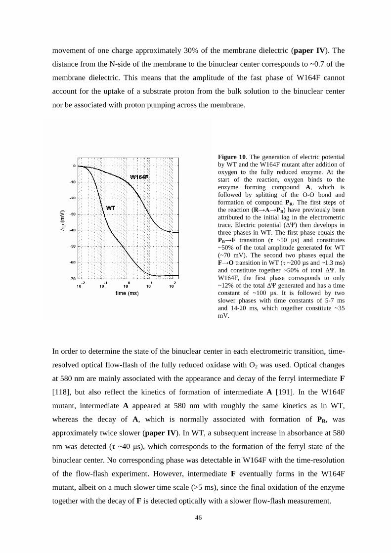
46
movement of one charge approximately 30% of the membrane dielectric (paper IV). The
distance from the N-side of the membrane to the binuclear center corresponds to ~0.7 of the
membrane dielectric. This means that the amplitude of the fast phase of W164F cannot
account for the uptake of a substrate proton from the bulk solution to the binuclear center
nor be associated with proton pumping across the membrane.
Figure 10. The generation of electric potentialby WT and the W164F mutant after addition ofoxygen to the fully reduced enzyme. At thestart of the reaction, oxygen binds to theenzyme forming compound A, which isfollowed by splitting of the O-O bond andformation of compound PR. The first steps ofthe reaction ( PR) have previously beenattributed to the initial lag in the electrometrictrace. Electric potential ( ) then develops inthree phases in WT. The first phase equals thePR F transition ( ~50 µs) and constitutes~50% of the total amplitude generated for WT(~70 mV). The second two phases equal theF O transition in WT ( ~200 µs and ~1.3 ms)and constitute together ~50% of total . InW164F, the first phase corresponds to only~12% of the total generated and has a timeconstant of ~100 µs. It is followed by twoslower phases with time constants of 5-7 msand 14-20 ms, which together constitute ~35mV.
In order to determine the state of the binuclear center in each electrometric transition, time-
resolved optical flow-flash of the fully reduced oxidase with O2 was used. Optical changes
at 580 nm are mainly associated with the appearance and decay of the ferryl intermediate F
[118], but also reflect the kinetics of formation of intermediate A [191]. In the W164F
mutant, intermediate A appeared at 580 nm with roughly the same kinetics as in WT,
whereas the decay of A, which is normally associated with formation of PR, was
approximately twice slower (paper IV). In WT, a subsequent increase in absorbance at 580
nm was detected ( ~40 s), which corresponds to the formation of the ferryl state of the
binuclear center. No corresponding phase was detectable in W164F with the time-resolution
of the flow-flash experiment. However, intermediate F eventually forms in the W164F
mutant, albeit on a much slower time scale (>5 ms), since the final oxidation of the enzyme
together with the decay of F is detected optically with a slower flow-flash measurement.

47
The optically detected decay of F in the W164F mutant (>5 ms) coincides with the
development of two slow phases ( ~5 ms and ~20 ms) in electrometry, which are
associated with generation of of the same magnitude as in the F O transition of WT.
The slow phases of electric potential generation in W164F are likely to comprise many
electrogenic events, i.e. a) the uptake of two substrate protons to the binuclear center, one
for formation of the F state and one for F O transition, b) electron transfer from CuA to
heme a and c) uptake of proton(s) that are pumped across the membrane.
The results from the optical and electrometrical flow-flash experiments appear at first sight
inexplicable. The fast phase of electric potential generation in the W164F mutant (in WT
corresponding to PR F transition) is only twice slower than WT. Yet, optically no F
intermediate develops with the same kinetics. The logical conclusion is that the small fast
electrometric phase does not include formation of the F intermediate, but reflects another
vectorial charge translocation in the W164F enzyme. In WT, the amplitude of the fast phase
(PR F transition) includes a small contribution from the partial eT between CuA and heme
a. The electron equilibration between CuA and heme a is dramatically slowed in the W164F
mutant (Figure 9) and would not attribute to the fast electrometic response. In the fast
optical flow-flash measurement, formation of the F state was not detected in the W164F
mutant during the time-resolution of the experiment. This means that the fast electrometric
phase in W164F precedes by far the formation of intermediate F in W164F. Part of the
explanation for the decrease in amplitude is hence a lack of proton uptake into the binuclear
center for the formation of F. Nevertheless, the origin of the small fast amplitude is W164F
cannot be explained by this observation.
As previously mentioned, there is a large separation in time between the fast electrometric
phase of W164F and the optically detected transition of PR state to F. Moreover, the initial
lag phase of membrane potential generation in W164F is shorter than in WT. When this is
considered, the kinetics of the development of the initial amplitude in the W164F mutant
actually coincides with the first step of the catalytic reaction in WT, the A PR transition.
PR formation is associated with transfer of an electron from heme a to the binuclear
center. This eT occurs in the plane of the membrane and does not contribute to the
generation of membrane potential. Only electrons and protons that move perpendicular to
the membrane will give rise to an electrometrically detectable signal. Since the oxidation of
CuA in W164F has a time constant of milliseconds, it is safe to assume that no electron

48
transfer events occurs perpendicular to the membrane during the fast electrometric phase.
Instead, the amplitude must originate from a vectorial proton transfer, which occurs
simultaneously with the eT from heme a to heme a3. A likely proton donor could be
Glu278, which is known to be involved in the transfer of both chemical and pumped protons
many times during enzyme turnover. The target of the proton transfer cannot be the
binuclear center. Proton delivery to the active site would immediately yield the F
intermediate, but according to our data, F is formed on a much slower time scale in the
mutant. The observed amplitude (equivalent to ~30% of the membrane dielectric) is
moreover too large to account for proton movement from Glu278 to the binuclear center,
which lay close to each other with respect to the membrane dielectric. We propose therefore
that the origin of the fast amplitude in W164F is a vectorial proton movement from Glu278
to a pump site at or above the heme propionates and that this proton movement occurs
simultaneously with the A PR transition of the catalytic cycle.
If the proton transfer that we see in the W164F mutant is associated with loading of the
pump site in the A PR transition, it should also occur in the wild-type enzyme. In WT,
such an event may be undetectable during normal experimental conditions due to the large
electrometric response of the PR F transition. However, it could be revealed when
formation of the F state is dramatically slowed, as in the W164F mutant. Recently, Belevich
and co-workers [192] studied the generation of electric potential in WT cytochrome c
oxidase at alkaline conditions, were the PR F and O transitions are slowed down
[178]. They noticed an acceleration of the rate of the fast electrometric phase and a
shortening of the initial lag, similar to our results with the W164F mutant. A comparison
with optical data revealed that the fast electrometric phase develops simultaneously with the
optically recorded A PR transition, which occurs when the electron at heme a is
transferred to the binuclear center. A previous study has shown that the reaction between
mixed-valence cytochrome c oxidase (where heme a and CuA are oxidized) and O2 does not
generate a membrane potential [137]. We can therefore conclude that the fast electric
amplitude generated in the W164F mutant, as well as in wild-type at high pH, is a result of
proton transfer from Glu278 to a proton acceptor above the hemes, and that this proton-
transfer is kinetically linked to electron transfer into the binuclear center.

49
5.7 A possible mechanism for redox-linked proton transfer
A molecular mechanism of proton translocation by cytochrome c oxidase can be proposed
based on the work presented in this thesis in combination with recent results from the
Helsinki Bioenergetics group (paper III, paper IV and [113,151,192]). The model, which
is schematically presented in Figure 11 relies heavily on the unique properties of water
molecules enclosed in hydrophobic cavities, such as the ones located between heme a and
the binuclear center. The proton pumping is initiated by loading of the pump site with a
proton, which enables the simultaneous electron transfer from heme a to the active site. Full
reduction of oxygen to water involves the step-wise transfer of four electrons to the
binuclear center. Each of these electron transfer steps are linked to proton pumping across
the membrane, which presumably occurs by the same mechanism four times during one
catalytic turnover. The details of the proposed mechanism are presented below, and are for
simplicity divided into different steps:
A. An electron is initially transferred from CuA to heme a. The reduction of heme a
raises the pKa of the pump site that is located presumably somewhere in the vicinity of heme
a3. The additional negative charge at heme a affects moreover the dipoles of the putative
water molecules in the cavity between heme a and the binuclear center. The water
molecules reorient themselves as a consequence of the electric field and form a hydrogen-
bonded water chain that connects Glu278 with the -propionate of heme a3. The strong salt
bridge between the -propionate and Arg473 is weakened by the influence of the water
chain and dissociates. The result is an increase in the pKa of the propionate that attracts a
proton from Glu278. The arriving proton can either stay on the propionate or alternatively
be further transferred to a pump site somewhere close.
B. The additional positive charge (the proton) at or near the propionate raises the
electron affinity (Em) of heme a3/CuB, after which the electron located at heme a is quickly
transferred to the binuclear center.

50
C. Glu278 is reprotonated from the bulk via the D-pathway. The change in the
orientation of the electric field between heme a and the binuclear center is sensed by the
water molecules in the cavity, which reorient themselves and form a hydrogen-bonded chain
that extend towards the active site. A proton can thereafter be transferred from Glu278 to
the active site.
D. Glu278 is again reprotonated from the D-pathway. This proton uptake together
with the neutralization of the negative charge at the binuclear center with the arrival of a
substrate proton in the previous step ultimately repels electrostatically the preloaded
pumped proton located at or near the heme a3 -propionate towards the P-side of the
membrane.
Figure 11. A schematic presentation of the proposed proton pumping mechanism. Electron transfer from hemea to the binuclear center is linked to proton transfer from E278, via water molecules, to a pump site located ator near the -propionate of heme a3 (A). Arg473 and the heme a3 -propionate form together a gate, whichregulates the access of protons to the P-side of the membrane. When the electron reaches the catalytic site itreverses the electric field between heme a and heme a3/CuB (B). The inverted field is sensed by the watermolecules, which are located in the hydrophobic cavity close by. Rearrangement of the water dipoles thenallows a proton to be transferred from Glu278 to the catalytic site (C). Upon its deprotonation, Glu278 israpidly reprotonated via the D-pathway. The reprotonation of Glu278 after transfer of a substrate proton to thebinuclear center expels the pumped proton to the P-side of the membrane (D).
H+
H+
H+
C
D124
Heme a3/CuB
A CuA
E278
Heme a
e- Mg2+
H+R473
e-
B
H+
D
H+
H+

51
5.7.1. The presented mechanism in relation to existing experimental data
In the above presented mechanism of proton transfer the pumped proton is transferred to the
pump site concurrently with the transfer of an electron from heme a to the active site. It has
been argued, that this type of mechanism cannot account for proton pumping in the PR F
transition of the catalytic cycle, since the PR F transition is not coupled to eT to the
binuclear center, which occurs already in the preceding A PR transition. Our study of the
generation of membrane potential in the W164F mutation revealed however a previously
undetected electrogenic event, which became visible due to the slow formation of the ferryl
state in the W164F mutant. This electrogenic event, which occurs kinetically simultaneous
with the optically detected A PR transition, was identified as loading of the pump site with
a proton (paper IV). The corresponding phase of charge transfer goes undetectable in WT
cytochrome c oxidase, due to the large generation of membrane potential in the subsequent
PR F transition [192]. But, in the W164F mutant, as well as in WT at high pH and in the
D124N mutant, a small electrometic phase becomes visible, which corresponds to transfer
of a charge about 30% of the membrane dielectric. This phase is absent in the Glu278 to
glutamine mutant, and may thus represent proton transfer from Glu278 to the -propionate
of heme a3 [192]. The discovery of this elementary step of the proton pumping machinery
shows that a common proton pumping mechanism can be applied to all proton-transferring
steps of the catalytic cycle, including the PR F transition, since a proton is already present
at the pump site at the onset of the transition.
During the reductive phase of the catalytic cycle two substrate protons are taken up
to the binuclear center via the K-pathway [79,80,90]. This observation is fully compatible
with our model of proton pumping. In the model, electron arrival at heme a results in the
formation of a hydrogen-bonded water chain that connects Glu278 to the -propionate of
heme a3 and allows proton transfer via a Grotthuss-type of mechanism. Next, the electron is
transferred to the active site, after which the arrival of a substrate proton to the active site
and the reprotonation of Glu278 expels the proton at the pump site. According to the model,
all pumped protons are donated from Glu278 and the D-pathway, whereas the substrate
protons can optionally be transferred via the D- or the K-pathway.
Mutation of amino acid residues in the D-pathway may create mutants, which lose
their ability to translocate protons across the membrane, but sustain varying degrees of O2
consumption activity. The D124N mutation for example severely impair catalytic activity
and abolishes proton pumping [82,83]. The catalytic cycle of D124N halts at the F

52
intermediate, since proton uptake through the D-pathway from the bulk is blocked. In the
recent electrometric study by Belevich and co-workers, a fast phase of membrane potential
generation was observed in the D124N mutant, equivalent to the one detected at pH 10.5 for
WT enzyme. The electrogenic phase was interpreted as proton transfer to the pump site
located close to the -propionate of heme a3 [192]. According to this interpretation, a proton
is initially transferred to the pump site also in the non-pumping D124N mutant. However,
since additional protons are not available, this proton is transferred to the binuclear center
and used for water formation. The lifetime of the protonated pump site in D124N is
presumably very short, since experimental data indicate that the PR F transition of the
D124N mutant occurs with the same rate as for WT [136]. The protonation state of the heme
a3 -propionate has been shown to influence the rate of eT from heme a to the binuclear
center [178]. A transient protonation of the -propionate of heme a3 in D124N would hence
insure fast eT from heme a to the active site.
Another example of a mutant in the D-pathway, which has lost its ability to
translocate protons across the membrane is the N131D mutant [193-195]. Quite contrary to
the D124N mutation, the N131D mutation yields an enzyme with higher O2 consumption
activity than WT. The high turnover activity of the N131D mutant does not necessarily
mean that the mutation has not affect the rate whereby protons are transferred through the
D-pathway. Another interpretation of the observed phenomenon may be that the N131D
mutation has affected the rate (increased) of a limiting step during catalytic turnover, for
example by un-coupling proton translocation from the chemical reaction. It is conceivable
that a proton is initially transferred to the pump site also in N131D, where after it is
instantly re-routed to the active site, due to a for instance a slowed down reprotonation of
Glu278. In the N131D mutant, the PR F transition shows WT transition rates, while the
rate of the F O transition is increased compared to WT enzyme [194]. We have shown
that loading of the pump site occurs already during A PR transition, which may be why
un-coupling of proton pumping from oxygen reduction does not affect the rate of PR F
transition in the N131D mutant. However, if the proton pumping is abolished in the
subsequent step, upon O formation, it can lead to an increased F O transition rate, even
though the rate of proton transfer via the D-pathway may be affected by the mutation.
53
5.7.2 Is the proton pumping mechanism common to all heme-copper oxidases?
It is appealing to think that the mechanism by which protons are pumped across the
membrane would be common to all heme-copper oxidases. The thought of a general
pumping mechanism is not far fetched considering that the genes encoding the heme-copper
oxidases have evolved from the same ancestor. The structural homology between the
mitochondrial-like cytochrome c oxidases and the more distantly related heme-copper
oxidases, such as the ba3 oxidase from T. thermophilus and the aa3 quinol oxidase from A.
ambivalens is however poor (discussed in chapter 5.1.1). A common pumping mechanism
would consequently have to be independent from protein structure and involve other
elements, such as water molecules. The importance of structurally conserved water
molecules in proton transfer has recently been discussed by Sharpe and co-workers [174].
Their comparison of the existing 3D structures of heme-copper oxidases revealed that
although only one amino acid residue from the D-pathway of the R. sphaeroides aa3 oxidase
is preserved in the T. thermophilus ba3 oxidase, both oxidases contain similar water
formations. An important role of water molecules in proton translocation is supported by
our results in paper I and II, were we showed that different structural motifs maintain
proton translocation in cytochrome c oxidase by providing the scaffold for formation of a
hydrogen-bonded water chain through which fast proton conduction can occur.
It is not unrealistic that the mechanism of proton transfer outlined in chapter 5.7 may
be generally applied to all heme-copper oxidases. The common structural feature of this
super family is a low-spin heme, a high-spin heme and a copper ion located close to each
other (see chapter 1.2). The near proximity of the redox centers in the heme-copper oxidases
insures fast electron delivery to the binuclear center, but may also be a criterion for redox-
linked proton translocation. In addition to the six histidine residues that ligate the redox
centers, only three amino acid residues seem to be totally conserved in subunit I of the
heme-copper oxidases [7]. Among these three residues are Arg474, which forms hydrogen
bonds to both propionates of heme a, but not Arg473, which forms a tight ion bond with the
-propionate of heme a3. Arg473 is however totally conserved amongst all other heme-
copper oxidases, but the cbb3 oxidases. The presence or absence of an arginine in the cbb3
oxidases that corresponds to Arg473 in cytochrome c oxidase from P. denitrificans may
perhaps be rethought by alternative sequence alignments. A mutational study targeted at
Arg473 have indicated that proton pumping by cytochrome c oxidase is sustained as long as
the -propionate of heme a3 receives a hydrogen bond from the surrounding protein [91]. A

54
recent homology model of the cbb3 oxidase from R. sphaeroides implies that a lysine and a
tryptophan residue are in hydrogen bonding distance of the high-spin heme [196].
A key role for water molecules in the proton pumping of the heme-copper oxidases
is supported by the structure of the ba3 oxidase from T. thermophilus, where one of the
proposed proton pathways ends at an internal cavity close to the binuclear center [166]. The
cavity contains structurally detected water molecules, which may take part in proton
transfer. Whether similar structural features are present in other heme-copper oxidase
remain to be seen with the appearance of x-ray structure of other non-canonical oxidases,
hopefully in the near future.

55
6. SUMMARY
This thesis is focused on three important aspects of the redox-linked proton transfer by
cytochrome c oxidase, namely a) the gating mechanism, whereby proton access to the
different sides of the membrane is regulated, b) the role of water molecules and the
conserved Glu278 in proton conduction, and c) the timing of proton transfer to the pump
site, located somewhere in the vicinity of the heme a3.
Theoretical calculations in combination with experimental data presented here
pinpointed the salt bride formed by the Arg473/heme a3 -propionate pair as a possible gate
that regulates proton access to the positively charged side of the membrane. Opening of the
gate is determined by the redox-state between heme a and the binuclear center and occurs
when the dipole moment of the water molecules in the cavity points towards the propionate
of heme a3. Dissociation of the Arg473/ -propionate pair is dependent on the hydrogen
bond between the -propionate and Trp164, but will not occur without the presence of water
molecules in the cavity. In addition, the electrometric measurements with the W164F
mutant revealed a new, undetected step in the proton pumping mechanism of cytochrome c
oxidase, i.e. loading of the pump site, located at or near the -propionate of heme a3, in the
A PR transition of the catalytic cycle.
Glu278, an invariant amino acid among the mitochondrial-like cytochrome c
oxidases, was shown to be dispensable for proton translocation, when other structural
solutions were present that could support a long, hydrogen-bonded chain of water through
which proton transfer by a Grotthuss-type of mechanism can occur.
Finally, a possible mechanism of proton transfer across the membrane was presented
in which proton transfer is initiated by the protonation of the pump site and the simultaneous
electron transfer between heme a and heme a3. It was speculated that a common proton
pumping mechanism could be applied to all heme-copper oxidases and that the mechanism
could be similar to the one presented in this thesis. A common proton pumping mechanism
is very likely to involve structural water molecules, since the protein structure of different
heme-copper oxidases can vary substantially.
56
7. ACKNOWLEDGEMENTS
This study was carried out in the Helsinki Bioenergetics Group at the Institute of
Biotechnology and at the Faculty of Biosciences, Division of Biochemistry, at the
University of Helsinki. The work was financially supported by the Academy of Finland, the
Sigrid Jusélius Foundation, the University of Helsinki, Biocentrum Helsinki, Viikki
Graduate School in Biosciences, and the Magnus Ehrnrooth Foundation.
I am deeply grateful to my supervisor, Docent Anne Puustinen, for all these years.
Thank you for your guidance and for sharing your knowledge of molecular biology and
protein chemistry with me.
I wish to express my gratitude to Professor Mårten Wikström, the leader of Helsinki
Bioenergetics Group, for providing excellent research conditions and allowing me to work
in the intriguing field of bioenergetics. Your enthusiasm in cytochrome c oxidase and in the
world of bioenergetics is inspiring and has motivated me to work hard in the laboratory.
I wish to sincerely thank Professor Michael Verkhovsky for being a patient teacher
in the kinetics lab and for educative discussions on biophysics.
Professor Ilmo Hassinen and Docent Moshe Finel are acknowledged for reading the
thesis manuscript and for their valuable comments and suggestions.
My warmest thanks go to all past and present members of the HBG that in any way
have helped me in my work, and have provided a nice and friendly working atmosphere. I
would like to especially express my appreciation to the co-authors of the articles, Marina
Verkhovskaya, Mika Molin, Liisa Laakkonen, Ilya Belevich and Dmitry Bloch from HGB,
and the external collaborators, Gerhard Hummer, Claudio Gomes and Miguel Teixeira.
A special thank you goes to Virve Rauhamäki, my office companion during the last
years, for good discussions about science and life, and to Pamela David, a former Post Doc
in HGB, for a memorable time during her years in Helsinki. The present and past
technicians of HGB, Eija Haasanen, Tarja Salojärvi, Katja Sissi and Anne Hakonen, require
a special mentioning due to their invaluable and excellent work. A huge thank you goes to
our former secretary Satu Sankkila for her encouragement and friendship, and for always
taking such good care of all the practical matters related to the laboratory.

57
In addition, I would like to thank all my friends and relatives, for your company on trips,
during holidays and weekends and for just being there every day to cheer up my life. My
sister Ann-Sofi and her family are acknowledged for their presence and support, and
especially Ann-Sofi for showing what one can do with enough enthusiasm and energy.
I have dedicated my thesis to my parents, Ulla and Kurt, who have always helped
and encouraged me and my sister in any way that they can. Tack för allt, mamma och
pappa!
Finally, I would like to thank my husband Johnny for being such a down-to-earth,
smart and supportive person that takes good care of me. Du är bäst, Johnny! And our little
bundle of joy, Wilmer, so eager to learn new things and explore the world. Det är bra
Wilmer att du håller mamma sysselsatt!
Helsinki, March 2007 Camilla Ribacka

58
8. REFERENCES
1. Brandt, U. (2006) Annu. Rev. Biochem. 75, 69-92.2. Cecchini, G. (2003) Annu. Rev. Biochem. 72, 77-109.3. Osyczka, A., Moser, C. C., and Dutton, P. L. (2005) TIBS 30, 176-182.4. Walker, J. E. and Dickson, V. K. (2006) Biochim. Biophys. Acta 1757, 286-296.5. Mitchell, P. (1961) Nature 191, 141-148.6. Garcia-Horsman, J. A., Barquera, B., Rumbley, J., Ma, J., and Gennis, R. B. (1994)
J. Bacteriol. 176, 5587-5600.7. Pereira, M. M., Santana, M., and Teixeira, M. (2001) Biochim. Biophys. Acta 1505,
185-208.8. Lübben, M. and Morand, K. (1994) J. Biol. Chem. 269, 21473-21479.9. Woese, C. R. (1987) Microbiol. Rev. 51, 221-271.10. Yang, D., Oyaizu, Y., Oyaizu, H., Olsen, G. J., and Woese, C. R. (1985) Proc. Natl.
Acad. Sci. USA 82, 4443-4447.11. John, P. and Whatley, F. R. (1977) Biochim. Biophys. Acta 463, 129-153.12. van Spanning, R. J., de Boer, A. P., Reijnders, W. N., De Gier, J. W., Delorme, C.
O., Stouthamer, A. H., Westerhoff, H. V., Harms, N., and van der Oost, J. (1995) J.Bioenerg. Biomembr. 27, 499-512.
13. Baker, S. C., Ferguson, S. J., Ludwig, B., Page, M. D., Richter, O. M., and vanSpanning, R. J. (1998) Microbiol. Mol. Biol. Rev. 62, 1046-1078.
14. Puustinen, A., Finel, M., Virkki, M., and Wikström, M. (1989) FEBS Lett. 249, 163-167.
15. Richter, O. M., Tao, J., Turba, A., and Ludwig, B. (1994) J. Biol. Chem. 269, 23079-23086.
16. Raitio, M., Jalli, T., and Saraste, M. (1987) EMBO J. 6, 2825-2833.17. De Gier, J. W., Schepper, M., Reijnders, W. N., van Dyck, S. J., Slotboom, D. J.,
Warne, A., Saraste, M., Krab, K., Finel, M., Stouthamer, A. H., van Spanning, R. J.,and van der Oost, J. (1996) Mol. Microbiol. 20, 1247-1260.
18. van Verseveld, H. W., Krab, K., and Stouthamer, A. H. (1981) Biochim. Biophys.Acta 635, 525-534.
19. Raitio, M. and Wikström, M. (1994) Biochim. Biophys. Acta 1186, 100-106.20. Wikström, M. (1977) Nature 266, 271-273.21. Iwata, S., Ostermeier, C., Ludwig, B., and Michel, H. (1995) Nature 376, 660-669.22. Tsukihara, T., Aoyama, H., Yamashita, E., Takashi, T., Yamaguichi, H., Shinzawa-
Itoh, K., Nakashima, R., Yaono, R., and Yoshikawa, S. (1996) Science 272, 1136-1144.
23. Ferguson-Miller, S. and Babcock, G. T. (1996) Chem. Rev. 7, 2889-2907.24. Kadenbach, B., Napiwotzki, J., Frank, V., Arnold, S., Exner, S., and Huttemann, M.
(1998) J. Bioenerg. Biomembr. 30, 25-33.25. Buse, G., Soulimane, T., Dewor, M., Meyer, H. E., and Bluggel, M. (1999) Protein
Science 8, 985-990.26. Ostermeier, C., Harrenga, A., Ermler, U., and Michel, H. (1997) Proc. Natl. Acad.
Sci. USA 94, 10547-10553.27. Yoshikawa, S., Shinzawa-Itoh, K., Nakashima, R., Yaono, R., Inoue, N., Yao, M.,
Fei, M. J., Libeu, C. P., Mizushima, T., Yamaguchi, H., Tomizaki, T., andTsukihara, T. (1998) Science 280, 1723-1729.

59
28. Svensson-Ek, M., Abramson, J., Larsson, G., Törnroth, S., Brzezinski, P., and Iwata,S. (2002) J. Mol. Biol. 321, 329-339.
29. Mills, D. A., Florens, L., Hiser, C., Qian, J., and Ferguson-Miller, S. (2000)Biochim. Biophys. Acta 1458, 180-187.
30. Florens, L., Schmidt, B., McCracken, J., and Ferguson-Miller, S. (2001)Biochemistry 40, 7491-7497.
31. Lee, A., Kirichenko, A., Vygodina, T., Siletsky, S. A., Das, T. K., Rousseau, D. L.,Gennis, R., and Konstantinov, A. A. (2002) Biochemistry 41, 8886-8898.
32. Riistama, S., Laakkonen, L., Wikström, M., Verkhovsky, M. I., and Puustinen, A.(1999) Biochemistry 38, 10670-10677.
33. Pfitzner, U., Kirichenko, A., Konstantinov, A. A., Mertens, M., Wittershagen, A.,Kolbesen, B. O., Steffens, G. C. M., Harrenga, A., Michel, H., and Ludwig, B.(1999) FEBS Lett. 456, 365-369.
34. Harrenga, A. and Michel, H. (1999) J. Biol. Chem. 274, 33296-33299.35. Humphrey, W., Dalke, A., and Schulten, K. (1996) J. Mol. Graph. 14, 33.36. Steffens, G. C. M., Soulimane, T., Wolff, G., and Buse, G. (1993) Eur. J. Biochem.
213, 1149-1157.37. Kelly, M., Lappalainen, P., Talbo, G., Haltia, T., Van der Oost, J., and Saraste, M.
(1993) J. Biol. Chem. 268, 16781-16787.38. Witt, H., Malatesta, F., Nicoletti, F., Brunori, M., and Ludwig, B. (1998) Eur. J.
Biochem. 251, 367-373.39. Riistama, S., Puustinen, A., Garcia-Horsman, A., Iwata, S., Michel, H., and
Wikström, M. (1996) Biochim. Biophys. Acta 1275, 1-4.40. Haltia, T., Finel, M., Harms, N., Nakari, T., Raitio, M., Wikström, M., and Saraste,
M. (1989) EMBO J. 8, 3571-3579.41. Echabe, I., Haltia, T., Freire, E., Goni, F. M., and Arrondo, J. L. (1995) Biochemistry
34, 13565-13569.42. Bratton, M. R., Pressler, M. A., and Hosler, J. P. (1999) Biochemistry 38, 16236-
16245.43. Gilderson, G., Salomonsson, L., Aagaard, A., Gray, J., Brzezinski, P., and Hosler, J.
(2003) Biochemistry 42, 7400-7409.44. Mills, D. A., Tan, Z., Ferguson-Miller, S., and Hosler, J. (2003) Biochemistry 42,
7410-7417.45. Witt, H. and Ludwig, B. (1997) J. Biol. Chem. 272, 5514-5517.46. Antalis, T. M. and Palmer, G. (1982) J. Biol. Chem. 257, 6194-6206.47. Zhen, Y., Hoganson, C. W., Babcock, G. T., and Ferguson-Miller, S. (1999) J. Biol.
Chem. 274, 38032-38041.48. Hill, B. C. (1991) J. Biol. Chem. 266, 2219-2226.49. Geren, L. M., Beasley, J. R., Fine, B. R., Saunders, A. J., Hibdon, S., Pielak, G. J.,
Durham, B., and Millett, F. (1995) J. Biol. Chem. 270, 2466-2472.50. Verkhovsky, M. I., Belevich, N., Morgan, J. E., and Wikström, M. (1999) Biochim.
Biophys. Acta 1412, 184-189.51. Nilsson, T. (1992) Proc. Natl. Acad. Sci. USA 89, 6497-6501.52. Verkhovsky, M. I., Tuukkanen, A., Backgren, C., Puustinen, A., and Wikström, M.
(2001) Biochemistry 40, 7077-7083.53. Boelens, R. and Wever, R. (1979) Biochim. Biophys. Acta 547, 296-310.54. Verkhovsky, M. I., Morgan, J. E., and Wikström, M. (1992) Biochemistry 31,
11860-11863.55. Oliveberg, M. and Malmström, B. G. (1991) Biochemistry 30, 7053-7057.56. Marcus, R. A. and Sutin, N. (1985) Biochim. Biophys. Acta. 811, 265-322.

60
57. Gray, H. B. and Winkler, J. R. (1996) Annu. Rev. Biochem. 65, 537-561.58. Moser, C. C., Keske, J. M., Warncke, K., Farid, R. S., and Dutton, P. L. (1992)
Nature 355, 796-802.59. Page, C. C., Moser, C. C., Chen, X. X., and Dutton, P. L. (1999) Nature 402, 47-52.60. Pilet, E., Jasaitis, A., Liebl, U., and Vos, M. H. (2004) Proc. Natl. Acad. Sci. USA
101, 16198-16203.61. Verkhovsky, M. I., Jasaitis, A., and Wikström, M. (2001) Biochim. Biophys. Acta
1506, 143-146.62. Verkhovsky, M. I., Morgan, J. E., and Wikström, M. (1995) Biochemistry 34, 7483-
7491.63. Agmon, N. (1995) Chem. Phys. Lett. 244, 456-462.64. Cukierman, S. (2006) Biochim. Biophys. Acta 1757, 876-885.65. Nagle, J. F. and Morowitz, H. J. (1978) Proc. Natl. Acad. Sci. USA 75, 298-302.66. Hummer, G., Rasaiah, J. C., and Noworyta, J. P. (2001) Nature 414, 188-190.67. Svensson, M., Hallen, S., Thomas, J. W., Lemieux, L., Gennis, R. B., and Nilsson,
T. (1995) Biochemistry 34, 5252-5258.68. Hosler, J. P., Shapleigh, J. P., Kim, Y., Pressler, M., Georgiou, C., Babcock, G. T.,
Alben, J. O., Ferguson-Miller, S., and Gennis, R. B. (1996) Biochemistry 35, 10776-10783.
69. Junemann, S., Meunier, B., Gennis, R. B., and Rich, P. (1997) Biochemistry 36,14456-14464.
70. Hofacker, I. and Schulten, K. (1998) Proteins 30, 100-107.71. Cukier, R. I. (2005) Biochim. Biophys. Acta 1706, 134-146.72. Brändén, M., Sigurdson, H., Namslauer, A., Gennis, R. B., Ädelroth, P., and
Brzezinski, P. (2001) Proc. Natl. Acad. Sci. USA 98, 5013-5018.73. Kannt, A., Lancaster, C. R. D., and Michel, H. (1998) Biophys. J. 74, 708-721.74. Ma, J. X., Tsatsos, P. H., Zaslavsky, D., Barquera, B., Thomas, J. W., Katsonouri,
A., Puustinen, A., Wikström, M., Brzezinski, P., Alben, J. O., and Gennis, R. B.(1999) Biochemistry 38, 15150-15156.
75. Brändén, M., Tomson, F., Gennis, R. B., and Brzezinski, P. (2002) Biochemistry 41,10794-10798.
76. Tomson, F. L., Morgan, J. E., Gu, G. P., Barquera, B., Vygodina, T. V., and Gennis,R. B. (2003) Biochemistry 42, 1711-1717.
77. Richter, O. M. H., Durr, K. L., Kannt, A., Ludwig, B., Scandurr, F. M., Giuffre, A.,Sarti, P., and Hellwig, P. (2005) Febs Journal 272, 404-412.
78. Tuukkanen, A., Verkhovsky, M. I., Laakkonen, L., and Wikström, M. (2006)Biochim. Biophys. Acta 1757, 1117-1121.
79. Ädelroth, P., Gennis, R. B., and Brzezinski, P. (1998) Biochemistry 37, 2470-2476.80. Wikström, M., Jasaitis, A., Backgren, C., Puustinen, A., and Verkhovsky, M. I.
(2000) Biochim. Biophys. Acta 1459, 514-520.81. Konstantinov, A. A., Siletskiy, S. A., Mitchell, D., Kaulen, A. D., and Gennis, R. B.
(1997) Proc. Natl. Acad. Sci. USA 94, 9085-9090.82. Thomas, J. W., Puustinen, A., Alben, J. O., Gennis, R. B., and Wikström, M. (1993)
Biochemistry 32, 10923-10928.83. Fetter, J. R., Qian, J., Shapleigh, J., Thomas, J. W., Garcia-Horsman, A., Schmidt,
E., Hosler, J., Babcock, G. T., Gennis, R. B., and Ferguson-Miller, S. (1995) Proc.Natl. Acad. Sci. USA 92, 1604-1608.
84. Garcia-Horsman, J. A., Puustinen, A., Gennis, R. B., and Wikström, M. (1995)Biochemistry 34, 4428-4433.
61
85. Riistama, S., Hummer, G., Puustinen, A., Dyer, R. B., Woodruff, W. H., andWikström, M. (1997) FEBS Lett. 414, 275-280.
86. Puustinen, A., Bailey, J. A., Dyer, R. B., Mecklenburg, S. L., Wikström, M., andWoodruff, W. H. (1997) Biochemistry 36, 13195-13200.
87. Zheng, X. H., Medvedev, D. M., Swanson, J., and Stuchebrukhov, A. A. (2003)Biochim. Biophys. Acta 1557, 99-107.
88. Ädelroth, P., Svensson, E. M., Mitchell, D. M., Gennis, R. B., and Brzezinski, P.(1997) Biochemistry 36, 13824-13829.
89. Verkhovskaya, M. L., García-Horsman, A., Puustinen, A., Rigaud, J. L., Morgan, J.E., Verkhovsky, M. I., and Wikström, M. (1997) Proc. Natl. Acad. Sci. USA 94,10128-10131.
90. Belevich, I., Tuukkanen, A., Wikström, M., and Verkhovsky, M. I. (2006)Biochemistry 45, 4000-4006.
91. Puustinen, A. and Wikström, M. (1999) Proc. Natl. Acad. Sci. USA 96, 35-37.92. Pomes, R., Hummer, G., and Wikstrom, M. (1998) Biochim. Biophys. Acta 1365,
255-260.93. Tsukihara, T., Shimokata, K., Katayama, Y., Shimada, H., Muramoto, K., Aoyama,
H., Mochizuki, M., Shinzawa-Itoh, K., Yamashita, E., Yao, M., Ishimura, Y., andYoshikawa, S. (2003) Proc. Natl. Acad. Sci. U. S. A 100, 15304-15309.
94. Pfitzner, U., Odenwald, A., Ostermann, T., Weingard, L., Ludwig, B., and Richter,O. M. H. (1998) J. Bioenerg. Biomembr. 30, 89-97.
95. Lee, H. M., Das, T. K., Rousseau, D. L., Mills, D., Ferguson-Miller, S., and Gennis,R. B. (2000) Biochemistry 39, 2989-2996.
96. Behr, J., Michel, H., Mäntele, W., and Hellwig, P. (2000) Biochemistry 39, 1356-1363.
97. Popovic, D. M. and Stuchebrukhov, A. A. (2004) FEBS Lett. 566, 126-130.98. Popovic, D. M. and Stuchebrukhov, A. A. (2004) J. Am. Chem. Soc. 126, 1858-
1871.99. Siegbahn, P. E. M., Blomberg, M. R. A., and Blomberg, M. L. (2003) J. Phys.
Chem. B 107, 10946-10955.100. Popovic, D. M. and Stuchebrukhov, A. A. (2005) J. Phys. Chem. B 109, 1999-2006.101. Busenlehner, L. S., Salomonsson, L., Brzezinski, P., and Armstrong, R. N. (2006)
Proc. Natl. Acad. Sci. U. S. A 103, 15398-15403.102. Schmidt, B., McCracken, J., and Ferguson-Miller, S. (2003) Proc. Natl. Acad. Sci
USA 100, 15539-15542.103. Riistama, S., Puustinen, A., Verkhovsky, M. I., Morgan, J. E., and Wikström, M.
(2000) Biochemistry 39, 6365-6372.104. Mitchell, R. and Rich, P. R. (1994) Biochim. Biophys. Acta 1186, 19-26.105. Rich, P. R. (1995) Aust. J. Plant Physiol. 22, 479-486.106. Moody, A. J. (1996) Biochim. Biophys. Acta 1276, 6-20.107. Moody, A. J., Cooper, C. E., and Rich, P. R. (1991) Biochim. Biophys. Acta 1059,
189-207.108. Brudvig, G. W., Stevens, T. H., Morse, R. H., and Chan, S. I. (1981) Biochemistry
20, 3912-3921.109. Baker, G. M., Noguchi, M., and Palmer, G. (1987) J. Biol. Chem. 262, 595-604.110. Morgan, J. E., Blair, D. F., and Chan, S. I. (1985) J. Inorg. Biochem. 23, 295-302.111. Antalik, M., Jancura, D., Palmer, G., and Fabian, M. (2005) Biochemistry 44, 14881-
14889.112. Verkhovsky, M. I., Jasaitis, A., Verkhovskaya, M. L., Morgan, L., and Wikström,
M. (1999) Nature 400, 480-483.

62
113. Bloch, D., Belevich, I., Jasaitis, A., Ribacka, C., Puustinen, A., Verkhovsky, M. I.,and Wikström, M. (2004) Proc. Natl. Acad. Sci. USA 101, 529-533.
114. Wikström, M. (2004) Biochim. Biophys. Acta 1655, 241-247.115. Ruitenberg, M., Kannt, A., Bamberg, E., Fendler, K., and Michel, H. (2002) Nature
417, 99-102.116. Chance, B., Saronio, C., and Leigh, J. S., Jr. (1975) J. Biol. Chem. 250, 9226-9237.117. Verkhovsky, M. I., Morgan, J. E., and Wikström, M. (1994) Biochemistry 33, 3079-
3086.118. Wikström, M. (1981) Proc. Natl. Acad. Sci. USA 78, 4051-4054.119. Babcock, G. T. and Wikström, M. (1992) Nature 356, 301-309.120. Proshlyakov, D. A., Ogura, T., Shinzawa-Itoh, K., Yoshikawa, S., Appelman, E. H.,
and Kitagawa, T. (1994) J. Biol. Chem. 269, 29385-29388.121. Proshlyakov, D. A., Ogura, T., Shinzawa-Itoh, K., Yoshikawa, S., and Kitagawa, T.
(1996) Biochemistry 35, 76-82.122. Proshlyakov, D. A., Pressler, M. A., and Babcock, G. T. (1998) Proc. Natl. Acad.
Sci. USA 95, 8020-8025.123. Fabian, M., Wong, W. W., Gennis, R. B., and Palmer, G. (1999) Proc. Natl. Acad.
Sci USA 96, 13114-13117.124. Hallen, S. and Nilsson, T. (1992) Biochemistry 31, 11853-11859.125. Mitchell, R., Mitchell, P., and Rich, P. R. (1992) Biochim. Biophys. Acta 1101, 188-
191.126. Oliveberg, M., Hallén, S., and Nilsson, T. (1991) Biochemistry 30, 436-440.127. Karpefors, M., Ädelroth, P., Namslauer, A., Zhen, Y. J., and Brzezinski, P. (2000)
Biochemistry 39, 14664-14669.128. Morgan, J. E., Verkhovsky, M. I., and Wikström, M. (1996) Biochemistry 35,
12235-12240.129. Morgan, J. E., Verkhovsky, M. I., Palmer, G., and Wikström, M. (2001)
Biochemistry 40, 6882-6892.130. Proshlyakov, D. A., Pressler, M. A., DeMaso, C., Leykam, J. F., Dewitt, D. L., and
Babcock, G. T. (2000) Science 290, 1588-1591.131. Iwaki, M., Breton, J., and Rich, P. R. (2002) Biochim. Biophys. Acta 1555, 116-121.132. Iwaki, M., Puustinen, A., Wikström, M., and Rich, P. R. (2003) Biochemistry 42,
8809-8817.133. Sucheta, A., Szundi, I., and Einarsdottir, O. (1998) Biochemistry 37, 17905-17914.134. Iwaki, M., Puustinen, A., Wikström, M., and Rich, P. R. (2006) Biochemistry 45,
10873-10885.135. Karpefors, M., Ädelroth, P., Zhen, Y. J., Ferguson-Miller, S., and Brzezinski, P.
(1998) Proc. Natl. Acad. Sci. USA 95, 13606-13611.136. Smirnova, I., Ädelroth, P., Gennis, R. B., and Brzezinski, P. (1999) Biochemistry 38,
6826-6833.137. Jasaitis, A., Verkhovsky, M. I., Morgan, J. E., Verkhovskaya, M. L., and Wikström,
M. (1999) Biochemistry 38, 2697-2706.138. Faxen, K., Gilderson, G., Ädelroth, P., and Brzezinski, P. (2005) Nature 437, 286-
289.139. Verkhovsky, M. I., Morgan, J. E., and Wikström, M. (1996) Proc. Natl. Acad. Sci.
USA 93, 12235-12239.140. Zaslavsky, D., Sadoski, R. C., Wang, K., Durham, B., Gennis, R. B., and Millett, F.
(1998) Biochemistry 37, 14910-14916.141. Wikström, M. (1998) Biochim. Biophys. Acta 1365, 185-192.142. Brzezinski, P. and Larsson, G. (2003) Biochim. Biophys. Acta 1605, 1-13.

63
143. Krab, K. and Wikström, M. (1987) Biochim. Biophys. Acta 895, 25-39.144. Morgan, J. E., Verkhovsky, M. I., and Wikström, M. (1994) J. Bioenerg. Biomembr.
26, 599-608.145. Malmström, B. G. (1989) FEBS Lett. 250, 9-21.146. Wikström, M. (1989) Nature 338, 776-778.147. Wikström, M. and Morgan, J. E. (1992) J. Biol. Chem. 267, 10266-10273.148. Verkhovsky, M. I., Morgan, J. E., Verkhovskaya, M., and Wikström, M. (1997)
Biochim. Biophys. Acta 1318, 6-10.149. Michel, H. (1999) Biochemistry 38, 15129-15140.150. Papa, S., Capitanio, N., Capitanio, G., and Palese, L. L. (2004) Biochim. Biophys.
Acta 1658, 95-105.151. Wikström, M., Verkhovsky, M. I., and Hummer, G. (2003) Biochim. Biophys. Acta
1604, 61-65.152. Raitio, M., Pispa, J. M., Metso, T., and Saraste, M. (1990) FEBS Lett. 261, 431-435.153. Kovach, M. E., Phillips, R. W., Elzer, P. H., Roop, R. M., and Peterson, K. M.
(1994) Biotechniques 16, 800-802.154. Mitchell, P., Moyle, J., and Mitchell, R. (1979) Methods Enzymol. 55, 627-640.155. Wikström, M. and Penttilä (1982) FEBS Lett. 144.156. Rigaud, J. L., Pitard, B., and Levy, D. (1995) Biochim. Biophys. Acta 1231, 223-246.157. Gibson, Q. H. and Greenwood, C. (1963) Biochem. J. 86, 541-554.158. Drachev, L. A., Jasaitis, A. A., Kaulen, A. D., Kondrashin, A. A., Liberman, E. A.,
Nemecek, I. B., Ostroumov, S. A., Semenov, A. Y., and Skulachev, V. P. (1974)Nature 249, 321-324.
159. Cornell, W. D., Cieplak, P., Bayly, C. I., Gould, I. R., Merz, K. M., Ferguson, D. M.,Spellmeyer, D. C., Fox, T., Caldwell, J. W., and Kollman, P. A. (1995) J. Am. Chem.Soc. 117, 5179-5197.
160. Pearlman, D. A., Case, D. A., Caldwell, J. W., Ross, W. S., Cheatham, T. E., Debolt,S., Ferguson, D., Seibel, G., and Kollman, P. (1995) Computer PhysicsCommunications 91, 1-41.
161. Thomas, J. W., Calhoun, M. W., Lemieux, L. J., Puustinen, A., Wikström, M.,Alben, J. O., and Gennis, R. B. (1994) Biochemistry 33, 13013-13021.
162. Pereira, M. M., Verkhovskaya, M. L., Teixeira, M., and Verkhovsky, M. I. (2000)Biochemistry 39, 6336-6340.
163. Kannt, A., Soulimane, T., Buse, G., Becker, A., Bamberg, E., and Michel, H. (1998)FEBS Lett. 434, 17-22.
164. Honnami, K. and Oshima, T. (1984) Biochemistry 23, 454-460.165. Pereira, M. M., Santana, M., Soares, C. M., Mendes, J., Carita, J. N., Fernandes, A.
S., Saraste, M., Carrondo, M. A., and Teixeira, M. (1999) Biochim. Biophys. Acta1413, 1-13.
166. Soulimane, T., Buse, G., Bourenkov, G. P., Bartunik, H. D., Huber, R., and Than, M.E. (2000) EMBO Journal 19, 1766-1776.
167. Keightley, J. A., Zimmermann, B. H., Mather, M. W., Springer, P., Pastuszyn, A.,Lawrence, D. M., and Fee, J. A. (1995) J. Biol. Chem. 270, 20345-20358.
168. Das, T. K., Gomes, C. M., Teixeira, M., and Rousseau, D. L. (1999) Proc. Natl.Acad. Sci. USA 96, 9591-9596.
169. Purschke, G. W., Schmidt, C. L., Petrsen, A., and Schafer, G. (1997) J. Bacteriol.179, 1344-1353.
170. Aagaard, A., Gilderson, G., Mills, D. A., Ferguson-Miller, S., and Brzezinski, P.(2000) Biochemistry 39, 15847-15850.
64
171. Salomonsson, L., Lee, A., Gennis, R. B., and Brzezinski, P. (2004) Proc. Natl. Acad.Sci. USA 101, 11617-11621.
172. Olkhova, E., Hutter, M. C., Lill, M. A., Helms, V., and Michel, H. (2004) Biophys.J. 86, 1873-1889.
173. Cukier, R. I. (2004) Biochim. Biophys. Acta 1656, 189-202.174. Sharpe, M. A., Qin, L., and Ferguson-Miller, S. (2005) in Biophysical and Structural
Aspects of Bioenergetics (Mårten Wikström, Ed.) pp 26-54, RSC Publishing,Cambridge, UK.
175. Warshel, A. (1981) Biochemistry 20, 3167-3177.176. Pereira, M. M., Sousa, F. L., Teixeira, M., Nyquist, R. M., and Heberle, J. (2006)
FEBS Letters 580, 1350-1354.177. Behr, J., Hellwig, P., Mäntele, W., and Michel, H. (1998) Biochemistry 37, 7400-
7406.178. Brändén, G., Brändén, M., Schmidt, B., Mills, D. A., Ferguson-Miller, S., and
Brzezinski, P. (2005) Biochemistry 44, 10466-10474.179. Lee, Y. S. and Krauss, M. (2004) J. Am. Chem. Soc. 126, 2225-2230.180. Seibold, S. A., Mills, D. A., Ferguson-Miller, S., and Cukier, R. I. (2005)
Biochemistry 44, 10475-10485.181. Mills, D. A., Geren, L., Hiser, C., Schmidt, B., Durham, B., Millett, F., and
Ferguson-Miller, S. (2005) Biochemistry 44, 10457-10465.182. Kornblatt, J. A., Kornblatt, M. J., Rajotte, I., Gaston, H. B. H., and Kahn, P. C.
(1998) Biophys. J. 75, 435-444.183. Mills, D. A. and Ferguson-Miller, S. (2002) Biochim. Biophys. Acta 1555, 96-100.184. Junemann, S., Heathcote, P., and Rich, P. R. (2000) Biochim. Biophys. Acta 1456,
56-66.185. Pecoraro, C., Gennis, R. B., Vygodina, T. V., and Konstantinov, A. A. (2001)
Biochemistry 40, 9695-9708.186. Watmough, N. J., Cheesman, M. R., Greenwood, C., and Thomson, A. J. (1994)
Biochem. J. 300, 469-475.187. Brittain, T., Little, R. H., Greenwood, C., and Watmough, N. J. (1996) FEBS Lett.
399, 21-25.188. Moody, A. J. and Rich, P. R. (1994) Eur. J. Biochem. 226, 731-737.189. Vygodina, T. and Konstantinov, A. (1989) Biochim. Biophys. Acta 973, 390-398.190. Fabian, M. and Palmer, G. (2001) Biochemistry 40, 1867-1874.191. Hill, B. C. and Greenwood, C. (1984) Biochem. J. 218, 913-921.192. Belevich, I., Verkhovsky, M. I., and Wikström, M. (2006) Nature 440, 829-832.193. Pfitzner, U., Hoffmeier, K., Harrenga, A., Kannt, A., Michel, H., Bamberg, E.,
Richter, O. M., and Ludwig, B. (2000) Biochemistry 39, 6756-6762.194. Pawate, A. S., Morgan, J., Namslauer, A., Mills, D., Brzezinski, P., Ferguson, M.,
and Gennis, R. B. (2002) Biochemistry 41, 13417-13423.195. Namslauer, A., Pawate, A. S., Gennis, R., and Brzezinski, P. (2003) Proc. Natl.
Acad. Sci. USA 100, 15543-15547.196. Sharma, V., Puustinen, A., Wikström, M., and Laakkonen, L. (2006) Biochemistry
45, 5754-5765.