recurrent aphthous ulceration - e-thesis /...
TRANSCRIPT

1
Department of Medicine (Invärtes medicin), and Department of Oral Medicine
Helsinki University Hospital,Oral Pathology Unit, Laboratory Diagnostics, Helsinki University Central Hospital;
Departments of Oral Medicine and Oral Pathology, Institute of Dentistry,ORTON Research Institute, Invalid Foundation; and
Inflammation Research Group, Biomedicum Helsinki/Anatomy, University of Helsinki, Helsinki, Finland
RECURRENT APHTHOUS ULCERATION
Immuno-pathological aspects
Sirajedin Sabri Natah
Academic dissertation
To be publicly discussed with the assent of the Faculty of Medicine,University of Helsinki, in the Auditorium of the
Haartman Institute on Thursday, June 7th, 2001, at 12 noon.
Helsinki 2001

2
Supervised by:
Prof. Yrjö T. Konttinen, MD, PhDDepartments of Medicine (Invärtes medicine) and Oral Medicine, Helsinki University CentralHospital and Institute of Dentistry, Head of Inflammation Research Group (TULES), Instituteof Biomedicine, Biomedicum Helsinki/AnatomyUniversity of Helsinki
Prof. Jarkko Hietanen, MD, DDS, PhD, MScDepartment of Oral Pathology, Institute of DentistryUniversity of Helsinki
Prof. (Emer) Maria Malmström, DDS, PhDDepartment of Oral Medicine, Institute of Dentistry,University of Helsinki
Reviewed by:
Docent Riitta Suuronen, MD, DDS, PhDDepartment of Oral and Maxillofacial Surgery,Helsinki University Central HospitalUniversity of Helsinki
Docent Pekka Laurila, MD, PhDDepartment of Pathology, Helsinki University Central HospitalUniversity of Helsinki
Official Opponent:
Prof. Auli Toivanen, MD, PhDDepartment of MedicineUniversity of Turku,Turku, Finland
ISBN 952-91-3499-1 Helsinki 2001Yliopistopaino

3
This work is dedicated to those whosuffer or might suffer from
recurrent aphthous ulceration
4
CONTENTS
ABBREVIATIONS ...................................................................................... 6
LIST OF ORIGINAL PUBLICATIONS ........................................................ 7
INTRODUCTION ........................................................................................ 8
REVIEW OF THE LITERATURE ................................................................ 10Definition, description and clinical forms of RAU ............................... 10Epidemiology of RAU ......................................................................... 11Factors predisposing to RAU ............................................................. 12
Age and sex ...................................................................... 12Family and heredity ........................................................... 12RAU and hormonal changes ............................................. 14Food hypersensitivity ........................................................ 14Drugs ................................................................................. 14Hematinic deficiencies ...................................................... 15Zinc deficiency .................................................................. 16Environmental factors ........................................................ 17
Stress ....................................................................... 17Local trauma ............................................................. 17Tobacco .................................................................... 17
Infectious factors ............................................................... 18Bacterial agents ......................................................... 18Viral agents ................................................................ 19
Heat shock proteins ............................................................................ 20Serology of RAU ................................................................................ 21Important systemic diseases associated with RAU ............................ 21
Coeliac disease ................................................................. 21Behçet’s disease (BD) ....................................................... 22HIV-associated RAU ......................................................... 23
Important effector cells participating in RAU inflammation ................. 25Neutrophils ........................................................................ 25Macrophages ..................................................................... 26Mast cells .......................................................................... 26Factor XIIIa+ dendrocytes as a memberof the subepithelial immune system .................................. 27Gamma/delta T-lymphocytes ............................................. 28TNF-" and RAU ................................................................ 30
Histopathology of recurrent aphthous ulcer ....................................... 30I. The ulcer area ........................................................................ 30II. The area lateral to the ulcer .................................................. 31
Immunohistopathology of RAU ........................................................... 31
THE AIMS OF THE STUDY ....................................................................... 34

5
PATIENTS AND METHODS ...................................................................... 35Patients and diagnostic criteria .......................................................... 35Sample collection, processing and storage ofcontrol and RAU specimens ............................................................... 41Experimental induction of traumatic ulcers ........................................ 41Histologic staining .............................................................................. 42Antigen-retrieval methods .................................................................. 42
Pepsin treatment ........................................................................ 42Microwave treatment .................................................................. 43
Primary antibodies used in the study ................................................. 43Immunohistochemistry protocols ........................................................ 44
Avidin-biotin-peroxidase complex (ABC) staining ...................... 44Peroxidase-anti-peroxidase (PAP) complex staining ................. 45Specificity .................................................................................. 46
Assessment and quantification of immunohistochemicalstaining ............................................................................................... 46Statistical analysis .............................................................................. 48
RESULTS ................................................................................................... 49Factor XIIIa+ dendrocytes in RAU (I) ................................................. 49Mast cells in RAU and TU (II) ............................................................. 51Tumor necrosis factor-" in RAU (III) .................................................. 52Expression of Ki-67 antigen in RAU and TU (IV) ............................... 54Lymphocytes bearing CD3, "$ and (* T-cell receptors in RAU (V) ...56
DISCUSSION .............................................................................................. 59Diagnosis ........................................................................................... 59Staining and evaluation methods ....................................................... 60Control specimens ............................................................................. 61Factor XIIIa+ dendrocytes (study I) .................................................... 62Mast cells (study II) ............................................................................ 64Tumor necrosis factor-" (study III) ..................................................... 68Cell proliferation in RAU lesions (study IV) ........................................ 71Intraepithelial lymphocytes (study V) ................................................. 74
SUMMARY & CONCLUSION .................................................................... 79
ACKNOWLEDGMENTS .......................................................................... 82
REFERENCES ........................................................................................... 85

6
LIST OF ABBREVIATIONS
ABC avidin-biotin-peroxidase complex"$ T cell alpha/beta T cellADCC antibody-dependent cellular cytotoxicityAEC 3-amino-9- ethylcarbazoleAECA anti-endothelial cell autoantibodiesBD Behçet’s diseaseCD cluster of differentiationCD4+ helper-inducer T lymphocyteCD8+ suppressor-cytotoxic T lymphocyteCMV cytomegalovirusDAB 3,3-diaminobenzidine tetrahydrochlorideEBV Epstein-Barr virusFGF-7 fibroblast growth factor-7(* T cell gamma/delta T cellG-CSF granulocyte-colony stimulating factorHCl hydrochloric acidHHV-6 herpes hominis virus-6HIV human immunodeficiency virusHLA human leucocyte antigenHsp heat shock proteinHSV herpes simplex virusHuRAU herpetiform ulcer RAUICAM-1 intercellular adhesion molecule-1IgG immunoglobulin G (also IgM, IgA, IgD, IgE)IL-10 interleukin 10IFN-( interferon-gammakDa kilodaltonLFA-3 lymphocyte function-antigen-3MaRAU major RAUMC mast cellMHC major histocompatibility complexMiRAU minor RAUMMPs matrix metalloproteinasesNK-cell natural killer cellNO nitric oxidePAP peroxidase-antiperoxidasePCR polymerase chain reactionRAU recurrent aphthous ulcerRNA ribonucleic acidTBS tris-HCl buffered salineTCR T-cell receptorTNF-" tumor necrosis factor-"TU traumatic ulcerVZV varicella-zoster virus
7
LIST OF ORIGINAL PUBLICATIONS
This dissertation is based on the following original publications, referred to in the text by theirRoman numerals (I-V).
I. Natah SS, Häyrinen-Immonen R, Hietanen J, Malmström M, Konttinen YT. FactorXIIIa-positive dendrocytes are increased in number and size in recurrent aphthous ulcers(RAU). J Oral Pathol Med 26: 408-13, 1997.
II. Natah SS, Häyrinen-Immonen R, Hietanen J, Malmström M, Konttinen YT. Quantitativeassessment of mast cells in recurrent aphthous ulcers (RAU). J Oral Pathol Med 27:124-9, 1998.
III. Natah SS, Häyrinen-Immonen R, Hietanen J, Malmström M, Konttinen YT.Immunolocalization of tumor necrosis factor-" expressing cells in recurrent aphthousulcer lesions (RAU). J Oral Pathol Med 29: 19-25, 2000.
IV. Natah SS, Hietanen J, Häyrinen-Immonen R, Malmström M, Konttinen YT. Expressionof cell proliferation-associated nuclear antigen (Ki-67) in recurrent aphthous ulcers.Oral Med Pathol 3: 29-34, 1998.
V. Natah SS, Häyrinen-Immonen R, Patinen P, Hietanen J, Malmström M, Savilahti E,Konttinen YT. Increased density of lymphocytes bearing (/* T-cell receptors inrecurrent aphthous ulceration (RAU). Int J Oral Maxillofac Surg 29: 375-80, 2000.
8
INTRODUCTION
“As it takes two to make a quarrel, so it takes two to make a disease, the microbe and its host”
Louis Pasteur, French chemist
Recurrent aphthous ulcer (RAU) seems to be as old as humanity itself. The Father of
Medicine, Hippocrates (460 to 370 BC) is credited with the first use of the term “aphthai” in
relation to focal painful inflammation of the oral mucosa, although valid clinical description of
RAU only appeared in 1898 in a paper published by Mikulicz and Kümmel (Mikulicz von and
Kümmel 1898, Sircus et al. 1957). RAU is one of the most common and poorly understood
mucosal disorders. It is found in men and women of all ages, races, and geographic regions
(Embil et al. 1975). It occurs more frequently in times of stress (Sibley 1899, Andrews and Hall
1990), and it is estimated that at least 1 in 5 individuals is afflicted with RAU (Axéll and
Henricsson 1985a). Much progress has been made over the last four decades on the
epidemiology, clinical description, predisposing factors, and symptomatic treatment of RAU.
Considerable research attention has been devoted to elucidating the etiology of RAU. Local
and systemic conditions, genetic, immunologic, and microbial factors all may play a role in
the pathogenesis of RAU. However, to date, no principal cause has been discovered (Ship
1996, Porter et al. 1998).
Since the etiology is unknown, the diagnosis is entirely based on history and clinical
criteria and no laboratory procedures exist to confirm the diagnosis (Ship 1996). There is no
curative therapy to prevent the recurrence of ulcers, and all available treatment modalities
can only reduce the frequency or severity of the lesions. Although RAU may be a marker of
an underlying systemic illness such as coeliac disease (Meini et al. 1993), or may be present
as one of the features of Behçet's disease (International Study Group for Behcet's Disease

9
1990), in most cases no additional body systems are affected, and patients remain otherwise
fit and well.
The aetiopathogenesis of RAU is not fully understood. Different etiologies and different
mechanisms might be operative in the aetiopathogenesis of aphthous ulceration, however,
pain, recurrence, self-limitation of the condition, and destruction of the epithelium seem to be
the ultimate outcomes. For better understanding of RAU, it is important to study the
inflammatory cytokines network and the cells involved in the initiation and progression of
inflammation. This information should provide clues to the cause(s) of RAU and may lead to
the development of effective and rational treatment for the control of this condition.
In this work, the findings of an expansion of the dendritic cell system, high density and
hyperactive mast cells, prominent expression of pro-inflammatory cytokine tumor necrosis
factor-" (TNF-"), marked cell proliferation in situ, and high counts of intraepithelial (/* T-
lymphocytes in RAU lesions are not the end of the RAU story, but the beginning of an
exciting new chapter in our attempts to understand the etiopathogenesis of this fascinating,
periodical and painful- and still enigmatic- condition.

10
REVIEW OF THE LITERATURE
“Let’s make use of our knowledge today,because tomorrow it may be too late”
Prof. Federico Mayor, The former Director General of UNESCO
Definition of RAU
An inflammatory condition of unknown etiology characterized by painful, recurrent
(single or multiple) ulcerations of the oral mucosa (Graykowski et al. 1966).
Description and clinical forms of RAU
RAU has three different variants – minor aphthous ulcers, major aphthous ulcers, and
herpetiform ulcers, according to the classification of Stanley (1972).
1) Minor RAU (MiRAU) is the common variety, affecting about 80% of RAU patients (Porter
et al.1998), and is characterized by painful round or oval shallow ulcers, regular in outline,
and usually less than 10 mm in diameter, with a gray-white pseudomembrane surrounded by
a thin erythematous halo. MiRAU usually occurs on non-keratinized labial, buccal mucosa
and the floor of the mouth, but is uncommon on the keratinized gingiva, palate, or dorsum of
the tongue. MiRAU is the most common form of childhood RAU (Field et al. 1992). These
lesions recur at varying frequencies (from every few years to almost constantly) and heal
within 10-14 days without scarring (Porter et al. 1998).
2) Major RAU (MaRAU), also known as periadenitis mucosa necrotica recurrens occurs in
approximately 10% of RAU patients (Rennie et al. 1985). These lesions are similar in
appearance to minor RAU, but they are larger than 10 mm in diameter, single or multiple and

11
very painful. MaRAU has a predilection for the lips, soft palate, and fauces, but can affect
any site (Scully and Porter 1989). The ulcers of MaRAU persist for up to 6 weeks or more and
often heal with scarring. MaRAU usually has its onset after puberty (Scully and Porter 1989).
3) The third and least common variety of RAU is herpetiform (HuRAU). The name is
derived from the supposed resemblance to the intraoral lesions of primary herpes simplex
virus (HSV) infection, but HSV cannot be isolated from HuRAU lesions or from any other
forms of RAU (Macphail et al. 1991). This form is characterized by multiple recurrent crops of
small, painful ulcers that are widely spread and may be distributed throughout the oral cavity.
As many as 100 ulcers may be present at a given time, each measuring 2-3 mm in diameter,
although they tend to fuse, producing large irregular ulcers. They usually heal without scar
formation, the healing time of an individual lesion being 7 to 10 days. HuRAU occurs more
often in women and has a later age of onset than other types of RAU (Lehner 1977, Scully and
Porter 1989, Porter and Scully 1991).
Recurrence is the hallmark of RAU, and one variant of the disease is generally present
in patients, but two forms may coexist, or a change in clinical expression may be seen with
time (VanHale et al. 1981).
Epidemiology of RAU
It has been estimated that 20% of the general population will suffer from RAU at some
time in their lives (Sircus et al. 1957; Axéll and Henricsson 1985a). In childhood, RAU is the most
common form of oral ulceration (Field et al. 1992). It seems to be more common in children
and adults of higher, rather than lower, socio-economic status (Ship 1966, Crivelli et al. 1988).

12
In cross sectional study RAU lesions were found in about 2% of Swedish adults (Axéll and
Henricsson 1985a). RAU prevalence varies from 5 to 66% of the population depending on the
group studied (Fahmy 1976; Miller et al. 1977a). RAU seems to be infrequent in Bedouin Arabs
(Fahmy 1976) and is more common in Western countries (Embil et al. 1975). The peak age
of onset is the second decade (Sircus et al. 1957, Lehner 1968), and a high prevalence and
severity of disease has been found in students with a high socio-economic background (Ship
1966, Ship 1972, Miller et al. 1977a).
Factors predisposing to RAU
Age and sex
The prevalence of RAU detected during oral examination (average time point
prevalence) was found to be about 1% in children of developed countries (Kleinman et al.
1994), but 40% of children (aged 15 years or less) may have a history of RAU, with ulceration
beginning before 5 years of age and the frequency of affected patients rising with age (Miller
et al. 1980, Peretz 1994). In the adult population, the first ulceration appears before the age of
30 in 60-85% of patients (Rennie et al. 1985). A slight predominance was found for females
(Axéll and Henricsson 1985a), and there may also be a female predisposition in affected
children (Field et al. 1992). A decreased prevalence has been noted in males, though not
females, over the age of 50 in the Scottish population (Sircus et al. 1957) whereas Axéll (1976)
found a decrease in prevalence with age in both sexes in the Swedish population.
Family and heredity
In some individuals, RAU may have a familial basis. Possibly more than 40% of
13
patients may have a familial history of RAU (Sircus et al. 1957). Patients with a positive family
history of RAU develop oral ulcers at an earlier age and have more severe symptoms than
individuals with no family history of RAU (Ship 1965, Miller et al. 1977a, Miller et al. 1980). The
probability of a sibling developing RAU is influenced by the parents' RAU status (Ship 1972)
with increased risk in children of two affected parents (67-90%), as well as there being a high
correlation of RAU in identical twins ( Miller et al. 1977b). Nevertheless, there is a clear
variability in host susceptibility, which can be explained by a polygenic inheritance, with the
penetrance being dependent on environmental factors (Ship 1965, 1972).
Genetic factors have been implicated by numerous studies on the association of RAU
and the genetically determined human leukocyte antigen (HLA) subtypes. An increase in the
frequency of HLA2 (Challacombe et al. 1977), B12 (Lehner et al. 1982, Malmström et al. 1983),
B51 and Cw7 in Jewish patients (Shohat-Zabarski et al. 1992), DR2 (Lehner et al. 1982, Özbakir
et al. 1987), DR4 in Turkish RAU patients (Özbakir et al. 1987), DR5 and A28 in Greeks
(Albanidou-Farmaki et al. 1988), DR7 and MT3 (Gallina et al. 1985) in Sicilians and DRw9 in
Chinese patients (Sun et al. 1991a) has been noted in patients with RAU. There may be a
negative association with HLA-B5 in Sicilians (Gallina et al. 1985) and DR4 in the Greek
population (Albanidou-Farmaki et al. 1988). Many studies have reported a variety of
associations or absence of associations (Platz et al. 1976, Dolby et al. 1977, Gallina et al. 1985,
Özbakir et al. 1987) between RAU and a particular HLA antigen. This could be explained by
the variable ethnic backgrounds of studied patients, or more likely the multiple etiologic basis
for RAU. The above mentioned literature, however, suggests that RAU, at least in certain
persons, has a genetic basis.

14
RAU and hormonal changes
It appears from different conflicting studies (Ship et al. 1961, Dolby 1968, Segal et al. 1974,
Boggess et al. 1990, McCartan and Sullivan 1992) that a minor subset of women with RAU have
cyclical oral ulceration related to the onset of menstruation or the luteal phase of menstrual
period. A complete remission during pregnancy have been reported (Sircus et al. 1957,
Vincent and Lilly 1992) but with exacerbations occurring in the puerperium (Dolby 1968). Sircus
and co-workers (1957) had reported that almost no men developed RAU after the age of 50,
whereas 10% of women had their first episode between 50-59. However, the association
between RAU and menopause (McCartan and Sullivan 1992) has not been established.
Food hypersensitivity
Some studies correlate the onset of ulcers with exposure to certain foods, such as
cow’s milk (Thomas et al. 1973), gluten (Wray 1981a, O'Farrelly et al. 1991), chocolate, nuts
(Wray et al. 1982b), cheese (Hay and Reade 1984), azo dyes, flavoring agents and
preservatives (Wright et al. 1986, Nolan et al. 1991b) but Eversole and co-workers (1982) did
not find any significant association of RAU with 3 specific food items (tomatoes, strawberries
and walnuts). Some studies have noted an increased prevalence of atopy among RAU
patients (Tuft and Ettleson 1956, Wilson 1980), whereas Wray and co-workers (1982b) found no
significant difference in the incidence of atopy in RAU patients compared with normal
population.
Drugs
Drugs such as non-steroidal anti-inflammatory drugs (NSAIDS) (the proprionic acid and
15
phenylacetic acid, diclofenac) rarely give rise to oral ulcers similar to those of RAU, along
with genital ulceration (Healy and Thornhill 1995) or only oral ulcers in case of piroxicam
(Siegel and Balciunas 1991). However, such type of ulcers usually occur as an adverse side
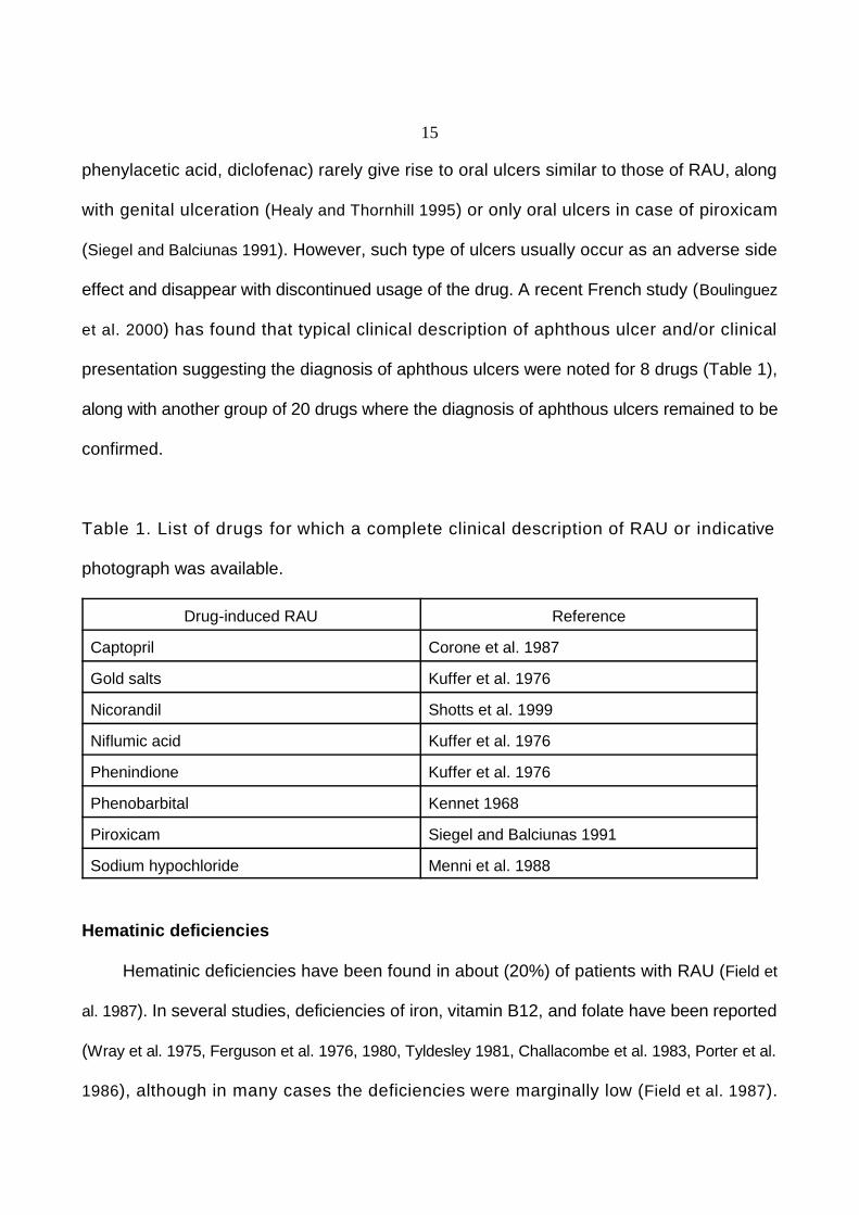
effect and disappear with discontinued usage of the drug. A recent French study (Boulinguez
et al. 2000) has found that typical clinical description of aphthous ulcer and/or clinical
presentation suggesting the diagnosis of aphthous ulcers were noted for 8 drugs (Table 1),
along with another group of 20 drugs where the diagnosis of aphthous ulcers remained to be
confirmed.
Table 1. List of drugs for which a complete clinical description of RAU or indicative
photograph was available.
Drug-induced RAU Reference
Captopril Corone et al. 1987
Gold salts Kuffer et al. 1976
Nicorandil Shotts et al. 1999
Niflumic acid Kuffer et al. 1976
Phenindione Kuffer et al. 1976
Phenobarbital Kennet 1968
Piroxicam Siegel and Balciunas 1991
Sodium hypochloride Menni et al. 1988
Hematinic deficiencies
Hematinic deficiencies have been found in about (20%) of patients with RAU (Field et
al. 1987). In several studies, deficiencies of iron, vitamin B12, and folate have been reported
(Wray et al. 1975, Ferguson et al. 1976, 1980, Tyldesley 1981, Challacombe et al. 1983, Porter et al.
1986), although in many cases the deficiencies were marginally low (Field et al. 1987).
16
However, Olson and colleagues (Olson et al. 1982) found that vitamin B12, folate and iron
deficiencies were not significantly different between the patients with RAU and controls.
Another pilot study on 22 HIV-infected patients with RAU suggested that vitamin B12 or
folate deficiencies were not risk factors for HIV-associated RAU (MacPhail and Greenspan
1997). Low serum ferritin levels were found in 8-12% of patients with RAU, compared with 3-
5% in controls, and the level did not differ in different subtypes of RAU (Challacombe et al.
1983). However, in the majority of cases, there was no identifiable underlying cause for those
RAU patients who had a ferritin deficiency (Porter et al. 1988). In a Scottish study, Nolan and
co-workers (1991a) found that 28.2% of patients with RAU had deficiencies of vitamins B1,
B2, and/or B6. They showed also that patients who have both RAU and a vitamin B
deficiency could benefit from vitamin replacement therapy. It appears from the above
mentioned literature that the wide variations in the findings may be due to differences in
genetic background and dietary habits of examined patients, or the multi-factorial etiology of
RAU.
Zinc deficiency
The improvement of RAU with zinc sulphate supplementation were described in an
open trial (Merchant et al. 1977) and in a case report (Endre 1991) of aphthous ulcers with zinc
deficiency and immunodeficiency, but such improvement could not be confirmed in later
studies (Merchant et al. 1981, Wray 1982a). In a Chinese study (Pang 1992) , the level of serum
zinc of 75 cases of RAU was found to be on a lower level within normal range, and serum
copper was also normal. So far no information exists on the association of RAU and other
trace elements.

17
Environmental factors
Stress
Earlier studies have documented an association between RAU and a variety of
psychological factors including anxiety, repressed hostility, as well as job related and other
stressors (Sircus et al. 1957, Ship et al. 1960, 1967, Miller et al. 1977a). Conversely, other studies
have failed to reveal any association between anxiety (Heft and Wray 1982), depression
(Ferguson et al. 1984), psychological life stress (Pedersen 1989) and recurrences of RAU. A
more recent study, in which the relaxation/imagery treatment program was used (Andrews
and Hall 1990) found a significant decrease in the frequency of ulcer recurrence for all treated
subjects. Although the majority of studies have been unable to validate the concept that
stress plays an important role in the development of RAU, the literature continues to report
that stress may play a role in precipitating RAU.
Local trauma
A subset of patients with RAU are predisposed to develop aphthae at sites of trauma
(Ross et al. 1958, Wray et al. 1981b). Why local trauma such as anaesthetic injections,
toothbrushing, and dental treatment (Kvam et al. 1987) would trigger aphthous ulceration in
these patients is still unknown.
Tobacco
Several reports document the negative association between smoking and the
occurrence of RAU (Shapiro et al. 1970, Axéll and Henricsson 1985b ). Such a negative
18
association has also been documented with use of smokeless tobacco (chewing tobacco
and snuff) (Grady et al. 1992), as well as in patients who are smokers and seropositive for HIV
(Greenspan et al. 1992). Paradoxically, the majority of patients with RAU are nonsmokers
(Rennie et al. 1985), for instance, in a more recent study (Tuzun et al. 2000) only 9% of RAU
patients were found to be active smokers compared with 25% among the control subjects.
Nicotine has been found to be beneficial in RAU (Bittoun 1991) and in inflammatory
bowel disease (Lashner et al. 1990), and its effects may result from influences on nerve
function, although these agents may also exert direct anti-inflammatory effects. However, the
mechanism by which cigarette smoking protects against RAU is still unknown.
Infectious factors
Bacterial agents
In 1963, Barile and co-workers (1963) isolated S. oralis (previously known as S.
Sanguis 2A) from an aphthous ulcer lesion. Other subsequent studies (Donatsky 1975,
Donatsky 1976 a,b) have found raised levels of antibodies against certain oral streptococcal
strains in patients with RAU when compared with controls. Cross-reaction of antibacterial
antibodies with oral mucosa has been postulated as an immunopathogenic mechanism in
RAU (Donatsky 1975). Later studies, however, have not confirmed these observations (Hoover
et al. 1984, Greenspan et al. 1985, Riggio et al. 2000). In a serological test, Helicobacter pylori
does not appear to be of etiologic significance in the development of RAU (Porter et al. 1997).
Another study had detected H. pylori DNA in swabs from 23 of 32 RAU lesions using
polymerase chain reaction (PCR) assay (Birek et al. 1999), but in a more recent study, the
culture of H. pylori from 12 RAU lesions were found to be negative (Shimoyama et al. 2000).

19
Suggesting that H. pylori might not have a direct association with RAU.
Viral agents
A possible viral cause for RAU has been suggested by several researchers. Sallay and
co-workers (1973) isolated adenoviruses from oral aphthae, but there was no antibody
response to adenovirus in RAU. Adenoviruses are ubiquitous organisms and these results
need confirmation. Studd et al. (1991) detected HSV-1 DNA in only 2 of 11 biopsies of oral
aphthae from RAU patients. Other studies failed to detect HSV antigen in the biopsies
(Poulter and Lehner 1989), and HSV cannot be cultured from the RAU lesions (Donatsky et al.
1977, Macphail et al. 1991). Antivirals such as acyclovir, highly effective against HSV, appear
to have only equivocal clinical effect on RAU (Wormser et al. 1988, Pedersen 1992). Patients
with RAU were found to have higher titers of IgM against varicella-zoster virus (VZV) and
cytomegalovirus (CMV) than control subjects (Pedersen and Hornsleth 1993a). Further studies
have detected VZV-like DNA (Pedersen et al. 1993b), CMV-DNA (Sun et al. 1996), Epstein-Barr
virus (EBV-DNA) (Sun et al. 1998) in some oral ulcer biopsy specimens from some RAU
and/or Behçet’s disease (BD) patients. However, VZV could not be cultivated from any of the
oral ulcer biopsies and VZV antigen was not detected in any of the smears. A further study
using PCR, has detected herpes hominis virus-6 (HHV-6- DNA) in six of 21 RAU lesions
(6/21), whereas VZV-DNA and CMV-DNA were not detected in any RAU samples
(Ghodratnama et al. 1997). The detection of human herpesvirus DNA from the oral mucosa
and peripheral blood mononuclear cells of patients with RAU appears to represent normal
viral shedding rather than a direct causal mechanism in this disorder (Brice et al. 2000).

20
Overall, the evidence for involvement of viruses such as HSV, VZV, CMV, EBV and
HHV-6 in RAU is conflicting. It is possible that RAU is a non-specific response with multiple
etiologies and represents the final common pathway of mucosal inflammation or,
alternatively, the dormant herpesviruses might be reactivated by the immuno-dysregulation
known to be associated with RAU.
Heat shock proteins
Cross-reactivity between mycobacterial 65-kDa heat shock protein (Hsp) and
Streptococcus sanguis has been demonstrated, and significantly elevated levels of serum
antibodies to recombinant 65-KDa mycobacterial Hsp were observed in RAU (Lehner et al.
1991). The lymphocytes of RAU patients have a significantly increased lymphoproliferative
response to peptide epitope 91-105 of the 65-kDa mycobacterial Hsp in the ulcerative stage
as opposed to the period of remission (Hasan et al. 1995). There is some cross-reactivity
between the microbial 65-kDa Hsp and the 60-kDa human mitochondrial Hsp. Thus, RAU
may be a T-cell-mediated response to antigens of S. sanguis that cross-react with the
mitochondrial Hsp and induce oral mucosal damage (Hasan et al. 1995). Conversely, other
studies (Van Eden et al. 1998) have suggested that immediate upregulation of Hsps in any cell
type, everywhere in the body, as a consequence of stress may trigger T cells with a
regulatory phenotype. This would provide the immune system with an immunoregulatory
mechanism which acts to monitor and control dangerous or potentially deleterious
inflammatory responses. However, whether Hsps in RAU are protective or destructive or
have a dual role is still unclear.
21
Serology of RAU
Increases in serum IgA, IgG, IgD and IgE have been reported in some groups of RAU
patients (Scully et al. 1983, Lehner 1969b, Ben-Aryeh et al. 1976), whereas in other groups of
RAU patients IgG, IgM and IgA were found to be normal or reduced (Malmström et al. 1983,
Bagg et al. 1987). A previous study by Porter et al. (1992) in a group of 71 RAU patients
showed no significant changes in serum levels of IgG1, IgG2, IgG3 or IgG4, but a more recent
study found low serum levels of IgG2 during the quiescent period of the disease (Vicente et al.
1996). The presence of raised levels of anti-endothelial cell auto-antibodies (AECA) lends
support to the hypothesis that a vasculitic process may underlie some cases of RAU (Healy
et al. 1996). Circulating immune complexes were found to be present in some patients
(Burton-Kee et al. 1981, Lehner et al. 1982). However, complexes have not reliably been
demonstrated in MiRAU (Bagg et al. 1987). Serum levels of C9 (Lehner and Adinolfi 1980) and
$2 microglobulin (Scully 1982b) have been reported to be raised in some patients, and may
represent a non-specific acute phase response (Rennie et al. 1985).
IMPORTANT SYSTEMIC DISEASES ASSOCIATED WITH RAU
Coeliac disease
Coeliac disease is characterized by inflammatory changes in the small intestinal
mucosa induced by a component of the gluten protein of wheat. Recent studies by
Lähteenoja et al. (2000a,b) have shown inflammatory changes with increased lamina propria
and intraepithelial helper-inducer T lymphocyte (CD4+) and suppressor-cytotoxic T
lymphocyte (CD8+) cells in the oral mucosa of coeliac disease patients after a local
challenge with gliadin. The prevalence of patients with coeliac disease who have concurrent

22
RAU ranges from 10% to 18% (Tyldesley 1981, Ferguson et al. 1980, Majorana et al. 1992, Meini
et al. 1993), with an increase in frequency of HLA-DRw10 and DQw1 in coeliac disease
associated with RAU (Majorana et al. 1992, Meini et al. 1993). However, the aphthae usually
disappear with appropriate management of the coeliac disease (Ferguson et al. 1980). On the
other hand, it has been found that about 5% of RAU patients suffer from coeliac disease
(Ferguson et al. 1976, Veloso and Saleiro 1987). Such RAU patients may have particularly IgA-
class reticulin and/or gliadin antibodies (Ferguson et al. 1980, Merchant et al. 1986).
Patients who have RAU with no detectable clinical or histological evidence of coeliac
disease on jejunal biopsy may respond to gluten withdrawal (Wray 1981a, Wright et al. 1986).
In contrast, another study failed to demonstrate any benefit from gluten withdrawal in
aphthous patients, suggesting that the improvements have been due to a placebo effect
(Hunter et al. 1993). So far no studies have used the new diagnostic markers of coeliac
disease such as anti-tissue transglutaminase and anti-endomysial antibodies for screening
patients with RAU.
Behçet’s disease
BD is a multisystem disorder that affects predominantly young men of Mediterranean,
Middle Eastern and Japanese descent. Classically, it features a triad of MiRAU, genital
ulcers and ocular lesions (Shimizu et al. 1979). In 1990 (International Study Group for Behcet's
Disease 1990), the criteria for the diagnosis of BD were redefined to include the presence of
oral aphthous ulcers plus any two of the following: genital ulcers, typical defined eye lesions
(such as uveitis, hypopyon and iridocyclitis), typical defined skin lesions and a positive
pathergy (cutaneous puncture hyperreactivity) test. Aphthous ulcers are present in 99% of

23
patients with BD and are the first symptoms to appear in 67% of patients (Lehner 1977). The
three types of RAU, minor, major, and herpetiform are also found in BD and there are no
features which differentiate the oral ulcers in BD from those of RAU (Lehner 1978). Although
the oral ulcerations in BD are both clinically and histologically identical to those seen in RAU,
the exact relationship of these diseases is still unknown.
Cases of complex aphthosis or bipolar aphthosis (present with oral and genital
aphthae, but no systemic signs or symptoms) may represent an atypical form of BD (Jorizzo
et al. 1985), and follow-up of such patients may eventually disclose more complete
expression of BD (Jorizzo et al. 1985). A high frequency of RAU was found among relatives of
patients with BD (Arber et al. 1991). Furthermore, RAU has some, but not all, of the
immunological abnormalities that arise in BD. In this respect, it has been suggested that
RAU and BD might be different degrees of the same disease spectrum (Lehner and Batchelor
1979). However, RAU is usually confined to the oral mucosa in otherwise healthy individuals
while in BD it affects the oro-genital mucosa. The cause of this extension of localization to
non-oral location is unclear.
HIV-associated RAU
Severe episodes of RAU have been observed in patients infected with HIV. The ulcers
are of the minor, major and herpetiform types and are often located on the soft palate,
tonsils or tongue, where they hinder eating and speaking. Macphail et al. (1991) showed that
66% of HIV patients affected by RAU had the usually uncommon herpetiform or major types
and that patients with MaRAU were significantly more immuno-suppressed than those with
MiRAU or HuRAU in that they had fewer CD4 and CD8 lymphocytes. The role played by the

24
marked neutropenia seen in most of the HIV patients with MaRAU is unclear, but the healing
of the ulcers without resolution of the neutropenia argues for the ulcers being MaRAU rather
than neutropenic ulcers. About half (44%) of the patients denied or could not recall having
had RAU during their childhood, which was presumably before they became infected with
HIV. The rest (56%) gave a definite history of childhood RAU and described the ulcers as
MiRAU (Macphail et al. 1991). Patients with HIV infection have an overall prevalence rate of
recurrent aphthae ranging from 1% to 4% (Phelan et al. 1991, Muzyka and Glick 1994).
Although the lesions are mainly oral, HIV-associated aphthae have been reported in
the esophagus and more distal gastrointestinal tract (Bach et al. 1990). HIV-associated RAU
lesions tended to be more severe and longer lasting, and may cause debilitating pain with
associated alteration of important oral functions such as speaking, chewing and swallowing
which ultimately lead to malnutrition and weight loss, compromise a patient's ability to take
medications and seriously interfere with the patients' quality of life (Muzyka and Glick 1994).
As progress in the treatment of HIV disease results in more patients living longer in a state
of significant immuno-suppression, managing severe RAU may become an increasing
challenge (Macphail et al. 1991).
Although it has not yet been definitely accepted that RAU-like lesions found in
association with HIV infection are indeed RAU, they meet the diagnostic criteria for RAU,
they respond to treatment like RAU, and therefore, until proven otherwise, they must be
considered RAU (MacPhail et al. 1992). Although HIV DNA has been identified in buccal
mucosal scrapings from apparently healthy mucosa of (18/45) HIV-seropositive subjects
(Qureshi et al. 1997), to my knowledge no studies have demonstrated the presence of HIV in
oral ulcers.
25
It is unknown whether such lesions represent a localized auto-immune reaction,
developing in response to an undefined antigen which triggers a normal immunologic
response or represent an overactive HIV in the mucosa of a T cell deficient host.
Important effector cells participating in the inflammatory events of RAU
Neutrophils
Although the chemotactic function of neutrophils is normal in RAU (Abdulla and Lehner
1979, Dagalis et al. 1987), their marked concentration at the ulcer area in the ulcerative phase
of the lesion suggests that may they play an active role in the pathogenesis and/or healing
of RAU. Indeed, the production of oxygen radicals by neutrophils in RAU was found to be
similar to controls (Wray and Charon 1991), and their phagocytic function does not seem to be
defective (Ueta et al. 1993). Oral aphthae are a prominent feature of cyclic neutropenia (Scully
et al. 1982a), and major aphthae in HIV-infected patients have been associated with a
depressed absolute neutrophil count (Macphail et al. 1991). The rapid healing of aphthae on
a regimen of granulocyte-colony stimulating factor (G-CSF) (Manders et al. 1995) and the
clinical response to similar regimens in patients with cyclic neutropenia (Fink-Puches et al.
1996) suggest that neutrophils are important in the healing of recurrent aphthae.
On the other hand, human neutrophil-type matrix metalloproteinase-8 (MMP-8) was
found intracellularly in the ulcer area, and extracellularly in the area of basement membrane
lateral to the ulcer (Häyrinen-Immonen et al. 1993) suggesting that neutrophils containing
MMP-8 are likely to be involved in the tissue destruction seen in aphthae.
However, the exact role of the neutrophils in the pathogenesis or healing of recurrent
aphthae is still not known and remains to be identified.

26
Macrophages
In spite of the fact that macrophages are likely to participate in every stage of the
inflammatory process, they have not yet been adequately studied to definitively establish
their exact role in RAU pathogenesis. Griffin (1982) has found that macrophages were seen
in the lamina propria but not in the epithelium of RAU lesions, and form a large proportion
of the infiltrate in the early phase of the ulcer ( VanHale and Rogers 1984). Another
histopathologic study of RAU (Schroeder et al. 1984) has found the presence of numerous
macrophages loaded with phagolysosomes containing debris of neutrophilic granulocytes,
implying that macrophages mainly function to clear the tissue of neutrophil remnants.
The results of Häyrinen-Immonen et al. (1991) indicated that CD11b and nonspecific
esterase-positive mature tissue macrophages formed about 14% of all inflammatory cells in
RAU lesions, with increased distribution around the periphery of the lymphoid cell infiltrates.
Mast cells
Mast cells (MCs) that have the ability to provide numerous mediators (Brody and
Metcalfe 1998) have long been regarded as potentially important cells in the inflammatory
events of RAU. In a histopathological study using Alcian blue/Safranin staining of 15 MiRAU,
Dolby and Allison (1969b) found that the MC count in the first 2 days did not differ from the
normal buccal mucosa. While there was approximately 50% reduction in the MC count in
lesions of more than 48 hours duration. In contrast to findings in Dolby and Allison’s study,
increased numbers of MCs were noted by Lehner (1969a) in all three types of RAU (minor,
major, and herpetiform) and in oral aphthae associated with BD, particularly with toluidine
blue stain. Such increase in MC numbers was also found in skin lesions and oral aphthae in
27
patients with active BD (Lichtig et al. 1980). The above mentioned results seem suggestive of
an active role of MCs in RAU pathogenesis.
Factor XIIIa+ dendrocytes as a member of the subepithelial immune system
Factor XIIIa-positive dendrocytes are normal residents of the dermis and subepithelial
connective tissues. They share a number of molecules, such as CD11b, CD14, CD18 and
CD36 with monocyte-macrophages (Caux et al. 1996). Their morphology and expression of
HLA-DR suggest that they are capable of processing and presenting antigens (Headington
1986, Drijkoningen et al. 1987, Cerio et al. 1989, Weber-Matthiesen and Sterry 1990). Negative
reactivity with antibodies recognizing CD1a and the absence of Birbeck granules and
ATPase activity distinguish the FXIIIa dendrocytes from Langerhans’ cells (Sontheimer et al.
1989, Headington and Cerio 1990, Moschella and Cropley 1990, Nestle et al. 1993a). Cytoplasmic
vacuoles containing hemosiderin and melanin implicate the FXIIIa+ dendrocyte as a
phagocytic cell (Headington and Cerio 1990, Altman et al. 1992). Furthermore, it has been
shown that isolated FXIIIa+ dermal dendrocytes were potent stimulators of resting T-cells
(Nestle et al. 1993a, b). There has been much speculation about the role of FXIIIa+
dendrocytes in various pathological conditions based on their increased number in such
diverse diseases as psoriasis (Nickoloff and Griffiths 1990), lichen planus (Regezi et al. 1994),
atopic dermatitis (Headington 1986), and Kaposi's sarcoma (Nickoloff and Griffiths 1989).
FXIIIa+ dendrocytes bear high amounts of major histocompatibility complex (MHC) class II
molecules on their surface, and are very potent antigen presenting cells in vitro.
A subpopulation of these cells acquires certain ultrastructural features of Langerhans
cells in vitro such as Birbeck granule and CD1a (Nestle and Nickoloff 1995a). These cells may
28
be precursors of epithelial Langerhans cells and may play a role in submucosal immune
responses. Given their prevalence in subepithelial connective tissue, and their in vitro
functional capacity, it is appropriate to conclude that FXIIIa+ dendrocytes are indeed
important members of the subepithelial immune system, and may have a role in stimulating
mucosal immunity via presenting antigens locally to infiltrating T-cells (Nestle and Nickoloff
1995b, Thomas 1996, Yoo et al. 1998).
Gamma/delta T-lymphocytes
Two classes of T cell antigen receptors (TCRs), the "$ TCR and the (* TCR, have
been identified. (/* T-cells share many cell surface proteins with "/$ T-cells and are able to
secrete lymphokines and express cytolytic activity in response to antigenic stimulation (Munk
et al. 1990). Moreover, (/* T-cells and natural killer (NK)-cells share similar responses upon
activation (Haas et al. 1993). However, differences between (/* T-cells and "/$ T-cells are
numerous. First, (/* TCRs are more closely related to immunoglobulins (Igs) than to "/$
TCRs (Rock et al. 1994). Thus, (/* TCRs bind antigens in a different manner than "/$ TCRs.
The second difference relates to their activation kinetics. A consistent feature of (/* T-cell
responses is a localized, rapid and transient release of bioactive polypeptides such as
interferon-gamma (IFN-() (Ferrick et al. 1995) following activation. This response can precede
"/$ T-cell activation by several hours to days. Third, (/* T-cells have the ability to recognize
non-peptidic molecules commonly associated with micro-organisms and stressed cells
(Boismenu and Havran 1997). In general, recognition of these antigens by (/* cells involves
the antigen receptor but does not require antigen processing and presenting cells or MHC
gene products.

29
Although in rodents (/* cells are preferentially localized in epithelial tissues such as
skin, intestine, and lung (De Libero 1997), they are rare in the normal human oral epithelia
(Pepin et al. 1993, Patinen et al. 1997). However, increased (/* T-cell numbers have been
found to be associated with a variety of infectious and auto-immune conditions such as
coeliac disease, multiple sclerosis and rheumatoid arthritis (Haas et al. 1993; Hayday and Geng
1997). Although (/* T-cell population constitutes only about 5% of circulating T cells, they
are much more common in the peripheral blood of patients affected by RAU or Behçet’s
disease, especially during the active phase of the disease (Suzuki et al. 1992, Pedersen and
Ryder 1994). Interestingly, Hasan and colleagues (1995) showed that (/* T-cells from
patients with RAU have a specific, proliferative response to heat shock protein peptides.
Cytokines, such as IL-7 released within the epithelial microenvironment, may play a
role in proliferation and differentiation of (/* T-cells in epithelia (Fujihashi et al. 1996a). On the
other hand, the epithelial-cell-specific fibroblast growth factor (FGF)-7 produced by (/* T-
cells may play a role in healing epithelia damaged by infection or inflammation, by prompting
cell growth and hence reinstalling tissue integrity (Boismenu and Havran 1994). However, the
role of (/* T-cells in mucosal immunity might not be restricted to nursing the epithelial injury.
A previous study using a murine model (Jones-Carson et al. 1995) showed that (/* T-
cells lining the orogastric tract can stimulate the production of nitric oxide (NO) by
neighboring epithelial cells. This finding may be important because human oral epithelial
cells might also be stimulated by neighbouring (/*-T-cells-derived IFN-( (Freysdottir et al.
1999) to produce NO via upregulation of inducible NO synthase (iNOS) expression.
As a gaseous free radical, NO could serve as a physiological cytoprotective agent for
the mucosa. However, large amounts of NO alone, or after reaction with superoxide released

30
from activated phagocytes (Beckman et al. 1990, Kimura et al. 1998), could lead to epithelial
autotoxicity (Flak and Goldman 1996) and possibly ulceration.
TNF-"""" and recurrent aphthous ulceration
The possible relevance of TNF-" to the pathogenesis of RAU stemmed from
observations that thalidomide, which reduces the activity of TNF-" by accelerating the
degradation of its messenger RNA (Moreira et al. 1993), and pentoxifylline, which inhibits
TNF-" production (Zabel et al. 1993), were found to be effective in the treatment of RAU in
HIV-infected patients and in otherwise healthy persons with RAU ( Revuz et al. 1990,
Thompson 1995, Pizarro et al. 1995). An enhanced release of TNF-" by peripheral blood
monocytes of patients with RAU has also been demonstrated (Taylor et al. 1992).
Furthermore, a recent study has shown low resting levels of interleukin-10 (IL-10)
mRNA in non-lesional mucosa of RAU patients and high levels of the pro-inflammatory
cytokines, IL-2, IFN-(, and TNF-" mRNAs in lesional and nonlesional mucosa of patients
with RAU compared with controls (Buno et al. 1998). However, the contribution of TNF-" to
RAU pathogenesis is not fully understood at present.
HISTOPATHOLOGY OF RECURRENT APHTHOUS ULCER
I. The ulcer area
Superficial tissue necrosis with fibrinopurulent exudate consisting of clotted fibrin,
numerous red blood cells forming hemorrhagic foci, neutrophils and cellular debris covers
the necrotic area. The epithelium is infiltrated with variable numbers of intraepithelial
lymphocytes and some neutrophils (Stanley 1972). Neutrophils predominate in the immediate
31
ulcerated area, although peripheral areas surrounding the ulcer remain mononuclear in
nature (Lehner 1969a; Mills et al. 1980; Schroeder et al. 1983; Häyrinen-Immonen et al. 1991).
II. The area lateral to the ulcer
Defined as the epithelium covered area from the edge of the ulcer and sideway to the
periphery of the biopsy. An intense leukocytic infiltration with predominance of lymphocytes
in non-ulcer regions, where they outnumbered neutrophils (Mills et al. 1980; Häyrinen-Immonen
et al. 1991). Monocyt/macrophages are also numerous in the tissue adjacent and lateral to
the ulcer. The density of MCs is increased in the lamina propria (Lehner 1969a, Schroeder et
al. 1984). The lymphocytes in RAU lesion are primarily T cells, and only 5-12% of all cells in
the lesion are B cells (Häyrinen-Immonen et al. 1991). A small proportion of plasma cells and
eosinophils can be found and more often in the older lesion ( Lehner 1969a). Dilatation of
blood vessels is a constant and prominent feature of RAU lesions as are foci of perivascular
mononuclear cell infiltrates (Lehner 1969a; Schroeder et al. 1984).
IMMUNOHISTOPATHOLOGY OF RAU
Immunological aberrations involving both cell-mediated and humoral immunity have
been reported in previous studies of RAU (Porter et al. 1998). Both class I and II MHC
antigens were found to be expressed on the epithelial basal cells in preulcerative RAU
lesions and more diffusely within the epithelium at the ulcer stage, consistent with active cell-
mediated inflammation (Savage et al. 1986, Poulter and Lehner 1989). In vitro studies have
shown that peripheral lymphocytes from patients suffering from RAU were found to be
directly cytotoxic against oral epithelial cells (Dolby 1969a, Rogers et al. 1976), which, however,
32
has not been confirmed by others (Peavy et al. 1982, Gadol et al. 1985, Burnett and Wray 1985).
RAU patients have significantly increased antibody-dependent cellular cytotoxic (ADCC)
activity in the early stage of the disease (Greenspan et al. 1981).
Immunofluorescence studies demonstrated deposits of IgG, IgM, IgA, and C3 in and
along mucosal blood vessels and in the cytoplasm of stratum spinosum cells in aphthous
ulcers lesions from patients with RAU and Behçet’s disease ( Lehner 1969a, VanHale et al.
1981, Malmström et al. 1983). Previous studies on peripheral NK-cells in patients with RAU
have been contradictory as their percentages have been reported to be either increased
(Greenspan et al. 1982) or similar to that of controls (Savage et al. 1988, Pedersen and Pedersen
1993c). Furthermore, Thomas et al. (1990) found that depletion of CD-16 positive cells (NK-
cells) produced no change in cytotoxicity towards the oral epithelial target cells. Another
report demonstrated that among patients with major RAU, NK cell activity is increased when
active oral lesions are seen, depressed during periods of resolution and normal in patients
in remission (Sun et al. 1991b).
Formation of perivascular lymphocyte infiltrates are probably in part mediated by
endothelial intercellular adhesion molecule-1 (ICAM-1) and lymphocyte function-antigen-3
(LFA-3)-binding to their counterpart ligands lymphocyte function-antigen-1 (LFA-1) and CD-2
on lymphocytes, respectively (Häyrinen-Immonen et al. 1992b, Verdickt et al. 1992). ICAM-1 is
expressed on the epithelium and submucosal capillaries and venules, suggesting that it may
support T-cell adhesion and control the trafficking of leukocytes into the submucosa and
epithelium (Savage et al. 1986, Häyrinen-Immonen et al. 1992b, Eversole 1994, Healy and Thornhill
1999), while LFA-3 and its counterpart ligand CD-2 are likely to be involved in T-cell
activation in RAU (Häyrinen-Immonen et al. 1992b). Increased numbers of CD1+ Langerhans

33
cells were found in the epithelium and lamina propria in BD and RAU (Poulter and Lehner
1989, Häyrinen-Immonen 1992a).
It is thus evident that there is no unifying theory of the immunopathogenesis of RAU.

34
AIMS OF THE STUDY
"It is not the answer that enlightens but the question"Eugène Ionesco, Romanian-born French playwright
1) To look for morphologic evidence of the involvement of factor-XIIIa+ dendrocytes in the
pathogenesis of RAU by assessing their frequency and spatial distribution in RAU
compared to traumatically induced oral ulcers.
2) By using specific immunohistochemical markers for MCs, we aimed to re-study the
density, distribution and degranulation activity of MCs in RAU and traumatic oral
ulcers.
3) To assess the cellular localization and extent of expression of the key pro-inflammatory
cytokine TNF-" in RAU lesions and traumatic oral ulcers.
4) To investigate whether the cell proliferation activity in RAU lesions differs from that of
the traumatic oral lesions, and to clarify whether the infiltrating inflammatory cells
proliferate locally by using the proliferation marker Ki-67.
5) To examine whether the density of lymphocytes bearing (/* T-cell receptors is
increased locally in RAU lesions compared to clinically normal appearing oral mucosa
from the same patients, and to correlate this density of lesional (/* T-cells to the
duration of RAU lesions.
35
PATIENTS AND METHODS
“What we observe is not nature itself,but nature exposed to our method of questioning”
Werner Heisenberg, German physicist and Nobel Prize winner
Patients and diagnostic criteria
The studied biopsy specimens consisted of four sets. The first set was comprised of 29
RAU lesions taken from 24 patients (14 women and 10 men, mean age=35 years, range=18-
54, Table 2) with longstanding RAU of the minor type (according to the classification by
Stanley, 1972). The second set is ulcerative lesions control consisted of 14 experimentally-
induced oral traumatic ulcers (TU) were taken from 14 healthy volunteers (women=7,
men=7, mean age=36 years, range 21-50) with no history of any mucosal disease (studies
l, III, IV). The third set contained 10 specimens (used only in study V) from the non-lesional
mucosa at the corresponding sites opposite to the RAU lesions, obtained from the same
RAU patients included in study (V) to demonstrate eventual differences between RAU
lesions and non-lesional mucosa. The fourth set, was clinically healthy oral mucosa (healthy
control) from 16 healthy individuals with a negative history of RAU (women=10, men=6,
mean age=35 years, range 15-58).
This work was carried out from 1995 to 2001 and the study protocol was approved by
the human research Ethics Committee of the Institute of Dentistry, Faculty of Medicine,
University of Helsinki, Finland, 1995. The informed consent was obtained from each
participant according to the Declaration of Helsinki (4th amendment, 1989).
A tobacco-smoking history was obtained from sixteen RAU patients included in the
study, only one had smoked tobacco in the past and the others had never smoked tobacco.
Of the thirty healthy individuals (16 control and 14 TU subjects) recruited to this study, only

36
two men currently smoked tobacco and four had stopped smoking. All the healthy control
subjects were in good general health.
All the patients included in this study had been investigated at the outpatient clinic of the
Department of Oral Medicine, Institute of Dentistry, Helsinki, Finland, during previous
episodes of ulceration. The results of full blood examination, serum vitamin B12 and folate
were within normal limits. None of the patients or controls had a history of any gastro-
intestinal signs or symptoms other than associated with minor illness. None of the patients
had a history of HIV infection. One of the patients had suffered from occasional genital ulcers
in association with oral aphthae, but she had no systemic signs or symptoms of BD. Apart
from the RAU complaint, all the patients were otherwise clinically healthy. All RAU lesions
studied were 2-10 days old, MiRAU, and painful. The histological picture was that of a
nonspecific ulcer.
During the study more than one sample of RAU lesions were obtained from some RAU
patients to match the location and duration of the TU lesions (Table 2).

37
Table 2. Distribution of the patients with RAU according to study, age, gender, duration of RAU, age
of the ulcer, and site of biopsy.
Patient Study Age(yrs)
Sex Duration ofRAU (yrs)
Age of RAU(days)
Site of RAU biopsy
1 II 37 F 22 10 Buccal
2 II, III, V 34 F 20 6 Labial
3 II, IIIV
2627
F 1314
22
Labial Buccal
4 II 29 F 15 7 Labial
5 I, II, IV 36 F 16 3 Labial
6 II, III, V 25 M 10 3-4 Buccal
7 I, II, III, IV, V 45 F 30 3 Buccal
8 I, II, IV, V 35 M 25 2 Buccal
9 IIIV
2830
F 1214
45
Buccal Labial
10 II, III, IV 40 F 33 2 Buccal
11 I, II 51 F 34 3 Labial
12 I, II, III, IV 33 F 19 3 Buccal
13 II 21 F 10 - Labial
14 IIII
22 M 12 13
BuccalBuccal
15 II, IIIV
4649
M 32 35
54
BuccalLabial
16 IIV
22 M 10 -7
BuccalBuccal
17 IV, V 37 F 23 3 Buccal
18 III 53 F 43 - Labial
19 I, II, III, IV 29 M 11 2-3 Buccal
20 IV 40 F 20 3 Buccal
21 I, III 54 M 37 3 Labial
22 IV 18 M 8 3 Buccal
23 IV 45 M 22 2-3 Buccal
24 V 21 M 12 3 Buccal

38
The inclusion criteria for the RAU patients selected in this study were as follows
1) The patients should have classical minor RAU: history of recurrent painful ulcers on
non-keratinized mucosa, clinical appearance of RAU and histopathological findings of
RAU.
2) Severe RAU condition: defined as at least one episode per month on average.
RAU Patients were excluded from the study if they had
1) a history or manifestation of any systemic illness or immunodeficiency state.
2) chronic or acute infection such as recent viral illness.
3) recurrent intraoral herpes simplex virus lesions or other oral mucous membrane
diseases.
4) been taking any medication that might interfere with the study parameters (such as
steroids or immunosuppressive drugs.
5) treated their current ulcer with any form of systemic or topical medication.
Inclusion/exclusion criteria for the healthy control subjects selected for this study
1) good general condition with absence of any health problem
2) comparable in age to RAU patients
3) negative history of RAU condition
4) lack of acute infection
5) negative history of medication or trauma-induced ulcers

39
Due to absence of a definitive etiology or diagnostic test for RAU, the identification of RAU in
a clinical practice usually relies on combinations of a history, clinical features and
histopathology. I propose in this thesis a set for diagnostic criteria for RAU which were meant
to distinguish RAU from other diseases, and to be practical, all were based on working
knowledge of aphthous ulcers and clinical experience. They were not tested for sensitivity
and specificity and further studies are required for the widespread use of these criteria.
Further refinement of the diagnostic criteria described here will depend on properly
conducted studies to validate them.
The diagnosis of primary RAU minor (idiopathic) or secondary RAU minor (that occur in
association with systemic diseases) can be made if the condition fulfills the four major criteria
(which are necessary to establish the diagnosis of RAU) plus at least one of the minor
(supportive) criteria.
Table 3. The major criteria for recognising and diagnosing the condition of RAU minor.
Major criteria Description
1. External appearance Single or multiple round-oval shaped ulcers, neverpreceded by vesicles. The ulcers are shallow and haveregular margins, yellow-gray base surrounded by thinerythematous halos, variable in size, but less than 1 cm indiameter.
2. Recurrence At least three attaks of RAU within the past 3 years anddo not recur at the same focal site.
3. Mechanical hyperalgesia The lesion is painful and the pain is exaggerated bymoving the area affected by the ulcer.
4. Self-limitation of the condition The ulcer heals spontaneously without seqeulae, eitherwith or without treatment.

40
Table 4. The minor criteria for recognising and diagnosing RAU minor.
Minor criteria Description
1. Family history of RAU A positive family history of RAU is present.
2. Age of onset The first RAU attack started before the age of 40.
3. Location of ulcers Occur on non-keratinized oral mucosa.
4. Duration of the lesion Each bout of ulceration lasts from a few days to twoweeks.
5. Pattern of recurrence Irregular
6. Histological examination Shows non-specific inflammation.
7. Presence of precipitation factor The attacks are triggered by hormonal changes,exposure to certain food or drug, intercurrent infections,stress and local trauma.
8 . P r e s e n c e o f h e m a t i n icdeficiencies
Laboratory investigations reveal an accompanyinghematinic deficiency. Particularly, ferritin, folate, iron,vitamin B and zinc.
9 . Negat ive assoc ia t ion w i thsmoking
RAU patient is a non-smoker or develops the ulcer afterstopping smoking.
10. Therapeutic trial with gluco-corticosteroids
Positive response to treatment with local or systemicsteroids.
41
Sample collection, processing and storage of control and RAU specimens
Control specimens of clinically healthy oral mucosa were obtained from buccal mucosa
(twelve cases) or labial mucosa (four cases). All TU lesions were obtained from the buccal
mucosa. RAU lesions were obtained from the buccal and/or labial mucosa. All biopsies were
performed under local anaesthesia (20mg/ml Xylocain + 12.5 :g/ml adrenalin, Astra Co.,
Södertälje, Sweden). The anesthetic solution was injected deep into the tissue in the area
from where the specimens were taken. Ulcer specimens were obtained as excisional
biopsies, and for control purposes, specimens from clinically healthy oral mucosa were
obtained from healthy individuals by the same technique. Those samples (studies I-IV), which
were fixed in 10% buffered neutral formalin, were embedded in paraffin and cut at 6 :m
thickness. Biopsy specimens of study V were divided into two pieces. One-half of each
specimen was fixed in buffered neutral formalin and embedded in paraffin for routine
histopathological examination using hematoxylin and eosin stain. The second half of each
specimen was snap frozen in liquid nitrogen, then embedded in Tissue-Tek OCT compound
(Lab-Tek Products, Elkhart, IN, U.S.A.) and stored at -70NC until used for further studies.
Experimental induction of traumatic ulcers (TU)
TU was induced experimentally in each normal volunteer by removing an ellipse of
normal buccal mucosa under local anaesthesia. No sutures were placed, the wound measure
0.5×1 cm, being left open for 24-72 h, after which time the induced lesion had the
appearance of traumatic ulcer. At this stage the ulcer was removed under local anaesthesia.
In study (IV), due to rapid resolution of TU lesions, 48 and 72 h were chosen as time
points following induction of the trauma for study and comparison with aphthae of similar

42
lesional duration. All biopsies were taken in the morning (9 am-12 noon) to avoid the diurnal
variation in the proliferation activity of the epithelium (Kellett et al.1989). For publications (I-IV),
all TU samples were processed as formalin-fixed specimens.
Histologic staining
All samples used in the present study were histologically examined using hematoxylin-
eosin staining. All aphthae samples were in ulcerative stage and showed histological signs of
inflammatory cell infiltration.
Paraffin-embedded tissue sections were deparaffinized three times in xylene,
rehydrated in graded ethanol from 100% to 70%, then stained with Delafield’s hematoxylin for
10 min. Sections were rinsed in distilled water, immersed in acidic ethanol (250 ml of 70%
ethanol + 2.5 ml of 37% HCl for 20 sec) and washed in running water for 10 min. The
sections were incubated in Eosin BA for 5 min. Finally, rinsed in a graded ethanol series from
70% to 100%, cleared in xylene and mounted in Diatex (Becker Industrifärg AB, Märsta,
Sweden).
Antigen-retrieval methods
The antigen epitopes hidden by aldehyde cross-links were disclosed either by
proteolytic digestion with pepsin enzyme (studies I-III) or by heating tissue sections in slightly
acidic citrate buffer solution using microwave oven (study IV) (Boon 1994; Mighell et al. 1998).
A) Pepsin treatment
Tissue sections were incubated in 0.4% pepsin (40 mg pepsin in 10 ml distilled water)
plus 100 :l 1N HCl (0.01N HCl) for 30 min at +37<C. Then washed three times with TBS.

43
B) Microwave treatment
Sections were cut onto slides coated with a strong adhesive (3-aminopropyl-
triethoxysilane). Tissue sections were deparaffinized, rehydrated and placed in a plastic box
filled with 10 mM tri-sodium citrate buffer, pH 6.0, and heated in a conventional microwave
oven for 10 min at 650W to unmask the antigen. While undergoing microwave processing,
slides were always covered with a buffer. After heating, slides were permitted to cool down
to room temperature over a period of 30-60 min.
Table 5. Primary antibodies used in the studies
Antigen Antibody Source subtype Concentration/dilution manufacturer
Factor-XIIIa Polyclonal Rabbit IgG 100U/ml; 1:900 Calbiochem, USA
Mast cell
tryptase
AA1
Monoclonal
Mouse IgG1 105 mg/L; 1:50 Dako, Denmark
IgE specific for
Epsilon-
Chains
Rabbit IgG 7.7 g/L; 1:800 Dako, Denmark
TNF-" Monoclonal Mouse IgG1 200 :g/ml; 1:50 Santa Cruz, USA
Ki-67 Polyclonal Rabbit IgG 0.25 g/L; 1:500 Dako, Denmark
T-cell
receptor (*
TCR *1
Monoclonal
Mouse IgG1 100 :g/ml; 1:100 T-cell Sciences,
USA
T-cell
receptor
"$
$F1
Monoclonal
Mouse IgG1 100 :g/ml; 1:100 T-cell Sciences,
USA
T-cell
receptor
CD3
Leu4
Monoclonal
Mouse IgG1 50 :g/ml; 1:400 Beckton-Dickinson,
USA

44
Immunohistochemistry protocol
Avidin-biotin-peroxidase complex (ABC) staining
The expression of antigen was evaluated by immuno-histochemical staining using the
avidin-biotin-peroxidase complex (ABC) method (Vectastain Elite-ABC-peroxidase, Vector
Lab).
Paraffin-embedded tissue sections were deparaffinized three times in xylene,
rehydrated in ethanol (absolute ethanol 5 min, 94% v/v ethanol 5 min, 70% v/v ethanol 5 min)
and rinsed in tap water. All sections were washed in 20 mM Tris-HCl, pH 7.5, containing 150
mM NaCl (TBS). Disclosing of antigen epitopes were done either by pepsin treatment
(studies I-III) or by microwave treatment (study IV). The intrinsic peroxidase activity was
abolished by pretreating tissue sections in 0.3% H2O2 in methanol for 30 min. Non-specifi c
binding sites were blocked by incubation in normal goat serum (studies I and IV) and normal
horse serum (for studies III and V) (1:50, Vector Laboratories, Burlingame, CA, USA) for 20
min. The sections were then incubated with the following primary antibodies: rabbit antiserum
against FXIII-a (1:900), monoclonal mouse anti-human TNF-" IgG1 (1:50), rabbit anti-human
Ki-67 IgG (1:500), mouse anti-human TCR *1 IgG1 (1:100), mouse anti-human "/$ TCR
IgG1 (1:100), and mouse anti-human CD3 TCR IgG1 (1:400) overnight at +4NC. Bound
antibodies were labeled with biotinylated goat anti-rabbit IgG (dilution 1:250-300, Vector
Laboratory, Burlingame, CA, USA) or biotinylated horse anti-mouse IgG (dilution 1:50, Vector
Lab) for 30 min followed by incubation in avidin-biotinylated peroxidase complex for 30 min.
Finally the peroxidase-binding sites were visualized by 3,3'-diaminobenzidine tetra
hydrochloride (DAB) (Sigma Chemical Co., St. Louis, MO, USA), 40 mg/150 mL TBS and
0.006% H2O2, treatment for 3 min (studies I, III, V) or revealed with a combination of 200:l
H2O2 and 3-amino-9- ethylcarbazole (AEC 40mg + 12 ml dimethyl formamide) for 20 min

45
(study IV). After immersion in DAB solution, the specimens were counterstained in Mayer's
hematoxylin (Mayer, Diagnostica Merck, Darmstadt, Germany) for 45 sec, dehydrated
through graded alcohol series, cleared in xylene and mounted in Diatex (Becker Industrifärg
AB, Märsta, Sweden) or in aqueous mounting medium (glycerin-gelatin solution, Dako, study
IV). Between each step, the slides were washed three times in TBS. All incubations were
performed at +22NC if not otherwise indicated.
For frozen tissue specimens (study V) 6 :m thick cryostat sections mounted on gelatin
coated slides were air dried for 30 min, and fixed in acetone for 20 min at +4<C, then in
chloroform for 20 min at 22<C. Endogenous peroxidase activity was blocked with 0.3% H2O2
in absolute methanol for 30 minutes at 22<C. Then the sections were processed for immuno-
histochemical ABC staining as described above.
Peroxidase-anti-peroxidase (PAP) complex staining
Paraffin embedded sections were deparaffinized three times in xylene, rehydrated in
ethanol (absolute ethanol 5 min, 94% v/v ethanol 5 min, 70% v/v ethanol 5 min) and rinsed
in tap water. All sections were washed in TBS. After disclosing of epitopes with pepsin, the
intrinsic peroxidase activity was abolished by H2O2-methanol pretreatment. Mast cell tryptase
and IgE were demonstrated with the unlabelled antibody enzyme method PAP (peroxidase-
anti-peroxidase). The tissue sections were treated sequentially with: 1) normal rabbit serum
(for MC staining) or normal goat serum (for anti-IgE staining) (dilution 1:66 in TBS) for 20
min. 2) the primary monoclonal mouse anti-human mast cell tryptase or the primary rabbit
anti-human IgE overnight at +4<C. 3) rabbit anti-mouse or goat anti-rabbit immunoglobulins
for 30 min. 4) incubation with the appropriate PAP (PAP mouse for MC tryptase staining and
PAP rabbit for IgE staining, (1:100; Dakopatts A/S, Glostrup, Denmark). The specimens were

46
washed with TBS three times for 5 min between each step. Finally the peroxidase-binding
sites were visualized by DAB and H2O2 treatment for 3 min. The specimens were
counterstained in Mayer's hematoxylin for 45 sec, dehydrated through graded ethanol series,
cleared in xylene and mounted in Diatex.
Specificity
The specificity of the reaction was tested by omission of the primary antibodies from the
staining sequence. In addition, normal goat, rabbit, or mouse serum, was used as
appropriate instead of the primary antibodies as an additional negative staining control
(studies I; II, IV). For studies III and V, an isotype specific IgG1 antibody raised against
Aspergillus niger glucose oxidase (monoclonal mouse anti-aspergillus niger IgG1), an enzyme
not present or inducible in mammalian tissues, was used at the same concentration as, and
instead of, the primary antibody as a negative staining control.
ABC staining was not used in study II because of the possibility of false positive staining
of MCs (Hsu and Raine 1984). No positive sample controls were used in this study.
Assessment and quantification of immuno-histochemical staining
In studies I-III the number of positively stained cells was calculated by means of the
video image digital analysis system (VIDAS, Kontron, Münich, Germany) linked to a low-light
charge screen coupled with a CCTV camera (HV/720K, Hitachi, Denshi, Osaka, Japan)
mounted on an Olympus BH-2 light microscope. The VIDAS system consisted of semi-
automatic Kontron image analysis and processing systems (Kontron Bildanalysis, Eching,
Germany) equipped with a VIDAS 2.1 programme (Kontron). After the adjustment of
illumination and focus, the images were recorded in digitized form in a computer. After the

47
parameters were set for the detection of positive cells, they were fixed and used during the
whole analysis procedure. The evaluation of the staining in studies (IV) and (V) was
undertaken using an Olympus BH 2 (Olympus Japan Corp., Ltd., Tokyo, Japan) light
microscope.
The number of positive cells was calculated in three serial sections in all study samples
(except in study III, where only one section from each sample was counted) to get an
estimate for a population mean. Using the formula:
it was calculated that the mean of five high-power fields is not significantly (p>0.05)
different from the population mean (t refers to t-statistics, x=the sample mean, µ=the
population mean, SEM=standard error of the mean). Results are expressed as the number
of positive cells per one mm2 of tissue (studies I, II, V) or averaged in each specified area and
expressed as count of cells/0.2 mm2 (studies III and IV).
The calculations were performed under 400× magnification and were done in the
surface epithelium at the edge of the ulcers and sideways, and in the subepithelial lamina
propria (defined as that immediately beneath the basal cell layer of the epithelium, to a
constant depth of 0.22 mm) in areas beneath and lateral to the ulcer, as well as from the
sites of mononuclear inflammatory cell infiltrations, and deeper connective tissue areas (I-V).
The overall positive cell counts were determined by counting 14 sequential high power fields
(field area 0.0704 mm 2), outlining an area of connective tissue that extended 1 mm
horizontally (starting just beneath the epithelium from the edge of the ulcer and laterally to the
periphery of the biopsy) and vertically to a depth of 1 mm.

48
Statistical analysis
The results are given as mean and standard deviation to describe the dispersion of the
data. The means of the three groups were compared by using the Kruskal-Wallis rank test,
and the comparisons between the pairs of means by using the Mann-Whitney U test, with a
downward adjustment of the " level to compensate for multiple comparisons. Spearman’s
rank correlation was used to measure the correlation between MC density and duration of the
lesions (study II), and linear correlation was calculated between the densities of intraepithelial
(/* T-cells and the ages of RAU lesions (study V). A P value of 0.01 or less was considered
statistically significant. BMDP-PC, version 7.01 (BMDP statistical software, Cork, Ireland) was
used for all calculations.

49
RESULTS
“A ship in port is safe, but that is not what ships are for,sail out to sea and do new things"
Admiral Grace Hopper, computer pioneer
Factor XIIIa+ dendrocytes in RAU (I)
In healthy controls, FXIIIa+ dendrocytes were mainly found in the lamina propria and
just beneath the basal epithelial cell layer and most of them had slender or spindle shapes.
Although the distribution and shape of FXIIIa+ cells in TU samples were similar to those seen
in healthy controls, many FXIIIa+ round-shaped dendrocytes were seen within the
inflammatory infiltrates of TUs.
In aphthae, FXIIIa+ cells were found in lamina propria, within and lateral to the
mononuclear inflammatory cell infiltrates and in perivascular areas. The cells had a spindle,
round or dendrite-like appearance. FXIIIa+ cells localized within the mononuclear cell
infiltrates were 80% more numerous in aphthae than in TU lesions (199±67 vs 110±31
cells/mm2, P<0.001). Dendrocytes in RAU were larger in size (up to 2 times as large as in
controls) and had more prominent dendritic processes (Fig. I) than those seen in TUs or in
healthy controls.
Overall, FXIIIa dendrocytes were 50% more numerous in RAU than in TU or healthy
controls (Fig. II).


51
Mast cells in RAU (II)
Tryptase staining
MC tryptase staining was either strong and confined to cytoplasm or was weaker and
diffuse, suggestive of degranulation.
Few or no MCs were seen at the base of the aphthous ulcer or close to the ulcer region.
In 3 of 15 RAU lesions, MCs were seen to infiltrate the epithelium and were localized in the
epithelial basal layer (Paper II: Fig. 2a). Many of these cells were degranulated and
surrounded by extracellular tryptase staining (paper II: Fig. 2b). Whereas in the TUs and
healthy control sections MCs were absent in the epithelial compartment.
MCs in the lamina propria were 30% more numerous in RAU than in TU lesions.
Numerous tryptase positive MCs were intermixed with the other infiltrating mononuclear cells.
The inflammatory infiltrates of the RAU lesions showed 1.6 times more MCs than
inflammatory cell infiltrates in TUs (118±31 vs 75±18 cells/mm2, P<0.001). Moreover, many
MCs at these sites were degranulated, as evidenced by diffuse extracellular tryptase staining
(halos of tryptase) around the MCs (paper II: Fig. 3), in contrast to only a few such cells in the
inflammatory cell infiltrates in TUs.
In aphthae, MCs were seen in connective tissue, particularly at the interface between
inflammatory cell infiltrates and connective tissue, where they often associated with sites of
connective tissue that appear disrupted (paper II: Fig. 4). The MC count in connective tissue
in RAU, lateral to the inflammatory infiltrates, was 70% to 80% greater than the count in the
connective tissue in TUs or controls. No difference was seen in the densities of MCs between
TUs and controls and no correlations could be found between the MC density and duration
of RAU or TU lesions. Overall, MCs were 63% more numerous in RAU lesions than in control
(Fig. III).
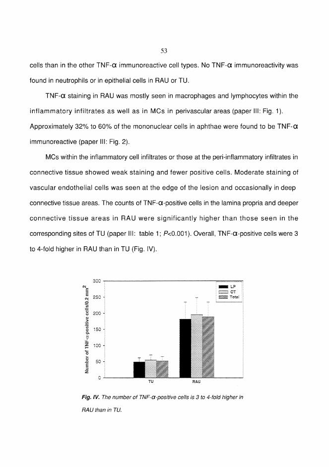

54
Expression of nuclear antigen Ki-67 in RAU (IV)
1) Epithelial compartment
The staining pattern of proliferation-associated antigen Ki-67 was strong and always
confined to the nuclei. Ki-67 positivity was mainly seen in the basal and suprabasal epithelial
cells. The epithelium surrounding RAU and TU showed an increase in the number of Ki-67
positive cells. There were few proliferating cells in the migrating epithelium at the edges of
the ulcer with absence of epithelial outgrowth in 6 out of 10 RAU. In the basal layer of both
RAU and TUs, Ki-67+ proliferating cells were more numerous among the cells in the deepest
epithelial rete ridges than among the cells overlying the connective tissue papillae.
Epithelium in RAU samples showed an increase in intraepithelial lymphocytes
compared with TU and some of the intraepithelial lymphocytes displayed Ki-67. Such cells
were located in the interepithelial cell spaces, as single cells or, more rarely, as very small
clusters. However, such cells were relatively few and could easily be distinguished from Ki-67
positive epithelial cells, which had intercellular bridges.
The Ki-67 positive cell count was 30% greater in the epithelium of RAU than in the
epithelium of TU (Fig. V).
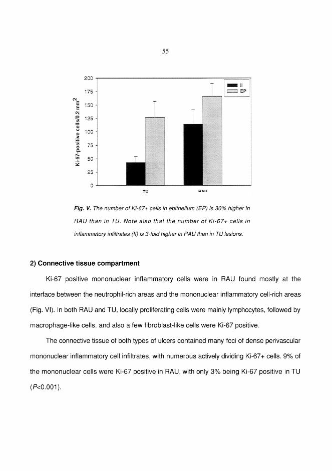
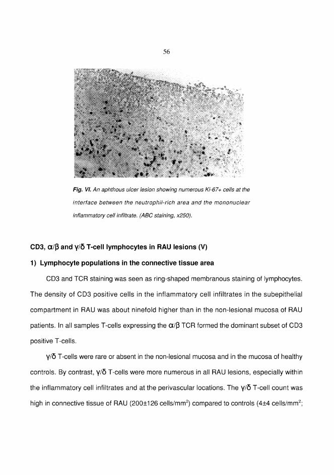
57
P<0.0001) or to non-lesional mucosa from RAU patients (5±7). Moreover, the mean
percentage of (/*+ T-cells among total CD3+ lymphocytes was increased in the connective
tissue area from 4% and 5% in controls and non-lesional mucosa, respectively, to 19% in
RAU.
2) Lymphocyte populations in the epithelium
In epithelium, CD3+ lymphocytes were approximately 3-fold more frequent in RAU
samples than in controls. Both "/$ and (/* T-cells were scattered as single cells between
basal and suprabasal epithelial cells. The count of "/$ T-cells was high in the RAU
epithelium (118±33 cells/mm2) compared to non-lesional mucosa (72±24 cells/mm2; P<0.001)
or to healthy controls (63±27 cells/mm2; P<0.001).
Intraepithelial (/*+ T-cells were seen in 5 of the 10 non-lesional mucosa, and in 4 of the
10 controls, whereas in aphthae they were seen in all 10 RAU samples. A wide variation was
observed in (/* T-cell counts in the epithelium of RAU, as they localized focally, homing
some epithelial ridges more than others. The highest densities of (/* T-cells were usually
seen in the lower epithelial layers and in the basement membrane zone (Fig. VII). The
number of (/* T-cells was high in the epithelium of RAU (70±34 cells/mm2) compared with
the epithelium of non-lesional mucosa (2.8±06 cells/mm2; P<0.0001) or epithelium of healthy
controls (1.2±1.5 cells/mm2; P<0.0001). In the epithelial area the proportion of (/* T-cells of
CD3 positive cells was 2% and 6% in controls and non-lesional mucosa, respectively, and
36% in RAU (P<0.0001; Fig. VIII). No significant differences were found in (/* T-cell counts
between the non-lesional mucosa and healthy controls, and no correlation was found
between the densities of intraepithelial (/* T-cells and the duration of RAU lesions.


59
DISCUSSION
"The probability of success is difficult to estimate,but if we never search, the chance of success is zero"
Astrophysicist Giuseppe Conconi concludes aboutsearching of extraterrestrial intelligence
Diagnosis
Although the criteria for diagnosing RAU were applied to all RAU patients recruited in
this study, the use of these criteria for diagnosing RAU has some limitations. First, there is no
known laboratory procedure available to establish the definite diagnosis, and
histopathological examination of the biopsies do not provide such a definitive diagnosis.
Second, detailed virological investigations of lesional tissue or serum are usually not
warranted, except to exclude atypical herpetic infection (Porter et al. 1998).
However, the high reproducibility and uniformity of the clinical picture and course in all
our patients suggests that all the RAU samples that were included in this study represent
recurrent aphthous ulcers. Furthermore, all patients with features suggesting systemic
disease, immunodeficiency state or neutropenic ulcers were excluded.
In spite of the fact that RAU is a common condition, its histopathology and time course
have been rather infrequently studied. For instance, studies on RAU lesions at the pre-
ulcerative stage are few in number and include very few subjects, probably because biopsy
specimens of true pre-ulcerative stage of RAU are extremely difficult to obtain (Stenman and
Heyden 1980). Therefore, in this study we dealt only with biopsies obtained from ulcerative
and painful lesions, 2-10 days after their development.
The selection of a severe form of RAU conditions (defined as one attack of RAU per
month or more for a period of at least one year) in this study was done to maximize the

60
possibility of observing significant changes that may occur at the cellular or subcellular level.
Although the patient group in each individual study was small, there were no remarkable
differences in the histopathologic features of RAU. This would suggest that the findings in
this study may be generally applicable.
Staining and evaluation methods
Most of the biopsy specimens in this study were stained by the ABC staining method
introduced by Hsu and et al. (1981). The strong affinity of avidin for biotin gives this method
high sensitivity and usually gives excellent results on fixed, paraffin-embedded specimens.
The peroxidase-anti-peroxidase (PAP) method has been used in study II instead of the
ABC method to avoid possible interference of tryptase staining with non-immunochemical
staining of MCs as a result of endogenous avidin-binding activity of MC granules (Hsu and
Raine 1984). To reduce the problem of non-specific background staining, we used mostly
monoclonal antibodies for their ability to bind specifically to one epitope on the immunogen,
and separate incubations with blocking normal sera prior to application of the primary
antibodies were used to inhibit non-specific immunoglobulin and other protein binding sites.
In this study, a 10% neutral buffered formalin solution was used as a fixative because
it is well tolerated by the tissues, has good penetration, and gives clear morphological details
(Leong 1993). Furthermore, it is suitable for identification of cytoplasmic and surface
membrane antigens (Farmilo and Stead 1989). Acetone was used in (study V) because it
produces good preservation of surface membrane antigens in cryostat sections (Farmilo and
Stead 1989). To achieve maximal staining sensitivity, the signal-to-noise ratio was increased
by suppressing the endogenous peroxidase activity with methanol-H2O2 treatment in addition

61
to the normal serum blocking step (Petrusz 1983). The semi-automatic VIDAS image analyzer
version 2.1 employed in this study has the advantages of accuracy, repeatability, and ease
of use.
Control specimens
Both of the control groups and RAU patients in this study were comparable in age factor
but were not well matched in gender factor as the controls and RAU groups were not equal
in sample size and the the number of female participants in this study were slightly more than
the males. A third set of control samples were obtained only for study V by taking specimens
from the normal appearing mucosa of RAU patients, at sites contralateral to RAU lesions.
These control groups were considered as very appropriate for detection of any specific
cellular changes (concerning the topics dealt with in this study) that have occurred at
inflammatory sites of RAU. Labial and/or buccal areas were selected as sampling sites for
clinically healthy control mucosa to match the same location areas of RAU lesions. Clinically
normal oral mucosa consisted of stratified squamous epithelium supported by fibrous
connective tissue. There was a small number of scattered chronic inflammatory cells in the
connective tissue areas. This could be explained by the fact that oral mucosa (due to its
anatomical state and function) is especially exposed to stimuli of physical, chemical and
microbiological origin. These noxious effects may increase the inflammatory cells pool within
the mucosal compartment.
Buccal mucosa was chosen as a site for induction of TUs to match the location of RAU
lesions, and for convenience, i.e. easy accessability and less painful than other areas. We
chose only those volunteers with a negative history of RAU for induction of TU lesions

62
because a local trauma may trigger RAU in people susceptible to aphthous ulcerations but
not in healthy controls (Wray et al. 1981b). The histology of TU differs from that of RAU in the
less marked mononuclear and perivascular mononuclear inflammatory cell infiltration, a more
pronounced neutrophil leukocyte and fewer MCs. The control tissue samples were fixed and
processed identically with the diseased RAU samples to avoid artefactual differences.
Factor XIIIa+ dendrocytes (I)
Factor XIIIa is expressed by subepithelial dendrocytes. Factor XIIIa as a marker has the
advantage of staining subepithelial dendrocytes but not epithelial Langerhans-type dendritic
cells. Langerhans cell-independent pathway is another pathway for handling the antigens that
are formed and/or enter via the subepithelial compartment (Streilein 1989, Yardy Tse and
Cooper 1990, Van Wilsem et al. 1994). It is conceivable that Langerhans cell-independent
pathway may play an important role in RAU inflammation because the starting focus of RAU
pathology lies in the subepithelial compartment with early accumulation of mononuclear
lymphoid cells (Stanley 1972).
FXIIIa + dendrocytes seem to be involved in the antigen handling in the connective
tissue, because they are much more frequent than Langerhans’ cells and because they are
capable of presenting antigen in vitro and possibly in vivo (Yardy Tse and Cooper 1990).
Moreover, the antigen-presenting potency of FXIIIa+ cells has been found to equal that of
dendritic cells isolated from epidermis or blood ( Nestle et al. 1994, Nestle and Nickoloff 1995b).
In this study we found that FXIIIa+ cells may acquire a spindle-shaped, rounded or
dendritic appearance, which may reflect differences in maturation development or cellular
and functional diversity of cells that express FXIIIa (Cerio et al. 1989, Ádany et al. 1988, Penneys

63
1990). Interestingly, the morphology of the FXIIIa+ cell population in RAU and TU lesions
had changed markedly towards dendrite-like (and, to a minor extent, round) at the expense
of the spindle-shaped cells. And many of these cells appeared to have enlarged (up to two
times as large as that of control) and revealed long processes.
A constant finding in the present study was the appearance of numerous FXIIIa+
dendrocytes interspersed among lymphocyte infiltrates and in close proximity to many blood
vessels. This finding contradicts partially previous results from other group (Regezi et al. 1993)
showing enlarged factor XIIIa+ dendrocytes but without increase in the the density of such
cells in RAU in HIV-positive patients. This discrepancy could perhaps be explained by the
differences in the subjects of study, as they have used only HIV-positive patients for study
and they did not use mucosa from healthy control for comparison.
We found that FXIIIa+ dendrocytes were in close contact with the infiltrating
lymphocytes. Such observations suggest that FXIIIa+ dendrocytes may serve as antigen-
presenting cells at the connective tissue compartment to infiltrating T lymphocytes after the
emigration of these T cells from the blood vessels into the site of inflammation (Cerio et al.
1989, Sontheimer et al. 1989, Lappin et al. 1996). This view might be further supported by our
previous finding in study (IV) that at least part of the infiltrating lymphocytes were replicating
locally in the perivascular areas in RAU lesions.
Moreover, it has been found that MCs are intimately associated with dermal
dendrocytes (Yoo et al. 1998) and that FXIIIa expression by dermal dendrocytes increases
when MCs degranulate and release TNF-" (Sueki et al. 1993). These data, in addition to our
findings on the functional activity status of MCs in RAU (study II), suggest a close relationship
between these cells at an anatomical and functional level. Furthermore, the XIIIa+

64
dendrocytes accumulated also in the connective tissue, lateral to inflammatory infiltrates of
RAU, which may suggest a regulatory function in this site since these cells are also believed
to have a role in extracellular matrix deposition and regulation of connective tissue
remodeling and repair that may accompany the inflammation process (Hermanns-Le et al.
1999, Monteiro et al. 1999).
Overall, the results demonstrated that FXIIIa+ dendrocytes were increased in number
and size in RAU, found in abundance in lamina propria, within the inflammatory cell infiltrates
and in deeper connective tissue, particularly in association with blood vessels. They may
have a prominent role as antigen presenting cells in the immuno-surveillance system of the
submucosa, and/or are involved in tissue repair and extracellular matrix deposition.
Mast cells (II)
MCs are effector cells of inflammation and immunity (Galli et al. 1999). Their specific
locations within tissues, immediately beneath and, in some cases, within epithelial surfaces
as well as near blood vessels, nerves and glands and their ability to respond quickly to
changes in the immune status of their microenvironments ( Wedemeyer et al. 2000), led us to
propose a potential role for MCs in RAU pathogenesis.
There are remarkable similarities between the triggering factors for MC degranulation
such as drugs (Bosso et al. 1991, Horinouchi et al. 1993), hormones (Tsakalos et al. 1993),
infections (Sugiyama 1977), stress (Gui 1998; Alexacos et al. 1999; Wilson and Baldwin 1999) and
trauma (Elsayed and Dyson 1993) and those factors precipitating RAU such as stress, food,
drugs, hormonal changes, trauma and infections (Porter et al. 1998). Previous studies on the
frequency of MCs in RAU lesions have yielded conflicting results, as being either increased
65
(Lehner 1969a), unchanged, or decreased (Dolby and Allison 1969b, Müller and Lehner 1982) in
RAU lesion.
Tryptase and chymase, are the two major MC proteinases. “Connective tissue” MC
population expresses both tryptase and chymase (MCTC), while “mucosal” MCs express
tryptase but no chymase (MCT). However, the oral mucosa contains a mixed population, with
a predominance of "connective tissue" phenotype (Walsh et al. 1995). In this study, tryptase,
which present almost exclusively in the granules of human MCs and expressed by both
subtypes of MCs of the oral mucosa is employed as a specific marker for MCs.
It is possible that some MCs have totally released their granular contents leading to the
formation of “phantom MCs”, and this could lead to an erroneously ''low number'' of MCs. I
therefore used surface bound IgE as a second marker, because it is not affected by
excessive degranulation of MCs.
The results obtained with the anti-IgE staining were similar to those found by tryptase
staining, suggesting that tryptase is a very sensitive marker for MCs.
In normal oral mucosa and in the majority of RAU samples, MCs were not found in
epithelium. It was an interesting finding that some MCs were localized intraepithelially, close
to the basal cell layer in 3 of 15 RAU lesions. MCs can infiltrate the epithelium in certain
circumstances, especially in parasitic and allergic inflammations (Fokkens et al. 1992). The
presence of MC in the epithelium of only 3 RAU lesions may have a number of
interpretations. One attractive possibility is the heterogeneity of RAU with respect to etiology.
RAU may represent a single condition with multiple causes and the etiology of these 3 RAU
lesions could be due to allergic cause.
Localization of MCs near, or even in, the epithelium may represent an interaction

66
between active degranulated MCs and epithelium of RAU. However, the absence of MCs in
the area close to the ulcer suggests that ulceration probably was not dependent upon direct
MC involvement. An indirect role in the early events cannot be ruled out, in view of our
observation of increased MC density and activity at the basement membrane zone, lateral to
the aphthous ulcers. The degranulation of MCs in close proximity to the basement membrane
results in the release of tryptase and chymase, which can promote both the inflammatory
reaction and the cleavage and activation of enzymes that are known to promote degradation
of the basement membrane component (Gruber et al. 1990a, Lohi et al. 1992, Häyrinen-Immonen
et al. 1993). They can therefore participate in the formation of the ulcer. Conversely, the mere
presence of MCs and their degranulation activity at the basement membrane zone does not
necessarily mean that they play only a destructive role. MCs might be involved in the repair
process. For instance, MC tryptase was found to be a mitogen for epithelial repair and to
facilitate recruitment of granulocytes (Cairns and Walls 1996). In this respect, higher
proliferation activity was found in the RAU epithelium than in TU epithelium of similar lesional
duration (study IV). Furthermore, Thompson and associates (1991) have provided evidence
that certain murine MC lines are capable of laminin synthesis. Thus MCs in regions lateral to
aphthous ulcers may contribute to increased epithelial mitosis and/or formation of the
basement membrane. The possibility that MCs were able to perform different or even
opposite funct ions during the course of RAU inf lammation or at the dif ferent
microenviromental areas in RAU is not surprising, as MCs can respond to antigens,
pathogens or their products by undergoing alterations in phenotype, function, survival, and as
some of these changes can be reversible (Galli et al. 1999).
Although this study showed that the majority of the MCs in RAU were apparently

67
degranulated, it was difficult to ascertain whether a partial degranulation of MCs in the tissue
was due to an inflammatory process or due to sampling and processing procedures.
However, the presence of a “halo” of tryptase around a proportion of MCs in the tissue
sections is a reliable indication of MC degranulation (i.e. the released granules lost their
round shape and appeared as diffuse staining around the cell indicating that dissolution of
the contents of such granules have occurred in a time preceded the sampling and processing
procedures). For this reason, this paper focuses mainly on those observations where halos
of tryptase around some MCs were encountered.
The significant increase in MC numbers and degranulation within the RAU mononuclear
cell infiltrates, compared with those seen in TU inflammatory cell infiltrates, suggests that
such an increase is not a response to ulceration in general but is more specific to aphthae.
The functional significance of MC accumulation in the connective tissue along the
borders of the inflammatory infiltrates, especially at the interfaces between the inflammatory
infiltrates and connective tissue, is unclear. MC degranulation at these interfaces may
contribute to localized degradation of extracellular matrix, both by the stimulation of matrix-
degrading enzymes, metalloproteinases (MMPs) production by resident connective tissue
cells (Yoffe et al. 1984, Yoffe et al. 1985, Häyrinen-immonen et al. 1993, Kanbe et al. 1999), and by
the subsequent activation of released inactive precursors (proMMPs) by the MC proteinases
(Gruber et al. 1990b, Lees et al. 1994, Suzuki et al. 1995). Recently, it was also found that
chymase is able to cleave and activate procollagenase directly (Saarinen et al. 1994), and can
function as an interleukin-1$ convertase (Mizutani et al. 1991). Thus, both tryptase and
chymase of MCs in RAU may initiate a sequence of events leading to degradation of the
extracellular matrix, which is an essential step for inflammatory cell movement and migration

68
through extracellular tissues.
Although the majority of RAU lesions were early, i.e. 2-3 days old, the results show that
the age of RAU and TU has no appreciable effect on MC density in such lesions. Our data
showed that MCs are 63% more numerous in RAU than in clinically healthy mucosa, which
is less than that reported by Lehner (1969a), who described a 100% rise in the number of
MCs in minor aphthous ulcers. However, it is in conflict with the results of Dolby & Allison
(1969b), who reported no difference in the number of MCs between controls and early (1-2
days old) RAU and also described a reduction of MC numbers in lesions of more than 2 days
duration. The most likely explanation for this discrepancy is the use of different stains like
toluidine blue or alcian blue/safranin to identify and localize MCs in the two previous studies.
However, the markers and methods employed in the present study were more sensitive and
allow all MCs in the tissues to be detected, regardless of their phenotype and state of
degranulation.
Our results, however, provide no clues as to the reasons for increased MCs in RAU
lesions. Finally, our results emphasize the importance of tryptase enzyme as a marker for the
localization and activation of MCs. The significant increase in number and degranulation
activity of MCs in aphthae suggests an active role for MCs in the pathogenesis of RAU.
TNF-"""" (III)
Although a previous study showed mRNA for TNF-" in inflamed RAU mucosal tissue,
little is known about its cellular distribution at sites of RAU inflammation. To address this point
immunohistochemistry was used to determine the cellular localization of TNF-" in RAU
lesions compared with TU lesions.

69
Our results showed that there was no TNF" immunoreactivity in the epithelial cells.
This was unexpected because epithelial cells participate in immune regulation and in the
maintenance of mucosal integrity by generating a range of biologically active mediators.
However, this does not mean that these cells are not producing and secreting this cytokine.
It is possible that such cell types may contain insufficient TNF-" to be demonstrated by our
immuno-histochemistry, or that the epithelium of RAU is not an important source of this
cytokine. However, we cannot rule out the possibility that epithelium may act as a target for
TNF-" that was produced by infiltrating inflammatory cells. TNF" has been found to possess
in vitro cytotoxic activity against a variety of tumor cells (Williamson et al. 1983, Deem et al.
1991) and to a lesser extent against some normal cell lines depending on the concentration
of TNF-" and on the simultaneous presence of other cytokines (Taverne et al. 1987, Wu et al.
1996). Although the hallmark of RAU is the destruction of epithelium leading to ulceration, the
relative importance of the role of cytotoxicity versus cytokine release in mediating epithelial
cell death and aphthous ulceration is still unknown.
Although it has been reported that neutrophils can produce TNF-" (Dubravec et al.
1990), the immunohistochemical results did not show TNF-" immunoreactivity in neutrophils
in either TU or RAU lesions. It may be that neutrophils do not produce TNF-" as a response
to surgically induced trauma or to the antigenic stimulation that is supposed to trigger
aphthous ulceration. However, TNF-" produced by other cells may act on neutrophils to
promote their adherence to endothelial cells and migration to the inflammatory areas. TNF-"
may also enhance phagocytosis, respiratory burst, reactive oxygen generation and
degranulation of the neutrophils (Gamble et al. 1985, Shalaby et al. 1985, Klebanoff et al. 1986,
Tsujimoto et al. 1986).
70
Because of the possibilty of slight nonspecific background staining it is difficult to make
comment on TNF-" release into the extracellular compartment of RAU and TU lesions.
The present study shows that the expression of TNF-" is marked in macrophages and
occurs mainly in mononuclear inflammatory cell infiltrates. TNF-" is secreted by activated
monocyte/macrophages and many other cells in response to bacterial toxins, inflammatory
products, and other stimuli (Vasalli 1992). But the precise factor(s) which activate
macrophages to produce TNF-" in the RAU lesions remain(s) uncertain.
Our findings show that numerous lymphocytes in mononuclear inflammatory cell
infiltrates in RAU biopsies were positive for TNF-". This means that these cells are activated,
and express TNF-" in RAU lesions (Kinkhabwala et al. 1990, Deem et al. 1991).
The finding of TNF-" localized to MCs adds to the previous studies demonstrating TNF-
" production by human MCs (Ohkawara et al. 1992, Walsh et al. 1991, Walsh et al. 1995). It is
worthwhile to mention that only MCs are known to store preformed TNF-" in significant
quantities and to release it upon immunologic stimulation (Gordon and Galli 1990). TNF-"
derived from MCs and other adjacent mononuclear inflammatory cells seems to be a
particularly important candidate for initiating the increased expression of cell adhesion
molecules like ICAM-1 that has been described in blood vessels close to the inflammatory
infiltrates and lateral to the aphthous ulcer sites (Häyrinen-Immonen et al. 1992b). MCs within
mononuclear inflammatory cell infiltrates showed weaker TNF-" immunoreactivity than those
MCs in deeper connective tissue. This weak TNF-" immunoreactivity in MCs might be due
to ongoing production and release of this cytokine. This is also in line with the previous
findings that showed prominent activation and degranulation of MCs in RAU inflammation
(study II).

71
Moderate immunoreactivity for TNF-" was observed in some vascular endothelial cells
at the lamina propria lateral to the ulcer and in the blood vessels of deep connective tissue.
This suggests that the production of TNF-" by RAU vascular endothelium is minimal and the
major sources of TNF-" that act on vascular endothelium in RAU are the adjacent
mononuclear inflammatory cells.
The difference in TNF-" immunoreactivity between RAU and TU controls appears to
relate to an increased expression of this cytokine in inflammatory cells in RAU. However, the
detection of TNF-" containing cells in TU lesions is not unexpected, since numerous
infiltrating inflammatory cells are usually present within the inflammatory infiltrates in TUs,
and local expression of inflammatory mediators is an essential component of the normal
healing process.
In view of the high density of TNF-" immunoreactive cells in RAU lesions and the well
characterized effects of this cytokine, it is probable that TNF-" makes a significant
contribution to inflammatory events in RAU lesions.
Cell proliferation in RAU lesions (IV)
As a result of mediators released in the vicinity of postcapillary venules, leukocyte
adhesion molecules are induced on endothelial cells. The circulating lymphocytes then roll,
adhere and transmigrate into inflammation areas via local blood vessels. Once in the
perivascular compartment, extravasated lymphocytes may produce a variety of pro-
inflammatory molecules, and the immune response is amplified by local T cell proliferation
stimulated by interaction between these cells and antigen-presenting cells in the connective
tissue compartment.
72
In this study we raised the issue of the relatively long persistence of RAU lesions and
whether the cell proliferation in RAU lesions differs from that of the TU lesions.
Ki-67 used in this study is a good marker of cell-cycling, has a short half-life (90 min), and the
nuclei can be scored as either positive or negative for Ki-67 immunoreactivity, (Bruno and
Darzynkiewicz 1992, Duchrow et al. 1994).
The absence of epithelial outgrowth in 6 out of 10 RAU might be due to ongoing
epithelial destruction or inability of the cells at the edge of the ulcer to switch from the
stationary to locomoting epithelial phenotype. For instance, Richards et al. (1996) found
disruption in the deposition of laminin-5 and an apparent lack of fibronectin (which provides
the provisional matrix for subsequent cell migration) at the edges of some of the RAU lesions
(Grinnell et al. 1981).
The low mitotic activity in the epithelial outgrowth of both RAU and TU suggests that the
advancing epithelial tongue, which extended over the ulcer margins, involves migratory,
nondividing cells and only to a lesser extent proliferating cells.
Our results with the Ki-67 proliferation marker were mostly in line with the previous
reports showing that DNA synthesizing cells in the oral mucosa are mainly localized at the
lowermost part of the epithelial rete ridges, with only few DNA synthesizing cells overlying the
connective tissue papillae (Hume 1989).
Few intraepithelial lymphocytes had displayed Ki-67 positivity, but their distinguished
appearance from epithelial cells made it possible to demonstrate that most of the proliferating
cells in RAU are epithelial cells.
Our finding of increased mitotic activity in RAU epithelium and the clinical observation
that RAU lesions last longer than TU (Stanley 1972, Richards et al. 1996) seem paradoxical.
73
However, epithelial cell proliferation is not the sole determinant of re-epithelialization (Garlick
et al. 1996). It seems likely that the epithelial cell migration is delayed in RAU lesion when
compared with TU, as suggested by the absence of epithelial outgrowth in 6 of 10 RAU
samples. The absence or inappropriate expression of keratinocyte integrins or their
extracellular ligands as a result of inflammation may explain such an apparent paradox
(Richards et al. 1996).
This immunostaining study gives no definite clues as to the type of inflammatory cells
involved in proliferation, but it confirms that there are proliferating cells in the inflammatory
infiltrates. Our results showed an inter-individual variation in the proportion of proliferating
cells in the inflammatory cell infiltrates in RAU (9±4% of inflammatory cells were proliferating),
which may reflect the dynamic nature of cell activation in RAU, the interpatient variations and
the possible variation in the concentration and presentation of the antigens, i.e. it may imply
that not all RAU lesions contain the same amount of triggering antigen(s).
Although the majority of infiltrating inflammatory cells in TU lesions were not
proliferating, the presence of some cycling lymphocytes in TU infiltrates could represent
physiological processes that act to stimulate epithelial repair and protect against infection. In
this respect Reusch and colleagues (1991) found that lymphocytes infiltrate the epidermis in
sterile wound healing, which suggests that in the absence of foreign antigenic stimulation, the
immune system can be activated by traumatic injury. The antigen presenting cell system has
been suggested to be activated by microenvironmental tissue damage (Ibrahim et al. 1995)
which leads to immune stimulation (Evavold et al. 1993) and subsequently to activation and
proliferation of the infiltrating lymphocytes.
In situ T cell proliferation has been shown to be a feature of several diseases believed

74
to be immunologically mediated (Wood et al. 1986, Séron et al. 1989, Morganroth et al. 1991),
which suggests that it is a mechanism which operates in different inflammatory lesions and
is not restricted to RAU inflammation.
It has been previously reported that 45% of all lymphoid cells in RAU lesions show signs
of previous antigenous contact (Häyrinen-Immonen 1991). Antigen-experienced cells can be
activated by a broader range of antigen presenting cells, respond faster and more effectively
upon new encounter with the antigen (even outside the context of the lymphoid tissues) than
naive cells (Byrne et al. 1988). They may represent the cellular basis for the high proliferative
response at sites of RAU lesions.
In conclusion, our results demonstrate that increased proliferative activity is present in
RAU epithelium and that the longer persistence of RAU lesions (compared to TU) cannot be
explained by a lack of new epithelial cell production. Furthermore, mononuclear cell
replication in situ represents one mechanism for the expansion of the inflammatory cell
infiltrates at the sites of RAU and TU lesions.
Intraepithelial lymphocytes (V)
Previous studies have found a high percentage and activity of (/* T cells in peripheral
blood of patients with RAU and Behçet's disease (Suzuki et al. 1992, Pedersen and Ryder 1994,
Freysdott ir et al. 1999). Such findings imply a possible role for (/* T cells in the
immunopathogenesis of RAU.
The early changes in RAU lesions are characterized by the early influx of lymphocytes
and neutrophils (Savage et al. 1985) into the inflammatory RAU site via blood vessels in
lamina propria. Initially, there is subepithelial aggregation and proliferation of lymphocytes in

75
situ and, subsequently, active intraepithelial migration.
The sequence of cellular inflammatory events in RAU inflammation culminates in a
marked migration of lymphocytes into the epithelia. It seemed therefore important to study
the proportion of (/* T-lymphocytes among the lymphocytes in the epithelium and in the
subepithelial compartment of RAU lesions. Furthermore, there have been, to my knowledge,
no reports concerning the occurrence of (/* T-cells in RAU lesions. In the present study, the
distribution and frequency of (/* T-cells in RAU lesions were analyzed immuno-
histochemically.
The present data showed that the epithelium of RAU lesions contains three times as
many CD3 positive lymphocytes as the epithelium of controls. These findings seem to agree
with most former pathological studies that have found frequent infiltration of lymphocytes to
the lower epithelial layers in RAU lesions (Lehner 1969a). In the present study, most of the
CD3 lymphocytes in all samples expressed the "/$ TCR. Although we found that the
absolute count of "$-TCR positive cells was increased in RAU lesions, their relative
proportion was reduced compared with the mucosa of controls, apparently because of the
relative increase of (/* T-cells.
Lymphocytes expressing (/* TCRs were found to comprise a small proportion (-2% of
CD3+ T-cells) in the healthy control mucosa, a value similar to that previously reported (Pepin
et al. 1993). In contrast to the results obtained in mice (Itohara et al. 1990), (/* T-cells were
uncommon in the epithelium and in the connective tissue of normal human oral mucosa.
Previous work has shown an increased proportion of (/* T-cells in the peripheral blood
of patients with RAU during both the inactive and active phase (Pedersen and Ryder 1994).
Furthermore, Suzuki et al. (1992) have shown that the concentration of circulating (/*+ T-

76
cells was higher in patients suffering from Behçet's disease with mucocutaneous RAU
compared to patients without mucocutaneous lesions. In the present retrospective study we
were unable to determine if the increased percentage of (/* cells in RAU lesions simply
reflected their high numbers in the peripheral blood.
Our results demonstrate a high density of (/* T-cells in inflammatory lesions in RAU,
whereas, in non-lesional mucosa from opposite sites of RAU, (/* T-cells were rarely seen in
the epithelium or in the connective tissue areas.
The requirements for (/* T-cell activation are less stringent than those for "/$ T-cell
activation. For instance, (/* T-cells can recognize antigens directly, without the need for
specialized antigen-presenting cells (Schild et al. 1994). Furthermore, (/* T-cells can also
react to non-peptide antigens such as a lipid fraction of M. tuberculosis (Tsuyuguchi et al.
1991) and various phosphorylated metabolites (Porcelli et al. 1996). Such features of (/* T-
cells would probably enable them to respond quickly to a variety of antigens before the "/$
TCR lymphocyte population begins to expand (De Libero 1997).
The bowel affected by coeliac disease contains higher than normal numbers of (/* T-
cells in either active or "silent" periods of the disease (when the pathology is mildest).
Furthermore, the density of such cells is higher in the epithelium than in the lamina propria
(Kutlu et al. 1993). Such an expansion of (/* TCR positive intraepithelial lymphocytes might be
driven by an intestinal allergy to cereal proteins associated with autoimmunity directed
against tissue transglutaminase. In aphthae, the high density of (/* T-cells was present
exclusively in RAU lesions and in both the epithelium and the subepithelial connective tissue.
Thus, it is possible that some of the (/* T-cells were responding in an antigen-specific
fashion and that antigenic stimuli inductive for (/* T-cells were expressed only during the
77
development and/or progression of RAU.
The exact biological function of (/* T-cells in the epithelium of RAU lesions remains
unclear. However, Boismenu & Havran (1994) have shown that activated (/* T-cells produce
the epithelial cell-specific fibroblast growth factor-7 (FGF-7). The effects of FGF-7 on the
proliferation and differentiation of epithelial cells may help to reduce mucosal damage
following tissue injury (Zeeh et al. 1996). This is in line with the findings of study (IV) on the
expression of the cell proliferation-associated nuclear antigen (Ki-67) in RAU lesions that
showed higher proliferation activity in the epithelium of RAU compared with that in the
epithelium of controls.
Many reports have shown that different sub-populations of (/* T-cells have different
functions and different patterns of cytokine secretion. For instance, Ferrick et al. (1995) found
that (/* T-cells can produce either T-helper type-1 (Th1) or Th2 cytokines depending on the
pathogen they are exposed to. However, the role of (/* T-cells may not be limited to merely
enhancing epithelial proliferation or modulating immune responses of "/$ T cells (Fujihashi et
al. 1996b). For instance, other investigators (Hamzaoui et al. 1994, Hasan et al. 1996) have
suggested that (/* T-cells may play a role in immunological damage in RAU. The "Janus-
like" coexistence of the cytotoxic and protective potential of (/* T-cells might enable them to
play a dual function in the pathogenesis of RAU, thereby acting as immunoregulators to
maintain the homeostasis and functional integrity of the epithelial barrier.
It should be pointed out that there was a wide variation in the density of (/* T-cells
between RAU samples in this study. Similar wide inter-individual variations were also noted
in the peripheral blood of Behçet's disease (Suzuki et al. 1992) and RAU patients (Pedersen
and Ryder 1994). The reasons for these variations are currently unknown.

78
Although our results provide no answers regarding the function of (/* T-cells in RAU
lesions, they showed that (/* T-cells are increased in RAU lesions and such an increase was
concomitant with the RAU inflammatory process and did not extend to mucosal areas other
than the inflammation sites.

79
SUMMARY & CONCLUSIONS
“Creativity is the ability to find the alternatives”Edward De Bono, a contemporary thinker
For better understanding of RAU, it will be necessary to study the immunocompetent
cells and the key pro-inflammatory cytokine TNF-" that are likely to have a role in aphthous
ulcer pathogenesis. The purpose of these studies was to investigate the distributions and
frequencies of the factor XIIIa-positive dendrocytes, mast cells, CD3+ lymphocytes and its
subtype "$ and (* T-cells, as well as to evaluate the expressions of the pro-inflammatory
cytokine TNF-" and the proliferation-associated nuclear antigen Ki-67 in RAU lesions
compared with clinically healthy oral mucosa and/or with induced traumatic oral ulcers.
Samples were incubated with the relevant antibodies (polyclonal rabbit anti-human factor
XIIIa, monoclonal antibody specific for MC tryptase (AA1), polyclonal anti-IgE antibody,
monoclonal antibodies specific for CD3, "/$ TCR and (/* TCR, monoclonal anti-human
TNF-" antibody, rabbit anti-human Ki-67 nuclear antigen) in avidin-biotin-peroxidase
complex (ABC) staining or with peroxidase-anti-peroxidase (PAP) complex staining. Results
were quantified by means of VIDAs image analyser or conventional microscopy.
Most of the FXIIIa-immunoreactive cells in TUs and normal mucosa were spindle-
shaped, whereas a relatively large, dendritic-like cell type was predominant in RAU lesions.
FXIIIa+ dendrocytes were increased in number, and apparently also in size, in RAU lesions
(274±68 cells/mm2) as compared to controls (177±74 cells/mm2, P<0.01), and to TU lesions
(183±50 cells/mm2, P<0.01). MCs were absent in the epithelium except in 3 out of 15 RAU
lesions. MC activation/degranulation as judged by diffuse extracellular tryptase staining, was
a common feature within RAU-inflammatory infiltrates and at RAU-inflammatory infiltrates-

80
connective tissue interfaces, which were often associated with connective tissue disruption.
MCs were significantly increased in aphthae (116±26 cells/mm2) compared with TU lesions
(72±11 cells/mm2, P<0.001) and healthy controls (71±16 cells/mm2, P<0.001).
The number of lymphocytes expressing (/* TCRs was very low in non-lesional mucosa
or in clinically healthy mucosa. By contrast, (/* T-cells were numerous and observed in all
RAU lesions especially intraepithelially, within the inflammatory infiltrates and at perivascular
locations. In epithelial areas, the average percentage of (/* T-cells among total CD3+
lymphocytes was increased from 2% and 6% in controls and non-lesional mucosa,
respectively, to 36% in RAU. TNF-" immunoreactivity was seen in monocyte/macrophages,
lymphocytes, MCs and vascular endothelial cells. TNF-"-containing cells were more
numerous in aphthae (188±46 cells/0.2 mm2) compared with controls (52±14 cells/0.2 mm2,
P<0.001). The epithelium of RAU contained 30% more proliferating cells than the epithelium
of TU and about 9±4% of the infiltrating inflammatory cells in RAU lesions were Ki-67+
proliferating cells, compared to only 3±1% in TU inflammatory infiltrates.
In conclusion, the results of the studies show significant increase in density and obvious
changes in the morphology and size of FXIIIa+ dendrocytes in RAU lesions. The increased
number of MCs in RAU lesions, and the local MCs show signs of activation and
degranulation suggesting an active involvement of this cell type in RAU pathogenesis.
High expression of TNF-" in RAU lesions was seen in mononuclear inflammatory cells, MCs
and vascular endothelial cells. This may reflect an active role for TNF-" in the recruitment
and activation of leukocytes and MCs that are found in RAU. The expression of cell
proliferation-associated nuclear antigen Ki-67 is increased in RAU compared with TU lesions.

81
The findings also indicate that local replication of the infiltrating mononuclear inflammatory
cells represents another mechanism that may contribute to the formation of the inflammatory
cell infiltrates at the inflammatorty sites of both RAU and TU. As in the peripheral blood of
patients with RAU and Behçet's disease, (/* T-lymphocytes are also increased locally at the
sites of RAU lesions suggesting that (/*T-lymphocytes may have a pathogenic and/or
regulatory role in aphthous ulceration.
Finally, the progress of RAU research in recent years has been remarkable. However,
many essential questions still remain: What is the etiology of RAU? Does RAU have a single
etiology or multiple etiologies? Is the association of RAU with coeliac disease, HIV infection,
and Behcet’s disease a meaningful guide towards elucidation of the etiology of RAU, or does
it only represent a similarity between RAU lesions and similar ulcers in these other diseases?
Future research may hold the answers to one or more of these questions.
82
ACKNOWLEDGMENTS
First of all, praise and thanks be to God, who provided me with the power and means toaccomplish this work. This study was carried out at the Department of Medicine (Invärtes medicin),Surgical Hospital, Helsinki University Central Hospital, Biomedicum Helsinki/Anatomy (InflammationResearch Group), Institute of Dentistry (Departments of Oral Medicine and Oral Pathology), HelsinkiUniversity Central Hospital, Laboratory diagnostics (Oral Pathology Unit), University of Helsinki, andORTON Research Institute, Invalid Foundation, during the years 1995-2001.
I wish to express my deepest gratitude to my first supervisor Professor Yrjö Tapio Konttinen, MD,PhD, Head of Inflammation Research Group (TULES Ryhmä), Biomedicum Helsinki, and Head ofOral Medicine Department, University of Helsinki. Without his encouraging attitude, his continuedsupport and broad understanding of inflammation, his constructive and timely discussions, and hiscreation of a free, yet stimulating, academic atmosphere, I would never have succeeded inaccomplishing this work.
I also wish to thank my second supervisor Professor Jarkko Hietanen, MD, DDS, PhD, M.Sc. for hissupport and valuable advice throughout the period of my study, for his suggestions during thepreparation of this manuscript and for providing research facilities, which was of utmost importancewhen this thesis was finalized.
My thanks are due to my third supervisor, Professor Maria Malmström, DDS, PhD, former head of theOral Medicine Department, for her continuous support and fruitful comments during all the phases ofthe study. She offered me the possibility to enter the fascinating world of RAU research.
I wish to express my sincere thanks to Dr. Ritva Häyrinen-Immonen, DDS, PhD, for excellentcollaboration and friendship, for her extensive knowledge of RAU and her admirable ability to raise myspirits even during the difficult moments of the study. Without her help in the collection of the samplesneeded for this study this thesis would not have been possible.
Thanks are due to Professor Frey Fyhrquist, MD, Head of the Department of Medicine (Invärtesmedicin), and to Professor Ismo Virtanen, the Head of the Anatomy Department, University ofHelsinki, for placing the research facilities of their departments at my disposal.
I also want to thank warmly all the volunteers, especially the RAU patients, for participating in thestudy, some of who agreed to biopsy three and even four times. Their excellent cooperation madethis work possible.
I owe my sincere thanks to Mrs. Marjatta Kivekäs and Mrs. Sirpa Kyllönen from the oral pathologylaboratory for their skillful technical assistance and enormous help in all stages of my work as well asfor creating the pleasant working atmosphere in the laboratory. I thank Prof. Timo Sorsa, TainaTervahartiala, DDS, Ravi Srinivas, DDS, Sirkku Nissalo, DDS, Pirjo-Lisa Lukinmaa, DDS and Kirsti


84

85
REFERENCES
Abdulla YH, Lehner T. The effect of immune complexes on chemotaxis in Behcet's syndrome andrecurrent oral ulcers. In: Lehner T, Barnes CG, editors. Behçet's syndrome: clinical andimmunological features. London: Academic Press, p 55-9, 1979.
Ádany R, Glukhova MA, Kabakov AY, Muszbek L. Characterisation of connective tissue cellscontaining factor XIII subunit a. J Clin Pathol 41: 49-56, 1988.
Albanidou-Farmaki E, Kayavis IG, Polymenidis Z, Papanayotou P. HLA-A, B, C and DR antigens inrecurrent oral ulcers. Ann Dent 47: 5-8, 1988.
Alexacos N, Pang X, Boucher W, Cochrane DE, Sant GR, Theoharides TC. Neurotensin mediatesrate bladder mast cell degranulation triggered by acute psychological stress. Urology 53: 1035-40,1999.
Altman DA, Fivenson DP, Lee MW. Minocyclin hyperpigmentation: model for in situ phagocytic activityof factor XIIIa positive dermal dendrocytes. J Cutan Pathol 19: 340-5, 1992.
Andrews VH, Hall HR. The effects of relaxation/imagery training on recurrent aphthous stomatitis: apreliminary study. Psychosom Med 52: 526-35, 1990.
Arber N, Klein T, Meiner Z, Pras E, Weinberger A. Close association of HLA-B51 and B52 in Israelipatients with Behçet's syndrome. Ann Rheum Dis 6: 351-3, 1991.
Axéll T. A prevalence study of oral mucosal lesions in an adult Swedish population. Odontol Rev 27(Suppl 36): 1S-103S, 1976.
Axéll T, Henricsson V. The occurrence of recurrent aphthous ulcers in an adult Swedish population.Acta Odontol Scand 43: 121-5, 1985(a).
Axéll T, Henricsson V. Association between recurrent aphthous ulcers and tobacco habits. Scand JDent Res 93: 239-42, 1985(b).
Bach MC, Howell DA, Valenti AJ, Smith TJ, Winslow DL. Aphthous ulceration of the gastrointestinaltract in patients with the acquired immunodeficiency syndrome (AIDS). Ann Intern Med 112: 465-7,1990.
Bagg J, Williams BD, Amos N, Dagalis P, Walker DM. Absence of circulating IgG immune complexesin minor recurrent aphthous ulceration. J Oral Pathol 16: 53-6, 1987.
Barile MF, Graykowski EA, Driscoll EJ, Riggs DB. L form of bacteria isolated from recurrent aphthousstomatitis lesions. Oral Surg Oral Med Oral Pathol 16: 1395-402, 1963.
Beckman JS, Beckman TW, Chen J, Marshall PA, Freeman BA. Apparent hydroxyl radical productionby peroxynitrite: implications for endothelial injury from nitric oxide and superoxide. Proc Natl Acad SciUSA 87: 1620-4, 1990.
Ben-Aryeh H, Malberger E, Gutman D, Szargel R, Anavi Y. Salivary IgA and serum IgG and IgA inrecurrent aphthous stomatitis. Oral Surg Oral Med Oral Pathol 42: 746-52, 1976.

86
Birek C, Grandhi R, McNeill K, Singer D, Ficarra G, Bowden G. Detection of Helicobacter pylori in oralaphthous ulcers. J Oral Pathol Med 28: 197-203, 1999.
Bittoun R. Recurrent aphthous ulcers and nicotine. Med J Aust 154: 471-2, 1991.
Boggess KA, Williamson HO, Homm RJ. Influence of the menstrual cycle on systemic diseases.Obstet Gynecol Clin North Am 17: 321-42, 1990.
Boismenu R, Havran WL. Modulation of epithelial cell growth by intraepithelial gamma delta T cells.Science 266: 1253-5, 1994.
Boismenu R, Havran W. An innate view of (* T cells. Curr Opin Immunol 9: 57-63, 1997.
Boon ME, Kok LP. Microwaves for immunohistochemistry. Micron 25: 151-70, 1994.
Bosso JV, Schwartz LB, Stevenson DD. Tryptase and histamine release during aspirin-inducedrespiratory reactions. J Allergy Clin Immunol 88: 830-7, 1991.
Boulinguez S, Cornée-Leplat I, Bouyssou-Gauthier M-L, Bedane C, Bonnetblanc J-M. Aphtes induitspar les médicaments: analyse de la littérature. Ann Dermatol Venereol 127: 155-8, 2000.
Brice SL, Cook D, Leahy M, Huff JC, Weston WL. Examination of the oral mucosa and peripheralblood cells of patients with recurrent aphthous ulceration for human herpesvirus DNA. Oral Surg OralMed Oral Pathol Oral Radiol Endod 89: 193-8, 2000.
Brody D, Metcalfe DD. Mast cells: A unique and functional diversity. Clin Exp Allergy 28: 1167-70,1998.
Brody HA, Silverman S Jr. Studies on recurrent oral aphthae. I. Clinical and laboratory comparisons.Oral Surg Oral Med Oral Pathol 27: 27-34, 1969.
Bruno S, Darzynkiewicz Z. Cell cycle dependent expression and stability of the nuclear proteindetected by Ki-67 antibody in HL-60 cells. Cell Prolif 25: 31-40, 1992.
Byrne JA, Butler JL, Cooper MD. Differential activation requirements for virgin and memory T cells. JImmunol 141: 3249-57, 1988.
Buno IJ, Huff JC, Weston WL, Cook DT, Brice SL. Elevated levels of interferon gamma, tumornecrosis factor ", interleukins 2, 4, and 5, but not interleukin 10, are present in recurrent aphthousstomatitis. Arch Dermatol 134: 827-31, 1998.
Burnett PR, Wray D. Lytic effects of serum and mononuclear leukocytes on oral epithelial cells inrecurrent aphthous stomatitis. Clin Immunol Immunopathol 34: 197-204, 1985.
Burton-Kee JE, Mowbray JF, Lehner T. Different cross-reacting circulating immune complexes inBehçets syndrome and recurrent oral ulcers. J Lab Clin Med 97: 559-67, 1981.
Cairns JA, Walls AF. Mast cell tryptase is a mitogen for epithelial cells. Stimulation of IL-8 productionand intercellular adhesion molecule-1 expression. J Immunol 156: 275-83, 1996.

87
Caux C, Vanbervliet B, Massacrier C, Dezutter-Dambuyant C, de Saint-Vis B, Jacquet C, Yoneda K,Imamura S, Schmitt D, Banchereau J. CD34+ hematopoietic progenitors from human cord blooddifferentiate along two independent dendritic cell pathways in response to GM-CSF+TNF alpha. J ExpMed 184: 695-706, 1996.
Cerio R, Griffiths CE, Cooper KD, Nickoloff BJ, Headington JT. Characterization of factor XIIIapositive dermal dendritic cells in normal and inflamed skin. Br J Dermatol 121: 421-31, 1989.
Challacombe SJ, Batchelor JR, Kennedy LA, Lehner T. HLA antigens in recurrent oral ulcerations.Arch Dermatol 113: 1717-9, 1977.
Challacombe SJ, Scully C, Keevil B, Lehner T. Serum ferritin in recurrent oral ulceration. J Oral Pathol12: 290-9, 1983.
Corone S, Davido A, Corone P. A rare complication of captopril: ulceration of the lingual and jugalmucosa. Rev Med Interne 8: 73-4, 1987.
Crivelli MR, Aguas S, Adler I, Quarracino C, Bazerque P. Influence of socioeconomic status on oralmucosa lesion prevalence in schoolchildren. Community Dent Oral Epidemiol 16: 58-60, 1988.
Dagalis P, Bagg J, Walker DM. Spontaneous migration and chemotactic activity of neutrophilpolymorphonuclear leukocytes in recurrent aphthous ulceration. Oral Surg Oral Med Oral Pathol 64:298-301, 1987.
Deem RL, Shanahan F, Targan SR. Triggered human mucosal T cells release tumor necrosis factor-alpha and interferon-gamma which kill human colonic epithelial cells. Clin Exp Immunol 83: 79-84,1991.
De Libero G. Sentinel function of broadly reactive human gamma delta T cells. Immunol Today 18:22-6, 1997.
Derrick EK, Barker NJ, Khan A, Price ML, Macdonald DM. The tissue distribution of factor XIIIapositive cells. Histopathology 22: 157-62, 1993.
Dolby AE. Recurrent Mikulicz's oral aphthae-their relationship to the menstrual cycle. Br Dent J 124:359-60, 1968.
Dolby AE. Recurrent aphthous ulceration. Effect of sera and peripheral blood lymphocytes upon oralepithelial tissue culture cells. Immunology 17: 709-14, 1969(a).
Dolby AE, Allison RT. Quantitative changes in the mast cell population in Mikulicz’s recurrent oralaphthae. J Dent Res 48: 901-3, 1969(b).
Dolby AE, Walker DM, Slade M, Allan C. HLA histocompatibility antigens in recurrent aphthousulceration. J Dent Res 56: 105-7, 1977.
Donatsky O. An immunofluorescence study on the cross-reaction between Strep. 2A and human oralmucosa. Scand J Dent Res 83: 111-9, 1975.
Donatsky O. A leukocyte migration study on the cell-mediated immunity against adult human oral

88
mucosa and streptococcal antigens in patients with recurrent aphthous stomatitis. Acta PatholMicrobiol Scand (C) 84: 227-34, 1976(a).
Donatsky O. Comparison of cellular and humoral immunity against streptococcal and adult oralmucosa antigens in relation to exacerbation of recurrent aphthous stomatitis. Acta Pathol MicrobiolScand 84: 270-82, 1976(b).
Donatsky O, Justesen T, Lind K, Vestergaard BF. Microorganisms in recurrent aphthous ulcerations.Scand J Dent Res 85: 426-33, 1977.
Drijkoningen M, De Wolf-Peeters C, Van Der Steen K, Moerman P, Desmet V. EpidermalLangerhans’ cel ls and dermal dendr i t ic ce l ls in human feta l and neonatal skin: animmunohistochemical study. Pediatr Dermatol 4: 11-7, 1987.
Dubravec DB, Spriggs DR, Mannick JA, Rodrick ML. Circulating human peripheral blood granulocytessynthesize and secrete tumor necrosis factor alpha. Proc Natl Acad Sci USA 87: 6758-61, 1990.
Duchrow M, Gerdes J, Schlüter C. The proliferation-associated Ki-67 protein: definition in molecularterms. Cell Prolif 27: 235-42, 1994.
Elsayed SO, Dyson M. Responses of dermal mast cells to injury. J Anat 182: 369-76, 1993.
Embil JA, Stephens RG, Manuel FR. Prevalence of recurrent herpes labialis and aphthous ulcersamong young adults on six continents. Can Med Assoc J 113: 627-30-, 1975.
Endre L. Recurrent aphthous ulceration with zinc deficiency and cellular immune deficiency. Oral SurgOral Med Oral Pathol 72: 559-61, 1991.
Evavold BD, Sloan-Lancaster J, Allen PM. Tickling the TCR: selective T-cell functions stimulated byaltered peptide ligands. Immunol Today 14: 602-9, 1993.
Eversole LR, Shopper TP, Chambers DW. Effects of suspected foodstuff challenging agents in theetiology of recurrent aphthous stomatitis. Oral Surg Oral Med Oral Pathol 54: 33-8, 1982.
Eversole LR. Immunopathology of oral mucosal ulcerative, desquamative, and bullous diseases.Selective review of the literature. Oral Surg Oral Med Oral Pathol 77: 555-71, 1994.
Fahmy MS. Recurrent aphthous ulcers in a mixed Arab community. Community Dent Oral Epidemiol4: 160-4, 1976.
Farmilo AJ, Stead RH. Fixation in immunocytochemistry. In: Naish SJ, editor. Hand book ofimmunochemical staining methods. Dako Corporation: Carpinteria, California, p 25-9, 1989.
Ferguson R, Basu MK, Asquith P, Cooke WT. Jejunal mucosal abnormalities in patients withrecurrent aphthous ulceration. Br Med J 1: 11-3, 1976.
Ferguson MM, Wray D, Carmichael HA, Russell RI, Lee FD. Coeliac disease associated withrecurrent aphthae. Gut 21: 223-6, 1980.
Ferguson MM, Carter J, Boyle P. An epidemiological study of factors associated with recurrent
89
aphthae in women. J Oral Med 39: 212-7, 1984.
Ferrick DA, Schrenzel MD, Mulvania T, Hsieh B, Ferlin WG, Lepper H. Differential production ofinterferon-( and interleukin-4 in response to Th1- and Th2-stimulating pathogens by (/* T cells invivo. Nature 373: 255-7, 1995.
Ficarra G. Oral ulcers in HIV-infected patients: an update on epidemiology and diagnosis. Oral Dis 3(suppl 1): S183-S9, 1997.
Field EA, Rotter E, Speechley JA, Tyldesley WR. Clinical and haematological assessment of childrenwith recurrent aphthous ulceration. Br Dent J 163: 19-22, 1987.
Field EA, Brookes V, Tyldesley WR. Recurrent aphthous ulceration in children: a review. Int J PediatrDent 2: 1-10, 1992.
Fink-Puches R, Kainz JT, Kahr A, Urban C, Smolle J, Kerl H. Granulocyte colony-stimulating factortreatment of cyclic neutropenia with recurrent oral aphthae. Arch Dermatol 132: 1399-400, 1996
Flak TA, Goldman WE. Autotoxicity of nitric oxide in airways disease. Am J Respir Crit Care Med 154:S202-6, 1996.
Fokkens WJ, Godthelp T, Holm AF, Blom H, Mulder PG, Vroom TM, Rijntjes E. Dynamics of mastcells in the nasal mucosa of patients with allergic rhinitis and non-allergic controls: a biopsy study.Clin Exp Allergy 22: 701-10, 1992.
Freysdottir J, Lau SH, Fortune F. Gammadelta T cells in Behçet's disease (BD) and recurrentaphthous stomatitis (RAS). Clin Exp Immunol 118: 451-7, 1999.
Fujihashi K, Kawabata S, Hiroi T, Yamamoto M, McGhee JR, Nishikawa S-I, Kiyono H. Interleukin 2(IL-2) and interleukin 7 (IL-7) reciprocally induce IL-7 and IL-2 receptors on (* T cell receptor-positiveintraepithelial lymphocytes. Proc Natl Acad Sci USA 93: 3613-8, 1996(a).
Fujihashi K, McGhee JR, Kweon MN, Cooper MD, Tonegawa S, Takahashi I, Hiroi T, Mestecky J,Kiyono H. (/* T cell-deficient mice have impaired mucosal immunoglobulin A responses. J Exp Med183: 1929-35, 1996(b).
Gadol N, Greenspan JS, Hoover CI, Olson J. Leukocyte migration inhibition in recurrent aphthousulceration. J Oral Pathol 14: 121-32, 1985.
Galli SJ, Maurer M, Lantz CS. Mast cells as sentinels of innate immunity. Curr Opin Immunol 11: 53-9, 1999.
Gallina G, Cumbo V, Messina P, Caruso C. HLA-A, B, C, DR, MT, MB antigens in recurrent aphthousstomatitis. Oral Surg Oral Med Oral Pathol 59: 364-70, 1985.
Gamble JR, Harlan JM, Klebanoff SJ, Vadas MA. Stimulation of the adherence of neutrophils toumbilical vein endothelium by human recombinant tumor necrosis factor. Proc Natl Acad Sci USA 82:8667-71, 1985.
Garlick JA, Parks WC, Welgus HG, Taichman LB. Re-epithelialization of human oral keratinocytes in

90
vitro. J Dent Res 75: 912-8, 1996.
Ghodratnama F, Riggio MP, Wray D. Search for human herpesvirus 6, human cytomegalovirus andvaricella zoster virus DNA in recurrent aphthous stomatitis tissue. J Oral Pathol Med 26: 192-7, 1997.
Gordon JR, Galli SJ. Mast cell as a source of both preformed and immunologically inducible TNF-alpha/cachectin. Nature 346: 274-6, 1990.
Grady D, Ernster VL, Stillman L, Greenspan J. Smokeless tobacco use prevents aphthous stomatitis.Oral Surg Oral Med Oral Pathol 74: 463-5, 1992.
Graykowski EA, Barile MF, Lee WB, Stanley HR Jr. Recurrent aphthous stomatitis. Clinical,therapeutic, histopathologic, and hypersensitivity aspects. JAMA 196: 637-44, 1966.
Greenspan JS, Gadol N, Olson JA, Talal N. Antibody-dependent cellular cytotoxicity in recurrentaphthous ulceration. Clin Exp Immunol 44: 603-10, 1981.
Greenspan JS, Daniels TE, Shillitoe EJ, Olson JA. Mononuclear cell subpopulations in recurrentaphthous ulceration enumerated using monoclonal antibodies, flow cytometry and the ABC technique[Abstract 1507]. J Dent Res 61: 346, 1982.
Greenspan JS, Gadol N, Olson JA, Hoover CI, Jacobsen PL, Shillitoe EJ, Armitage GC. Lymphocytefunction in recurrent aphthous ulceration. J Oral Pathol 14: 592-602, 1985.
Greenspan D, Hilton JF, Westenhouse JL. Association between oral lesions and smoking in a clinicpopulation (Abstract no. PoB 3362). Amsterdam, Netherland, Int Conference AIDS VIII: B147, 1992.
Griffin CJ. An electron microscopis study on the aetiopathogenesis of recurrent aphthous ulcers of theoral mucosa: the fine structure of mucosal lymphocytes, reticular or reticuloid cells, macrophages andbasal epithelial cells. Aust Dent J 27: 176-88, 1982.
Grinnell F, Billingham RE, Burgess L. Distribution of fibronectin during wound healing in vivo. J InvestDermatol 76: 181-9, 1981.
Gruber BL, Marchese MJ, Carsons SE, Schechter NM. Human mast cell chymase degradesbasement membrane components laminin, fibronectin, and type IV collagen. Clin Res 38: 578[abstrct], 1990(a).
Gruber BL, Schwartz LB. The mast cell as an effector of connective tissue degradation: a study ofmatrix susceptibility to human mast cells. Biochem Biophys Res Commun 171: 1272-8, 1990(b).
Gui XY. Mast cells: a possible link between psychological stress, enteric infection, food allergy andgut hypersensitivity in the irritable bowel syndrome. J Gastroenterol Hepatol 13: 980-9, 1998.
Haas W, Pereira P, Tonegawa S. Gamma/delta cells. Annu Rev Immunol 11: 637-85, 1993.
Hamzaoui K, Hamzaoui A, Hentati F, Kahan A, Ayed K, Chabbou A, Ben Hamida M, Hamza M.Phenotype and functional profile of T cells expressing gamma delta receptor from patients with activeBehçet's disease. J Rheumatol 21: 2301-6, 1994.

91
Hasan A, Childerstone A, Pervin K, Shinnick T, Mizushima Y, van der Zee R, Vaughen R, Lehner T.Recognition of a unique peptide epitope of the mycobacterial and human heat shock protein 65-60antigen by T cells of patients with recurrent oral ulcers. Clin Exp Immunol 99: 392-7, 1995.
Hasan A, Fortune F, Wilson A, Warr K, Shinnick T, Mizushima Y, Van Der Zee R, Stanford MR,Sanderson J, Lehner T. Role of (/* T cells in pathogenesis and diagnosis of Behçet’s disease.Lancet 347: 789-94, 1996.
Hayday A, Geng L. (* cells regulate autoimmunity. Curr Opin Immunol 9: 884-9, 1997.
Hay KD, Reade PC. The use of an elimination diet in the treatment of recurrent aphthous ulcerationof the oral cavity. Oral Surg Oral Med Oral Pathol 57: 504-7, 1984.
Häyrinen-Immonen R, Nordström D, Malmström M, Hietanen J, Konttinen YT. Immune-inflammatorycells in recurrent oral ulcers (ROU). Scand J Dent Res 99:510-8, 1991.
Häyrinen-Immonen R, Immune-activation in recurrent oral ulcers (ROU). Scand J Dent Res 100: 222-7, 1992(a).
Häyrinen-Immonen R, Malmström M, Nordström D, Sorsa T, Konttinen YT. Distribution of adhesionreceptors in recurrent oral ulcers. J Oral Pathol Med 21:199-202, 1992(b).
Häyrinen-Immonen R, Sorsa T, Nordström D, Malmström M, Konttinen YT. Collagenase andstromelysin in recurrent aphthous ulcers (RAU). Int J Oral Maxillofac Surg 22: 46-9, 1993.
Headington JT. The dermal dendrocytes. In: Callen JP, Dahl MV, Golitz LE, Rasmussen JE, StegmenSJ, editors. Advances in Dermatology. Chicago: Year Book Medical Publishers Inc., p 159-71, 1986.
Headington JT, Cerio R. Dendritic cells and the dermis. Am J Dermatopathol 12: 217-20, 1990.
Healy CM, Thornhill MH. An association between recurrent oro-genital ulceration and non-steroidalanti-inflammatory drugs. J Oral Pathol Med 24: 46-8, 1995.
Healy CM, Carvalho D, Pearson JD, Thornhill MH. Raised anti-endothelial cell autoantibodies (AECA),but not anti-neutrophil cytoplasmic autoantibodies (ANCA), in recurrent oral ulceration: modulation ofAECA binding by tumor necrosis factor-alpha (TNF-") and interferon-gamma (IFN-(). Clin ExpImmunol 106: 523-8, 1996.
Healy CM, Thornhill MH. Induction of adhesion molecule expression on blood vessels andkeratinocytes in recurrent oral ulceration. J Oral Pathol Med 28: 5-11, 1999.
Heft M, Wray D. Anxiety levels in recurrent aphthous stomatitis (RAS) patients. J Dent Res 61: 264,1982.
Hermanns-Le T, Paquet P, Pierard-Franchimont C, Arrese JE, Pierard GE. Regulatory function offactor-XIIIa-positive dendrocytes in incipient toxic epidermal necrolysis and graft-versus-host reaction,a hypothesis. Dermatology 198:184-6, 1999.
Horinouchi Y, Abe K, Kubo K, Oka M. Mechanisms of vancomycin-induced histamine release from ratperitoneal mast cells. Agents Actions 40: 28-36, 1993.

92
Hoover CI, Olson JA, Greenspan JS. Humoral responses and cross-reactivity to oral streptococci inrecurrent aphthous ulceration. J Dent Res 63 (special issue): 279 [abstract]: 972, 1984.
Hsu SM, Raine L, Fanger H. Use of avidin-biotin-peroxidase complex (ABC) in immunoperoxidasetechniques: A comparison between ABC and unlabeled antibody (PAP) procedures. J HistochemCytochem 29: 577-80, 1981.
Hsu SM and Raine L. In DeLellis, editors. Advances in immunohistochemistry. New York: Masson; p31-42, 1984.
Hume WJ. DNA-synthesizing cells in oral epithelium have a range of cell cycle durations: evidencefrom double-labelling studies using tritiated thymidine and bromodeoxyuridine. Cell Tissue Kinet 22:377-82, 1989.
Hunter IP, Ferguson MM, Scully C, Galloway AR, Main AN, Russell RI. Effects of dietary glutenelimination in patients with recurrent minor aphthous stomatitis and no detectable gluten enteropathy.Oral Surg Oral Med Oral Pathol 75: 595-8, 1993.
Ibrahim MA, Chain BM, Katz DR. The injured cell: the role of the dendritic cell system as a sentinelreceptor pathway. Immunol Today 16: 181-6, 1995.
International Study Group for Behcet's Disease. Criteria for diagnosis of Behçet's disease. Lancet335: 1078-80, 1990.
Itohara S, Farr AG, Lafaille JJ, Bonneville M, Takagaki Y, Haas W, Tonegawa S. Homing of a gammadelta thymocyte subset with homogenous T-cell receptors to mucosal epithelia. Nature 343: 754-57,1990.
Jones-Carson J, Torres-Vazquez A, van der Heyde HC, Warner T, Wagner RD, Balish E. Gammadelta T cell-induced nitric oxide production enhances resistance to mucosal candidiasis. Nature Med1:552-7, 1995.
Jorizzo JL, Taylor RS, Schmalstieg FC, Solomon AR Jr, Daniels JC, Rudloff HE, Cavallo T. Complexaphthosis: a forme fruste of Behçet's syndrome? J Am Acad Dermatol 13: 80-4, 1985.
Kanbe N, Tanaka A, Kanbe M, Itakura A, Kurosawa M, Matsuda H. Human mast cells produce matrixmetalloproteinase 9. Eur J Immunol 29: 2645-9, 1999.
Kellett M, Hume WJ, Potten CS. A topographical study of the circadian rhythm in labelling index ofmouse gingival and floor-of-mouth epithelium, including changes in labelling activity with individual cellposition on the epithelial ridges. Arch oral Biol 34: 321-8, 1989.
Kennet S. Stomatitis medicamentosa due to barbiturates. Oral Surg Oral Med Oral Pathol 25: 351-6,1968.
Kimura H, Hokari R, Miura S, Shigematsu T, Hirokawa M, Akiba Y, Kurose I, Higuchi H, Fujimori H,Tsuzuki Y, Serizawa H, Ishii H. Increased expression of an inducible isoform of nitric oxide synthaseand the formation of peroxynitrite in colonic mucosa of patients with active ulcerative colitis. Gut42:180-7, 1998.

93
Kinkhabwala M, Sehajpal P, Skolink E, Smith D, Sharma VK, Vlassara H, Cerami A, Suthanthiran M.A novel addition to the T cell repertory: cell surface expression of tumor necrosis factor/cachectin byactivated normal human T cells. J Exp Med 171: 941-46, 1990.
Klebanoff SJ, Vadas MA, Harlan JM, Sparks LH, Gamble JR, Agosti JM, Waltersdorph AM.Stimulation of neutrophils by tumor necrosis factor. J Immunol 136: 4220-5, 1986.
Kleinman DV, Swango PA, Pindborg JJ. Epidemiology of oral mucosal lesions in United Statesschoolchildren. Community Dent Oral Epidemiol 22: 243-53, 1994.
Kuffer R, Baumont M, Brocheriou C. Oral aphthoid toxic dermatoses. Rev Stomatol Chir Maxillofac77: 747-55, 1976.
Kutlu T, Brousse N, Rambaud C, Le Deist F, Schmitz J, Cerf-Bensussan N. Numbers of T cellreceptor (TCR) alpha beta+ but not of TcR gamma delta+ intraepithelial lymphocytes correlate withthe grade of villous atrophy in coeliac patients on a long term normal diet. Gut 34: 208-14, 1993.
Kvam E, Gjerdet NR, Bondevik O. Traumatic ulcers and pain during orthodontic treatment.Community Dent Oral Epidemiol 15: 104-7, 1987.
Lähteenoja H, Maki M, Viander M, Toivanen A, Syrjanen S. Local Challenge of oral mucosa withgliadin in patients with coeliac disease. Clin Exp Immunol 120: 38-45, 2000(a).
Lähteenoja H, Maki M, Viander M, Raiha I, Vilja P, Rantala I, Toivanen A, Syrjanen S. Local challengeon oral mucosa with an alpha-gliadin related synthetic peptide in patients with celiac disease. Am JGastroenterol 95: 2880-7, 2000(b).
Lappin MB, Kimber I, Norval M. The role of dendritic cells in cutaneous immunity. Arch Dermatol Res288: 109-21, 1996.
Lashner BA, Hanauer SB, Silverstein MD. Testing nicotine gum for ulcerative colitis patients:experience with single patient trials. Dig Dis Sci 35: 827-32, 1990.
Lees M, Taylor DJ, Woolley DE. Mast cell proteinases activate precursor forms of collagenase andstromelysin, but not of gelatinase A and B. Eur J Biochem 223: 171-7, 1994.
Lehner T. Autoimmunity in oral diseases with special reference to recurrent oral ulceration. Proc RSoc Med 61: 515-24, 1968.
Lehner T. Pathology of recurrent oral ulceration and oral ulceration in Behçet’s syndrome: light,electron and fluorescence microscopy. J Pathol 97: 481-93, 1969(a).
Lehner T. Immunoglobulin estimation of blood and saliva in human recurrent oral ulceration. ArchOral Biol 14: 351-64, 1969(b).
Lehner T. Oral ulceration and Behçet's syndrome. Gut 18: 491-511, 1977.
Lehner T. Immunological aspects of recurrent oral ulceration and Behçet’s syndrome. J Oral Pathol7: 424-30, 1978.
94
Lehner T, Batchelor JR: In Lehner T, editor. Behçet syndrome. New York: Academic press Inc., p 13,1979.
Lehner T, Adinolfi M. Acute phase proteins, C9, factor B, and lysozyme in recurrent oral ulcerationand Behçet's syndrome. J Clin Pathol 33: 269-75, 1980.
Lehner T, Welsh KI, Batchelor JR. The relationship of HLA-B and DR phenotypes to Behçet'ssyndrome, recurrent oral ulceration and the class of immune complexes. Immunology 47: 581-7,1982.
Lehner T, Lavery E, Smith R, van der Zee R, Mizushima Y, Shinnick T. Association between the 65-kilodalton heat shock protein, Streptococcus sanguis, and the corresponding antibodies in Behçet'ssyndrome. Infect Immun 59: 1434-41, 1991.
Leong AS-Y. Immunohistochemistry: Theoretical and practical aspects. In Leong AS-Y, editors.Applied immunohistochemistry for the surgical pathologist. London: Edward Arnold; p 1-22, 1993.
Lichtig C, Haim S, Hammel I, Friedman-Birnbaum R. The quantification and significance of mast cellsin lesions of Behcet’s disease. Br J Dermatol 102: 255-9, 1980.
Lohi J, Harvima IT, Keski-Oja J. Pericellular substrates of human mast cell tryptase: 72000 daltongelatinase and fibronectin. J Cell Biochem 50: 337-49, 1992.
Macphail LA, Greenspan D, Feigal DW, Lennette ET, Greenspan JS. Recurrent aphthous ulcers inassociation with HIV infection: description of ulcer types and analysis of T-lymphocyte subsets. OralSurg Oral Med Oral Pathol 71: 678-83, 1991.
MacPhail LA, Greenspan D, Greenspan JS. Recurrent aphthous ulcers in association with HIVinfection. Diagnosis and treatment. Oral Surg Oral Med Oral Pathol 73: 283-8, 1992.
MacPhail LA, Greenspan JS. Oral ulceration in HIV infection: investigation and pathogenesis. Oral Dis3 Suppl 1: S190-93, 1997.
Majorana A, Sapelli PL, Malagoli A, Meini A, Pillan MN, Duse M, Ugazio AG. Celiac disease andrecurrent aphthous stomatitis: the clinical and immunogenetic aspects. Minerva Stomatol 41: 33-40,1992.
Malmström M, Salo OP, Fyhrquist F. Immunogenetic markers and immune response in patients withrecurrent oral ulceration. Int J Oral Surg 12: 23-30, 1983.
Manders SM, Kostman JR, Mendez L, Russin VL. Thalidomide-resistent HIV-associated aphthaesuccessfully treated with granulocyte colony-stimulating factor. J Am Acad Dermatol 33: 380-2, 1995.
McCartan BE, Sullivan A. The association of menstrual cycle, pregnancy, and menopause withrecurrent oral aphthous stomatitis: a review and critique. Obstet Gynecol 80: 455-8, 1992.
Meini A, Pillan MN, Plebani A, Ugazio AG, Majorana A, Sapelli PL. High prevalence of DRw10 andDQw1 antigens in celiac disease associated with recurrent aphthous stomatitis. Am J Gastroenterol88: 972, 1993.

95
Menni S, Piccinno R, Pistritto MG. Ulcerative stomatitis in a neonate due to a chlorine antiseptic.Contact Dermatitis 18: 320-1, 1988.
Merchant HW, Gangarosa LP, Glassman AB, Sobel RE. Zinc sulphate supplementation for thetreatment of recurring oral ulcers. South Med J 70: 559-61, 1977.
Merchant HW, Gangarosa LP, Morse PK, Strain WH, Baisden CR. Zinc sulphate as a preventive ofrecurrent aphthous ulcers. J Dent Res 60A: 609, 1981.
Merchant NE, Ferguson MM, Ali A, Hole DJ, Gillis CR. The detection of IgA-reticulin antibodies andtheir incidence in patients with recurrent aphthae. J Oral Med 41: 31-4, 1986.
Mighell AJ, Hume WJ, Robinson PA. An overview of the complexities and subtleties ofimmunohistochemistry. Oral Dis 4: 217-23, 1998.
Mikulicz von J, Kümmel W. Die Krankheiten des Mundes, Jena 1898.
Miller MF, Ship II. A retrospective study of the prevalence and incidence of recurrent aphthous ulcersin a professional population, 1958-1971. Oral Surg Oral Med Oral Pathol 43: 532-7, 1977(a).
Miller MF, Garfunkel AA, Ram C, Ship II. Inheritance patterns in recurrent aphthous ulcers: twin andpedigree data. Oral Surg Oral Med Oral Pathol 43: 886-91, 1977(b).
Miller MF, Garfunkel AA, Ram CA, Ship II. The inheritance of recurrent aphthous stomatitis.Observations on susceptibility. Oral Surg Oral Med Oral Pathol 49: 409-12, 1980.
Mills MP, Mackler BF, Nelms DC, Peavy DL. Quantitative distribution of inflammatory cells in recurrentaphthous stomatitis. J Dent Res 59: 562-6, 1980.
Mizutani H, Schechter N, Lazarus G, Black RA, Kupper TS. Rapid and specific conversion of inactiveprecursor IL-1 beta to mature active IL-1 by human mast cell chymase. J Exp Med 174: 821-5, 1991.
Monteiro MR, Shapiro SS, Takafuta T, Menezes DW, Murphy GF. Von Willebrand factor receptorGPIb alpha is expressed by human factor XIIIa-positive dermal dendrocytes and is upregulated bymast cell degranulation. J Invest Dermatol 113: 272-6, 1999.
Moreira AL, Sampaio EP, Zmuidzinas A, Frindt P, Smith KA, Kaplan G. Thalidomide exerts itsinhibitory action on tumor necrosis factor alpha by enhancing mRNA degradation. J Exp Med177:1675-80, 1993.
Morganroth GS, Chan LS, Weinstein GD, Voorhees JJ, Cooper KD. Proliferating cells in psoriaticdermis are comprised primarily of T cells, endothelial cells, and factor XIIIa+ perivascular dendriticcells. J Invest Dermatol 96: 333-40, 1991.
Moschella SL, Cropley TG. Mononuclear phagocytic and dendritic cell systems. J Am Acad Dermatol22: 1091-7, 1990.
Munk ME, Gatrill AJ, Kaufmann SHE. Target cell lysis and IL-2 secretion by (* T lymphocytes afteractivation with bacteria. J Immunol 145: 2434-9, 1990.

96
Muzyka BC, Glick M. Major aphthous ulcers in patients with HIV disease. Oral Surg Oral Med OralPathol 77: 116-20, 1994.
Müller W, Lehner T. Quantitative electron microscopical analysis of leukocyte infiltration in oral ulcersof Behçet’s syndrome. Br J Dermatol 106: 535-44, 1982.
Nestle F, Turka L, Nickoloff BJ. Purification and immunological characterization of dermaldendrocytes obtained from normal skin. J Invest Dermatol 100: 521 [abstract], 1993(a).
Nestle F, Turka L, Nickoloff BJ. Comparative functional analysis of dendritic cells obtained fromhuman epidermis (Langerhans cells), dermis (dermal dendrocytes), and peripheral blood. J InvestDermatol 100: 562 [abstract], 1993(b).
Nestle FO, Thompson C, Shimizu Y, Turka LA, Nickoloff BJ. Co-stimulation of superantigen-activatedT-lymphocytes by autologous dendritic cells is dependent on B7. Cell Immunol 156: 220-9, 1994.
Nestle FO Nickoloff BJ. A fresh morphological and functional look at dermal dendritic cells. J CutanPathol 22: 385-93, 1995(a).
Nestle FO, Nickoloff BJ. Dermal dendritic cells are important members of the skin immune system.Adv Exp Med Biol 378: 111-6, 1995(b).
Nickoloff BJ, Griffiths CEM. Factor XIIIa-expressing dermal dendrocytes in AIDS-associatedcutaneous Kaposi’s sarcoma. Science 243: 1736-7, 1989.
Nickoloff BJ, Griffiths CE. Lymphocyte trafficking in psoriasis: a new perspective emphasizing thedermal dendrocytes with active dermal recruitment mediated via endothelial cells followed by intra-epidermal T-cell activation. J Invest Dermatol 95: 35S-7S, 1990.
Nolan A, McIntosh WB, Allam BF, Lamey PJ. Recurrent aphthous ulceration: Vitamin B1, B2 and B6status and response to replacement therapy. J Oral Pathol Med 20: 389-91, 1991(a).
Nolan A, Lamey PJ, Milligan KA, Forsyth A. Recurrent aphthous ulceration and food sensitivity. J OralPathol Med 20: 473-5, 1991(b).
O'Farrelly C, O'Mahony C, Graeme-Cook F, Feighery C, McCartan BE, Weir DG. Gliadin antibodiesidentify gluten-sensitive oral ulceration in the absence of villous atrophy. J Oral Pathol Med 20: 476-8,1991.
Ohkawara Y, Yamauchi K, Tanno Y, Tamura G, Ohtani H, Nagura H, Ohkuda K, Takishima T. Humanlung mast cells and pulmonary macrophages produce tumor necrosis factor " in sensitized lungtissue after IgE receptor triggering. Am J Respir Cell Mol Biol 7: 385-92, 1992.
Olson JA, Feinberg I, Silverman S, Abrams D, Greenspan JS. Serum vitamin B12, folate, and ironlevels in recurrent aphthous ulceration. Oral Surg Oral Med Oral Pathol 54: 517-20, 1982.
Özbakir F, Yazici H, Mat C, Tüzün Y, Yurdakul S, Yilmazer S. HLA antigens in recurrent oralulcerations: evidence against a common disease spectrum with Behçet's syndrome. Clin ExpRheumatol 5: 263-5, 1987.
97
Pang JF. Relation between treatment with traditional Chinese medicine for recurrent aphthous ulcerand human zinc and copper. Zhongguo Zhong Xi Yi Jie He Za Zhi 12: 280-2, 1992.
Patinen P, Savilahti E, Hietanen J, Malmström M, Maki M, Reunala T. Intraepithelial lymphocytesbearing the gamma/delta receptor in the oral and jejunal mucosa in patients with dermatitisherpetiformis. Eur J Oral Sci 105: 130-5, 1997.
Peavy DL, Nelms DC, Mackler BF. Failure of autologous oral epithelia to activate RAU lymphocytes.Clin Immunol Immunopathol 22: 291-5, 1982.
Pedersen A. Psychologic stress and recurrent aphthous ulceration. J Oral Pathol Med 18: 119-22,1989.
Pedersen A. Acyclovir in the prevention of severe aphthous ulcers. Arch Dermatol 128: 119-20, 1992.
Pedersen A, Hornsleth A. Recurrent aphthous ulceration: a possible clinical manifestation ofreactivation of varicella zoster or cytomegalovirus infection. J Oral Pathol Med 22: 64-8, 1993(a).
Pedersen A, Madsen HO, Vestergaard BF, Ryder LP. Varicella zoster virus DNA in recurrentaphthous ulcers. Scand J Dent Res 101: 311-3, 1993(b).
Pedersen A, Pedersen BK. Natural killer cell function and number of peripheral blood are not alteredin recurrent aphthous ulceration. Oral Surg Oral Med Oral Pathol 76: 616-9, 1993(c).
Pedersen A, Ryder LP. Gamma delta T-cell fraction of peripheral blood is increased in recurrentaphthous ulceration. Clin Immunol Immunopathol 72: 98-104, 1994.
Penneys NS. Factor XIII expression in the skin: observations and hypothesis. J Am Acad Dermatol22: 484-8, 1990.
Pepin LF, Roger T, Morisset J, Seman M. Preferential V delta 1 expression among TcR gamma/delta-bearing T cells in human oral epithelium. Scand J Immunol 37: 289-94, 1993.
Peretz B. Major recurrent aphthous stomatitis in an 11 year old girl: case report. J Clin Pediatr Dent18: 309-12, 1994.
Petrusz P. Essential requirements for the validity of immunocytochemical staining procedures. JHistochem Cytochem 31 (1A Suppl): 177-9, 1983.
Phelan JA, Eisig S, Freedman PD, Newsome N, Klein RS. Major aphthous-like ulcers in patients withAIDS. Oral Surg Oral Med Oral Pathol 71: 68-72, 1991.
Pizarro A, Navarro A, Fonseca E, Vidaurrazaga C, Herranz P. Treatment of recurrent aphthousstomatitis with pentoxifylline. Br J Dermatol 133: 659-60, 1995.
Platz P, Ryder LP, Donatsky O. No deviations of HLA-A and -B antigens in patients with recurrentaphthous stomatitis. Tissue Antigens 8: 279-80, 1976.
Porcelli SA, Morita CT, Modlin RL. T-cell recognition of non-peptide antigens. Curr Opin Immunol 8:510-16, 1996.
98
Porter SR, Cassar A, Flint G, Scully C. Haematological assessment of patients with recurrentaphthous stomatitis. Br Society Dent Res 344: [abstract], 1986.
Porter SR, Scully C, Flint S. Hematologic status in recurrent aphthous stomatitis compared with otheroral disease. Oral Surg Oral Med Oral Pathol 66: 41-4, 1988.
Porter SR, Scully C. Aphthous stomatitis- an overview of aetiopathogenesis and management. ClinExp Dermatol 16: 235-43, 1991.
Porter SR, Scully C, Bowden J. Immunoglobulin G subclasses in recurrent aphthous stomatitis. J OralPathol Med 21: 26-7, 1992.
Porter SR, Barker GR, Scully C, Macfarlane G, Bain L. Serum IgG antibodies to Helicobacter pyloriin patients with recurrent aphthous stomatitis and other oral disorders. Oral Surg Oral Med OralPathol Oral Radiol Endod 83: 325-8, 1997.
Porter SR, Scully C, Pedersen A. Recurrent aphthous stomatitis. Crit Rev Oral Biol Med 9: 306-21,1998.
Poulter LW, Lehner T. Immunohistology of oral lesions from patients with recurrent oral ulcers andBehçet's syndrome. Clin Exp Immunol 78: 189-95, 1989.
Qureshi MN, Barr CE, Hewlitt I, Boorstein R, Kong F, Bagasra O, Bobroski LE, Joshi B. Detection ofHIV in oral mucosal cells. Oral Dis 3 Suppl 1:S73-8, 1997.
Regezi JA, Macphail LA, Richards DW, Greenspan JS. A study of macrophages, macrophage-relatedcells, and endothelial adhesion molecules in recurrent aphthous ulcers in HIV-positive patients. JDent Res 72:1549-53, 1993.
Regezi JA, Daniels TE, Saeb F, Nickoloff BJ. Increased submucosal factor XIIIa-positive dendrocytesin oral lichen planus. J Oral Pathol Med 23: 114-8, 1994.
Rennie JS, Reade PC, Hay KD, Scully C. Recurrent aphthous stomatitis-review. Br Dent J 159: 361-7, 1985.
Reusch MK, Mansbridge JN, Nickoloff BJ, Morhenn VB. Immunophenotyping of skin cells duringhealing of suction blister injury. Dermatologica 183: 179-83, 1991.
Revuz J, Guillaume JC, Janier M, Hans P, Marchand C, Souteyrand P, Bonnetblanc JM, Claudy A,Dallac S, Klene C. Crossover study of thalidomide vs placebo in severe recurrent aphthous stomatitis.Arch Dermatol 126: 923-7, 1990.
Richards DW, MacPhail LA, Dekker N, Greenspan D, Greenspan JS, Lozada-Nur F, Regezi JA.Expression of laminin 5, fibronectin, and epithelium-associated integrins in recurrent aphthous ulcers.J Dent Res 75: 1512-7, 1996.
Riggio MP, Lennon A, Ghodratnama F, Wray D. Lack of association between Streptococcus oralisand recurrent aphthous stomatitis. J Oral Pathol Med 29: 26-32, 2000.
Rock EP, Sibbald PR, Davis MM, Chien YH. CDR3 length in antigen-specific immune receptors. J

99
Exp Med 179: 323-8, 1994.
Rogers RS 3rd, Movius DL, Pierre RV. Lymphocyte-epithelial cell interactions in oral mucosalinflammatory diseases. J Invest Dermatol 67: 599-602, 1976.
Ross R, Kutscher AH, Zegarelli EV, Silvers H, Piro JD. Relationship of mechanical trauma torecurrent ulcerative (aphthae) stomatitis. NY State Dent J 24: 101-2, 1958.
Saarinen J, Kalkkinen N, Welgus HG, Kovanen PT. Activation of human interstitial procollagenasethrough direct cleavage of the Leu83-Thr84 bond by mast cell chymase. J Biol Chem 269: 18134-40,1994.
Sallay K, Kulcsár G, Nász I, Dán P, Geck P. Adenovirus isolation from recurrent oral ulcers. JPeriodontol 44: 712-4, 1973.
Savage NW, Seymour GJ, Kruger BJ. T-lymphocyte subset changes in recurrent aphthous stomatitis.Oral Surg Oral Med Oral Pathol 60: 175-81, 1985.
Savage NW, Seymour GJ, Kruger BJ. Expression of Class I and Class II major histocompatibilitycomplex antigens on epithelial cells in recurrent aphthous stomatitis. J Oral Pathol 15: 191-5, 1986.
Savage NW, Mahanonda R, Seymour GJ, Bryson GJ, Collins RJ. The proportion of suppressor-inducer T-lymphocytes is reduced in recurrent aphthous stomatitis. J Oral Pathol 17: 293-7, 1988.
Schild H, Mavaddat N, Litzenberger C, Ehrich EW, Davis MM, Bluestone JA, Matis L, Draper RK,Chien YH. The nature of major histocompatibility complex recognition by gamma delta T cells. Cell76: 29-37, 1994.
Schroeder HE, Müller-Glauser W, Sallay K. Stereologic analysis of leukocyte infiltration in oral ulcersof developing Mikulicz aphthae. Oral Surg Oral Med Oral Pathol 56: 629-40, 1983.
Schroeder HE, Müller-Glauser W, Sallay K. Pathomorphologic features of the ulcerative stage of oralaphthous ulcerations. Oral Surg Oral Med Oral Pathol 58: 293-305, 1984.
Scully C, MacFadyen E, Campbell A. Oral manifestations in cyclic neutropenia. Br J Oral Surg 20: 96-101, 1982(a).
Scully C. Serum beta 2 microglobulin in recurrent aphthous stomatitis and Behçet's syndrome. ClinExp Dermatol 7: 61-4, 1982(b).
Scully C, Yap PL, Boyle P. IgE and IgD concentrations in patients with recurrent aphthous stomatitis.Arch Dermatol 119: 31-4, 1983.
Scully C, Porter S. Recurrent aphthous stomatitis: current concepts of etiology, pathogenesis andmanagement. J Oral Pathol Med 18: 21-7, 1989.
Segal AL, Katcher AH, Brightman KJ, Miller MF. Recurrent herpes labialis, recurrent aphthous ulcers,and the menstrual cycle. J Dent Res 53: 797-803, 1974.
Séron D, Alexopoulos E, Raftery MJ, Hartley RB, Cameron JS. Diagnosis of rejection in renal allograft

100
biopsies using the presence of activated and proliferating cells. Transplantation 47: 811-6, 1989.
Shalaby MR, Aggarwal BB, Rinderknecht E, Svedersky LP, Finkle BS, Palladino MA Jr. Activation ofhuman polymorphonuclear neutrophil functions by interferon-( and tumor necrosis factors. J Immunol135: 2069-73, 1985.
Shapiro S, Olsson DL, Chellemi SJ. The association between smoking and aphthous ulcers. OralSurg Oral Med Oral Pathol 30: 624-30, 1970.
Shimizu T, Ehrlich GE, Inaba G, Hayashi K. Behçet's disease (Behçet's syndrome). Semin ArthritisRheum 8: 223-60, 1979.
Ship II, Morris AW, Durocher RT, Burket LW. Recurrent aphthous ulceration and recurrent herpeslabialis in a professional school student population. Oral Surg Oral Med Oral Pathol 13: 17-29, 1960.
Ship II, Moris AL, Durocher RT, Burket LW. Recurrent aphthous ulceration in a professional schoolstudent population. IV- Twelve months study of natural disease patterns. Oral Surg Oral Med OralPathol 14: 30-9, 1961.
Ship II. Inheritance of aphthous ulcers of the mouth. J Dent Res 44: 837-44, 1965.
Ship II. Socioeconomic status and recurrent aphthous ulcers. J Am Dent Assoc 73: 120-3, 1966.
Ship II, Brightman VJ, Laster LL. The patient with recurrent aphthous ulcers and the patient withherpes labialis: a study of two population samples. J Am Dent Assoc 75: 645-54, 1967.
Ship II. Epidemiologic aspects of recurrent aphthous ulcerations. Oral Surg Oral Med Oral Pathol 33:400-6, 1972.
Ship JA. Recurrent aphthous stomatitis. An update. Oral Surg Oral Med Oral Pathol 81: 141-7, 1996.
Shohat-Zabarski R, Kalderon S, Klein T, Weinberger A. Close association of HLA-B51 in persons withrecurrent aphthous stomatitis. Oral Surg Oral Med Oral Pathol 74: 455-8, 1992.
Shotts RH, Scully C, Avery CM, Porter SR. Nicorandil-induced severe oral ulceration: a newlyrecognized drug reaction. Oral Surg Oral Med Oral Pathol Oral Radiol Endod 87: 706-7, 1999.
Sibley WK. Neurotic ulcers of the mouth. Brit Med J 900-901, 1899.
Siegel MA, Balciunas BA. Medication can induce severe ulcerations. J Am Dent Assoc 122: 75-7,1991.
Sircus W, Church R, Kelleher J. Recurrent aphthous ulceration of the mouth: a study of the naturalhistory, aetiology, and treatment. Quart J Med 26: 235-49, 1957.
Sontheimer RD, Matsubara T, Seelig LL Jr. A macrophage phenotype for a constitutive, class IIantigen-expressing, human dermal perivascular dendritic cell. J Invest Dermatol 93: 154-9, 1989.
Stanley HR. Aphthous lesions. Oral Surg Oral Med Oral Pathol 33: 407-16, 1972.

101
Stenman G, Heyden G. Premonitory stages of recurrent aphthous stomatitis. I. Histological andenzyme histochemical investigations. J Oral Pathol 9:155-62, 1980.
Streilein JW. Antigen-presenting cells in the induction of contact hypersensitivity in mice: evidencethat Langerhans cells are sufficient but not required. J Invest Dermatol 93: 443-8, 1989.
Studd M, McCance DJ, Lehner T. Detection of HSV-1 DNA in patients with Behçet's syndrome andin patients with recurrent oral ulcers by the polymerase chain reaction. J Med Microbiol 34: 39-43,1991.
Sueki H, Whitaker D, Buchsbaum M, Murphy GF. Novel interactions between dermal dendrocytes andmast cells in human skin. Implications for hemostasis and matrix repair. Lab Invest 69: 160-72, 1993.
Sugiyama K. Histamine release from rat mast cells induced by sendai virus. Nature 270: 614-5, 1977.
Sun A, Hsieh RP, Chu CT, Wu YC. Strong association of HLA DRw9 in Chinese patients withrecurrent oral ulcers. J Am Acad Dermatol 24: 195-8, 1991(a).
Sun A, Chu CT, Wu YC, Yuan JH. Mechanisms of depressed natural killer cell activity in recurrentaphthous ulcers. Clin Immunol Immunopathol 60: 83-92, 1991(b).
Sun A, Chang JG, Kao CL, Liu BY, Wang JT, Chu CT, Yuan JH, Chiang CP. Human cytomegalovirusas a potential etiologic agent in recurrent aphthous ulcers and Behçet's disease. J Oral Pathol Med25: 212-8, 1996.
Sun A, Chang JG, Chu CT, Liu BY, Yuan JH, Chiang CP. Preliminary evidence for an association ofEpstein-Barr virus with pre-ulcerative oral lesions in patients with recurrent aphthous ulcers orBehçet’s disease. J Oral Pathol Med 27: 168-75, 1998.
Suzuki Y, Hoshi K, Matsuda T, Mizushima Y. Increased peripheral blood gamma delta+ T cells andnatural killer cells in Behçet’s disease. J Rheumatol 19: 588-92, 1992.
Suzuki K, Lees M, Newlands GFJ, Nagase H, Woolley DE. Activation of precursors for matrixmetalloproteinases 1 (interstitial collagenase) and 3 (stromelysin) by rat mast-cell proteinases I andII. Biochem J 305: 301-6, 1995.
Taverne J, Rayner DC, Van Der Meide PH, Lydyard PM, Bidey SP, Cooke A. Cytotoxicity of tumornecrosis factor for thyroid epithelial cells and its regulation by interferon gamma. Eur J Immunol 17:1855-8, 1987.
Taylor LJ, Bagg J, Warker DM. Increased production of tumor necrosis factor by peripheral bloodleukocytes in patients with recurrent oral aphthous ulceration. J Oral Pathol Med 21:21-5, 1992.
Thomas HC, Ferguson A, McLennan JG, Mason DK. Food antibodies in oral disease: a study ofserum antibodies to food proteins in aphthous ulceration and other oral diseases. J Clin Pathol 26:371-4, 1973.
Thomas DW, Bagg J, Walker DM. Characterisation of the effector cells responsible for the in vitrocytotoxicity of blood leucocytes from aphthous ulcer patients for oral epithelial cells. Gut 31: 294-9,1990.

102
Thomas R, Quinn C. Functional di fferentiation of dendritic cells in rheumatoid arthritis: role of CD68in the synovium. J Immunol 156: 3074-86, 1996.
Thompson HL, Burbelo PD, Gabriel G, Yamada Y, Metcalfe DD. Murine mast cells synthesizebasement membrane components. A potential role in early fibrosis. J Clin Invest 87:619-23, 1991.
Thompson C. Thalidomide effective for AIDS-related oral ulcers. Lancet 346: 1289, 1995.
Tsakalos ND, Theoharides TC, Kops SK, Askenase PW. Induction of mast cell secretion byparathormone. Biochem Pharmacol 32: 355-60, 1983.
Tsujimoto M, Yokota S, Vilcek J, Weissmann G. Tumor necrosis factor provokes superoxide aniongeneration from neutrophils. Biochem Biophys Res Commun 137: 1094-100, 1986.
Tsuyuguchi I, Kawasumi H, Ueta C, Yano I, Kishimoto S. Increase of T-cell receptor gamma/delta-bearing T cells in cord blood of newborn babies obtained by in vitro stimulation with mycobacterialcord factor. Infect Immun 59: 3053-9, 1991.
Tuft L, Ettleson LN. Canker sores from allergy to weak organic acids (citric and acetic). J Allergy 27:536-43, 1956.
Tuzun B, Wolf R, Tuzun Y, Serdaroglu S. Recurrent aphthous stomatitis and smoking. Int J Dermatol39: 358-60, 2000.
Tyldesley WR. Recurrent oral ulceration and coeliac disease. A review. Br Dent J 151: 81-3, 1981.
Ueta E, Umazume M, Yamamoto T, Osaki T. Leukocyte dysfunction in oral mucous membranediseases. J Oral Pathol Med 22: 120-5, 1993.
Van Eden W, Van der Zee R, Paul AG, Prakken BJ, Wendling U, Anderton SM, Wauben MH. Do heatshock proteins control the balance of T-cell regulation in inflammatory diseases? Immunol Today 19:303-7, 1998.
VanHale HM, Rogers RS 3rd, Doyle JA, Schroeter AL. Immunofluorescence microscopic studies ofrecurrent aphthous stomatitis. Arch Dermatol 117: 779-81, 1981.
VanHale HM, Rogers RS 3rd. Light and fluorescent microscopic studies of recurrent aphthous ulcers.Cutis 34: 284-90, 1984.
Van Wilsem EJ, Brevé J, Kleijmeer M, Kraal G. Antigen-bearing Langerhans cells in skin draininglymph nodes: phenotype and kinetics of migration. J Invest Dermatol 103: 217-20, 1994.
Vasalli P. The pathophysiology of tumor necrosis factor. Ann Rev Immunol 10: 411-52, 1992.
Veloso FT, Saleiro JV. Small-bowel changes in recurrent u lcerat ion of the mouth.Hepatogastroenterology 34: 36-7, 1987.
Verdickt GM, Savage NW, Dodd NM, Walsh LJ. Expression of the CD54 (ICAM-1) and CD11a (LFA-1) adhesion molecules in oral mucosal inflammation. J Oral Pathol Med 21: 65-9, 1992.

103
Vicente M, Soria A, Mosquera A, Pérez J, Lamas A, Castellano T, Ramos A. Immunoglobulin Gsubclass measurements in recurrent aphthous stomatitis. J Oral Pathol Med 25: 538-40, 1996.
Vincent SD, Lilly GE. Clinical, historic, and therapeutic features of aphthous stomatitis. Literaturereview and open clinical trial employing steroids. Oral Surg Oral Med Oral Pathol 74: 79-86, 1992.
Walsh LJ, Trinchieri G, Waldorf HA, Whitaker D, Murphy GF. Human dermal mast cells contain andrelease tumor necrosis factor alpha, which induces endothelial leukocyte adhesion molecule-1. ProcNatl Acad Sci USA 88: 4220-4, 1991.
Walsh LJ, Davis MF, Xu LJ, Savage NW. Relationship between mast cell degranulation andinflammation in the oral cavity. J Oral Pathol Med 24: 266-72, 1995.
Weber-Matthiesen K, Sterry W. Organization of the monocyte/macrophage system of normal humanskin. J Invest Dermatol 95: 83-9, 1990.
Wedemeyer J, Tsai M, Galli SJ. Roles of mast cells and basophils in innate and acquired immunity.Curr Opin Immunol 12: 624-31, 2000.
Williamson BD, Carswell EA, Rubin BY, Prendergast JS, Old LJ. Human tumor necrosis factorproduced by human B-cell lines: synergistic cytotoxic interaction with human interferon. Proc NatlAcad Sci USA 80: 5397-401, 1983.
Wilson CW. Food sensitivities, tast changes, aphthous ulcers and atopic symptoms in allergicdisease. Ann Allergy 44: 302-7, 1980.
Wilson LM, Baldwin AL. Environmental stress causes mast cell degranulation, endothelial andepithelial changes, and edema in the rat intestinal mucosa. Microcirculation 3: 189-98, 1999.
Wood GS, Volterra AS, Abel EA, Nickoloff BJ, Adams RM. Allergic contact dermatitis: novelimmunohistologic features. J Invest Dermatol 87: 688-93, 1986.
Wormser GP, Mack L, Lenox T, Hewlett D, Goldfarb J, Yarrish RL, Reitano M. Lack of effect of oralacyclovir on prevention of aphthous stomatitis. Otolaryngol Head Neck Surg 98: 14-7, 1988.
Wray D, Ferguson MM, Mason DK, Hutcheon AW, Dagg JH. Recurrent aphthae: treatment withvitamin B12, folic acid, and iron. Br Med J 2: 490-3, 1975.
Wray D. Gluten-sensitive recurrent aphthous stomatitis. Dig Dis Sci 26: 737-40, 1981(a).
Wray D, Graykowski EA, Notkins AL. Role of mucosal injury in initiating recurrent aphthous ulceration.Br Med J 283: 1569-70, 1981(b).
Wray D. A double-blind trial of systemic zinc sulfate in recurrent aphthous stomatitis. Oral Surg OralMed Oral Pathol 53: 469-72, 1982(a).
Wray D, Vlagopoulus TP, Siraganian RP. Food allergens and basophil histamine release in recurrentaphthous stomatitis. Oral Surg Oral Med Oral Pathol 54: 388-95, 1982(b).
Wray D, Charon J. Polymorphonuclear neutrophil function in recurrent aphthous stomatitis. J Oral
104
Pathol Med 20: 392-4, 1991.
Wright A, Ryan FP, Willingham SE, Holt S, Page AC, Hindle MO, Franklin CD. Food allergy orintolerance in severe recurrent aphthous ulceration of the mouth. Br Med J 292: 1237-8, 1986.
Wu A, Chen ZJ, Tsokos M, O’connell BC, Ambudkar IS, Baum BJ. Interferon-gamma induced celldeath in a cultured human salivary gland cell line. J Cell Physiol 167: 297-304, 1996.
Yardy Tse BA, Cooper KD. Cutaneous dermal Ia+ cells are capable of initiating delayed typehypersensitivity responses. J Invest Dermatol 94: 267-72, 1990.
Yoffe JR, Taylor DJ, Woolley DE. Mast cell products stimulate collagenase and prostaglandin Eproduction by cultures of adherent rheumatoid synovial cells. Biochem Biophys Res Commun 122:270-6, 1984.
Yoffe JR, Taylor DJ, Woolley DE. Mast cell products and heparin stimulate the production ofmononuclear-cell factor by cultured human monocyte-macrophages. Biochem J 230: 83-8, 1985.
Yoo YH, Park BS, Whitaker-Menezes D, Korngold R, Murphy GF. Dermal dendrocytes participate inthe cellular pathology of experimental acute graft-versus-host disease. J Cutan Pathol 25: 426-34,1998.
Zabel P, Schade FU, Schlaak M. Inhibition of endogenous TNF formation by pentoxifylline.Immunobiology 187: 447-63, 1993.
Zeeh JM, Procaccino F, Hoffmann P, Aukerman SL, McRoberts JA, Soltani S, Pierce GF,Lakshmanan J, Lacey D, Eysselein VE. Keratinocyte growth factor ameliorates mucosal injury in anexperimental model of colitis in rats. Gastroenterology 110: 1077-83, 1996.

105
Figure legends
Fig. I. Immunohistochemical staining of RAU lesion revealing FXIIIa+ dendrocytes within and
at the periphery of the mononuclear inflammatory infiltrate of RAU. Note that cells are large
with prominent dendritic processes, x160.
Fig. II. A diagram showing that the number of FXIIIa-positive cells is 50% higher in RAU than
in TU or healthy control mucosa.
Fig. III. MCs are 63% more numerous in RAU than in control mucosa.
Fig. IV. The number of TNF-"-positive cells is 3 to 4-fold higher in RAU than in TU.
Fig. V. Tthe number of Ki-67+ cells in epithelium (EP) is 30% higher in RAU than in TU. Note
also that the number of Ki-67+ cells in inflammatory infiltrates (II) is 3-fold higher in RAU than
in TU lesions.
Fig. VI. An aphthous ulcer lesion showing numerous Ki-67+ cells at the interface between the
neutrophil-rich area and the mononuclear inflammatory cell infiltrate. (ABC staining, x250).
Fig. VII. Numerous (/* T-cells are found in RAU lesion, particularly in the lower epithelial
layers and in the basement membrane zone, x250.
Fig. VIII. Percentage of (/* T-cell counts expressed as a proportion of CD3+ T-cells in
clinically healthy mucosa (CHM), non-lesional mucosa (NLM) and RAU.