recurrence and disease-free survival in head and neck
TRANSCRIPT

Review began 10/29/2020 Review ended 10/29/2020 Published 11/08/2020
© Copyright 2020Hashmi et al. This is an open accessarticle distributed under the terms of theCreative Commons Attribution LicenseCC-BY 4.0., which permits unrestricteduse, distribution, and reproduction in anymedium, provided the original author andsource are credited.
Recurrence and Disease-Free Survival in Headand Neck Squamous Cell Carcinoma AfterMargin-Free Resection on Frozen Section: AnInstitutional PerspectiveAtif A. Hashmi , Syeda N. Iftikhar , Rimsha Haider , Nabeel N. Baig , Muhammad Ghani Asif ,Muhammad Irfan
1. Pathology, Liaquat National Hospital and Medical College, Karachi, PAK 2. Internal Medicine, Liaquat NationalHospital and Medical College, Karachi, PAK 3. Emergency Medicine, National Institute of Blood Diseases and BoneMarrow Transplantation, Karachi, PAK 4. Research and Development, College of Physicians and Surgeons, Karachi,PAK 5. Pathology, Multan Medical and Dental College, Multan, PAK 6. Statistics, Liaquat National Hospital andMedical College, Karachi, PAK
Corresponding author: Atif A. Hashmi, [email protected]
AbstractIntroductionThe most important factor determining survival in patients with head and neck squamous cell carcinoma(HNSCC) is a disease recurrence. A high rate of recurrence was noted in previous studies conducted inPakistan; however, these studies did not consider margin status as inadequate margin clearance leads todisease recurrence. In this study, we determined cancer recurrence in patients with HNSCC after nullifyingthis factor.
MethodsThis cross-sectional observational study was conducted in Liaquat National Hospital (LNH) for a duration ofthree years. Data collection period was from January 2015 to December 2017. A total of 150 patients thatunderwent surgery at LNH for HNSCC with margin-free frozen sections were included in the study.Pathological tumor characteristics such as tumor type, size, depth of invasion and nodal status weredetermined.
ResultsThe mean age of the patients was 50.31±12.90 with mean tumor size of 3.38±1.76. Nodal metastases werepresent in 45.3% cases with 17.3% showing extranodal extension. Recurrence was observed in 66% of caseswith median disease-free survival of 12 months and perineural invasion was noted in 12% cases. We found asignificant association of disease recurrence with larger tumor size, depth of invasion and extranodalextension. Moreover, younger age (<30 years) and older age (>50 years) groups showed higher rates ofrecurrence than the middle age group (30-50 years). Similarly, univariate and multivariate analyses revealedthat tumors with ≥1 cm depth of invasion and the presence of extranodal extension were more likely to havedisease recurrence than tumors with <1 cm depth of invasion and without extranodal extension. Survivalanalysis using the Kaplan-Meier method for HNSCC revealed a significant difference in disease-free survivalin patients with more than 2 cm tumor size and ≥1 cm depth of invasion than cases with ≤ 2cm tumor sizeand <1 cm depth of invasion.
ConclusionA high rate of disease recurrence for HNSSC was noted in our study, despite margin-free primary tumorresection. Apart from tumor size and depth of invasion, extranodal extension was significantly associatedwith disease recurrence in HNSCC. This signifies a need for margin evaluation of neck dissection specimenin cases with extranodal extension.
Categories: Otolaryngology, Pathology, OncologyKeywords: head and neck squamous cell carcinoma (hnscc), oral squamous cell carcinoma, frozen section,recurrence, disease-free survival, margin-free resection, gutka, areca nut, human papilloma virus (hpv)
IntroductionOur region (South-east Asia) is considered a high-risk area for head and neck squamous cell carcinoma(HNSCC) owing to the wide use of areca-nut chewing in the form of gutka and pan [1-3]. It was proposed thatareca-nut induced HNCC is more prevalent than human papilloma virus (HPV)-induced HNSCC in this region[4]. These cancers are considered more aggressive than HPV-induced oral cancers in western countries.
1 1 2, 3 4 5
6
Open Access OriginalArticle DOI: 10.7759/cureus.11385
How to cite this articleHashmi A A, Iftikhar S N, Haider R, et al. (November 08, 2020) Recurrence and Disease-Free Survival in Head and Neck Squamous CellCarcinoma After Margin-Free Resection on Frozen Section: An Institutional Perspective. Cureus 12(11): e11385. DOI 10.7759/cureus.11385

Alternatively, a low frequency of HPV-induced HNSCC was depicted in a few studies conducted in Pakistan[5,6].
The most important factor determining survival in patients with HNSCC is disease recurrence. A high rate ofrecurrence was noted in previous studies conducted in Pakistan [7]. Whether this high rate of recurrence issecondary to inadequate margin clearance or not is unknown as margin-free resection is the most importantfactor determining recurrence in most cancers. Most of the previous studies that evaluated recurrence inHNSCC in our population did not consider margin status as inadequate margin clearance leads to diseaserecurrence, and thus is an important confounding factor. In this study, we determined cancer recurrence inpatients with HNSCC after nullifying this factor.
Materials And MethodsThis cross-sectional observational study was conducted in Liaquat National Hospital (LNH) for a duration ofthree years. Data collection period was from January 2015 to December 2017. Patients that underwentsurgery at LNH for HNSCC with margin-free frozen sections were included in the study. More than 1 mmtumor-free margin on the frozen section was taken as tumor-free resection margin (Figure 1).
FIGURE 1: (A) Gross specimen of oral cancer specimen sent for frozensection, tumor present on buccal mucosa (white arrow), and marginsare marked with sutures. (B) Microscopic frozen sections showingtumor (white arrow) more than 1 mm from the resection margin (blackarrow).
All the cases included in the study had the diagnosis of squamous cell carcinoma on incisional biopsy beforedefinitive surgical resection. All these patients presented in the Otorhinology clinics of LNH with head andneck mass or ulcerated lesions. After clinical examination and further workup, including computedtomography (CT) scans, incisional biopsy of the lesion was taken. Cases with post-neoadjuvantchemotherapy or radiation were excluded from the study along with patients having any evidence ofsystemic metastasis. Any patient with the positive margin on frozen section was also excluded from thestudy. Tumor present on resected margin or less than 1 mm distance of the tumor from the resected marginwas considered a positive margin on the frozen section. Any case with positive margin on the final (paraffin)section was also excluded from the study. After approval from the research and ethical committee of thehospital, pathological and oncological records were evaluated and data were entered. Frozen section reportswere assessed along with final histopathology reports of all patients. Pathological tumor characteristics suchas tumor type, size, depth of invasion and nodal status were noted. The disease-free survival wasdetermined by reviewing the oncological records.
All specimens (whole resected specimen) were received fresh for frozen section examination. Afterevaluation of size and dimensions of the specimen, the tumor was located and size and extent of the tumorwere assessed. Gross distances of the tumor from all the marked (by the surgeon) surgical resection marginswere measured. All margins were inked with different colored inks. If the resected margin was more than 1cm from the tumor, then shaved sections were taken. Alternatively, if the gross distance of the tumor fromthe resected margin was less than 1 cm, multiple radial sections were taken from the margin to measure thedistance of the tumor from the margin microscopically. After frozen section reporting, specimens were putinto the formalin-filled containers and kept for 24 h for fixation. After 24 h, specimens were examined againand additional sections were taken from the tumor to assess tumor differentiation, perineural invasion anddepth of invasion. Moreover, sections that were submitted for frozen sections were again assessedmicroscopically after formalin fixation to note any discrepancy from frozen section reports.
Data analysis was performed using Statistical Package for Social Sciences (Version 26.0, IBM Inc., Armonk,NY). Chi-square and Fisher exact tests were used to check the association. The odds ratio was computedusing univariate logistic regression. The multivariate binary logistic regression was applied for variables that
2020 Hashmi et al. Cureus 12(11): e11385. DOI 10.7759/cureus.11385 2 of 10

were significant on univariate logistic regression. Survival analysis was done by the Kaplan-Meiermethod. P-values ≤ 0.05 were considered as significant.
ResultsClinicopathologic features of population under studyA total of 150 cases of HNSCC, were included in the study. The mean age of the patients was 50.31±12.90with mean tumor size of 3.38±1.76. Most of the tumors were between 2 and 4 cm tumor sizes (49.3%), and42.7% cases had a depth of invasion ≥ 1 cm. Majority of the patients were male (75.3%) with the oral cavitybeing the most common site of the tumor (69.3%). Nodal metastases were present in 45.3% cases with 17.3%showing extranodal extension. Most of the tumors were keratinizing (60%) and 62.7% were grade II(moderately differentiated). Recurrence was observed in 66% of cases with median disease-free survival of12 months and perineural invasion was noted in 12% cases (Table 1).
Clinicopathological characteristic Frequency (%)
Age (years)
Mean ± SD 50.31±12.90
Age groups
≤30 years 17(11.3)
31–50 years 68(45.3)
>50 years 65(43.3)
Tumor size (cm)
Mean ± SD 3.38±1.76
Tumor size groups
≤2 cm 32(21.3)
2.1–4 cm 74(49.3)
>4 cm 44(29.3)
Depth of invasion (cm)
Mean ± SD 1.08±0.68
Depth of invasion groups
<1 cm 79(52.7)
≥1 cm 71(47.3)
Disease-free survival (months)
Mean± SD 25.27±22.51
Median 12.00
Gender
Male 113(75.3)
Female 37(24.7)
Site of tumor
Oral cavity 104(69.3)
Lip 2(1.3)
Tongue 36(24)
Soft palate 8(5.3)
Nodal metastasis
Present 68(45.3)
2020 Hashmi et al. Cureus 12(11): e11385. DOI 10.7759/cureus.11385 3 of 10

Absent 82(54.7)
Extranodal extension
Present 26(17.3)
Absent 124(82.7)
Histological subtypes
Non-keratinizing 14(9.3)
Keratinizing 90(60)
Non-keratinizing with maturation 46(30.7)
Histological grade
Grade-I 42(28)
Grade-II 94(62.7)
Grade-III 14(9.3)
Perineural invasion
Present 18(12)
Absent 132(88)
Recurrence
Yes 99(66)
No 51(34)
TABLE 1: Clinicopathologic features of population under studySD, standard deviation
Among 99 cases in which recurrence was observed, 61 cases (61.6%) had recurrence at the primary tumorsite and 38 cases (38.3%) had recurrence in the neck.
The most common risk factor was pan/ gutka usage (62.0%) followed by smoking (10.7%). Adjuvantchemotherapy and radiation therapy were given in 57.3% and 59.3% cases, respectively (Table 2).
2020 Hashmi et al. Cureus 12(11): e11385. DOI 10.7759/cureus.11385 4 of 10

Risk factor Frequency (%)
History of Pan/ gutka usage
Present 93 (62.0%)
Absent 57 (38.0%)
Smoking history
Present 16 (10.7%)
Absent 134 (89.3%)
Alcohol history
Present 3 (2.0%)
Absent 147 (98.0%)
Adjuvant therapy
Chemotherapy
Given 86 (57.3%)
Not given 64 (42.6%)
Radiation
Given 89 (59.3%)
Not given 61 (40.7%)
TABLE 2: Frequency of risk factors and adjuvant therapy in patients with head and necksquamous cell carcinoma included in the study (n = 150)
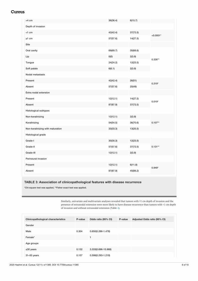
Association of clinicopathologic features with disease recurrenceTable 3 shows the association of clinicopathologic features with disease recurrence. We found a significantassociation of disease recurrence with larger tumor size, depth of invasion and extranodal extension.Moreover, younger age (<30 years) and older age (>50 years) groups showed a higher rate of recurrencecompared to the middle age group (30-50 years). No significant association of disease recurrence with anyother clinicopathological characteristic was noted in our study.
Clinicopathological characteristicsRecurrence
P-valuen (%)
Yes No
Gender
Male 72(72.7) 41(80.4)0.302*
Female 27(27.3) 10(19.6)
Age group
≤30 years 15(15.2) 2(3.9)
0.043*31–50 years 39(39.4) 29(56.9)
>50 years 45(45.5) 20(39.2)
Tumor size
≤2 cm 12(12.1) 20(39.2)
<0.0001*2.1–4 cm 51(51.5) 23(45.1)
2020 Hashmi et al. Cureus 12(11): e11385. DOI 10.7759/cureus.11385 5 of 10
>4 cm 36(36.4) 8(15.7)
Depth of invasion
<1 cm 42(42.4) 37(72.5)<0.0001*
≥1 cm 57(57.6) 14(27.5)
Site
Oral cavity 69(69.7) 35(68.6)
0.326**Lip 0(0) 2(3.9)
Tongue 24(24.2) 12(23.5)
Soft palate 6(6.1) 2(3.9)
Nodal metastasis
Present 42(42.4) 26(51)0.319*
Absent 57(57.6) 25(49)
Extra nodal extension
Present 12(12.1) 14(27.5)0.019*
Absent 87(87.9) 37(72.5)
Histological subtypes
Non-keratinizing 12(12.1) 2(3.9)
0.107**Keratinizing 54(54.5) 36(70.6)
Non-keratinizing with maturation 33(33.3) 13(25.5)
Histological grade
Grade-I 30(30.3) 12(23.5)
0.131**Grade-II 57(57.6) 37(72.5)
Grade-III 12(12.1) 2(3.9)
Perineural invasion
Present 12(12.1) 6(11.8)0.949*
Absent 87(87.9) 45(88.2)
TABLE 3: Association of clinicopathological features with disease recurrence*Chi-square test was applied, **Fisher exact test was applied.
Similarly, univariate and multivariate analyses revealed that tumors with ≥1 cm depth of invasion and thepresence of extranodal extension were more likely to have disease recurrence than tumors with <1 cm depthof invasion and without extranodal extension (Table 4).
Clinicopathological characteristics P-value Odds ratio (95% CI) P-value Adjusted Odds ratio (95% CI)
Gender
Male 0.304 0.650(0.286-1.478)
Female* 1
Age groups
≤30 years 0.132 3.333(0.696-15.968)
31–50 years 0.157 0.598(0.293-1.219)
2020 Hashmi et al. Cureus 12(11): e11385. DOI 10.7759/cureus.11385 6 of 10

>50 years* 1
Tumor size
≤2 cm <0.0001 0.133(0.04-0.380)
2.1–4 cm 0.128 0.493(0.198-1.225)
>4 cm* 1
Depth of invasion
<1 cm 0.001 0.279(0.134-0.580) 0.001 0.276(0.131-0.584)
≥1 cm* 1 1
Site
Oral cavity 0.618 0.657(0.126-3.426)
Lip 0.999 0.000(0.000-0.000)
Tongue 0.649 0.667(0.117-3.813)
Soft palate* 1
Nodal metastasis
Present 0.319 0.709(0.360-1.396)
Absent* 1
Extranodal extension
Present 0.022 0.365(0.154-0.863) 0.027 0.359(0.145-0.887)
Absent* 1
Histological subtypes
Non-keratinizing 0.301 2.364(0.464-12.048)
Keratinizing 0.179 0.591(0.274-1.274)
Non-keratinizing with maturation* 1
Histological grade
Grade-I 0.295 0.417(0.081-2.148)
Grade-II 0.086 0.257(0.054-1.213)
Grade-III* 1
Perineural invasion
Present 0.949 0.967(0.340-2.746)
Absent* 1
TABLE 4: Univariate and multivariate analysis for disease recurrence with clinicopathologicalfeatures of population under studyUnivariate and multivariate binary logistic regression were applied. *Reference group. CI, confidence interval.
Survival analysis using the Kaplan-Meier method for HNSCC revealed a significant difference in disease freesurvival in patients with more than 2 cm tumor size and ≥1 cm depth of invasion than cases with ≤2 cmtumor size and <1 cm depth of invasion (Figures 2, 3, 4).
2020 Hashmi et al. Cureus 12(11): e11385. DOI 10.7759/cureus.11385 7 of 10

FIGURE 2: Survival analysis using the Kaplan-Meier method for headand neck squamous cell carcinoma with respect to tumor sizeLog rank p-value = 0.002
FIGURE 3: Survival analysis using the Kaplan-Meier method for headand neck squamous cell carcinoma with respect to depth of invasionLog rank p-value = <0.0001
2020 Hashmi et al. Cureus 12(11): e11385. DOI 10.7759/cureus.11385 8 of 10

FIGURE 4: Survival analysis using the Kaplan-Meier method for headand neck squamous cell carcinoma with respect to nodal metastasisLog rank p-value = 0.526
DiscussionIn this study, we found a high rate of recurrence in our cases of HNSCC despite tumor-free resectionmargins on the frozen section. Moreover, we noted that the presence of recurrence was significantlyassociated with tumor size, depth of invasion and extranodal extension.
Various studies have evaluated the recurrence of HNSCC in locoregional population. A study conducted inChina assessed disease recurrence in 275 patients with oral squamous cell carcinoma. They reported 32.7%recurrence rate in their study; however, in their study, 66.2% cases were early-stage cancers (T1+T2).Recurrence in late-stage cancers (T3+T4) in their study was 57%. They also found that tumor (T) stage, nodal(N) stage and degree of differentiation were the major factors determining disease recurrence [8]. We alsofound a significant association of tumor size and depth of invasion with disease recurrence; however, nosignificant association of disease recurrence was noted with the degree of differentiation in our study.Another study, conducted in Brazil, compared cases of HNSCC with and without recurrence. They found thatthe site (tongue) and degree of differentiation were the factors associated with disease recurrence [9].However, we did not find any significant association of tumor site with disease recurrence in our study. Animportant factor that was not assessed carefully in these studies was extranodal extension as a recurrence ofHNSCC typically occurs either at the primary site or in the neck and presence of extranodal extension is animportant factor that can lead to disease recurrence in the neck. Although margins of primary tumor arecarefully assessed and closed margins are re-shaved to get a margin-free resection, neck specimen istypically not assessed for margins and therefore, in the presence of extranodal extension, the chances ofrecurrence in the neck increase.
An important prognostic factor in oral cancers is margin-free resection, and the frozen section has pivotalimportance in this regard. There are two ways for intra-operative pathological consultation. The first andmore prevalent is tumor-bed margin evaluation and second is sending a whole resected specimen for frozensection assessment (tumor-specific margin). In tumor-bed evaluation for the frozen section, the surgeonafter removal of tumor sends shaving from resected tumor bed. Any tumor present in the shaving specimenis considered positive and re-shaving is done. The disadvantage of this technique is that the differentiationbetween a positive margin and close margin (i.e. <5 mm) is not possible. Second, in case of large tumors,tumor bed is huge and evaluation of whole tumor bed is impossible. The second method, i.e. evaluation ofwhole specimen by frozen section, although cumbersome and time-consuming has recently been proved tobe more effective in reducing the chances of recurrence and improving disease-free survival in patients withoral squamous cell carcinoma [10-12]. We at LNH routinely practice this technique, in which all the marginsof the resection specimen are evaluated on frozen section and rapidly communicated to the surgeon. Despitemargin-free resections, we found a high rate of recurrence in our study. This signifies the importance of
2020 Hashmi et al. Cureus 12(11): e11385. DOI 10.7759/cureus.11385 9 of 10

other factors that affect disease recurrence, that is, tumor size and depth of invasion. Moreover, the poorprognostic importance of extranodal extension should not be underscored in these circumstances.
One of the major limitations of our study was limited sample size. Second, molecular analysis for HPV statuswas not determined, as HPV-induced HNSCC behave in a less aggressive fashion than non-HPV inducedcancers. Although we found significant associations in our study, there were multiple confounding factorsthat could not be controlled in such study design, for instance, different combinations of chemotherapy andradiation therapies were given to different patients based on oncologist decision and multidisciplinary teammeetings that could also alter individual outcomes.
ConclusionsA high rate of disease recurrence was noted in HNSCC in our study after tumor-free resection margins onthe frozen section. A substantial proportion of recurrence occurred in the neck in our study. Apart fromtumor size and depth of invasion, which are known risk factors of disease recurrence in HNSCC, we alsonoted a significant association of disease recurrence with extranodal extension. Therefore, we suggest that,in the presence of extranodal extension, resection margins of neck dissection specimen should also beassessed along with primary specimen resection margins.
Additional InformationDisclosuresHuman subjects: Consent was obtained by all participants in this study. Ethical Review Committee ofLiaquat National Hospital issued approval 0555-2020 LNH-ERC. Ethical Review Committee of LiaquatNational Hospital approved the study. Animal subjects: All authors have confirmed that this study did notinvolve animal subjects or tissue. Conflicts of interest: In compliance with the ICMJE uniform disclosureform, all authors declare the following: Payment/services info: All authors have declared that no financialsupport was received from any organization for the submitted work. Financial relationships: All authorshave declared that they have no financial relationships at present or within the previous three years withany organizations that might have an interest in the submitted work. Other relationships: All authors havedeclared that there are no other relationships or activities that could appear to have influenced thesubmitted work.
References1. Hashmi AA, Hussain ZF, Hashmi SK, et al.: Immunohistochemical over expression of p53 in head and neck
Squamous cell carcinoma: clinical and prognostic significance. BMC Res Notes. 2018, 11:433.10.1186/s13104-018-3547-7
2. Hashmi AA, Hussain ZF, Aijaz S, et al.: Immunohistochemical expression of epidermal growth factorreceptor (EGFR) in South Asian head and neck squamous cell carcinoma: association with various riskfactors and clinico-pathologic and prognostic parameters. World J Surg Oncol. 2018, 16:118.10.1186/s12957-018-1425-3
3. Naz S, Salah K, Khurshid A, Hashmi AA, Faridi N: Head and neck squamous cell carcinoma - comparativeevaluation of pathological parameters in young and old patients. Asian Pac J Cancer Prev. 2015, 16:4061-4063. 10.7314/apjcp.2015.16.9.4061
4. Hashmi AA, Aijaz S, Irfan M, Hussain ZF, Hashmi SK, Asif H, Faridi N: Low p27kip1 expression in head andneck squamous cell carcinoma: association with risk factors and adverse outcomes. Appl Cancer Res. 2019,39:5. 10.1186/s41241-019-0074-3
5. Akram S, Shabbir A, Mirza T: Association of high risk human papilloma virus (HPV-16/18) with P16 proteinin oral premalignant lesions and oral squamous cell carcinoma. Pak J Med Dent. 2019, 9:4-11.10.36283/PJMD8-4/002
6. Hashmi AA, Younus N, Naz S, et al.: p16 immunohistochemical expression in head and neck squamous cellcarcinoma: association with prognostic parameters. Cureus. 2020, 13:e8601. 10.7759/cureus.8601
7. Kazmi FN, Adil A, Ghaffar S, Ahmed F: Association between tumour volume and recurrence of squamouscell carcinoma of the head and neck. J Pak Med Assoc. 2012, 62:1129-1133.
8. Wang B, Zhang S, Yue K, Wang XD: The recurrence and survival of oral squamous cell carcinoma: a reportof 275 cases. Chin J Cancer. 2013, 32:614-618. 10.5732/cjc.012.10219
9. Camisasca DR, Silami MA, Honorato J, Dias FL, de Faria PA, Lourenço Sde Q: Oral squamous cell carcinoma:clinicopathological features in patients with and without recurrence. ORL J Otorhinolaryngol Relat Spec.2011, 73:170-176. 10.1159/000328340
10. Chiosea SI: Intraoperative margin assessment in early oral squamous cell carcinoma . Surg Pathol Clin. 2017,10:1-14. 10.1016/j.path.2016.10.002
11. Thomas Robbins K, Triantafyllou A, Suárez C, et al.: Surgical margins in head and neck cancer: intra- andpostoperative considerations. Auris Nasus Larynx. 2019, 46:10-17. 10.1016/j.anl.2018.08.011
12. Buchakjian MR, Tasche KK, Robinson RA, Pagedar NA, Sperry SM: Association of main specimen and tumorbed margin status with local recurrence and survival in oral cancer surgery. JAMA Otolaryngol Head NeckSurg. 2016, 142:1191-1198. 10.1001/jamaoto.2016.2329
2020 Hashmi et al. Cureus 12(11): e11385. DOI 10.7759/cureus.11385 10 of 10