recist 1.1: applying the rules -...
TRANSCRIPT

RECIST 1.1: Applying the Rules
Developed by Center for Cancer Research, National Cancer Institute Endorsed by the CTN SIG Leadership Group
(reviewed and revised January 2014)

Assessing response to therapy allows for prospective end point evaluation in clinical trials and serves as a guide for decision making for clinician and patient/study subject. In oncology clinical trials there are several standards that are used. This module will focus on the standard used for solid tumors: Response Evaluation Criteria for Solid Tumors (RECIST). By then end of the module you will be able to:
1. Describe criteria used to select target lesions when using RECIST 1.1.
2. Discuss how to use target and non-target lesions when determining overall response for RECIST 1.1.

RECIST is being used in most solid tumor protocols to assess tumor response. However, not all studies will be using RECIST. This module is intended to assist you in understanding how to apply the RECIST “rules” using RECIST version 1.1. Consult your protocol for specifics of assessing tumor response.

Background
• Initial attempts to standardize assessing tumor response began in 1960s
• 1979 World Health Organization • Standardized criteria for response
assessment
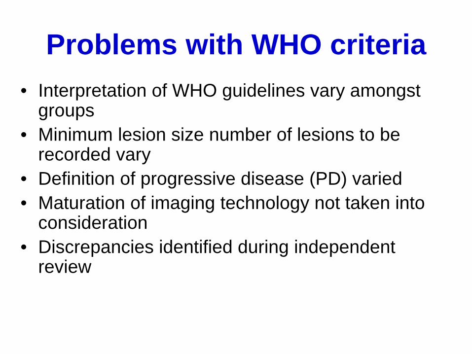
Problems with WHO criteria • Interpretation of WHO guidelines vary amongst
groups • Minimum lesion size number of lesions to be
recorded vary • Definition of progressive disease (PD) varied • Maturation of imaging technology not taken into
consideration • Discrepancies identified during independent
review

Development of RECIST 1.0 • 1994 international task force
• European Organization for Research and Treatment of Cancer (EORTC) • National Cancer Institute (NCI) of the U.S. • National Cancer Institute of Canada Clinical Trials Group
• Review of 4000 patients for tumor response • Recommendation to simplify response evaluation • 1999: Criteria was publicly presented/accepted the
American Society for Clinical Oncology meeting • 2000: Published in Journal of the National Cancer
Institute in 2000 • Intended for solid tumor response assessment in Phase
II clinical trials but is actually being used for response assessment in all Phases
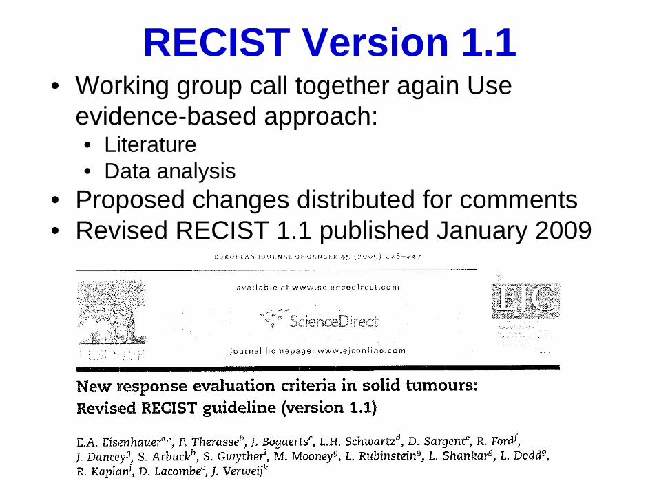
RECIST Version 1.1 • Working group call together again Use
evidence-based approach: • Literature • Data analysis
• Proposed changes distributed for comments • Revised RECIST 1.1 published January 2009

Measurability of Tumor at Baseline
• Measurable disease • Non-measurable disease

Measurable Disease • Tumor lesion measured in longest
diameter • EXCEPT for lymph nodes: use short axis
• Minimum size of measurable non-nodal lesions • CT scan 5 mm slice: ≥10 mm • CT scan > 5 mm slice: 2x slice thickness • Calibers (clinical exam): ≥10 mm • Chest x-ray: ≥20 mm
• Up to 5 measurable lesions (2/organ)

Imaging and RECIST… • Window settings refer to the brightness of
the image. Window settings can be adjusted to accentuate various anatomical structures.
• Consistency is important when following these lesions over time, as measurements should be performed using the same window setting on each follow-up imaging study

This is an example of a chest CT, shown with chest windows. Note that the structures in the mediastinum and the borders on the lesion in the L lung can be easily identified. The L lung lesion measurement is 10.5 mm.
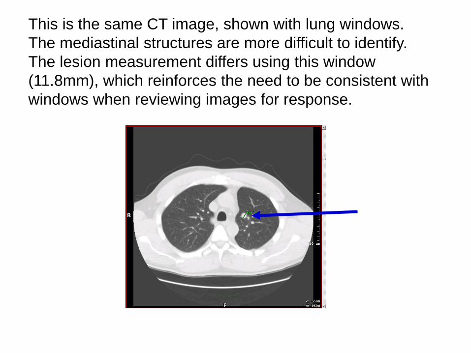
This is the same CT image, shown with lung windows. The mediastinal structures are more difficult to identify. The lesion measurement differs using this window (11.8mm), which reinforces the need to be consistent with windows when reviewing images for response.

… Imaging and RECIST … • All images in a series should be reviewed for new
disease rather than reviewing selected target lesion images only
• Oral contrast to help differentiate the bowel from other soft tissue in the abdomen
• MRI scans may be used to identify target lesions, perform lesion measurements, and follow the lesions over time, although CT is the preferred modality of choice. • Recommended that ideally the same MRI scanner be used
to obtain repeat images and the same anatomic place when following lesions over time using MRI images

…Imaging and RECIST • Ultrasound examinations may be used to perform
superficial target lesions measurements, such as subcutaneous lesions and thyroid • Should not be used to follow deeper lesions
• Chest x-ray may be used to identify, measure, and follow lesions over time, as long as the lesion borders are clearly defined and surrounded by aerated lung. • CT scanners are readily available and are the preferred
imaging modality since CT images can also be used to follow mediastinal and thoracic wall lesions.

Clinical Exam and RECIST • Clinical examination may be used to follow
superficial lesions over time. • Recommended that skin lesion assessment
include taking a lesion color picture with a ruler to document the size of the lesion

Non-measurable Disease • All other lesions that do not meet the
criteria to be measurable and: • Bone lesions • Leptomeningeal disease • Ascites • Pleural/pericardial effusion • Inflammatory breast disease • Cystic lesions

Tumor Response Evaluation • Overall tumor burden at baseline
including lymph nodes • Target lesions • Non-target lesions

Target Lesions • All measurable lesions up to 5 total
(max of 2/organ) • Must be representative of all involved
organs • Selected on the basis of their size and
suitability for accurate repeated measurements
• Sum of diameters
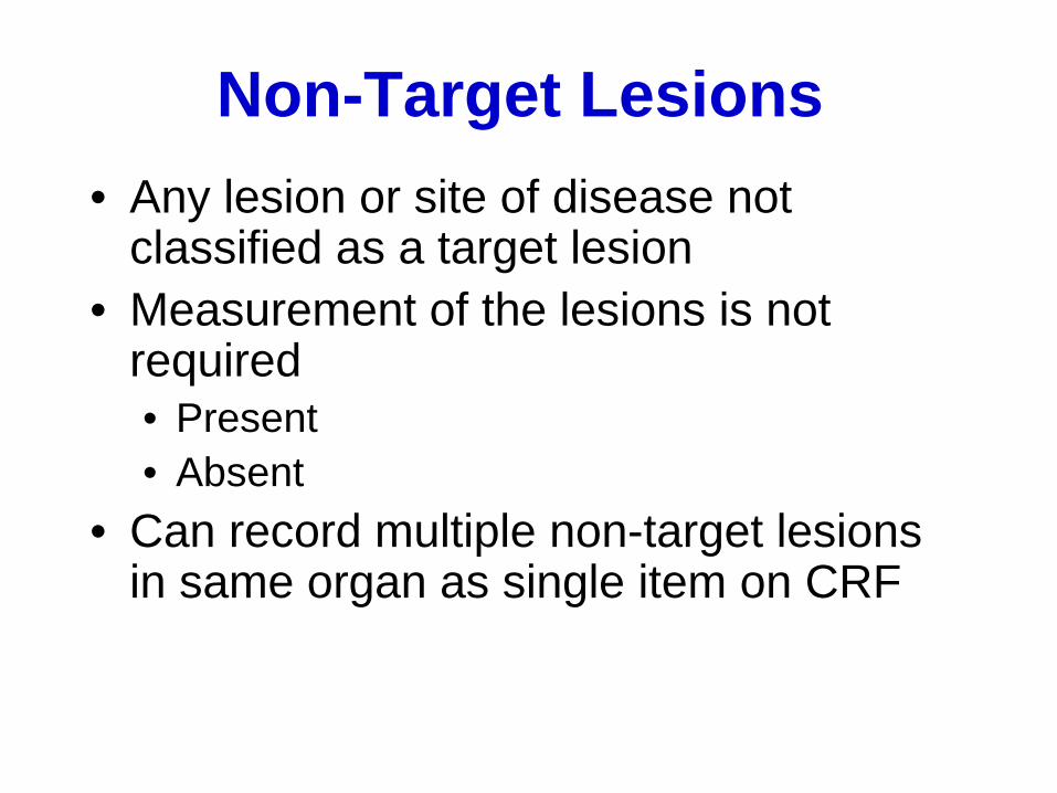
Non-Target Lesions • Any lesion or site of disease not
classified as a target lesion • Measurement of the lesions is not
required • Present • Absent
• Can record multiple non-target lesions in same organ as single item on CRF

Assessment of Lymph Node • Target lesion:
• Lymph node ≥ 15 mm • Non-target lesion
• Lymph node <15 mm • Normal if lymph node is <10 mm
REMEMBER Lymph nodes use short axis for
measuring

Response Assessment • Determine:
• target lesion response • non-target lesion response • appearance of new lesions

Target Lesion Response • Complete Response (CR)
• Disappearance of all target lesions • Any pathological lymph nodes (whether target or non-target) must have
reduction in short axis to <10 mm • Partial Response (PR)
• At least a 30% decrease in the sum of the diameters of target lesions, taking as reference the baseline sum diameters
• Progressive Disease (PD) • At least a 20% increase in the sum of the diameters of target lesions, taking
as reference the smallest sum on study (this includes the baseline sum if that is the smallest on study).
• In addition to the relative increase of 20%, sum must also demonstrate an absolute increase of at least 5 mm. (Note: the appearance of one or more new lesions is also considered progressions).
• Stable Disease (SD) • Neither sufficient shrinkage to qualify for PR nor sufficient increase to
qualify for PD, taking as reference the smallest sum diameters while on study
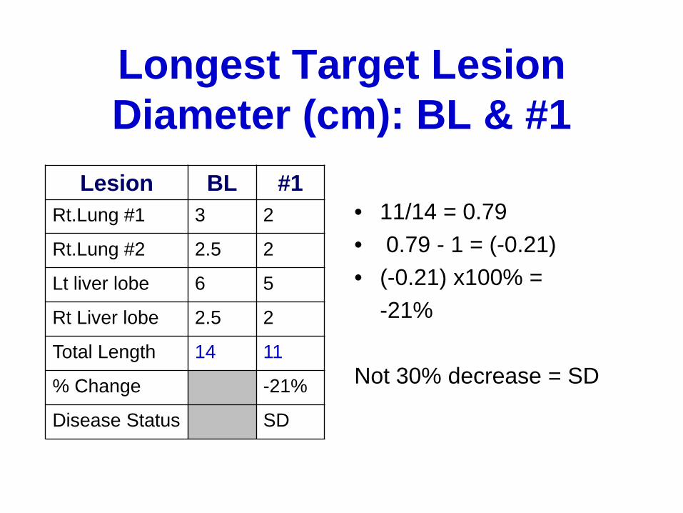
Longest Target Lesion Diameter (cm): BL & #1
• 11/14 = 0.79 • 0.79 - 1 = (-0.21) • (-0.21) x100% = -21% Not 30% decrease = SD
Lesion BL #1 Rt.Lung #1 3 2
Rt.Lung #2 2.5 2
Lt liver lobe 6 5
Rt Liver lobe 2.5 2
Total Length 14 11
% Change -21%
Disease Status SD

Longest Target Lesion Diameter (cm): BL, #1, #2
• 9/14 = 0.64 • 0.64 - 1 = (-0.35) • (-0.355) x100% = -36% PR = > 30% decrease
Lesion BL #1 #2 Rt.Lung #1 3 2 2
Rt.Lung #2 2.5 2 2
Lt liver lobe 6 5 3
Rt Liver lobe 2.5 2 2
Total Length 14 11 9
% Change -21% -36%
Disease Status SD PR

Longest Target Lesion Diameter (cm): BL, #1, #2, #3 Lesion BL #1 #2 #3
Rt.Lung #1 3 2 2 2
Rt.Lung #2 2.5 2 2 2
Lt liver lobe 6 5 3 3
Rt Liver lobe 2.5 2 2 2
Total Length 14 11 9 9
% Change -21% -36% -36% Disease Status SD PR PR
• 9/14 = 0.64 • 0.64 - 1 = (-0.35) • (-0.36) x100% = • -36%
• PR = > 30%
decrease
Note: Evaluation #3 done 4 weeks after #2 to confirm the PR
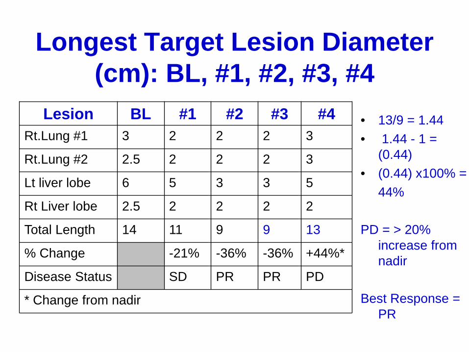
Longest Target Lesion Diameter (cm): BL, #1, #2, #3, #4
Lesion BL #1 #2 #3 #4 Rt.Lung #1 3 2 2 2 3
Rt.Lung #2 2.5 2 2 2 3
Lt liver lobe 6 5 3 3 5
Rt Liver lobe 2.5 2 2 2 2
Total Length 14 11 9 9 13
% Change -21% -36% -36% +44%*
Disease Status SD PR PR PD
* Change from nadir
• 13/9 = 1.44 • 1.44 - 1 =
(0.44) • (0.44) x100% = 44% PD = > 20%
increase from nadir
Best Response =
PR
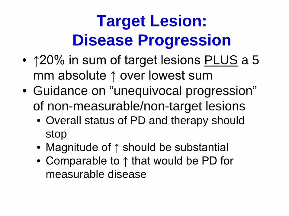
Target Lesion: Disease Progression
• ↑20% in sum of target lesions PLUS a 5 mm absolute ↑ over lowest sum
• Guidance on “unequivocal progression” of non-measurable/non-target lesions • Overall status of PD and therapy should
stop • Magnitude of ↑ should be substantial • Comparable to ↑ that would be PD for
measurable disease

Non-target Lesion Response • Complete Response (CR)
• Disappearance of all non-target lesions and normalization of tumor marker level. All lymph nodes must be non-pathological in size (<10 mm short axis)
• Note: If tumor markers are initially above the upper normal limit, they must normalize for a patient to be considered in complete clinical response.
• Non-CR/Non-PD • Persistence of one or more non-target lesion(s) and/or maintenance of
tumor marker level above the normal limits • Progressive Disease (PD)
• Appearance of one or more new lesions and/or unequivocal progression of existing non-target lesions.
• Unequivocal progression should not normally trump target lesion status. • It must be representative of overall disease status change, not a single
lesion increase.
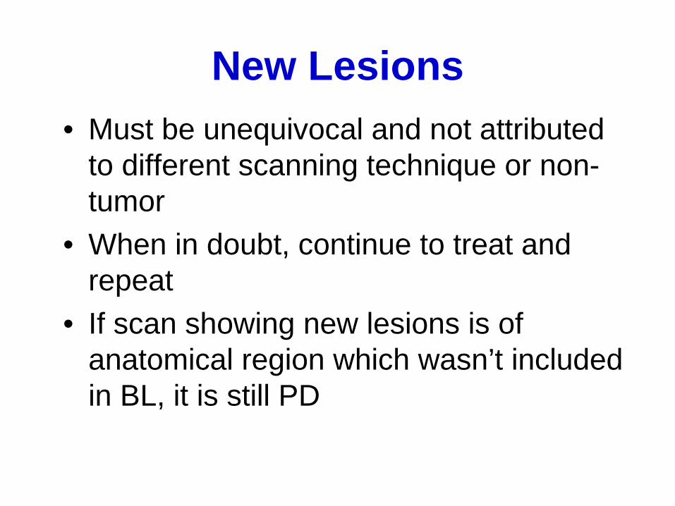
New Lesions • Must be unequivocal and not attributed
to different scanning technique or non-tumor
• When in doubt, continue to treat and repeat
• If scan showing new lesions is of anatomical region which wasn’t included in BL, it is still PD

Imaging Guidance: FDG-PET
• FDG-PET “-” at BL and “+” at follow-up = PD
• No FDG-PET at BL and “+” at follow-up: • PD: corresponds to new site in CT • Equivocal: no new site on CT. Repeat CT
and if new site, PD date is that of initial “+” FDG-PET
• Not PD: corresponds to pre-existing site on CT that is not progressing

Confirmation of Response
• If response is primary endpoint (e.g.,Phase II), confirmation IS required
• If response is secondary endpoint (e.g, RCT w/PFS or OS): confirmation IS NOT required • Control arm provides ability to interpret
results

Best Response for Patients with Measurable Disease
Target Lesions Non-Target Lesions
New Lesions Overall Response Best Overall Response when Confirmation is
Required* CR CR No CR >4 wks.
Confirmation** CR Non-CR/Non-PD No PR
>4 wks. Confirmation** CR Not evaluated No PR
PR Non-CR/Non-PD/not evaluated
No PR
SD Non-CR/Non-PD/not evaluated
No SD documented at least once >4 wks. from
baseline** PD Any Yes or No PD
no prior SD, PR or CR Any PD*** Yes or No PD
Any Any Yes PD
* See RECIST 1.1 manuscript for further details on what is evidence of a new lesion. ** Only for non-randomized trials with response as primary endpoint. *** In exceptional circumstances, unequivocal progression in non-target lesions may be accepted as disease progression. Note: Patients with a global deterioration of health status requiring discontinuation of treatment without objective evidence of
disease progression at that time should be reported as “symptomatic deterioration.” Every effort should be made to document the objective progression even after discontinuation of treatment.
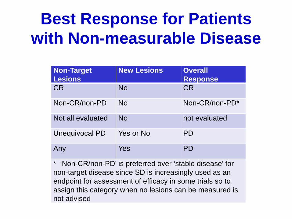
Best Response for Patients with Non-measurable Disease
Non-Target Lesions
New Lesions Overall Response
CR No CR
Non-CR/non-PD No Non-CR/non-PD*
Not all evaluated No not evaluated
Unequivocal PD Yes or No PD
Any Yes PD
* ‘Non-CR/non-PD’ is preferred over ‘stable disease’ for non-target disease since SD is increasingly used as an endpoint for assessment of efficacy in some trials so to assign this category when no lesions can be measured is not advised
Documentation • Ideally all radiology reports should include
tumor measurements but this may not be done using RECIST
• Response assessment and documentation should be addressed at the protocol specific time points to make a decision if therapy is to continue • Target lesion measurements should be
documented at that time

Resources
• RECIST version 1.1 resources: • http://www.eortc.be/recist/ (link to publications and
presentation) • http://www.recist.com/

References • James, K., Eisenhauer, E., Christian, M., Terenziani, M., Vena,
D., Muldal, A., and Therasse, P. (1999). Measuring response in solid tumors: unidimensional versus bidimensional measurement. Journal of the National Cancer Institute, 91, 523-528.
• Therasse, P., Arbuck, S., Eisenhauer, E., Wanders, J., Kaplan, R., Rubinstein, L., Verweij, J., Van Glabbeke, M., van Oosterom, A., Christian, M., and Gwyther, S. (2000). New guidelines to evaluate the response to treatment in solid tumors. Journal of the National Cancer Institute, 92, 205-216.
• Eisenhauer, E,Therasse, P., Bogaerts, J., Schwartz, L.H., Sargent, D., Ford, R., Dancey, J., et al. (2009) New response evaluation criteria in solid tumours: Revised RECIST guideline (version 1.1). European Journal of Cancer , 45, 228-247.

Module Evaluation
The CTN SIG would greatly appreciate your feedback on this learning module. Please complete the evaluation form and fax to Elizabeth Ness at 301-496-9020.