receptor-interactingproteinkinase4andinterferon ... · kinase c pathway. therefore, our findings...
TRANSCRIPT
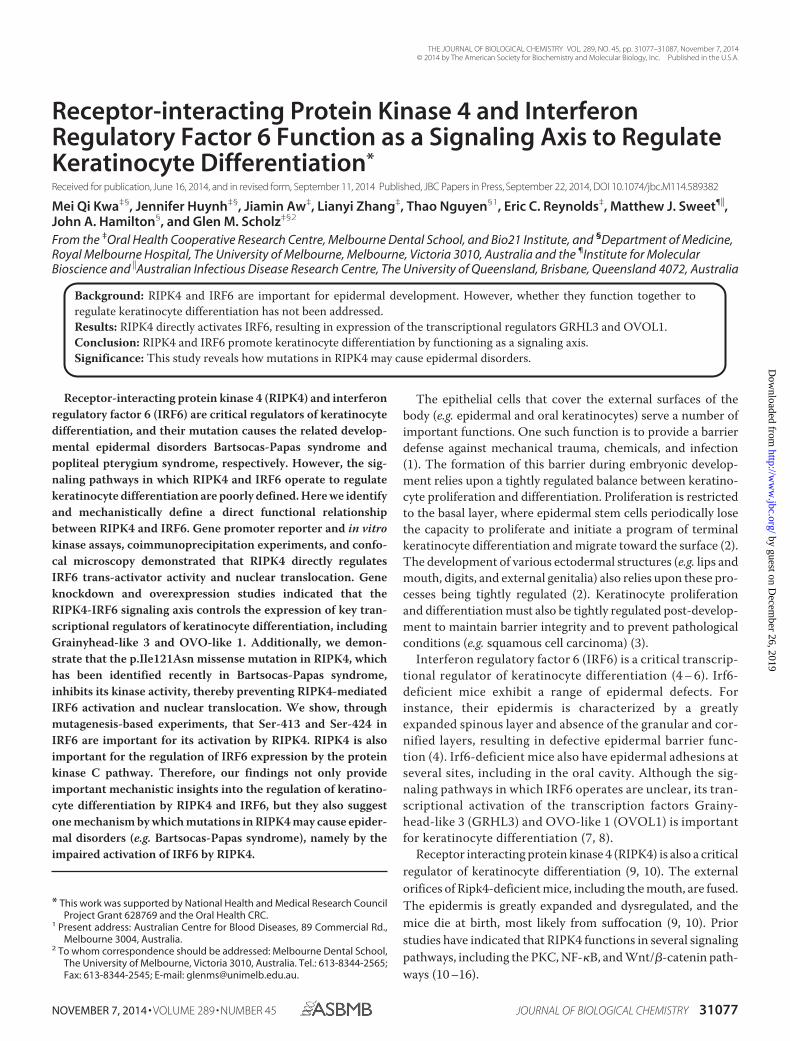
Receptor-interacting Protein Kinase 4 and InterferonRegulatory Factor 6 Function as a Signaling Axis to RegulateKeratinocyte Differentiation*
Received for publication, June 16, 2014, and in revised form, September 11, 2014 Published, JBC Papers in Press, September 22, 2014, DOI 10.1074/jbc.M114.589382
Mei Qi Kwa‡§, Jennifer Huynh‡§, Jiamin Aw‡, Lianyi Zhang‡, Thao Nguyen§1, Eric C. Reynolds‡, Matthew J. Sweet¶�,John A. Hamilton§, and Glen M. Scholz‡§2
From the ‡Oral Health Cooperative Research Centre, Melbourne Dental School, and Bio21 Institute, and §Department of Medicine,Royal Melbourne Hospital, The University of Melbourne, Melbourne, Victoria 3010, Australia and the ¶Institute for MolecularBioscience and �Australian Infectious Disease Research Centre, The University of Queensland, Brisbane, Queensland 4072, Australia
Background: RIPK4 and IRF6 are important for epidermal development. However, whether they function together toregulate keratinocyte differentiation has not been addressed.Results: RIPK4 directly activates IRF6, resulting in expression of the transcriptional regulators GRHL3 and OVOL1.Conclusion: RIPK4 and IRF6 promote keratinocyte differentiation by functioning as a signaling axis.Significance: This study reveals how mutations in RIPK4 may cause epidermal disorders.
Receptor-interacting protein kinase 4 (RIPK4) and interferonregulatory factor 6 (IRF6) are critical regulators of keratinocytedifferentiation, and their mutation causes the related develop-mental epidermal disorders Bartsocas-Papas syndrome andpopliteal pterygium syndrome, respectively. However, the sig-naling pathways in which RIPK4 and IRF6 operate to regulatekeratinocyte differentiation are poorly defined. Here we identifyand mechanistically define a direct functional relationshipbetween RIPK4 and IRF6. Gene promoter reporter and in vitrokinase assays, coimmunoprecipitation experiments, and confo-cal microscopy demonstrated that RIPK4 directly regulatesIRF6 trans-activator activity and nuclear translocation. Geneknockdown and overexpression studies indicated that theRIPK4-IRF6 signaling axis controls the expression of key tran-scriptional regulators of keratinocyte differentiation, includingGrainyhead-like 3 and OVO-like 1. Additionally, we demon-strate that the p.Ile121Asn missense mutation in RIPK4, whichhas been identified recently in Bartsocas-Papas syndrome,inhibits its kinase activity, thereby preventing RIPK4-mediatedIRF6 activation and nuclear translocation. We show, throughmutagenesis-based experiments, that Ser-413 and Ser-424 inIRF6 are important for its activation by RIPK4. RIPK4 is alsoimportant for the regulation of IRF6 expression by the proteinkinase C pathway. Therefore, our findings not only provideimportant mechanistic insights into the regulation of keratino-cyte differentiation by RIPK4 and IRF6, but they also suggestone mechanism by which mutations in RIPK4 may cause epider-mal disorders (e.g. Bartsocas-Papas syndrome), namely by theimpaired activation of IRF6 by RIPK4.
The epithelial cells that cover the external surfaces of thebody (e.g. epidermal and oral keratinocytes) serve a number ofimportant functions. One such function is to provide a barrierdefense against mechanical trauma, chemicals, and infection(1). The formation of this barrier during embryonic develop-ment relies upon a tightly regulated balance between keratino-cyte proliferation and differentiation. Proliferation is restrictedto the basal layer, where epidermal stem cells periodically losethe capacity to proliferate and initiate a program of terminalkeratinocyte differentiation and migrate toward the surface (2).The development of various ectodermal structures (e.g. lips andmouth, digits, and external genitalia) also relies upon these pro-cesses being tightly regulated (2). Keratinocyte proliferationand differentiation must also be tightly regulated post-develop-ment to maintain barrier integrity and to prevent pathologicalconditions (e.g. squamous cell carcinoma) (3).
Interferon regulatory factor 6 (IRF6) is a critical transcrip-tional regulator of keratinocyte differentiation (4 – 6). Irf6-deficient mice exhibit a range of epidermal defects. Forinstance, their epidermis is characterized by a greatlyexpanded spinous layer and absence of the granular and cor-nified layers, resulting in defective epidermal barrier func-tion (4). Irf6-deficient mice also have epidermal adhesions atseveral sites, including in the oral cavity. Although the sig-naling pathways in which IRF6 operates are unclear, its tran-scriptional activation of the transcription factors Grainy-head-like 3 (GRHL3) and OVO-like 1 (OVOL1) is importantfor keratinocyte differentiation (7, 8).
Receptor interacting protein kinase 4 (RIPK4) is also a criticalregulator of keratinocyte differentiation (9, 10). The externalorifices of Ripk4-deficient mice, including the mouth, are fused.The epidermis is greatly expanded and dysregulated, and themice die at birth, most likely from suffocation (9, 10). Priorstudies have indicated that RIPK4 functions in several signalingpathways, including the PKC, NF-�B, and Wnt/�-catenin path-ways (10 –16).
* This work was supported by National Health and Medical Research CouncilProject Grant 628769 and the Oral Health CRC.
1 Present address: Australian Centre for Blood Diseases, 89 Commercial Rd.,Melbourne 3004, Australia.
2 To whom correspondence should be addressed: Melbourne Dental School,The University of Melbourne, Victoria 3010, Australia. Tel.: 613-8344-2565;Fax: 613-8344-2545; E-mail: [email protected].
THE JOURNAL OF BIOLOGICAL CHEMISTRY VOL. 289, NO. 45, pp. 31077–31087, November 7, 2014© 2014 by The American Society for Biochemistry and Molecular Biology, Inc. Published in the U.S.A.
NOVEMBER 7, 2014 • VOLUME 289 • NUMBER 45 JOURNAL OF BIOLOGICAL CHEMISTRY 31077
by guest on Decem
ber 26, 2019http://w
ww
.jbc.org/D
ownloaded from
Mutations in IRF6 cause popliteal pterygium syndrome (17),a developmental epidermal disorder characterized by orofacialclefting, skin webbing, syndactyly, and genital deformities (18).Mutations in RIPK4 have been identified recently in Bartsocas-Papas syndrome (BPS)3 (19, 20). BPS is a more severe form ofpopliteal pterygium and causes death early in life (21, 22).RIPK4 and IRF6 mutations have also been identified in squa-mous cell carcinoma (23).
How RIPK4 and IRF6 regulate keratinocyte differentiation isnot well understood. However, given the phenotypic similari-ties between Ripk4- and Irf6-deficient mice, along with the sim-ilarities in the developmental defects in individuals with muta-tions in RIPK4 and IRF6, we sought to establish whether a directfunctional relationship exists between these two proteins. Wedemonstrate here that not only do RIPK4 and IRF6 function inthe same PKC-regulated signaling pathway to promote kerati-nocyte differentiation, but RIPK4 also directly activates IRF6.We also provide insights into how a specific missense mutationin RIPK4 may cause BPS.
EXPERIMENTAL PROCEDURES
Reagents—Cell culture medium and supplements, FCS,SuperScript III reverse transcriptase, random primers, dNTPs,TaqMan Universal Master Mix II, Lipofectamine RNAiMAX,Lipofectamine 2000, Silencer Select RIPK4 siRNA and controlnon-targeting siRNA, precast 10% NuPAGE gels, mouseanti-V5 antibodies, Alexa Fluor 488-conjugated goat anti-rab-bit IgG and Alexa Fluor 594-conjugated goat anti-mouse IgGantibodies, and ProLong Gold Antifade reagent (containingDAPI) were from Invitrogen. [�-32P]ATP (3000 Ci/mmol) wasfrom PerkinElmer Life Sciences. FuGENE6, Passive LysisBuffer, and the Dual-Glo luciferase assay system were from Pro-mega. The QuikChange II site-directed mutagenesis kit wasfrom Agilent Technologies, and the mutagenic primers weresynthesized by GeneWorks. cOmplete protease inhibitors werefrom Roche. The ON-TARGETplus IRF6 siRNA and non-tar-geting control siRNA were from Millennium Science. The anti-IRF6 and anti-PCNA antibodies were from Cell Signaling Tech-nology. Anti-�-tubulin and HRP-conjugated anti-FLAG (M2)antibodies and anti-FLAG-agarose were from Sigma-Aldrich.Anti-HSP90 antibody was from BD Biosciences, and anti-ERK2antibody was from Santa Cruz Biotechnology.
Expression Vectors and Mutagenesis—The IRF6 expressionvectors pEF-HA-IRF6 (expresses an N-terminal HA-taggedversion of human IRF6), pEF-HA-IRF6 S413A (Ser-413replaced by alanine), pEF-HA-IRF6 S424A (Ser-424 replaced byalanine), pEF-HA-IRF6 S413A/S424A (Ser-413 and Ser-424replaced by alanine), pEF-HA-IRF6 S413E (Ser-413 replaced byglutamic acid), pEF-HA-IRF6 S424E (Ser-424 replaced by glu-tamic acid), pEF-HA-IRF6 S413E/S424E (Ser-413 and Ser-424replaced by glutamic acid), and pEF-V5-IRF6 (expresses anN-terminal V5-tagged version of IRF6) have been describedpreviously (24). Expression vectors encoding FLAG-tagged ver-sions of wild-type and kinase-dead (K51R) mouse Ripk4 were
provided by Dr. Shiv Pillai (Harvard Medical School) (12). TheRipk4 expression vector pFLAG-Ripk4 I121N (Ile-121 replacedby asparagine) was created using the primer pair 5�-GGG ACCTGC GCT TTC GCA ACG TGC ACG AG-3� and 5�-CTCGTG CAC GTT GCG AAA GCG CAG GTC CC-3� and aQuikChange II site-directed mutagenesis kit. The GST expres-sion vector pGEX-IRF6 CTD (expresses the C-terminal domainof IRF6 fused to GST) was created by PCR using the primer pair5�-ACG GAA TTC GCT CGG ATG ATC TAC GAG ATG-3�and 5�-CAG AAG CTT TTA CTG GGG AGG CAG GGCAG-3�. The PCR product was digested with EcoRI and HindIIIand cloned into pGEX2TH, and then the sequence was verified.
Bacterial Expression and Purification of GST Proteins—Transformed BL21(DE3) Escherichia coli were grown at 37 °Cin 2YT broth (1.6% tryptone, 1% yeast extract, 0.5% NaCl, pH7.0) with constant shaking. When the culture A600 reached�0.6, protein expression was induced with isopropyl 1-thio-�-D-galactopyranoside (0.4 mM), and the culture was incubated at25 °C for 2–3 h with constant shaking. The bacteria were lysedby sonication in ice-cold PBS containing 1% Triton X-100,DNase I (30 �g/ml) and protease inhibitors. The lysates wereclarified by centrifugation (27,000 � g for 40 min at 4 °C), andthe GST proteins (GST-IRF6 CTD and GST) were purified onan FPLC system using GSTrap FF columns (GE Healthcare)and concentrated using Amicon Ultra-4 centrifugal filters(Millipore).
Cell Culture—The human oral keratinocyte cell line OKF6/TERT-2 (25) was cultured in keratinocyte serum-free mediumsupplemented with 25 �g/ml bovine pituitary extract, 2 ng/mlEGF, 0.4 mM CaCl2, 100 units/ml penicillin, 100 �g/ml strepto-mycin, and 2 mM GlutaMax-1. Primary human epidermal kera-tinocytes (Lonza) were cultured in KGM-GoldTM mediumaccording to the protocol of the supplier. HEK293T cells werecultured in DMEM supplemented with 10% FCS, 100 units/mlpenicillin, 100 �g/ml streptomycin, and 2 mM GlutaMax-1.Cells were cultured at 37 °C in a humidified atmosphere of 5%CO2.
Cell Lysis and Western Blotting—Cells were washed twicewith ice-cold PBS and then lysed (20 mM Tris-HCl (pH 7.4), 150mM NaCl, 1 mM EDTA, 1% Nonidet P-40, 10% glycerol, 10 mM
NaF, 10 mM �-glycerol phosphate, and cOmplete proteaseinhibitors) on ice for 60 min. The lysates were clarified by cen-trifugation (13,000 � g for 10 min at 4 °C), and protein concen-trations were measured using a protein assay kit (Bio-Rad).Lysates were subjected to electrophoresis on 10% NuPAGEgels, followed by Western blotting according to standard pro-tocols. Immunoreactive bands were visualized using ECLreagents (Millipore) and a LAS-3000 Imager (Fujifilm) or byexposure to x-ray film (Fujifilm). Films were scanned using aGS-800 calibrated imaging densitometer (Bio-Rad).
Immunoprecipitation Assays—V5-IRF6 was immunoprecipi-tated by incubating 1 mg of cell lysate with 1 �g of anti-V5antibody and 20 �l of protein G-Sepharose for 4 h at 4 °C withconstant mixing. FLAG-Ripk4 was immunoprecipitated byincubating 1 mg of cell lysate with 10 �l of anti-FLAG-agarosefor 4 h at 4 °C with constant mixing. In both cases, the beadswere then washed four times with lysis buffer.
3 The abbreviations used are: BPS, Bartsocas-Papas syndrome; PMA, phorbol12-myristate 13-acetate; CTD, C-terminal domain; PCNA, proliferating cellnuclear antigen; IVL, involucrin; DMSO, dimethyl sulfoxide.
A RIPK4-IRF6 Signaling Axis in Keratinocytes
31078 JOURNAL OF BIOLOGICAL CHEMISTRY VOLUME 289 • NUMBER 45 • NOVEMBER 7, 2014
by guest on Decem
ber 26, 2019http://w
ww
.jbc.org/D
ownloaded from

In Vitro Kinase Assays—Anti-FLAG (Ripk4) immunoprecipi-tates were incubated for 30 min at 30 °C in kinase assay buffer(20 mM Hepes (pH 7.4), 25 mM MgCl2, 3 mM MnCl2, 10 mM
�-glycerol phosphate, 10 �M ATP, and 10 �Ci of [�-32P]ATP)containing 2 �g of GST-IRF6 CTD or GST. Reactions wereterminated by the addition of SDS-PAGE sample buffer andheating at 95 °C for 5 min. Aliquots of the reactions were thensubjected to SDS-PAGE. The gels were stained with colloidalCoomassie G-250 and dried, and 32P incorporation was detectedusing a Typhoon Trio PhosphorImager (GE Healthcare).
Gene Promoter Reporter Assays—HEK293T cells were seededin 12-well tissue culture plates at a density of 3 � 105 cells/welland transfected (in duplicate) the next day using FuGENE6transfection reagent. The total amount of plasmid in eachtransfection was kept constant using empty vector. The cellswere lysed 24 h post-transfection with passive lysis buffer andassayed for firefly and Renilla luciferase activity using the Dual-Glo luciferase assay system. Renilla luciferase activity was usedto normalize transfection efficiencies. The IFN� gene promoterreporter plasmid was provided by Dr. Ashley Mansell (MonashInstitute of Medical Research). The NF-�B-dependent E-selec-tin (ELAM-1) gene promoter reporter plasmid was as describedpreviously (26). The control Renilla reporter plasmid, pRL-TK,was from Promega.
Immunofluorescent Staining and Confocal Microscopy—OKF6/TERT-2 cells and HEK293T cells (seeded on glass cov-erslips) were fixed with 4% paraformaldehyde, solubilized with0.1% Triton X-100, and blocked in 5% goat serum. The cellswere then incubated overnight (at 4 °C) with a rabbit anti-IRF6antibody, a mouse anti-FLAG antibody, or a mouse anti-�-tu-bulin antibody for 16 h at 4 °C. Following three washes withPBS, the cells were incubated with an Alexa Fluor 488-conju-gated goat anti-rabbit IgG antibody or an Alexa Fluor 594-con-jugated goat anti-mouse IgG antibody for 60 min at room tem-perature. The cells were washed three times with PBS andfinally mounted on glass microscope slides using ProLong GoldAntifade reagent containing DAPI. Mounted coverslips wereallowed to cure in the dark for 24 h. Images were acquired on anOlympus FV1000 scanning confocal microscope. No anti-IRF6or anti-FLAG staining was apparent in HEK293T cells trans-fected with empty vector only. The effects of phorbol 12-myris-tate 13-acetate (PMA) on IRF6 subcellular localization wereanalyzed using ImageJ software. Three random fields of cellswere analyzed for each treatment condition. Binary imageswere obtained by thresholding to define the nuclear region ofinterest, which was subsequently used for measuring IRF6nuclear fluorescence. Thereafter, IRF6 cytoplasmic fluores-cence was determined by subtracting the nuclear fluorescencefrom the total cell fluorescence. IRF6 nuclear localization wascalculated by dividing the nuclear fluorescence by the cytoplas-mic fluorescence and is presented as a ratio of nuclear to cyto-plasmic IRF6.
Real-time PCR—Total RNA was purified using an RNAeasymini kit (Qiagen). RNA (1 �g) was reverse-transcribed intocDNA using random primers and SuperScript III reverse tran-scriptase. Real-time PCR was performed (in triplicate) with anApplied Biosystems Prism 7900HT sequence detection systemand TaqMan assays (Invitrogen) for the following genes:
GRHL3 (Hs00297962_m1), IRF6 (Hs00196213_m1), IVL(Hs00902520_m1),KRT13(Hs00999762_m1),MAD1(Hs00965581_m1), OVOL1 (Hs00190060_m1), RIPK4 (Hs01062501_m1),and Ripk4 (Mm00458366_m1). Messenger RNA levels relativeto those of the endogenous control gene, HPRT, were calcu-lated using the ��Ct method (27).
Gene Silencing and Overexpression—A reverse transfectionprotocol was used for the transfection of OKF6/TERT-2 cellsand primary human epidermal keratinocytes. For gene silenc-ing, siRNAs were diluted to 120 nM with 100 �l of Opti-MEM Ireduced serum medium (Invitrogen) and then mixed with 100�l of Opti-MEM medium containing 1 �l of LipofectamineRNAiMAX transfection reagent, followed by incubation atroom temperature for 20 min. For gene overexpression, theexpression plasmid (1 �g of total plasmid) was diluted in 100 �lof Opti-MEM medium and then mixed with 100 �l of Opti-MEM medium containing 1 �l of Lipofectamine 2000 transfec-tion reagent, followed by incubation at room temperature for20 min. For both gene silencing and overexpression, keratino-cytes (2–5 � 105 cells in 1 ml of antibiotic-free growth medium)were seeded into 12-well plates, and the transfection mixturewas added. For gene knockdown experiments, the medium wasreplaced after 16 h, and the cells were treated with PMA 48 or72 h post-transfection. For gene overexpression experiments,the cells were treated with PMA 24 h post-transfection withoutmedium change.
Statistical Analysis—Data combined from three or moreindependent experiments are given as mean � S.E. Statisticalanalyses were performed using GraphPad Prism software ver-sion 6.01 (GraphPad Software, La Jolla, CA). Differencesbetween two groups were evaluated using Student’s t test. Formultiple comparisons, statistical analysis was performed usinga one-way analysis of variance and Sidak’s or Dunnett’s test as apost hoc test. p �0.05 was considered to be statisticallysignificant.
RESULTS
Regulation of IRF6 by RIPK4 —To establish whether a func-tional relationship exists between RIPK4 and IRF6, we firstinvestigated whether the trans-activator activity of IRF6 is reg-ulated by RIPK4. IRF6 trans-activator activity, which was mea-sured using an IFN� gene promoter reporter plasmid (24), wasstrongly induced, in a concentration-dependent manner, bywild-type RIPK4 (Fig. 1A) but not by the kinase-dead RIPK4mutant RIPK4 K51R (Fig. 1B). Consistent with earlier findings(13, 15), RIPK4 also activated NF-�B in a concentration- (Fig.1C) and kinase-dependent (Fig. 1D) manner.
The phosphorylation of regulatory serine residues in theC-terminal domain (CTD) of IRFs represents a critical step intheir inducible activation (28). To determine whether RIPK4can directly phosphorylate the IRF6 CTD, in vitro kinase assayswere performed using a GST-IRF6 CTD fusion protein as sub-strate. RIPK4 phosphorylated the GST-IRF6 CTD protein butnot the GST control (Fig. 1E). Because of the instability of theGST-IRF6 CTD fusion protein, a degradation product was alsoevident in the Coomassie-stained gel (Fig. 1E). The ability ofRIPK4 to interact with IRF6 was also investigated by perform-ing coimmunoprecipitation experiments. IRF6 interacted,
A RIPK4-IRF6 Signaling Axis in Keratinocytes
NOVEMBER 7, 2014 • VOLUME 289 • NUMBER 45 JOURNAL OF BIOLOGICAL CHEMISTRY 31079
by guest on Decem
ber 26, 2019http://w
ww
.jbc.org/D
ownloaded from

either directly or as part of a complex, with wild-type RIPK4.However, no interaction with the RIPK4 K51R mutant wasdetected (Fig. 1F, top panel). Because of autophosphorylation(12), a doublet was detected when wild-type, but not kinase-dead, RIPK4 was expressed in HEK293T cells (Fig. 1F, bottompanel).
The ability of RIPK4 to regulate IRF6 nuclear translocationwas also tested. IRF6 was detected in the cytoplasm of trans-fected cells. However coexpression of wild-type RIPK4, but notRIPK4 K51R, resulted in the partial nuclear translocation ofIRF6 (Fig. 1G). RIPK4 localized exclusively to the cytoplasm,where it exhibited a degree of colocalization with the cytoplas-mic pool of IRF6 (Fig. 1G). Collectively, the results in Fig. 1indicate that RIPK4 can directly activate IRF6 and induce itsnuclear translocation.
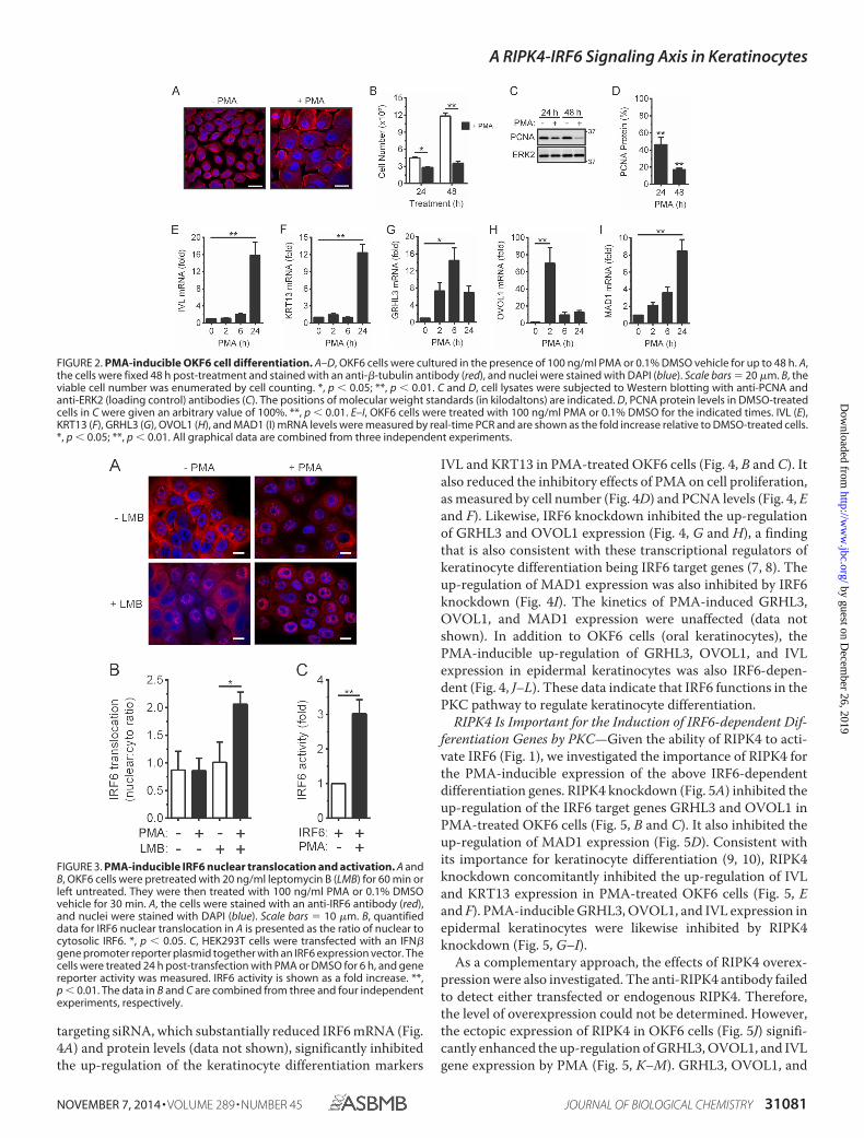
Characterization of PKC-regulated Oral Keratinocyte Differentia-tion—PKC is an important regulator of keratinocyte differenti-ation (29 –33), and previous studies have indicated that RIPK4functions in the PKC pathway (10 –14). PMA is a potent PKCactivator and a strong inducer of keratinocyte terminal differ-entiation (34). Therefore, we assessed whether PMA treatmentof OKF6/TERT-2 cells (hereafter referred to as OKF6 cells),which have the characteristics of primary human oral keratino-cytes (25), would provide a suitable model for oral keratinocytedifferentiation. PMA treatment induced marked morphologi-cal changes (Fig. 2A) and strongly suppressed cell proliferation(Fig. 2B). This correlated with decreased levels of the prolifer-ation marker proliferating cell nuclear antigen (PCNA) (Fig. 2,C and D). Differentiation was further assessed by measuring theexpression levels of the early and late terminal differentiationmarkers involucrin (IVL) and keratin 13 (KRT13), respectively(35, 36). Both genes were strongly up-regulated by PMA treat-ment (Fig. 2, E and F). GRHL3, OVOL1, and mitotic arrest-deficient 1 (MAD1), which themselves are important transcrip-tional regulators of keratinocyte differentiation (37– 42), werealso up-regulated in PMA-treated cells (Fig. 2, G–I). OVOL1expression was most rapidly up-regulated (Fig. 2H), whereasthe induction of MAD1 gene expression was slowest (Fig. 2I).Collectively, these data demonstrate that PMA treatment ofOKF6 cells provides a good model for investigating the regula-tion of keratinocyte differentiation by RIPK4 and IRF6.
IRF6 Is Important for PKC-regulated Keratinocyte Differentia-tion—To establish whether IRF6 mediates PKC-regulated kera-tinocyte differentiation, we first investigated the ability of PMAto induce IRF6 nuclear translocation. The staining of OKF6cells with anti-IRF6 antibodies revealed that IRF6 was presentin both the cytoplasm and nucleus of a substantial proportion ofcells, whereas, in other cells, it localized exclusively to the cyto-plasm (Fig. 3A). PMA did not appear to cause an overallincrease in IRF6 nuclear localization (Fig. 3, A and B). However,a significant increase in IRF6 nuclear translocation wasobserved when the cells were pretreated with the nuclearexport inhibitor leptomycin B (LMB) (Fig. 3, A and B). There-fore, PKC activation induces the transient nuclear transloca-tion of IRF6. Gene promoter reporter assays demonstrated thatPKC activation also induces IRF6 activity (Fig. 3C).
We next investigated the importance of IRF6 for PKC-regu-lated keratinocyte differentiation. As shown in Fig. 4, the IRF6-
FIGURE 1. Activation of IRF6 by RIPK4. A–D, HEK293T cells were transfectedwith an IFN� (A and B) or ELAM-1 (C and D) gene promoter reporter plasmidtogether with expression vectors encoding IRF6 (A and B) and the indicatedRipk4 proteins (A–D). Gene reporter activity was measured 24 h post-trans-fection. IRF6 (A and B) and NF-�B (C and D) activity are shown as fold increases.The data are combined from three independent experiments. **, p � 0.01. E,HEK293T cells transiently expressing FLAG-Ripk4 and FLAG-Ripk4 K51R werelysed 24 h post-transfection. The Ripk4 proteins were immunoprecipitatedfrom the lysates using anti-FLAG antibodies, and their ability to phosphory-late GST-IRF6 CTD was measured. The lysates (input) were subjected to West-ern blotting with an anti-FLAG antibody. Data are representative of two inde-pendent experiments. F, HEK293T cells transiently expressing the indicatedproteins were lysed 24 h post-transfection. IRF6 was immunoprecipitated (IP)from the lysates using anti-V5 antibodies, followed by Western blotting withanti-FLAG and anti-V5 antibodies. The asterisk indicates the position of theanti-V5 antibody heavy chain. The lysates (input) were subjected to Westernblotting with an anti-FLAG antibody. The positions of molecular weightstandards (in kilodaltons) are indicated. The data are representative of threeindependent experiments. G, HEK293T cells transiently expressing V5-IRF6alone or together with either FLAG-Ripk4 or FLAG-Ripk4 K51R were stainedwith anti-IRF6 (green) and anti-FLAG (red) antibodies. Scale bars � 10 �m.Data are representative of four independent experiments.
A RIPK4-IRF6 Signaling Axis in Keratinocytes
31080 JOURNAL OF BIOLOGICAL CHEMISTRY VOLUME 289 • NUMBER 45 • NOVEMBER 7, 2014
by guest on Decem
ber 26, 2019http://w
ww
.jbc.org/D
ownloaded from
targeting siRNA, which substantially reduced IRF6 mRNA (Fig.4A) and protein levels (data not shown), significantly inhibitedthe up-regulation of the keratinocyte differentiation markers
IVL and KRT13 in PMA-treated OKF6 cells (Fig. 4, B and C). Italso reduced the inhibitory effects of PMA on cell proliferation,as measured by cell number (Fig. 4D) and PCNA levels (Fig. 4, Eand F). Likewise, IRF6 knockdown inhibited the up-regulationof GRHL3 and OVOL1 expression (Fig. 4, G and H), a findingthat is also consistent with these transcriptional regulators ofkeratinocyte differentiation being IRF6 target genes (7, 8). Theup-regulation of MAD1 expression was also inhibited by IRF6knockdown (Fig. 4I). The kinetics of PMA-induced GRHL3,OVOL1, and MAD1 expression were unaffected (data notshown). In addition to OKF6 cells (oral keratinocytes), thePMA-inducible up-regulation of GRHL3, OVOL1, and IVLexpression in epidermal keratinocytes was also IRF6-depen-dent (Fig. 4, J–L). These data indicate that IRF6 functions in thePKC pathway to regulate keratinocyte differentiation.
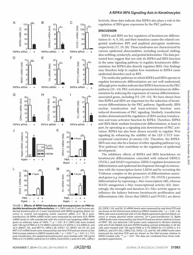
RIPK4 Is Important for the Induction of IRF6-dependent Dif-ferentiation Genes by PKC—Given the ability of RIPK4 to acti-vate IRF6 (Fig. 1), we investigated the importance of RIPK4 forthe PMA-inducible expression of the above IRF6-dependentdifferentiation genes. RIPK4 knockdown (Fig. 5A) inhibited theup-regulation of the IRF6 target genes GRHL3 and OVOL1 inPMA-treated OKF6 cells (Fig. 5, B and C). It also inhibited theup-regulation of MAD1 expression (Fig. 5D). Consistent withits importance for keratinocyte differentiation (9, 10), RIPK4knockdown concomitantly inhibited the up-regulation of IVLand KRT13 expression in PMA-treated OKF6 cells (Fig. 5, Eand F). PMA-inducible GRHL3, OVOL1, and IVL expression inepidermal keratinocytes were likewise inhibited by RIPK4knockdown (Fig. 5, G–I).
As a complementary approach, the effects of RIPK4 overex-pression were also investigated. The anti-RIPK4 antibody failedto detect either transfected or endogenous RIPK4. Therefore,the level of overexpression could not be determined. However,the ectopic expression of RIPK4 in OKF6 cells (Fig. 5J) signifi-cantly enhanced the up-regulation of GRHL3, OVOL1, and IVLgene expression by PMA (Fig. 5, K–M). GRHL3, OVOL1, and
FIGURE 2. PMA-inducible OKF6 cell differentiation. A–D, OKF6 cells were cultured in the presence of 100 ng/ml PMA or 0.1% DMSO vehicle for up to 48 h. A,the cells were fixed 48 h post-treatment and stained with an anti-�-tubulin antibody (red), and nuclei were stained with DAPI (blue). Scale bars � 20 �m. B, theviable cell number was enumerated by cell counting. *, p � 0.05; **, p � 0.01. C and D, cell lysates were subjected to Western blotting with anti-PCNA andanti-ERK2 (loading control) antibodies (C). The positions of molecular weight standards (in kilodaltons) are indicated. D, PCNA protein levels in DMSO-treatedcells in C were given an arbitrary value of 100%. **, p � 0.01. E–I, OKF6 cells were treated with 100 ng/ml PMA or 0.1% DMSO for the indicated times. IVL (E),KRT13 (F), GRHL3 (G), OVOL1 (H), and MAD1 (I) mRNA levels were measured by real-time PCR and are shown as the fold increase relative to DMSO-treated cells.*, p � 0.05; **, p � 0.01. All graphical data are combined from three independent experiments.
FIGURE 3. PMA-inducible IRF6 nuclear translocation and activation. A andB, OKF6 cells were pretreated with 20 ng/ml leptomycin B (LMB) for 60 min orleft untreated. They were then treated with 100 ng/ml PMA or 0.1% DMSOvehicle for 30 min. A, the cells were stained with an anti-IRF6 antibody (red),and nuclei were stained with DAPI (blue). Scale bars � 10 �m. B, quantifieddata for IRF6 nuclear translocation in A is presented as the ratio of nuclear tocytosolic IRF6. *, p � 0.05. C, HEK293T cells were transfected with an IFN�gene promoter reporter plasmid together with an IRF6 expression vector. Thecells were treated 24 h post-transfection with PMA or DMSO for 6 h, and genereporter activity was measured. IRF6 activity is shown as a fold increase. **,p � 0.01. The data in B and C are combined from three and four independentexperiments, respectively.
A RIPK4-IRF6 Signaling Axis in Keratinocytes
NOVEMBER 7, 2014 • VOLUME 289 • NUMBER 45 JOURNAL OF BIOLOGICAL CHEMISTRY 31081
by guest on Decem
ber 26, 2019http://w
ww
.jbc.org/D
ownloaded from
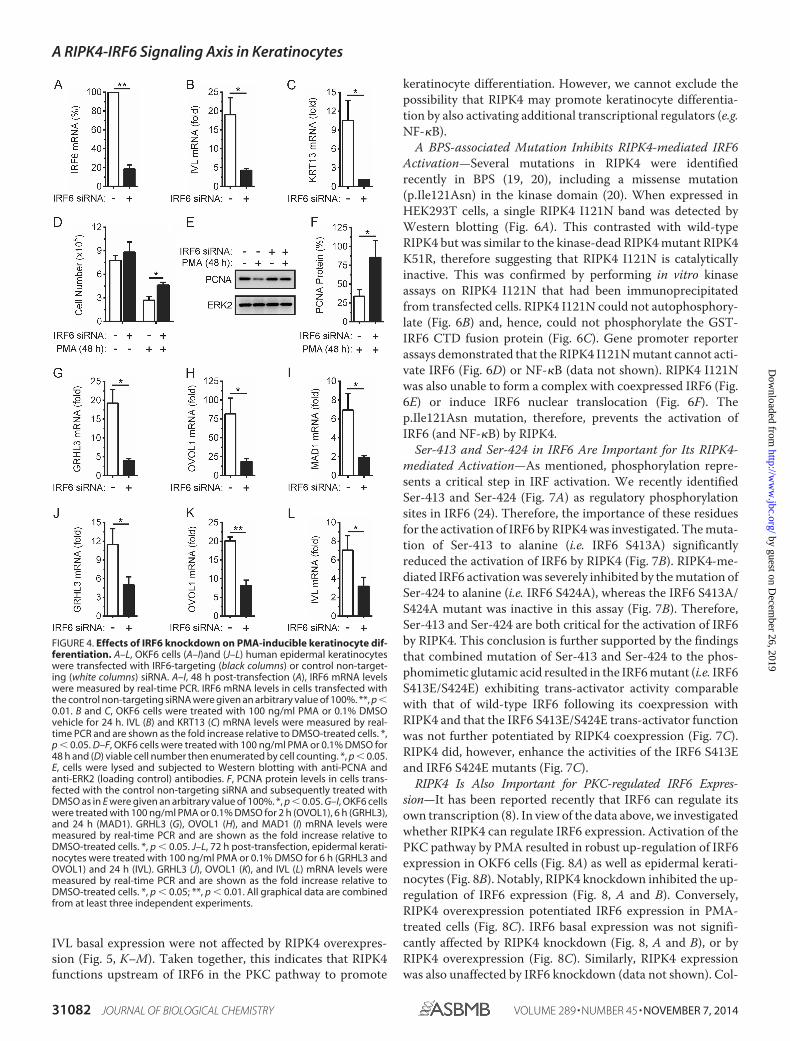
IVL basal expression were not affected by RIPK4 overexpres-sion (Fig. 5, K–M). Taken together, this indicates that RIPK4functions upstream of IRF6 in the PKC pathway to promote
keratinocyte differentiation. However, we cannot exclude thepossibility that RIPK4 may promote keratinocyte differentia-tion by also activating additional transcriptional regulators (e.g.NF-�B).
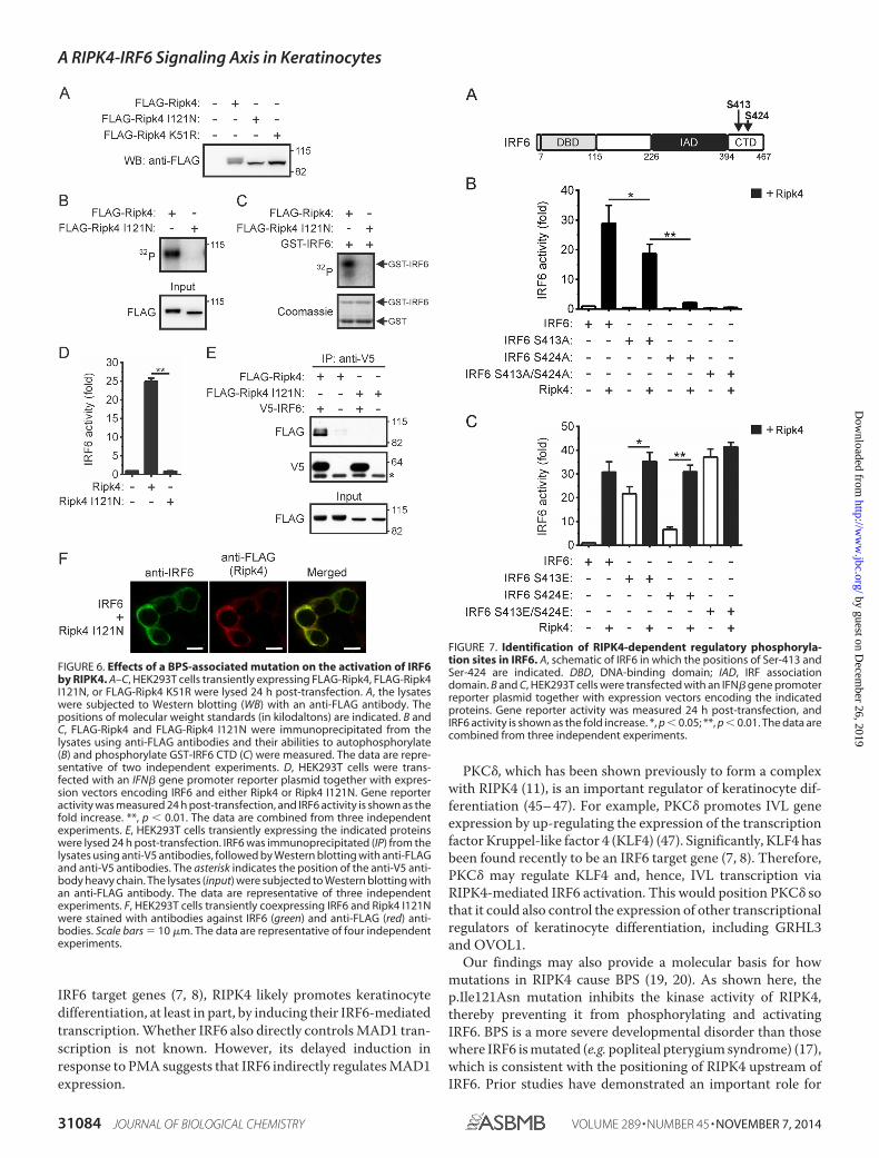
A BPS-associated Mutation Inhibits RIPK4-mediated IRF6Activation—Several mutations in RIPK4 were identifiedrecently in BPS (19, 20), including a missense mutation(p.Ile121Asn) in the kinase domain (20). When expressed inHEK293T cells, a single RIPK4 I121N band was detected byWestern blotting (Fig. 6A). This contrasted with wild-typeRIPK4 but was similar to the kinase-dead RIPK4 mutant RIPK4K51R, therefore suggesting that RIPK4 I121N is catalyticallyinactive. This was confirmed by performing in vitro kinaseassays on RIPK4 I121N that had been immunoprecipitatedfrom transfected cells. RIPK4 I121N could not autophosphory-late (Fig. 6B) and, hence, could not phosphorylate the GST-IRF6 CTD fusion protein (Fig. 6C). Gene promoter reporterassays demonstrated that the RIPK4 I121N mutant cannot acti-vate IRF6 (Fig. 6D) or NF-�B (data not shown). RIPK4 I121Nwas also unable to form a complex with coexpressed IRF6 (Fig.6E) or induce IRF6 nuclear translocation (Fig. 6F). Thep.Ile121Asn mutation, therefore, prevents the activation ofIRF6 (and NF-�B) by RIPK4.
Ser-413 and Ser-424 in IRF6 Are Important for Its RIPK4-mediated Activation—As mentioned, phosphorylation repre-sents a critical step in IRF activation. We recently identifiedSer-413 and Ser-424 (Fig. 7A) as regulatory phosphorylationsites in IRF6 (24). Therefore, the importance of these residuesfor the activation of IRF6 by RIPK4 was investigated. The muta-tion of Ser-413 to alanine (i.e. IRF6 S413A) significantlyreduced the activation of IRF6 by RIPK4 (Fig. 7B). RIPK4-me-diated IRF6 activation was severely inhibited by the mutation ofSer-424 to alanine (i.e. IRF6 S424A), whereas the IRF6 S413A/S424A mutant was inactive in this assay (Fig. 7B). Therefore,Ser-413 and Ser-424 are both critical for the activation of IRF6by RIPK4. This conclusion is further supported by the findingsthat combined mutation of Ser-413 and Ser-424 to the phos-phomimetic glutamic acid resulted in the IRF6 mutant (i.e. IRF6S413E/S424E) exhibiting trans-activator activity comparablewith that of wild-type IRF6 following its coexpression withRIPK4 and that the IRF6 S413E/S424E trans-activator functionwas not further potentiated by RIPK4 coexpression (Fig. 7C).RIPK4 did, however, enhance the activities of the IRF6 S413Eand IRF6 S424E mutants (Fig. 7C).
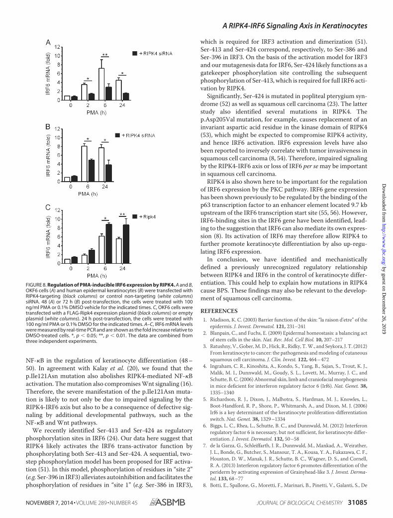
RIPK4 Is Also Important for PKC-regulated IRF6 Expres-sion—It has been reported recently that IRF6 can regulate itsown transcription (8). In view of the data above, we investigatedwhether RIPK4 can regulate IRF6 expression. Activation of thePKC pathway by PMA resulted in robust up-regulation of IRF6expression in OKF6 cells (Fig. 8A) as well as epidermal kerati-nocytes (Fig. 8B). Notably, RIPK4 knockdown inhibited the up-regulation of IRF6 expression (Fig. 8, A and B). Conversely,RIPK4 overexpression potentiated IRF6 expression in PMA-treated cells (Fig. 8C). IRF6 basal expression was not signifi-cantly affected by RIPK4 knockdown (Fig. 8, A and B), or byRIPK4 overexpression (Fig. 8C). Similarly, RIPK4 expressionwas also unaffected by IRF6 knockdown (data not shown). Col-
FIGURE 4. Effects of IRF6 knockdown on PMA-inducible keratinocyte dif-ferentiation. A–L, OKF6 cells (A–I)and (J–L) human epidermal keratinocyteswere transfected with IRF6-targeting (black columns) or control non-target-ing (white columns) siRNA. A–I, 48 h post-transfection (A), IRF6 mRNA levelswere measured by real-time PCR. IRF6 mRNA levels in cells transfected withthe control non-targeting siRNA were given an arbitrary value of 100%. **, p �0.01. B and C, OKF6 cells were treated with 100 ng/ml PMA or 0.1% DMSOvehicle for 24 h. IVL (B) and KRT13 (C) mRNA levels were measured by real-time PCR and are shown as the fold increase relative to DMSO-treated cells. *,p � 0.05. D–F, OKF6 cells were treated with 100 ng/ml PMA or 0.1% DMSO for48 h and (D) viable cell number then enumerated by cell counting. *, p � 0.05.E, cells were lysed and subjected to Western blotting with anti-PCNA andanti-ERK2 (loading control) antibodies. F, PCNA protein levels in cells trans-fected with the control non-targeting siRNA and subsequently treated withDMSO as in E were given an arbitrary value of 100%. *, p � 0.05. G–I, OKF6 cellswere treated with 100 ng/ml PMA or 0.1% DMSO for 2 h (OVOL1), 6 h (GRHL3),and 24 h (MAD1). GRHL3 (G), OVOL1 (H), and MAD1 (I) mRNA levels weremeasured by real-time PCR and are shown as the fold increase relative toDMSO-treated cells. *, p � 0.05. J–L, 72 h post-transfection, epidermal kerati-nocytes were treated with 100 ng/ml PMA or 0.1% DMSO for 6 h (GRHL3 andOVOL1) and 24 h (IVL). GRHL3 (J), OVOL1 (K), and IVL (L) mRNA levels weremeasured by real-time PCR and are shown as the fold increase relative toDMSO-treated cells. *, p � 0.05; **, p � 0.01. All graphical data are combinedfrom at least three independent experiments.
A RIPK4-IRF6 Signaling Axis in Keratinocytes
31082 JOURNAL OF BIOLOGICAL CHEMISTRY VOLUME 289 • NUMBER 45 • NOVEMBER 7, 2014
by guest on Decem
ber 26, 2019http://w
ww
.jbc.org/D
ownloaded from
lectively, these data indicate that RIPK4 also plays a role in theregulation of IRF6 gene expression by the PKC pathway.
DISCUSSION
RIPK4 and IRF6 are key regulators of keratinocyte differen-tiation (4 – 6, 9, 10), and their mutation causes the related con-genital syndromes BPS and popliteal pterygium syndrome,respectively (17, 19, 20). These syndromes are characterized byvarious epidermal abnormalities, including orofacial clefting,skin webbing, syndactyly, and genital deformities. The data pre-sented here suggest that not only do RIPK4 and IRF6 functionin the same signaling pathway to regulate keratinocyte differ-entiation, but RIPK4 also directly regulates IRF6. Our findingsmay therefore help to explain how mutations in RIPK4 causeepidermal disorders such as BPS.
The molecular pathways in which RIPK4 and IRF6 operate toregulate keratinocyte differentiation are not well understood,although prior studies indicate that RIPK4 functions in the PKCpathway (10 –14). PKC activation promotes keratinocyte differ-entiation by inducing the expression of various differentiation-associated genes, including IVL (29 –33). We have shown herethat RIPK4 and IRF6 are important for the induction of kerati-nocyte differentiation by the PKC pathway. Significantly, IRF6nuclear translocation and trans-activator function wereinduced downstream of PKC signaling. Similarly, transfectionstudies demonstrated the regulation of IRF6 nuclear transloca-tion and trans-activator function by RIPK4. Therefore, RIPK4and IRF6 likely mediate keratinocyte differentiation, at least inpart, by operating as a signaling axis downstream of PKC acti-vation. RIPK4 has also been shown recently to regulate Wntsignaling by enhancing the stability of the LEF-1/TCF tran-scriptional coactivator �-catenin (16). Therefore, the RIPK4-IRF6 axis may also be a feature of other signaling pathways (e.g.Wnt pathway) that contribute to the regulation of epidermaldevelopment.
The inhibitory effects of RIPK4 and IRF6 knockdown onkeratinocyte differentiation coincided with reduced GRHL3,OVOL1, and MAD1 expression. GRHL3 regulates keratinocytedifferentiation and epidermal development through its interac-tion with the transcription factor LMO4 and by recruiting theTrithorax complex to the promoters of differentiation-associ-ated genes (e.g. transglutaminase 1) (37–39). OVOL1 promotesdifferentiation by repressing c-Myc transcription (40), whereasMAD1 antagonizes c-Myc transcriptional activity (43). Inter-estingly, the strength and duration of c-Myc activity appear toinfluence the balance between keratinocyte proliferation anddifferentiation (44). Given that GRHL3 and OVOL1 are direct
FIGURE 5. Effects of RIPK4 knockdown and overexpression on PMA-in-ducible keratinocyte differentiation. A–I, OKF6 cells (A–F) and human epi-dermal keratinocytes (G–I) were transfected with RIPK4-targeting (black col-umns) or control non-targeting (white columns) siRNA. A–F, 48 h post-transfection (A) RIPK4 mRNA levels were measured by real-time PCR. RIPK4mRNA levels in cells transfected with the control non-targeting siRNA weregiven an arbitrary value of 100%. **, p � 0.01. B–F, OKF6 cells were treatedwith 100 ng/ml PMA or 0.1% DMSO vehicle for 2 h (OVOL1), 6 h (GRHL3), and24 h (MAD1, IVL, and KRT13). GRHL3 (B), OVOL1 (C), MAD1 (D), IVL (E), andKRT13 (F) mRNA levels were measured by real-time PCR and are shown as thefold increase relative to DMSO-treated cells. *, p � 0.05; **, p � 0.01. G–I, 72 hpost-transfection, the epidermal keratinocytes were treated with 100 ng/mlPMA or 0.1% DMSO vehicle for 6 h (GRHL3 and OVOL1) and 24 h (IVL). GRHL3
(G), OVOL1 (H), and IVL (I) mRNA levels were measured by real-time PCR andare shown as the fold increase relative to DMSO-treated cells. *, p � 0.05. J–M,OKF6 cells were transfected with a FLAG-Ripk4 expression plasmid (black col-umns) or empty plasmid (white columns). 24 h post-transfection (J), Ripk4mRNA levels were measured by real-time PCR and are shown relative to thoseof HPRT. ND, not detected. Cell lysates were subjected to Western blottingwith anti-FLAG (Ripk4) and anti-HSP90 (loading control) antibodies. K–M, thecells were treated with 100 ng/ml PMA or 0.1% DMSO for 2 h (OVOL1), 6 h(GRHL3), and 24 h (IVL). GRHL3 (K), OVOL1 (L), and IVL (M) mRNA levels weremeasured by real-time PCR and are shown as the fold increase relative toDMSO-treated cells. **, p � 0.01. The data are combined from three inde-pendent experiments.
A RIPK4-IRF6 Signaling Axis in Keratinocytes
NOVEMBER 7, 2014 • VOLUME 289 • NUMBER 45 JOURNAL OF BIOLOGICAL CHEMISTRY 31083
by guest on Decem
ber 26, 2019http://w
ww
.jbc.org/D
ownloaded from
IRF6 target genes (7, 8), RIPK4 likely promotes keratinocytedifferentiation, at least in part, by inducing their IRF6-mediatedtranscription. Whether IRF6 also directly controls MAD1 tran-scription is not known. However, its delayed induction inresponse to PMA suggests that IRF6 indirectly regulates MAD1expression.
PKC�, which has been shown previously to form a complexwith RIPK4 (11), is an important regulator of keratinocyte dif-ferentiation (45– 47). For example, PKC� promotes IVL geneexpression by up-regulating the expression of the transcriptionfactor Kruppel-like factor 4 (KLF4) (47). Significantly, KLF4 hasbeen found recently to be an IRF6 target gene (7, 8). Therefore,PKC� may regulate KLF4 and, hence, IVL transcription viaRIPK4-mediated IRF6 activation. This would position PKC� sothat it could also control the expression of other transcriptionalregulators of keratinocyte differentiation, including GRHL3and OVOL1.
Our findings may also provide a molecular basis for howmutations in RIPK4 cause BPS (19, 20). As shown here, thep.Ile121Asn mutation inhibits the kinase activity of RIPK4,thereby preventing it from phosphorylating and activatingIRF6. BPS is a more severe developmental disorder than thosewhere IRF6 is mutated (e.g. popliteal pterygium syndrome) (17),which is consistent with the positioning of RIPK4 upstream ofIRF6. Prior studies have demonstrated an important role for
FIGURE 6. Effects of a BPS-associated mutation on the activation of IRF6by RIPK4. A–C, HEK293T cells transiently expressing FLAG-Ripk4, FLAG-Ripk4I121N, or FLAG-Ripk4 K51R were lysed 24 h post-transfection. A, the lysateswere subjected to Western blotting (WB) with an anti-FLAG antibody. Thepositions of molecular weight standards (in kilodaltons) are indicated. B andC, FLAG-Ripk4 and FLAG-Ripk4 I121N were immunoprecipitated from thelysates using anti-FLAG antibodies and their abilities to autophosphorylate(B) and phosphorylate GST-IRF6 CTD (C) were measured. The data are repre-sentative of two independent experiments. D, HEK293T cells were trans-fected with an IFN� gene promoter reporter plasmid together with expres-sion vectors encoding IRF6 and either Ripk4 or Ripk4 I121N. Gene reporteractivity was measured 24 h post-transfection, and IRF6 activity is shown as thefold increase. **, p � 0.01. The data are combined from three independentexperiments. E, HEK293T cells transiently expressing the indicated proteinswere lysed 24 h post-transfection. IRF6 was immunoprecipitated (IP) from thelysates using anti-V5 antibodies, followed by Western blotting with anti-FLAGand anti-V5 antibodies. The asterisk indicates the position of the anti-V5 anti-body heavy chain. The lysates (input) were subjected to Western blotting withan anti-FLAG antibody. The data are representative of three independentexperiments. F, HEK293T cells transiently coexpressing IRF6 and Ripk4 I121Nwere stained with antibodies against IRF6 (green) and anti-FLAG (red) anti-bodies. Scale bars � 10 �m. The data are representative of four independentexperiments.
FIGURE 7. Identification of RIPK4-dependent regulatory phosphoryla-tion sites in IRF6. A, schematic of IRF6 in which the positions of Ser-413 andSer-424 are indicated. DBD, DNA-binding domain; IAD, IRF associationdomain. B and C, HEK293T cells were transfected with an IFN� gene promoterreporter plasmid together with expression vectors encoding the indicatedproteins. Gene reporter activity was measured 24 h post-transfection, andIRF6 activity is shown as the fold increase. *, p � 0.05; **, p � 0.01. The data arecombined from three independent experiments.
A RIPK4-IRF6 Signaling Axis in Keratinocytes
31084 JOURNAL OF BIOLOGICAL CHEMISTRY VOLUME 289 • NUMBER 45 • NOVEMBER 7, 2014
by guest on Decem
ber 26, 2019http://w
ww
.jbc.org/D
ownloaded from
NF-�B in the regulation of keratinocyte differentiation (48 –50). In agreement with Kalay et al. (20), we found that thep.Ile121Asn mutation also abolishes RIPK4-mediated NF-�Bactivation. The mutation also compromises Wnt signaling (16).Therefore, the severe manifestation of the p.Ile121Asn muta-tion is likely to not only be due to impaired signaling by theRIPK4-IRF6 axis but also to be a consequence of defective sig-naling by additional developmental pathways, such as theNF-�B and Wnt pathways.
We recently identified Ser-413 and Ser-424 as regulatoryphosphorylation sites in IRF6 (24). Our data here suggest thatRIPK4 likely activates the IRF6 trans-activator function byphosphorylating both Ser-413 and Ser-424. A sequential, two-step phosphorylation model has been proposed for IRF activa-tion (51). In this model, phosphorylation of residues in “site 2”(e.g. Ser-396 in IRF3) alleviates autoinhibition and facilitates thephosphorylation of residues in “site 1” (e.g. Ser-386 in IRF3),
which is required for IRF3 activation and dimerization (51).Ser-413 and Ser-424 correspond, respectively, to Ser-386 andSer-396 in IRF3. On the basis of the activation model for IRF3and our mutagenesis data for IRF6, Ser-424 likely functions as agatekeeper phosphorylation site controlling the subsequentphosphorylation of Ser-413, which is required for full IRF6 acti-vation by RIPK4.
Significantly, Ser-424 is mutated in popliteal pterygium syn-drome (52) as well as squamous cell carcinoma (23). The latterstudy also identified several mutations in RIPK4. Thep.Asp205Val mutation, for example, causes replacement of aninvariant aspartic acid residue in the kinase domain of RIPK4(53), which might be expected to compromise RIPK4 activity,and hence IRF6 activation. IRF6 expression levels have alsobeen reported to inversely correlate with tumor invasiveness insquamous cell carcinoma (8, 54). Therefore, impaired signalingby the RIPK4-IRF6 axis or loss of IRF6 per se may be importantin squamous cell carcinoma.
RIPK4 is also shown here to be important for the regulationof IRF6 expression by the PKC pathway. IRF6 gene expressionhas been shown previously to be regulated by the binding of thep63 transcription factor to an enhancer element located 9.7 kbupstream of the IRF6 transcription start site (55, 56). However,IRF6-binding sites in the IRF6 gene have been identified, lead-ing to the suggestion that IRF6 can also mediate its own expres-sion (8). Its activation of IRF6 may therefore allow RIPK4 tofurther promote keratinocyte differentiation by also up-regu-lating IRF6 expression.
In conclusion, we have identified and mechanisticallydefined a previously unrecognized regulatory relationshipbetween RIPK4 and IRF6 in the control of keratinocyte differ-entiation. This could help to explain how mutations in RIPK4cause BPS. These findings may also be relevant to the develop-ment of squamous cell carcinoma.
REFERENCES1. Madison, K. C. (2003) Barrier function of the skin: “la raison d’etre” of the
epidermis. J. Invest. Dermatol. 121, 231–2412. Blanpain, C., and Fuchs, E. (2009) Epidermal homeostasis: a balancing act
of stem cells in the skin. Nat. Rev. Mol. Cell Biol. 10, 207–2173. Ratushny, V., Gober, M. D., Hick, R., Ridky, T. W., and Seykora, J. T. (2012)
From keratinocyte to cancer: the pathogenesis and modeling of cutaneoussquamous cell carcinoma. J. Clin. Invest. 122, 464 – 472
4. Ingraham, C. R., Kinoshita, A., Kondo, S., Yang, B., Sajan, S., Trout, K. J.,Malik, M. I., Dunnwald, M., Goudy, S. L., Lovett, M., Murray, J. C., andSchutte, B. C. (2006) Abnormal skin, limb and craniofacial morphogenesisin mice deficient for interferon regulatory factor 6 (Irf6). Nat. Genet. 38,1335–1340
5. Richardson, R. J., Dixon, J., Malhotra, S., Hardman, M. J., Knowles, L.,Boot-Handford, R. P., Shore, P., Whitmarsh, A., and Dixon, M. J. (2006)Irf6 is a key determinant of the keratinocyte proliferation-differentiationswitch. Nat. Genet. 38, 1329 –1334
6. Biggs, L. C., Rhea, L., Schutte, B. C., and Dunnwald, M. (2012) Interferonregulatory factor 6 is necessary, but not sufficient, for keratinocyte differ-entiation. J. Invest. Dermatol. 132, 50 –58
7. de la Garza, G., Schleiffarth, J. R., Dunnwald, M., Mankad, A., Weirather,J. L., Bonde, G., Butcher, S., Mansour, T. A., Kousa, Y. A., Fukazawa, C. F.,Houston, D. W., Manak, J. R., Schutte, B. C., Wagner, D. S., and Cornell,R. A. (2013) Interferon regulatory factor 6 promotes differentiation of theperiderm by activating expression of Grainyhead-like 3. J. Invest. Derma-tol. 133, 68 –77
8. Botti, E., Spallone, G., Moretti, F., Marinari, B., Pinetti, V., Galanti, S., De
FIGURE 8. Regulation of PMA-inducible IRF6 expression by RIPK4. A and B,OKF6 cells (A) and human epidermal keratinocytes (B) were transfected withRIPK4-targeting (black columns) or control non-targeting (white columns)siRNA. 48 (A) or 72 h (B) post-transfection, the cells were treated with 100ng/ml PMA or 0.1% DMSO vehicle for the indicated times. C, OKF6 cells weretransfected with a FLAG-Ripk4 expression plasmid (black columns) or emptyplasmid (white columns). 24 h post-transfection, the cells were treated with100 ng/ml PMA or 0.1% DMSO for the indicated times. A–C, IRF6 mRNA levelswere measured by real-time PCR and are shown as the fold increase relative toDMSO-treated cells. *, p � 0.05; **, p � 0.01. The data are combined fromthree independent experiments.
A RIPK4-IRF6 Signaling Axis in Keratinocytes
NOVEMBER 7, 2014 • VOLUME 289 • NUMBER 45 JOURNAL OF BIOLOGICAL CHEMISTRY 31085
by guest on Decem
ber 26, 2019http://w
ww
.jbc.org/D
ownloaded from
Meo, P. D., De Nicola, F., Ganci, F., Castrignanò, T., Pesole, G., Chimenti,S., Guerrini, L., Fanciulli, M., Blandino, G., Karin, M., and Costanzo, A.(2011) Developmental factor IRF6 exhibits tumor suppressor activity insquamous cell carcinomas. Proc. Natl. Acad. Sci. U.S.A. 108, 13710 –13715
9. Holland, P., Willis, C., Kanaly, S., Glaccum, M., Warren, A., Charrier, K.,Murison, J., Derry, J., Virca, G., Bird, T., and Peschon, J. (2002) RIP4 is anankyrin repeat-containing kinase essential for keratinocyte differentia-tion. Curr. Biol. 12, 1424 –1428
10. Rountree, R. B., Willis, C. R., Dinh, H., Blumberg, H., Bailey, K., Dean, C.,Jr., Peschon, J. J., and Holland, P. M. (2010) RIP4 regulates epidermaldifferentiation and cutaneous inflammation. J. Invest. Dermatol. 130,102–112
11. Bahr, C., Rohwer, A., Stempka, L., Rincke, G., Marks, F., and Gschwendt,M. (2000) DIK, a novel protein kinase that interacts with protein kinaseC�. Cloning, characterization, and gene analysis. J. Biol. Chem. 275,36350 –36357
12. Chen, L., Haider, K., Ponda, M., Cariappa, A., Rowitch, D., and Pillai, S.(2001) Protein kinase C-associated kinase (PKK), a novel membrane-as-sociated, ankyrin repeat-containing protein kinase. J. Biol. Chem. 276,21737–21744
13. Muto, A., Ruland, J., McAllister-Lucas, L. M., Lucas, P. C., Yamaoka, S.,Chen, F. F., Lin, A., Mak, T. W., Núñez, G., and Inohara, N. (2002) Proteinkinase C-associated kinase (PKK) mediates Bcl10-independent NF-� Bactivation induced by phorbol ester. J. Biol. Chem. 277, 31871–31876
14. Moran, S. T., Haider, K., Ow, Y., Milton, P., Chen, L., and Pillai, S. (2003)Protein kinase C-associated kinase can activate NF�B in both a kinase-de-pendent and a kinase-independent manner. J. Biol. Chem. 278,21526 –21533
15. Meylan, E., Martinon, F., Thome, M., Gschwendt, M., and Tschopp, J.(2002) RIP4 (DIK/PKK), a novel member of the RIP kinase family, acti-vates NF-� B and is processed during apoptosis. EMBO Rep. 3, 1201–1208
16. Huang, X., McGann, J. C., Liu, B. Y., Hannoush, R. N., Lill, J. R., Pham, V.,Newton, K., Kakunda, M., Liu, J., Yu, C., Hymowitz, S. G., Hongo, J. A.,Wynshaw-Boris, A., Polakis, P., Harland, R. M., and Dixit, V. M. (2013)Phosphorylation of Dishevelled by protein kinase RIPK4 regulates Wntsignaling. Science 339, 1441–1445
17. Kondo, S., Schutte, B. C., Richardson, R. J., Bjork, B. C., Knight, A. S.,Watanabe, Y., Howard, E., de Lima, R. L., Daack-Hirsch, S., Sander, A.,McDonald-McGinn, D. M., Zackai, E. H., Lammer, E. J., Aylsworth, A. S.,Ardinger, H. H., Lidral, A. C., Pober, B. R., Moreno, L., Arcos-Burgos, M.,Valencia, C., Houdayer, C., Bahuau, M., Moretti-Ferreira, D., Richieri-Costa, A., Dixon, M. J., and Murray, J. C. (2002) Mutations in IRF6 causeVan der Woude and popliteal pterygium syndromes. Nat. Genet. 32,285–289
18. Gorlin, R. J., Sedano, H. O., and Cervenka, J. (1968) Popliteal pterygiumsyndrome: a syndrome comprising cleft lip-palate, popliteal and intercru-ral pterygia, digital and genital anomalies. Pediatrics 41, 503–509
19. Mitchell, K., O’Sullivan, J., Missero, C., Blair, E., Richardson, R., Anderson,B., Antonini, D., Murray, J. C., Shanske, A. L., Schutte, B. C., Romano,R. A., Sinha, S., Bhaskar, S. S., Black, G. C., Dixon, J., and Dixon, M. J. (2012)Exome sequence identifies RIPK4 as the Bartsocas-Papas syndrome locus.Am. J. Hum. Genet. 90, 69 –75
20. Kalay, E., Sezgin, O., Chellappa, V., Mutlu, M., Morsy, H., Kayserili, H.,Kreiger, E., Cansu, A., Toraman, B., Abdalla, E. M., Aslan, Y., Pillai, S., andAkarsu, N. A. (2012) Mutations in RIPK4 cause the autosomal-recessiveform of popliteal pterygium syndrome. Am. J. Hum. Genet. 90, 76 – 85
21. Bartsocas, C. S., and Papas, C. V. (1972) Popliteal pterygium syndrome:evidence for a severe autosomal recessive form. J. Med. Genet. 9, 222–226
22. Hennekam, R. C., Huber, J., and Variend, D. (1994) Bartsocas-Papas syn-drome with internal anomalies: evidence for a more generalized epithelialdefect or new syndrome? Am. J. Med. Genet. 53, 102–107
23. Stransky, N., Egloff, A. M., Tward, A. D., Kostic, A. D., Cibulskis, K., Si-vachenko, A., Kryukov, G. V., Lawrence, M. S., Sougnez, C., McKenna, A.,Shefler, E., Ramos, A. H., Stojanov, P., Carter, S. L., Voet, D., Cortés, M. L.,Auclair, D., Berger, M. F., Saksena, G., Guiducci, C., Onofrio, R. C., Parkin,M., Romkes, M., Weissfeld, J. L., Seethala, R. R., Wang, L., Rangel-Es-careño, C., Fernandez-Lopez, J. C., Hidalgo-Miranda, A., Melendez-Za-jgla, J., Winckler, W., Ardlie, K., Gabriel, S. B., Meyerson, M., Lander, E. S.,
Getz, G., Golub, T. R., Garraway, L. A., and Grandis, J. R. (2011) Themutational landscape of head and neck squamous cell carcinoma. Science333, 1157–1160
24. Kwa, M. Q., Nguyen, T., Huynh, J., Ramnath, D., De Nardo, D., Lam, P. Y.,Reynolds, E. C., Hamilton, J. A., Sweet, M. J., and Scholz, G. M. (2014)Interferon regulatory factor 6 differentially regulates Toll-like receptor2-dependent chemokine gene expression in epithelial cells. J. Biol. Chem.289, 19758 –19768
25. Dickson, M. A., Hahn, W. C., Ino, Y., Ronfard, V., Wu, J. Y., Weinberg,R. A., Louis, D. N., Li, F. P., and Rheinwald, J. G. (2000) Human keratino-cytes that express hTERT and also bypass a p16(INK4a)-enforced mech-anism that limits life span become immortal yet retain normal growth anddifferentiation characteristics. Mol. Cell Biol. 20, 1436 –1447
26. Schindler, U., and Baichwal, V. R. (1994) Three NF-� B binding sites in thehuman E-selectin gene required for maximal tumor necrosis factor �-in-duced expression. Mol. Cell Biol. 14, 5820 –5831
27. Pfaffl, M. W. (2001) A new mathematical model for relative quantificationin real-time RT-PCR. Nucleic Acids Res. 29, e45
28. Tamura, T., Yanai, H., Savitsky, D., and Taniguchi, T. (2008) The IRFfamily transcription factors in immunity and oncogenesis. Annu. Rev. Im-munol. 26, 535–584
29. Denning, M. F. (2004) Epidermal keratinocytes: regulation of multiple cellphenotypes by multiple protein kinase C isoforms. Int. J. Biochem. CellBiol. 36, 1141–1146
30. Matsui, M. S., Chew, S. L., and DeLeo, V. A. (1992) Protein kinase C innormal human epidermal keratinocytes during proliferation and calcium-induced differentiation. J. Invest. Dermatol. 99, 565–571
31. Długosz, A. A., and Yuspa, S. H. (1994) Protein kinase C regulates kerati-nocyte transglutaminase (TGK) gene expression in cultured primarymouse epidermal keratinocytes induced to terminally differentiate by cal-cium. J. Invest. Dermatol. 102, 409 – 414
32. Eckert, R. L., Crish, J. F., Efimova, T., Dashti, S. R., Deucher, A., Bone, F.,Adhikary, G., Huang, G., Gopalakrishnan, R., and Balasubramanian, S.(2004) Regulation of involucrin gene expression. J. Invest. Dermatol. 123,13–22
33. Chew, Y. C., Adhikary, G., Wilson, G. M., Reece, E. A., and Eckert, R. L.(2011) Protein kinase C (PKC) � suppresses keratinocyte proliferation byincreasing p21(Cip1) level by a KLF4 transcription factor-dependentmechanism. J. Biol. Chem. 286, 28772–28782
34. Watt, F. M. (1989) Terminal differentiation of epidermal keratinocytes.Curr. Opin. Cell Biol. 1, 1107–1115
35. Watt, F. M. (1983) Involucrin and other markers of keratinocyte terminaldifferentiation. J. Invest. Dermatol. 81, 100s-103s
36. Fuchs, E. (1988) Keratins as biochemical markers of epithelial differentia-tion. Trends Genet. 4, 277–281
37. Ting, S. B., Caddy, J., Hislop, N., Wilanowski, T., Auden, A., Zhao, L. L.,Ellis, S., Kaur, P., Uchida, Y., Holleran, W. M., Elias, P. M., Cunningham,J. M., and Jane, S. M. (2005) A homolog of Drosophila grainy head isessential for epidermal integrity in mice. Science 308, 411– 413
38. Yu, Z., Lin, K. K., Bhandari, A., Spencer, J. A., Xu, X., Wang, N., Lu, Z., Gill,G. N., Roop, D. R., Wertz, P., and Andersen, B. (2006) The Grainyhead-likeepithelial transactivator Get-1/Grhl3 regulates epidermal terminal differ-entiation and interacts functionally with LMO4. Dev. Biol. 299, 122–136
39. Hopkin, A. S., Gordon, W., Klein, R. H., Espitia, F., Daily, K., Zeller, M.,Baldi, P., and Andersen, B. (2012) GRHL3/GET1 and trithorax groupmembers collaborate to activate the epidermal progenitor differentiationprogram. PLoS Genet. 8, e1002829
40. Nair, M., Teng, A., Bilanchone, V., Agrawal, A., Li, B., and Dai, X. (2006)Ovol1 regulates the growth arrest of embryonic epidermal progenitor cellsand represses c-myc transcription. J. Cell Biol. 173, 253–264
41. Teng, A., Nair, M., Wells, J., Segre, J. A., and Dai, X. (2007) Strain-depen-dent perinatal lethality of Ovol1-deficient mice and identification of Ovol2as a downstream target of Ovol1 in skin epidermis. Biochim. Biophys. Acta1772, 89 –95
42. Werner, S., Beer, H. D., Mauch, C., Lüscher, B., and Werner, S. (2001) TheMad1 transcription factor is a novel target of activin and TGF-� action inkeratinocytes: possible role of Mad1 in wound repair and psoriasis. Onco-gene 20, 7494 –7504
A RIPK4-IRF6 Signaling Axis in Keratinocytes
31086 JOURNAL OF BIOLOGICAL CHEMISTRY VOLUME 289 • NUMBER 45 • NOVEMBER 7, 2014
by guest on Decem
ber 26, 2019http://w
ww
.jbc.org/D
ownloaded from
43. Lüscher, B. (2012) MAD1 and its life as a MYC antagonist: an update. EurJ. Cell Biol. 91, 506 –514
44. Watt, F. M., Frye, M., and Benitah, S. A. (2008) MYC in mammalianepidermis: how can an oncogene stimulate differentiation? Nat. Rev. Can-cer 8, 234 –242
45. Deucher, A., Efimova, T., and Eckert, R. L. (2002) Calcium-dependentinvolucrin expression is inversely regulated by protein kinase C (PKC)�and PKC�. J. Biol. Chem. 277, 17032–17040
46. Adhikary, G., Chew, Y. C., Reece, E. A., and Eckert, R. L. (2010) PKC-� and-�, MEKK-1, MEK-6, MEK-3, and p38-� are essential mediators of theresponse of normal human epidermal keratinocytes to differentiatingagents. J. Invest. Dermatol. 130, 2017–2030
47. Chew, Y. C., Adhikary, G., Xu, W., Wilson, G. M., and Eckert, R. L. (2013)Protein kinase C � increases Kruppel-like factor 4 protein, which drivesinvolucrin gene transcription in differentiating keratinocytes. J. Biol.Chem. 288, 17759 –17768
48. Bushdid, P. B., Brantley, D. M., Yull, F. E., Blaeuer, G. L., Hoffman, L. H.,Niswander, L., and Kerr, L. D. (1998) Inhibition of NF-�B activity results indisruption of the apical ectodermal ridge and aberrant limb morphogen-esis. Nature 392, 615– 618
49. Kanegae, Y., Tavares, A. T., Izpisúa Belmonte, J. C., and Verma, I. M.(1998) Role of Rel/NF-�B transcription factors during the outgrowth ofthe vertebrate limb. Nature 392, 611– 614
50. Gugasyan, R., Voss, A., Varigos, G., Thomas, T., Grumont, R. J., Kaur, P.,Grigoriadis, G., and Gerondakis, S. (2004) The transcription factors c-reland RelA control epidermal development and homeostasis in embryonic
and adult skin via distinct mechanisms. Mol. Cell Biol. 24, 5733–574551. Panne, D., McWhirter, S. M., Maniatis, T., and Harrison, S. C. (2007)
Interferon regulatory factor 3 is regulated by a dual phosphorylation-de-pendent switch. J. Biol. Chem. 282, 22816 –22822
52. Matsuzawa, N., Kondo, S., Shimozato, K., Nagao, T., Nakano, M., Tsuda,M., Hirano, A., Niikawa, N., and Yoshiura, K. (2010) Two missense muta-tions of the IRF6 gene in two Japanese families with popliteal pterygiumsyndrome. Am. J. Med. Genet. A 152A, 2262–2267
53. Meylan, E., and Tschopp, J. (2005) The RIP kinases: crucial integrators ofcellular stress. Trends Biochem. Sci. 30, 151–159
54. Restivo, G., Nguyen, B. C., Dziunycz, P., Ristorcelli, E., Ryan, R. J., Özuysal,Ö. Y., Di Piazza, M., Radtke, F., Dixon, M. J., Hofbauer, G. F., Lefort, K., andDotto, G. P. (2011) IRF6 is a mediator of Notch pro-differentiation andtumour suppressive function in keratinocytes. EMBO J. 30, 4571– 4585
55. Thomason, H. A., Zhou, H., Kouwenhoven, E. N., Dotto, G. P., Restivo, G.,Nguyen, B. C., Little, H., Dixon, M. J., van Bokhoven, H., and Dixon, J.(2010) Cooperation between the transcription factors p63 and IRF6 isessential to prevent cleft palate in mice. J. Clin. Invest. 120, 1561–1569
56. Rahimov, F., Marazita, M. L., Visel, A., Cooper, M. E., Hitchler, M. J.,Rubini, M., Domann, F. E., Govil, M., Christensen, K., Bille, C., Melbye, M.,Jugessur, A., Lie, R. T., Wilcox, A. J., Fitzpatrick, D. R., Green, E. D.,Mossey, P. A., Little, J., Steegers-Theunissen, R. P., Pennacchio, L. A.,Schutte, B. C., and Murray, J. C. (2008) Disruption of an AP-2� bindingsite in an IRF6 enhancer is associated with cleft lip. Nat. Genet. 40,1341–1347
A RIPK4-IRF6 Signaling Axis in Keratinocytes
NOVEMBER 7, 2014 • VOLUME 289 • NUMBER 45 JOURNAL OF BIOLOGICAL CHEMISTRY 31087
by guest on Decem
ber 26, 2019http://w
ww
.jbc.org/D
ownloaded from

Reynolds, Matthew J. Sweet, John A. Hamilton and Glen M. ScholzMei Qi Kwa, Jennifer Huynh, Jiamin Aw, Lianyi Zhang, Thao Nguyen, Eric C.
Function as a Signaling Axis to Regulate Keratinocyte DifferentiationReceptor-interacting Protein Kinase 4 and Interferon Regulatory Factor 6
doi: 10.1074/jbc.M114.589382 originally published online September 22, 20142014, 289:31077-31087.J. Biol. Chem.
10.1074/jbc.M114.589382Access the most updated version of this article at doi:
Alerts:
When a correction for this article is posted•
When this article is cited•
to choose from all of JBC's e-mail alertsClick here
http://www.jbc.org/content/289/45/31077.full.html#ref-list-1
This article cites 56 references, 21 of which can be accessed free at
by guest on Decem
ber 26, 2019http://w
ww
.jbc.org/D
ownloaded from