recent advances in understanding the antibacterial
TRANSCRIPT
1
Recent advances in understanding the antibacterial properties of flavonoids
T.P. Tim Cushnie a*
Andrew J. Lamb b
a Faculty of Medicine, Mahasarakham University, Khamriang. Kantarawichai. Maha
Sarakham 44150. Thailand.
b School of Pharmacy and Life Sciences, Robert Gordon University, Schoolhill, Aberdeen.
AB10 1FR. UK.
* Corresponding author
e-mail [email protected] or [email protected]
telephone +66 (0)43 754 32240 ext. 1159
fax +66 (0)43 754 245

2
Abstract
Antibiotic resistance is a major global problem and there is a pressing need to develop new
therapeutic agents. Flavonoids are a family of plant-derived compounds with potentially
exploitable activities including direct antibacterial activity, synergism with antibiotics, and
suppression of bacterial virulence. In this review, recent advances towards understanding
these properties are described. Information is presented on the ten most potently antibacterial
flavonoids, and the five most synergistic flavonoid-antibiotic combinations tested in the last
six years (identified from PubMed and ScienceDirect). Top of these respective lists are
panduratin A with MICs of 0.06 to 2.0 µg/mL against Staphylococcus aureus, and epicatechin
gallate which reduces oxacillin MICs as much as 512-fold. Research seeking to improve such
activity, and understand structure-activity relationships is discussed. Proposed mechanisms of
action are discussed too. In addition to direct and synergistic activities, flavonoids inhibit a
number of bacterial virulence factors including quorum sensing signal receptors, enzymes,
and toxins. Evidence of these molecular effects at the cellular level include in vitro inhibition
of biofilm formation, inhibition of bacterial attachment to host ligands, and neutralisation of
toxicity toward cultured human cells. In vivo evidence of bacterial pathogenesis being
disrupted includes demonstrated efficacy against Helicobacter pylori infection and S. aureus
α-toxin intoxication.
Keywords: flavonoids; antibacterial; structure-activity; mechanism of action; synergy;
antivirulence

3
1. Introduction
With antibiotic resistance reaching crisis point in many hospitals around the world [1]
and resistance increasing in community acquired infections also [2], there is an urgent need to
replenish our arsenal of anti-infective agents. Ideally, this should be in the form of new
classes of antibacterial agent [3], as the structural alteration of drugs to which resistance has
already developed rarely provides a major solution [4]. Inhibition of resistance mechanisms
through the development of novel adjuncts represents an important strategy also. The β-
lactamase inhibitor clavulanate, launched in 1981, remains effective today in spite of many
years of extensive use [5, 6]. A third promising but unproven approach is the development of
drugs that target bacterial virulence factors. Rather than inhibiting cellular components
necessary for growth or viability, these compounds would ameliorate infection by interfering
with aspects of bacterial pathogenesis eg. attachment to host tissue [7].
Natural products are a major source of chemical diversity and have provided important
therapeutic agents for many bacterial diseases [8]. Most of these agents have been of
microbial origin, but the antibacterial properties of plant-derived compounds are attracting
increasing attention [9, 10]. This is attributed, in part, to the fact that plants can be rationally
selected for antibacterial testing based on ethnomedicinal use [11]. Flavonoids are a group of
heterocyclic organic compounds present in plants and related products eg. propolis and honey
[12]. Poultices, infusions, balms, and spices containing flavonoids as active constituents have
been used in many cultures for centuries. Traditional uses include treatment and prevention
of various infectious and toxin-mediated diseases eg. sores, wound infections [13], acne,
respiratory infections [14], gastrointestinal disease [15], and urinary tract infections [16]. Not
surprisingly, this family of compounds is the subject of much antibacterial research.
There are fourteen classes of flavonoid in total, differentiated on the basis of the
chemical nature and position of substituents on the A, B and C rings [17]. The skeleton
structures of six of these classes are shown in Figure 1 with rings named and positions
numbered. Most of the reports of flavonoids possessing antibacterial properties can be
attributed to these six structures or their isoflavonoid counterparts (flavonoids where ring B is
4
joined at position 3 of ring C instead of position 2). Potential applications for these
compounds include modern agents [18] and adjuncts [19] for the treatment of bacterial
infection, drugs for treating toxin-mediated disease [20], antivirulence therapies [21], and
capture molecules for removing endotoxin from pharmaceutical preparations [22].
In this paper, reports on the diverse range of antibacterial properties exhibited by
flavonoids are reviewed. Emphasis is on important developments in the last six years as
earlier research has already been discussed [13, 23]. The activity of naturally occurring
flavonoids is covered, as well as that of semi-synthetic and synthetic flavonoids. Proposed
structure-activity relationships and mechanisms of action (MOAs) are reviewed too. The
structures of all the flavonoids discussed are presented in Supplementary Table 1. Readers
interested in the more specific topic of antibacterial tea flavonoids or broader topic of
medicinal flavonoids are directed to reviews by Friedman [24] and Cazarolli et al. [25].
2. Direct antibacterial activity
2.1 Naturally occurring flavonoids
For several decades, the antibacterial activity of flavonoid-rich natural products has
been reported in the scientific literature. This has continued in recent years, with some plant
and propolis extracts being identified with MICs <100 µg/mL [26-28] or in one case <10
µg/mL [29]. Antibacterial flavonoids have been successfully isolated in over 50 such studies,
and a list of compounds with the lowest reported MICs is presented (Table 1). To put these
values into context, compounds with MICs ≤100 µg/mL are considered noteworthy, and those
with MICs ≤10 µg/mL, very interesting [30]. Caution is always necessary when comparing
flavonoid MICs determined in different laboratories [13] but, this caveat notwithstanding,
some of the flavonoids isolated since 2005 have very impressive antibacterial activity.
2.2 Semi-synthetic and synthetic flavonoids
Synthetic modification of natural flavonoid structures has been reported as early as 1981
[31], but it is only in recent years that there has been a real surge of work in this area.
5
Particularly successful alterations include linking an N-heterocyclic ring to the A ring of
chrysin (position 7). This derivative was 16- to 32-fold more active than its parent
compound, with MICs of 1.56 and 3.13 µg/mL against Escherichia coli and Staphylococcus
aureus respectively [32]. Similar enhancements of activity were observed when dibutylamine
was linked to the A ring of genistein (position 7) [18]. Alkylation of (-)-epigallocatechin
gallate dramatically improved the activity of this compound against Gram positive pathogens.
The derivative 3-O-decyl-(+)-catechin was 64- to 128-fold more active than its parent
structure, with MICs of 1.0 and 2.0 µg/mL against S. aureus and Enterococcus faecalis [33].
2.3 Structure-activity relationship
The findings of recent structure-activity investigations are summarised below. These
correspond well with relationships summarised previously [13], and shed further light on this
subject. Structural components which improve the activity of open chain flavonoids
(chalcones) generally improve the activity of other flavonoids. However, in the interests of
clarity these two groups are discussed separately.
2.3.1 Chalcones
For the A ring, Avila et al. have confirmed that hydroxylation at position 2’ is important
for antibacterial activity, though it is hypothesised that this feature indirectly affects activity
by promoting structural stability [34]. Hydroxylation at other A ring positions [35] including
position 4’ [36] improve activity too. Interestingly, carboxylation at position 4’ has been
shown to cause a 60-fold improvement in aqueous solubility with negligible loss of
antibacterial activity [37]. Reports by Avila [34] and Batovska [38] suggest that A ring
lipophilicity is important however, with chalcones possessing prenyl or geranyl groups at
position 3’ displaying good activity [34]. Substitutions which decrease activity include
acetoxylation or methoxylation at position 2’ [34] and fluorination at positions 3’ and 5’ [37].
On the B ring, substitution at position 4 is important for antibacterial activity. For
example, chalcones with a 6-carbon alkyl chain and piperidine group at this position have

6
good activity [39] as do compounds with a hydroxyl group [34]. Batovska et al. report that
hydroxylation of the B ring is not sufficient for activity on its own though, and suggest that a
lipophilic A ring is necessary [38]. Presence of the lipophilic substituents trifluoromethyl or
bromo at position 3 of the B ring has also been reported to improve antibacterial activity, with
activity increasing further if one of these groups is present at position 5 [37].
2.3.2 Other flavonoid classes
On the A ring, the presence of an O-acyl [40] or O-alkylamino chain [41] at position 7
improves the antibacterial activity of compounds in the flavone class. Šmejkal and colleagues
report that hydroxylation at position 5 is also important [42], a finding which corresponds
well with previous studies of flavones and flavanones [43, 44]. The presence of a lipophilic
group (eg. geranyl) at position 6 or 8 of the A ring improves activity too [42]. This supports
previous findings by Tsuchiya and colleagues [44].
There has been comparatively little work on the B ring but Šmejkal et al. report that, as
is the case with chalcones [34], methoxylation decreases activity [42]. This correlates well
with previous work by Alcaraz and colleagues, who found that 4’-oxymethylflavanone had an
MIC in excess of 1000 µg/mL against S. aureus [43]. Older research, worth mentioning here
because it corresponds well with recent data for the chalcones [37], indicates that bromo- (and
chloro-) groups at positions 2’, 3’ and 4’ improve flavanone activity [31].
For the C ring, hydroxylation at position 3 improves the activity of flavanones [42].
There is growing evidence to suggest that an O-acyl or O-alkyl chain at this position improves
activity even further, at least in the case of flavonols [45] and flavan-3-ols [33, 46]. Recent
work by Mughal et al., which sought to improve the activity of flavones, found that
replacement of the oxygen atom at position 4 with sulphur or nitrogen was effective [47].
2.4 Identification of flavonoid activity as bacteriostatic or bactericidal
With increasing numbers of immunocompromised patients [48], there is understandable
interest in the identification of compounds which kill bacteria rather than just inhibiting their
7
growth. Bactericidal activity in this context is usually defined as activity resulting in a 99.9%
reduction in bacterial numbers, and is tested for using the time-kill method or minimum
bactericidal concentration (MBC) assay [49, 50]. Such methods have frequently been used to
test flavonoids, and on many occasions bactericidal activity was reportedly detected [51-53].
Studies with model membranes indicate that flavonoids cause aggregation though [54, 55],
and in 2007 it was confirmed that the flavonol galangin has this effect on bacterial cells [56].
The flavan-3-ol epicatechin gallate has also been reported to cause bacterial aggregation [19],
though it is not yet clear if this observation was due to genuine aggregation (cells clumping
together) or pseudomulticellular bacteria (cells failing to separate following binary fission
[46]). An important ramification of this aggregatory effect is that conventional methods are
no longer sufficient for demonstrating the bactericidal activity of flavonoids. This is because
decreases in colony forming unit (CFU) numbers may be attributable to bacteria clumping
together and not cell death (Figure 2). There are no immediately apparent solutions to this
methodological problem, but an interim approach might be to cease MBC testing in favour of
time-kill studies, with microscopic analysis of treated bacteria [57].
2.5 Mechanism(s) of action
Early flavonoid research (1987 to 2004; reviewed previously [13]) suggested that their
direct antibacterial activity may be attributable to up to three mechanisms. These were
cytoplasmic membrane damage (caused by perforation [54] and / or a reduction in membrane
fluidity [58]), inhibition of nucleic acid synthesis [59] (caused by topoisomerase inhibition
[60, 61]), and inhibition of energy metabolism (caused by NADH-cytochrome c reductase
inhibition [62]). In the period since (2005 to 2010), additional evidence has been presented in
support of each of the proposed mechanisms. Work with compounds in the flavonol [63],
flavan-3-ol [64, 65], and flavolan classes [66] suggests these damage the cytoplasmic
membrane (possibly by generating hydrogen peroxide [67]), and work with flavan-3-ols [68,
69] and isoflavones [70] suggests these inhibit nucleic acid synthesis (through topoisomerase
[68, 71] and / or dihydrofolate reductase [69] inhibition). In addition, compounds in the

8
flavonol, flavan-3-ol, and flavone classes have been shown to inhibit energy metabolism
(through ATP synthase inhibition [72]). Evidence has also been presented for two new
mechanisms. These are inhibition of cell wall synthesis (caused by D-alanine:D-alanine
ligase inhibition [73]) and inhibition of cell membrane synthesis (caused by inhibition of
FabG [74-76], FabI [74], FabZ [77], Rv0636 [78] or KAS III [79]).
Probably for logistical reasons, most of the above studies were conducted with just one
or two compounds. For a long time, this meant it was not clear whether the findings from
these studies were due to (a) flavonoids of one structure having a single MOA and flavonoids
of a different structure having a different MOA, (b) all flavonoids having multiple MOAs, or
(c) all flavonoids having the same single MOA, with the suggestion of multiple MOAs
attributable to errors in experimental design, data interpretation etc. The possibility that
different flavonoids have different MOAs [option (a)] was always the least probable of the
three hypotheses because all flavonoids share broad structural similarity. Furthermore, the
number of flavonoid studies has now grown to the extent that the activity of some compounds
eg. quercetin [61, 73, 80] has been investigated several times and attributed to numerous
mechanisms. Essentially, this leaves two viable hypotheses: either flavonoids have multiple
MOAs, or flavonoids have a single MOA which remains to be convincingly identified.
Several reports suggest flavonoids have multiple MOAs [45, 73, 81] and, on face value,
that is what the evidence suggests. Recent developments mean the findings of some MOA
studies are not as reliable as first thought though. One such development is the discovery that
epigallocatechin gallate causes aggregation of FabG enzyme purified from E. coli [75]. It is
not yet clear if this effect occurs with other enzymes, or if other flavonoids induce similar
enzyme aggregation. Importantly though, this finding raises doubts about conclusions drawn
by studies which examined the inhibitory effect of flavonoids on purified bacterial enzymes
[60-62]. Perceived inhibitory effects may have been due to enzyme aggregation rather than
specific inhibition, so the MOA of the tested flavonoids may not involve enzymes at all.
A second development is the finding that flavonoids have an aggregatory effect on
whole bacterial cells. This was shown to occur in cells treated with flavonol [56] and
9
possibly flavan-3-ol [19] compounds. Prior to this, research was conducted on the basis that
decreases in CFU numbers equated to decreases in viability. This can no longer be assumed
(Figure 2). Therefore, studies where supposed decreases in bacterial viability were correlated
with events like potassium leakage [63], inhibition of nucleic acid synthesis [70] or inhibition
of dihydrofolate reductase [69] to draw inferences about MOA may require re-examination.
Another consideration is that this aggregatory effect may have directly interfered with the
results of some MOA assays. If bacterial cells clump together when treated with flavonoids,
this will decrease the surface area of the bacterial population. This, in turn, is likely to result
in decreased oxygen consumption by the bacteria, an observation previously thought to
indicate disruption to the electron transport chain [62]. Decreased surface area is also likely
to result in decreased uptake of nutrients such as uridine and thymidine, an observation
previously thought to indicate inhibition of nucleic acid synthesis [70].
In addition, the possibility exists that ‘cause’ and ‘effect’ have been confused in some
MOA studies. If an antibacterial agent damages the cytoplasmic membrane, for example, this
will disrupt the proton-motive force. This, in turn, will affect ATP generation and transport of
solutes into the bacterial cell [82]. If the cell’s ability to generate energy and acquire nutrients
is impaired, then it follows that the bacterium’s ability to synthesise DNA, peptidoglycan etc.
will also be impaired. In this way, a single MOA may be misinterpreted as multiple MOAs.
Similarly, if an agent inhibits a bacterial enzyme like DNA gyrase, then this may trigger
programmed cell death and lysis [83]. In this way an antibacterial agent which inhibits
nucleic acid synthesis could be mistaken for one that damages the cytoplasmic membrane.
3. Synergistic and antibiotic resistance modulating activity
3.1 Naturally occurring flavonoids
There have been many recent reports of flavonoids increasing the activity of antibiotics,
and information on the five most potent combinations is presented (Table 2). Methods used
in these studies have varied, but investigations which determined FIC index values concluded
the effect is genuinely synergistic as opposed to just additive [84-86]. Of all the flavonoid

10
classes reported to have synergistic activity, it is the flavan-3-ols which have received most
attention and been investigated in greatest depth. Galloyl flavan-3-ols such as (-)-epicatechin
gallate reduce the MICs of β-lactam antibiotics against some strains of methicillin resistant
Staphylococcus aureus (MRSA) more than 512-fold [87]. A recent development in this area
is the finding that nongalloylated flavan-3-ols, which are themselves unable to sensitise
strains of MRSA to β-lactams, can potentiate galloyl flavan-3-ol mediated sensitisation [88].
3.2 Semi-synthetic flavonoids
(-)-Epicatechin gallate is known to sensitise MRSA isolates to a range of β-lactam
antibiotics [87, 89, 90], but the susceptibility of this flavan-3-ol to bacterial esterases had
raised doubts about its clinical usefulness. Recently, a hydrolytically more stable structure
was prepared by substituting the ester linkage between the C-ring and the galloyl D-ring with
an amide. This semi-synthetic flavonoid possesses a similar level of synergistic activity to its
parent compound, reducing oxacillin MICs against strains of MRSA 32- to 512-fold [91].
3.3 Structure-activity relationship
Structure-activity relationships for synergism and antibiotic resistance modulation are
less well characterised than for direct antibacterial activity. However, there is compelling
evidence to suggest that flavan-3-ols require a gallo- or gallate group to potentiate β-lactam
antibiotics against MRSA [87]. There has also been a study into the ability of flavones,
flavonols, flavanones and flavan-3-ols to potentiate the effect of isoniazid against different
Mycobacterium spp. [86]. Results from this suggest that hydroxylation of the A ring at
positions 5 and 7 is important. Hydroxyl groups in ring B are also thought to contribute.
3.4 Mechanism(s) of action
Several MOAs have been proposed for the synergistic and antibiotic resistance
modulating activity of flavonoids. For the galloyl flavan-3-ols, it has been suggested these
modulate β-lactam resistance by reducing D-alanylation of cell wall teichoic acid [resulting in

11
inactivation of penicillin binding protein 2a (PBP2a)] [92], or by intercalating into the
cytoplasmic membrane [19] and inducing structural changes that result in delocalisation of
PBP2a [93]. Mechanisms which have been discounted are inhibition of PBP2a expression,
and binding of the flavan-3-ol to peptidoglycan [19].
For less studied compounds in the flavone [94, 95], isoflavone [96], flavonol [95, 97],
and flavolan [66] classes, it has been suggested these increase antibiotic efficacy through β-
lactamase inhibition [66, 95], efflux pump inactivation [94, 96], cytoplasmic membrane
destabilisation [66, 95], disruption of PBP2a synthesis [95], and topoisomerase inhibition
[97]. On the basis that some of these mechanisms do not fully account for detected activity, it
has been proposed that these flavonoids exert their effect via multiple mechanisms [66, 94].
4. Attenuation of bacterial pathogenicity by flavonoids
4.1 Inhibition of the quorum sensing signal receptors TraR and RhlR
Quorum sensing is a cell-to-cell communication system bacteria use to regulate aspects
of virulence including biofilm formation. Bacteria release signal molecules which bind to cell
density-responsive receptors in neighbouring cells, resulting in activation of virulence genes
[98]. Two recent studies suggest flavonoids disrupt the interaction between acyl-homoserine
lactones (AHLs; signal molecules used by Gram negative bacteria) and their receptors. Zeng
et al. report that baicalein inhibits the cytoplasmic membrane-associated [99] receptor TraR
[100]. Evidence presented included docking scores from computer modelling, and bioassay
data showing receptor degradation [100]. In the second study, catechin was shown to inhibit
the cytoplasm-associated [101] receptor RhlR [21]. This study used an RhlR-based biosensor
to show catechin affects the rhlRI system, and P. aeruginosa reporter strains to demonstrate
reduced expression of associated genes [21]. In both studies, sub-MIC levels of flavonoid
reduced P. aeruginosa adhesion and biofilm formation, and this was attributed to quorum
sensing inhibition [21, 100]. Other studies have shown flavonoids to inhibit surface adhesion
by Gram positive bacteria [19] and even latex microspheres though [102]. Inhibition of
biofilm formation by flavonoids must therefore be attributable to an additional mechanism(s).

12
4.2 Inhibition of sortase
Sortases are enzymes found in the cytoplasmic membrane of Gram positive bacteria
which catalyse the assembly of surface proteins eg. adhesins and internalins [103]. Studies
with knockout mutants suggest that sortases are important for the establishment of infection,
but not bacterial viability [104]. Using purified S. aureus enzymes, Kang and colleagues
recently demonstrated that sub-MIC quantities of morin inhibit sortases A and B [105].
Encouragingly, there were indications of this activity at the cellular level. Whole cells of S.
aureus treated with morin exhibited decreased binding to fibrinogen, one of the host ligands
to which bacteria attach during infection [105]. This suggests that the enzyme inhibition
detected by Kang et al. is specific and not due to aggregation. If this is the case, then sortase
inhibition (and its knock-on effect on cell surface proteins eg. FruA and WapA [106]) may
contribute to the ability of some flavonoids eg. flavan-3-ols [107] and flavolans [108] to
inhibit biofilm formation by Gram positive bacteria.
4.3 Inhibition of urease
The gastric pathogen Helicobacter pylori secretes urease during infection to survive the
low pH of the stomach. Recent reports suggest that compounds in the isoflavone [109] and
chalcone [110] classes inhibit this enzyme. This may, to some extent, explain the in vivo
activity of the flavonol quercetin against H. pylori in guinea pigs [111] and the clinical
efficacy of sofalcone (a chalcone derivative) in multidrug treatment of human H. pylori
infection [112]. Other flavonoid effects may be responsible for this in vivo activity too
though. These include neutralisation of VacA [113] and interference with TLR-4 signalling
[114]. It is also possible that some of the tested flavonoids have direct antibacterial activity
against H. pylori or work synergistically with antibiotics used against this bacterium.
4.4 Inhibition of listeriolysin O
Listeriolysin O (LLO) is a virulence factor of the intracellular pathogen Listeria
monocytogenes. Secretion of this protein enables bacteria to escape from phagosomes and

13
enter the cytosol of host cells, where they can multiply [115]. Kohda et al. recently showed
that sub-MIC levels of epigallocatechin gallate inhibit growth of L. monocytogenes within
macrophages [116]. This was attributed to LLO inhibition, following observation that the
flavan-3-ol prevented LLO from binding to membrane lipid and inhibited LLO-induced lysis
[116]. If this is the case, then LLO inhibition may also be partially responsible for the activity
recently detected from β-naphthoflavone against L. monocytogenes in hepatocytes [117].
4.5 Neutralisation of bacterial toxins
Toxins play an important role in bacterial pathogenesis, sometimes causing fatal disease
long after the bacteria themselves have been killed [118]. Recent studies indicate flavonoids
can neutralise these virulence factors. Choi et al. demonstrated that polymerised catechin
negates the effect of S. aureus α-toxin both in vitro and in vivo [20]. The isoflavone genistein
inhibits exotoxin too [119]. Oh et al. showed that pretreatment of Hela cells with genistein
protected them from the Vibrio vulnificus toxin RtxA1. Genistein also had a protective effect
against V. vulnificus infection in vivo, as demonstrated using CD-1 mice [119].
In addition, Delehanty et al. [22] have shown that polymers of catechin and epicatechin
neutralise endotoxin (lipopolysaccharide; LPS). This effect was detected by incubating LPS
with the flavonoids, then demonstrating a decrease in the quantity of LPS attaching to beads
coated with binding agent. Activity was detected against LPS from multiple species [22], and
was retained when the flavolans were immobilised on beads [120], suggesting a possible use
for these compounds in removing endotoxin from pharmaceutical preparations. Furthermore,
the flavonoids blocked interaction between LPS and its receptors TLR4/MD2 and CD14, an
early step in the development of septic shock. Possibly, therefore, these compounds could
also be employed for in vivo treatment of Gram negative bacterial infections [22].
4.6 Inhibition of virulence factor secretion
The capacity of S. aureus to cause disease is largely attributable to its ability to secrete
enzymes and toxins. Recent studies have shown that sub-MIC levels of flavonoid inhibit

14
release of this bacterium’s virulence factors. Shah et al. found that epicatechin gallate
prevents secretion of coagulase and α-toxin [121]. This was demonstrated by examining
supernatants from treated S. aureus cultures for the ability to coagulate blood plasma and lyse
erythrocytes. As in the Choi et al. study [20], some direct activity was detected from the
flavonoid against α-toxin, but not enough to account for the results obtained [121]. The
decrease in secretion is unlikely to be related to cell aggregation (and diminished surface area)
either, because secretion of the enzyme protease was not inhibited.
More recent studies by Qiu et al. show that S. aureus α-toxin secretion is also reduced
by licochalcone A [122]. Decreased secretion of enterotoxins was noted too [123]. Real time
PCR indicated these effects were accompanied by a decrease in transcription of the agrA
gene, suggesting the observed results may actually be due to inhibition of virulence factor
synthesis rather than virulence factor secretion [122, 123]. In light of these PCR results, and
the recent demonstration that epicatechin gallate binds predominantly to the cytoplasmic
membrane [93], it seems plausible that the above flavonoids may interfere with AgrC (a
cytoplasmic membrane-associated [101] quorum sensing signal receptor found in S. aureus).
5. Concluding remarks
There have been considerable advances in antibacterial flavonoid research since 2005,
and it is important we take stock of these developments and move forward effectively.
Recent studies have identified some flavonoids with MICs as low as 0.06 µg/mL, and others
with impressive levels of synergistic activity. Whilst promising, many of these compounds
will require further analysis to determine if the detected activity is selective. Improvements in
the way this fundamental research is performed would facilitate the development process.
The importance of various experimental parameters, in particular inoculum size [49, 50], is
not always recognised. In some laboratories, the bacterial cell density being used is too low
(<105 CFU/mL), and in others it is not being reported. Details of such variables and other
pertinent aspects of antibacterial screening are discussed in a review by Cos et al. [11].
Recent medicinal chemistry studies have identified several structural features which

15
improve the antibacterial properties of flavonoids. The establishment of such relationships is
essential if flavonoid activity is to be optimised. As with MIC testing however, there is room
for improvement in the way these studies are executed. It is important to bear in mind that
increases in antibacterial activity can be accompanied by decreases in selectivity. Future
structure-activity studies should therefore perform cytotoxicity testing in parallel with
antibacterial testing.
Definitive identification of the antibacterial MOA of flavonoids is key to the
development of these compounds. Identification of their cellular target(s) would permit
anticipation of problems relating to clinical safety and drug resistance [8], and facilitate
optimisation by means of ligand-target structure activity relationship and cocrystallography
[124]. It has been proposed by some groups that the antibacterial properties of flavonoids are
due to interference with specific intracellular or surface enzymes. Future studies examining
this possibility using cell-free assays should be wary of false positive results due to
promiscuous inhibition of the purified enzymes via aggregation. MOA studies with whole
bacterial cells should be similarly wary of cell aggregation and the manifold ways this could
influence results. Where possible, new technologies such as genetically engineered target-
specific screening [125], ‘reporter’ strains of bacteria [126], and gene overexpression and
inactivation studies [127] should be used to facilitate identification of flavonoid MOA.
In addition to direct and synergistic antibacterial activity, there is growing evidence that
flavonoids interfere with various bacterial virulence factors including enzymes, toxins, and
signal receptors. This opens the possibility of flavonoids being developed as antivirulence
therapies. It should be noted that there are inherent limitations with this as-yet-theoretical
approach to infection treatment. For example, opsonophagocytosis would need to take place
for host clearance, so patients would need to be immune-competent [103]. Nevertheless, this
finding does add a new dimension to antibacterial flavonoid research. Importantly, in vitro
assays currently used to assess direct and synergistic antibacterial activity of flavonoids are
likely to be underestimating the in vivo potential of compounds possessing these additional
activities. This point underscores recent concerns raised regarding the limitations of

16
pharmacology studies performed with single bioassays [128], and adds weight to wider calls
[129] for a less reductionist approach to drug development.
Acknowledgements
We would like to thank everyone who has been kind enough to work with us or provide
bacterial strains or reagents to us on past flavonoid projects. We apologise to those authors
whose work could not be included in this review due to limitations of space.
Funding: None
Competing interests: None declared
Ethical approval: Not required
17
References
1. Gould IM, The epidemiology of antibiotic resistance. Int J Antimicrob Agents 2008;
32:S2-S9.
2. Moellering RC, Jr. The growing menace of community-acquired methicillin-resistant
Staphylococcus aureus. Ann Intern Med 2006; 144:368-370.
3. Fischbach MA, Walsh CT, Antibiotics for emerging pathogens. Science 2009;
325:1089-1093.
4. Gould IM, Antimicrobials: an endangered species? Int J Antimicrob Agents 2007;
30:383-384.
5. Geddes AM, Klugman KP, Rolinson GN, Introduction: historical perspective and
development of amoxicillin/clavulanate. Int J Antimicrob Agents 2007; 30:S109-
S112.
6. Bonsignori F, Chiappini E, De Martino M, The infections of the upper respiratory
tract in children. Int J Immunopathol Pharmacol 2010; 23:16-19.
7. Cegelski L, Marshall GR, Eldridge GR, Hultgren SJ, The biology and future
prospects of antivirulence therapies. Nat Rev Microbiol 2008; 6:17-27.
8. Payne DJ, Gwynn MN, Holmes DJ, Pompliano DL, Drugs for bad bugs: confronting
the challenges of antibacterial discovery. Nat Rev Drug Discov 2007; 6:29-40.
9. Guzman JD, Gupta A, Evangelopoulos D, Basavannacharya C, Pabon LC, Plazas EA
et al. Anti-tubercular screening of natural products from Colombian plants: 3-
methoxynordomesticine, an inhibitor of MurE ligase of Mycobacterium tuberculosis.
J Antimicrob Chemoth 2010; 65:2101-2107.
10. Rukayadi Y, Lee K, Han S, Yong D, Hwang J-K, In vitro activities of panduratin A
against clinical Staphylococcus strains. Antimicrob Agents Ch 2009; 53:4529-4532.
11. Cos P, Vlietinck AJ, Vanden Berghe D, Maes L, Anti-infective potential of natural
products: How to develop a stronger in vitro 'proof-of-concept'. J Ethnopharmacol
2006; 106:290-302.

18
12. Havsteen BH, The biochemistry and medical significance of the flavonoids.
Pharmacol Ther 2002; 96:67-202.
13. Cushnie TPT, Lamb AJ, Antimicrobial activity of flavonoids. Int J Antimicrob
Agents 2005; 26:343-356.
14. Gutierrez RM, Mitchell S, Solis RV, Psidium guajava: a review of its traditional uses,
phytochemistry and pharmacology. J Ethnopharmacol 2008; 117:1-27.
15. Shan B, Cai YZ, Brooks JD, Corke H, Antibacterial properties and major bioactive
components of cinnamon stick (Cinnamomum burmannii): Activity against
foodborne pathogenic bacteria. J Agr Food Chem 2007; 55:5484-5490.
16. Ngueyem TA, Brusotti G, Caccialanza G, Finzi PV, The genus Bridelia: a
phytochemical and ethnopharmacological review. J Ethnopharmacol 2009; 124:339-
349.
17. Hendrich AB, Flavonoid-membrane interactions: possible consequences for
biological effects of some polyphenolic compounds. Acta Pharmacol Sinica 2006;
27:27-40.
18. Zhang L-N, Cao P, Tan S-H, Gu W, Shi L, Zhu H-L, Synthesis and antimicrobial
activities of 7-O-modified genistein derivatives. Eur J Med Chem 2008; 43:1543-
1551.
19. Stapleton PD, Shah S, Ehlert K, Hara Y, Taylor PW, The β-lactam-resistance
modifier (-)-epicatechin gallate alters the architecture of the cell wall of
Staphylococcus aureus. Microbiology 2007; 153:2093-2103.
20. Choi O, Yahiro K, Morinaga N, Miyazaki M, Noda M, Inhibitory effects of various
plant polyphenols on the toxicity of staphylococcal α-toxin. Microb Pathog 2007;
42:215-224.
21. Vandeputte OM, Kiendrebeogo M, Rajaonson S, Diallo B, Mol A, El Jaziri M et al.
Identification of catechin as one of the flavonoids from Combretum albiflorum bark
extract that reduces the production of quorum-sensing-controlled virulence factors in
Pseudomonas aeruginosa PAO1. Appl Environ Microbiol 2010; 76:243-253.

19
22. Delehanty JB, Johnson BJ, Hickey TE, Pons T, Ligler FS, Binding and neutralization
of lipopolysaccharides by plant proanthocyanidins. J Nat Prod 2007; 70:1718-1724.
23. Taylor PW, Hamilton-Miller JMT, Stapleton PD, Antimicrobial properties of green
tea catechins. Food Sci Tech Bull Funct Foods 2005; 2:71-81.
24. Friedman M, Overview of antibacterial, antitoxin, antiviral, and antifungal activities
of tea flavonoids and teas. Mol Nutr Food Res 2007; 51:116-134.
25. Cazarolli LH, Zanatta L, Alberton EH, Figueiredo MS, Folador P, Damazio RG et al.
Flavonoids: prospective drug candidates. Mini Rev Med Chem 2008; 8:1429-1440.
26. Fabri RL, Nogueira MS, Braga FG, Coimbra ES, Scio E, Mitracarpus frigidus aerial
parts exhibited potent antimicrobial, antileishmanial, and antioxidant effects.
Bioresource Technol 2009; 100:428-433.
27. Aremu AO, Fawole OA, Chukwujekwu JC, Light ME, Finnie JF, Van Staden J, In
vitro antimicrobial, anthelmintic and cyclooxygenase-inhibitory activities and
phytochemical analysis of Leucosidea sericea. J Ethnopharmacol 2010; 131:22-27.
28. Koru O, Toksoy F, Acikel CH, Tunca YM, Baysallar M, Uskudar Guclu A et al. In
vitro antimicrobial activity of propolis samples from different geographical origins
against certain oral pathogens. Anaerobe 2007; 13:140-145.
29. Uzel A, Sorkun K, Oncag O, Cogulu D, Gencay M, Salih B, Chemical compositions
and antimicrobial activities of four different Anatolian propolis samples. Microbiol
Res 2005; 160:189-195.
30. Ríos JL, Recio MC, Medicinal plants and antimicrobial activity. J Ethnopharmacol
2005; 100:80-84.
31. Ward FE, Garling DL, Buckler RT, Lawler DM, Cummings DP, Antimicrobial 3-
methyleneflavanones. J Med Chem 1981; 24:1073-1077.
32. Li HQ, Shi L, Li QS, Liu PG, Luo Y, Zhao J et al. Synthesis of C(7) modified chrysin
derivatives designing to inhibit beta-ketoacyl-acyl carrier protein synthase III (FabH)
as antibiotics. Bioorg Med Chem 2009; 17:6264-6269.

20
33. Park KD, Cho SJ, Synthesis and antimicrobial activities of 3-O-alkyl analogues of
(+)-catechin: improvement of stability and proposed action mechanism. Eur J Med
Chem 2010; 45:1028-1033.
34. Avila HP, Smânia EdFA, Monache FD, Smânia Júnior A, Structure-activity
relationship of antibacterial chalcones. Bioorg Med Chem 2008; 16:9790-9794.
35. Liu XL, Xu YJ, Go ML, Functionalized chalcones with basic functionalities have
antibacterial activity against drug sensitive Staphylococcus aureus. Eur J Med Chem
2008; 43:1681-1687.
36. Alvarez MD, Zarelli VEP, Pappano NB, Debattista NB, Bacteriostatic action of
synthetic polyhydroxylated chalcones against Escherichia coli. Biocell 2004; 28:31-
34.
37. Nielsen SF, Boesen T, Larsen M, Schonning K, Kromann H, Antibacterial chalcones-
-bioisosteric replacement of the 4'-hydroxy group. Bioorg Med Chem 2004; 12:3047-
3054.
38. Batovska D, Parushev S, Stamboliyska B, Tsvetkova I, Ninova M, Najdenski H,
Examination of growth inhibitory properties of synthetic chalcones for which
antibacterial activity was predicted. Eur J Med Chem 2009; 44:2211-2218.
39. Nowakowska Z, Kedzia B, Schroeder G, Synthesis, physicochemical properties and
antimicrobial evaluation of new (E)-chalcones. Eur J Med Chem 2008; 43:707-713.
40. Babu KS, Babu TH, Srinivas PV, Sastry BS, Kishore KH, Murty USN et al.
Synthesis and in vitro study of novel 7-O-acyl derivatives of oroxylin A as
antibacterial agents. Bioorg Med Chem Lett 2005; 15:3953-3956.
41. Babu KS, Babu TH, Srinivas PV, Kishore KH, Murthy USN, Rao JM, Synthesis and
biological evaluation of novel C(7) modified chrysin analogues as antibacterial
agents. Bioorg Med Chem Lett 2006; 16:221-224.
42. Šmejkal K, Chudik S, Klouček P, Marek R, Cvačka J, Urbanova M et al.
Antibacterial C-geranylflavonoids from Paulownia tomentosa fruits. J Nat Prod 2008;
71:706-709.

21
43. Alcaraz LE, Blanco SE, Puig ON, Tomas F, Ferretti FH, Antibacterial activity of
flavonoids against methicillin-resistant Staphylococcus aureus strains. J Theor Biol
2000; 205:231-240.
44. Tsuchiya H, Sato M, Miyazaki T, Fujiwara S, Tanigaki S, Ohyama M et al.
Comparative study on the antibacterial activity of phytochemical flavanones against
methicillin-resistant Staphylococcus aureus. J Ethnopharmacol 1996; 50:27-34.
45. Otsuka N, Liu M-H, Shiota S, Ogawa W, Kuroda T, Hatano T et al. Anti-methicillin
resistant Staphylococcus aureus (MRSA) compounds isolated from Laurus nobilis.
Biol Pharm Bull 2008; 31:1794-1797.
46. Stapleton PD, Shah S, Hamilton-Miller JMT, Hara Y, Nagaoka Y, Kumagai A et al.
Anti-Staphylococcus aureus activity and oxacillin resistance modulating capacity of
3-O-acyl-catechins. Int J Antimicrob Agents 2004; 24:374-380.
47. Mughal EU, Ayaz M, Hussain Z, Hasan A, Sadiq A, Riaz M et al. Synthesis and
antibacterial activity of substituted flavones, 4-thioflavones and 4-iminoflavones.
Bioorg Med Chem 2006; 14:4704-4711.
48. Corti M, Palmero D, Eiguchi K, Respiratory infections in immunocompromised
patients. Curr Opin Pulm Med 2009; 15:209-217.
49. Clinical and Laboratory Standards Institute (CLSI), Methods for determining
bactericidal activity of antimicrobial agents; Approved guideline (1st edition; M26-
A). Wayne: CLSI, 1999.
50. Amsterdam D, Susceptibility testing of antimicrobials in liquid media. In: Lorian V,
ed. Antibiotics in laboratory medicine (5th edition). London: Lippincott, Williams &
Wilkins, 2005:84-89.
51. Cha JD, Jeong MR, Jeong SI, Lee KY, Antibacterial activity of sophoraflavanone G
isolated from the roots of Sophora flavescens. J Microbiol Biotechnol 2007; 17:858-
864.
52. Kuete V, Simo IK, Ngameni B, Bigoga JD, Watchueng J, Kapguep RN et al.
Antimicrobial activity of the methanolic extract, fractions and four flavonoids from

22
the twigs of Dorstenia angusticornis Engl. (Moraceae). J Ethnopharmacol 2007;
112:271-277.
53. Rukayadi Y, Han S, Yong D, Hwang JK, In vitro antibacterial activity of panduratin
A against enterococci clinical isolates. Biol Pharm Bull 2010; 33:1489-1493.
54. Ikigai H, Nakae T, Hara Y, Shimamura T, Bactericidal catechins damage the lipid
bilayer. Biochim Biophys Acta 1993; 1147:132-136.
55. Hendrich AB, Malon R, Pola A, Shirataki Y, Motohashi N, Michalak K, Differential
interaction of Sophora isoflavonoids with lipid bilayers. Eur J Pharm Sci 2002;
16:201-208.
56. Cushnie TPT, Hamilton VES, Chapman DG, Taylor PW, Lamb AJ, Aggregation of
Staphylococcus aureus following treatment with the antibacterial flavonol galangin. J
Appl Microbiol 2007; 103:1562-1567.
57. Cushnie TPT, Taylor PW, Nagaoka Y, Uesato S, Hara Y, Lamb AJ, Investigation of
the antibacterial activity of 3-O-octanoyl-(-)-epicatechin. J Appl Microbiol 2008;
105:1461-1469.
58. Tsuchiya H, Iinuma M, Reduction of membrane fluidity by antibacterial
sophoraflavanone G isolated from Sophora exigua. Phytomedicine 2000; 7:161-165.
59. Mori A, Nishino C, Enoki N, Tawata S, Antibacterial activity and mode of action of
plant flavonoids against Proteus vulgaris and Staphylococcus aureus. Phytochemistry
1987; 26:2231-2234.
60. Bernard FX, Sable S, Cameron B, Provost J, Desnottes JF, Crouzet J et al.
Glycosylated flavones as selective inhibitors of topoisomerase IV. Antimicrob Agents
Ch 1997; 41:992-998.
61. Plaper A, Golob M, Hafner I, Oblak M, Solmajer T, Jerala R, Characterization of
quercetin binding site on DNA gyrase. Biochem Biophys Res Commun 2003;
306:530-536.
23
62. Haraguchi H, Tanimoto K, Tamura Y, Mizutani K, Kinoshita T, Mode of
antibacterial action of retrochalcones from Glycyrrhiza inflata. Phytochemistry 1998;
48:125-129.
63. Cushnie TPT, Lamb AJ, Detection of galangin-induced cytoplasmic membrane
damage in Staphylococcus aureus by measuring potassium loss. J Ethnopharmacol
2005; 101:243-248.
64. Sirk TW, Brown EF, Sum AK, Friedman M, Molecular dynamics study on the
biophysical interactions of seven green tea catechins with lipid bilayers of cell
membranes. J Agric Food Chem 2008; 56:7750-7758.
65. Tamba Y, Ohba S, Kubota M, Yoshioka H, Yoshioka H, Yamazaki M, Single GUV
method reveals interaction of tea catechin (-)-epigallocatechin gallate with lipid
membranes. Biophys J 2007; 92:3178-3194.
66. Kusuda M, Inada K, Ogawa TO, Yoshida T, Shiota S, Tsuchiya T et al. Polyphenolic
constituent structures of Zanthoxylum piperitum fruit and the antibacterial effects of
its polymeric procyanidin on methicillin-resistant Staphylococcus aureus. Biosci
Biotechnol Biochem 2006; 70:1423-1431.
67. Arakawa H, Maeda M, Okubo S, Shimamura T, Role of hydrogen peroxide in
bactericidal action of catechin. Biol Pharm Bull 2004; 27:277-281.
68. Gradisar H, Pristovsek P, Plaper A, Jerala R, Green tea catechins inhibit bacterial
DNA gyrase by interaction with its ATP binding site. J Med Chem 2007; 50:264-271.
69. Navarro-Martinez MD, Navarro-Peran E, Cabezas-Herrera J, Ruiz-Gomez J, Garcia-
Canovas F, Rodriguez-Lopez JN, Antifolate activity of epigallocatechin gallate
against Stenotrophomonas maltophilia. Antimicrob Agents Ch 2005; 49:2914-2920.
70. Ulanowska K, Tkaczyk A, Konopa Gy, Węgrzyn G, Differential antibacterial activity
of genistein arising from global inhibition of DNA, RNA and protein synthesis in
some bacterial strains. Arch Microbiol 2006; 184:271-278.
71. Wang Q, Wang H, Xie M, Antibacterial mechanism of soybean isoflavone on
Staphylococcus aureus. Arch Microbiol 2010; 192:893-898.

24
72. Chinnam N, Dadi PK, Sabri SA, Ahmad M, Kabir MA, Ahmad Z, Dietary
bioflavonoids inhibit Escherichia coli ATP synthase in a differential manner. Int J
Biol Macromol 2010; 46:478-486.
73. Wu D, Kong Y, Han C, Chen J, Hu L, Jiang H et al. D-alanine:D-alanine ligase as a
new target for the flavonoids quercetin and apigenin. Int J Antimicrob Agents 2008;
32:421-426.
74. Zhang Y-M, Rock CO, Evaluation of epigallocatechin gallate and related plant
polyphenols as inhibitors of the FabG and FabI reductases of bacterial type II fatty-
acid synthase. J Biol Chem 2004; 279:30994-31001.
75. Li BH, Zhang R, Du YT, Sun YH, Tian WX, Inactivation mechanism of the β-
ketoacyl-[acyl carrier protein] reductase of bacterial type-II fatty acid synthase by
epigallocatechin gallate. Biochem Cell Biol 2006; 84:755-762.
76. Zhang F, Luo SY, Ye YB, Zhao WH, Sun XG, Wang ZQ et al. The antibacterial
efficacy of an aceraceous plant [Shantung maple (Acer truncatum Bunge)] may be
related to inhibition of bacterial β-oxoacyl-acyl carrier protein reductase (FabG).
Biotechnol Appl Biochem 2008; 51:73-78.
77. Zhang L, Kong YH, Wu DL, Zhang HT, Wu J, Chen J et al. Three flavonoids
targeting the β-hydroxyacyl-acyl carrier protein dehydratase from Helicobacter
pylori: Crystal structure characterization with enzymatic inhibition assay. Protein Sci
2008; 17:1971-1978.
78. Brown AK, Papaemmanouil A, Bhowruth V, Bhatt A, Dover LG, Besra GS,
Flavonoid inhibitors as novel antimycobacterial agents targeting Rv0636, a putative
dehydratase enzyme involved in Mycobacterium tuberculosis fatty acid synthase II.
Microbiology 2007; 153:3314-3322.
79. Jeong K-W, Lee J-Y, Kang D-I, Lee J-U, Shin SY, Kim Y, Screening of flavonoids
as candidate antibiotics against Enterococcus faecalis. J Nat Prod 2009; 72:719-724.

25
80. Mirzoeva OK, Grishanin RN, Calder PC, Antimicrobial action of propolis and some
of its components: the effects on growth, membrane potential and motility of bacteria.
Microbiol Res 1997; 152:239-246.
81. Gordon NC, Wareham DW, Antimicrobial activity of the green tea polyphenol (-)-
epigallocatechin-3-gallate (EGCG) against clinical isolates of Stenotrophomonas
maltophilia. Int J Antimicrob Agents 2010; 36:129-131.
82. Ryan KJ, Ray CJ, Sherris Medical Microbiology. London: McGraw-Hill, 2004.
83. Rice KC, Bayles KW, Molecular control of bacterial death and lysis. Microbiol Mol
Biol Rev 2008; 72:85-109.
84. Chang P-C, Li H-Y, Tang H-J, Liu J-W, Wang J-J, Chuang Y-C, In vitro synergy of
baicalein and gentamicin against vancomycin-resistant Enterococcus. J Microbiol
Immunol 2007; 40:56-61.
85. Lee Y-S, Kang O-H, Choi J-G, Oh Y-C, Chae H-S, Kim J et al. Synergistic effects of
the combination of galangin with gentamicin against methicillin-resistant
Staphylococcus aureus. J Microbiol 2008; 46:283-288.
86. Lechner D, Gibbons S, Bucar F, Modulation of isoniazid susceptibility by flavonoids
in Mycobacterium. Phytochem Lett 2008; 1:71-75.
87. Stapleton PD, Shah S, Anderson JC, Hara Y, Hamilton-Miller JMT, Taylor PW,
Modulation of β-lactam resistance in Staphylococcus aureus by catechins and
gallates. Int J Antimicrob Agents 2004; 23:462-467.
88. Stapleton PD, Shah S, Hara Y, Taylor PW, Potentiation of catechin gallate-mediated
sensitization of Staphylococcus aureus to oxacillin by nongalloylated catechins.
Antimicrob Agents Ch 2006; 50:752-755.
89. Yam TS, Hamilton-Miller JMT, Shah S, The effect of a component of tea (Camellia
sinensis) on methicillin resistance, PBP2' synthesis, and β-lactamase production in
Staphylococcus aureus. J Antimicrob Chemoth 1998; 42:211-216.

26
90. Hamilton-Miller JMT, Shah S, Activity of the tea component epicatechin gallate and
analogues against methicillin-resistant Staphylococcus aureus. J Antimicrob Chemoth
2000; 46:852-853.
91. Anderson JC, Headley C, Stapleton PD, Taylor PW, Synthesis and antibacterial
activity of hydrolytically stable (-)-epicatechin gallate analogues for the modulation
of β-lactam resistance in Staphylococcus aureus. Bioorg Med Chem Lett 2005;
15:2633-2635.
92. Bernal P, Zloh M, Taylor PW, Disruption of D-alanyl esterification of
Staphylococcus aureus cell wall teichoic acid by the β-lactam resistance modifier (-)-
epicatechin gallate. J Antimicrob Chemoth 2009; 63:1156-1162.
93. Bernal P, Lemaire S, Pinho MG, Mobashery S, Hinds J, Taylor PW, Insertion of
epicatechin gallate into the cytoplasmic membrane of methicillin-resistant
Staphylococcus aureus disrupts penicillin-binding protein (PBP) 2a-mediated β-
lactam resistance by delocalizing PBP2. J Biol Chem 2010; 285:24055-24065.
94. Fujita M, Shiota S, Kuroda T, Hatano T, Yoshida T, Mizushima T et al. Remarkable
synergies between baicalein and tetracycline, and baicalein and β-lactams against
methicillin-resistant Staphylococcus aureus. Microbiol Immunol 2005; 49:391-396.
95. Eumkeb G, Sakdarat S, Siriwong S, Reversing β-lactam antibiotic resistance of
Staphylococcus aureus with galangin from Alpinia officinarum Hance and synergism
with ceftazidime. Phytomedicine 2010; 18:40-45.
96. Lechner D, Gibbons S, Bucar F, Plant phenolic compounds as ethidium bromide
efflux inhibitors in Mycobacterium smegmatis. J Antimicrob Chemoth 2008; 62:345-
348.
97. Liu MH, Otsuka N, Noyori K, Shiota S, Ogawa W, Kuroda T et al. Synergistic effect
of kaempferol glycosides purified from Laurus nobilis and fluoroquinolones on
methicillin-resistant Staphylococcus aureus. Biol Pharm Bull 2009; 32:489-492.

27
98. Raina S, De Vizio D, Odell M, Clements M, Vanhulle S, Keshavarz T, Microbial
quorum sensing: a tool or a target for antimicrobial therapy? Biotechnol Appl
Biochem 2009; 54:65-84.
99. Qin Y, Luo Z-Q, Smyth AJ, Gao P, von Bodman SB, Farrand SK, Quorum-sensing
signal binding results in dimerization of TraR and its release from membranes into
the cytoplasm. EMBO J 2000; 19:5212-5221.
100. Zeng Z, Qian L, Cao L, Tan H, Huang Y, Xue X et al. Virtual screening for novel
quorum sensing inhibitors to eradicate biofilm formation of Pseudomonas aeruginosa.
Appl Microbiol Biot 2008; 79:119-126.
101. Rasko DA, Sperandio V, Anti-virulence strategies to combat bacteria-mediated
disease. Nat Rev Drug Discov 2010; 9:117-128.
102. Eydelnant IA, Tufenkji N, Cranberry derived proanthocyanidins reduce bacterial
adhesion to selected biomaterials. Langmuir 2008; 24:10273-10281.
103. Maresso AW, Schneewind O, Sortase as a target of anti-infective therapy. Pharmacol
Rev 2008; 60:128-141.
104. Paterson GK, Mitchell TJ, The biology of Gram-positive sortase enzymes. Trends
Microbiol 2004; 12:89-95.
105. Kang SS, Kim JG, Lee TH, Oh KB, Flavonols inhibit sortases and sortase-mediated
Staphylococcus aureus clumping to fibrinogen. Biol Pharm Bull 2006; 29:1751-1755.
106. Levesque CM, Voronejskaia E, Huang YC, Mair RW, Ellen RP, Cvitkovitch DG,
Involvement of sortase anchoring of cell wall proteins in biofilm formation by
Streptococcus mutans. Infect Immun 2005; 73:3773-3777.
107. Blanco AR, Sudano-Roccaro A, Spoto GC, Nostro A, Rusciano D, Epigallocatechin
gallate inhibits biofilm formation by ocular staphylococcal isolates. Antimicrob
Agents Ch 2005; 49:4339-4343.
108. Koo H, Duarte S, Murata RM, Scott-Anne K, Gregoire S, Watson GE et al. Influence
of cranberry proanthocyanidins on formation of biofilms by Streptococcus mutans on

28
saliva-coated apatitic surface and on dental caries development in vivo. Caries Res
2010; 44:116-126.
109. Xiao Z-P, Shi D-H, Li H-Q, Zhang L-N, Xu C, Zhu H-L, Polyphenols based on
isoflavones as inhibitors of Helicobacter pylori urease. Bioorg Med Chem 2007;
15:3703-3710.
110. Ansari FL, Umbreen S, Hussain L, Makhmoor T, Nawaz SA, Lodhi MA et al.
Syntheses and biological activities of chalcone and 1,5-benzothiazepine derivatives:
promising new free-radical scavengers, and esterase, urease, and alpha-glucosidase
inhibitors. Chem Biodivers 2005; 2:487-496.
111. Gonzalez-Segovia R, Quintanar JL, Salinas E, Ceballos-Salazar R, Aviles-Jimenez F,
Torres-Lopez J, Effect of the flavonoid quercetin on inflammation and lipid
peroxidation induced by Helicobacter pylori in gastric mucosa of guinea pig. J
Gastroenterol 2008; 43:441-447.
112. Isomoto H, Furusu H, Ohnita K, Wen CY, Inoue K, Kohno S, Sofalcone, a
mucoprotective agent, increases the cure rate of Helicobacter pylori infection when
combined with rabeprazole, amoxicillin and clarithromycin. World J Gastroenterol
2005; 11:1629-1633.
113. Tombola F, Campello S, De Luca L, Ruggiero P, Del Giudice G, Papini E et al. Plant
polyphenols inhibit VacA, a toxin secreted by the gastric pathogen Helicobacter
pylori. FEBS Letters 2003; 543:184-189.
114. Lee KM, Yeo M, Choue JS, Jin JH, Park SJ, Cheong JY et al. Protective mechanism
of epigallocatechin-3-gallate against Helicobacter pylori-induced gastric epithelial
cytotoxicity via the blockage of TLR-4 signaling. Helicobacter 2004; 9:632-642.
115. Pamer EG, Immune responses to Listeria monocytogenes. Nat Rev Immunol 2004;
4:812-823.
116. Kohda C, Yanagawa Y, Shimamura T, Epigallocatechin gallate inhibits intracellular
survival of Listeria monocytogenes in macrophages. Biochem Biophys Res Comm
2008; 365:310-315.

29
117. Shi LZ, Czuprynski CJ, β-naphthoflavone causes an AhR-independent inhibition of
invasion and intracellular multiplication of Listeria monocytogenes in murine
hepatocytes. Microb Pathog 2009; 47:258-266.
118. Garrett DO, McDonald LÂC, Wanderley A, Wanderley C, Miller P, Carr J et al. An
outbreak of neonatal deaths in Brazil associated with contaminated intravenous fluids.
J Infect Dis 2002; 186:81-86.
119. Oh DR, Kim JR, Kim YR, Genistein inhibits Vibrio vulnificus adhesion and
cytotoxicity to HeLa cells. Arch Pharm Res 2010; 33:787-792.
120. Johnson BJ, Delehanty JB, Lin B, Ligler FS, Immobilized proanthocyanidins for the
capture of bacterial lipopolysaccharides. Anal Chem 2008; 80:2113-2117.
121. Shah S, Stapleton PD, Taylor PW, The polyphenol (-)-epicatechin gallate disrupts the
secretion of virulence-related proteins by Staphylococcus aureus. Lett Appl Microbiol
2008; 46:181-185.
122. Qiu J, Jiang Y, Xia L, Xiang H, Feng H, Pu S et al. Subinhibitory concentrations of
licochalcone A decrease α-toxin production in both methicillin-sensitive and
methicillin-resistant Staphylococcus aureus isolates. Lett Appl Microbiol 2010;
50:223-229.
123. Qiu J, Feng H, Xiang H, Wang D, Xia L, Jiang Y et al. Influence of subinhibitory
concentrations of licochalcone A on the secretion of enterotoxins A and B by
Staphylococcus aureus. FEMS Microbiol Lett 2010; 307:135-141.
124. Gwynn MN, Portnoy A, Rittenhouse SF, Payne DJ, Challenges of antibacterial
discovery revisited. Ann NY Acad Sci 2010; 1213:5-19.
125. DeVito JA, Mills JA, Liu VG, Agarwal A, Sizemore CF, Yao Z et al. An array of
target-specific screening strains for antibacterial discovery. Nat Biotechnol 2002;
20:478-483.
126. Hutter B, Fischer C, Jacobi A, Schaab C, Loferer H, Panel of Bacillus subtilis
reporter strains indicative of various modes of action. Antimicrob Agents Ch 2004;
48:2588-2594.

30
127. Huber J, Donald RG, Lee SH, Jarantow LW, Salvatore MJ, Meng X et al. Chemical
genetic identification of peptidoglycan inhibitors potentiating carbapenem activity
against methicillin-resistant Staphylococcus aureus. Chem Biol 2009; 16:837-848.
128. Houghton PJ, Howes MJ, Lee CC, Steventon G, Uses and abuses of in vitro tests in
ethnopharmacology: visualizing an elephant. J Ethnopharmacol 2007; 110:391-400.
129. Verpoorte R, Choi YH, Kim HK, Ethnopharmacology and systems biology: A perfect
holistic match. J Ethnopharmacol 2005; 100:53-56.
130. Mbaveng AT, Ngameni B, Kuete V, Simo IK, Ambassa P, Roy R et al. Antimicrobial
activity of the crude extracts and five flavonoids from the twigs of Dorstenia barteri
(Moraceae). J Ethnopharmacol 2008; 116:483-489.
131. Ozcelik B, Orhan I, Toker G, Antiviral and antimicrobial assessment of some selected
flavonoids. Z Naturforsch C 2006; 61:632-638.
132. Radwan MM, Rodriguez-Guzman R, Manly SP, Jacob M, Ross SA, Sepicanin A- a
new geranyl flavanone from Artocarpus sepicanus with activity against methicillin-
resistant Staphylococcus aureus (MRSA). Phytochem Lett 2009; 2:141-143.
133. Sato M, Tanaka H, Tani N, Nagayama M, Yamaguchi R, Different antibacterial
actions of isoflavones isolated from Erythrina poeppigiana against methicillin-
resistant Staphylococcus aureus. Lett Appl Microbiol 2006; 43:243-248.
134. Basile A, Conte B, Rigano D, Senatore F, Sorbo S, Antibacterial and antifungal
properties of acetonic extract of Feijoa sellowiana fruits and its effect on Helicobacter
pylori growth. J Med Food 2010; 13:189-195.
135. Clinical and Laboratory Standards Institute (CLSI), Methods for dilution
antimicrobial susceptibility tests for bacteria that grow aerobically; approved standard
(7th edition; M7-A7). Wayne: CLSI, 2006.

Figure 1 The skeleton structures of six of the main classes of antibacterial flavonoids: flavones, flavonols,
flavanones, chalcones, flavan-3-ols (chiral centres at C-2 and C-3 mean that these compounds have 4
diastereoisomers; the 2R3S diastereoisomer is depicted above) and flavolans [occur as oligomers or polymers; R =
0, 1, or >1 flavan-3-ol unit(s)]. Note: The convention for numbering chalcones is different to that of the other five
flavonoid classes shown (in chalcones, the A ring positions are primed instead of the B ring positions).

Figure 2 Two mechanisms by which flavonoids may reduce colony forming unit (CFU) numbers of bacteria in
time-kill and MBC assays.

Table 1 Information on the ten most potently antibacterial natural flavonoids tested in recent years as identified by
PubMed and ScienceDirect searches for studies published between January 2005 and December 2010 (studies
marked with an asterisk isolated more than one highly antibacterial flavonoid).
Flavonoid MIC assay Cell density
(CFU/mL)
MIC (µg/mL) Reference
Gram
positive
Gram
negative
Panduratin A
Isobavachalcone
Bartericin A
Scandenone
Kaempferol 3-O-α-L-(2’’,4’’-
di-E-p-coumaroyl)-rhamnoside
Sepicanin A
Isolupalbigenin
Flavone
3’-O-methyldiplacol
Licochalcone A
BMID
BMID
BMAD
BMID
BMID
BMID
BMAD
BMID
BMID
BMID
5 x 10⁵
3.75 x 10⁴
NS
1 x 10⁵
1 x 10⁵
5 x 10⁵
1 x 10⁵
5 x 10⁵
5 x 10⁵
5 x 10⁵
0.06 to 2.0
0.3 to 0.6
0.6 to 2.4
0.5 to 8
0.5 to >16
1.2
1.6 to 3.1
7.8 to 31.3
2 to 4
2 to 8
NT
0.3 to >39.1
0.3 to 39.1
2 to 32
>16
NT
NT
1.95 to 31.3
>32
NT
[10, 53]
[130]*
[52]*
[131]*
[45]
[132]
[133]
[134]
[42]
[122]
BMID, broth microdilution assay; BMAD, broth macrodilution assay; NS, not stated; NT, not tested
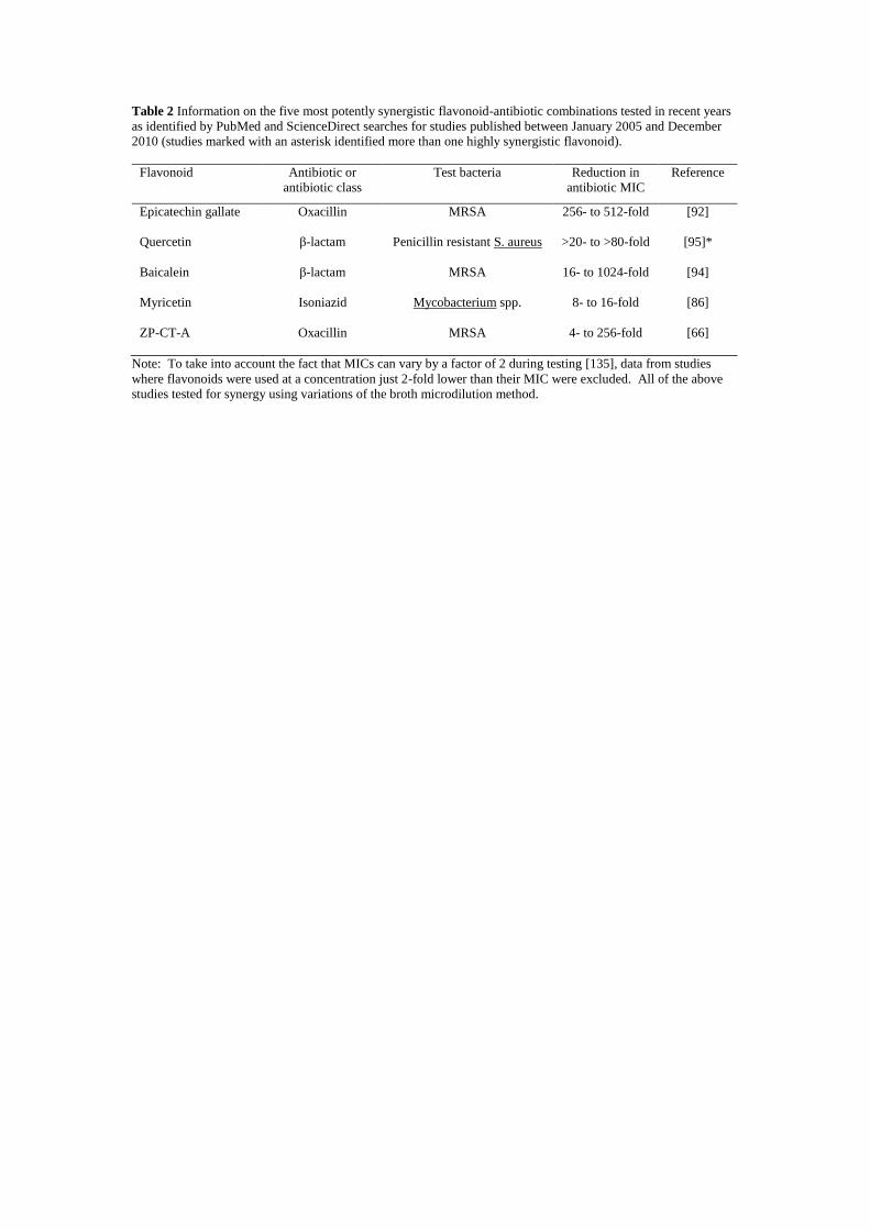
Table 2 Information on the five most potently synergistic flavonoid-antibiotic combinations tested in recent years
as identified by PubMed and ScienceDirect searches for studies published between January 2005 and December
2010 (studies marked with an asterisk identified more than one highly synergistic flavonoid).
Flavonoid Antibiotic or
antibiotic class
Test bacteria Reduction in
antibiotic MIC
Reference
Epicatechin gallate
Quercetin
Baicalein
Myricetin
ZP-CT-A
Oxacillin
β-lactam
β-lactam
Isoniazid
Oxacillin
MRSA
Penicillin resistant S. aureus
MRSA
Mycobacterium spp.
MRSA
256- to 512-fold
>20- to >80-fold
16- to 1024-fold
8- to 16-fold
4- to 256-fold
[92]
[95]*
[94]
[86]
[66]
Note: To take into account the fact that MICs can vary by a factor of 2 during testing [135], data from studies
where flavonoids were used at a concentration just 2-fold lower than their MIC were excluded. All of the above
studies tested for synergy using variations of the broth microdilution method.

Supplementary Table 1 Structures of flavonoids discussed within the review (compiled from individual research papers)
Compound Substituents at carbon position
2 3 4 5 6 7 8 2’ 3’ 4’ 5’ 6’
Flavone:
Baicalein
Chrysin
Flavone
β-naphthoflavone
-
-
-
-
-
-
-
-
-
-
-
-
OH
OH
-
*
OH
-
-
*
OH
OH
-
-
-
-
-
-
-
-
-
-
-
-
-
-
-
-
-
-
-
-
-
-
-
-
-
-
Isoflavone:
Genistein
Isolupalbigenin
Scandenone
-
-
-
-
-
-
-
-
-
OH
OH
OH
-
-
Φ
OH
OH
Φ
-
R1
R1
-
-
-
-
R1
-
OH
OH
OH
-
-
-
-
-
-
Flavonol :
Kaempferol 3-O-α-L-(2’’,4’’-di-E-p-
coumaroyl)-rhamnoside
Galangin
Morin
Myricetin
Quercetin
-
-
-
-
-
R2
OH
OH
OH
OH
-
-
-
-
-
OH
OH
OH
OH
OH
-
-
-
-
-
OH
OH
OH
OH
OH
-
-
-
-
-
-
-
OH
-
-
-
-
-
OH
OH
OH
-
OH
OH
OH
-
-
-
OH
-
-
-
-
-
-
Flavanone:
3’-O-methyldiplacol
Hesperetin
Naringenin
Pinocembrin
Sepicanin A
-
-
-
-
-
OH
-
-
-
-
-
-
-
-
-
OH
OH
OH
OH
OH
R3
-
-
-
R3
OH
OH
OH
OH
OH
-
-
-
-
-
-
-
-
-
OH
OCH3
OH
-
-
-
OH
OCH3
OH
-
OH
-
-
-
-
-
-
-
-
-
-
Flavan-3-ol:
Catechin
Epicatechin
Epicatechin gallate
Epigallocatechin gallate
-
-
-
-
OH
OH
R4
R4
-
-
-
-
OH
OH
OH
OH
-
-
-
-
OH
OH
OH
OH
-
-
-
-
-
-
-
-
OH
OH
OH
OH
OH
OH
OH
OH
-
-
-
OH
-
-
-
-
Flavolan:
ZP-CT-A
-
OH
R6
OH
-
OH
-
-
OH
OH
-
-
Compound
Substituents at carbon position
2 3 4 5 6 α β β' 2’ 3’ 4’ 5’ 6’
Chalcone:
Bartericin A
Isobavachalcone
Licochalcone A
Panduratin A
Sofalcone
-
-
OCH3
-
-
R1
-
-
-
-
OH
OH
OH
-
R9
R7
-
R8
-
-
-
-
-
-
-
-
-
-
Ψ
-
-
-
-
Ψ
-
O
O
O
O
O
OH
OH
-
OH
R10
-
R1
-
-
-
OH
OH
OH
OCH3
R9
-
-
-
-
-
-
-
-
OH
-
-: no substitution; *: benzene ring attached at positions C-5 and C-6; Φ: 2,2-dimethylpyran ring attached at positions C-6 and C-7; Ψ:
1-methyl-2-(3-methyl-2-butenyl)-benzene ring attached at positions α and β; R1: prenyl group; R2: O-α-L-(2’’,4’’-di-E-p-coumaroyl)-
rhamnoside group; R3: geranyl group; R4: gallate group; R5: octanoyl group; R6: 15 epicatechin units with catechin or epicatechin as
the terminal unit; R7: 2-hydroxy-3-methylbut-3-enyl group; R8: 3,3-dimethyl-1-butene group; R9: prenyloxy group; R10:
carboxymethoxy group