recent advances and applications of transgenic animal technology
TRANSCRIPT

13
Recent Advances and Applications of Transgenic Animal Technology
Xiangyang Miao Institute of Animal Sciences, Chinese Academy of Agricultural Sciences
China
1. Introduction
Transgenic animal technology is one of the fastest growing biotechnology areas. It is used to integrate exogenous genes into the animal genome by genetic engineering technology so that these genes can be expressed and inherited by offspring. The transgenic efficiency and precise control of gene expression are the key limiting factors in the production of transgenic animals. A variety of transgenic techniques are available, each of which has its own advantages and disadvantages and needs further study because of unresolved technical and safety issues. Further studies will allow transgenic technology to explore gene function, animal genetic improvement, bioreactors, animal disease models, and organ transplantation. This article reviews the recent developments in animal gene transfer techniques, including microinjection method, sperm vector method, Embryonic stem cell, somatic cell nuclear transplantation method, retroviral vector method, germ line stem cell mediated method to improve efficiency, gene targeting to improve accuracy, RNA interference-mediated gene silencing technology, zinc-finger nucleases–gene targeting technique and induced pluripotent stem cell technology. These new transgenic techniques can provide a better platform to develop transgenic animals for breeding new animal varieties, and promote the development of medical sciences, livestock production, and other fields.
2. Microinjection
In the past 20 years, DNA microinjection has become the most widely applied method for gene transfer in animals. The mouse was the first animal to undergo successful gene transfer using DNA microinjection. This method involves: 1) transfer of a desired gene construct (of a single gene or a combination of genes that are recombined and then cloned) from another member of the same species or from a different species into the pronucleus of a reproductive cell; 2) in vitro culture of the manipulated cells to develop to a specific embryonic phase; and 3) then transfer of the embryonic cells to the recipient female.
Microinjection equipments include microscopes and micromanipulators. Various microscope configurations from upright to inverted styles made by different companies (e.g. Leica, Zeiss, Nikon, Olympus) afford excellent differential interference contrast. Micromanipulator systems are grouped into either air-driven systems (e.g. Nikon, Zeiss, Eppendorf) or oil-driven hydraulic systems. Microinjection needles and slides are also needed during experiment.
www.intechopen.com

Polymerase Chain Reaction
256
Steps for getting transgene production and evaluation from DNA microinjection are: collection of fertilized eggs from superovulated donors, injection of interested genes into male pronuclei, surgical transfer of 20-25 eggs into oviduct of pseudopregnant recipients that carry eggs to term, and PCR or southern blot analyses to detect offspring carrying the transgenes.
Transgenic frequencies obtained from pronuclear microinjection are about 5%-30%. Some factors influence transgenesis production. The studies in mouse done by Brinster et al. (1985) showed that: 1) linear DNA fragments integrated with greater efficiency than circular or supercoiled DNA; 2) transgene DNA should be injected in low amounts othervise it had toxic effects on the embryos; 3) nuclear injection was dramatically more efficient than cytoplasmic injection. In general, good results may be obtained with equipment systems, injector’s experience and skills, and the technique. A major advantage of this method is its applicability to a wide variety of species.
3. Sperm-mediated gene transfer
The finding that mature spermatozoa act as vectors of genetic materials, not only for their own genome, but also for exogenous DNA molecules, has suggested a strategy for animal transgenesis alternative to DNA microinjection. Exploiting this possibility, protocols for sperm-mediated gene transfer (SMGT) have been developed in a variety of animal species with extremely variable results.
In 1989 Lavitrano et al. described a simple and efficient technique, sperm-mediated gene transfer to produce transgenic mice. In this technique, DNA was mixed with sperm cells before in vitro. 30% of offspring mouse were integrated foreign DNA. In subsequent years, however, many successful reports of SMGT have been published.
The basic principle of sperm-mediated gene transfer is quite straightforward: seminal plasma-free sperm cells are suspended in the appropriate medium, and then incubated with DNA. The resultant DNA-carrying sperms are then used to fertilize eggs, via in vitro fertilization or artificial insemination or, in the case of aquatic animals, via waterborne (natural) fertilization. To improve transgenic efficiency, ‘augmentation’ techniques such as electroporation or liposomes to ‘force’ sperm to capture transgenes were used in some studies.
Here we briefly introduced methods of SMGT. 1) Sperm cells directly incubated with exogenous DNA. Lavitrano et al. (1989) first incubated mouse sperms with DNA to integrate foreign genes into the germ cells and produce transgenic mice. But as low efficiency of the method, now researchers use this method in combination with other methods of sperm carrier techniques to obtain transgenes. 2) Transfection of DNA into the sperm cells mediated by liposomes. These cationic lipids interact with the negatively charged nucleic acid molecules forming complexes that the nucleic acid is coated by the lipids. The positive outer surface of the complex can then associate with the negatively charged cell membrane, allowing the internalization of the nucleic acid. 3) Electroporation-mediated import of foreign DNA into sperms. High voltage electric field can induce a temporary reversible membrane permeability changes, which allowes the foreign DNA into the cells more easily. Studies showed that this method can improve the transgenic efficiency up to 22% of the embryos in pigs, cattle, chickens and other animals. However, sperm cells underwent electroporation have two aspects. On one hand, some channels temporarily
www.intechopen.com

Recent Advances and Applications of Transgenic Animal Technology
257
opened on cell membranes are conducive to the entry of foreign gene; on the other hand, the shock is also an injury to cells that causes irreversible damage to sperm motility. 4) Adenoviral vector mediated gene transfer. Farre et al. (1999) tested the ability of adenoviral vectors to transfer DNA into boar spermatozoa and to offspring. Exposure of spermatozoa to adenovirus bearing the E. coli lacZ gene resulted in the transfer of the gene to the head of the spermatozoa. Of the 2-to 8-cell embryos obtained after in vitro fertilization with adenovirus-exposed sperm, 21.7% expressed the LacZ product. Four out of 56 piglets (about 7%) obtained foreign gene in PCR analyses after artificial insemination with adenovirus-exposed spermatozoa. SMGT is based on the intrinsic ability of sperm cells to bind and internalise exogenous DNA and to transfer it into the egg at fertilisation as illustrated in Fig. 1.
Fig. 1. Sperm-mediated gene transfer in the pig.
Mechanisms of nucleic acid uptaken by sperm are not known very well. It is suggested that the interaction of exogenous molecules may trigger an endogenous reverse transcriptase (RT) activity in spermatozoa. Such RT activity is able to reverse transcribe exogenous RNA molecules (specifically, the human poliovirus RNA genome was used in the study that provided the first set of evidence) into cDNA, which are transferred to embryos following in vitro fertiliszation (Giordano et al., 2000). That finding suggested for the first time that a sperm endogenous RT is implicated in the generation of newly reverse-transcribed sequences and, more generally, established the notion that the retrotransposon/ retroviral machinery is involved in SMGT.
Now it is well established that spermatozoa can play a role in transgenesis in all species. Their ability to take up exogenous DNA molecules can be exploited to transmit novel genetic information to the offspring after fertilization. This potential is highlighted by the recent development of SMGT variant protocols.
4. Embryonic stem cell-mediated gene transfer
This method involves isolation of totipotent stem cells, which are undifferentiated cells that have the potential to differentiate into any type of cells (somatic and germ cells) and
www.intechopen.com
Polymerase Chain Reaction
258
therefore to give rise to a complete organism. Then the desired DNA sequences are inserts into the genome of embryonic stem (ES) cells cultured in vitro by homologous recombination. The cells containing the desired DNA are incorporated into the host’s embryo, resulting in a chimeric animal. This technique is of particular importance for the study of the genetic control of developmental processes and works well in mice.
It has the advantage of allowing precise targeting of defined mutations in the gene via homologous recombination. Based on the resultant function of the targeted gene, gene targeting methods have opened two lines of investigation: the gene knock-out (KO) to disrupt the existing gene, and the gene knock-in (KI) to insert a functional new gene.
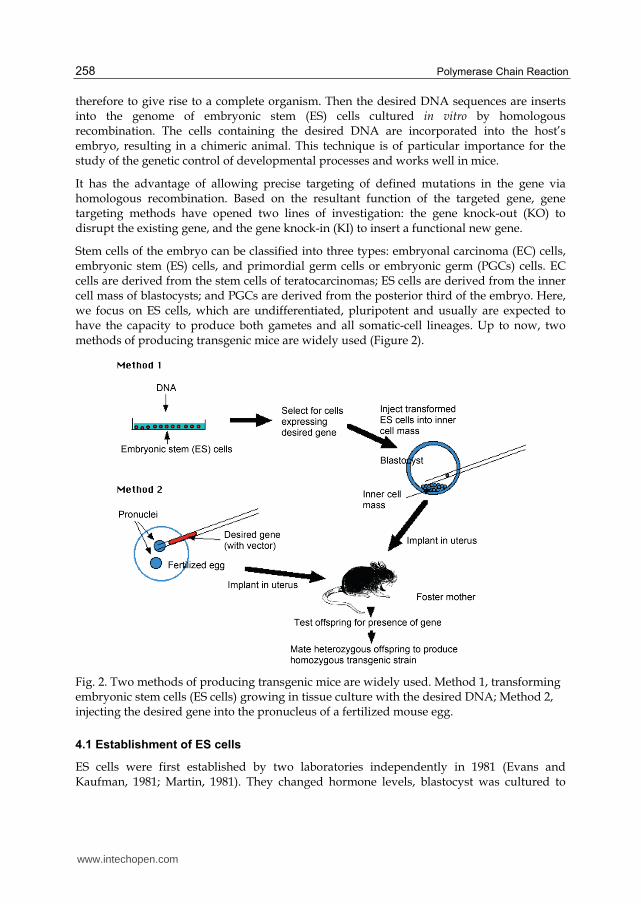
Stem cells of the embryo can be classified into three types: embryonal carcinoma (EC) cells, embryonic stem (ES) cells, and primordial germ cells or embryonic germ (PGCs) cells. EC cells are derived from the stem cells of teratocarcinomas; ES cells are derived from the inner cell mass of blastocysts; and PGCs are derived from the posterior third of the embryo. Here, we focus on ES cells, which are undifferentiated, pluripotent and usually are expected to have the capacity to produce both gametes and all somatic-cell lineages. Up to now, two methods of producing transgenic mice are widely used (Figure 2).
Fig. 2. Two methods of producing transgenic mice are widely used. Method 1, transforming embryonic stem cells (ES cells) growing in tissue culture with the desired DNA; Method 2, injecting the desired gene into the pronucleus of a fertilized mouse egg.
4.1 Establishment of ES cells
ES cells were first established by two laboratories independently in 1981 (Evans and Kaufman, 1981; Martin, 1981). They changed hormone levels, blastocyst was cultured to
www.intechopen.com

Recent Advances and Applications of Transgenic Animal Technology
259
obtain delay in its development. 4 to 6-day blastocyst inner cell mass (ICM) was separated, then co-cultured on the mitomycin C-treated Unlimited lines of fibroblasts (STO) feeder layer (Feeder). After cell proliferation and inhibition of differentiation of passage, the first mouse undifferentiated ES cell lines were established. It was shown that mouse ES cells by blastocyst injection could widely induce variety of organizations involved in the formation of chimeric animals at rate of 61%.
Currently, mouse fibroblasts unlimited line (STO) or mouse embryonic fibroblasts (Mouse Embryonic Fibroblast, namely MEF) was widely used to prepare the feeder layer. These cells can secrete fibroblast growth factor (FGF), differentiation inhibitory factor (DIA), white leukemia inhibitory factor (LIF) and other substances. They also promote the growth and colonization of ES cells and suppress their differentiation. The STO or MEF is treated with mitomycin vitamin C or other mitotic inhibitor, early embryos or PGCs is cultured on the feeder layer and ES cells can be obtained. Rat liver cell conditioned medium (buffalo liver conditioned medium) is another wildly used differentiation inhibiting medium. In addition, sheep oviduct epithelial (oTE), goat oviduct epithelial (cTE), sheep uterine epithelium (oUE), goat uterine epithelium (cUE), bovine granulosa cells (bG), bovine uterine fibroblasts (bUF) and fetal bovine testes, kidney and liver fibroblasts (fbTF, fbKF, fbLF), etc., are also used as feeder layers in laboratories.
Several conditions must be met for isolating ES cells. 1) Present cells in culture must be undifferentiated and pluripotent. 2) The pluripotent cells must be deprived of differentiation signals in culture. 3) The cells must be stimulated, or at least be allowed, to proliferate.
4.2 Characteristics of ES cells
ES cells are small, aggregated and unpolarized cells forming islands on the feeder layers and have large nucleoli and a high nucleo-cytoplasmic rate. Cell markers have been used to characterize undifferentiated or differentiated ES cells. Alkaline phosphatase is equivalent to the cell surface nonspecific alkaline phosphatase of the inner cell mass of the mouse blastocyst. Other markers are surface glycolipids (ECMA-7), embryoglycans (SSEA-1) and so on.
As gene expression characteristics of ES cells, they express all the “house-keeping” genes involved in the machinery of cell cycling and some receptors to factors which allow them escaping cycling and differentiating.
4.3 ES cells for transgenesis
ES cells are highly efficient materials for animal cloning. With the development of chimeric nuclear transfer technology and production technology, ES cells are widely used in animal cloning. Proliferation of ES cells derived from donor as the nucleus, which in chimeric germline, produce following generations by sperm or egg cell proliferation.
Introduction of foreign DNA by electroporation into ES cells is very efficient. The DNA integrates into genome at a frequency of -10-3, then numerous markers are used for efficient selection. The gene-modified cell clones are introduced back into preimplantation stage embryos, either by blastocyst injection or by morula aggregation, to produce chimeras.
www.intechopen.com

Polymerase Chain Reaction
260
There are two main methods of embryonic stem cell-mediated gene transfer: one is gene trap, another is gene targeting. We will discuss in the following section.
5. Somatic cell nuclear transfer
ESCs are totipotent in development, capable of limitless passage and proliferation, and have become the ideal cells for gene targeting. However, in many species, especially the large agricultural animals, ESCs have not been successfully isolated or cultured. For these animals, somatic cells are easily obtained in large numbers and can be cultured in vitro.
Somatic cell nuclear transfer (SCNT) is a technique for cloning. The nucleus is removed from a healthy egg. The enucleated egg becomes the host for a nucleus that is transplanted from another cell, such as a skin cell. The resulting embryo can be used to generate embryonic stem cells with a genetic match to the nucleus donor (therapeutic cloning), or can be implanted into a surrogate mother to create a cloned individual, such as Dolly the sheep (reproductive cloning) (Figure 3).
Fig. 3. Diagram of the nuclear transfer procedure that produced the dolly sheep.
www.intechopen.com

Recent Advances and Applications of Transgenic Animal Technology
261
Cloning by nuclear transfer from adult somatic cells is a remarkable demonstration of developmental plasticity. When a nucleus is placed in oocyte cytoplasm, the changes in chromatin structure that govern differentiation can be reversed, and the changed nucleus can control the development of oocyte to term.
Dolly was cloned by SCNT with a nucleus from a cultured mammary gland cell. In the
Edinburgh experiment, three types of cells were used as karyoplasts: (i) mammary
epithelium, (ii) fetal fibroblasts, and (iii) embryo-derived cells. The efficiencies of fusion
were relatively high for all three cell types, which were 63.8%, 84.7% and 82.8% respectively.
5.1 The main methods of nuclear transfer
According to the different donor cells, somatic cell nuclear transfer divided into early
embryo nuclear transplantation, nuclear transfer and differentiated embryonic stem cell
nuclear transfer. From the present point of view, there are two somatic cell nuclear transfer
technology: one is Roslin technology, another is Honolulu technique. Compared with
previous techniques, the major breakthrough of Roslin method is the use of blood starvation
method, which enables the proliferation of cultured cells temporarily in the G0 phase week.
To ensure that the donor nucleus and development of the cytoplasm of recipient cells
synchronized, electric pulse method was used. The fusion and activation of donor nucleus
and enucleated oocyte were triggered by oocyte.
In Honolulu technique, a slight modification was made in the direct use of G0 phase or G1
phase of somatic cell nuclear as donor to avoid serum starvation. Donor nucleus was
transferred into the oocyte cytoplasm and stayed for some time (6h). Then the oocyte was
activated and stimulated to proliferation with strontium ions (chemical activation). More
details of nuclear transplantation technology include the following steps:
5.1.1 Choose of recipient cells and the nuclear removing
MII oocytes are the most use of nuclear transplantation donor, but different laboratories
used different activation time: before the activation of nuclear transfer, nuclear transfer
when activated, delayed activation of nuclear transfer, and so on. No matter which methods
using the blastomeres of early embryos, ES cells or somatic cell, nuclear transfer ported to
this type of recipient, all get the offspring.
Nuclear removing from oocytes are mainly done by mechanical and chemical methods.
Earlier mechanical method is the use of glass needle under the microscope, including the
blind absorption and fluorescence dye staining under UV to remove nuclear. In 1998,
Wakayama et al. first applied piezoelectric microinjection (PEM) to remove nuclear. Because
of its high frequency of vibration control, more easily through the zona pellucida, and the
small damage to the fertilized egg, the success rate to get transgenes was greatly improved.
Compared with the mechanical method, chemical method is much simpler and easier to
remove nuclear. In the early time, researchers mainly used early etopside (ethylene grapes
pyran sugar) and cycloheximide treatment. However, the success rate was low. Recently
Gasparrini et al. (2003) chose the decarboxylation youthful acid base to successfully remove
the nucleus from the mouse oocyte. But whether this method is applicable to other species
needs to further experiments.
www.intechopen.com

Polymerase Chain Reaction
262
5.1.2 The choice of donor cells and transplantation
Currently cells mainly used in the nuclear donor are: cumulus cells, testicular pillar cells, sperm cells, brain cells, fetal or adult fibroblasts, breast cells, embryonic stem cells (ES cells) and so on. The synchronization of donor and recipient cell cycle is an important factor affecting the development of nuclear transfer. Early studies suggest that donor cells in the G0 is essential to the success of somatic cell nuclear transplantation, and that cells in the G0 phase can be selected or artificially induced. However, the recent discovery indicated that the nuclear transfer from donor cells in G1 or G2 or M phase was also a success way. Meanwhile, the study also found that in mice, somatic cell nuclear transfer had a greater efficiency of transplantation.
There are two ways to transplant donor nuclei: fusion and injection. Fusion includes the chemical fusion, sendai virus mediated fusion and electro-fusion. And injection includes glass needle injection and piezoelectric microinjection.
5.1.3 Oocyte activation
Mature oocytes as recipients are lack of nuclear migration and the fertilization, so they must be manually activated to promote their further development. The activation methods are currently mostly used with electrical activation, ethanol, ionomycin, calcium ionophore A23187, chlorine strontium, 3-phosphatidylinositol (IP3), sperm extract and so on. These methods are often used in combination or with protein synthesis inhibitors (actinomycetes ketone, puromycin), serine threonine protein kinase inhibitor DMAP. However, studies have found that all of these methods can only lead to increased concentration of intracellular calcium in oocytes, and calcium can not form vibration, which might be the reason of low efficiency of nuclear transfer.
5.2 Application of SCNT
This technology will be helpful in understanding the most important issues such as nuclear matter interaction, the nucleus division and reprogramming, changes in mitochondria of reconstructed embryos, cell aging. Nuclear transfer technology also provides a powerful tool to wildlife, endangered species’ protection.
In agricultural area, this technology will further enrich the quality of breedings. Nuclear transfer technology can be used to accelerate the breeding process and expand population within the effective number in a short time. Both nuclear transfer and gene targeting can be used to modify target genes and produce new varieties with superior traits (such as increased fecundity, increased milk yield, strengthen resistance to disease, etc.).
In the medical field, SCNT was used to clone tissues and organs for patient transplant, such as the treatment of Parkinson's disease, etc. Meanwhile, the combination of gene targeting and nuclear transfer technology established various animal models of human disease for medical and pharmaceutical research.
But lots of questions of nuclear transplantation in mammals are still not well solved, including cytoplast aging, cytoplast cell-cycle stage, activation procedure, source of karyoplasts and its differentiation, karyoplast cell cycle stage, serial transfer, karyoplast: cytoplast (nucleocytoplasmic interactions), species specific differences. Instead, we have
www.intechopen.com

Recent Advances and Applications of Transgenic Animal Technology
263
aimed at highlighting some of the unanswered questions relating to somatic cell cloning that will require resolution before the procedure becomes useful for practical purposes.
6. Retrovirus-mediated gene transfer
A retrovirus is a virus that carries its genetic material in the form of RNA rather than DNA. In this method, retroviruses are used as vectors to transfer genetic material into the host cell, resulting in a chimera, an organism consisting of tissues or parts of diverse genetic constitution. Chimeras are inbred for as many as 20 generations until homozygous (carrying the desired transgene in every cell) transgenic offspring are born.
Retrovirus-mediated expression cloning was developed in mid 1990s. The most important features of retrovirus as vectors are the technical ease and effectiveness of gene transfer and target cells specificity. When cells are infected by retroviruses, the resultant viral DNA, after reverse transcription and integration, becomes a part of the host cell genome to be maintained for the life of the host cell (Ponder, 2001). It is also reported that DNase hypersensitive regions are the preferred targets for retrovirus integration. Unlike DNA microinjection, integration of a viral gene does not seem to induce rearrangements of the host genome, except for a short duplication at the site of integration (Jaenisch, 1988).
6.1 Retrovirus biology and Retroviral vector design
Retroviruses are animal viruses contain two positive-strand RNA genomes. The word “retro” means, when the virus vectors infect a host cell, the viral RNA is reverse transcribed in the cytoplasm making linear double-stranded DNA. This dsDNA then is transported into host cell mucleus and integrates into a chromosome directly with no change of its original linear form.
The retrovirus genome can be divided into trans- and cis- acting sequences. Trans-acting protein-coding genes are gas, pol and env. The gas gene encodes the structural components of the virus. The pol gene encodes the RNA-dependent DNA polymerase (reverse-transcriptase), integrase for the integration of reverse-transcribed viral DNA into the host cell chromosome, and the protease for posttranslational cleavage of viral proteins. The env gene encodes the surface envelope glycoproteins.
Cis region are located at the 5’ and 3’ ends of the genome. 1) The long terminal repeat (LTR) contains transcription and integration signals. 2) Primer binding site and polypurine sequence are for reverse transcription. 3) Posttranscriptional splicing sites include splicing donor and acceptor sites along with two short fragments within the viral intron for env mRNA production. 4) E signal is for encapsidation of murine leukemia virus (MLV) and reticuloendotheliosis virus (REV), respectively.
The viral vector based on Moloney murine leukemia virus (Mo-MLV) is the most widely used retroviral vector systems. Mo-MLV retroviral vector system is constructed based on the Mo-MLV genes for packaging, reverse transcription and integration of the required cis-acting elements and trans-acting protein coding sequence of separation, respectively, as well as recombinant retroviral vector elements and packaging cell line. In the molecule level of recombinant retroviral vector, the foreign gene replace the original virus structural protein coding region, but essential components, the virus replication, transcription and packaging
www.intechopen.com

Polymerase Chain Reaction
264
sequences are preserved. Retrovirus structural protein coding genes are provided in trans from the packaging cells. Target cells are infected with this virus particles, so that the target gene stably integrated in the genome or chromosomes of target cells, in order to achieve the transfer of foreign genes.
6.2 Packing cell lines
Packing cells are designed to synthetize all retroviral proteins necessary for the production infectious particles. The purpose of a packing cell is to provide Gag, Pol, and Env protein to the retroviral vectors having no trans-acting sequences. NIH3T3 cells transformed with appropriate MLV genes are the most popular system for gene transfer in mammalian cells.
In earlier packaging cell construction, NIH3T3 cells were transfected with the gag, pol, and env genes of MLV in a single transcriptional unit, causing production of replication-competent helper virus. In the later work, decreased the possibility of homologous recombination were made. For instance, in ampli-GPE packaging cells, the 5’ LTR promoter replaced with mouse metallothionein promoter in controlling of the gag, pol, and env genes. Then the PG13 packing cell line, the BOSC23 packing cell line, and the 293GP/VSV-G packing cell line, have the advantage of low replication competent virus production, high DNA transfection efficiency and wide host cell ranges.
Retroviral vectors have been mainly used in somatic transgenesis for gene expression studies by using reporter genes, cell lineage, and for antisense sequences inhibition of gene expression in specific cell types.
6.3 Problems of retroviral vectors in gene transfer
The maximum size for reverse transcription of each vector is about 10kb, which may affect the expression level in transgenic animals. Another problem is the recombination, which is production of replication competent retrovirus from virus-producing cells. So nowdays, reducing the homologous sequences between DNAs for packing cells and vector and by using different plasmids to separate different genes gets over this problem.
7. Germ line stem cell technique
7.1 Spermatogonial stem cell technique
Spermatogonial stem cells (SSCs) are a population of cells in mammalian testes that have high potential to self-renew and differentiate similarly to embryonic stem cells. SSCs transplantation is a recently developed animal reproduction technique that involves the injection of in vitro cultured spermatogonial stem cells from an age-matched male donor into the seminiferous tubule of age-matched host animal to produce germ cells. During the in vitro culture of spermatogonial stem cells, positive spermatogonial stem cells that will be transferred can be screened and, thus, the transgenic efficacy can be significantly enhanced.
7.1.1 Origin of spermatogonial stem cells
Spermatogonial stem cells derive from the birth of the original sex cells, the original sex cells are from primordial germ cells (primordial germ cells, PGCs). PGCs are generated in early
www.intechopen.com

Recent Advances and Applications of Transgenic Animal Technology
265
embryo development in mammals, processing in earlier period independent of other cell lines on a small group of cells. They have been integrated into the base from the yolk sac formed after the intestine. After along intestinal active migration, and migration on the way to proliferate in pregnancy 10.5 d, PGCS eventually arrives at the genital ridge to form a gonad. PGCs then differentiate and form germ cell precursor cells known as the the original sex cells. In the male animals, the original cells of strong mitotic activity are not stationary for a long time, until the animal was born. In mice, spermatogonial stem cells appear in the course of 6 d after birth, the earliest spermatogonial stem cells appeared probably after birth 3-4 d. The specific time for the original cell into spermatogonial stem cells in other species is unclear. Livestock may take a few months, and spiritual may take about a few years. Some studies have shown that there are two types of primary cells in neonatal mouse testis, one directly differentiates into spermatogonia and completes the first form of spermatogenesis, and another form of spermatogonial stem cells, in the later time provides the basis for spermatogenesis.
7.1.2 Spermatogonial stem cell proliferation and differentiation
Spermatogonial stem cells are located in the testis seminiferous tubule basement membrane, and are round, less cytoplasm. Their nucleus were round or slightly oval, often dominant with chromatin, little heterochromatin. They have abundant of cytoplasmic ribosomal cores, mitochondria. Based on cell arrangement characteristics, mice type A spermatogonia can be divided into three types: A single spermatogonia (As); A paired spermatogonia (Apr); and 8, 16 or 32 cells of A aligned spermatogonia (Aal). As having stem cell activity, As, Apr, Aal are referred as undifferentiated spermatogonia. After division, daughter cells of As separate from each other as two new As stem cells, or due to incomplete cytokinesis, two daughter cells form Apr by cytoplasmic bridges between connected to each other. Under normal circumstances, about half of the As cell divides and forms Apr cells, while the other half through the proliferation renews themselves and keeps the number of stem cells. Apr cells, by four further division, form 8, 16 or 32 cells Aal-based original cells, which then divide into the Al type A spermatogonia. After over six consecutive division, Al type A spermatogonia differentiate into type A2 spermatogonia, which in turn gradually differentiate from the A2 → A3 → A4 → In → B, and finally into B Type A spermatogonia. Proliferation and differentiation of stem cells generally are subject to balance to their microenvironment (niche) of the regulation. Current study suggests that, spermatogonial stem cell micro-environment is seminiferous tubules and interstitial blood vessels around the area. As /Apr / Aal tends to distribution there, and will move out of this area when they differentiate into A1 spermatogonial cells.
GDNF from supporting (Sertoli) cells is one of the most important cytokines regulating spermatogonial stem cell proliferation. Studies have shown that over-expression GDNF expression in mice leads to accumulate undifferentiated spermatogonia. When the GDNF-/+ mouse spermatogonial stem cells is exhausted, the process of spermatogenesis is damaged. More importantly, GDNF has become a necessary factor for culturing spermatogonial stem cells. GDNF binds to GFRA1, induces activation of RET, and then recruits other molecules to the RET intracellular domain. The recent study showed that BCL6 and Etv5 genes were regulated by GDNF. BCL6 and Etv5 knockout mice are showing spermatogenesis and sertoli cell degeneration syndrome phenotype. In addition, genetic models in mice also observed that two non GDNF regulated genes: Plzf and Taf4b, play an important role in maintaining
www.intechopen.com
Polymerase Chain Reaction
266
the proliferation of spermatogonial stem cells in body. Plzf knockout mice with aging, accompanied by the original cell degeneration, and gradually lose the structure of the seminiferous tubule. And Plzf - / - mouse spermatogonial cell transplantation can not be re-formed seminiferous epithelium. Taf4b knockout mice become sterile at 3 months, also appear cell syndrome phenotype.
There are three important regulatory points in the differentiation of spermatogonial cells: 1) the change between As and Apr; 2) Aal changes to the A1 and A1 to B spermatogonial cell transformation; 3) Apr spermatogonia without completing cytokinesis. The cytoplasmic bridge connecting two daughter cells is considered to be first visible sign of spermatogonia differentiation. However, little is known about how to control differentiation of spermatogonia process. Current studies showed that Vitamin A (RA), c-Kit and other genes in the germ cell differentiation play an important role. If RA defects, only undifferentiated testicular spermatogonia exist, when RA renew, Aal spermatogonia after block of re-entered the cell cycle differentiate into A1 spermatogonia. RA can induce cultured undifferentiated spermatogonia to express large amount of Stra8 and c-Kit, and these two genes are considered as the molecular markers for spermatogonial stem cells starting to differentiate. C-kit oncogene is the original W locus, encoding the tyrosine kinase receptor and its ligand is stem cell factor (SCF). C-kit point mutation in male mice results in the initial stage of spermatogenesis DNA synthesis disorder, no DNA synthesis in the process of Aal to A1 differentiation, and complete infertility.
7.1.3 Pluripotency of spermatogonial stem cell
It has long been considered a single spermatogonial stem cells can only differentiate into
sperm-specific manner. But in recent years, studies have shown that the original stem cells
in vitro can be induced to become pluripotent stem cells. In 2003, Kanatsu-Shinohara first
began research in this area and found that spermatogonial stem cells isolated from newborn
mouse could produce embryonic-like stem cells (embryonic stem cell-like, ES-like) cells
when cultured in vitro. These ES-like cells further cultured in vitro produced ES-like clones,
which formed teratomas when injected into mice. Subsequently a number of scientific
researchers discovered that adult mouse spermatogonial stem cells ccould be induced into
ES-like pluripotent stem cells named multipotent adult germline stem cells (maGSCs). Ko et
al. (2009) recently made an outstanding contribution by first proving that maGSCs cells was
indeed changing from spermatogonial stem cells, and differentiated and formed the three
germ layers both in vitro and in vivo, and the reproductive system could transfer to the next
generation. Other study also showed that spermatogonial stem cells were very malleable
and could direct transdifferentiation into other cell types. In 2007, Boulang et al. tried
spermatogonial stem cell transplantation to the breast, and achieved the breast epithelium in
vivo. In 2009, U.S. scientists fusioned spermatogonial stem cells and appropriate cells, and
then grafted the hybrids into the body. Results showed that spermatogonial stem cells could
differentiate directly into the prostate epithelium, uterine epithelium and skin epithelium in
newborn mouse. Although the molecular mechanisms of the transition of spermatogonial
stem cells to pluripotent cells are not clear, scientists successfully induced spermatogonial
stem cells into pluripotent stem cells in the adult testis, which suggested that
spermatogonial stem cells will become an important source of pluripotent stem cells in
medical field.
www.intechopen.com

Recent Advances and Applications of Transgenic Animal Technology
267
7.2 Primordial germ cell technique
Primordial germ cells (PGCs) refer to the ancestral cells that can develop into sperm or ovum cells. PGCs reside in the recipient’s gonads, migrate and proliferate in the recipient embryonic gonads. Because PGCs at different stages of development can serve as transgenic recipient cells (Honaramooz etal., 2011), transgenic studies using PGCs as vectors are simple, highly effective and will likely gain favor for the production of transgenic animals. Development of Mouse Embryonic Primordial Germ Cells was shown in Figure 4.
Fig. 4. Development of Mouse Embryonic Primordial Germ Cells.
7.2.1 Origin and characteristics of PGCs
The PGCs originate in the epiblast of the stage X blastoderm. The fusion of an egg and a sperm, at fertilization gives rise to a zygote that is totipotent and capable of giving rise to all embryonic lineages. In the early embryo, PGCs can be distinguished from the somatic cells at 7 days post coitus (d.p.c), because they express an alkaline phosphatase izozyme, tissue non-specific alkaline phosphatase (TNAP).
PGCs are ordinarily identified through morphological characteristics, e.g. large size (12-20um in diameter), large eccentrically placed nuclei with prominent, often fragmented nucleoli. Also some histochemical markers such as periodic acid-schiff (PAS), which stains for glycogen, or immunohistochemical markers such as EMA-1 and SSEA-1 (stage-specific embryonic antigen 1), which recognize cell-surface carbohydrate epitopes, are used for identifing PGCs.
Recent studies demonstrated that the normal progression of the germ cell lineage during gonadogenesis involved a delicate balance of primordial germ cell survival and death
www.intechopen.com
Polymerase Chain Reaction
268
factors generated by surrounding somatic cells. This balance operates in a different fashion in females and males. The tuning primordial germ cell specification in the wall of the yolk sac, migration through the hindgut and dorsal mesentery, and colonization in the urogenital ridges involves the temporal and spatial activation of the following signaling pathways. Primordial germ cell specification involves bone morphogenetic proteins 2, 4 and 8b, and their migration is facilitated by the c-kit receptor-ligand duet. When colonization occurs: (1) neuregulin-b ligand is expressed and binds to an ErbB2-ErbB3 receptor tyrosine kinase heterodimer on primordial germ cells; (2) Vasa, an ortholog of the Drosophila gene vasa, a member of an ATP-dependent RNA helicase of the DEAD (Asp-Glu-Ala-Asp)-box family protein is also expressed by primordial germ cells; (3) Bcl-x (cell survival factor) and Bax (cell death factor) join forces to modulate the first burst of primordial germ cell apoptosis; (4) Cadherins, integrins, and disintegrins bring together primordial germ cells and somatic cells to organize testis and ovary. Information on other inducers of primordial cell survival, such as teratoma (TER) factor, is beginning to emerge.
7.2.2 Transgenesis using PGCs
Several methods of inserting DNA into PGCs are available. Mueller et al. (1999) isolated PGCs from pig fetuses, generated hemizygous transgenic cells for a human growth hormone (hGH) gene, and obtained chimeric pigs following blastocyst injection of transgenic porcine PGCs. Van de Lavoir et al. (2006) targeted chicken PGCs with a GFP gene construct, and transplanted the targeted cells into primordial gonads of stage XIII–XV chicken embryos that had been incubated for 3 days. A total of eight male chicks were obtained. Once fully matured, seven sired transgenic chicks carried and expressed the foreign GFP gene. These results indicated that it is feasible to use transgenic or gene targeted PGCs to produce transgenic animals, even in large animals. In early study, only retroviral infection of germinal crescent PGCs had been successfully produced transgenic chicken. DNA complexed with liposomes provided a convenient method both in situ and in vitro. Recently, electroporation of germinal crescent or blood PGCs or gonagal tissues was used as transfection method.
8. Gene targeting
Gene targeting is a technology to specifically modify a particular gene in the chromosome through homologous recombination and the integration of extrinsic gene into the specific target site. Gene targeting technology overcomes random integration events and, therefore, is ideal for the modification and reconstruction of biological genetic materials.
The advent of pronuclear injection, ES cells, and gene knockout technology led to the generation of mice harboring gain-of-function or loss-of-function mutations.
The integrase family consisted of 28 proteins from bacteria, phage, and yeast that have a common invariant His-Arg-Tyr triad. These proteins have the function of DNA recognition, synapsis, cleavage strand exchange, and relegation. There are four wildly used site-specific recombination system in eukaryotic applications: 1) Cre-loxP from bacteriophage P1, 2) FLP-FRT from plasmid of Saccharomyces cerevisiae, 3) R-RS from Zygosaccharomyces rouxii, 4) Gin-Gix from bacteriophage Mu. The Cre-loxP and FLP-FRT systems have been developed as wildly applied tools in Drosophila and mouse genetics.
www.intechopen.com
Recent Advances and Applications of Transgenic Animal Technology
269
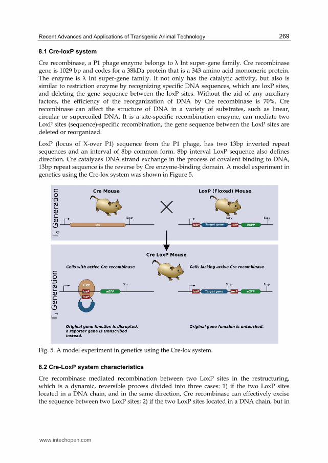
8.1 Cre-loxP system
Cre recombinase, a P1 phage enzyme belongs to λ Int super-gene family. Cre recombinase gene is 1029 bp and codes for a 38kDa protein that is a 343 amino acid monomeric protein. The enzyme is λ Int super-gene family. It not only has the catalytic activity, but also is similar to restriction enzyme by recognizing specific DNA sequences, which are loxP sites, and deleting the gene sequence between the loxP sites. Without the aid of any auxiliary factors, the efficiency of the reorganization of DNA by Cre recombinase is 70%. Cre recombinase can affect the structure of DNA in a variety of substrates, such as linear, circular or supercoiled DNA. It is a site-specific recombination enzyme, can mediate two LoxP sites (sequence)-specific recombination, the gene sequence between the LoxP sites are deleted or reorganized.
LoxP (locus of X-over P1) sequence from the P1 phage, has two 13bp inverted repeat sequences and an interval of 8bp common form. 8bp interval LoxP sequence also defines direction. Cre catalyzes DNA strand exchange in the process of covalent binding to DNA, 13bp repeat sequence is the reverse by Cre enzyme-binding domain. A model experiment in genetics using the Cre-lox system was shown in Figure 5.
Fig. 5. A model experiment in genetics using the Cre-lox system.
8.2 Cre-LoxP system characteristics
Cre recombinase mediated recombination between two LoxP sites in the restructuring, which is a dynamic, reversible process divided into three cases: 1) if the two LoxP sites located in a DNA chain, and in the same direction, Cre recombinase can effectively excise the sequence between two LoxP sites; 2) if the two LoxP sites located in a DNA chain, but in
www.intechopen.com
Polymerase Chain Reaction
270
the opposite direction, Cre recombinase can lead to two LoxP sites between the sequence inversion; 3) if the two LoxP sites are located in two different strands of DNA or chromosomes, Cre enzyme can mediate the exchange of two strands of DNA or chromosomal translocations. In addition, Cre/LoxP can not only recognize the two 13bp inverted repeat sequences and 8bp interval region, but also a 13bp inverted repeat sequence of the interval or 8bp able when changes occurred and recombined. Using this feature, people can build a LoxP site carrier as needed transformation sequences for specific gene mutations or repair, to increase the scope of application of the system.
8.3 Cre-LoxP and transgenic models
The Cre / LoxP system is wildly used in transgenic technologies in a loss-of-function model.
The use of Cre / LoxP system to achieve knockout a particular gene in vivo under certain conditions needs two transgenic mice. The first mice commonly are obtained with embryonic stem cell technology. In both ends of each gene locus contains a loxP sequence, then this sequence inserts into embryonic stem cells by homologous recombination, replaced the original genome sequence. After this treatment, the embryonic stem cells are re-implanted into pseudo-pregnant mouse uterus, to re-develop into a complete embryo, eventually becoming a transgenic mouse. In this transgenic mouse, loxP sites are introduced into the corresponding gene’s intron, which does not affect the function of the corresponding gene, so under normal circumstances, the phenotype of mice is normal. Second mice are obtained by using transgenic mice injected oocytes or embryonic stem cell technology. In this mice, Cre recombinase is placed in a particular gene under the regulation, making its expression in a particular condition. Finally, these two mice were mated and produced at the same time. With the offspring of these mice, two genotypes in a particular type of cells results in the absence of a specific gene.
Obviously, in different cells or organs a specific gene knockout depends on the chosen promoter. Selecting the appropriate promoter to control the expression of Cre recombinase in specific parts of the organism under certain conditions, can be achieved under the conditions corresponding to a specific gene knockout. So far, several different promoters under different conditions are successfully used to achieve gene knockout. These promoters can be cell type-specific, such as lck promoter (thymocytes), alphaA crystallin promoter (eye lens), calmodulin-dependent kinase II promoter (hippocampus and neocortex), whey acidic protein promoter (mammary gland), aP2 promoter (adipose tissue), AQP2 promoter (kidney collecting duct) and sarcoplasmic protein promoter (skeletal muscle), etc. Promoter can also be subject to certain exogenous chemicals regulation such as interferon response Mxl promoter, Mosey phenol-dependent mutant estrogen promoter and tetracycline regulation system. Regulation of exogenous gene knockout can be avoided in the early embryo because the abnormal gene function may have side-effects.
9. Gene silencing mediated by RNA interference
RNA interference (RNAi) is the silencing of specific gene expression mediated by the formation of double-stranded RNA that results in the inhibition of gene expression by degrading mRNA. Therefore, RNAi can achieve the spatiotemporality and reversibility of gene expression modulation.
www.intechopen.com

Recent Advances and Applications of Transgenic Animal Technology
271
Gene silencing has been found to be an important methods of regulating gene expression. In gene transfer studies, it is easier to insert a foreign gene into an animal genome than to remove an existing gene, unless by a knock-out technique. Gene knock-out is complex, difficult and irreversible, once the gene knocked out, it cannot be recovered. The development of RNA interference (RNAi) technology, through blocking of gene expression or cleavage of the expressed mRNA, allows specific, partial and reversible knock-down of the desired gene, and can also achieve spatio-temporal regulation of gene expression.
The introduction of 21 nt small RNA, which is fully or partially complementary to an endogenous gene, into animal cells or tissues will interfere with the expression of the endogenous gene, or trigger the cleavage of the expressed mRNA. Consequently, RNAi impairs gene function or alters an animal trait. For example, Acosta et al. (2005) microinjected myostatin small interference RNA (siRNA) into fertilized eggs of zebra fish, which resulted in virtually eliminating myostatin mRNA, reducing the inhibitory effect of myostatin on muscle growth and creating muscle hypergrowth fish. Such microinjected siRNA molecules, although they might be carried over to progenies and remain functional, were not integrated into the genome and not inheritable. Dann et al. (2006) microinjected a rat fertilized egg with a lentiviral vector that directed expression of a short hairpin RNA (shRNA) of the Dazl gene, which is normally expressed by germ cells and is critical for fertility. Result showed that Dazl protein was substantially depressed in the testes of pups, and the pups were sterile. This RNA interference effect by the transferred gene was inherited for at least three generations.
By using the Cre–loxP system and combining RNA polymerase promoter sequence and complementary sequence of the targeted gene, a new method of spatio-temporal RNA interference was invented (Ventura et al., 2004; Yu and McMahon, 2006). However, this gene targeting based technique is not reversible. Turning the RNAi on and off at will should not involve the genomic modification, but may be achieved through the regulation of transcription of the RNA interfering gene. Kistner et al. (1996) developed a transcription factor (tTA) which was only activated in the presence of tetracycline. Such a transcription factor was transferred into the mouse genome and, in the presence of tetracycline, bound with the specific promoter, tetracycline response element (TRE), and activated transcription of the downstream gene of interest. Dickins et al. (2007) combined the TRE with the RNAi sequence, and obtained transgenic mice having the hybrid gene. Using a proper mating system, transgenic mice with both the TRE-RNAi and tTA transgenes were prepared. Administration of tetracycline to such mice activated the transcription factor tTA, and subsequently activated transcription of the RNAi gene, which silenced the expression of the target gene (Dickins et al., 2007). Withdrawal of tetracycline terminated the expression of the RNAi gene and the interference with the target gene, thus achieving reversible RNA interference. Using the reversible RNAi theory to create transgenic animals, so to reversibly silence or knock down genes, may help to change reversibly physiological activities of animals and even humans.
10. Zinc-finger nucleases – Gene targeting technique
Recently the emergence of the zinc finger nuclease (ZFN) technique signified a qualitative leap in gene targeting techniques. ZFN is comprised of one DNA binding domain and one non-specific endonuclease domain. ZFN can bind and cut DNA at specific sites, introduce
www.intechopen.com

Polymerase Chain Reaction
272
double-stranded DNA break at a specific location, transfer extrinsic DNA by induction of the endogenous DNA repair procedure, homology-directed repair or non-homology terminal junction, and modify the cellular endogenous gene. This technique breaks through the limitation of gene targeting efficacy which is enhanced by five orders of magnitude.
Hence the development of ZFN-mediated gene targeting technology provides molecular biologists with the ability to site-specifically modify mammalian genomes, including the human genome, via the homology-directed repair of a targeted genomic double-strand break (DSB). ZFNs are showing promise as reagents that can create gene-specific DSBs. ZFNs are artificial proteins by fusing a specific zinc finger DNA-binding domain with a nonspecific endonuclease domain from the FokI restriction enzyme. ZFNs can create specific DSBs in vitro. Some studies showed that DSBs stimulate gene targeting or homologous recombination in Xenopus oocytes, Drosophila melanogaster, even in plants. In mammalian cells, model ZFNs stimulate gene targeting by a factor of several thousand, as observed using a green fluorescent protein (GFP) reporter system. In addition, ZFNs have been designed to recognize an endogenous gene (IL2RG) and stimulate gene targeting at one allele of the endogenous IL2RG locus in 11% of the cells and at both alleles in 6.5% of the cells.
Mechanism of DSB by ZFNs requires: 1) two different ZFN monomers to bind to their adjacent cognate sites on DNA; 2) the Fokl nuclease domains to dimerize to form the active catalytic site for the induction of the DSB.
ZFN-mediated gene transformation has been successfully achieved in different kinds of cells from diverse species, for example, frog oocytes, nematodes, zebra fish, mice, rats and humans. Through this approach, the endogenous gene modification efficiency reached significant high (>10%).
10.1 Application of ZFN introduction of foreign genes or fragments of target gene mutation
Up to now, there are very few published papers describing the use of ZFNs to stimulate the targeting of natural sites in mammalian cells. For instance, two fundamental biological processes: DNA recognition by C2H2 zinc-finger proteins and homology-directed repair of DNA double-strand breaks were used in Urnov et al. (2005) research. Zinc-finger proteins recognizing a unique chromosomal site can be fused to a nuclease domain, and a double-strand break induced by the resulting zinc-finger nuclease can create specific sequence alterations by stimulating homologous recombination between the chromosome and an extrachromosomal DNA donor. Result showed that zinc-finger nucleases designed against mutation in the IL2Rgamma gene in an X-linked severe combined immune deficiency (SCID) yielded more than 18% gene-modified human cells without selection.
Foley et al. (2009) adapted this technology to create targeted mutations in the zebrafish germ line. ZFNs were engineered that recognize sequences in the zebrafish ortholog of the vascular endothelial growth factor-2 receptor, kdr (also known as kdra). Co-injection of mRNAs encoding these ZFNs into one-cell-stage zebrafish embryos led to mutagenic lesions at the target site that were transmitted through the germ line with high frequency. The use of engineered ZFNs to introduce heritable mutations into a genome obviates the need for embryonic stem cell lines and should be applicable to most animal species for which early-stage embryos are easily accessible.
www.intechopen.com
Recent Advances and Applications of Transgenic Animal Technology
273
So far the development of ZFN technology has gradually matured. The technology is based on the ZFN specifically identifiable target DNA. Therefore, the future of ZFP study will focus on finding more highly specific ZFP, ZFP and the optimal combination, which can greatly reduce the workload of experimental design and validation.
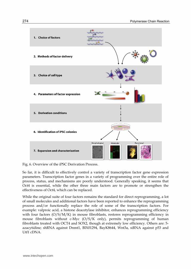
11. Induced pluripotent stem cell technique
Induced pluripotent stem cells (iPS) are a cell type that the differentiated body cell is transfected with several transcriptional factors and is re-programmed as an embryonic-like stem cell. Similarly, iPS has the totipotency of self-renew and differentiation like embryonic stem cells.
iPS cells can be used as target cells for transgenes with extrinsic genes through transgenic techniques or be genetically modified through gene targeting or gene knock-out. The iPS cells can be used as donor cells for somatic cell nuclei and fused with suitable recipient somatic cells to produce transgenic animals.
iPSCs are adult cells that have been genetically reprogrammed to an embryonic stem cell–like state by being forced to express genes and factors important for maintaining the defining properties of embryonic stem cells. Although these cells meet the defining criteria for pluripotent stem cells, it is not known if iPSCs and embryonic stem cells differ in clinically significant ways. Mouse iPSCs were first reported in 2006, and human iPSCs were first reported in late 2007. Mouse iPSCs demonstrate important characteristics of pluripotent stem cells, including expressing stem cell markers, forming tumors containing cells from all three germ layers, and being able to contribute to many different tissues when injected into mouse embryos at a very early stage in development. Human iPSCs also express stem cell markers and are capable of generating cells characteristic of all three germ layers. The Figure 6 addresses each of iPSC steps in detail.
11.1 Choice of reprogramming factors
Direct reprogramming was initially performed in mouse fibroblasts through retroviral transduction of 24 candidate genes that were all implicated in the establishment and maintenance of the pluripotent state. Four transcription factors, Oct4 (Pou5f1), Sox2, c-Myc, and Klf4, play important roles in process.
However, with further research, it found that in certain circumstances, four transcription factors Oct4, Sox2, Nanog, Lin28 could be used to reprogramm human fibroblasts into iPSCs. Similarly, in rat fibroblasts transformed by renumbering process, we can use Sox1 or Sox3 alternative Sox2, Klf4 can also be replaced with Klf2. According to the target cells to different levels of gene expression, transcription factor for re-programming can also be adjusted, such as neural stem cells (NSCs) themselves high expression of Sox2 and c-Myc, only Oct4 can re-compiled NSCs into iPSCs, only Oct4 and Sox2 are enough to induce human umbilical cord blood stem cells into iPSCs. In reprogramming human fibroblast to iPSCs, no c-Myc factor involved in can also get successful result, indicating that c-Myc is not the necessary iPSCs transcription factor of human fibroblasts. This finding has important meaning because c-Myc is a transcription factor highly expressed in tumor cells, import of exogenous human c-Myc may activate the new iPSCs into tumor cells at inappropriate time.
www.intechopen.com
Polymerase Chain Reaction
274
Fig. 6. Overview of the iPSC Derivation Process.
So far, it is difficult to effectively control a variety of transcription factor gene expression parameters. Transcription factor genes in a variety of programming over the entire role of process, status, and mechanisms are poorly understood. Generally speaking, it seems that Oct4 is essential, while the other three main factors are to promote or strengthen the effectiveness of Oct4, which can be replaced.
While the original suite of four factors remains the standard for direct reprogramming, a lot of small molecules and additional factors have been reported to enhance the reprogramming process and/or functionally replace the role of some of the transcription factors. For example: valproic acid, a histone deacetylase inhibitor, enhances reprogramming efficiency with four factors (O/S/M/K) in mouse fibroblasts, restores reprogramming efficiency in mouse fibroblasts without c-Myc (O/S/K only), permits reprogramming of human fibroblasts treated with OCT4 and SOX2, though at extremely low efficiency. Others are: 5-azacytidine; shRNA against Dnmt1, BIX01294, BayK8644, Wnt3a, siRNA against p53 and Utf1 cDNA.
www.intechopen.com

Recent Advances and Applications of Transgenic Animal Technology
275
11.2 Methods of factor delivery
Once the transcription factors are selected to transfer to target cells, delivery methods is
another key to success. From the initial use of retroviral vectors to Lentiviral vector, these
carriers can ensure the expression pattern of transcription factors in transformation of cells.
However, this viral vector may change or even increase the potential of cell differentiation.
For iPSCs safely used in clinical, non-integrated carriers must be taken. The new carrier
called Cre-recombinant virus can be renumbered 5 cases for Parkinson's disease skin
fibroblast dimensional cells. This vector has the advantage that you can remove one of the
transcription factor when iPSCs turn to success. PiggyBac transposon renumbering systems
are also used. When the piggyBac transposon re-compiled successfully, then transposase
expression transposon is removed to obtain iPSCs without exogenous viral vectors or gene
transcription.
The use of integrating viruses for iPSC induction has represented a major roadblock in
the pursuit of clinically relevant applications. For HIV-based lentivirus method, it is
constitutive, transduction of both dividing and nondividing cells, temporal control over
factor expression. But lower expression levels than integrated form is main disadvantage.
In short, in order to be more conducive to clinical iPSCs, the carrier of transcription factors
becomes hot spots in the field of iPSCs research.
11.3 Choice of cell type
As the capacity of skin fibroblasts derived easily, and culture conditions similar to ESCs,
skin fibroblasts can be used as trophoblast cells. But fibroblasts are not the only option, a
variety of somatic cells, such as stomach, liver, pancreas, blood cells and bone marrow
stromal cells, neural progenitor cells, and even in the adult division at the end of the angle
stromal cells have been successfully re-compiled into the iPSC.
Several factors must be considered in determining the optimal cell type for a given
application: 1) the ease at which reprogramming factors can be introduced, which varies
both by cell type and delivery approach; 2) the availability and ease of derivation of the
given cell type; and 3) the age and source of the cell.
Although additional research is needed, iPSCs are already useful tools for drug
development and modeling of diseases, and scientists hope to use them in transplantation
medicine. Viruses are currently used to introduce the reprogramming factors into adult
cells, and this process must be carefully controlled and tested before the technique can lead
to useful treatments for humans. In animal studies, the virus used to introduce the stem cell
factors sometimes causes cancers. Researchers are currently investigating non-viral delivery
strategies. In any case, this breakthrough discovery has created a powerful new way to "de-
differentiate" cells whose developmental fates had been previously assumed to be
determined. In addition, tissues derived from iPSCs will be a nearly identical match to the
cell donor and thus probably avoid rejection by the immune system. The iPSC strategy
creates pluripotent stem cells that, together with studies of other types of pluripotent stem
cells, will help researchers learn how to reprogram cells to repair damaged tissues in the
human body.
www.intechopen.com
Polymerase Chain Reaction
276
12. Applications
Transgenic animals have potentially broad application for the improvement of animal
production quality, the enhancement of production capacity, the studies of human disease
models and the production of biomedical materials.
The benefits of these animals to human welfare can be grouped into the following areas:
12.1 Agricultural applications
The application of biotechnology to farm animals has the potential to benefit both humans
and animals in significant ways.
a. Breeding: Farmers have always used selective breeding to produce animals that exhibit desired traits (e.g., increased milk production, high growth rate). Traditional breeding is a time-consuming, difficult task. When technology using molecular biology was developed, it became possible to develop traits in animals in a shorter time and with more precision. In addition, it offers the farmer an easy way to increase yields. Take ES cell technology as an example, chimeric nuclear transfer technology and production technology is improving, as ES cells are widely used in animal cloning. Proliferation of ES cells derived from donor as the nucleus, produced cloned animals. ES cells in germline chimeric, then develop into sperm or eggs to produce offspring. Animal cloning technology can produce excellent breeding, combination of genes and their high proportion in the population in short time.
b. Quality: Transgenic cows exist that produce more milk or milk with less lactose or cholesterol, pigs and cattle that have more meat on them, and sheep that grow more wool. In the past, farmers used growth hormones to spur the development of animals but this technique was problematic, especially since residue of the hormones remained in the animal product.
At present the production of transgenic animals in low efficiency is one of the main problems. The results of the testing work are carried out at the individual level. Using ES cells as a carrier, directed transformation of ES cells, the integration of inserted genes, expression level and stability of interested genes can be screened. The work is carried out at the cellular level, which is easy to obtain stable cell line with expression of satisfaction, accessing to the target gene carrying the transgene for animals. One success story is artificial insemination: the use of this technology from 1950s to 1990s in US, increased the average milk production per cow over 300%.
12.2 Medical applications
a. Xenotransplantation: Transplant organs may soon come from transgenic animals. Transgenic pigs may provide the transplant organs needed to alleviate the shortfall. Currently, xenotransplantation is hampered by a pig protein that can cause donor rejection but research is underway to remove the pig protein and replace it with a human protein. For organ and tissue transplantation, which is known as a "species of daughter cells ", for the clinical organization, organ transplantation offers great amount of material knockout cells. U.S. ACT companies put the nucleus of human skin into bovine oocytes without the genetic information, nurturing issued totipotency cell. If they could be
www.intechopen.com

Recent Advances and Applications of Transgenic Animal Technology
277
successfully used in clinical, in future, many difficult diseases such as Parkinson's disease will be cured.
b. Nutritional supplements and pharmaceuticals: Milk-producing transgenic animals are especially useful for medicines. Products such as insulin, growth hormone, and blood anti-clotting factors may soon be or have already been obtained from the milk of transgenic cows, sheep, or goats. Research is also underway to manufacture milk through transgenesis for treatment of debilitating diseases such as phenylketonuria (PKU), hereditary emphysema, and cystic fibrosis. ES cell culture techniques are used in some special body, then the cost can be a huge improvement. For example, some special drugs (interferon, antithrombin, erythropoietin and other biological systems agents or genetically modified), in body fluids from animals (milk, blood, etc.) or tissue extract achieve the body of the animal drug production factory.
c. Human gene therapy: A transgenic cow exists that produces a substance to help human red cells grow. Human gene therapy involves in adding a normal copy of a gene (transgene) to the genome of a person carrying defective copies of the gene. The potential for treatments for the 5,000 named genetic diseases is huge and transgenic animals could play a role.
The most current human serious medical diseases are cancer, genetic diseases, including birth defects, These diseases are caused by abnormal cell transformation and differentiation, such as Lesch, Nyhan. Fully understanding the process of cell differentiation and development will be able to cure the diseases. Many scientists have established many mouse disease models, and expressed human disease gene in mice for further treatment of human disease. For example, U.S. National Institute of Molecular Neurology Laboratory used mice ESC to induce neuroepithelial cells, implanted them into the brain, and got a large number of small conflicts like cells and glial cells. It can be envisaged to treat multiple sclerosis diseases.
13. Problems and prospects
Transgenic animals have potentially broad application for the improvement of animal production quality, the enhancement of productivity, the studies of human disease models and the production of pharmaceuticals. However, there are many pressing problems that need to be resolved for transgenic animal studies.
a. Dietary and food safety concerns: Food safety of bioengineered products is always a significant public topic. For the transgenic animals, some of the foreign gene and its promoter sequences from the virus may occur in the recipient animals. Homologous recombination or integration may cause the formation of new virus. Foreign gene inserted in the chromosome locus may also result in different genetic changes in different degrees, causing unintended effects. Transgenic animals may also increase the risk of zoonotic disease, and cause human allergic reactions.
b. Environmental impacts: If transgenic animals are in the external environment and mating with wildlife, foreign gene may spread, which results in changing the species composition of the original genes, causing confusion in species resources. It may also lead to the loss of the wild allele, resulting in a decline in genetic diversity. Once released into the environment, transgenic animals can disrupt the ecological balance of species, genetic diversity of threatened species. For example, once the transgenic fishes are into ponds or rivers and out of control, they may affect the balance of ecology.
www.intechopen.com

Polymerase Chain Reaction
278
c. Respect for life and “unnaturalness” of genetic engineering: Ethical concern has also been discussed about the “unnaturalness” of genetic engineering and the ways it might devalue nature and commercialize life. Here we quoted Strachan Donnelley’s view:
“Animal biotechnology, inspired by of often genuine and legitimate desires to meet human and animal need and interests, must beware that it does not pre-empt ‘nature natural’ in the minds and hearts of us human beings and replace it with its own ‘nature contrived’…This would be the end of us as seekers after ‘living’ natural norms and ways of being human, and given the press of our present technological powers, no doubt the end of nature’s richness and goodness itself. This would decidedly be a double moral disaster and irresponsibility.”
With the fast development of animal gene transfer technology, scientists had well improved the efficiency of making transgenic animals as well as the control of the transgene. Combination of gene targeting with somatic cell cloning or RNAi techniques had created a powerful platform for preparation of transgenic animals. However, cloning was still a highly unpredictable laboratory protocol, which existed uncertainty results in the experiments. These questions deserved each scientist careful attention.
14. Conclusion
Transgenic animal techniques have developed rapidly and provided more and improved platforms for the preparation of transgenic animals since their emergence. These techniques provide an entirely new pathway for the accurate modulation of genes. In addition, transgenic animal research may provide the tools for a series of research hotspots like microRNA function and iPS cells. All of these developments will provide new ideas and bring forth important changes in fields like medicine, health and livestock improvement. In particular, the economic and social benefits from the production of bioreactors, drug production, and organ culture for human transplantation will be great.
In summary, this review has attempted to present a comprehensive comparison of the currently available transgenic animal technology. Transgenic animal research involves consistent exploration and creation, and searching simple, reliable and efficient transgenic techniques is the key for transgenic animals. It is conceivable that the development of more simple and novel animal transgenic techniques will lead to more transgenic animals and related products that will likely improve our livelihood and wellbeing.
15. References
Aasen, T.; Raya, A.; Barrero, M.J.; Garreta, E.; Consiglio, A.; Gonzalez, F.; Vassena, R.; Billic, J.; Pekarik, V. & Tiscornia, G. (2008). Efficient and rapid generation of induced pluripotent stem cells from human keratinocytes. Nat. Biotechnol., Vol.26, No.11, (October 2008), pp. 1276–1284, ISSN 1087-0156.
Abremski, K.E. & Hoess, R.H. (1992). Evidence for a second conserved arginine residue in the integrase family of recombination proteins. Protein Eng., Vol.5, No.1, (October 1991), pp. 87-91, ISSN 1741-0134.
Acosta, J; Carpio, Y; Borroto, I; Gonzalez, O & Estrada, M.P. (2005). Myostatin gene silenced by RNAi show a zebrafish giant phenotype. J Biotechnol., Vol.119, No.4, (October 2005), pp. 324–331, ISSN 0168-1656.
www.intechopen.com

Recent Advances and Applications of Transgenic Animal Technology
279
Aponte, P.M; van Bragt, M.P.; de Rooij, D.G. & van Pelt, A.M. (2005). Spermatogonial stem cells: characteristics and experimental possibilities. APMIS, Vol.113, No.11-12, (November 2005), pp. 727-742, ISSN 1600-0463.
Baubonis, W. & Sauer, B. (1993). Genomic targeting with purified Cre recombinase. Nucleic Acids Res., Vol.21, No.9, (May 1993), pp. 2025-2029, ISSN 1362-4962.
Blelloch, R.; Venere, M.; Yen, J. & Ramalho-Santos, M. (2007). Generation of induced pluripotent stem cells in the absence of drug selection. Cell Stem Cell, Vol.1, No.3, (September 2007), pp. 245–247, ISSN 1934-5909.
Bosnali, M. & Edenhofer, F. (2008). Generation of transducible versions of transcription factors Oct4 and Sox2. Biol. Chem., Vol.389, No.7, (July 2008), pp. 851–861, ISSN 1437-4315.
Boulanger, C.A.; Mack, D.L.; Booth, B.W. & Smith, G.H. (2007). Interaction with the mammary microenvironment redirects spermatogenic cell fate in vivo. Proc. Natl. Acad. Sci., Vol.104, No.10, (March 2007), pp. 3871-3876, ISSN 1091-6490.
Brinster, R.L.; Chen, H.Y.; Trumbauer, M.E.; Yagle, M.K. & Palmiter, R.D. (1985). Factors affecting the efficiency of introducing foreign DNA into mice by microinjection eggs. Proc. Natl. Acad. Sci., Vol.82, No.13, (July 1985), pp. 4438-4442, ISSN 1091-6490.
Chen, C.; Ouyang, W.; Grigura, V.; Zhou, Q.; Carnes, K.; Lim, H.; Zhao, G.Q.; Arber, S.; Kurplos, N.; Murphy, T.L.; Cheng, A.M.; Hassell, J.A; Chandrashekar, M.C.; Hess, R.A. & Murphy, K.M. (2005). ERM is required for transcriptional control of the spermatogonial stem cell niche. Nature, Vol.436, No.7053, (August 2005), pp. 1030-1034, ISSN 0028-0836.
Cibelli, J.B.; Stice, S.L.; Golueke, P.J.; Kane, J.J.; Jerry, J.; Blackwell, C.; Deleon, F.A. &, Robl, J.M. (1998). Transgenic bovine chimeric offspring produced from somatic cell-derived stem-like cells. Nature Biotechnol., Vol.16, No.7, (July 1998), pp. 642–646, ISSN 1087-0156.
Costoya, J.A.; Hobbs, R.M.; Barna, M. Cattoretti, G.; Manova, K.; Sukhwani, M.; Orwiq, K.E.; Wolgemuth, D.J. & Pandolfi, P.P. (2004). Essential role of Plzf in maintenance of spermatogonial stem cells. Nat Genet., Vol.36, No.6, (June 2004), pp. 653-659, ISSN 1061-4036.
Dai, Y.F.; Vaught, T.D. & Boone, J. (2002). Targeted disruption of the alpha-1,3-galactosyltransferase gene in cloned pigs. Nature Biotechnol., Vol.20, No.3, (March 2002), pp. 251–255, ISSN 1087-0156.
Dann, C.T.; Alvarado, A.L.; Hammer, R.E. & Garbers, D.L. (2006). Heritable and stable gene knockdown in rats. Proc. Natl. Acad. Sci., Vol.103, No.30, (July 2006), pp. 11246–11251, ISSN 1091-6490.
de Rooij, D.G. (2001). Proliferation and differentiation of spermatogonial stem cells. Reproduction, Vol.121, No.3, (March 2001), pp. 347-354, ISSN 1470-1626.
Dexter, M. & Allen, T. (1992). Haematopoiesis. Multi-talented stem cells? Nature, Vol.360, No.6406, (December 1992), pp. 709-710, ISSN 0028-0836.
Dickins, R.A.; McJunkin, K.; Hernando, E.; Premsrirut, P.K.; Krizhanovsky, V.; Burgess, D.J.; Kim, S.Y.; Cordon-Cardo, C.; Zender, L.; Hannon, G.J.; Lowe, S.W. (2007). Tissuespecific and reversible RNA interference in transgenic mice. Nat Genet., Vol.39, No.7, (July 2007), pp. 914–921, ISSN 1061-4036.
Evans, M.J. & Kaufman, M.H. (1981). Establishment in culture of plurpotential cells from mouse embryos. Nature, Vol.292, No.2819, (July 1981), pp. 154-156, ISSN 0028-0836.
www.intechopen.com
Polymerase Chain Reaction
280
Farre, L.; Riqau, T.; Garcia-Rocha, M.; Canal, M.; Gomez-Foix, A.M. & Rodriquez-Gil, J.E. (1999). Adenovirus-mediated introduction of DNA into pig sperm and offspring. Mol. Reprod.Dev., Vol.53, No.2, (June 1999), pp. 149-158, ISSN 1098-2795.
Foley, J.E.; Yeh, J.R.J; Maeder, M.L.; Reyon, D.; Sander, J.D.; Pe-terson, R.T. & Joung, J.K. (2009). Rapid mutation of endogenous ze-brafish genes using zinc finger nucleases made by Oli-gomerized Pool ENgineering (OPEN). PLoS One, Vol.4, No.2, (February 2009), pp. e4348, ISSN 1932-6203.
Fulka, J. Jr.; First, N.L.; Loi, P. & Moor, R.M. (1998). Cloning by somatic cell nuclear transfer. BioEssays, Vol.20, No.10, (October 1998), pp. 847-851, ISSN 1521-1878.
Fulka, J. Jr.; First, N.L.; Moor, R.M. (1996). Nuclear transplantation in mammals: Remodelling of transplanted nuclei under the influence of maturation promoting factor. BioEssays, Vol.18, No.10, (October 1996), pp. 835–840, ISSN 1521-1878.
Gasparrini, B.; Gao, S.; Ainslie, A.; Fltcher, J.; McGarry, M.; Ritchie, W.A.; Springbett, A.J.; Overstrom, E.W.; Wilmut, I.; DeSousa, P.A. (2003). Cloned mice derived from embryonic stem cell karyoplasts and activated cytoplasts prepared by induced enucleation. Biol Reprod, Vol.68, No.4, (April 2003), pp. 1259-1266, ISSN 0006-3363.
Giordano, R.; Magnano, A.R.; Zaccagnini, G.; Pittoggi, C. & Moscufo, N. (2000). Reverse transcriptase activity in mature spermatozoa of mouse. J Cell Biol., Vol.148, No.6, (March 2000), pp. 1107–1113, ISSN 1540-8140.
Guan, K.; Nayernia, K.; Maier, L.S.; Wagner, S.; Dressel, R.; Lee, J.H.; Nolte, J.; Wolf, F.; Li, M.Y.; Engel, W. & Hasenfuss, G. (2006). Pluripotency of spermatogonial stem cells from adult mouse testis. Nature, Vol.440, No.7088, (April 2006), pp. 1199-203, ISSN 0028-0836.
Huangfu, D.; Maehr, R.; Guo, W.; Eijkelenboom, A.; Snitow, M.; Chen, A.E. & Melton, D.A. (2008a). Induction of pluripotent stem cells by defined factors is greatly improved by small-molecule compounds. Nat. Biotechnol., Vol.26, No.7, (July 2008), pp. 795–797, ISSN 1087-0156.
Huangfu, D.; Osafune, K.; Maehr, R.; Guo, W.; Eijkelenboom, A.; Chen, S.; Muhlestein, W. & Melton, D.A. (2008b). Induction of pluripotent stem cells from primary human fibroblasts with only Oct4 and Sox2. Nat. Biotechnol. Vol.26, No.11, (November 2008), pp. 1269–1275, ISSN 1087-0156.
Jaenisch, R. (1988). Transgenic animals. Science, Vol.240, No.4858, (November 1998), pp. 1468-1474, ISSN 0036-8075.
Kim, J.B.; Zaehres, H.; Wu, G.; Gentile, L.; Ko, K.; Sebastiano, V.; Arauzo- Bravo, M.J.; Ruau, D.; Han, D.W. & Zenke, M. (2008). Pluripotent stem cells induced from adult neural stem cells by reprogramming with two factors. Nature, Vol.454, No.7204, (July 2008), pp. 646–650, ISSN 0028-0836.
Kitamura, T.; Koshino, Y.; Shibata, F.; Oki, T.; Nakajima, H.; Nosaka, T. & Kumagai, H. (2003). Retrovirus-mediated gene transfer and expression cloning: powerful tools in functional genomics. Exp Hematol., Vol.31, No.11, (November 2003), pp. 1007-1014, ISSN 0301-472X.
Ko, K.; Tapia, N.; Wu, G.; Kim, J.B.; Bravo, M.J.; Sasse, P.; Glaser, T.; Han, D.W.; Hausdorfer, K.; Sebastiano, V.; Stehling, M.; Fleischmann, B.K.; Brustle, O.; Zenke, M. U. & Scholer, H.R. (2009). Induction of pluripotency in adult unipotent germline stem cells. Cell Stem Cell, Vol.5, No.1, (July 2009), pp. 87-96, ISSN 1934-5909.
Kubota, H.; Avarbock, M.R. & Brinster, R.L. (2004). Growth factors essential for self-renewal and expansion of mouse spermatogonialmstem cells. Proc Natl Acad Sci, Vol.101, No.47, (November 2004), pp. 16489-16494, ISSN 1091-6490.
www.intechopen.com

Recent Advances and Applications of Transgenic Animal Technology
281
Kubota, H.; Avarbock, M.R. & Brinster RL. (2003). Spermatogonial stem cells share some, but not all, phenotypic and functional characteristics with other stem cells. Proc Natl Acad Sci, Vol.100, No.11, (May 2003), pp. 6487-6492, ISSN 1091-6490.
Kuroiwa, Y.; Kasinathan, P. & Matsushita H. (2004). Sequential targeting of the genes encoding immunoglobulin-m and prion protein in cattle. Nat Genet., Vol.36, No.7, (July 2004), pp. 775–780, ISSN 1061-4036.
Lavitrano, M.; Busnelli, M.; Cerrito, M.G.; Giovannoni, R.; Manzini, S.; Vargiolu, A. (2006) Sperm-mediated gene transfer. Reproduction Fertility and Development, Vol.18, No.2, (January 2006), pp. 19–23, ISSN 1031-3613.
Lavitrano, M.; Camaioni, A.; Fazio, V.M.; Dolci, S., Farace M.G. & Spadafora, C. (1989). Sperm cells as vectors for introducing foreign DNA into eggs: Genetic transformation of mice. Cell, Vol.57, No.5, (June 1989), pp. 717-723, ISSN 0092-8674.
Maherali, N. & Hochedlinger, K. (2008). Guidelines and techniques for the generation of induced pluripotent stem cells. Cell Stem Cell, Vol. 3, No. 6, (December 2008), pp. 595-605, ISSN 1934-5909.
Martin, G.R. (1981). Isolation of a pluripotent cell lines from early mouse embryos cultured in medium conditioned by teratocarcinoma stem cells. Proc. Natl. Acad. Sci., vol.78, No. 12, (December 1981), pp. 7634-7638, ISSN 1091-6490.
McCreath, K.J.; Howcroft, J.; Campbell, K.H.; Colman, A.; Schnieke, A.E. & Kind, A.J. (2000). Production of gene-targeted sheep by nuclear transfer from cultured somatic cells. Nature, Vol. 405, No. 6790, (June 2000), pp. 1066–1069, ISSN 0028-0836.
McLaren, A. (2003). Primordial germ cells in the mouse. Dev Biol., Vol. 262, No. 1, (October 2003), pp. 1-15, ISSN 0012-1606.
McLean, D.J.; Friel, P.J.; Johnston, D.S. & Griswold, M.D. (2003). Characterization of spermatogonial stem cell maturation and differentiation in neonatal mice. Biol Reprod, Vol. 69, No. 6, (September 2003), pp. 2085-2091, ISSN 0006-3363.
Mikkelsen, T.S.; Hanna, J.; Zhang, X.; Ku, M.; Wernig, M.; Schorderet, P.; Bernstein, B.E.; Jaenisch, R.; Lander, E.S. & Meissner, A. (2008). Dissecting direct reprogramming through integrative genomic analysis. Nature Vol. 454, No. 7200, (May 2008), pp. 49–55, ISSN 0028-0836.
Mueller, S.; Prelle, K.; Rieger, N.; Petznek, H.; Lassnig, C.; Luksch, U.; Aigner, B.; Baetscher, M.; Wolf, E.; Mueller, M. & Brem, G. (1999). Chimeric pigs following blastocyst injection of transgenic porcine primordial germ cells. Mol Reprod Dev., Vol. 54, No. 3, (November 1999), pp. 244–254, ISSN 1040-452X.
Oatley, J.M. & Brinster, R.L. (2008). Regulation of spermatogonial stem cell self-renewal in mammals. Annu Rev Cell Dev Biol, Vol. 24, (June 2008), pp. 263-286, ISSN 1081-0706.
Petolino, J.F.; Worden ,A.; Curlee, K.; Connell, J.; Strange Moynahan, T.L. & Larsen C, Russell. (2010). Zine finger nuclease-mediated transgene deletion. Plant Mol Biol, Vol. 73, No. 6, (May 2010), pp. 617–628, ISSN 0167-4412.
Phillips, B.T.; Gassei, K.; Orwig, K.E. (2010). Spermatogonial stem cell regulation and spermatogenesis. Philos Trans R Soc Lond B Biol Sci, Vol. 365, No. 1546, (May 2010), pp. 1663-1678, ISSN 0962-8436.
Porteus, M.H. & Baltimore, D. (2003). Chimeric nucleases stimulate gene targeting in human cells. Science, Vol. 300, No. 5620, (May 2003), pp. 763, ISSN 0036-8075.
Seandel, M.; James, D.; Shmelkov, S.V,; Falciatori, I.; Kim, J.; Chavala, S.; Scherr, D.S.; Zhang, F.; Torres, R.; Gale, N.W.; Yancopoulos, G.D.; Murphy, A.; Valenzuela, D.M. Hobbs, R.M.; Pandolfi, P.P. & Rafii, S. (2007). Generation of functional multipotent
www.intechopen.com

Polymerase Chain Reaction
282
adult stem cells from GPR125+ germline progenitors. Nature, Vol. 449, No. 7160, (September 2007), pp. 346-50, ISSN 0028-0836.
Smith, K. & Spadafora, C. (2005). Sperm-mediated gene transfer: application and implications. Bioessays, Vol. 27, No. 5, (May 2005), pp. 551-562, ISSN 0265-9247.
Stadtfeld, M.; Brennand, K. & Hochedlinger, K. (2008a). Reprogramming of pancreatic beta cells into induced pluripotent stem cells. Curr. Biol.,Vol. 18, No. 12, (May 2008), pp. 890–894, ISSN 0960-9822.
Stadtfeld, M.; Maherali, N.; Breault, D.T. & Hochedlinger, K. (2008b). Defining molecular cornerstones during fibroblast to iPS cell reprogramming in mouse. Cell Stem Cell, Vol.2, No. 3, (February 2008), pp. 230–240, ISSN 1934-5909.
Stadtfeld, M.; Nagaya, M.; Utikal, J.; Weir, G. & Hochedlinger, K. (2008c). Induced Pluripotent Stem Cells Generated Without Viral Integration. Science, Vol. 322, No. 5903, (September 2008), pp. 945–949, ISSN 0036-8075.
Streckfuss-Bomeke, K.; Vlasov, A.; Hulsmann, S.; Yin, D.; Nayernia, K.; Engel, W.; Hasenfuss, G. & Guan, K. (2009). Generation of functional neurons and glia from multipotent adult mouse germ-line stem cells. Stem Cell Res, Vol. 2, No. 2, (March 2009), pp. 139-154, ISSN 1873-5061.
Takahashi, K. & Yamanaka, S. (2006). Induction of pluripotent stem cells from mouse embryonic and adult fibroblast cultures by defined factors. Cell, Vol. 126, No. 4, (August 2006), pp. 663–676, ISSN 0092-8674.
Tegelenbosch, R.A.; de Rooij, D.G. (1993). A quantitative study of spermatogonial multiplication and stem cell renewal in the C3H/101 F1 hybrid mouse. Mutat Res, Vol. 290, No. 2, (December 1993), pp. 193-200, ISSN 0027-5107.
Tong-Starksen, S.E. & Peterlin, B.M. (1990). Mechanism of retroviral transcriptional activation. Semin. Virol. 1, pp. 215-227.
Urnov, F.D.; Miller, J.C.; Lee, Y.L.; Beausejour, C.M.; Rock, J.M.; Augustus, S; Jamieson A.C.; Porteus, M.H.; Gregory, P.D. & Holmes, M.C. (2005). Highly efficient endogenous human gene correction using designed zinc-finger nucleases. Nature, Vol. 435, No. 7042, (April 2005), pp. 646–651, ISSN 0028-0836.
Van de Lavoir, M.C.; Diamond, J.H. & Leighton, P.A. (2006). Germline transmission of genetically modified primordial germ cells. Nature, Vol. 441, No. 6907, (April 2006), pp. 766–769, ISSN 0028-0836.
Wakayama, T.; Perry, A.C.F.; Zuccotti, M.; Johnson, K.R. & Yanagimachi, R. (1998), Full-term development of mice from enucleated oocytes injected with cumulus cell nuclei. Nature, Vol. 394, No. 6691, (June 1998), pp. 369-374, ISSN 0028-0836.
Willadsen, S.M. (1986). Nuclear transplantation in sheep embryos. Nature, Vol. 320, No. 6057, (March 1986), pp. 63–65, ISSN 0028-0836.
Wilmut, I.; Beaujean, N.; de Sousa, P.A.; Dinnyes, A.; King, T.J.; Paterson, L.A.; Wells, D.N. & Young, L.E. (2002). Somatic cell nuclear transfer. Nature, Vol. 419, No. 6907, (October 2002), pp. 583-586, ISSN 0028-0836.
Yoshida, S.; Sukeno, M. & Nabeshima, Y. (2007). A Vasculature-Associated Niche for Undifferentiated Spermatogonia in the Mouse Testis. Science, Vol. 317, No. 5845, (August 2007), pp. 1722-1726, ISSN 0036-8075.
Yoshida, S. (2010). Stem cells in mammalian spermatogenesis. Dev Growth Differ, Vol.52, No.3, (January 2010), pp. 311-317, ISSN 0012-1592.
www.intechopen.com
Polymerase Chain ReactionEdited by Dr Patricia Hernandez-Rodriguez
ISBN 978-953-51-0612-8Hard cover, 566 pagesPublisher InTechPublished online 30, May, 2012Published in print edition May, 2012
InTech EuropeUniversity Campus STeP Ri Slavka Krautzeka 83/A 51000 Rijeka, Croatia Phone: +385 (51) 770 447 Fax: +385 (51) 686 166www.intechopen.com
InTech ChinaUnit 405, Office Block, Hotel Equatorial Shanghai No.65, Yan An Road (West), Shanghai, 200040, China
Phone: +86-21-62489820 Fax: +86-21-62489821
This book is intended to present current concepts in molecular biology with the emphasis on the application toanimal, plant and human pathology, in various aspects such as etiology, diagnosis, prognosis, treatment andprevention of diseases as well as the use of these methodologies in understanding the pathophysiology ofvarious diseases that affect living beings.
How to referenceIn order to correctly reference this scholarly work, feel free to copy and paste the following:
Xiangyang Miao (2012). Recent Advances and Applications of Transgenic Animal Technology, PolymeraseChain Reaction, Dr Patricia Hernandez-Rodriguez (Ed.), ISBN: 978-953-51-0612-8, InTech, Available from:http://www.intechopen.com/books/polymerase-chain-reaction/recent-advances-and-applications-of-transgenic-animal-technology

© 2012 The Author(s). Licensee IntechOpen. This is an open access articledistributed under the terms of the Creative Commons Attribution 3.0License, which permits unrestricted use, distribution, and reproduction inany medium, provided the original work is properly cited.