real-time electromagnetically navigated breast cancer...
TRANSCRIPT

Real-time electromagnetically navigated breast cancer surgery
Tamas Ungi, Gabrielle Gauvin, Andras Lasso, Caitlin T. Yeo, John Rudan, C. Jay
Engel, Gabor Fichtinger
Laboratory for Percutaneous Surgery & Department of Surgery, Queen's University, Kingston, ON, Canada

Breast conserving surgery
• Breast cancer kills more women than any other cancer
• Detected at earlier stage due to screening programs
• Early stage breast cancer is treated by breast conserving surgery (aka) lumpectomy

Image-guided wire localization
• Localization needle and wire are placed in the tumor under X-ray or ultrasound
• But tumor margins are not directly marked by the needle, and it is invisible during surgery

Problem: positive margins
• Tumor at excision borders
• Additional surgery
– delays adjuvant treatments
– causes further trauma
– increases cost
• Positive margins are reported in 15% - 47% of cases after wire-localization guidance
• Repeat surgery is no longer conserving, 1/3 of those is full mastectomy

Method derived from “Wire-Guide” & “EM beacon”
EM-tracked tissue locking needle
Needle
Tissue lock
EM sensor
Beforeresection
Duringresection

Navigation system schematics
Laboratory for Percutaneous Surgery (The Perk Lab) – Copyright © Queen’s University, 2015
Ultrasound
Localization needle
CauteryPatient reference
Navigation computer
Network connection
Position tracker
Breast
Navigationtablet
Ultrasound machine
Overhead display

Resulting navigation system
Note: In the actual experiments the phantom was opaque; here we use transparent phantom to show the simulated lesion inside

Phantom experiment
Screenshot of SlicerIGTinterface with registered
tumor model
Example of EM navigated model resection showing clear margins on all sides of implanted tumor

Phantom experiment results• N = 42 synthetic tumors resected
by 8 surgical trainees and 2 staff
breast surgeons
– 21 using control method
(wire-localization)
– 21 using EM-navigation
• Positive margin rate
– Control 42.9% (9 of 21)
– EM 19.0% (4 of 21)
– p = 0.18
• Average amount of tissue
resected (“cosmesis”)
– Control 36.3 g (SD =14.5 g)
– EM 37.7g (SD =9.8 g)

Human cadaver – workflow refinement

Feasibility in the operating room
- 11 -

Infection control
• Designed tracker attachments that are usable through plastic bags
• CAD files available at www.plustoolkit.org

User interactions• Touchscreen tablet on the operating table, in a
sterile bag (X-ray cassette bag)
• Screen sharing and user interaction forwarding (TightVNC)
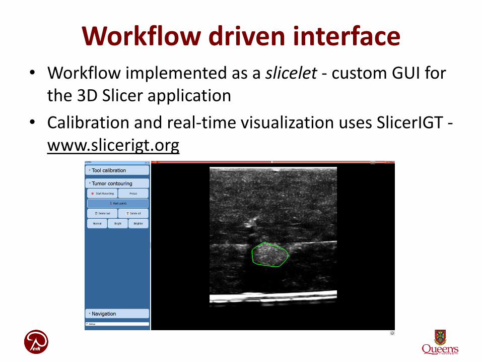
Workflow driven interface• Workflow implemented as a slicelet - custom GUI for
the 3D Slicer application
• Calibration and real-time visualization uses SlicerIGT -www.slicerigt.org

Dynamic tumor contouring
• Tumor model interactively edited by surgeon. Tapping on the ultrasound images (sterile tablet) extends the existing 3D contour.

Finally: clinical trial

Trial 1: Palpable tumors
Laboratory for Percutaneous Surgery (The Perk Lab) – Copyright © Queen’s University, 2015
Patient Number
1 2 3 4 5 6
Sex F F F F F F
Age 92 46 63 48 43 29
Side Right Left Left Left Right Right
Procedure L+SLNBx L+SLNBx L+SLNBx L+SLNBx L L
Anesthetic Sedation GA GA GA Sedation Sedation
Sterility maintained
Yes Yes Yes Yes Yes Yes
Operative time 48 min 65 min 65 min 58 min 29 min 42 min
Setup time n/a n/a 10 min 12 min 10 min 6 min
Pathology
Invasive ductal carcinoma and
non invasive solid papillary
carcinoma
In situ and invasive ductal
carcinoma
Invasive ductal
carcinoma
In situ and invasive ductal
carcinoma
Benign phyllodes
tumor with florid ductal
hyperplasia
Benign fibroepithelial lesion
Margins Negative Negative Negative Negative Negative Negative
Stage IA IA IIIA IIA n/a n/a
Gauvin et al. The Breast Surgery Journal (in review)

Non-palpable tumorsNon-palpable tumor trial
Number of cases 27
Positive margin for invasive cancer
2 (7.4%)
Excised tissue volume
113.1 ± 90.4 cm3
(43% reduction)
Problematic aspects:
• 65 user interactions per case
• Technician in the OR
Ungi et al. CARS/ISCAS, 2017

Real-time spatial mapping of electromagnetic tracking error
Youtube Video: https://www.youtube.com/watch?v=R78Dxi5exO4
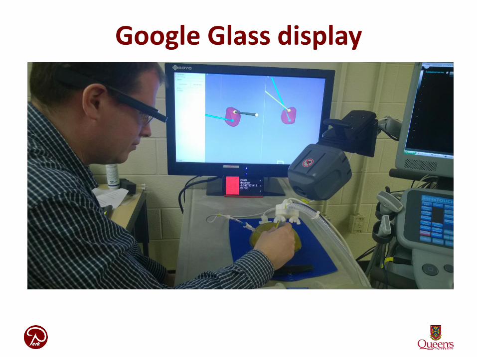
Google Glass display

Sonic ConstraintsYoutube Video: https://www.youtube.com/watch?v=gSz8IHmogMo
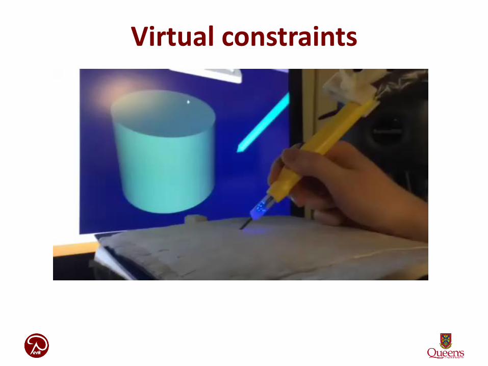
Virtual constraints
Navigated margin probe
Youtube Video: https://www.youtube.com/watch?v=ag7fWY27lus

X-ray mammography

Breast Tomosynthesis
Youtube Video: https://www.youtube.com/watch?v=VhvSYl_ZMEs&feature=youtu.be

X-ray breast biopsy

Marking the breast biopsy site
Mammogram following the biopsy shows the
clips (arrows) placed to mark the biopsy site.Mammogram following the biopsy with
injectable hydrogel markers placed to mark
the biopsy site.
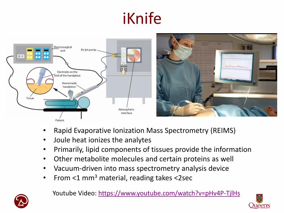
iKnife
Youtube Video: https://www.youtube.com/watch?v=pHv4P-TjlHs
• Rapid Evaporative Ionization Mass Spectrometry (REIMS)• Joule heat ionizes the analytes• Primarily, lipid components of tissues provide the information• Other metabolite molecules and certain proteins as well• Vacuum-driven into mass spectrometry analysis device• From <1 mm3 material, reading takes <2sec

Navigated iKnife in Perk Lab
• Only 1 units exists outside the inventor’s lab• Funding for 3 units (for lab, 1 clinical) at $2.5M• 2 delivered, 1 still to come• 2 labs at KGH, 1 operating room• Medtronic Stealth Station ($300,000)

Desorption electrospray ionization (DESI)• Ambient ionization coupled to mass spectrometry• fast-moving charged solvent stream, at an angle wrt sample surface• extracts ionized analytes from surface • propels these secondary ions to mass spec
• High resolution 0.01mm• Works on fresh and fixed tissue

“Molecular navigation” challenge
DESI
iKnife / REIMS
Genetic info
Pathology proven condition • Cancer• Normal• Atypical• etc,…
AI / ML
Spatio / temporal registration
US / X-ray / MRI
Optical spectr.