rd-2012-3773 molecular characterisation of the rhynchosporium … pa… · combination with other...
TRANSCRIPT
December 2018
Student Report No. SR48
Molecular characterisation of the Rhynchosporium commune
interaction with barley
Louise Gamble
The James Hutton Institute, Invergowrie, Dundee, DD2 5DA
Supervisors: Anna Avrova and Adrian Newton
This is the final report of a PhD project (RD-2012-3773) that ran from October 2012 to December 2015. The work was funded by the James Hutton Institute and a contract for £37,500 from AHDB Cereals & Oilseeds.
While the Agriculture and Horticulture Development Board seeks to ensure that the information contained within this document is
accurate at the time of printing, no warranty is given in respect thereof and, to the maximum extent permitted by law, the Agriculture and
Horticulture Development Board accepts no liability for loss, damage or injury howsoever caused (including that caused by negligence)
or suffered directly or indirectly in relation to information and opinions contained in or omitted from this document.
Reference herein to trade names and proprietary products without stating that they are protected does not imply that they may be
regarded as unprotected and thus free for general use. No endorsement of named products is intended, nor is any criticism implied of
other alternative, but unnamed, products.
AHDB Cereals & Oilseeds is a division of the Agriculture and Horticulture Development Board (AHDB).
RD-2012-3773

3
CONTENTS
1. ABSTRACT ....................................................................................................................... 1
2. INTRODUCTION ............................................................................................................... 2
3. MATERIALS AND METHODS .......................................................................................... 4
3.1. Plant growth .......................................................................................................... 4
3.2. Culturing and storage of micro organisms ......................................................... 5
3.2.1. Fungi 5
3.2.2. Bacteria .................................................................................................................... 5
3.2.3. Harvesting of fungal spores ...................................................................................... 5
3.3. R. commune inoculation of barley ...................................................................... 5
3.3.1. Detached leaf assay ................................................................................................. 5
3.3.2. Barley spray inoculation and trypan blue staining of R. commune in planta .............. 5
3.4. Confocal laser scanning microscopy (CLSM) .................................................... 6
3.5. Molecular biology protocols ................................................................................ 6
3.5.1. DNA extraction, RNA extraction and cDNA synthesis ............................................... 6
3.5.2. Polymerase chain reaction ........................................................................................ 6
3.5.3. Quantitative RT-PCR (qRT-PCR) ............................................................................. 6
3.6. Yeast re-combinational cloning (YRC) ................................................................ 7
3.7. Transformation protocols .................................................................................... 7
3.7.1. E. coli and yeast transformation ................................................................................ 7
3.7.2. Electroporation transformation of R. commune ......................................................... 7
3.8. Proteomics protocols ........................................................................................... 9
3.8.1. Protein extraction ...................................................................................................... 9
3.8.2. Protein visualisation .................................................................................................. 9
3.8.3. Proteome analysis of barley apoplast ....................................................................... 9
3.9. Polysaccharide binding assay ............................................................................. 9
4. RESULTS ........................................................................................................................ 10
5. DISCUSSION .................................................................................................................. 26
6. REFERENCES ................................................................................................................ 28

1
1. Abstract
The interaction between Rhynchosporium commune and its host barley were studied to gain a better
understanding of how the pathogen infects its host and to provide further characterisation of
resistance in barley, using a combination of bioinformatics, transcript expression analysis,
proteomics and confocal microscopy.
Expression analysis of potential effector sequences identified novel candidate effectors
Rc_10934, Rc_2091 and Rc_2835 which showed the highest abundance during the biotrophic
infection. A further two novel candidates Rc07_03591 and Rc07_02334 and a LysM domain
containing protein (RcLysM3) were identified using a proteomic analysis of infected plant apoplast.
Functional assays were used to characterise one of the LysM domain containing proteins indicating
its potential involvement in the evasion of plant immune responses. Further analysis of the apoplast
revealed some of the most abundant molecules that are present in R. commune’s infection toolkit.
Cell wall degrading enzymes (CWDEs), virulence factors and proteins involved in detoxification were
all highlighted as some of the key players of pathogenesis.
A R. commune strain expressing green fluorescent expressing (GFP) was used to characterise
differences in pathogen growth and colony morphology in response to different genetic backgrounds
of barley using lines carrying the Rrs3 (Abyssinian), Rrs4 (CI11549) and Rrs13 (BC line 30) genes
and barley landraces with uncharacterised resistance. This study also identified R. commune strains
recognised by barley genotypes containing Rrs3 (Abyssinian), Rrs4 (CI11549), and Rrs13 (BC
Line30) resistance genes as well as two super virulent strains that overcome these resistances.
Rrs1 resistance was further analysed using comparative proteomics to identify proteins
differentially expressed in resistant and susceptible cultivars. Pathogenesis related proteins -
chitinase, glucanase and thaumatin-like protease, were identified in the barley apoplastic fluid and
were shown to be upregulated during infection. In addition, serine carboxypeptidase and purple acid
phosphatase proteins were identified that were novel to the barley resistance interaction but have
been identified in other incompatible interactions as defence related proteins.
Asymptomatic growth of R. commune on the model dicotyledonous plant Nicotiana
benthamiana was shown to be confined to the leaf surface making it a good model for
characterisation of non-host interactions.

2
2. Introduction
Fungal plant pathogens represent a group of agronomically important microorganisms causing
devastating diseases on some of the most important world crops. Among these pathogens, the
fungus Rhynchosporium commune causes one of the most damaging diseases of barley worldwide.
Barley was one of the first cultivated grains and is a major food source for developing countries and
is known for its nutritional value and versatility. In addition, early maturation coupled with a high level
of adaptability to stressful conditions allows it to grow in a wide variety of environmental conditions
(Saisho & Takeda, 2011). Worldwide, barley production amounted to just under 150 million tonnes
in 2015/2016 (http://www.statista.com/statistics/271973/world-barley-production-since-2008/).
Barley crops infected with R. commune have lower yields and produce lower quality seeds. Across
the globe there can be losses averaging 10% due to pathogen infection (Zhan et al., 2008). In the
United Kingdom, around two thirds of the barley crop is used for animal feed and barley is a major
element of the malting and brewing distilleries (Newton et al., 2011; Newman & Newman, 2006).
Yield loss associated with the presence of this disease equates to £7.2 million a year, despite
treatment (HGCA, 2013).
A relatively high genetic variation rate is a characteristic of this pathogen which has enabled it
to overcome resistance genes deployed in attempts to control it (McDermott et al., 1988). However,
utilising resistant cultivars is one of the most economically and environmentally beneficial methods
of controlling the disease, providing a low input, cost effective strategy that can be used in
combination with other control methods as part of an integrated disease management approach.
There is a need to develop more effective and sustainable resistance to this pathogen and a deeper
understanding of the molecular basis of host-pathogen interactions is a prerequisite.
The first research objective was to gain an understanding of how R. commune colonises its
host and evades barley immunity. Pathogen proteins, termed effectors, are secreted by the pathogen
to aid with the infection of the host plant. They can also be recognised by plants and can therefore
activate a plant immune response resulting in resistance to the pathogen (avirulence proteins)
(Jones & Dangl, 2006). Hence the identification of the pathogen proteins is a crucial first step in the
discovery of barley resistance to R. commune. Many pathogen effectors that have been identified
from fungal plant pathogens are typically secreted proteins that are often host specific, induced upon
host colonisation and highly abundant during infection (Jonge et al., 2011; Zhu et al., 2013).
Initially, R. commune genome and transcriptome sequence data were used to predict a panel
of candidate effector sequences (Avrova, unpublished). This was achieved by applying criteria to
select for known features of fungal effectors. The NCBI BLASTp tool was used to compare the
candidate effector sequences to the databases which identifies similarities to other sequences in the
database. The Pfam online tool was used to detect any regions within the candidate effector
sequences that could provide insights into their function. Sequences which were conserved between
nine R. commune sequenced strains were selected. The durability of R genes will depend on how

3
quickly the pathogen can alter the effector which is being recognised, therefore sequences which
are conserved are likely to be essential for the pathogen and less likely to be altered or deleted.
In order to select candidate genes which are highly expressed during a compatible interaction
for further characterisation an infection time course using strain L2A on susceptible barley cultivar
Optic was set up. Extraction of gene transcripts was carried out on each of the samples obtained
from the infection time course. A further infection time course using a green fluorescent expressing
(GFP) isolate, 214GFP, containing three replicates was also conducted. To finalise the research of
effector identification, apoplastic fluid extracted from barley leaves inoculated with R. commune
strain L73A was analysed to confirm the presence of candidate effector proteins during a susceptible
interaction. Two time points were selected, 4 dpi (days post infection) - which represents the initial
colonisation of the apoplast - and 7 dpi in which growth of the fungus would be well developed.
After prioritising candidate effectors it was necessary to determine if any were essential for
pathogenicity and to gain an understanding of the function. In filamentous fungi, a common
application to analyse the function of a gene is by its replacement or disruption with a marker gene
for antibiotic resistance (Yang et al., 2004; Kück & Hoff, 2010; Chung & Lee, 2014). Targeted gene
disruption was used to functionally characterise novel candidate effectors to determine if their
function is essential for pathogenesis.
In contrast to candidate effectors that share no similarity to known effectors from other species,
it was possible to use a different approach to predict the function of any genes that contained regions
(domains) that are known to have a specific function. There have been many effectors from other
plant pathogens which have been functionally characterised. An example is fungal effectors which
contain a LysM domain and have been shown to bind chitin. Chitin is a component of the fungal cell
wall and during infection plants can recognise fragments of fungal chitin and mount an immune
response (Jones & Dangl, 2006). Therefore LysM containing proteins have been shown to play a
fundamental role in the infection process of apoplastic pathogens through their ability to bind chitin
and prevent host immune responses to the pathogen infection (Kombrink & Thomma, 2013). The
ability to conceal chitin and protect the fungal cell wall within the apoplast is an effective strategy and
there has been much research dedicated to the understanding of the LysM fungal protein effectors.
The second part of the research focused on identifying new barley resistance to R. commune
which has become a top priority since the breakdown of Rrs1 resistance occurred (Schürch et al.,
2004; Zhan et al., 2008). However, due to the pathogen’s high genetic variability, one of the biggest
challenges is finding cultivars with longer lasting resistance (Zaffarano et al., 2006). Despite the
economic importance of the disease no R genes have been cloned and the understanding of a
resistant response is limited to the Rrs1-AvrRrs1 interaction (Rohe et al., 1995). Evaluation of cultivar
resistance has generally been scored using qualitative and subjective methods based upon the
presence of visual disease symptoms on barley leaves after inoculation with the pathogen (Ayliffe et
al., 2013). However due to the long asymptomatic phase of infection this approach fails to provide
much insight into asymptomatic infection and how the pathogen is colonising the host. To investigate
4
the mechanisms of other barley resistant genotypes fluorescent confocal microscopy was used to
visualise growth of R. commune during infection of barley lines containing R genes other than Rrs1.
Analysis of lesion formation using a detached leaf assay was also assessed. During an incompatible
interaction the pathogen was shown to be highly restricted in growth and a change in fungal
morphogenesis characterised the Rrs1 resistance response. The establishment of defence requires
the fine regulation of a wide variety of apoplastic proteins which can act rapidly and effectively to
restrict the pathogen’s spread. Some studies have used proteomics to screen the apoplast for
proteins involved in resistance, identifying extracellular enzymes involved in defence and cell wall
metabolism (van der Westhuizen et al., 1998; Floerl et al., 2008; Delaunois et al., 2012). It has
become evident that numerous approaches are required to obtain a more detailed picture of
resistance and to gain a better understanding of the type of resistance barley confers against this
pathogen. Furthermore, our knowledge is still limited regarding the mechanisms of other barley major
R gene resistance to this pathogen. In addition, a comparative proteomics approach to identifying
proteins present during an Rrs1 resistant interaction may highlight some interesting proteins that can
be used to assess the resistance of other barley genotypes.
Understandably, most of the research to date has focused on the narrow host range of R.
commune due to the damage it causes as a pathogen (Zhan et al., 2008). Interestingly, a recent
study conducted by King et al. (2013) showed R. commune to be pathogenic on Italian ryegrass,
which was not previously classified as a host. In addition, R. commune can in fact grow
asymptomatically on its host barley. No research has been conducted that has investigated the
growth on non-grass species, despite the fact that many dicotyledonous plants like Nicotiana
benthamiana have now been used for many years as model plants within the laboratory (Goodin et
al., 2012). Plant species on which disease symptoms have not been observed are a non-host for a
pathogen (Malcom et al., 2012). Non-host resistance (NHR) is more durable than host resistance
but the mechanism of NHR to R. commune has not been previously addressed (Lee et al., 2016).
There are multiple factors that contribute to NHR of non-adapted pathogens including induced and
preformed plant defence mechanisms (Uma et al., 2011; Fan & Doerner, 2012; Stam et al., 2014).
The development of a GFP expressing R. commune strain has been a valuable tool for
understanding the mechanisms of the pathogen’s growth during infection and in response to barley
Rrs1 genotypes (Kirsten et al., 2011; Thirugnanasambandam et al., 2011). Hence, the growth of R.
commune on a dicotyledonous plant can now be analysed.
3. Materials and methods
3.1. Plant growth
All plants used in this research were grown in a general compost mix from the James Hutton Institute
(JHI) containing Intercept insecticide. Barley plants were grown under glasshouse conditions at 19oC

5
with a 16-h day photoperiod for approximately 8-11 days. N. benthamiana plants were grown under
glasshouse conditions at 22-24°C with a 14-hour day photoperiod for 4-5 weeks.
3.2. Culturing and storage of micro organisms
3.2.1. Fungi
R. commune strains from the culture collection at the JHI were grown on CZV8CM agar medium
(Newton, 1989) at 17oC in the dark. Saccharomyces cerevisiae strain FY834 and Pichia pastoris
strain GS115 were grown from glycerol stock stored at -80oC on Yeast Extract Peptone Dextrose
(YPD) media at 28oC for 2-3 days.
3.2.2. Bacteria
Escherichia coli cells (MAX Efficiency DH5α™ Competent Cells, Invitrogen) were grown overnight
at 37oC on Luria-Bertani (LB) agar medium with the addition of appropriate antibiotics.
3.2.3. Harvesting of fungal spores
R. commune conidia were harvested from approximately 14-day-old cultures by scraping the
mycelial mat with a spatula following the addition of 5 mL of sterile distilled water (SDW). The
suspension was filtered through glass wool or a filter unit containing 30 µm filter (Millipore). The
suspension was centrifuged for 3 min at 1600 g and washed with SDW. This step was repeated
three times. Spore concentration was measured using a haemocytometer.
3.3. R. commune inoculation of barley
3.3.1. Detached leaf assay
The assay was performed as described in Newton et al. (1989). Inspection of lesion formation began
at 10 days post inoculation (dpi) and measurements continued until the leaf segment became too
chlorotic to assess.
3.3.2. Barley spray inoculation and trypan blue staining of R. commune in planta
Ten day old barley plants were inoculated with a suspension of R. commune conidia (106 spores/mL,
0.1% Tween 20) and kept in plastic boxes at 100% humidity for 72 h with the first 24 h in the dark.
After 72 h the inoculated plants were kept at 80% relative humidity.
Leaf samples were taken before inoculation, and at 1, 2, 3, 4, 6, 8, 10 and 13 dpi. To allow for
variation in infection, leaf sections from five plants were collected for each time point, frozen in liquid
nitrogen and stored at -70 oC prior to RNA extraction. Additional inoculated plants kept for 22 days
after inoculation showed high levels of infection (results not shown). Uninoculated plants remained
symptomless. Leaf samples were also taken at 3, 4, 6, 8, 10 and 13 dpi for trypan blue staining (Koch
& Slusarenko, 1990) and light microscopy, to confirm the stages of infection as conidia germination

6
and penetration (1-3 dpi), the biotrophic interaction with internal hyphae spreading under the cuticle
(3-8 dpi), and a transition phase between biotrophy and necrotrophy (10-13 dpi).
3.4. Confocal laser scanning microscopy (CLSM)
Leaf segments inoculated with isolate 214-GFP were mounted onto a glass slide using double sided
tape to secure the sample. 10-20 µL of silicone oil was pipetted onto the barley leaf surface and a
glass cover slip was placed on top. The Leica SP2 confocal microscope, controlled via software
Leica Confocal Software (LCS) was used to capture images of 214-GFP strain growth on barley and
on N. benthamiana at an excitation of 488 nm and emission collection of 500-530 nm. At the same
time the autofluorescence signal from plant chlorophyll was collected with an emission range of 650-
700 nm.
3.5. Molecular biology protocols
3.5.1. DNA extraction, RNA extraction and cDNA synthesis
DNA extractions were conducted using the Qiagen DNeasy kit following the manufacturer’s
guidelines.
Total RNA was extracted from barley leaves, conidia prepared as described above and conidia
germinated in SDW for 24 h using a Qiagen RNeasy Plant mini kit, following the manufacturer’s
protocol. The extraction of mRNA from inoculated leaf samples was carried out in accordance with
Dynabeads® mRNA DIRECT™ Kit protocol (Invitrogen). Prior to cDNA synthesis, RNA samples
were DNaseI treated using Ambion Turbo DNA-free™ DNA Removal Kit following the manufacturer’s
protocol. First strand cDNA for real-time RT-PCR was synthesised from 10-15 μg of total RNA or
150 ng of mRNA by oligo dT priming using the SuperScript® III Reverse Transcriptase (Invitrogen),
following the manufacturer's protocol.
3.5.2. Polymerase chain reaction
Polymerase chain reactions (PCR) were carried out using the Biorad T100TM Thermal cycler. The
PCR cycle was dependent on the Tm of the primers, template, amplicon size and type of polymerase
used. All primers used are listed in Table 4.1.
3.5.3. Quantitative RT-PCR (qRT-PCR)
SYBR green qRT-PCR assays for gene expression analysis were carried out as described in Avrova
et al. (2003). R. commune actin was used as a constitutively expressed endogenous control gene.
Relative expression of R. commune transcripts was normalized against expression levels in conidia
(assigned a relative expression value of 1.0) as described in Grenville-Briggs et al. (2008). Assays
were repeated on two independent occasions, using cDNA from two independent infection time
courses. Primer sequences are provided in Table 4.1.

7
3.6. Yeast re-combinational cloning (YRC)
YRC was conducted using the procedure described by Oldenburg (Oldenburg et al., 1997).
Plasmids used in this study were constructed using standard techniques (Sambrook and Russell,
2001).
3.7. Transformation protocols
3.7.1. E. coli and yeast transformation
MAX Efficiency® DH5α™ Competent E. coli cells (Invitrogen) were used for all E. coli
transformations and the procedure followed the guidelines provided.
Transformation of S. cerevisiae was based on the protocol detailed in Knop et al. (1999).
Pichia pastoris was transformed following the protocol described in the Invitrogen Pichia expression
kit manual.
Strategies for analysing protein expression in selected clones are described in detail in the
Pichia expression kit manual (https://tools.thermofisher.com/content/sfs/manuals/pich_man.pdf).
3.7.2. Electroporation transformation of R. commune
R. commune conidia were harvested using the previously described method. The pellet obtained
from centrifugation was suspended in 10 mL of SDW with 10 µL of ampicillin and left in the dark for
24-48 hours at 17°C for the conidia to germinate. The conidial suspension was washed 3 times with
10 mL of 1 M sorbitol and centrifuged at 1600 g for 3 min. The pellet was re-suspended in 100 µL
of 1M sorbitol, transferred to an ice-cold 2 mL Eppendorf tube containing 1 µg of DNA and mixed
gently. The mixture was kept on ice for 5 min before being transferred to an ice-cold electroporation
cuvette. The germinated conidia and DNA were electroporated at 1.25 kV and transferred into a 50
mL falcon tube with 10 mL of Potato Dextrose Broth (PDB), 1mL of sorbitol, 10 µL of 100 mg/mL
ampicillin and placed onto a rolling shaker for 24 hours. The suspension was centrifuged at 700 g
for 5 min and re-suspended in 2 mL of PDB and 1 mL of 1M sorbitol. The sample was plated onto
CZV8CM agar medium, containing 100 g/mL of hygromycin and ampicillin. After 2-3 weeks,
antibiotic resistant colonies were transferred onto fresh medium containing antibiotics as stated
above.
Primer Name Sequence Primer Name Sequence
Rc_1097 F ATCCTCAGCACCGCAACATC Rc_11301 F CCCCAGTTACAGGCCCAATT
Rc_1097 R TCGCAGCAATCCACGAATT Rc_11301 R CACGTATCGCTTGATGAAACCA
Rc_1130 F CTTCGCGGCCTGTGGAT Rc_11752 F CAACTCTTCTATCGATCGTTCTCATG
Table 4.1: List of primer sequences.

8
Rc_1130 R TTGCAGCCAGCGTCACAAT Rc_11752 R CATACAGTCGTCCTCCTCACAGTCT
Rc_1176 F CTCACACTCCTTCTATCTATGCATCTG Rc_11935 F ATATTGTTAAGAGCCTAGGGCAGAGT
Rc_1176 R TGGGCATCCGTCATTCTTG Rc_11935 R TTTGTGTCGCACTTATAATGGATGT
Rc_2091 F CATCACTCTTCCTCGCTTTCCTT Rc_11976 F TCCGTCGCCTCCACCAT
Rc_2091 R TCCCCAGATGCGTGGTATTC Rc_11976 R CCGCGCAGTTGTTCCAA
Rc_2410 F CTCGTGGTGCGCAATCC Rc_12364 F GGCCTGGAAAACCCTCAAG
Rc_2410 R CGCTTGTGACCTTGCTTCAAG Rc_12364 R TCGGAGGCCAAGGGATTAC
Rc_2608 F CCCGTTTCCACCAAATCATC Rc_LysM1 F CGCTCTAGCCTGTTCAGC
Rc_2608 R CGGCCTCGTCTTCTTTCTCA Rc_LysM1 R CGATTGAGGTAAACACCACT
Rc_2835 F CGCATGTCGAGTCACGTATGA Rc_LysM2 F GCAACTCTGGCAACTCAGG
Rc_2835 R ACGAAAATCGACTTGGGACAA Rc_LysM2 R CAATAGCATCCGGATTCTTG
Rc_4755 F CGGGAGGCCGAGACAAA Rc_LysM3 F CGCTTCTCTCCTAGCAGTTG
Rc_4755 R CAGCGCCTTTTAGTACTTGATGAA Rc_LysM3 R CGATTTGAGTGTTTGCCGC
Rc_5049 F CAATGAGAACGCAGACGAGAAA Rc_LysM4 F GGCAGATCTACTCTTAGGCTGC
Rc_5049 R ACTTCCGGCCCTCAGTACCT Rc_LysM4 R GCTTAGTTGGGGTGTGGC
Rc_5109 F TAAGCGCTGCATCAATCGAAT Rc_LysM5 F AGAACAGTCGTCATACCTGG
Rc_5109 R GCCACCATTACCAGGGATACAA Rc_LysM5 R CTCAAATAGCGTCGTCTGAG
Rc_5673 F CGAGAGAGGCCAATGCAAA Rc_LysM6 F CTTCGGATATGATGAAGAGTTGG
Rc_5673R CACACATAAAGAGCTCAGCCTTGT Rc_LysM6 R GCAGTTGCAGTAGCAGTAACG
Rc_5783 F GCCTTATCAGCCGCAATCA Rc_LysM7 F TGTAAGGTGGGATTCACG
Rc_5783 R CTATGCAATGGCAACTAGCGTAA Rc_LysM7 R CACGGTCGTGTGCAATC
Rc_6721 F CAGAGGCACCAAAATGCAAA Rc_Chi F CGATGTGGAATATCGCAGAC
Rc_6721 R CGCCGCAGAAGATGTTGTTT Rc_Chi R GAGGCAAGGTGCTAGGA
Rc_7108 F GCTCAAGCAGTCCCAGAAACA Rc_CAZy F CGGCAGAATTACACCATTGC
Rc_7108 R TCGTGGGAATCGGATCCA Rc_CAZy R CCATTGTGAGCTTGCATCAAG
Rc_7354 F CACTCCATTGCTTCAAAGTCTCCTA Rc_2091 G1 GGGCTGCTACTGTAACCACTAGC
Rc_7354 R GCCTCAATGACCGAGACAATTT Rc_2091 G2 CCATTCATCCAAGAGCGCTT
Rc_7612 F GCACACCTACTGCTGCTCTGAT Rc_10934_G1 AGTCAGCCACATCCATGAGC
Rc_7612 R TGGCGCTCCTTTTGGATTC
Rc_10934_G2 GCAATCTGAGGCTTTCTTGCA
Rc_8075 F CGCAGCCTCCCAAGAAGA Rc_2835_G1 ACCGAGCATGAAAGGCCAC
Rc_8075 R CGGCCAATTCCCAAACTACAT Rc_2835_G2 CGTCGCAACATCATCGAAAC
Rc_8731 F TCCGGCCAGCCAGACTACT Rc_2091_P1 GGAAGGGCGATCGGTGCGGGCCGTTTAAACGCCTAATCTACTCGACGCCG
Rc_8731 R GAAGCGCTTGTCGGAACTG Rc_2091_P2 TTGTGTCATGAATTAACAGTTAACGAATACTGAAGGGAATGAATGTGGTG
Rc_9760 F GGTGGTTCTCCCAACAATTGTAA Rc_2091_P3 TTAGTGTCAAACAGTCAAACCAGTTCTACGGGATTCCTCTAGCGACTGAG
Rc_9760 R TAAACTCCCTCGGCAAGCA Rc_2091_P4 TGGAATTGTGAGCGGATAACAAGTTTAAACCCTACTGCCAAGACATCCG
Rc_10317 F CTGCAGTGCAAGCTGAAGAGA Rc_10934_P1 GGAAGGGCGATCGGTGCGGGCCGTTTAAACACCAGGGAAAGCCTAGAAGG
Rc_10317 R CATCGATCGCATCCTTCAGA Rc_10934_P2 TTGTGTCATGAATTAACAGTTAACGAATACCAAGTGTCAGGCAATGTAACG
Rc_10900 F GGCTCCGGTACATACAAGTTCTG Rc_10934_P3 TTAGTGTCAAACAGTCAAACCAGTTCTACGCTGTCTACCCGGAGAGAAGG
Rc_10900 R TTCCAAAACCAACTGCATTTTCT Rc_10934_P4 TGGAATTGTGAGCGGATAACAAGTTTAAACAGCATCTTTCATACACGCAG
Rc_10934 F CTCGGGCTTAGCACCTTGAC Rc_2835_P1 GGAAGGGCGATCGGTGCGGGCCGTTTAAACTACCTCTGCACCATCGTACG
Rc_10934 R TGCGGCATTCGCCTCTAT Rc_2835_P2 TTGTGTCATGAATTAACAGTTAACGAATACCCTGCTTACGAAGTACGGAG
Rc_11163 F TTCACAACATCCACCACTCTTCTC Rc_2835_P3 TTAGTGTCAAACAGTCAAACCAGTTCTACGGATGACGAGTCCTGCTTTGG
Rc_11163 R TGATGGCGAATATTCCATTGC Rc_2835_P4 TGGAATTGTGAGCGGATAACAAGTTTAAACGGTTGTCCGCGTCTCTTAGTC
9
3.8. Proteomics protocols
3.8.1. Protein extraction
Leaves were placed into a mortar, covered with liquid nitrogen and ground to a fine powder.
Extraction buffer in a 1:1 ratio of m/v was added and plant leaf material was further grinded ensuring
no thawing occurred. Samples were centrifuged at 8000 g for 5 min at 4°C and the supernatant was
transferred to 1.5-mL Eppendorf tube and used immediately for enzymatic and protein assays.
3.8.2. Protein visualisation
Samples were prepared using the NuPAGE® protocol. SYPRO ® Ruby Protein Gel Stain
(Invitrogen) was used to visualise proteins. Standard western blotting procedure was conducted
(http://www.biorad.com/webroot/web/pdf/lsr/literature/Bulletin_6376.pdf).
3.8.3. Proteome analysis of barley apoplast
Apoplastic fluid was extracted using vacuum infiltration as described with slight modifications
(Vanacker, H et al., 1998; Bolton et al., 2008). 8-10 day old cotyledons were gently removed from
the plant stem. Approximately 20 leaves were placed into a 2 L glass beaker and covered with SDW.
A second smaller glass beaker was placed on top of the leaves to prevent them rising. Vacuum was
applied until the leaves were completely infiltrated using a vacuum infiltrator/freeze drier (Edwards
Modulyo). The infiltrated leaves were blotted dry with paper tissue and were rolled in muslin cloth
and placed leaf tip first into a 20 mL syringe which was introduced into a 50 mL conical tube. The
apoplast extract was collected by centrifuging at 1000 g for 15 min at 4°C. The fraction collected in
the 1.5 mL Eppendorf tube was transferred to a new 2 mL Eppendorf tube and centrifuged again for
10 min at 1600 g at 4°C. The supernatant was decanted into a clean 1.5 mL Eppendorf tube and
filter sterilised using 0.2-μm Millipore filter. The samples were concentrated to approximately 1/5th
of their original volume and stored at -80°C.
3.9. Polysaccharide binding assay
A polysaccharide affinity precipitation assay was used to determine the affinity of LysM domain
containing proteins to various polysaccharides: crab shell chitin, chitosan, xylan or cellulose (all from
Sigma Aldrich), following the protocol described in de Jonge et al. (2010).

10
4. Results
Computer based prediction models helped to prioritise R. commune genes for further analysis.
BLASTp search matched gene Rc_6721 to a putative aldehyde dehydrogenase from the fungal plant
pathogen Diaporthe (Phomopsis) species disease complex. Aldehyde dehydrogenases (ALDHs)
help to protect the pathogen against plant immune responses (Singh et al., 2012). Another eight
candidate effectors matched hypothetical proteins from other fungi (Table 4.2). The remaining 13
candidate effectors did not match any sequences in NCBI database. Most of BLASTp matches were
to protein sequences from the foliar fungal endophyte Phialocephala scopiformis. In addition, there
were similarities between some of the candidates to hypothetical proteins from Marssonina brunnea
an important fungus that causes Marssonina leaf spot on all species of poplar, the soil borne
pathogen F. oxysporum and a fungal plant pathogen that causes root rot in flax and wheat
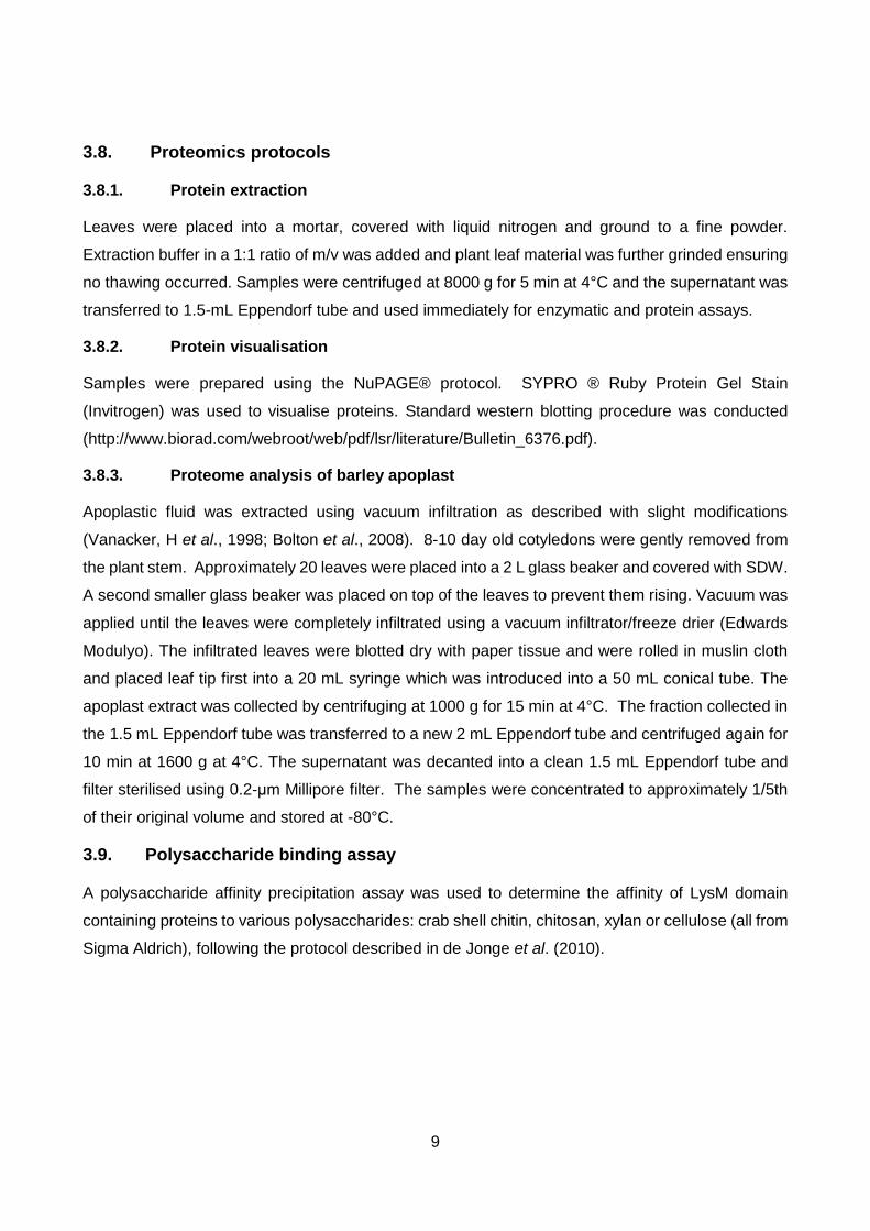
Microdochium bolleyi. BLASTp searches revealed the presence of varying numbers of LysM
domains within some of the sequences (Figure 4.1). Four LysM domain proteins identified (RcLysM1,
RcLysM5, RcLysM7 and RcChi) contained one LysM domain, while RcLysM2 and RcCAZy
contained two LysM domains. Similar to the well characterised Ecp6 effector from the tomato
pathogen C. fulvum, RcLysM3 contained three LysM domains, whereas RcLysM4 and RcLysM6
contain five and four domains respectively. BLASTp results are detailed in Table 4.3. A total of 31
R. commune candidate effectors were selected for transcription profiling during infection.
To help further prioritise R. commune molecules involved in infection the level of their presence
during infection was determined. The candidate effectors expression profiles were split into four
groups, based on the infection stage in which their expression peaked (Figure 4.2). The largest
proportion of candidate effector genes were upregulated at the biotrophic stage. Exactly half of the
candidate genes were most highly expressed between 6-8 dpi when the fungus would have already
established a mycelial network within the apoplast. All of the candidates within this group exhibited
a similar profile - a gradual increase from 1-2 dpi with a distinct maximum between 3-6 dpi, continuing
expression at 8 dpi and a subsequent decline (Figure 4.2).
Three genes, Rc_10934, Rc_2091 and Rc_2835, were selected for further analysis as they
were some of the highest expressed during infection. The selected candidates shared the same
expression profile, inclining from 1 dpi with highest expression at 6 dpi before declining at 8 dpi and
subsequently at 10 dpi (Figure 4.5 H, I & J). The increase in transcript abundance between the three
candidates varied considerably. Rc_2835 showed the highest level of upregulation, reaching a
1200-fold increase at 6 dpi compared to its level in conidia. At the peak of its expression, Rc_2835
transcript was almost as abundant as actin. Both Rc_10934 and Rc2091 were highly upregulated
during barley infection compared to their levels in conidia, with a 150-fold and 25-fold increase
respectively. At the peak of their expression, Rc_10934 and Rc2091 transcripts were 1.5 and 5.5
times as abundant as actin, respectively. All LysM fungal effectors were expressed at a time
corresponding to the potential release of chitin fragments from the fungal cell walls into the apoplast

11
and thus may play a role in chitin sequestration (Figure 4.5, A-G), apart from LysM4 and RcCazy,
which were not expressed during infection. In addition, expression at this stage of infection suggests
other possible roles in the colonisation of the plant apoplast aiding in the protection against plant
immunity like that of C. fulvum effector Avr4 (van den Burg et al., 2006).
Table 4.2. Sequence analysis of Rhynchosporium commune candidate effectors and homology to
other fungal proteins.
Candidate gene Id
Protein length
Cysteines Top BLASTp hit Species
Rc_01097 103 8 hypothetical protein MBM_09244
Marssonina brunnea f. sp.
Rc_01130 157 14 No significant similarities
Rc_01776 91 8 hypothetical protein FOCG_15424
Fusarium oxysporum f. sp.
Rc_02091 138 10 No significant similarities
Rc_02410 149 6 No significant similarities
Rc_2835 125 6 No significant similarities
Rc_05049 194 4 No significant similarities
Rc_05109 116 6 hypothetical protein LY89DRAFT_729122
Phialocephala scopiformis
Rc_5673 157 8 No significant similarities
Rc_05783 121 6 hypothetical protein LY89DRAFT_579580
Phialocephala scopiformis
Rc_06721 104 8 putative aldehyde dehydrogenase
Diaporthe ampelina
Rc_07354 151 8 hypothetical protein LY89DRAFT_723264
Phialocephala scopiformis
Rc_07612 129 8 No significant similarities
Rc_08075 160 6 hypothetical protein MBM_08646
Marssonina brunnea f. sp.
Rc_08731 145 8 hypothetical protein Micbo1qcDRAFT_180629
Microdochium bolleyi
Rc_10317 67 6 No significant similarities
Rc_10933 137 8 No significant similarities
Rc_10934 117 6 No significant similarities
Rc_11163 126 4 hypothetical protein LY89DRAFT_730227
Phialocephala scopiformis
Rc_11301 191 7 No significant similarities
Rc_11752 59 6 No significant similarities
Rc_11935 93 5 No significant similarities

12
Table 4.3: Amino acid sequence analysis of Rhynchosporium commune LysM domain proteins.
Sequence Id Protein length, aa Top BLASTp match Species
RcLysM1 688 LysM domain-containing protein Colletotrichum graminicola
RcLysM2 332 putative cell wall-associated hydrolase
Marssonina brunnea
RcLysM3 232 putative cell wall-associated hydrolase
Marssonina brunnea
RcLysM4 449 LysM domain-containing protein Colletotrichum tofieldiae
RcLysM5 269 hypothetical protein Phialocephala scopiformis
RcLysM6 672 LysM domain-containing protein Colletotrichum graminicola
RcLysM7 164 carbohydrate-binding module family
Glonium stellatum
RcCAZy 317 hypothetical protein Marssonina brunnea
RcChi 979 glycosyl hydrolase family 18 Colletotrichum incanum
13
Figure 4.1: Schematic amino acid sequence diagrams of LysM domain proteins (not drawn to
scale). Domains are highlighted as follows: LysM, orange; signal peptide (SP), blue;
unconventional signal peptide (USP), light blue; transmembrane (TM), green; chitin binding domain
(CBD), purple; Chitinase-like superfamily, yellow-green; and Lysozyme like superfamily, red.
RcLysM1
RcLysM5
RcLysM3
RcLysM2
RcLysM4
RcLysM6
RcLysM7
RcChi
RcCazY
USP
USP
CBD

14
Figure 4.2: Relative transcript abundance of Rhynchosporium commune candidate effectors during infection of barley with R. commune strain L2A. Error bars indicate confidence intervals of the 3 technical repetitions.
020406080
100120140160180
C gC
1d
pi
2d
pi
3d
pi
4d
pi
6d
pi
8d
pi
10
dp
i
12
dp
i
14
dp
i
21
dp
i
Re
l. e
xpre
ssio
n
Days post inoculation
Group 2: 3 dpi
Rc_1097
Rc_11935
Rc_1130
Rc_6721
Rc_5049
0
50
100
150
200
250
300
C gC
1d
pi
2d
pi
3d
pi
4d
pi
6d
pi
8d
pi
10
dp
i
12
dp
i
14
dp
i
21
dp
i
Re
l. e
xpre
ssio
n
Days post inoculation
Group 1: 1-2dpi
Rc_ 5109
Rc_ 2410
Rc_7354
020406080
100120140160180
C gC
1d
pi
2d
pi
3d
pi
4d
pi
6d
pi
8d
pi
10
dp
i
12
dp
i
14
dp
i
21
dp
i
Re
l. e
xpre
ssio
n
Days post inoculation
Group 3: 4-8 dpi 2091
1776
7621
5673
10933
11301
5783
11752
8731
050
100150200250300
C gC
1d
pi
2d
pi
3d
pi
4d
pi
6d
pi
8d
pi
10
dp
i
12
dp
i
14
dp
i
21
dp
i
Re
l. e
xpre
ssio
n
Days post inoculation
Group 4: 10-21 dpi
Rc_8075
Rc_10934
Rc_11163

15
Figure 4.3: Relative expression of low abundance transcripts of Rhynchosporium commune candidate effectors during infection of barley with R. commune strain L2A. Error bars indicate confidence intervals of the 3 technical repetitions. Rc_7354 - Group1, 1-2 dpi; Rc_1097 – Group 2, 3dpi; Rc_5783, Rc_11301, Rc_10933, Rc_5763, Rc_8731 & Rc_11752 Group 3, 4-8dpi; Rc_11163 Group 4, 10-21dpi.
Figure 4.4: Relative transcript abundance of Rhynchosporium commune candidate effector Rc_2835 during infection of barley with R. commune strain L2A. Error bars indicate confidence intervals of the 3 technical repetitions.
0
500
1000
1500
2000
Re
l. e
xpre
ssio
n
Days post inoculation
Rc_2835
0
2
4
6
8
10
C gC 1dpi 2dpi 3dpi 4dpi 6dpi 8dpi 10dpi 12dpi 14dpi 21dpi
Re
l. e
xpre
ssio
n
Days post inoculation
Low abundance transcriptsRc_1097
Rc_7354
Rc_8731
Rc_11163
Rc_11752
Rc_5783
Rc_11301
Rc_10933
Rc_5673

16
Figure 4.5: Relative expression of selected Rhynchosporium commune genes during infection of susceptible barley cultivar Optic with R. commune strain L2A. Error bars indicate the confidence interval for the average of technical repetition.
0
0.5
1
1.5
2
0
50
100
150
200
C
gC
1d
pi
2d
pi
3d
pi
4d
pi
6d
pi
8d
pi
10
dpi
Tra
nsc
rip
t ab
un
dan
ce a
s a
pro
port
ion
of
Act
in
Rel
. ex
pre
ssio
n
Days post inoculation
Rc_10934
A C D B
E F G
I H J

17
To finalise the research of effector identification, apoplastic fluid extracted from the barley
leaves inoculated with R. commune strain L73A was analysed to confirm the presence of candidate
effector proteins during a susceptible interaction. Two time points were selected: 4 dpi, which
represents the initial colonisation of the apoplast, and 7 dpi, in which growth of the fungus would be
well developed.
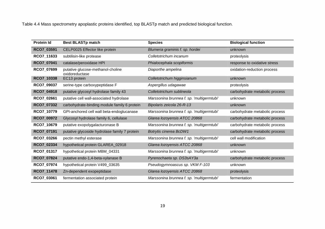
Plant cell wall degrading enzymes (CWDEs) were the most highly abundant proteins in the
apoplast during infection. Enzymes involved in the breakdown of xylan (Rc_07824), lignin
(Rc_07699), pectin (Rc_03266) and cellulose (Rc_00972) were identified (Table 4.4). This was not
surprising as CWDEs play a significant role in pathogenesis with the ability to depolymerize the main
structural polysaccharide components of the plant cell wall (Kubicek et al., 2014). Furthermore,
amongst the most abundant proteins was a putative glucose-methanol-choline (GMC)
oxidoreductase which has been suggested to be a lignocellulose acting enzyme (Couturier et al.,
2015). Two different types of proteases were identified, a serine type carboxypeptidase and a
subtilisin like protease (Table 4.4). In many cases, proteases are considered to be virulence factors
of many pathogenic species (Hoge et al., 2010).
Similar to many plant pathogens, R. commune secretes a probable catalase peroxidase at
both 4 and 7 dpi with a high up-regulation of the protein at the latter time point of infection (Table
4.3). The importance of catalase peroxidases to circumvent the effects of plant defence have been
highlighted in numerous studies (Zámocký et al., 2009). Catalase-peroxidase proteins are known to
detoxify the products of the oxidative burst in the apoplast upon the triggering of plant immunity.
Tanabe et al. (2011) demonstrated that one of the three catalase peroxidase genes identified in Z.
tritici plays an important role in pathogenicity. MgDCat-1 is also upregulated during infection and is
most abundant at 8 dpi.
Although none of the candidate effectors from the research described in this chapter were
detected in apoplastic fluid from barley leaves infected with R. commune, proteomics analysis
identified four other potential effectors. These included two proteins which had been previously
highlighted as candidate effectors but not in the original panel: Rc07_03591, which showed
homology to an effector like protein from powdery mildew; B. graminis f. sp. Hordei; and
Rc07_02334, a hypothetical fungal protein from the anamorphic fungus Glarea lozoyensis. The best
BLASTp hit for RC07_10338 was to EC13 protein from anthracnose leaf spot which has been shown
to be expressed during the establishment of biotrophic hyphae (Kleemann et al., 2008). Lastly,
protein Rc_LysM3 which contained three LysM domains was also identified. Interestingly LysM
domain proteins have been well characterised in several plant pathogens and shown to play a
fundamental role in fungal pathogenesis (Kombrink, 2013).
Candidates Rc_10934, Rc_2091 and Rc_2835 were selected in the attempt to knock out the
gene of interest and obtain a phenotype. Each candidate knockout was attempted three times and
resulted in the creation of between 60 to 100 transformants for each attempt. Amplification and
sequencing of the deletion cassette from the DNA extracted from the transformants confirmed its

18
successful integration into the fungal genome. Primers were used to amplify the deletion cassette
and determine if the original gene was disrupted. A typical result of genotyping of the transformants
is detailed in Figure 4.6. No candidate effector genes were knocked out and time restrictions limited
any further continuation of the approach.
To confirm the chitin binding prediction RcLysM3 protein tagged with V5 peptide at the C
terminus to allow detection, was produced in P. pastoris and affinity binding to a range of
polysaccharides was examined. RcLysM3 co-precipitated with crab shell chitin and, interestingly,
with chitosan but not with any of the plant cell wall polysaccharides, xylan or cellulose (Figure 4.7).
RcLysM3 was also identified as a potential avirulence protein. A change in an amino acid at position
67 from a glutamic acid (Q) to glutamine (E) within the protein sequence of RcLysM3 was identified
that correlated with a change in virulence/avirulence of 9 sequenced R. commune strains on cultivar
La Mesita (Table 4.5). Isolate L43D carrying the E allele was avirulent on cultivar La Mesita. A
detached leaf assay confirmed the lack of macroscopic symptoms. RcLysM3 sequence was
analysed in a further four isolates L101B, L90A, L43A and L43B. Both L101B and L90A contained
the Q allele whereas L43A and L43B contained the E allele (Table 4.5). The latter two were isolated
from the same plant and are possibly the same strain as L43D. While both L101B and L90A isolates
containing the Q allele were virulent on La Mesita in line with Q allele being a virulent allele, virulence
testing of the isolates L43A and L43B contained the E allele on La Mesita still needs to be conducted
to determine if the correlation is valid for these isolates.
To identify novel resistance, detached barley leaves were inoculated with a conidial
suspension of R. commune to obtain a phenotype of each barley line. A total of nine sequenced
strains were used to infect a set of barley lines containing the resistance genes Rrs3, Rrs4 and
Rrs13. Each assay included a very susceptible cultivar Optic as a control to determine isolates’
aggressiveness. Inspection of lesion formation began around 10 dpi and lesion measurements
continued until 21 dpi. All barley lines tested including Abyssinian (Rrs3), CI11549 (Rrs4) and BC
Line30 (Rrs13) were shown to be susceptible to L77 and AU2 which were the most virulent in
comparison to other strains (Table 4.5). Strain AU2 caused early lesion development, susceptible
barley lines inoculated with strain L77 also developed lesions quickly and produced symptoms that
were comparable to the highly susceptible control Optic, although lesions did take longer to develop
on Cl11549 which contains the Rrs4 gene. In contrast strains UK7, L32B, L43D, L73A and 214-GFP
caused no lesions on barley plants containing Rrs3 or Rrs4 and Rrs13 (Table 4.5). The lack of lesions
may indicate that these strains contain avirulence genes recognised by Rrs3 or Rrs4 and Rrs13, or
the presence of extra resistance to these strains which can be further assessed.
19
Table 4.4 Mass spectrometry apoplastic proteins identified, top BLASTp match and predicted biological function.
Protein Id Best BLASTp match Species Biological function
RCO7_03591 CELP0025 Effector like protein Blumeria graminis f. sp. hordei unknown
RCO7_11633 subtilisin-like protease Colletotrichum incanum proteolysis
RCO7_07041 catalase/peroxidase HPI Phialocephala scopiformis response to oxidative stress
RCO7_07699 putative glucose-methanol-choline oxidoreductase
Diaporthe ampelina oxidation-reduction process
RCO7_10338 EC13 protein Colletotrichum higginsianum unknown
RCO7_09037 serine-type carboxypeptidase F Aspergillus udagawae proteolysis
RCO7_04918 putative glycosyl hydrolase family 43 Colletotrichum sublineola carbohydrate metabolic process
RCO7_02661 putative cell wall-associated hydrolase Marssonina brunnea f. sp. 'multigermtubi' unknown
RCO7_07332 carbohydrate-binding module family 6 protein Bipolaris zeicola 26-R-13 unknown
RCO7_10779 GPI-anchored cell wall beta-endoglucanase Marssonina brunnea f. sp. 'multigermtubi' carbohydrate metabolic process
RCO7_00972 Glycosyl hydrolase family 6, cellulase Glarea lozoyensis ATCC 20868 carbohydrate metabolic process
RCO7_10679 putative exopolygalacturonase B Marssonina brunnea f. sp. 'multigermtubi' carbohydrate metabolic process
RCO7_07191 putative glycoside hydrolase family 7 protein Botrytis cinerea BcDW1 carbohydrate metabolic process
RCO7_03266 pectin methyl esterase Marssonina brunnea f. sp. 'multigermtubi' cell wall modification
RCO7_02334 hypothetical protein GLAREA_02918 Glarea lozoyensis ATCC 20868 unknown
RCO7_01317 hypothetical protein MBM_04331 Marssonina brunnea f. sp. 'multigermtubi' unknown
RCO7_07824 putative endo-1,4-beta-xylanase B Pyrenochaeta sp. DS3sAY3a carbohydrate metabolic process
RCO7_07974 hypothetical protein V499_03635 Pseudogymnoascus sp. VKM F-103 unknown
RCO7_11478 Zn-dependent exopeptidase Glarea lozoyensis ATCC 20868 proteolysis
RCO7_03061 fermentation associated protein Marssonina brunnea f. sp. 'multigermtubi' fermentation

20
Figure 4.6: Genotyping of Rhynchosporium commune transformants. A) Primer locations used for genotyping strategy to determine targeted gene disruption and hygromycin resistance gene insert. Red_5’ UTR G1. Orange- G2, wild type ORF reverse. Green hygromycin forward, Blue hygromycin reverse. B) PCR. Lane 1: 1kb ladder. PCR products produced using Lane 2: Hygromycin F&R primers. Lane 3: G1 &G2 primers – amplification of wild type. Lane 4: No amplification with G1and HYG R primers. Amplification of actin was used as a loading control.
Figure 4.7 Protein gel showing RcLysM3-V5 protein co-precipitating in the pellet (P) of chitin and chitosan, but only present in the supernatant (S) of cellulose and xylan. Table 4.4: Correlation of the Gln (E) and Glu (Q) allele with the virulence and avirulence of
Rhynchosporium commune isolates on barley cultivar La Mesita.
R. commune isolates
13-13 214 L2A L32B
L43D
L73A
L77 UK7 AU2 101B
90 B
L43A
L43 B
La Mesita V V V V A V V V V V V ? ?
RcLysM3 allele
Q Q Q Q E Q Q Q Q Q Q E E
P
S
1kbb
5’ UTR 3’UTR ORF
Resistance gene 3’ UTR 5’ UTR
1.5kb
500bp
G1 G2
B)
Actin loading control
Hyg F Hyg R
G1
A)

21
Barley lines containing Rrs4 and Rrs13 showed a moderately high level of resistance in terms
of lack of lesion formation to 5 out of 7 R. commune strains. Further analysis to determine how the
fungus proliferates during asymptomatic infection was conducted using R. commune strain 214-
GFP. Growth after 10 dpi was investigated to determine the extent of the mycelial network. In
comparison to the susceptible barley line the amount of growth at 10 dpi was much less for BC
Line30 carrying the Rrs13 resistance gene (Figure 4.8 D). Although the growth was less, it followed
the same pattern of growth as seen in a susceptible cultivar (Figure 4.8 H). Despite the fact that
pathogen growth on Rrs4 line Cl11549 was evident, the type of growth differed. Instead of the
mycelium forming lines between the epidermal cells, the fungal growth was random. The mycelium
did not travel far from the inoculation spot suggesting line Cl11549 is resistant to strain 214 (Figure
4.8 B).
Table 4.5: Virulence testing results of barley lines containing Rrs3, Rrs4 & Rrs13 resistance genes
inoculated with Rhynchosporium commune strains L32B, L43D, L73A, L77, UK7, AU2 & 214-GFP.
Barley R gene
R. commune isolates
L32B L43D L73A L77 UK7 AU2 214-GFP
Rrs3
A A A V A V A
Rrs4
A A A V A V A
Rrs13
A A A V A V A

22
Figure 4.8: Confocal LASER microscopy images of Rhynchosporium commune strain 214-GFP infection on: A & B CI11549; C & D BC Line 30; E & F Optic. Scale bars A, C, E = 50µm & B, D, F = 100 µm.
In addition, a further two barley lines were analysed for asymptomatic growth. Syrian
landraces were used to look at response to infection and were included in this research as they are
genetically more diverse than cultivated barley which increases the chance of finding novel barley
resistance. It was evident that the interaction between SLB 66_024 (unknown R gene) and 214-
GFP was not compatible. The early stages of growth showed a similar pattern to a resistant line
(Figure 5.2 B) and although there was quite a substantial amount of growth at 21 dpi, the mycelium
did not grow along the epidermal cell walls (Figure 4.9 C). Instead, the growth was randomly
dispersed. In contrast, growth of 214-GFP on SLB67-015 (unknown resistance) was established
after 8 dpi (Figure 4.9 E) and continued throughout the assay resulting in a bidirectional mycelial
A
d)
c)
e)
j)
h)
d)
f)
C
D
E
B
F
A
E
C

23
growth out with the inoculum spot (Figure 4.9 F). The pattern of growth was similar to a susceptible
interaction.
Figure 4.9 Confocal LASER microscopy images of Rhynchosporium commune strain 214-GFP infection on SLB 66-024 A-B and SLB 67-015 C-D at 3 dpi, 8 dpi and 21dpi. Scale bars A & B = 50µm, C, E, F = 100µm, D = 25µm.
Although microscopy can distinguish between lack of growth and the presence of
morphological differences, to gain a better understanding of the molecules involved in resistance to
R. commune, a quantitative proteomics approach was taken to determine the change in abundance
or absence of proteins. Three biological experiments were used for the extraction of infected and
uninfected apoplastic fluid. The infection of inoculated cultivars was analysed using R. commune
strain 214-GFP. Leaf samples were viewed under a confocal microscope before taking samples for
apoplastic extraction to confirm colonisation of the leaves of susceptible cultivars Optic and Atlas
and restricted growth on the leaves of resistant cultivar Atlas 46. Growth of 214-GFP was as
expected (Thirgnanasbanadam et al., 2011) – Optic contained the highest level of colonisation
whereas resistant Atlas 46 showed very restricted growth with random colony morphology, growth
was identified on Atlas but not to the extent of Optic.
The intensity of each of the proteins was compared in 3 different cultivars, highly susceptible
Optic, Atlas 46 which contains the Rrs1 and Rrs2 gene and the NIL Atlas which does not contain the
Rrs1 gene, uninfected and infected with R. commune strain 214-GFP at 4 dpi and 7 dpi. Four
a)
b)
D
C
E F
B A C
F

24
proteins were highly abundant and showed a distinct increase in infected apoplastic fluid of Atlas 46
at 4 dpi. The remaining six proteins were most highly expressed in 7 dpi infected apoplastic fluid.
This included an α-L-arabinofuranosidase involved in cell wall reorganisation which has been
suggested as a putative defence related protein and was highly abundant in Atlas46 at 4 dpi infected
apoplastic fluid (Figure 4.10). The protein has also been found in pathogens to aid with plant cell
wall breakdown (Morant et al., 2008). In addition, pathogenesis related (PR) proteins which are well
known to participate in complex plant defence responses to pathogens were also identified including
glucan endo-1,3-beta-glucosidase- PR2 playing a role in the hydrolysis of fungal cell walls (Figure
4.10).
To investigate the possibility of R. commune growth on other plant species with the absence
of any visual disease symptoms, R. commune inoculations were carried out on the model plant
species N. benthamiana. Drop inoculations of spores from R. commune strain 214-GFP was carried
out on leaves of N. benthamiana plants.
N. benthamiana plants with plasma membrane protein tagged with a red fluorscent protein
were used to determine if any signs of damage were occurring inside the leaf tissue. At 5 dpi,
microscopic anlaysis of R. commune revealed the germination of fungal conidia. By 9 dpi fungal
mycelium had started to develop and the growth of the fungus from the original inoculation spot had
increased. At 15 dpi there was a noticable increase in the amount of mycelium. From this point
and until the last day of analysis the fungal mycelium did not grow in the same manner as it would
on its host barley, outlining the epidermal cells. In fact the growth resembled that of an incompatible
infection on barley with explorative hyphae growing in all directions (Thirugnanasambandam et al.,
2011). The spread of the fungus did persist over time resulting in a sizeable colony by 28 dpi (Figure
4.11 A-C). The plant showed no evidence of plasma membrane deterioration, as would be seen
during the late stages of infection in barley. The plant plasma membrane was unimpaired which was
clearly evident at the later time point, 28 dpi (Figure 4.11). Throughout the entire experiment no
macroscopic signs of infection were visible.
e) f)

25
0
2
4
6
8
4DPI NONINFECTED
4DPIINFECTED
7DPI NONINFECTED
7DPIINFECTED
log
tra
nd
form
ed
in
tesn
sity
va
lues
apoplast sample
Purple acid phosphotase
Optic Atlas Atlas 46
0
10
20
30
4DPI NONINFECTED
4DPIINFECTED
7DPI NONINFECTED
7DPIINFECTED
log
tra
nd
form
ed
in
tesn
sity
va
lues
apoplast sample
Subtil isin l ike serine
protease
Optic Atlas Atlas 46
0
2
4
6
8
4DPI NONINFECTED
4DPIINFECTED
7DPI NONINFECTED
7DPIINFECTED
log
tra
nd
form
ed
in
tesn
sity
va
lues
apoplast sample
Glucan endo-1,3-beta
glucosidase
Optic Atlas Atlas 46
0
1
2
3
4DPI NONINFECTED
4DPIINFECTED
7DPI NONINFECTED
7DPIINFECTED
log
tran
sfo
rme
d in
ten
sity
val
ue
s
apoplast sample
Disease resistant protein
Optic Atlas Atlas 46
020406080
100
4DPI NONINFECTED
4DPIINFECTED
7DPI NONINFECTED
7DPIINFECTED
log
tra
nd
form
ed
in
tesn
sity
va
lues
apoplast sample
Chitinase
Optic Atlas Atlas 46
0
2
4
6
8
10
4DPI NONINFECTED
4DPIINFECTED
7DPI NONINFECTED
7DPIINFECTED
log
tra
nd
form
ed
in
tesn
sity
va
lues
apoplast sample
Thuamat in l ike protein
Optic Atlas Atlas 46
0
2
4
6
8
4DPI NONINFECTED
4DPIINFECTED
7DPI NONINFECTED
7DPIINFECTED
log
tra
nd
form
ed
in
tesn
sity
va
lues
apoplast sample
Beta-glucosidase
Optic Atlas Atlas 46
D
0
5
10
15
4DPINON
INFECTED
4DPIINFECTED
7DPINON
INFECTED
7DPIINFECTED
log
tra
nd
form
ed
in
tesn
sity
va
lues
apoplast sample
(1-3) beta glucanase
Optic Atlas Atlas 46
00.5
11.5
2
4DPI NONINFECTED
4DPIINFECTED
7DPI NONINFECTED
7DPIINFECTED
log
tra
nd
form
ed
in
tesn
sity
va
lues
apoplast sample
Alpha -L
arabinofuranosidase
Optic Atlas Atlas 46
B
02468
10
4DPI NONINFECTED
4DPIINFECTED
7DPI NONINFECTED
7DPIINFECTED
log
tra
nd
form
ed
inte
snsi
ty v
alu
es
apoplast sample
Serine carboxypeptidase
Optic Atlas Atlas 46
E
C
F
Figure 4.10: Proteins highly abundant at 4 dpi and 7 dpi.
Intensity values in apoplastic fluid samples from cultivars Optic,
blue line, Atlas, red line, and Atlas 46, green line, from non-
infected samples and infected samples (inoculated with
Rhynchosporium commune strain 214-GFP) at 4 and 7 dpi.
G H
I J

26
Figure 4.11: A-C Confocal microscopy images of Rhynchosporium commune strain 214-GFP on Nicotiana benthamiana line CB173 expressing red plasma membrane at 28 dpi. Orange arrow shows intact plant plasma membrane. N. benthamiana plants inoculated with R. commune strain 214-GFP D) at 9 dpi and E) 28 dpi, showing no macroscopic symptoms. Scale bars = 25 µm.
5. Discussion
As the global population increases rapidly, agriculture struggles to maintain the levels of crop
production required for the immense rise in food demands. Plant pathogens have a high capacity to
cause substantial disease levels on food crops, reducing the production and quality of food. Hence,
greater emphasis to reduce the impact of crop disease is required. In many cases chemical
treatments to limit or eradicate diseases are used, however the environmental impact of the
applications can result in consequences to non-target organisms, pesticide drift and residues on food
(Kilbrew & Wolff, 2010). Agriculture is faced with the challenge to maximise crop yields while
decreasing negative environmental impacts. However, several factors influence the reduction of food
security imposed by pathogens. The lack of well-developed diagnostic tools to identify asymptomatic
pathogen infection can lead to severe disease implications later in the growing season. In addition,
the level of disease severity can be overlooked due to subjective rather than quantitative methods
A
E D
C B

27
to detect pathogen biomass accumulation. Furthermore, experimental obstacles preventing the
mapping and cloning of plant resistance genes in conjunction with the variation and vast amounts of
evolving pathogen molecules, results in the lack of complete understanding of the mechanisms of
resistance and pathogen infection. Therefore, the development of methods to identify pathogens,
experimental research to gain an understanding of how the pathogen infects and the molecules
involved in plant defence against pathogens will result in better understanding of how we can
improve methods for diagnostics and predicting crop resistance durability.
As pathogens are known to use effector molecules to overcome plant resistance (Dangl &
Jones, 2006), this study began with the exploitation of the genome and transcriptome sequences of
R. commune to identify novel candidate effectors. The importance of effector discovery is high as
the research into R. commune effector repertoire is still in its infancy. The expression profiling was
effective in determining the timing and levels of gene expression that can be used to indicate the
involvement of candidate effectors in pathogenesis. Extremely low efficiency of targeted gene
disruption in R. commune limited the possibility of functional characterisation. However new
approaches are now being developed in fungi, which can be used in the future (Matsurura et al.,
2015). The identification of RcLysM3 was an important discovery, indicating its high abundance
within the apoplast. Further characterisation revealing chitin binding abilities and avirulence
correlation indicates an essential nature for this protein. Revealing that effectors are essential for
pathogenicity and potentially recognised by the host plant (Avr genes) is an important factor. Barley
R gene resistance to R. commune has not proved durable; therefore the discovery of novel
avirulence genes that are required for pathogenicity is a critical step to identify more durable forms
of resistance to this devastating fungal disease.
Despite the identification of AvrRrs1 recognition by the Rrs1 over 20 years ago (Rohe et al.,
1995), there is very little information on the intricate molecular mechanisms that occur in a resistant
response. To characterise other types of resistance to R. commune, cultivars were selected
containing different R genes to that of Rrs1. The virulence testing approach helped to prioritise barley
lines for further analysis using the 214-GFP strain. Microscopic assessment of the extent of the
growth and the colony morphology were used to distinguish between susceptibility and potential
resistance to R. commune. This is one of the characteristics of Rrs1 containing plants that has been
previously highlighted. Although barley lines presenting no symptoms and a decrease in biomass,
physically restricted growth and / or random colony morphology could be a sign of resistant
interaction, it still remains difficult to determine the durability of the plant defence. It is possible that
some R. commune strains develop much slower throughout the growing season but their
accumulation in barley leaves may still have an impact on the crop yield although no research has
looked into this possibility. In addition, a range of R. commune strains need to be used to distinguish
the level of asymptomatic infection. The production of some other fungal strains expressing
fluorescent proteins would be highly beneficial for future research, especially for highly virulent stains
such as AU2. Partial resistance could also be potentially at play, as it is also characterised by

28
reduced growth of the pathogen. Again, there is a need to gain a better understanding of this type
of resistance.
Due to the lack of evidence to allow full confirmation of resistant lines, a proteome approach
to identify the key players in Rrs1 resistance was conducted. Initial research began on the contents
of apoplast and its importance in plant pathogen interactions was identified almost 30 years ago.
However, only a few studies have focused on plant-pathogen interactions in the apoplast (Mehta et
al., 2008). The identification of known defence proteins present in all of the cultivars used indicated
similar components of a basal defence mechanism. However, the abundance of defence proteins
in the resistant line was slightly higher. Only the disease related protein and α-L arabinofuriodase
were highly upregulated in comparison to Optic and Atlas suggesting a specific role in the Rrs1-
controlled resistance. Some proteins which were down regulated in Atlas 46 might represent
susceptibility factors for disease and therefore were not upregulated in a resistant response. Only
one biological repetition was available for analysis due to the inefficient labelling, other repetitions
would be required to provide rigidity to the results. Although the extraction of the apoplastic fluid is
relatively laborious and in some cases protein identification can be limited, this work has identified
some important plant molecules that could be further analysed for their potential use as markers of
barley resistance to R. commune.
Major R gene resistance is an important factor of sustainable agriculture, although protection
against several strains of a pathogen may be incompletely effective. However, the use of R gene
pyramids may provide an alternative and more effective strategy to control various R. commune
pathotypes (Zhan et al., 2012). More recently the investigation of NHR has become more prominent
in the literature. NHR is only beginning to be understood but in contrast to major R gene resistance
the response involves multiple pathways (Gill et al., 2015) and is known to provide simultaneous
resistance to many pathogens. The results of this research have indicated N. benthamiana as a non-
host. Furthermore, the inability of R. commune to infect non-grass species highlights the importance
of crop rotations for prevention of the build-up of the pathogen inoculum in the field. Further
identification and characterisation of components of NHR will provide an effective alternative for the
future development of crops with a wide range of more durable resistance.
6. References
Ayliffe M, Singh D., Park R., Moscou M., Pryor T, 2013. Infection of Brachypodium distachyon with selected grass rust pathogens. Mol. Plant Microbe Interact 26, 946–957.
Chung, K.-R. & Lee, M.-H., 2015. Split-Marker-Mediated Transformation and Targeted Gene Disruption in Filamentous Fungi. In A. M. van den Berg & K. Maruthachalam, eds. Genetic Transformation Systems in Fungi, Volume 2. Cham: Springer International Publishing, pp. 175–180.
Couturier M, Navarro D., Chevret D., Henrissat B., Piumi F., Ruiz-Dueñas F., Martinez A., Grigoriev I., Riley R., Lipzen A., Berrin J., Master E., Rosso M-N, 2015.

29
Enhanced degradation of softwood versus hardwood by the white-rot fungus Pycnoporus coccineus. Biotechnology for Biofuels, 8(1), pp.1–16.
Delaunois B, Jeandet P, Clément C, Baillieul F, Dorey S, Cordelier, S, 2014. Uncovering plant-pathogen crosstalk through apoplastic proteomic studies. Frontiers in Plant Science 5, 249.
Fan J, Doerner P, 2011. Genetic and molecular basis of nonhost disease resistance: complex, yes; silver bullet, no. Curr. Opin. Plant Biol. 15, 400–406.
Floerl S Druebert,C, Majcherczyk A, Karlovsky P,Kües U,Polle A, 2008. Defence reactions in the apoplastic proteome of oilseed rape (Brassica napus var. napus) attenuate Verticillium longisporum growth but not disease symptoms. BMC Plant Biology 8, 1–15.
Goodin M, Zaitlin D, Naidu R, Lommel, S 2008. Nicotiana benthamiana: its history and future as a model for plant-pathogen interactions. Mol. Plant Microbe Interact 21, 1015–1026.
Gill U, Lee S, Mysore K, 2015a. Host versus nonhost resistance: distinct wars with similar arsenals. Phytopathology 105, 580–587.
Jonge R, Bolton M, Thomma B, 2011. How filamentous pathogens co-opt plants: the ins and outs of fungal effectors. Current opinion in plant biology 14, 400-406.Jones J, Dangl J, 2006. The plant immune system. Nature 444, 323-329.
King K, West J, Brunner P, Dyer P, Fitt B 2013. Evolutionary Relationships between Rhynchosporium lolii sp. nov. and other Rhynchosporium Species on Grasses. PLoS ONE 8
Kirsten, S., Siersleben, S. & Knogge, W., 2011. A GFP-based assay to quantify the impact of effectors on the ex planta development of the slowly growing barley pathogen Rhynchosporium commune. Mycologia , 103(5), pp.1019–1027.
Kombrink A, Thomma B, 2013. LysM Effectors: Secreted Proteins Supporting Fungal Life. PLoS Pathog 9, e1003769.
Kubicek C. P., Starr T. L., Glass N. L. 2014. Plant cell wall-degrading enzymes and their secretion in plant-pathogenic fungi. Ann. Rev. Phytopathol. 52 427–451.
Kück, U. & Hoff, B., 2010. New tools for the genetic manipulation of filamentous fungi. Applied Microbiology and Biotechnology, 86(1), pp.51–62.
Lee, HK Tewari, J, Turkington T, 2001. Symptomless infection of barley seed by Rhynchosporium secalis. Canadian Journal of Plant Pathology 23, 315-317.
Matsu-ura, T et al., 2015. Efficient gene editing in Neurospora crassa with CRISPR technology. Fungal Biology and Biotechnology 2, 1–7.
McDermott J, McDonald B, Allard R, Webster R, 1989. Genetic variability for pathogenicity, isozyme, ribosomal DNA and colony color variants in populations of Rhynchosporium secalis. Genetics 122, 561-565.
Newton AC, 1989. Somatic recombination in Rhynchosporium secalis. Plant Pathology 38, 71-74.
Newton A, Searle J, Guy D, Hackett C, Cooke D, 2001. Variability in pathotypes, aggressiveness, RAPD profile, and rDNA ITS1 sequences of UK isolates of Rhynchosporium secalis. Journal of Plant Diseases and Protection 108, 446-458.
Newman C, Newman R, 2006. A brief history of barley foods. Cereal Foods World 51, 4–7. Newman R, Newman C, Graham H, (1989). The hypocholesterolemic function of barley B-
glucan. Cereal Foods World 34, 883–886. Saisho D, Takeda K, 2011.Barley: Emergence as a New Research Material of Crop Science.
Plant Cell Physiol 52, 724-727. Stam R, Mantelin S, McLellan H, Thilliez G, 2014. The role of effectors in nonhost resistance
to filamentous plant pathogens. Frontiers in Plant Science 5, 582. Schurch S, Linde CC, Knogge W, Jackson LF, McDonald BA, 2004. Molecular population
genetic analysis differentiates two virulence mechanisms of the fungal avirulence gene NIP1. Molecular Plant-Microbe Interactions 17, 1114-1125.
Thirugnanasambandam A, Wright K, Atkins S, Whisson S, Newton A, 2011. Infection of Rrs1 barley by an incompatible race of the fungus Rhynchosporium secalis expressing the green fluorescent protein. Plant Pathology 60, 513-521.
Uma B, Swaroopa Rani T, Podile A, 2011. Warriors at the gate that never sleep: non-host resistance in plants. J. Plant Physiol 168, 2141–2152.
van der Westhuizen J, Qian X, Botha A, 1998. Differential induction of apoplastic peroxidase and chitinase activities in susceptible and resistant wheat cultivars by Russian wheat aphid infestation. Plant Cell Reports, 18, 132–137.
30
van den Burg H, Harrison S, Joosten M, Vervoort J, de Wit P, 2006. Cladosporium fulvum Avr4 protects fungal cell walls against hydrolysis by plant chitinases accumulating during infection. Mol. Plant-Microbe Interact. 19, 1420-1430.
Yang, L et al., 2004 Rapid Production of Gene Replacement Constructs and Generation of a Green Fluorescent Protein-Tagged Centromeric Marker in Aspergillus nidulans Eukaryotic Cell. 3, 1359-1362.
Zámocký, M, Hallberg M, Ludwig R, Divne C, Haltrich D, 2004. Ancestral gene fusion in cellobiose dehydrogenases reflects a specific evolution of GMC oxidoreductases in fungi. Gene 338, 1–14.
Zaffarano P, McDonald B, Zala M, Linde C, 2006. Global hierarchical gene diversity analysis suggests the Fertile Crescent is not the centre of origin of the barley scald pathogen Rhynchosporium secalis. Phytopathology 96, 941-950.
Zhan J, Fitt B, Pinnschmidt H, Oxley S, Newton A, 2008. Resistance, epidemiology and sustainable management of Rhynchosporium secalis populations on barley. Plant Pathology 57, 1-14.
Zhu W, Wei W, Fu Y, Cheng J, Xie J, Li G, Yi X Kang, Z Dickman M, Jiang D, 2013. A secretory protein of the necrotrophic fungus Sclerotinia sclerotiorum that supresses host resistance. PLoS One 8, e53901.