rasmol - institute for molecular virology · note: we are using rasmol in macosx under x-windows....
TRANSCRIPT

Biochem 660 – 2008 165
Rasmol‐page‐165
Rasmol
✔ INFO - RasMol (shadowed RASter MOLecules) is a program for molecular graphics visualization well suited for Protein Data Bank formatted files. Please review the four pages dedicated to Rasmol in the previous section. Manual: the HTML online manual is the most direct source for information about any command: http://www.openrasmol.org/doc/rasmol.html Getting your own copy: The original author, Roger Sayle, stopped developing Rasmol at version 2.6beta2, and further development is done as open source, currently at version 2.7.x. Therefore there are 2 sources for downloading Rasmol regardless of the operating system: For version 2.6beta2: http://www.umass.edu/microbio/rasmol/getras.htm For version 2.7.x: http://arcib.dowling.edu/software/rasmol/index.shtml and http://www.openrasmol.org/OpenRasMol.html#Software
Note: within the exercises, Bold text shows what actions are taken by the user (you!): typing text or clicking the mouse.
- e -
1 RasMol - Exercise A: Download a PDB from the database repository
Reminder: Structures have a PDB ID code made of a combination of 4 letters and/or numbers. PDB files contain coordinates pertinent to the crystallographic arrangement of the molecules within the crystal.
✔ TASK 1) Open a web browser such as Safari or Firefox (bottom task bar) 2) Point your web browser to www.rcsb.org 3) In the Search box enter/type the following ID: 1ccr and click the
SEARCH button 4) On the left column click
Download Files to show submenus
5) Click on PDB File 6) The file is automatically saved
on the desktop as 1CCR.pdb. If it is not, saved automatically, manually save the file on the desktop and name it 1CCR.pdb.

Biochem 660 – 2008 166
Rasmol‐page‐166
You can look at some of the information displayed by the web page pertinent to this molecule, such as its name (rice ferricytochrome C), journal reference, etc. You can now close or hide (Command-H or ⌘R) the web browser. Create a directory called MyRasmol on the desktop, and move the file 1CCR.pdb into it. (Use the menu cascade: File > New Folder to create new directory from the Finder. Hint: if you don’t see Finder, click anywhere on the desktop.
- e -
2 RasMol - Exercise B: Opening Rasmol and Basic graphics (manipulation and renderings)
Note: we are using Rasmol in MacOSX under X-windows. It needs to be launched by line-command as shown below. In Windows and Mac Classic you can launch Rasmol by double clicking either on the PDB file or on the Rasmol (Rasmac / Raswin) icon.
✔ TASK
1) Open the Rasmol program and a molecule at the same time Click on the X11 logo within the Dock (bottom of screen) A terminal window will appear. The window is labeled “xterm” it will become the Rasmol command line.
At the % or $ prompt type on 2 lines: cd Desktop/MyRasmol /sw/bin/rasmol 1CCR.pdb
Note: This is case sensitive.
A window will appear with the name of the PDB file (1CCR) and harboring Rasmol menus (File, Display, Colours, Options, Export). The terminal also becomes the Rasmol text-window:
Biochem 660 – 2008 167
Rasmol‐page‐167
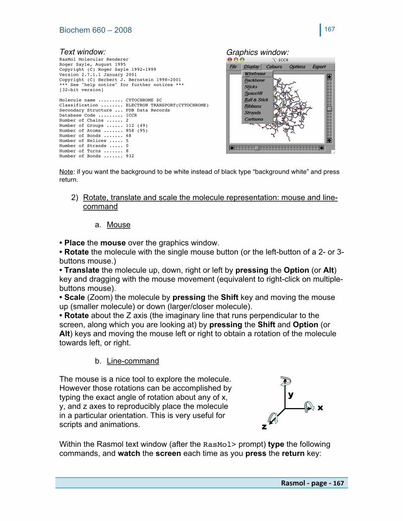
Text window: Graphics window: RasMol Molecular Renderer Roger Sayle, August 1995 Copyright (C) Roger Sayle 1992-1999 Version 2.7.1.1 January 2001 Copyright (C) Herbert J. Bernstein 1998-2001 *** See "help notice" for further notices *** [32-bit version] Molecule name ......... CYTOCHROME $C Classification ........ ELECTRON TRANSPORT(CYTOCHROME) Secondary Structure ... PDB Data Records Database Code ......... 1CCR Number of Chains ...... 2 Number of Groups ...... 112 (49) Number of Atoms ....... 858 (95) Number of Bonds ....... 68 Number of Helices ..... 5 Number of Strands ..... 0 Number of Turns ....... 8 Number of Bonds ....... 932 Note: if you want the background to be white instead of black type “background white” and press return.
2) Rotate, translate and scale the molecule representation: mouse and line-command
a. Mouse
• Place the mouse over the graphics window. • Rotate the molecule with the single mouse button (or the left-button of a 2- or 3-buttons mouse.) • Translate the molecule up, down, right or left by pressing the Option (or Alt) key and dragging with the mouse movement (equivalent to right-click on multiple-buttons mouse). • Scale (Zoom) the molecule by pressing the Shift key and moving the mouse up (smaller molecule) or down (larger/closer molecule). • Rotate about the Z axis (the imaginary line that runs perpendicular to the screen, along which you are looking at) by pressing the Shift and Option (or Alt) keys and moving the mouse left or right to obtain a rotation of the molecule towards left, or right.
b. Line-command The mouse is a nice tool to explore the molecule. However those rotations can be accomplished by typing the exact angle of rotation about any of x, y, and z axes to reproducibly place the molecule in a particular orientation. This is very useful for scripts and animations. Within the Rasmol text window (after the RasMol> prompt) type the following commands, and watch the screen each time as you press the return key:

Biochem 660 – 2008 168
Rasmol‐page‐168
reset rotate y 10 rotate x 10 rotate z 10 zoom zoom 200 zoom 5 reset
3) Change the representation and color of the molecule
a. Mouse This is a simple exploration that you can do quickly: with the mouse select the menu Display and choose the various options successively and observe how this affects the graphical representation. Remain or return to Cartoon. Select the menu Colours and try the various menu options. Return to the Colours menu Group which displays a rainbow color from blue (N-terminal) to red (C-terminal).
b. Line-command
The Display menus are also available as line-commands, also useful for scripting as we shall see shortly. Within the Rasmol text window (after the RasMol> prompt) type the following commands, and observe the screen as you press the return key: Play with structural styles: cartoon off wireframe cartoon cartoon off wireframe off backbone backbone 0.3 backbone off spacefill spacefill 0.5 wireframe 0.2
Note that when using the mouse, selecting an alternate representation (e.g. switching from cartoon to wireframe) automatically removes the other style. With the line command, each command is additive, for example after the 2 commands “wireframe” and “cartoon” both representations are shown. With the line command the name of a style turns it on, and it is removed by adding off to its name. Ball and Stick requires the addition of 2 styles: spacefill and wireframe
Biochem 660 – 2008 169
Rasmol‐page‐169
Play with color styles: color cpk color chain color group color structure background [200,200,200] background white
CPK= color scheme inspired by the original plastic models developed by Corey, Pauling and Kultun. See the previous section or the manual for a list of preset color names (e.g. red, yellow, cyan etc.) and color numbers for the red-green-blue triplets (e.g. yellow is [255,255, 200]
- e -
3 RasMol - Exercise C: PDB file structure and atom selection
✔ TASK Review the Protein Data Bank (PDB) paragraph in background information to understand the words ATOM and HETATM as well as the meaning and name of the space-delimited columns of a PDB file (Record name, Atom serial number, Atom name, Residue name, Chain identifier, Residue sequence number, xyz values, occupancy, and temperature factor.)
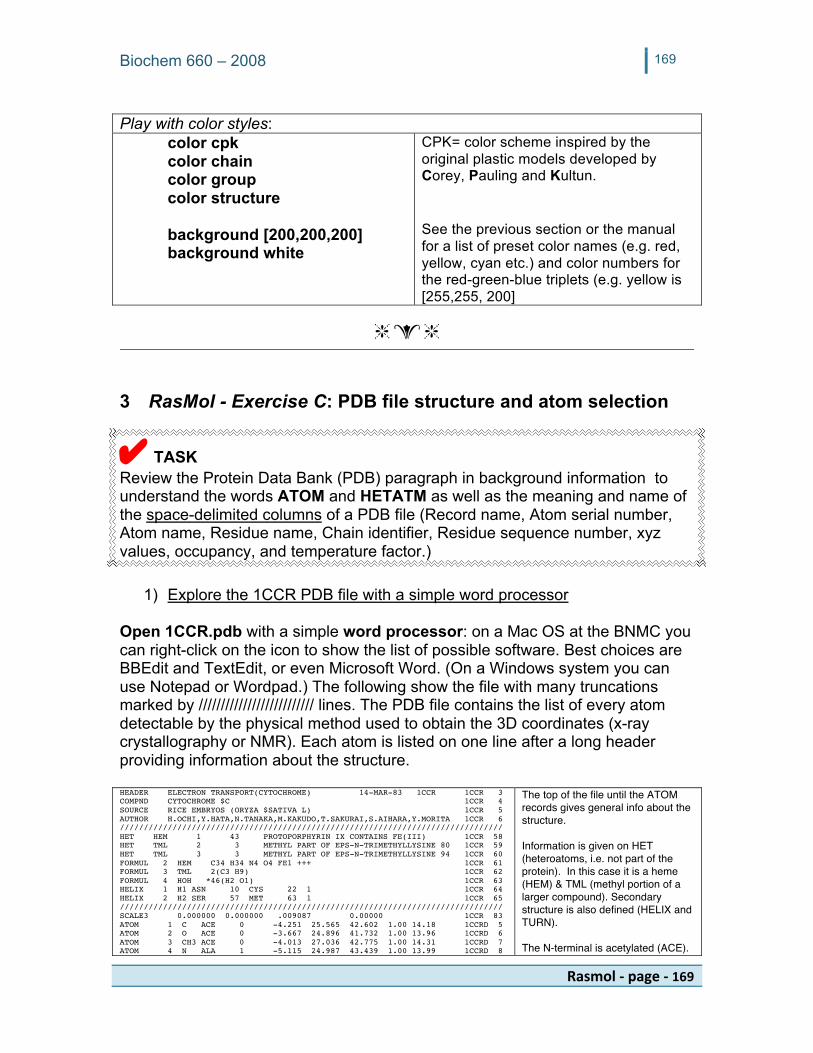
1) Explore the 1CCR PDB file with a simple word processor Open 1CCR.pdb with a simple word processor: on a Mac OS at the BNMC you can right-click on the icon to show the list of possible software. Best choices are BBEdit and TextEdit, or even Microsoft Word. (On a Windows system you can use Notepad or Wordpad.) The following show the file with many truncations marked by ////////////////////////// lines. The PDB file contains the list of every atom detectable by the physical method used to obtain the 3D coordinates (x-ray crystallography or NMR). Each atom is listed on one line after a long header providing information about the structure. HEADER ELECTRON TRANSPORT(CYTOCHROME) 14-MAR-83 1CCR 1CCR 3 COMPND CYTOCHROME $C 1CCR 4 SOURCE RICE EMBRYOS (ORYZA $SATIVA L) 1CCR 5 AUTHOR H.OCHI,Y.HATA,N.TANAKA,M.KAKUDO,T.SAKURAI,S.AIHARA,Y.MORITA 1CCR 6 //////////////////////////////////////////////////////////////////////////////// HET HEM 1 43 PROTOPORPHYRIN IX CONTAINS FE(III) 1CCR 58 HET TML 2 3 METHYL PART OF EPS-N-TRIMETHYLLYSINE 80 1CCR 59 HET TML 3 3 METHYL PART OF EPS-N-TRIMETHYLLYSINE 94 1CCR 60 FORMUL 2 HEM C34 H34 N4 O4 FE1 +++ 1CCR 61 FORMUL 3 TML 2(C3 H9) 1CCR 62 FORMUL 4 HOH *46(H2 O1) 1CCR 63 HELIX 1 H1 ASN 10 CYS 22 1 1CCR 64 HELIX 2 H2 SER 57 MET 63 1 1CCR 65 //////////////////////////////////////////////////////////////////////////////// SCALE3 0.000000 0.000000 .009087 0.00000 1CCR 83 ATOM 1 C ACE 0 -4.251 25.565 42.602 1.00 14.18 1CCRD 5 ATOM 2 O ACE 0 -3.667 24.896 41.732 1.00 13.96 1CCRD 6 ATOM 3 CH3 ACE 0 -4.013 27.036 42.775 1.00 14.31 1CCRD 7 ATOM 4 N ALA 1 -5.115 24.987 43.439 1.00 13.99 1CCRD 8
The top of the file until the ATOM records gives general info about the structure. Information is given on HET (heteroatoms, i.e. not part of the protein). In this case it is a heme (HEM) & TML (methyl portion of a larger compound). Secondary structure is also defined (HELIX and TURN). The N-terminal is acetylated (ACE).

Biochem 660 – 2008 170
Rasmol‐page‐170
ATOM 5 CA ALA 1 -5.273 23.535 43.450 1.00 14.03 1CCRD 9 ATOM 6 C ALA 1 -6.043 23.098 42.188 1.00 14.06 1CCRD 10 ATOM 7 O ALA 1 -5.962 21.921 41.763 1.00 14.65 1CCRD 11 ATOM 8 CB ALA 1 -6.032 23.081 44.672 1.00 13.89 1CCRD 12 //////////////////////////////////////////////////////////////////////////////// ATOM 852 N SER 111 -6.048 28.458 18.110 1.00 15.85 1CCRD856 ATOM 853 CA SER 111 -7.241 28.969 17.544 1.00 18.25 1CCRD857 ATOM 854 C SER 111 -7.213 28.966 15.998 1.00 19.11 1CCRD858 ATOM 855 O SER 111 -8.242 28.477 15.513 1.00 20.22 1CCRD859 ATOM 856 CB SER 111 -7.463 30.465 17.929 1.00 18.63 1CCRD860 ATOM 857 OG SER 111 -8.472 30.406 18.910 1.00 19.81 1CCRD861 ATOM 858 OXT SER 111 -6.211 29.484 15.471 1.00 20.32 1CCRD862 TER 859 SER 111 1CCR 942 HETATM 860 FE HEM 1 2.886 16.923 24.535 1.00 6.49 1CCRD863 HETATM 861 CHA HEM 1 .478 14.744 23.614 1.00 5.28 1CCRD864 HETATM 862 CHB HEM 1 .587 19.307 25.293 1.00 5.33 1CCRD865 HETATM 863 CHC HEM 1 5.329 18.833 25.864 1.00 6.37 1CCRD866 HETATM 864 CHD HEM 1 5.265 14.858 23.135 1.00 5.76 1CCRD867 //////////////////////////////////////////////////////////////////////////////// HETATM 899 CBD HEM 1 1.041 11.117 23.763 1.00 7.11 1CCRD902 HETATM 900 CGD HEM 1 .272 9.835 23.656 1.00 8.09 1CCRD903 HETATM 901 O1D HEM 1 -.207 9.534 22.556 1.00 8.87 1CCRD904 HETATM 902 O2D HEM 1 .176 9.077 24.679 1.00 7.89 1CCRD905 HETATM 903 CH1 TML 2 8.745 9.202 30.551 1.00 21.57 1 1CCRD906 HETATM 904 CH2 TML 2 7.964 8.370 32.710 1.00 21.65 1 1CCRD907 HETATM 905 CH3 TML 2 7.618 7.057 30.621 1.00 21.85 1 1CCRD908 HETATM 906 CH1 TML 3 7.984 16.673 38.397 1.00 19.56 2 1CCRD909 HETATM 907 CH2 TML 3 6.915 16.739 40.569 1.00 19.47 2 1CCRD910 HETATM 908 CH3 TML 3 5.565 17.049 38.545 1.00 19.24 2 1CCRD911 HETATM 909 O HOH 4 -2.402 19.505 9.833 1.00 13.32 1CCRD912 HETATM 910 O HOH 5 -5.510 8.889 11.007 1.00 32.11 1CCRD913 HETATM 911 O HOH 6 5.824 19.189 11.159 1.00 8.44 1CCRD914 HETATM 912 O HOH 7 -3.634 28.132 12.148 1.00 20.43 1CCRD915 HETATM 913 O HOH 8 .298 7.948 11.425 1.00 33.89 1CCRD916
The first amino acid is an ALAnine, the first AA of the protein sequence. The last amino acid is a SERine, number 111 on the protein sequence. The last 2 atoms are the carbon beta (CB) and terminal oxygen (OXT). After all the ATOM are shown, a TERminator keyword announces that something else is starting. Here it is the list of all the HETATM, starting with the iron (FE) of the heme. The heme is 43 lines long (see numbers at extreme right, from line 863 to line 905). Only the methyl part of TML is present as 3 carbon atoms (CH-). Finally the water molecules are shown.
Note: Because there is a single protein chain, the column for the chain identifier (before the sequence number) has been left blank.
2) Making atom selection (line command only)
✔ INFO - In addition to the 3D xyz coordinates, the various columns and keywords within the PDB data file provide extra clues to the software so that the data can be organized in logical groups of various sizes: one atom, one amino acid, one protein chain, etc. The groups can also be combined. Rasmol also organizes the data internally in various predefined sets (e.g. charged, hydrophobic, sheet, helix, nucleic, protein, purine, etc.). Rasmol operates only on the current atom selection. When a PDB file is opened, the current default atom selection is everything. In order to make changes, we will want to select various portions of the PDB file and change the way they are shown or colored. For example we may want to select the protein and show it as a cartoon drawing in a specific color, and then select the region that forms an active site and show the amino acid side chains and a ligand as thick wireframes in another color. In the following examples, atoms are selected and “something” is done to them to verify that we selected them. Within the Rasmol text window (after the RasMol> prompt) type the following commands, and observe the screen as you press the return key:
✔ TASK Simple selections after some reset commands: reset cartoon off wireframe off backbone off
These commands should remove the current representations and prepare for the next steps.

Biochem 660 – 2008 171
Rasmol‐page‐171
spacefill off select protein cartoon select helix color red wireframe 0.3 select helix and not backbone color cpk wireframe off select * color gray
“protein” is a useful predefined set. helix is a predefined set as described previously.
Select only the helix side chains by not selecting what constitutes the backbone, making up the helix turns. Hence the helix remains red..
Reset everything and color all gray. Note: the * selects everything but is not mandatory.
select HEM wireframe 0.3 color red select HEM.FE spacefill color green
Select the heme. So far with the previous commands (cartoon) only the protein was shown. We know that the heme has an iron (FE) within it.
More complex selections and definitions: select within(5.0, HEM.FE) and protein
The “select wihin” command is a very powerful tool to select atoms in a given radius of an atom selection. Note that there is no space between within and the parenthesis and that the distance value, and angstroms, requires a decimal point.
define myatoms selected spacefill myatoms
Creates a new predefined set that contains all the atoms that were selected with the “within” command above. The word “myatoms” is a

Biochem 660 – 2008 172
Rasmol‐page‐172
made-up word. select myatoms and basic color blue
This expression selects the common amino acids between my defined set myatoms and the predefined set basic. The result is colored in blue.
Rasmol atom selection syntax: AaaNum:Chain.Atom designating the residue in 3-letter code, its sequence number, a colon (:) (optional if chain is a letter) the chain value (e.g. A, B, C), a dot (.) is added to specify an atom or group of atoms (e.g. *.FE, *.CA). The names of atoms, heteroatoms, and protein chains are determined by the PDB file. Note: clicking on an atom within the graphic window echoes its name within the text window. With the mouse randomly click on one atom of each of the blue amino acids. Within the Rasmol text window, you will see their atom name (e.g. CB), serial number and amino acid with its sequence number.
RasMol> Atom: CB 154 Group: LYS 21 RasMol> Atom: CD2 192 Group: HIS 26 RasMol> Atom: CB 663 Group: LYS 87 RasMol>
Quit Rasmol by selecting the File > Exit menu. We will restart it in the next exercise.
- e -
4 RasMol - Exercise D: Working with protein and nucleic acids ✔ TASK
a. Find or retrieve the file 3cro.pdb or download it with PDB ID code 3CRO (phage 434 cro/or1 complex at 2.5 angstroms resolution), and save the file on the desktop.
b. Launch Rasmol as previously: (% or $) rasmol 3CRO.pdb (case
sensitive!)
Biochem 660 – 2008 173
Rasmol‐page‐173
Orient the molecule :
reset rotate z -125 rotate y -17 rotate x -85
c. Atom selection with multiple chains, nucleic acids
Within the PDB file (which you can explore with a word processor) the DNA strands are given the chain names A and B, and the protein chains are given the names L and R.
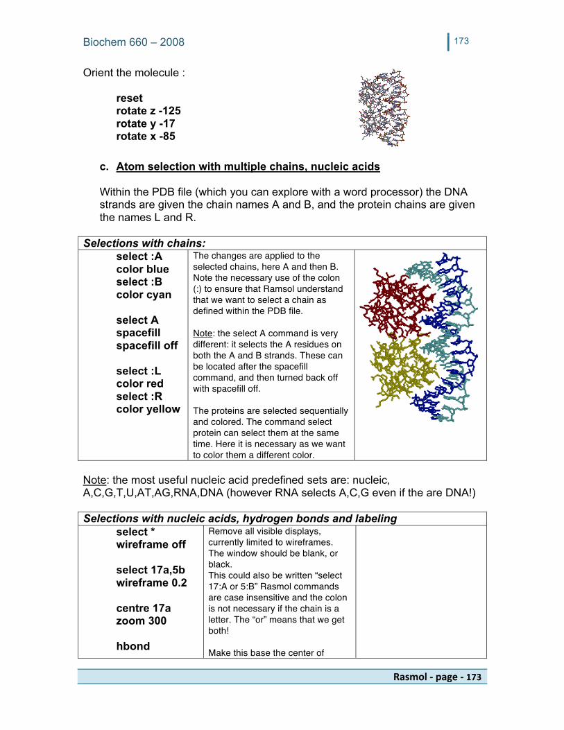
Selections with chains: select :A color blue select :B color cyan select A spacefill spacefill off select :L color red select :R color yellow
The changes are applied to the selected chains, here A and then B. Note the necessary use of the colon (:) to ensure that Ramsol understand that we want to select a chain as defined within the PDB file. Note: the select A command is very different: it selects the A residues on both the A and B strands. These can be located after the spacefill command, and then turned back off with spacefill off. The proteins are selected sequentially and colored. The command select protein can select them at the same time. Here it is necessary as we want to color them a different color.
Note: the most useful nucleic acid predefined sets are: nucleic, A,C,G,T,U,AT,AG,RNA,DNA (however RNA selects A,C,G even if the are DNA!) Selections with nucleic acids, hydrogen bonds and labeling select * wireframe off select 17a,5b wireframe 0.2 centre 17a zoom 300 hbond
Remove all visible displays, currently limited to wireframes. The window should be blank, or black. This could also be written “select 17:A or 5:B” Rasmol commands are case insensitive and the colon is not necessary if the chain is a letter. The “or” means that we get both! Make this base the center of
Biochem 660 – 2008 174
Rasmol‐page‐174
select C5:B.C5 label %n%r “chain B” select G17:A.N7 label %n%r “chain A”
rotation and zoom on this display. Rasmol echoes: Number of H-Bonds ..... 130 Select the 5th carbon (C5 after the dot) on the sugar of the C with sequence number 5 (first C5) Add a label attached to this atom. Make a similar selection on the other nucleotide and label it too.
Note: when an atom is clicked, all the relevant information is given within the Rasmol text output as seen above.
Rasmol atom selection syntax for nucleic acid: XNum:Chain.Atom where X is the nucleotide name (A,C,G,T,U).
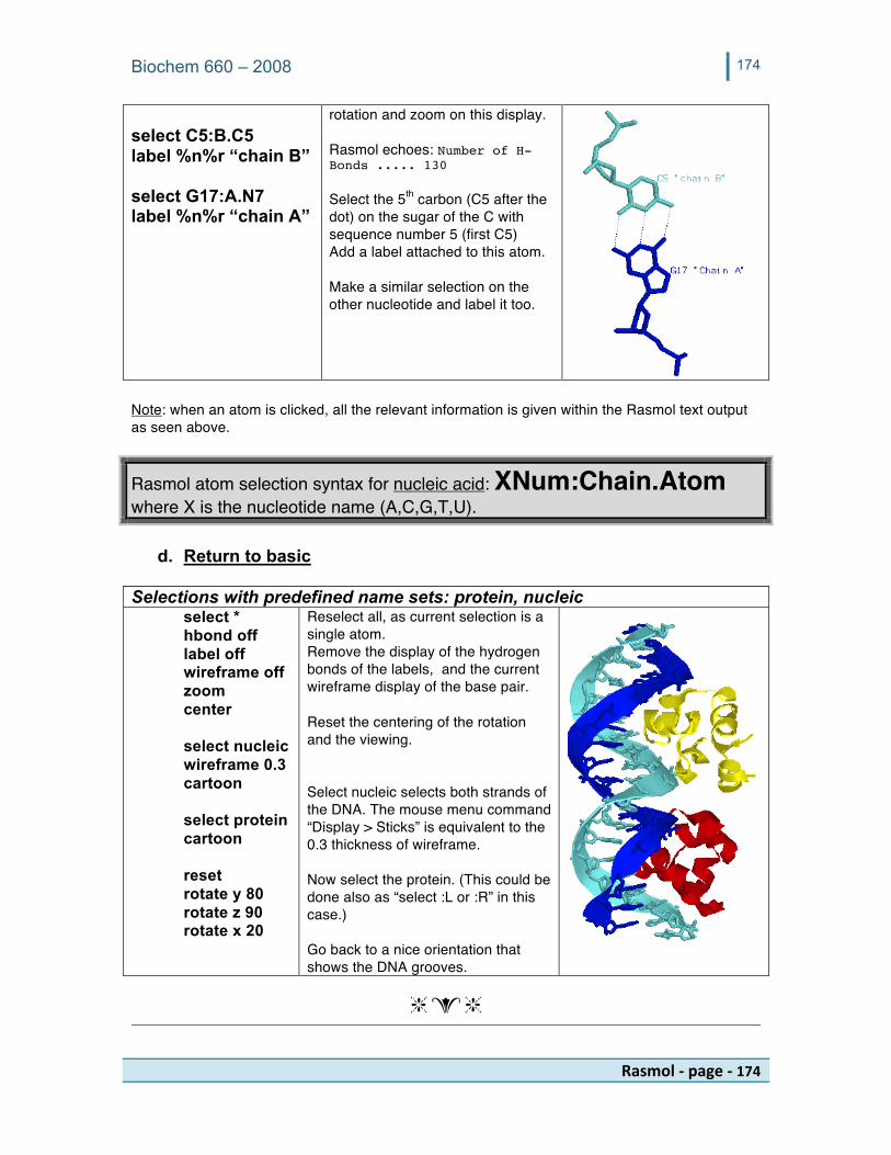
d. Return to basic Selections with predefined name sets: protein, nucleic select * hbond off label off wireframe off zoom center select nucleic wireframe 0.3 cartoon select protein cartoon reset rotate y 80 rotate z 90 rotate x 20
Reselect all, as current selection is a single atom. Remove the display of the hydrogen bonds of the labels, and the current wireframe display of the base pair. Reset the centering of the rotation and the viewing. Select nucleic selects both strands of the DNA. The mouse menu command “Display > Sticks” is equivalent to the 0.3 thickness of wireframe. Now select the protein. (This could be done also as “select :L or :R” in this case.) Go back to a nice orientation that shows the DNA grooves.
- e -

Biochem 660 – 2008 175
Rasmol‐page‐175
5 RasMol - Exercise E: Working with Rasmol scripts Clearly it would be advantageous to reuse some of this typing. This is possible with scripts: simple, plain text files containing Rasmol commands, one per line. Note: More on-line information on this subject (scripts) can be found at: http://www.umass.edu/microbio/rasmol/makescrp.htm and http://www.umass.edu/microbio/rasmol/maketuts.htm while demo scripts are at http://www.umass.edu/microbio/rasmol/scripts.htm
✔ READ - Background info about Rasmol scripts: a. Scripts are plain text files (saved as text-only e.g. from MSWord or other text editor.) b. Scripts location: scripts may need to be located in a specific place, depending on the OS:
a. Macintosh OS X, Unix, Linux: scripts need to be in the directory/folder where the line-command was originally started from (here we used “cd Desktop” hence script should be on the desktop
b. Windows and Mac Classic: the script needs to be within the same folder as the running copy of Rasmol
c. Exceptions: if you give the full path to the location of the script, it can be stored elsewhere on the computer.
c. Scripts contain one Rasmol command per line. Lines starting with # are comments lines. d. Rasmol scripts are activated at the Rasmol> prompt line with the command “script”
followed by the name of the script file e.g. “script myfirstscript.scr” e. Scripts can be calling other scripts: this is useful for complex or repetitive tasks.
1 Using scripts to animate a structure within Rasmol Important note: Rasmol will not refresh the display screen until all the calculations are done. For example the line “rotate y 10” repeated on each line of an 18-lines script will create an 18 x 10 = 180 degrees rotation…. However Rasmol will not display the intermediate rotations images and will only display the final image, in this case a half-turn. How then can we see the intermediate frames to see the rotation? The answer is the “refresh” command that forces Rasmol to recalculate the image with the current, new coordinate rotations and update the display within the graphics window.
✔ TASK
1.1 Copy the script xyz-360-36.scr to the MyRasmol directory (find the script either online on http://virology.wisc.edu/acp under “Class Resources,” or on the BNMC Macintosh hard drive within the Applications > Classes > Biochemistry PDB directory. This script (reproduced in the table below) will rotate the molecule a complete turn (10 times 36 degrees) around each of the 3 axes x, y, and z. Note the use of the command “refresh” after each rotation.

Biochem 660 – 2008 176
Rasmol‐page‐176
#!rasmol -script # File: xyz-360-36.scr # 10x -36degrees for 360 degrees # 10y -36degrees for 360 degrees # 10z +36degrees for 360 degrees # clockwise #(SHOWN AS COLUMS TO SAVE SPACE # AND PAPER....) # #Also only a few commands are shown
#x rotate x -36 refresh rotate x -36 refresh rotate x -36 refresh rotate x -36 refresh rotate x -36 refresh rotate x -36 refresh rotate x -36 refresh
#y rotate y -36 refresh rotate y -36 refresh rotate y -36 refresh rotate y -36 refresh rotate y -36 refresh rotate y -36 refresh rotate y -36 refresh
#z rotate z 36 refresh rotate z 36 refresh rotate z 36 refresh rotate z 36 refresh rotate z 36 refresh rotate z 36 refresh rotate z 36 refresh
1.2 Type the following command at the Rasmol> prompt:
Rasmol> script xyz-360-36.scr
Observe the rotation about all 3 axes within the display window. The rotation may be going very fast!
This technique is useful to animate molecules within Rasmol itself. The script can be more complex than a simple rotation and all Rasmol commands can be made part of a script. But what if we want to show the animation within a PowerPoint presentation without having to run Rasmol? Answer: we save the frames, as explained next…. 2 Using scripts to animate a structure and save each frame into a graphics file Rasmol can save files in various graphics format, shown within the Export menu. The most useful formats are GIF and PICT. However GIF files only contain 256 colors (8-bit) and can only be saved from a version of Rasmol that is compiled as 8-bit (8-,16-, and 32-bit versions of Rasmol are available from download web sites). The Rasmol version running at the BNMC is a 32-bit Rasmol which cannot save GIF files. PICT is the Macintosh Classic file format and is suitable for these exercises. On a PC it is sometimes preferred to save as GIF. The other formats are specific to other systems not discussed here. In a script we obviously do not use the mouse: the graphics frames will be saved into files by line command with the “write” command.
Important note: Rasmol will not save the graphics files until the following command is given:
✔ TASK Rasmol> set write on This is useful to test the script before rendering all the frames, (and was also a safeguard for web sites running rasmol script automatically.)
Note: without this command no image will be saved.

Biochem 660 – 2008 177
Rasmol‐page‐177
2.1 Copy the script xyz-360-36-PICT2-images.scr to MyRasmol
directory on the desktop (find the script in the same spot as in the previous exercise). This script contains additional lines for saving the images in PICT format e.g. “write pict frame.0010.pict” (GIF script also available). You view the script content by double clicking on its icon or opening it with a word processor.
2.2 Type the following commands at the Rasmol> prompt:
Rasmol> set write on Rasmol> script xyz-360-36-PICT2-images.scr
2.3 Frame location: the frames will be saved within the MyRasmol
directory.
Note: on a PC or Macintosh Classic, the frames would be found within the directory where the running Rasmol is located. Here, in X-windows, we have a different result: the frames are saved within the location from which we started the line command.
Rasmol can only create the frames. These need to be assembled into a movie by another software such as Apple’s QuickTimePro ($30). Freeware such as ImageMagik and Gimp can also be useful for this, but are not covered here.
- END- - e -
Special Help Notes for Installation of Rasmol 2.7.x X11 on Macintosh OS X
X11: starting with 10.4 (Tiger) X11 is located on the original Mac OS install disk or DVD and is no longer available as a download from Apple. If X11 is not installed, use the original OS CD or DVD to install X11 as this is an optional installation when the system was first installed. Rasmol download page: http://arcib.dowling.edu/software/rasmol/index.shtml Rasmol 2.7.x versions: As of this writing the latest version is 2.7.3.1.
Download the compressed or uncompressed version labeled “NOXF” in either of the 8-, 16-, or 32-bit versions (8-bit version is limited to 256 colors and is necessary for creating GIF files.)
Rasmol is an application. Starting with Tiger you have to acknowledge that you are downloading a software. Simply click “Yes” on the dialog box. However running of the software is not yet allowed and this privilege has to be given manually with a series of line commands. Open a Terminal and issue the following commands after the % or $ prompt, assuming that the file is called “rasmol:”

Biochem 660 – 2008 178
Rasmol‐page‐178
cd Desktop chmod a+x rasmol
If you want to run rasmol from the command line from any directory, you can move the executable rasmol to a higher level directory: sudo mv rasmol /usr/bin/ Your admin password will be requested. Alternatively, if you have installed Fink (see end of VMD animation section) you may want to place rasmol into the /sw directry with the alternate command:
sudo mv rasmol /sw/bin/ From this point forward (after either of the sudo commands has been given) you can always start rasmol with the simple command “rasmol” at the % or $ prompt from within any directory (folder.) Note: you could install all 3 rasmol versions and call them rasmol8, rasmol16, and rasmol32 and use that same name at the prompt to launch it within an X11 terminal, e.g. rasmol32.
- e -
Class notes