radiolucent wrist fixator - orthofix,...
TRANSCRIPT

VOICE OF DES IGN
Radiolucent Wrist Fixator
Orthofix approach to Evidence Based Medicine
For years, clinical decision-making was based primarily on physician knowledge and expert opinion. Now, the medical community is searching for measurable outcomes “validating” efficacy of treatments. Evidence Based Medicine (EBM) is an approach that integrates individual clinical expertise with the best available evidence when making decisions about patient treatment. (Nierengarten MB et al. Using Evidence Based Medicine in Orthopaedic Clinical Practice: The Why, When, and How-To Approach. Medscape Orthopaedics & Sports Medicine. 2001; 5[1]). Over the last few years, there has been a significant growth in Evidence Based Medicine.
To receive a digital copy of this Product in detail please submit your request to:
CLINICAL AFFAIRS DEPT:Email: [email protected]: +39 045 6719000
To receive a digital copy of the “Quick Reference PG 150”, please submit your request to:
CUSTOMER CARE:Email: [email protected]: +39 045 6719000

Radiolucent Wrist Fixator
INDEX
SUMMARY: RADIOLUCENT WRIST FIXATOR I
1. Introduction P. 1
2. Technical features and benefits P. 2
3. Mechanical features P. 6
4. Conclusions P. 8
5. References P. 8
Radiolucent Wrist Fixator
I
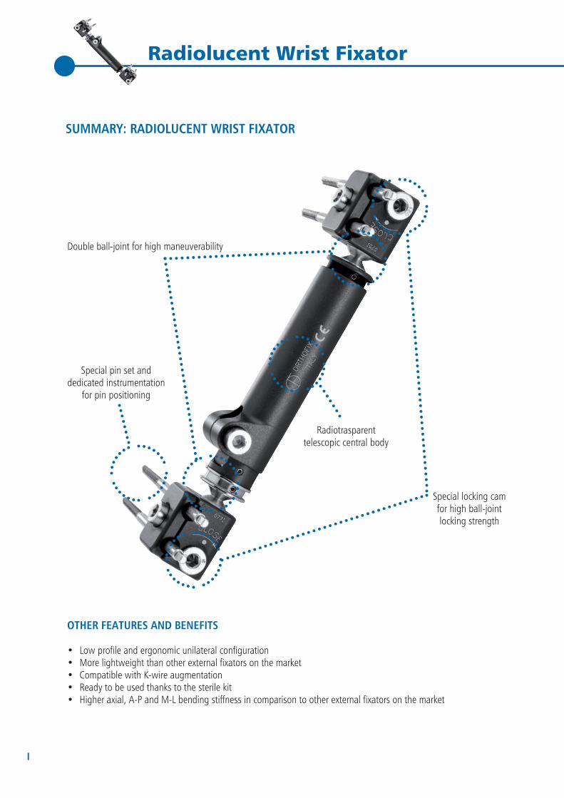
OTHER FEATURES AND BENEFITS
• Low profile and ergonomic unilateral configuration• More lightweight than other external fixators on the market• Compatible with K-wire augmentation• Ready to be used thanks to the sterile kit• Higher axial, A-P and M-L bending stiffness in comparison to other external fixators on the market
Double ball-joint for high maneuverability
Special pin set and dedicated instrumentation
for pin positioning
Special locking cam for high ball-joint locking strength
Radiotrasparenttelescopic central body
SUMMARY: RADIOLUCENT WRIST FIXATOR
Voice of Design | 1
Radiolucent Wrist Fixator
1. INTRODUCTION
The Radiolucent Wrist Fixator is part of Orthofix External Fixation Systems which are intended as a means to stabilize bone segments in a broad range of indications, including fractures, joint fusion, joint distraction, bone transport, lengthening and angular corrections.
In particular, the Radiolucent Wrist Fixator indications include:
• Fractures of the distal radius - intrarticular or extrarticular• Preliminary fixation before ORIF• Fractures with open or closed soft tissue damage• Polytrauma (“damage controlled surgery“, temporary or definitive)• Burns or other skin damage – wrist, carpus, forearm• Fractures and dislocation in combination with - severe soft tissue damage - bone-loss or other reconstructive procedures - damage to nerves-and/or blood vessels• Loss of reduction after initial treatment• Infections
This aim of this document is to give a detailed description of the Radiolucent Wrist Fixator and to underline the advantages and benefits that it can offer to both surgeons and patients.

2 | Voice of Design
Radiolucent Wrist Fixator
2. TECHNICAL FEATURES AND BENEFITS
LOW PROFILE AND ERGONOMIC UNILATERAL CONFIGURATION
Most external fixation devices are bulky and inconvenient for patients, interfering with daily activities, personal hygiene and clothing [1]. It has been suggested that external fixators for distal radius fractures should be preferably low profile and unilateral configurations are considered more desirable [2]. Ergonomics is another important aspect for patient comfort and safety.
The Radiolucent Wrist Fixator has been designed to have low profile and advanced ergonomics for enhanced patient comfort. The frame is compact and has rounded edges without sharp protrusions.
In addition, pins can be mounted so that they do not stick out from the clamps. Therefore there is no need to use pin caps to protect patients from accidental injuries.
RADIOTRANSPARENT TELESCOPIC CENTRAL UNIT
The central unit of the Radiolucent Wrist Fixator is composed of a thin tube sliding into a cylinder. This design makes the central unit telescopic, so from an overall length of 13 cm, it can expand up to 19 cm. Thanks to the telescopic unit, it is possible to perform fracture reduction when pins and fixator have been already mounted [3].
Uncontrolled rotations of the external tube around the cylinder are impeded by a blocking screw.
The internal cylinder is made of PEEK, whereas the external tube is made of aluminum that is only 0.6 mm thick. Material choice and thickness have been optimized to have satisfactory radiotransparency, a feature generally recognized as important for visualization of the fracture site.
If a unilateral external fixator for distal radius fractures is not radiotransparent, pins need to be placed properly to allow for AP and ML view of the fracture site. If visualization in one plane remains difficult, additional procedures such as placing a dental film between fixator and skin to obtain an unobstructed view [2] or supplementary oblique views, that can be difficult to interpret, need to be performed [4]. The amount of time both surgeons and patients are exposed to X-rays is prolonged and the total operative time increases [4].
The Radiolucent Wrist Fixator has satisfactory radiotransparency to allow for visualization of fracture reduction and scaphoid and carpal alignment during healing [3] (Fig. 1).
Fig. 1: Radiolucent Wrist Fixator radiotransparency

Voice of Design | 3
Radiolucent Wrist Fixator
LIGHTER THAN OTHER FIXATORS ON THE MARKET
One of the advantages of external fixators for the treatment of wrist fractures in comparison to casts is that they are lighter in weight [5]. It is well-accepted that a lightweight design improves wearing comfort for the patients [6].The Radiolucent Wrist Fixator is lighter than other external fixators for distal radial fractures on the market (Fig. 2). This results from the low specific gravity materials. Pins and closing mechanisms are made of stainless steel for mechanical and structural reasons.
The Radiolucent Wrist Fixator has an additional unit which is mounted on the fixator intra-operatively and helps the surgeon to perform compression-distraction (Fig.3). Once the procedure is completed, the compression-distraction device can be removed. This solution permits further reductions in the weight of the fixator.
DOUBLE BALL-JOINT FOR HIGH MANEUVERABILITY
The Radiolucent Wrist Fixator is composed of two clamps connected to the central unit through a ball-joint. Thanks to this particular feature, it is possible to move the clamp in all axes with a total angulation of up to 64°. This improves maneuverability of the system during fracture reduction.
+64%
+77%
Radiolucent WristFixator
GRA
MS
Competitor 1
+64%
Fig. 2: Radiolucent Wrist Fixation System is lighter than other systems on the market
Fig. 3: Compression-distraction unit
Competitor 2
+77%
FIXATION WEIGHT

4 | Voice of Design
Radiolucent Wrist Fixator
SPECIAL LOCKING CAM FOR HIGH-BALL JOINT LOCKING STRENGTH
In the Radiolucent Wrist Fixator ball-joints are locked by tightening a locking cam characterized by a special geometry (Fig. 4). Its design assures high ball-joint locking strength [internal reports].This closing mechanism has been designed to reduce the probability of ball-joint locking failure that can lead to loss of stability and consequently loss of reduction.
SPECIAL PIN SET AND DEDICATED INSTRUMENTATION FOR PIN POSITIONING
Anchoring pins used in external fixation, since they are subjected to bending force, should be sufficiently large and strong, but should not exceed a third of the bone diameter to prevent secondary pin hole fractures [7]. In regard to distal radius fractures, the fixator pins should have an adequate size: a 3-3.5 mm diameter combines stable fixation with minimal bone damage and an adequate pin-bone interface [2].
The Radiolucent Wrist Fixation System pins have a standard Orthofix tapered thread design. Before inserting the pin, predrilling is required.
Predrilling of fixator pins has shown to reduce the degree of thermal necrosis, which may contribute to pin loosening [2].
The Radiolucent Wrist Fixator is supplied with a special instrumentation for pin positioning. The screw template consists of a fixed screw guide and a threaded screw guide the position of which can be regulated according to the bone anatomy. It has been shown that this instrumentation ensures that the pins are positioned parallel and 14 mm apart, which is the spacing in the fixator pin clamps [3].
Fig. 4: Details of the particular geometry of the cam
Fig. 5: Screw guide template

Voice of Design | 5
Radiolucent Wrist Fixator
COMPATIBLE WITH K-WIRE AUGMENTATION
Augmentation of external fixation with K-wires has shown to increase stability of distal radial fractures [7, 8], reduce the need for excessive traction [7, 8] and avoid late collapse [2]. K-wires also help to maintain palmar tilt [7].Furthermore, severely impacted fragments may not be reduced simply with traction and require percutaneous manipulation using supplementary K-wires [7, 9].
The Radiolucent Wrist Fixator can be successfully used in combination with K-wire fixation [3].
READY TO BE USED THANKS TO THE STERILE KIT
Thanks to the sterile kit, the Radiolucent Wrist Fixator is ready to be used.
Fig. 6: Sterile kit blister

6 | Voice of Design
Radiolucent Wrist Fixator
3. MECHANICAL FEATURES
HIGH STIFFNESS
It is not known which rigidity is required for adequately stabilizing a distal radius fracture [10]. It is thought that a stiff external fixator promotes primary fracture healing while a less rigid device promotes more of a secondary fracture healing [11].Rigid external fixators help to obtain and maintain an acceptable reduction until the fracture has gained sufficient stability [7]. In highly unstable fractures, rigid fixation is required to maintain a satisfactory anatomical position [12].
In order to evaluate Radiolucent Wrist Fixator bending stiffness, mechanical tests comparing different external fixators for distal radius fractures were performed.
These tests were performed internally during the development of the device.
AXIAL, A-P AND M-L BENDING STIFFNESS TESTS
Testing ProtocolThe fixators were mounted on stainless steel alloy bars and a gap of 20 mm was maintained to present the fracture site. Load was applied, and stiffness values were calculated from the load-deformation curve as reported in [13]. For each test type, fixators were tested in three different configurations: Configuration 1: D = 10 mmConfiguration 2: D = 15 mmConfiguration 3: D = 20 mm where D is the distance between the frame and the bars.Each configuration was tested 5 times.
ResultsThe results demonstrate that the Radiolucent Wrist Fixator is characterized by high axial, A-P and M-L bending stiffness, comparable to Pennig Dynamic Wrist Fixator and higher than other competitors on the market (Fig. 7, 8 and 9).
MEA
N S
TIFF
NES
S [N
/mm
]
Radiolucent Wrist Fixator
Pennig Dynamic Wrist Fixator Competitor 1 Competitor 2 Competitor 3
D= 10mm
D= 15mm
D= 20mm
Fig. 7: Axial Bending Stiffness
AXIAL BENDING STIFFNESS

Voice of Design | 7
Radiolucent Wrist FixatorM
EAN
STI
FFN
ESS
[N/m
m]
MEA
N S
TIFF
NES
S [N
/mm
]
Radiolucent Wrist Fixator
Radiolucent Wrist Fixator
Pennig Dynamic Wrist Fixator
Pennig Dynamic Wrist Fixator
Competitor 1
Competitor 1
Competitor 2
Competitor 2
Competitor 3
Competitor 3
A-P BENDING STIFFNESS
M-L BENDING STIFFNESS
Fig. 8: A-P Bending Stiffness
Fig.9: M-L Bending Stiffness
D= 10mm
D= 15mm
D= 20mm
D= 10mm
D= 15mm
D= 20mm

8 | Voice of Design
Radiolucent Wrist Fixator
4. CONCLUSIONS
This document was created to give a detailed description of the Radiolucent Wrist Fixator features. The technical features were studied to provide a simple, low profile, radiolucent and lightweight external fixator. Mechanical tests prove that the Radiolucent Wrist Fixator is characterized by high stiffness comparable to the Pennig Dynamic Wrist Fixator and higher than other external fixators for distal radial fractures on the market.In conclusion, Radiolucent Wrist Fixator is a simple, but advantageous system for the treatment of fractures of the distal radius.
5. REFERENCES
1. Kerkhoffs GM, Kuipers MM, Marti RK, Van der Werken C. External fixation with standard AO-plates: technique, indications, and results in 31 cases. J Orthop Trauma. 2003 Jan;17(1):61-4.
2. Gausepohl T, Pennig D, Mader K. Principles of external fixation and supplementary techniques in distal radius fractures. Injury. 2000;31 Suppl 1:56-70.
3. Nelson DL. The radiolucent wrist fixator for distal radius fractures. In: De Bastiani G, Apley AG, Goldberg A, eds. Orthofix External Fixation in Trauma and Orthopaedics. Springer; 2000, pp. 181-191.
4. Dall’Oca C, Christodoulidis A, Bortolazzi R, Bartolozzi P, Lavini F. Treatment of 103 displaced tibial diaphyseal fractures with a radiolucent unilateral external fixator. Arch Orthop Trauma Surg. 2010 Nov;130(11):1377-82. doi: 10.1007/s00402-010-1090-7. Epub 2010 Apr 2.
5. Frykman GK, Tooma GS, Boyko K, Henderson R. Comparison of eleven external fixators for treatment of unstable wrist fractures. J Hand Surg Am. 1989 Mar;14(2 Pt 1):247-54.
6. Burgers PT, Van Riel MP, Vogels LM, Stam R, Patka P, Van Lieshout EM. Rigidity of unilateral external fixators--a biomechanical study. Injury. 2011 Dec;42(12):1449-54. doi: 10.1016/j.injury.2011.05.024. Epub 2011 Jun 23.
7. Siripakarn Y, Siripakarn Z. Multipurpose external fixator for intraarticular fracture of distal radius. J Med Assoc Thai. 2010 Dec;93 Suppl 7:S324-31.
8. Wolfe SW, Austin G, Lorenze M, Swigart CR, Panjabi MM. A biomechanical comparison of different wrist external fixators with and without K-wire augmentation. J Hand Surg Am. 1999 May;24(3):516-24.
9. Pennig DW. Dynamic external fixation of distal radius fractures. Hand Clin 1993; 9: 587-602.
10. Pennig D, Gausepohl T. External fixation of the wrist. Injury. 1996 Jan;27(1):1-15. Review.
11. Chang D, Kummer FJ, Egol K, Tejwani N, Wolinsky P, Koval KJ. Biomechanical comparison of five external wrist fixators. Bull Hosp Jt Dis. 2002-2003;61(1-2):40-4.
12. Wilcke MK, Abbaszadegan H, Adolphson PY. Wrist function recovers more rapidly after volar locked plating than after external fixation but the outcomes are similar after 1 year. Acta Orthop. 2011 Feb;82(1):76-81.
13. Chao EY, Hein TJ. Mechanical performance of the standard Orthofix external fixator. Orthopedics. 1988 Jul;11(7):1057-69.

6. PAPERS ON RADIOLUCENT WRIST FIXATOR
- Nelson DL. The radiolucent wrist fixator for distal radius fractures. In: De Bastiani G, Apley AG, Goldberg A, eds. Orthofix External Fixation in Trauma and Orthopaedics. Springer; 2000, pp. 181-191.
- Sato K, Furumachi K, Nishida J, Tajima K, Kaiyama J, Suzuki Y, Shimamura T. Comparison of the volar locking plate and the bridging external fixator in the treatment of distal radius fracture based on range of wrist motion assessed by functional radiography. Med Sci Monit. 2010 May;16(5):CR207-12.
Radiolucent Wrist Fixator


Distributor:
www.o r tho f i x . comRW-1301-VOD-E0 A 08/13
Manufactured by: ORTHOFIX SrlVia Delle Nazioni 937012 Bussolengo (Verona)Italy
Telephone +39-0456719000Fax +39-0456719380
0123