radiolaria: achievements and unresolved issues: taxonomy ... · radiolaria: achievements and...
TRANSCRIPT
Introduction
Radiolarians are planktonic unicellular Protoctista with asymmetrical or asymmetrical skeleton such as in a spheri-cal, discoidal, or conical shape. Their skeleton size rangesfrom 40 to 400 m m, and colonial Radiolaria form gelatinpacks a few meters in length. Radiolarians appeared earlyin the Cambrian (ca. 530 million years ago) and inhabitshallow-to-open oceans at depths of up to 4,000 m in themodern ocean. Although no typical freshwater members ofthe Radiolaria have been found, at least 2,500 genera/sub-genera and 15,000 species (living and fossil) have been re-ported since radiolarians were first recognized and de-scribed by Meyen (1834). Of these, approximately 800–1000 radiolarian species are currently recognized in theoceans (N Suzuki unpubl). Radiolaria are important fossilsfor determining geological age at a resolution of 1,000–10,000 years and for understanding oceanographic history.However, they have long been ignored because of discus-sions suggesting the relative unimportance of geology held
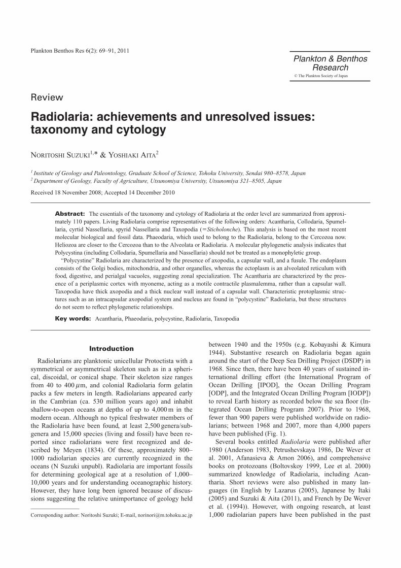
between 1940 and the 1950s (e.g. Kobayashi & Kimura1944). Substantive research on Radiolaria began againaround the start of the Deep Sea Drilling Project (DSDP) in1968. Since then, there have been 40 years of sustained in-ternational drilling effort (the International Program ofOcean Drilling [IPOD], the Ocean Drilling Program[ODP], and the Integrated Ocean Drilling Program [IODP])to reveal Earth history as recorded below the sea floor (In-tegrated Ocean Drilling Program 2007). Prior to 1968,fewer than 900 papers were published worldwide on radio-larians; between 1968 and 2007, more than 4,000 papershave been published (Fig. 1).
Several books entitled Radiolaria were published after1980 (Anderson 1983, Petrushevskaya 1986, De Wever etal. 2001, Afanasieva & Amon 2006), and comprehensivebooks on protozoans (Boltovskoy 1999, Lee et al. 2000)summarized knowledge of Radiolaria, including Acan-tharia. Short reviews were also published in many lan-guages (in English by Lazarus (2005), Japanese by Itaki(2005) and Suzuki & Aita (2011), and French by De Weveret al. (1994)). However, with ongoing research, at least1,000 radiolarian papers have been published in the past
Plankton Benthos Res 6(2): 69–91, 2011
Radiolaria: achievements and unresolved issues:taxonomy and cytology
NORITOSHI SUZUKI1,* & YOSHIAKI AITA2
1 Institute of Geology and Paleontology, Graduate School of Science, Tohoku University, Sendai 980–8578, Japan2 Department of Geology, Faculty of Agriculture, Utsunomiya University, Utsunomiya 321–8505, Japan
Received 18 November 2008; Accepted 14 December 2010
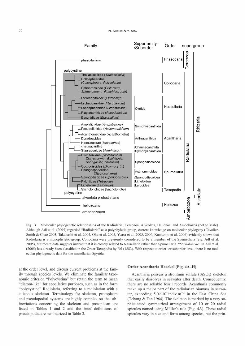
Abstract: The essentials of the taxonomy and cytology of Radiolaria at the order level are summarized from approxi-mately 110 papers. Living Radiolaria comprise representatives of the following orders: Acantharia, Collodaria, Spumel-laria, cyrtid Nassellaria, spyrid Nassellaria and Taxopodia (�Sticholonche). This analysis is based on the most recentmolecular biological and fossil data. Phaeodaria, which used to belong to the Radiolaria, belong to the Cercozoa now.Heliozoa are closer to the Cercozoa than to the Alveolata or Radiolaria. A molecular phylogenetic analysis indicates thatPolycystina (including Collodaria, Spumellaria and Nassellaria) should not be treated as a monophyletic group.
“Polycystine” Radiolaria are characterized by the presence of axopodia, a capsular wall, and a fusule. The endoplasmconsists of the Golgi bodies, mitochondria, and other organelles, whereas the ectoplasm is an alveolated reticulum withfood, digestive, and perialgal vacuoles, suggesting zonal specialization. The Acantharia are characterized by the pres-ence of a periplasmic cortex with myoneme, acting as a motile contractile plasmalemma, rather than a capsular wall.Taxopodia have thick axopodia and a thick nuclear wall instead of a capsular wall. Characteristic protoplasmic struc-tures such as an intracapsular axopodial system and nucleus are found in “polycystine” Radiolaria, but these structuresdo not seem to reflect phylogenetic relationships.
Key words: Acantharia, Phaeodaria, polycystine, Radiolaria, Taxopodia
Review
Corresponding author: Noritoshi Suzuki; E-mail, [email protected]
Plankton & Benthos Research
© The Plankton Society of Japan
decade (1997–2007). This explosion of new knowledge hasresulted in these reviews and textbooks becoming outdated.In particular, the concept of “Radiolaria” has been drasti-cally revised in recent years, with increasing knowledge oftaxonomic relationships based on molecular biologicalanalyses (e.g. Kunitomo et al. 2006). Thus, almost all sys-tematic treatments of “Radiolaria” published prior to 2006are outdated.
The object of this review is to summarize the current un-derstanding of Radiolaria for those who are interested inpractical knowledge for reading radiolarian papers. First,we focus on radiolarian taxonomy and cytology. Morpho-logical terminology mainly follows Petrushevskaya(1984a), Suzuki (1998, 2006), De Wever et al. (2001), andOgane & Suzuki (2006). As a field, radiolarian taxonomy isas yet so immature that specialists tend to avoid discussionat genus and family levels because they are strongly con-scious of the hazardous interim application of genus names.Cytology is important in understanding radiolarians at thecellular level, as well as for understanding their life history,and physiology.
Taxonomy
The term “Radiolaria” (Müller 1858) initially referred tosmall organisms with a special membrane called a “capsu-
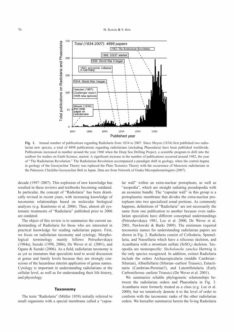
lar wall” within an extra-nuclear protoplasm, as well as“axopodia”, which are straight radiating pseudopodia withan axoneme bundle. The “capsular wall” in this group is aprotoplasmic membrane that divides the extra-nuclear pro-toplasm into two specialized zonal portions. As commonlyhappens, definitions of “Radiolaria” are not necessarily thesame from one publication to another because even radio-larian specialists have different conceptual understandings(Petrushevskaya 1981, Lee et al. 2000, De Wever et al.2001, Pawlowski & Burki 2009). The minimum requiredtaxonomic names for understanding radiolarian papers areshown in Fig. 2. Radiolaria consist of Collodaria, Spumel-laria, and Nassellaria which have a siliceous skeleton, andAcantharia with a strontium sulfate (SrSO4) skeleton. Tax-opodia are monospecific: Sticholonche zanclea Hertwig isthe only species recognized. In addition, extinct Radiolariainclude the orders Archaeospicularia (middle Cambrian–Silurian), Albaillellaria (Silurian–earliest Triassic), Entacti-naria (Cambrian-Permian?), and Latentifistularia (EarlyCarboniferous–earliest Triassic) (De Wever et al. 2001).
We summarize reliable phylogenetic relationships be-tween the radiolarian orders and Phaeodaria in Fig. 3.Acantharia were formerly treated as a class (e.g. Lee et al.2000), but we tentatively demote it to the level of order toconform with the taxonomic ranks of the other radiolarianorders. We hereafter summarize herein the living Radiolaria
70 N. SUZUKI & Y. AITA
Fig. 1. Annual number of publications regarding Radiolaria from 1834 to 2007. Since Meyen (1834) first published two radio-larian new species, a total of 4898 publications regarding radiolarians (including Phaeodaria) have been published worldwide.Publications increased in number around the year 1968 when the Deep Sea Drilling Project, a scientific program to drill into theseafloor for studies on Earth Science, started. A significant increase in the number of publications occurred around 1982, the yearof “The Radiolarian Revolution.” The Radiolarian Revolution accompanied a paradigm shift in geology, when the central dogmain geology of the Geosyncline Theory was replaced the Plate Tectonics Theory with the occurrence of Mesozoic radiolarians inthe Paleozoic Chichibu Geosyncline Belt in Japan. Data are from Network of Osaka Micropaleontologists (2007).
Review on taxonomy and cytology of Radiolaria 71
Fig
.2.
Key
to r
adio
lari
ans
at th
e or
der-
and
sub
orde
r-le
vels
. Rad
iola
ria
incl
ude
the
Ord
ers
Col
loda
ria,
Nas
sell
aria
, Spu
mel
lari
a, A
cant
hari
a an
d Ta
xopo
dia.
The
firs
tth
ree
orde
rs a
re c
hara
cter
ized
by
the
poss
essi
on o
f si
lice
ous
skel
eton
, A
cant
hari
a ha
ve s
kele
tons
of
stro
ntiu
m s
ulfa
te,
and
Taxo
podi
a ha
s si
lice
ous
mat
ter
insi
de o
ar-
like
pse
udop
odia
. C
ollo
dari
a us
ed t
o be
inc
lude
d w
ithi
n th
e S
pum
ella
ria.
Mai
n so
urce
s of
inf
orm
atio
n: S
chew
iako
ff (
1926
), C
ampb
ell
(195
4),
Cac
hon
& C
acho
n(1
978)
, Pet
rush
evsk
aya
(198
1), B
erns
tein
et a
l. (1
999)
, De
Wev
er e
t al.
(200
1), a
nd A
dl e
t al.
(200
5).
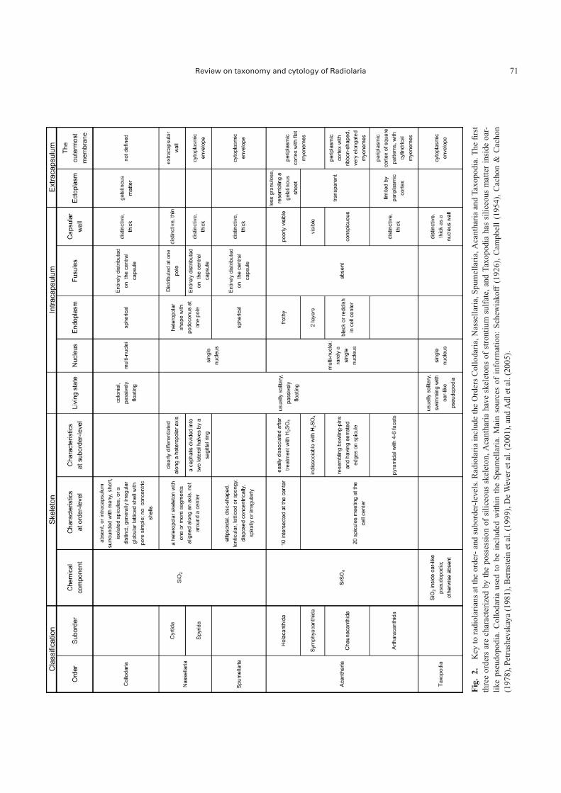
at the order level, and discuss current problems at the fam-ily through species levels. We eliminate the familiar taxo-nomic criterion “Polycystina” but retain the term to mean“diatom-like” for appellative purposes, such as in the form“polycystine” Radiolaria, referring to a radiolarian with asiliceous skeleton. Terminology for skeleton, protoplasmand pseudopodial systems are highly complex so that ab-breviations concerning the skeleton and protoplasm arelisted in Tables 1 and 2 and the brief definitions ofpseudopodia are summarized in Table 3.
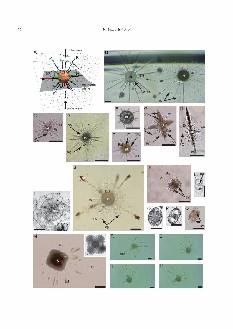
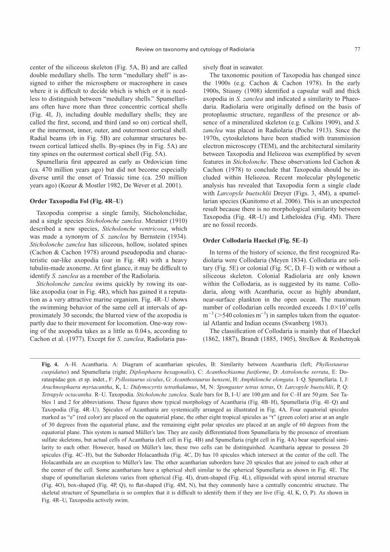
Order Acantharia Haeckel (Fig. 4A–H)
Acantharia possess a strontium sulfate (SrSO4) skeletonthat easily dissolves in seawater after death. Consequently,there are no reliable fossil records. Acantharia commonlymake up a major part of the radiolarian biomass in seawa-ter, exceeding 5.0�104 indiv. m�3 in the East China Sea(Tchang & Tan 1964). The skeleton is marked by a very so-phisticated symmetrical arrangement of 10 or 20 radialspicules named using Müller’s rule (Fig. 4A). These radialspicules vary in size and form among species, but the prin-
72 N. SUZUKI & Y. AITA
Fig. 3. Molecular phylogenetic relationships of the Radiolaria: Cercozoa, Alveolata, Heliozoa, and Amoebozoa (not to scale).Although Adl et al. (2005) regarded “Radiolaria” as a polyphyletic group, current knowledge on molecular phylogeny (Cavalier-Smith & Chao 2003, Takahashi et al. 2004, Oka et al. 2005, Yuasa et al. 2005, 2006, Kunitomo et al. 2006) evidently shows thatRadiolaria is a monophyletic group. Collodaria were previously considered to be a member of the Spumellaria (e.g. Adl et al.2005), but recent data suggests instead that it is closely related to Nassellaria rather than Spumellaria. “Sticholonche” in Adl et al.(2005) has already been classified in the Order Taxopodia by Fol (1883). With respect to order- or suborder-level, there is no mol-ecular phylogenetic data for the nassellarian Spyrida.
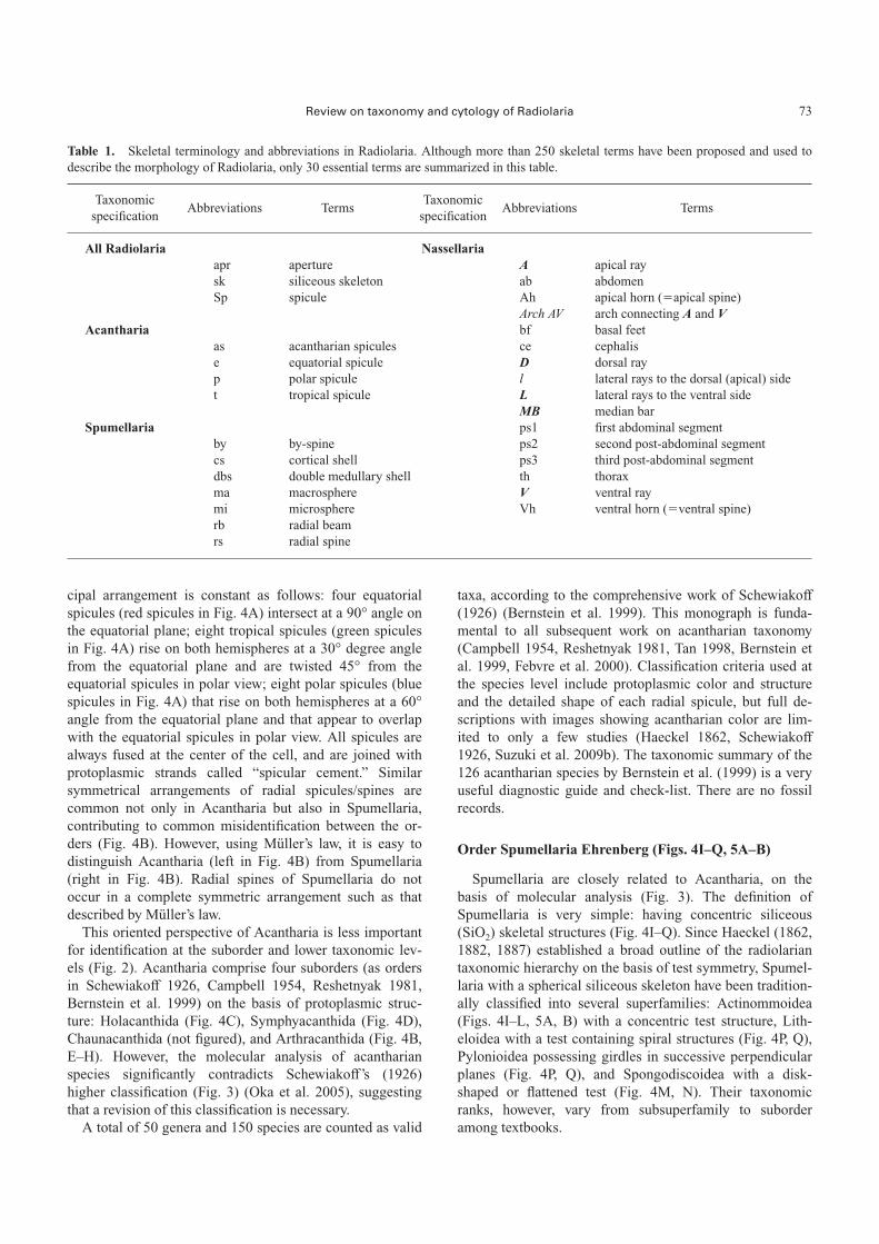
cipal arrangement is constant as follows: four equatorialspicules (red spicules in Fig. 4A) intersect at a 90° angle onthe equatorial plane; eight tropical spicules (green spiculesin Fig. 4A) rise on both hemispheres at a 30° degree anglefrom the equatorial plane and are twisted 45° from theequatorial spicules in polar view; eight polar spicules (bluespicules in Fig. 4A) that rise on both hemispheres at a 60°angle from the equatorial plane and that appear to overlapwith the equatorial spicules in polar view. All spicules arealways fused at the center of the cell, and are joined withprotoplasmic strands called “spicular cement.” Similarsymmetrical arrangements of radial spicules/spines arecommon not only in Acantharia but also in Spumellaria,contributing to common misidentification between the or-ders (Fig. 4B). However, using Müller’s law, it is easy todistinguish Acantharia (left in Fig. 4B) from Spumellaria(right in Fig. 4B). Radial spines of Spumellaria do notoccur in a complete symmetric arrangement such as thatdescribed by Müller’s law.
This oriented perspective of Acantharia is less importantfor identification at the suborder and lower taxonomic lev-els (Fig. 2). Acantharia comprise four suborders (as ordersin Schewiakoff 1926, Campbell 1954, Reshetnyak 1981,Bernstein et al. 1999) on the basis of protoplasmic struc-ture: Holacanthida (Fig. 4C), Symphyacanthida (Fig. 4D),Chaunacanthida (not figured), and Arthracanthida (Fig. 4B,E–H). However, the molecular analysis of acantharianspecies significantly contradicts Schewiakoff’s (1926)higher classification (Fig. 3) (Oka et al. 2005), suggestingthat a revision of this classification is necessary.
A total of 50 genera and 150 species are counted as valid
taxa, according to the comprehensive work of Schewiakoff(1926) (Bernstein et al. 1999). This monograph is funda-mental to all subsequent work on acantharian taxonomy(Campbell 1954, Reshetnyak 1981, Tan 1998, Bernstein etal. 1999, Febvre et al. 2000). Classification criteria used atthe species level include protoplasmic color and structureand the detailed shape of each radial spicule, but full de-scriptions with images showing acantharian color are lim-ited to only a few studies (Haeckel 1862, Schewiakoff1926, Suzuki et al. 2009b). The taxonomic summary of the126 acantharian species by Bernstein et al. (1999) is a veryuseful diagnostic guide and check-list. There are no fossilrecords.
Order Spumellaria Ehrenberg (Figs. 4I–Q, 5A–B)
Spumellaria are closely related to Acantharia, on thebasis of molecular analysis (Fig. 3). The definition ofSpumellaria is very simple: having concentric siliceous(SiO2) skeletal structures (Fig. 4I–Q). Since Haeckel (1862,1882, 1887) established a broad outline of the radiolariantaxonomic hierarchy on the basis of test symmetry, Spumel-laria with a spherical siliceous skeleton have been tradition-ally classified into several superfamilies: Actinommoidea(Figs. 4I–L, 5A, B) with a concentric test structure, Lith-eloidea with a test containing spiral structures (Fig. 4P, Q),Pylonioidea possessing girdles in successive perpendicularplanes (Fig. 4P, Q), and Spongodiscoidea with a disk-shaped or flattened test (Fig. 4M, N). Their taxonomicranks, however, vary from subsuperfamily to suborderamong textbooks.
Review on taxonomy and cytology of Radiolaria 73
Table 1. Skeletal terminology and abbreviations in Radiolaria. Although more than 250 skeletal terms have been proposed and used todescribe the morphology of Radiolaria, only 30 essential terms are summarized in this table.
Taxonomic Abbreviations Terms
Taxonomic Abbreviations Terms
specification specification
All Radiolaria Nassellariaapr aperture A apical raysk siliceous skeleton ab abdomenSp spicule Ah apical horn (�apical spine)
Arch AV arch connecting A and VAcantharia bf basal feet
as acantharian spicules ce cephalise equatorial spicule D dorsal rayp polar spicule l lateral rays to the dorsal (apical) sidet tropical spicule L lateral rays to the ventral side
MB median barSpumellaria ps1 first abdominal segment
by by-spine ps2 second post-abdominal segmentcs cortical shell ps3 third post-abdominal segmentdbs double medullary shell th thoraxma macrosphere V ventral raymi microsphere Vh ventral horn (�ventral spine)rb radial beamrs radial spine
74 N. SUZUKI & Y. AITA
Table 2. Cytological terminology and abbreviations in Radiolaria. Since no recent list of cytological terminology exists, we list here im-portant cytological terms from more than 50 references.
Protoplasmic Abbreviations Terms
Protoplasmic Abbreviations Terms
part part
Both in extracapsulum and intracapsulum Pseudopodia for Acanthariacpl color particles M myonemeCt cytokalymmaG Golgi body Pseudopodia for Cyrtid NassellariaMc mitochondria AJ apical projectionsMt microtubulines Ccp projection from concavityPM perispicular membrane CvP projection from convexityPS perinuclear cisterna CZ cortical junction zonePT protoplasm TC terminal conePV perialgal vacuole TP terminal projectionR electron-dense reserve droplet VP ventral projectionSb granular storage substance XP axial projectionSY associated organisms (symbionts, parasites)VA vacuole Pseudopodia for NassellariaWV waste vacuole PF porochora (�pore field)
PL protoplasmic lobeIntracapsulum PN podoconus
CA canalCV central vacuole Pseudopodia for Collodaria & SpumellariaCW capsular wall Af axoflagellumEnd endoplasmInt intracapsulum Pseudopodia for TaxopodiaLa electron translucent lacune Aj axopodial jointN nucleusNL nuclear lobe Ax(D) dorsal axopodiaNM nuclear membrane Ax(Dl) dorsolateral axopodiaPL protoplasmic lobe Ax(l) lateral axopodiaPP vacuolated protoplasmic process Ax(V) ventral axopodiaSC spicular cement JF joint filament
Mf microfilamentExtracapsulum (exclusive of pseudopodia) oar oar-like axopodia
BA bubble-like alveoliCM cell membrane Fusule structuresDV digestive vacuole ER endoplasmic reticulumExt extracapsulum FA funneled appendageEW extracapsular wall FM fusule membraneGM gelatin matrix IF interseptal fusulePC periplasmic cortex IOT inner osmiophilic tubePE perispicular ectoplasm IOZ inner osmiophilic zonePO protoplasmic envelope IS internal emptyRN reticulopodia network OOT outer osmiophilic tube
OOZ outer osmiopihilic zonePseudopodia for all Radiolaria PELZ peripheral electron-lucent zone
AF axopodial filaments RC rheoplasmitic canalAM axoflagellar filaments Sl slit-like narrow fissuresAp axoplastAx axopodiaFp filopodiaFS fusulePs pseudopodia
Determination to genus as well as species for Spumel-laria requires examination of the internal skeletal structure(mi, ma in Fig. 5A, B) (e.g. De Wever et al. 2001, Suzuki2006), but this is as yet poorly understood for describedspecies as well as “new species.” This taxonomic uncer-tainty forces radiolarian specialists to apply tentativegeneric identifications. Many attempts to refine spumellar-ian classification have been made by micropaleontologists(e.g. Kozur & Mostler 1972, 1978, 1979, Dumitrica 1984,Petrushevskaya 1984b, De Wever et al. 2001, Afanasieva &Amon 2006), but these attempts are far from a consensusresolution. Not only that, it is very difficult to assignspecies names for Spumellaria. A review of available litera-ture shows that for all documented “polycystine” morpho-types (See Fig. 3), 34.7% (50/144 species) of livingSpumellaria (Takahashi 1991) and 50.8% (33/65 species) of
subantarctic Eocene Spumellaria (ca. 30–40 million yearsago) (Suzuki et al. 2009a) are still in open nomenclature.
Spumellaria have such complex internal skeleton struc-ture that more than 250 morphological features and termshave been proposed (Petrushevskaya 1984a, Suzuki 1998,Ogane & Suzuki 2006). However, only a few terms are es-sential for understanding the principal skeletal morphologyof Spumellaria. The skeleton of the superfamily Actinom-moidea consists mainly of concentric latticed shells with ra-dial beams (rb in Fig. 5B), radial spines (rs in Fig. 5A), andby-spines (by in Fig. 5A). Concentric latticed shells have amicrosphere, a macrosphere, and cortical shells (from in-nermost to outermost). The microsphere, or innermedullary shell (mi in Fig. 5A), and the macrosphere, orouter medullary shell (ma in Figs. 4I, 5A, B), are generallyvery concentric (cs in Figs. 4I, 5A) in appearance at the
Review on taxonomy and cytology of Radiolaria 75
Table 3. Brief definition of pseudopodial systems for Radiolaria. The pseudopodial system is variable and significantly different at thesuborder and order-level. Since no list including definitions of pseudopodial systems exists, approximately 25 terms are listed here as essen-tial terms for describing the pseudopodial systems based on more than 50 references.
Terminology Taxonomy specified Characters
axial projections Nassellaria a single thick pseudopodium from the aperture of the siliceous test, similar toaxoflagellum
axoflagellar filaments Radiolaria electrically dense matter inside an axoflagellum. This term is usually usedwhen the component of the filament is not determiend.
axoflagellum Spumellaria a significantly thick, and longer pseudopodia, differing from other pseudopodiaaxoneme Radiolaria a microtubular axis of shaft, exclusive of the covering membrane, composed of
the [9(2)+2] arrangement of microtubulesaxoneme bundle Radiolaria bundles of axonemeaxoplast Spumellaria, Nassellaria a fine fibrillar mass or central granule as a microtubule-organizing center that
may enable microtubules to extend axopodia through fusulesaxopodium Radiolaria pseudopodia stiffened by microtubuline bundles or axonemes inside pseudopo-
dia, and including vacuoles, mitochondria and protoplasmic reticulumaxopodial filaments Radiolaria electrically dense matter which is connected to the axopodia in the endoplas-
mic reticulumaxopodial system Radiolaria microtubuline network comprising axopodia, axoplast and axopodial filamentsfilopodium Spumellaria temporary, slender protruding part of the ectoplasm, appearing like fine hairintracapsular axopodial system Radiolaria microtubuline bundles of axopodial filaments, axoflagellar filaments and axo-
plast; always situated within the endoplasmic reticulummicrotubuline Radiolaria slender, hollow structure made primarily of tubulin proteinsmicrotubular bundle Radiolaria fibrous structure composed of longitudinally aligned 24 nm microtubulesmyoneme Acantharia retractable fibrillar, ribbon-like organelles which consist of densely packed 2–3
nm microfibrils.nucleus-axoplast relationship Spumellaria, Nassellaria the connecting patterns between axoplast and nucleusperispicular membrane Acantharia a protoplasmic membrane that envelopes the acantharian spiculespodoconus Nassellaria cone-shaped organelle which consists of axopodial filamentsprojections Nassellaria nassellarian pseudopodiaprojections from concavities Nassellaria projections extending from concavities between segments of siliceous testprojections from convexities Nassellaria projections extending from the middle part of a segment of siliceous testpseudopodium Radiolaria temporary protoplasmic protrusion of radiolarian cell, a common term despite
the different internal structures contained within itpseudopodial filament Radiolaria electrically dense matter inside a pseudopodia. This term is usually used when
the component of the filament is not determined.reticulopodial network Collodaria network of cross-connected pseudopodiaterminal cone Nassellaria cone-shaped arrangement of terminal projectionsterminal projections Nassellaria fine pseudopodia radiating from the aperture of siliceous test
76 N. SUZUKI & Y. AITA
center of the siliceous skeleton (Fig. 5A, B) and are calleddouble medullary shells. The term “medullary shell” is as-signed to either the microsphere or macrosphere in caseswhere it is difficult to decide which is which or it is need-less to distinguish between “medullary shells.” Spumellari-ans often have more than three concentric cortical shells(Fig. 4I, J), including double medullary shells; they arecalled the first, second, and third (and so on) cortical shell,or the innermost, inner, outer, and outermost cortical shell.Radial beams (rb in Fig. 5B) are columnar structures be-tween cortical latticed shells. By-spines (by in Fig. 5A) aretiny spines on the outermost cortical shell (Fig. 5A).
Spumellaria first appeared as early as Ordovician time(ca. 470 million years ago) but did not become especiallydiverse until the onset of Triassic time (ca. 250 millionyears ago) (Kozur & Mostler 1982, De Wever et al. 2001).
Order Taxopodia Fol (Fig. 4R–U)
Taxopodia comprise a single family, Sticholonchidae,and a single species Sticholonche zanclea. Meunier (1910)described a new species, Sticholonche ventricosa, whichwas made a synonym of S. zanclea by Bernstein (1934).Sticholonche zanclea has siliceous, hollow, isolated spines(Cachon & Cachon 1978) around pseudopodia and charac-teristic oar-like axopodia (oar in Fig. 4R) with a heavytubulin-made axoneme. At first glance, it may be difficult toidentify S. zanclea as a member of the Radiolaria.
Sticholonche zanclea swims quickly by rowing its oar-like axopodia (oar in Fig. 4R), which has gained it a reputa-tion as a very attractive marine organism. Fig. 4R–U showsthe swimming behavior of the same cell at intervals of ap-proximately 30 seconds; the blurred view of the axopodia ispartly due to their movement for locomotion. One-way row-ing of the axopodia takes as a little as 0.04 s, according toCachon et al. (1977). Except for S. zanclea, Radiolaria pas-
sively float in seawater.The taxonomic position of Taxopodia has changed since
the 1900s (e.g. Cachon & Cachon 1978). In the early1900s, Stiasny (1908) identified a capsular wall and thickaxopodia in S. zanclea and indicated a similarity to Phaeo-daria. Radiolaria were originally defined on the basis ofprotoplasmic structure, regardless of the presence or ab-sence of a mineralized skeleton (e.g. Calkins 1909), and S.zanclea was placed in Radiolaria (Poche 1913). Since the1970s, cytoskeletons have been studied with transmissionelectron microscopy (TEM), and the architectural similaritybetween Taxopodia and Heliozoa was exemplified by sevenfeatures in Sticholonche. These observations led Cachon &Cachon (1978) to conclude that Taxopodia should be in-cluded within Heliozoa. Recent molecular phylogeneticanalysis has revealed that Taxopodia form a single cladewith Larcopyle buetschlii Dreyer (Figs. 3, 4M), a spumel-larian species (Kunitomo et al. 2006). This is an unexpectedresult because there is no morphological similarity betweenTaxopodia (Fig. 4R–U) and Litheloidea (Fig. 4M). Thereare no fossil records.
Order Collodaria Haeckel (Fig. 5E–I)
In terms of the history of science, the first recognized Ra-diolaria were Collodaria (Meyen 1834). Collodaria are soli-tary (Fig. 5E) or colonial (Fig. 5C, D, F–I) with or without asiliceous skeleton. Colonial Radiolaria are only knownwithin the Collodaria, as is suggested by its name. Collo-daria, along with Acantharia, occur as highly abundant,near-surface plankton in the open ocean. The maximumnumber of collodarian cells recorded exceeds 1.0�105 cellsm�3 (�540 colonies m�3) in samples taken from the equator-ial Atlantic and Indian oceans (Swanberg 1983).
The classification of Collodaria is mainly that of Haeckel(1862, 1887), Brandt (1885, 1905), Strelkov & Reshetnyak
Review on taxonomy and cytology of Radiolaria 77
Fig. 4. A–H. Acantharia. A: Diagram of acantharian spicules, B: Similarity between Acantharia (left; Phyllostauruscuspidatus) and Spumellaria (right; Diplosphaera hexagonalis), C: Acanthochiasma fusiforme, D: Astrolonche serrata, E: Do-rataspidae gen. et sp. indet., F: Pyllostaurus siculus, G: Acanthostaurus henseni, H: Amphilonche elongata. I–Q. Spumellaria. I, J:Arachnosphaera myriacantha, K, L: Didymocyrtis tetrathalamus, M, N: Spongaster tetras tetras, O: Larcopyle buetschlii, P, Q:Tetrapyle octacantha. R–U. Taxopodia. Sticholonche zanclea. Scale bars for B, I–U are 100 mm and for C–H are 50 mm. See Ta-bles 1 and 2 for abbreviations. These figures show typical morphology of Acantharia (Fig. 4B–H), Spumellaria (Fig. 4I–Q) andTaxopodia (Fig. 4R–U). Spicules of Acantharia are systemically arranged as illustrated in Fig. 4A. Four equatorial spiculesmarked as “e” (red color) are placed on the equatorial plane, the other eight tropical spicules as “t” (green color) arise at an angleof 30 degrees from the equatorial plane, and the remaining eight polar spicules are placed at an angle of 60 degrees from theequatorial plane. This system is named Müller’s law. They are easily differentiated from Spumellaria by the presence of strontiumsulfate skeletons, but actual cells of Acantharia (left cell in Fig. 4B) and Spumellaria (right cell in Fig. 4A) bear superficial simi-larity to each other. However, based on Müller’s law, these two cells can be distinguished. Acantharia appear to possess 20spicules (Fig. 4C–H), but the Suborder Holacanthida (Fig. 4C, D) has 10 spicules which intersect at the center of the cell. TheHolacanthida are an exception to Müller’s law. The other acantharian suborders have 20 spicules that are joined to each other atthe center of the cell. Some acantharians have a spherical shell similar to the spherical Spumellaria as shown in Fig. 4E. Theshape of spumellarian skeletons varies from spherical (Fig. 4I), drum-shaped (Fig. 4L), ellipsoidal with spiral internal structure(Fig. 4O), box-shaped (Fig. 4P, Q), to flat-shaped (Fig. 4M, N), but they commonly have a centrally concentric structure. Theskeletal structure of Spumellaria is so complex that it is difficult to identify them if they are live (Fig. 4J, K, O, P). As shown inFig. 4R–U, Taxopodia actively swim.
78 N. SUZUKI & Y. AITA

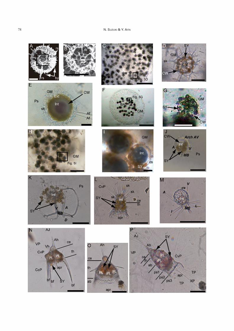
(1971), and Anderson (1983). Thalassosphaeridae is a soli-tary collodarian, possessing isolated tiny spicules (sk inFig. 5E) around the cell. Similar individuals with many iso-lated, tiny spicules (sk in Fig. 5D) are also observed inSphaerozoidae (Fig. 5D), but they always form colonies, asshown in Fig. 5C. Isolated, tiny spicules are occasionallyfound in sediments and seawater, but cannot be differenti-ated as solitary Thalassosphaeridae or colonial Sphaero-zoidae. Cells of Collosphaeridae have a simple, single, thin-walled spherical or subspherical latticed shell (cs in Fig. 5I)and most make colonies (Fig. 5H). Collosphaeridae is amajor identifiable group of fossil Collodaria. The samespecies forms a variety of gelatinous colony shapes, andcolony size ranges from several tens of microns to a fewmeters. For instance, Collozoum longiforme Swanberg &Harbison and Collozoum caudatum Swanberg & Andersonreach lengths of up to 2–3 m with tail-like strands of wastematerial (Swanberg & Harbison 1980, Swanberg & Ander-son 1981).
Collodaria were used to study fine protoplasmic struc-tures that are typical of Radiolaria (Huth 1913, Anderson &Botfield 1983), because the structures are easily observedin ultrathin sections due to the absence of or minimal pres-ence of hard parts. However, there is some doubt as towhether Collodaria can be regarded as a typical representa-tive of “polycystine” Radiolaria (see Fig. 3). The reason forthis is that most Radiolaria are solitary, whereas most Col-lodaria are colonial.
Collodaria are commonly associated with algal sym-bionts (SY in Fig. 5D, G, I) and a nutrient-based alga–hostinteraction has been demonstrated in laboratory experi-
ments (Anderson, 1978b). Accordingly, Collodaria are ex-clusively common in oligotrophic environments (Swanberg1983). Collodaria appeared in the Eocene (339–558 millionyears ago) (De Wever et al. 2001).
Order Nassellaria Ehrenberg (Fig. 5J–P)
Nassellaria are probably the best-known “polycystine”Radiolaria possessing conical siliceous tests with segmenta-tions (e.g. Fig. 5P). Nassellaria are traditionally subdividedinto two suborders: Spyrida Ehrenberg (Fig. 5J, K) and Cyr-tida (or Cyrtellaria) Haeckel (Figs. 2, 5L–P). Spyrida arecharacterized by a skeleton that generally possesses a sagit-tal ring. The sagittal ring is the D-shaped ring that is com-prised of a median bar (MB in Fig. 5J, K), apical ray (A inFig. 5J, K), vertical ray (V in Fig. 5J, K), and an arch con-necting the apical and vertical rays (Arch AV in Fig. 5J) andlatticed lateral chambers that form a bilobate cephalis (Fig.5K), whereas Cyrtida have a skeleton with a conical (Fig.5O, P) or cap-shaped test (Fig. 5L, M), consisting of unise-rially and usually rectilinearly arranged segments (Petru-shevskaya 1971, Sanfilippo et al. 1985). Superficial featuresof Spyrida (Fig. 5J, K) appear quite different from those ofCyrtida (Fig. 5L–P), but they have a common skeletalstructure. It is surprising that the same bony framework in-cluding the apical ray (A in Fig. 5J, K), vertical ray (V inFig. 5J, K), and median bar (MB in Fig. 5J, K, M) is en-crypted in the first segment (cephalis) (ce in Fig. 5L–P) ofmulti-segmented Nassellaria. This framework is referred toas the “initial spicular system” or “cephalic structure”which is considered a very conservative structure in family-
Review on taxonomy and cytology of Radiolaria 79
Fig. 5. A, B. SEM photographs showing the internal structure of the spumellarian Actinomma sp. from the Miocene (ca. 14million years ago). A: entire view showing the internal skeletal structure. B: close-up view of double medullary shells from theopen box in Fig. 5A. C–I. Collodaria. C: colony form of Sphaerozoum fuscum, D: close-up view of a single radiolarian cell fromthe open box in Fig. 5C, E: solitary form of Thalassosphaera curvicornis, F: colony form of Collozoum inerme, G: close-up viewof a single radiolarian cell from the open box in Fig. 5F, H: colony form of Collosphaera tuberosa, I: close-up view of single radi-olarian cells from the open box in Fig. 5H. J–M. Spyrid Nassellaria. J: Zygocircus productus, K: Acanthodesmia vinculata, L:Lophophaena hispida, M: Peromelissa phalacra. N–P. Cyrtid Nassellaria. N: Pterocanium praetextum, O: Lipmanelladictyoceras, P: Eucyrtidium hexagonatum. Scales for A, B, D, G, I, J, L, M are 50 mm, for C, F, H are 0.5 mm, and for E, K, N–Pare 100 mm, respectively. See Tables 1 and 2 for abbreviations. Superfamily Actinommoidea (Spumellaria) are characterized bythe possession of a double medullary shell (microsphere and macrosphere) (Fig. 5A, B). The architecture of the microsphere isconsidered to be of a common pattern within the same family. Fig. 5C–I represents three major families of Collodaria. Collodariaare characterized by a colonial form with numerous radiolarian cells (Fig. 5C, F, H), but the family Thalassosphaeridae (Fig. 5E)is an exception being a solitary collodarian group. Thalassosphaeridae have a gelatinous matrix (GM in Fig. 5E) and isolatedsiliceous spicules (sk in Fig. 5E) as in the Sphaerozoidae (Fig. 5D). It is extremely difficult to differentiate between the Thalas-sosphaeridae and Sphaerozoidae after their protoplasmic part is lost. Identifiable collodarians largely belong to the family Col-losphaeridae (Fig. 5H, I) because this family has a spherical cortical shell (rarely two shells). Collodaria commonly harbor algalsymbionts (SY in Fig. 5D, G, I). Spyrid Nassellaria (Fig. 5J, K) appear to be different from the Cyrtid Nassellaria (Fig. 5L–P), butthe internal spicular system composed of A, V and MB is commonly recognized throughout the Spyrida (Fig. 5J, K) and Cyrtida(Fig. 5M). The internal spicular system is the main distinguishing feature for the both Spyrida and Cyrtida at the family–level.Spyrida are characterized by the presence of a “D-ring” which is comprised of A, Arch-AV or a relevant arch, V and MB (Fig. 5J,K). In the Cyrtid Nassellaria, the Superfamily Plagiacanthoidea have a significantly large cephalis (ce in Fig. 5L, M), comparedwith the overall size of the skeleton. Cyrtids have a smaller cephalis (Fig. 5N–P). Nassellarians shown here are easily recogniz-able with their yellowish brown associated organisms (SY in Fig. 5J–L, N–P) and yellowish to reddish matter outside the proto-plasmic lobes (PL in Fig. 5N–P).
or genus-based fossil evidence (Petrushevskaya 1971). Al-though this assumption is still in dispute among micropale-ontologists, the current scheme for Nassellaria at highertaxonomic levels is mainly based on the initial spicular sys-tem as well as morphologic changes through time (DeWever et al. 2001). This structure is rarely used for speciesclassification. Other important morphological terms are“cephalis” (ce in Fig. 5L–P), “thorax” (th in Fig. 5L, N–P),“abdomen” (ab in Fig. 5O, P), and “post-abdominal seg-ments” (ps1, ps2, ps3 in Fig. 5P) from the apex of the testto the widened aperture.
Classification of Nassellaria at the species level seems tohave gained consensus among radiolarian specialists, butgenus and family designations are still disputed. For in-stance, a well-known extant high-latitude marker species,Cycladophora davisiana Ehrenberg, has been placed in oneof four genera. Even worse, a given genus can be attributedto a variety of families. Such confusion is mainly due to in-sufficient traces of phylogeny at the species–level in eachgenus. Since taxonomic classification at the genus and fam-ily levels is still confused, we maintain that discussion ofnassellarian radiolarians must be at the species level. Al-though taxonomic confusion continues, 52 families and 400genera (fossil and living) are listed in Nassellaria (DeWever et al. 2001). The continuous fossil records go backto the Early Triassic (250 million years ago), although iso-lated occurrences of nassellarian–like “polycystine” Radio-laria (families Archocyrtiidae and Popofskyellidae) areknown between the Late Devonian (380 million years ago)to Late Carboniferous (315 million years ago) (Cheng1986).
Cytology
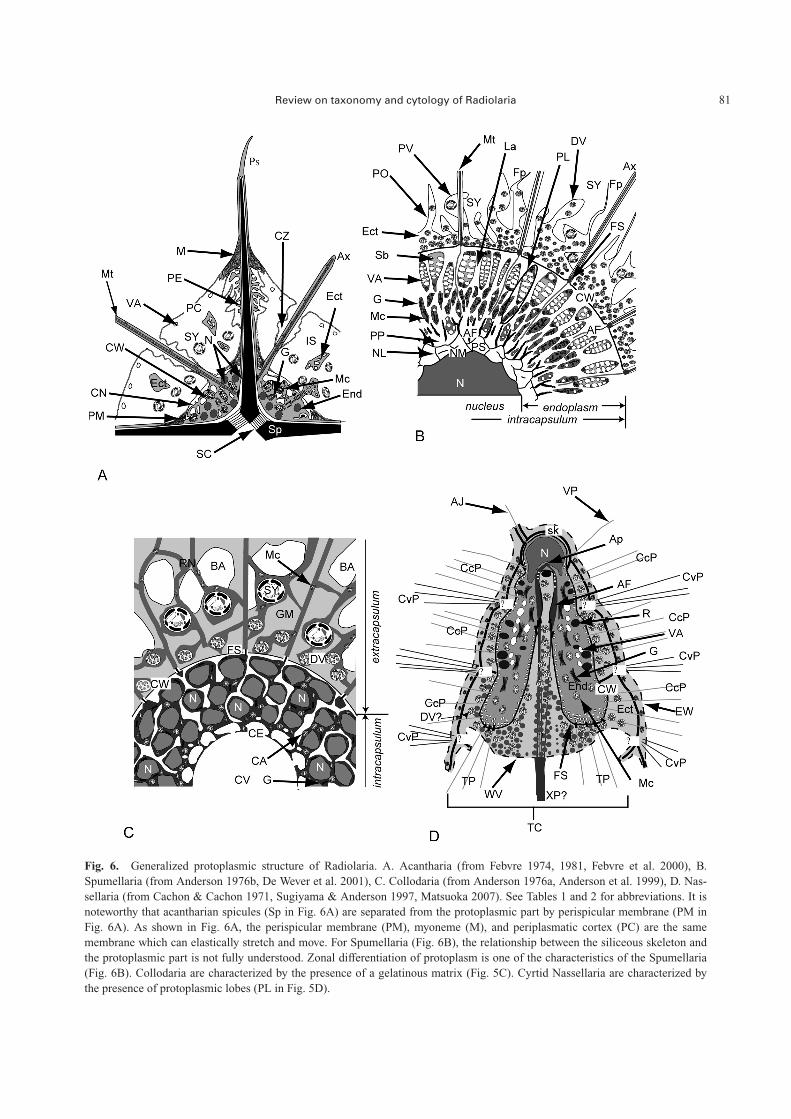
The protoplasm is roughly divided into a nucleus/nuclei(N in Fig. 6A–D), nuclear wall, endoplasm (End in Fig. 6A,D), capsular wall (or capsular membrane) (CW in Fig.6A–D), ectoplasm (Ect in Fig. 6A–D), and pseudopodia(Table 3). The capsular wall divides the protoplasm into anendoplasm and an ectoplasm. An interconnecting organellebetween the endoplasm and ectoplasm termed the fusule(FS in Fig. 6B–D) is also characteristic of Radiolaria. Themost significant feature of the radiolarian protoplasm is adense, colored section that corresponds to the protoplasm inthe capsular wall. For practical recognition of radiolarianprotoplasm, the region that includes the nucleus and endo-plasm is called the “intracapsulum” (�intracapsular proto-plasm, or central capsule) (Int in Figs. 4A–H, J, K, P, 5D, E,I, K–M), whereas the protoplasm outside the capsular wallis the “extracapsulum” (�extracapsular protoplasm) (Ext inFigs. 4B–D, F–H, J, 5E) (Campbell 1954, Anderson 1983,De Wever et al. 2001).
Taxopodia (Fig. 4R–U) are distinctly different from allother radiolarians because of the absence of a capsular wall,which is an important characteristic of the Radiolaria. Thethick protoplasmic wall of Taxopodia was considered to be
a capsular wall in early studies, but this is now regarded asa nuclear wall. This was established to be a nuclear wall be-cause of the absence of other protoplasmic organelles (Hol-lande et al. 1967). Cachon & Cachon (1978) demonstratedthe presence of a thick, double nuclear membrane (600 nmin thickness) called the “nuclear capsule.”
Intracapsulum
Nucleus: The nucleus of Radiolaria is interesting be-cause of its spatial relationship within the skeleton.Spicules protrude through the center in acantharian species,so the nucleus is never found there (Fig. 6A). The intracap-sulum of some Symphyacanthoidea (Haliommatidium andDicranophora) contains a single large nucleus that is proba-bly polyploid during the trophont life stage, whereas thereare numerous small round or oblong nuclei in all otherAcantharia (N in Fig. 6A) (Febvre 1977, Febvre et al. 2000,Suzuki et al. 2009b).
A similar multinucleated intracapsulum is also observedin some colonial Collodaria (Sphaerozoum fuscum Meyen,Collosphaera globularis Haeckel, Collozoum caudatumSwanberg & Anderson) at both reproductive and trophontstages (N in Fig. 6C) (Anderson 1976a, 1978b, Swanberg &Anderson 1981, Suzuki et al. 2009b). The diameter of thenuclei ranges from 5 to 20 µm. Other collodarian species,including Thalassicolla nucleata Huxley, have a single nu-cleus at the center of the intracapsulum (Fig. 7B) (Huth1913).
A single central nucleus is common in Spumellaria (Fig.7B, 7D–F) (Hollande & Enjumet 1960, Suzuki et al.2009b). In some spongiose Spumellaria (e.g. Styptosphaeraspumacea Haeckel), a dense siliceous skeletal meshwork isfully developed inside the test and the 20–60-µm-diameternucleus is located at the center (Swanberg et al. 1990).Actinommoidea possess double medullary shells (with asiliceous skeletal part in the center of the test), and the nu-cleus wraps around the microsphere and is encrypted in themacrosphere (Sugiyama & Anderson 1998a).
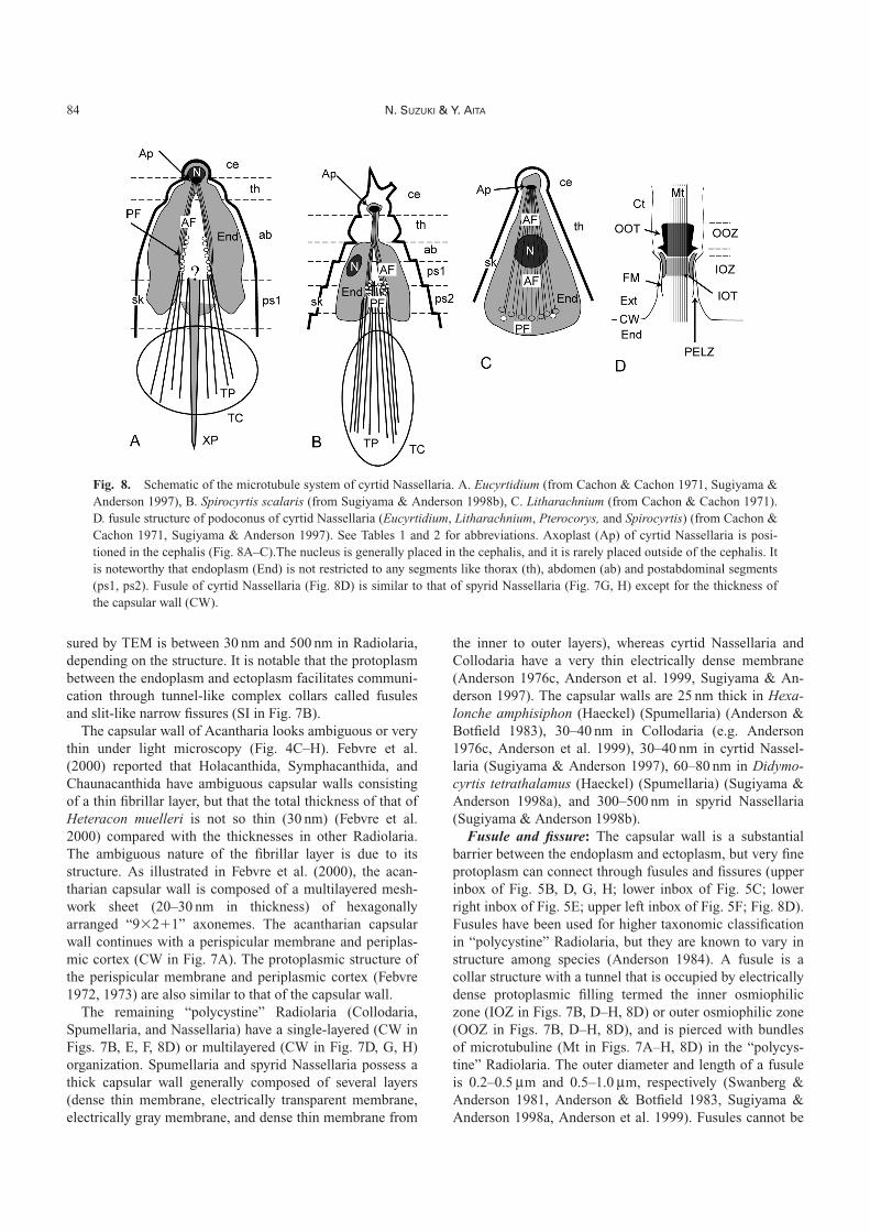
The Nassellaria suborder Cyrtida consist of a cephalis(ce in Fig. 8A–C), thorax (th in Fig. 8A–C), abdomen (ab inFig. 8A, B), and post-abdominal segments (ps1, ps 2 in Fig.8A, B), but this segmentation does not appear to corre-spond to the protoplasmic structure very well (Fig. 8A–C).The apex of the intracapsulum is always in the cephalis, butthe entire nucleus may not always be in the cephalis (N inFig. 8B, C) (Hollande & Enjumet 1960, Sugiyama & An-derson 1997). In contrast, the nucleus in the Nassellariasuborder Spyrida (Acanthodesmia vinculata (Müller) andLithocircus reticulatus (Ehrenberg)) is spherical to elliptical(N in Fig. 7G, H) (Sugiyama & Anderson 1998b), probablydue to a lack of skeletal coverage (Fig. 5J, K).
The shape and position of the nucleus are different inTaxopodia from all other Radiolaria (N in Fig. 7C). Sti-cholonche zanclea is generally elongate in shape with anelongated nucleus that is always positioned on the proto-
80 N. SUZUKI & Y. AITA
Review on taxonomy and cytology of Radiolaria 81
Fig. 6. Generalized protoplasmic structure of Radiolaria. A. Acantharia (from Febvre 1974, 1981, Febvre et al. 2000), B.Spumellaria (from Anderson 1976b, De Wever et al. 2001), C. Collodaria (from Anderson 1976a, Anderson et al. 1999), D. Nas-sellaria (from Cachon & Cachon 1971, Sugiyama & Anderson 1997, Matsuoka 2007). See Tables 1 and 2 for abbreviations. It isnoteworthy that acantharian spicules (Sp in Fig. 6A) are separated from the protoplasmic part by perispicular membrane (PM inFig. 6A). As shown in Fig. 6A, the perispicular membrane (PM), myoneme (M), and periplasmatic cortex (PC) are the samemembrane which can elastically stretch and move. For Spumellaria (Fig. 6B), the relationship between the siliceous skeleton andthe protoplasmic part is not fully understood. Zonal differentiation of protoplasm is one of the characteristics of the Spumellaria(Fig. 6B). Collodaria are characterized by the presence of a gelatinous matrix (Fig. 5C). Cyrtid Nassellaria are characterized bythe presence of protoplasmic lobes (PL in Fig. 5D).
82 N. SUZUKI & Y. AITA
plasm “backbone” (Cachon & Cachon 1978). Figure 7Cshows the axial transverse section and the lateral view of S.zanclea, as shown in Fig. 4R–U.
Endoplasm: The endoplasmic reticulum (ER) of Radio-laria contains a variety of organelles including Golgi bodies(G in Fig. 6A, B), mitochondria (Mc in Fig. 6A–D), vac-uoles, and reserve droplets with a granular storage sub-stance matrix (R in Fig. 6D). Free ribosomes are liberallydistributed throughout the ER (Anderson 1977). In somecolonial Collodaria and Spumellaria, orthorhombic ordipyramidal crystals are scattered in the endoplasm (Müller1858, Haeckel 1862, Hollande & Enjumet 1960). Thesecrystals are celestite (strontium sulfate), according to spec-tral analysis and crystal morphology of Sphaerozoumneapolitatum Brandt (Hollande & Martoja 1974). Similarcrystals have also been obtained from Acantharia (Bern-stein et al. 1999).
In Acantharia, the ER is clearly parceled with the capsu-lar wall (CW in Fig. 6A) in three suborders (Symphyacan-thida, Chaunacanthida and Arthracanthida) but protrudesoutside the intracapsulum as ectoplasm through a relativelylarge aperture on the capsular wall (Ect in Fig. 6A) (Febvreet al. 2000). The ER of Collodaria surrounds the nucleuswhen a single large nucleus is present, whereas the ER ofCollodaria, which is within the intracapsulum, is coveredwith a wrinkled protoplasmic envelope to form a canal (CAin Fig. 6C) or is an irregularly shaped protoplasmic enve-lope (Anderson et al. 1999). In colonial Collodaria, thesespaces may enhance the exchange of material and gases,such as oxygen and carbon dioxide, with the surroundingenvironment (Anderson et al. 1999).
The ER of Spumellaria is illustrated for spherical forms
only in Figs. 6B, 7D–F, and is packed in radially elongatedprotoplasmic lobes inside the intracapsulum. In somespecies, the Golgi bodies (G in Fig. 6B) and mitochondria(Mc in Fig. 6B) tend to be concentrated in the inner portionof the protoplasmic lobe (PL in Fig. 6B), whereas manyvacuoles (VA in Fig. 6B) and granular storage substancesare present in the outer portion, suggesting spherically sym-metric, functionally differentiated tissue in the sphericalSpumellaria. A similar protoplasmic structure is recognizedin spyrid Nassellaria (Sugiyama & Anderson 1998b).
The ER of Cyrtida is divided into four to six protoplas-mic lobes (PL in Fig. 5N–P). Relatively large electron-dense reserve droplets (R in Fig. 6D) are recognized in thelobe (Sugiyama & Anderson 1997), and small vacuoles arescattered near the capsular wall (VA in Fig. 6D).
Capsular wall and associated structures
Capsular wall: “Capsular membrane” is the most widelyused name for the protoplasmic divider between the endo-plasm and ectoplasm. Holacanthida (Fig. 4C), Symphya-canthida (Fig. 4D), and Chaunacanthida (not figured)(Acantharia) possess thin fibrillar layers (CW in Fig. 7A)instead of a firm chitinous or pseudochitinous membrane(CW in Fig. 7B–H), and subsequently, Febvre et al. (2000)recommended using the term “capsular wall,” which in-cludes the terms “capsular membrane” and “fibrillar layer.”
The capsular wall is a firm, chitinous or pseudochitinouscellular envelope. This firm membrane can remain intactfor several months after death under normal seawater con-ditions, and this property is occasionally misunderstood asevidence of life. The thickness of the capsular wall as mea-
Review on taxonomy and cytology of Radiolaria 83
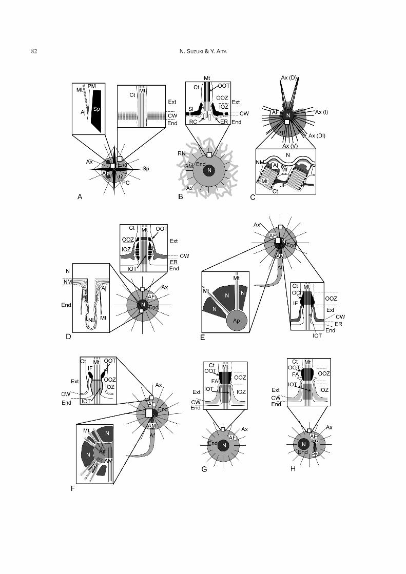
Fig. 7. Schematic of the microtubule systems of Acantharia, Collodaria, Taxopodia, Spumellaria, and spyrid Nassellaria. A.Acantharia (from Febvre & Febvre-Chevalier 1982, Febvre et al. 2000), B. Exo-axoplastid Collodaria (from Anderson 1976a, b, c,Cachon & Cachon 1976a, 1977, Swanberg & Anderson 1981, Anderson et al. 1999). C. Taxopodia (from Hollande et al. 1967,Cachon et al. 1977), D. Cryptoaxoplastid spherical Spumellaria (from Cachon & Cachon 1972b), E. Centroaxoplastid Spumel-laria (Actinosphaera and Rhizosphaera) (from Hollande & Enjumet 1960, Cachon & Cachon 1972a), F. Periaxoplastid sphericalRadiolaria (spumellarian Cenosphaera reticulata) (from Cachon & Cachon 1972b), G. Spyrida of the Nassellaria (Lithocircusreticulatus) (from Sugiyama & Anderson 1998b), H. Spyrida of the Nassellaria (Acanthodesmia vinculata) (from Sugiyama &Anderson 1998b). See Tables 1 and 2 for abbreviations.The microtubule system of Radiolaria is considered to be important because the axopodial system is a unique protoplasmic struc-ture. Studies on the pseudopodial system mainly focus on fusules, the passage structure in the capsular wall (CW) between the in-tracapsulum and extracapsulum and the relationships between the intracellular axopodial system (AP, AM and Mt) and nucleus(N). The axopodia of Acantharia (Fig. 7A) arise from the perispicular membrane (PM) but are not attached to the acantharianspicules (Sp). Fusules of Acantharia are very simple (upper left inbox of Fig. 7A), differing from other Radiolaria (Figs. 7B – H,8D). In Collodaria (Fig. 7B), a bundle of microtubuline (Mt) is present inside the axopodia but is absent in the intracapsulum.Taxopodia (Fig. 7C) have thick, oar-like axopodia (lower inbox of Fig. 7C) which arise from the nuclear membrane (NM). Oar-like axopodia can simultaneously move with axopodial joints (Aj) and joint filaments (JF). Spherical Spumellaria (Fig. 7D–F)have different arrangements of nucleus (N) and axoplast (AP). Axoplasts are absent in cryptoaxoplastids (Fig. 7D), present in thecenter of the protoplasm in centroaxoplastids (Fig. 7E), and situated beside the nucleus or open in one direction in periaxoplastids(Fig. 7F). The fusule structure also differs in cryptoaxoplastids, centroaxoplastids and periaxoplastids (Inbox figures around cap-sular wall [CW] in Fig. 7D–F). The microtubule system of spyrid Nassellaria (Fig. 7G, H) is similar to cryptoaxoplastid Spumel-laria (Fig. 7D) regardless of the phylogenetically close relationship to cyrtid Nassellaria (Fig. 8A–C). Densely thick outer osmio-philic tubes (OOT) of spyrid Nassellaria (inbox of Fig. 7G, H) are similar to those in centroaxoplastid Spumellaria (lower rightinbox of Fig. 7E) rather than cryptoaxoplastid Spumellaria (upper inbox of Fig. 7D).
sured by TEM is between 30 nm and 500 nm in Radiolaria,depending on the structure. It is notable that the protoplasmbetween the endoplasm and ectoplasm facilitates communi-cation through tunnel-like complex collars called fusulesand slit-like narrow fissures (SI in Fig. 7B).
The capsular wall of Acantharia looks ambiguous or verythin under light microscopy (Fig. 4C–H). Febvre et al.(2000) reported that Holacanthida, Symphacanthida, andChaunacanthida have ambiguous capsular walls consistingof a thin fibrillar layer, but that the total thickness of that ofHeteracon muelleri is not so thin (30 nm) (Febvre et al.2000) compared with the thicknesses in other Radiolaria.The ambiguous nature of the fibrillar layer is due to itsstructure. As illustrated in Febvre et al. (2000), the acan-tharian capsular wall is composed of a multilayered mesh-work sheet (20–30 nm in thickness) of hexagonallyarranged “9�2�1” axonemes. The acantharian capsularwall continues with a perispicular membrane and periplas-mic cortex (CW in Fig. 7A). The protoplasmic structure ofthe perispicular membrane and periplasmic cortex (Febvre1972, 1973) are also similar to that of the capsular wall.
The remaining “polycystine” Radiolaria (Collodaria,Spumellaria, and Nassellaria) have a single-layered (CW inFigs. 7B, E, F, 8D) or multilayered (CW in Fig. 7D, G, H)organization. Spumellaria and spyrid Nassellaria possess athick capsular wall generally composed of several layers(dense thin membrane, electrically transparent membrane,electrically gray membrane, and dense thin membrane from
the inner to outer layers), whereas cyrtid Nassellaria andCollodaria have a very thin electrically dense membrane(Anderson 1976c, Anderson et al. 1999, Sugiyama & An-derson 1997). The capsular walls are 25 nm thick in Hexa-lonche amphisiphon (Haeckel) (Spumellaria) (Anderson &Botfield 1983), 30–40 nm in Collodaria (e.g. Anderson1976c, Anderson et al. 1999), 30–40 nm in cyrtid Nassel-laria (Sugiyama & Anderson 1997), 60–80 nm in Didymo-cyrtis tetrathalamus (Haeckel) (Spumellaria) (Sugiyama &Anderson 1998a), and 300–500 nm in spyrid Nassellaria(Sugiyama & Anderson 1998b).
Fusule and fissure: The capsular wall is a substantialbarrier between the endoplasm and ectoplasm, but very fineprotoplasm can connect through fusules and fissures (upperinbox of Fig. 5B, D, G, H; lower inbox of Fig. 5C; lowerright inbox of Fig. 5E; upper left inbox of Fig. 5F; Fig. 8D).Fusules have been used for higher taxonomic classificationin “polycystine” Radiolaria, but they are known to vary instructure among species (Anderson 1984). A fusule is acollar structure with a tunnel that is occupied by electricallydense protoplasmic filling termed the inner osmiophiliczone (IOZ in Figs. 7B, D–H, 8D) or outer osmiophilic zone(OOZ in Figs. 7B, D–H, 8D), and is pierced with bundlesof microtubuline (Mt in Figs. 7A–H, 8D) in the “polycys-tine” Radiolaria. The outer diameter and length of a fusuleis 0.2–0.5 mm and 0.5–1.0 mm, respectively (Swanberg &Anderson 1981, Anderson & Botfield 1983, Sugiyama &Anderson 1998a, Anderson et al. 1999). Fusules cannot be
84 N. SUZUKI & Y. AITA
Fig. 8. Schematic of the microtubule system of cyrtid Nassellaria. A. Eucyrtidium (from Cachon & Cachon 1971, Sugiyama &Anderson 1997), B. Spirocyrtis scalaris (from Sugiyama & Anderson 1998b), C. Litharachnium (from Cachon & Cachon 1971).D. fusule structure of podoconus of cyrtid Nassellaria (Eucyrtidium, Litharachnium, Pterocorys, and Spirocyrtis) (from Cachon &Cachon 1971, Sugiyama & Anderson 1997). See Tables 1 and 2 for abbreviations. Axoplast (Ap) of cyrtid Nassellaria is posi-tioned in the cephalis (Fig. 8A–C).The nucleus is generally placed in the cephalis, and it is rarely placed outside of the cephalis. Itis noteworthy that endoplasm (End) is not restricted to any segments like thorax (th), abdomen (ab) and postabdominal segments(ps1, ps2). Fusule of cyrtid Nassellaria (Fig. 8D) is similar to that of spyrid Nassellaria (Fig. 7G, H) except for the thickness ofthe capsular wall (CW).

observed with light-transmission microscopy. This generalfusule structure is common among “polycystine” Radio-laria, but Collodaria has unique fusules in which the axopo-dial axoneme is disconnected from the internal axoneme;instead, a rheoplasmic canal extends below the fusule(upper inbox of Fig. 7B) (Anderson et al. 1999). CyrtidNassellaria have a similar fusule construction, as shown inFig. 8D, and the fusule is relatively large (width, 0.5–2.0mm; length, 1.0–1.6 mm) (Sugiyama & Anderson 1997,1998b). Cyrtid Nassellaria are characterized by the pres-ence of porochora (Fig. 8), which appear under light-trans-mitted microscopy as a pore field (PF in Fig. 8A–C) withclosely spaced pores coming from the fusules. In cyrtidNassellaria, microtubules of 1 mm in diameter lie in afusule at intervals of approximately 0.12 mm. The acanthar-ian “fusule” is exceptionally simple represented by a tube-like protoplasm that surrounds bundles of axonemes (upperright inbox of Fig. 7A). The fusule structure of Spumellariais specific to the different axopodial systems (upper inboxof Fig. 7D, G; lower right inbox of Fig. 7D; upper left inboxof Fig. 7F).
Narrow slits or fissures (ca. 50 nm in width) are found inthe solitary Collodaria (Thalassicolla nucleata) (SI in Fig.5B) (Anderson & Botfield 1983) and cyrtid Nassellaria(Eucyrtidium hexagonatum Haeckel) (Sugiyama & Ander-son 1997), but are not recognized in the Spumellaria (H.amphisiphon) (Anderson et al. 1998).
Pseudopodia and axopodial system
The pseudopodia of Radiolaria are well known as axopo-dia and contain significant numbers of microtubuline bun-dles (e.g. Cachon & Cachon 1972a, b), but not all thepseudopodia are axopodia. The pseudopodial system is socomplex that the definitions and terms are summarized inTable 3. Axopodia (Ax in Figs. 6A, B, 7B–H), axoflagella(Af in Fig. 4M, 7E, F), filopodia (Fp in Fig. 6B), projec-tions (AJ, Ccp, Cvp, P, VP, XP in Figs. 5N, P, 6D), reticu-lopodia networks (RN in Figs. 6C, 7B), and otherpseudopodia are recognized in Radiolaria (Figs. 5, 7) on thebasis of their cytological structure. The axopodia of Radio-laria, except for those of Collodaria (Fig. 7B), are con-nected with the intracapsular axopodial system throughfusules (Ax in Fig. 7C–H, 8A–C). The term “projection” isonly applied for characteristic nassellarian pseudopodia thatemerge from particular parts of nassellarian tests (Mat-suoka 2007, Sugiyama et al. 2008).
Intracapsular axopodial system (Figs. 7, 8): In contrastto simple radiating axopodia, the intracapsular axopodialsystem inside the protoplasm exhibits characteristic distrib-ution patterns. This intracapsular axopodial system is com-prised of microtubuline bundles of axopodial filaments (AFin Figs. 6B, D, 7A, C–H, 8A–C), axoflagellar filaments(AM in Fig. 7E, F), and axoplasts (Ap in Figs. 6D, 7E, F, H,8A–C). The axoplast (Ap in Figs. 6D, 7E, F, H, 8A–C) is afine fibrillar mass that may enable microtubules to extend
the axoneme of axopodia through fusules (Anderson 1977,1983). The intracapsular axopodial system is situatedwithin the ER as determined from TEM images, althoughthe proximal part of the intracapsular axopodial system ap-pears to be within or projecting out of the nucleus in Acan-tharia (upper left of Fig. 7A) (Febvre et al. 2000), Spumel-laria (lower left of Fig. 7E, F) (Cachon & Cachon 1972a,b), Nassellaria (Figs. 6D, 8A–C) (Cachon & Cachon 1971,Sugiyama & Anderson, 1997, 1998b), and Taxopodia(lower inbox of Fig. 7C) (Cachon & Cachon 1978). The mi-crotubuline bundles in the axopodia are connected with theaxopodial system, but it is unknown whether the protoplas-mic part of the axopodia is derived from endoplasm or ec-toplasm (Figs. 7, 8). The intracapsular axopodial system isrecognized in Spumellaria (Fig. 7E, F), spyrid Nassellaria(Fig. 7G, H), and cyrtid Nassellaria (Fig. 8A–C).
On the basis of the relationship between the axoplast (Apin Fig. 7) and the nucleus (N in Fig. 7), spherical Radiolariaare divided into three groups: “cryptoaxoplastid” (�anaxo-plastid) (Fig. 7D), “centroaxoplastid” (Fig. 7E), and “peri-axoplastid” (Fig. 7F) (Hollande & Enjumet 1960, Cachon& Cachon 1972a, b). Collodaria are also spherical Radio-laria but lack an intracapsular axopodial system, which iscalled an “exo-axoplastid” (Fig. 7B) (Cachon & Cachon1976a). The pattern of the intracapsular axopodial systemhas been considered as a key character in the higher taxo-nomic classification of spherical “polycystines” (Hollande& Enjumet 1960, Anderson 1983, Cachon & Cachon1985), but the axopodial system is not concordant with theclassification of Spumellaria and their molecular phylogeny(Yuasa et al. 2009).
The axopodial system of Nassellaria varies betweenSpyrida (Fig. 7G, H) (Sugiyama & Anderson 1998b) andCyrtida (Fig. 8A–C) (Sugiyama & Anderson 1997). Fur-thermore, Spyrida have a different axopodial system evenwithin the same family. For instance, L. reticulus and A.vinculata belong to the family Acanthodesmidae, but theformer has an axopodial system with a cryptoaxoplastid(Fig. 7D) or exo-axoplastid structure (Fig. 7B), whereas thelatter’s system (Fig. 7H) is similar to the periaxoplastid type(Fig. 7F). Cachon & Cachon (1985) regarded both “cryp-toaxoplastid” (Fig. 7D) and “periaxoplastid” (Fig. 7F) asorder-level characters but failed to apply them in their clas-sification of the Nassellaria. Sugiyama & Anderson (1998b)noted that the nucleus–axoplast relationship can be thesame among even phylogenetically independent lineages.
In Cyrtida (E. hexagonatum, Spirocyrtis scalarisHaeckel, and Pterocorys zancleus Müller), the axoplast ispositioned above the median bar (MB) of the internal spicu-lar systems (Figs. 5D, 7A–C) (Cachon & Cachon 1971,Sugiyama & Anderson 1997). The axoplast of Cyrtida isperiaxoplastid (Fig. 7F). Significant axopodial filaments(AF in Fig. 8A–C) tend to extend diversely into the centerof the conical siliceous test and probably connect with ter-minal projections (TP in Fig. 6D, TP and AF in Fig. 8A–C).The axopodial filament (AF in Fig. 8A–C) was recognized
Review on taxonomy and cytology of Radiolaria 85
in Cyrtida as early as the late 19th century as a cone-shapedorganelle (e.g. Hertwig 1879), now called a podoconus (PNin Fig. 7).
Pseudopodia: The cytoskeleton of the radiolarian axopo-dia has been well studied in the context of higher taxo-nomic classifications in the “Actinopoda,” ever since sym-metrically arranged axoneme bundles were first recognizedin Heliozoa (Actinophrys and Actinosphaerium) (Eagles1967). A unit of an axoneme bundle consists of six hexago-nally arranged axonemes, and these hexagonal bundles areconnected to each other. The internal axopodial structurehas been described in a variety of radiolarian groups, andthese studies all assume that the axopodial structure reflectshigher classification; however, this assumption has receivedlittle attention in subsequent taxonomic studies.
Acantharia have three types of pseudopodia; axopodia(Ax in Fig. 6A), the pseudopodia from the tip of acanthar-ian spicules (Ps in Fig. 6A), and pseudopodial filaments(not shown). Acantharian axopodia are more or less invisi-ble (Fig. 4C–H), presumably due to there being fewer ax-oneme bundles in the axopodia (Febvre et al. 2000). Acan-tharia lack an intracapsular axopodial system (Fig. 7A). In-stead, bundles of microtubules (Mt in Fig. 7A) attach di-rectly to the perispicular membrane (PM in Fig. 7A) thatenvelopes the central region of the acantharian spicules (Mtand PM in Fig. 6A). Microtubules simply pierce throughthe capsular wall to form axopodia (upper right inbox ofFig. 7A) (Febvre et al. 2000); thus, the acantharian axopo-dia are part of the endoplasm (Ax and Mt in Fig. 6A).Acantharian spicules extend the pseudopodia (Ps in Fig.6A), which are connected with the myoneme (M in Fig.6A). The myoneme consists of bundles of retractable fibril-lar cytoskeletons and is a unique motile protoplasmic struc-ture in Acantharia (Febvre 1971, 1974, 1981).
Spumellaria have axopodia (Ax in Fig. 6B), filopodia (Fpin Fig. 6B), and an axoflagellum (Af in Fig. 4K, M) (Hol-lande & Enjumet 1960, Cachon & Cachon 1972a, b). Theaxopodia consist of microtubules, vacuoles, mitochondria,and protoplasmic reticulum within the protoplasm (Ander-son 1976b). Food inclusion has never been observed alongor within the axopodia, therefore it is concluded that phago-cytosis mainly occurs on or in the ectoplasm (Cachon &Cachon 1976b). Some axopodia also contain a rather gran-ular protoplasmic matrix (cpl in Fig. 4B, J, P). The axofla-gellum (Af in Figs. 4K, M, 7E, F) is a significantly thickand longer pseudopodia. In most cases, if an axoflagellumis present in a cell, it is a single axoflagellum. Filopodia (Fpin Fig. 6B) are a slender extension of the ectoplasm.Pseudopodia also extend from the tip of radial spines andother spiny structures (De Wever et al. 1994, Suzuki 2005),but their physiological functions are unknown.
Bundles of microtubules (Mt in Fig. 7B) are recognizedin the pseudopodia of solitary Collodaria (Thalassolampeand Thalassicolla) (Cachon & Cachon 1976a, 1977),whereas microtubules never extend into the pseudopodia ofcolonial Collodaria (Sphaerozoum and Collozoum) (Ander-
son 1976a, c). Instead of axopodia, colonial Collodaria de-velop a reticulopodial network (RN in Fig. 6C). ColonialCollodaria occasionally possess large bubble-like alveoli inthe extracapsulum (BA in Figs. 5F, 6C), and these bubble-like alveoli are generally attached to the reticulopodial net-work near the mitochondria (Mc in Fig. 6C) (Anderson1976a, b, c), suggesting some biological functions for thesealveoli.
Cyrtid Nassellaria radiate many pseudopodia, called pro-jections (Matsuoka 2007, Sugiyama et al. 2008) (AJ, Ccp,CvP, CZ, TP, VP, XP in Figs. 5N–P, 6D) but the cytoskele-ton of these projections is poorly understood. The projec-tions (apical, dorsal, primary lateral, or ventral) extendfrom the cephalis (AJ and VP in Figs. 5N, P, 6B) and arelikely related to initial spicular systems and other cephalicstructures. The axial projection (XP in Figs. 5P, 6D, 8A, B)is a single thick pseudopodium from the aperture, similar tothe axoflagellum of Spumellaria, and cyrtid nassellariansuse the axial projection for capturing prey (Sugiyama et al.2008). The terminal projections (TP in Figs. 5P, 6D, 8A, B)are the fine pseudopodia that radiate from the aperture.They often form a terminal cone (TC in Figs. 6D, 8A, B).The projections extending from concavities between seg-ments are “projections from concavities” (Ccp in Figs. 5N,6D) whereas those from a convex part of the test such asthe middle part of a barrel-shaped segment are “projectionsfrom convexities” (CvP in Figs. 5N, P, 6D).
Spyrid Nassellaria also radiate numerous pseudopodiathroughout a cell, but most are presumed to be non-ax-oneme pseudopodia (Ps in Fig. 5J, K). Sugiyama & Ander-son (1998b) have shown that there are few fusules con-nected with the axoneme bundles of the axopodia, in con-trast with the huge number of pseudopodia.
The axopodia of Taxopodia have been well studied be-cause of their interesting oar-like movement (Oar in Fig.4R–U) (e.g. Cachon et al. 1977, Cachon & Cachon 1978).Sticholonche zanclea has axopodia that are on average150 mm long, and that can be classified as dorsal axopodia(Ax(D) in Fig. 7C), dorsolateral axopodia (Ax(Dl) in Fig.7C), lateral axopodia (Ax(l) in Fig. 7C), and ventral axopo-dia (Ax(V) in Fig. 7C) on the basis of their location. Dorsalaxopodia (Ax(D) in Fig. 7C) are arranged into two halvesby a central dorsal midline space, where approximately 160axopodia are present. The axopodia on the anterior side ofthe cell are larger than those on the posterior side. The tax-opod axopodia differ from those in other Radiolaria in thatthey arise from the nuclear capsule instead of the capsularwall (lower inbox of Fig. 7C). The axopodial root is con-cave on the nuclear membrane surface and appears as a lat-tice pattern under light transmission microscopy. The denseaxopodial head lies on the surface of the nuclear membranewith strands of microfilaments (Mf in Fig. 7C). Axopodiaare connected with joint filaments (Aj in Fig. 7C) that prob-ably control their simultaneous movement. These microfila-ments and fibrils are not composed of actin, because oftheir size and structure. Axopodia can move by means of
86 N. SUZUKI & Y. AITA
contractile microfilaments under calcium control.
Extracapsulum
The ectoplasm (Ect in Fig. 6A–D), cytokalymma (Ct inFig. 6A–H, 8D), gelatinous matrix (GM in Figs. 5D–I, 6D),periplasmic cortex (PC in Figs. 4D, G, H, 6A), andpseudopodia (Ps in Figs. 4J, K, P, 5E, J, K, 6A) are recog-nized as extracapsular organelles of the extracapsulum (Extin Figs. 4B–D, F–H, J, 5E, 6B, C), but the relationships be-tween the ectoplasmic membrane, axopodial membrane,and cytokalymma is not fully understood. The ectoplasm isthe outer protoplasm surrounding the capsular wall, and thecytokalymma is a protoplasmic sheath enveloping the ax-opodial reticulum (Ct in Figs. 6A–H, 8D). The gelatinousmatrix (GM in Figs. 5D–I, 6D) is named for the colonialgelatin of colonial Collodaria, and the periplasmic cortex(PC in Figs. 4D, G, H, 6A) is the tent-like plasmalemma ofAcantharia. The density and size of extracapsular mito-chondria in Thalassicolla nucleata are 10 mitochondria 100mm�2 and 0.92 mm in length, whereas those in the intracap-sulum are 26 mitochondria 100 mm�2 and 1.4 mm in length,respectively.
Ectoplasm: The ectoplasm of Spumellaria (Ect in Fig.6B), Nassellaria (Ect in Fig. 6D), and solitary Collodaria(Ect in Fig. 5E) is generally alveolated, transparent, orslightly milky in color and contains ectoplasmic reticulum,digestive vacuoles (DV in Figs. 5B, 6C) or waste vacuoles(WV in Fig. 6D), perialgal vacuoles (PV in Fig. 6B), andfilopodia (Fp in Fig. 6B). As early as the late 19th century,Haeckel (1887) presumed the extracapsulum to be a majorsite of catabolism, and this hypothesis has since been sup-ported by biochemical studies (e.g. Anderson & Botfield1983). Acid aryl phosphatase, a marker enzyme for diges-tive activity, has been detected in the extracapsulum of asolitary collodarian (Thalassicolla sp.) (Anderson 1984).The volume of ectoplasm changes significantly even withinthe same cell, depending on conditions (Suzuki 2005) andafter predation (Suzuki & Sugiyama 2001), which can beexplained by acquisition of food in the ectoplasm.
The alveolate ectoplasm is recognized mainly within theinnermost ectoplasm. Food vacuoles (DV in Fig. 6B) havebeen examined by TEM, and diatoms, dinoflagellates, non-thecate algae, and microheterotrophs were found in thespumellarian D. tetrathalamus (Anderson et al. 1990,Sugiyama & Anderson 1998a). Larger Radiolaria (Ander-son 1978a, 1983, Anderson et al. 1984) contain metazoanprey (such as segments of muscle, adipose tissue, or clumpsof cells) in their food vacuoles.
Organelle composition in the ectoplasm is similar amongSpumellaria, solitary Collodaria, and Nassellaria, but cyrtidNassellaria have eccentrically located organelles. The extra-capsulum of cyrtid Nassellaria is highly alveolated, mainlybelow the protoplasmic lobes where numerous waste vac-uoles (WP in Fig. 6D) and perialgal vacuoles with algalsymbionts are concentrated (Sugiyama & Anderson 1997).
In Spumellaria (D. tetrathalamus and H. amphisiphon)(Anderson & Botfield 1983, Sugiyama & Anderson 1998a),the extracapsular membrane is initially enclosed within theinnermost cortical shell. In the grown spumellarian cell, theectoplasm forms a variety of lobes that project out from thepores of the cortical shells (Hollande & Enjumet 1960).Haeckel (1862) used the term “exosphere” to describe thetaxonomically important structure of concentric shells pre-sent outside of the ectoplasm, but it is considered less im-portant now because it is known that the extent and volumeof ectoplasm varies significantly.
The extracapsular membrane (EW in Fig. 6D) enclosesthe siliceous skeleton in the cyrtid Nassellaria. The cyrtidextracapsular membrane differs from that of Spumellaria inthat it is composed of small, electron-dense spherules inter-connected by fine filamentous strands. This membrane hasbeen called the extracapsular wall by Sugiyama & Ander-son (1997).
Gelatinous matrix: Colonial Collodaria develop a webbyreticulopodial network (RN in Fig. 6C) throughout thegelatinous colony matrix (GM in Figs. 5B–I, 6B) (Ander-son 1976a) and bubble-like alveoli (BA in Figs. 5F, 6C)(Anderson & Botfield 1983) (Fig. 6C). The reticulopodialnetwork (RN if Fig. 6C) of colonial Collodaria serves thesame physiological function as the pseudopodia and ecto-plasm of other “polycystine” Radiolaria, and its gelatinousmatrix is clearly separated from the reticulopodial network(Swanberg & Anderson 1981). In contrast to Spumellariaand Nassellaria, food vacuoles, digestive vacuoles, andwaste vacuoles occur within the reticulopodial network (DVin Fig. 6C) of colonial Collodaria (Collozoum caudatum),not within the ectoplasm (Swanberg & Anderson 1981).The gelatinous matrix is secreted from membrane-boundgranules (not shown) (Anderson 1976a). The bubble-likealveoli (BA in Fig. 5F) break up when a colonial Collodaria(Sphaerozoum fuscum) is disturbed, suggesting a buoyancycontrol function (Anderson 1976c). The alveoli are not sim-ply hollow, because mitochondria (Mc in Fig. 6C) aresparsely distributed throughout.
Cytokalymma: The radiolarian skeleton is wrappedwithin a protoplasmic sheath called the cytokalymma (An-derson 1976a, 1983, Swanberg et al. 1985). A silica-se-creted cytokalymma is called the “silikalyomma” (Ander-son 1984). Ogane et al. (2009, 2010) proved the presence ofthe silica secretion site (probably silica deposition vacuole:SV) on the skeletons of Nassellaria and spheroidal Spumel-laria.
Periplasmic cortex: The periplasmic cortex (or ectoplas-mic cortex) is a peculiar protoplasmic wall joined with 10or 20 acantharian spicules (PC in Fig. 6A) (Febvre et al.2000). It consists of elastic thin fibrils arranged in a check-ered hexagonal or polygonal pattern of 20 polygonal piecesthat join each other through a junction area (Febvre 1972,1973). This wall resembles an ectoplasmic membrane but isconnected with a perispicular membrane (PM in Fig. 7A)and capsular walls. The protoplasm of Acantharia is en-
Review on taxonomy and cytology of Radiolaria 87
closed by another plasmalemma that is beneath the capsularwall.
A transparent zone is present between the capsular wall(CW in Fig. 6A) and the periplasmic cortex (PC in Fig.6A), and is considered to be a space occupied by seawater.The periplasmic cortex is a very motile membrane that con-trols buoyancy by contraction of the myoneme along theacantharian spicules.
Symbionts: Algal symbionts are generally present in theextracapsulum (SY in Fig. 6A–D). Probable symbioticalgae and bacteria are enclosed in the ectoplasm of someradiolarians (e.g. Didymocyrtis tetrathalamus) (Sugiyama& Anderson 1998a). Perialgal vacuoles (PV in Fig. 6B)contain algal symbionts, but they are clearly separated fromthe food vacuoles (SY in Fig. 6C) (Swanberg et al. 1985).
Summary
We have emphasized the differences in cytological struc-ture among the radiolarians. For example, Collodaria havebeen considered to be a typical example of Spumellaria, butthis assumption is not acceptable on the basis of cytologicaland geological considerations. The historic use of criteriasuch as the capsular wall and axopodia do not satisfy thecurrent taxonomy either. The long evolutionary history ofRadiolaria from Cambrian time permits ample opportunityfor both convergence and differentiation in cytologicalstructure. Taxonomic issues can be easily solved by exam-ining living “fossil” species whose geological ranges arewell understood. There is also potential with molecularanalysis, although such research has yet to be apllied to dis-criminate classifications at the species and genus levels.
Radiolaria are known for possessing beautiful symmetricskeletal morphologies, as shown in Haeckel (1887). Manyplates of Haeckel (1887) have been cited as proof of thissymmetry, but it creates a transient and rather misleadingimpression of perfection for those who are not radiolarianspecialists. Almost all specimens illustrated in Haeckel(1887) have failed to be located despite considerable effort.We have visited the Natural History Museum of Londonand the Haeckel Haus in Jena (Germany), where his origi-nal radiolarian slides are allegedly archived, and failed tofind such “beautiful” specimens (Aita et al. 2009, Sakai etal. 2009). Only a few original slides are still there and it ispresumed that many have been lost or destroyed (Lazarus &Suzuki 2009). Haeckel (1887) is still an important refer-ence for Radiolaria, but we do not recommend use of his il-lustrations as exemplary for Radiolaria.
While summarizing the cytology and molecular biologi-cal analysis of Radiolaria, we encountered many difficultiesand inconsistencies with identification. The taxonomy of“polycystines” has been significantly advanced by micropa-leontologists. However, we have had to omit citations in ourreview if, in our opinion, the identification is indeter-minable in terms of the current taxonomic sense. We foundseveral papers in which Acantharia was erroneously identi-
fied as Spumellaria, even with the presence of Müller’s rule.This misidentification may be attributable in part to the ex-traordinarily symmetrical images of Haeckel (1887).
Our review presents relatively advanced knowledge onradiolarian cytology. However, the cytological structure offewer than 25 “polycystine” species, estimated to be only4% of the 800 living “polycystine” species, have been ex-amined with electron transmission microscopy. Molecularbiological analysis has been conducted for even fewer radi-olarian species. Phylogenetic and taxonomic data on “poly-cystine” Radiolaria has been accumulated from micropale-ontological studies but has not yet been fully assimilatedinto our understanding of Radiolaria. However, Radiolariaare one of the most attractive protistan groups, not only formicropaleontologists but also for biologists and ecologists.
Acknowledgments
We would like to sincerely thank Susumu Ohtsuka (Hi-roshima Univ.), the chief editor of this journal, for invitingus to write a review on Radiolaria. Akihiro Tuji (NationalMuseum of Nature and Science) was also very helpful to uswith respect to introductory biological knowledge of protis-tan groups, because our knowledge is largely micropaleon-tology-based. Some images of living Radiolaria were pho-tographed by attendants at the Observation Tour of LivingRadiolarians at Sesoko Islands, presided over by AtsushiMatsuoka (Niigata Univ.). We are greatly indebted toYoshikatsu Nakano and the staff of the Sesoko TropicalBiosphere Research Center and the University of theRyukyus for their generous hospitality and assistance withsampling. Several living radiolarian images were also takenduring the 2008–04 cruise of the TRV Toyoshio-Maru (Hi-roshima Univ.). Captain Akio Go and the crew of the TRVToyoshio-Maru provided us with many opportunities to col-lect numerous valuable samples. The manuscript wasgreatly improved by the constructive comments of two re-viewers, Hamish Campbell and Atsushi Matsuoka. In par-ticular, we gratefully acknowledge Campbell for revisingmuch of our manuscript.
References
Adl SM, Simpson GB, Farmer MA, Andersen RA, Anderson OR,Barta JR, Bowser SS, Brugerolle G, Fensome RA, Fredericq S,James TY, Karpov S, Kugrens P, Krug J, Lane CE, Lewis LA,Lodge J, Lynn DH, Mann DG, Mccourt RM, Mendoza L,Moestrup Ø, Mozley-Standridge SE, Nerad TA, Shearer CA,Smirnov AV, Spiegel FW, Taylor MFJR (2005) The new higherlevel classification of Eukaryotes with emphasis on the taxon-omy of Protists. J Eukaryot Microbiol 52: 399–451.
Afanasieva MS, Amon EO (2006) Radiolaria. Russian Academyof Sciences, Paleontological Institute, Institute of Geology andGeochemistry, PIN RAS, Moscow, 320 pp. (in Russian)
Aita Y, Suzuki N, Ogane K, Sakai T, Lazarus D, Young J, Tan-imura Y (2009) Haeckel Radiolaria Collection and the H.M.S.
88 N. SUZUKI & Y. AITA
Challenger plankton collections. Nat Mus Natl Sci Monogr 40:35–45.
Anderson OR (1976a) Ultrastructure of a colonial radiolarian Col-lozoum inerme and a cytochemical determination of the role ofits zooxanthellae. Tiss Cell 8: 195–208.
Anderson OR (1976b) A cytoplasmic fine-structure study of twospumellarian Radiolaria and their symbionts. Mar Micropalen-tol 1: 81–99.
Anderson OR (1976c) Fine structure of a collodarian radiolarian(Sphaerozoum punctatum Müller 1858) and cytoplasmicchanges during reproduction. Mar Micropaleontol 1: 287–297.
Anderson OR (1977) Cytoplasmic fine structure of nassellarianRadiolaria. Mar Micropaleontol 2: 251–264.
Anderson OR (1978a) Light and electron microscopic observa-tions of feeding behavior, nutrition, and reproduction in labora-tory cultures of Thalassicolla nucleata. Tiss Cell 10: 401–412.
Anderson OR (1978b) Fine structure of a symbiont-bearing colo-nial radiolarian, Collosphaera globularis, and 14C isotopic evi-dence for assimilation of organic substances from its zooxan-thellae. J Ultrast Res 62: 181–189.
Anderson OR (1983) Radiolaria. Springer-Verlag, New York, 355pp.
Anderson OR (1984) Cellular specialization and reproduction inplanktonic foraminifera and Radiolaria. In: Marine PlanktonLife Cycle Strategies (eds Steidinger K, Walker L). ChemicalRubber Co. Press, pp. 36–66.
Anderson OR, Botfield M (1983) Biochemical and fine structureevidence for cellular specialization in a large spumellarian radi-olarian, Thalassicolla nucleata. Mar Biol 72: 235–241.
Anderson OR, Bryan M, Bennett P (1990) Experimental and ob-servational studies of radiolarian physiological ecology: 4. Fac-tors determining the distribution and survival of Didymocyrtistetrathalamus tetrathalamus with implication for paleoecologi-cal interpretations. Mar Micropaleontol 16: 155–167.
Anderson OR, Danelian T, Langdon C (1998) Cytoplasmic andshell fine structure of Tetrapetalon elegans (Polycystinea) andcomparisons to Hexacontium spp. with implications for phy-logeny and taxonomy of the Spumellarida. Mar Micropaleontol33: 299–307.
Anderson OR, Gastrich MD, Amaral-Zettler LA (1999) Finestructure of the colonial radiolarian Collozoum serpentinum(Polycystinea: Spumellaria) with a reconsideration of its taxo-nomic status and re-establishment of the genus Collophidium(Haeckel). Micropaleontology 36: 81–89.
Anderson OR, Swanberg NR, Bennett P (1984) An estimation ofpredation rate and relative preference for algal versus crus-tacean prey by a spongiose skeletal radiolarian. Mar Biol 78:205–207.
Bernstein RE, Kling SK, Boltovskoy D (1999) Acantharia. In:South Atlantic Zooplankton (ed Boltovskoy D). Backhuys Pub-lishers, Leiden, pp. 75–147.
Bernstein T (1934) Zooplankton des nördlichen Teiles desKarischen Meeres. Trans Arct Inst 9: 3–58.
Boltovskoy D (1999) Radiolaria Polycystina. In: South AtlanticZooplankton (ed Boltovskoy D). Backhuys Publishers, Leiden,pp. 149–212.
Brandt K (1885) Die koloniebilden Radiolarien (Sphaerozoeen)des Golfes von Neapel. Fauna Flora Golfes Neapel 13: 1–276.
Brandt K (1905) Zur Systematik der kolonienbildenden Radio-laria. Z Jahrb, Suppl 8: 311–352.
Cachon J, Cachon M (1971) Le système axopodial des RadiolariesNassellaires. Arch Protistenkd 113: 80–97.
Cachon J, Cachon M (1972a) Le système axopodial des Radio-laires Sphaeroïdés. I. Centroaxoplastidiés. Arch Protistenkd114: 51–64.
Cachon J, Cachon M (1972b) Le système axopodial des Radio-laires Sphaeroidés. II. Les Périaxoplastidiés, III. Les Cryptoax-oplastidiés (Anaxopastidiés), IV. Les fusules et le systèmerhéoplasmique. Arch Protistenkd 114: 291–307.
Cachon J, Cachon M (1976a) Le Système axopodial des Collo-daires (Radiolaires Polycystines) 1. Les Exo-axoplastidíes.Arch Protistenkd 118: 227–234.
Cachon J, Cachon M (1976b) Les axopodes de Radiolaires dansleur partie libre et ectoplasmique. Structure et fonction. ArchProtistenkd 118: 310–320.
Cachon J, Cachon M (1977) Le système axopodial des Colladaires(Radiolaires Polycystines). 2. Thalassolampe margarodesHaeckel. Arch Protistenkd 119: 401–406.
Cachon J, Cachon M (1978) Sticholonche zanclea Hertwig: Areinterpretation of its phylogenetic position based upon new ob-servations on its ultrastructure. Arch Protistenkd 120: 148–168.
Cachon J, Cachon M (1985) 2. Class Polycystinea. In: IllustratedGuide to the Protozoa (eds Lee JJ, Hutner SH, Bovee EC). Soci-ety of Protozoologists, Lawrence, Kansas, pp. 283–295.
Cachon J, Cachon M, Tilney LG, Tilney MS (1977) Movementgenerated by interactions between the dense material at the endsof microtubules and non-actin-containing microfilaments in Sti-cholonche zanclea. J Cell Biol 72: 314–338.
Calkins G (1909) Protozoölogy. Lea & Febiger, New York, 349pp.
Campbell AS (1954) Radiolaria. In: Treatise on Invertebrate Pale-ontology, Part D Protista 3. Protozoa (Chiefly Radiolaria andTintinnina) (ed Moore RC). Geological Society of America andUniversity of Kansas Press, Kansas, pp. D1–D163.
Cavalier-Smith T, Chao EEY (2003) Molecular phylogeny of cen-trohelid Heliozoa, a novel lineage of Bikont Eukaryotes thatarose by ciliary loss. J Mol Evol 56: 387–396.
Cheng YN (1986) Taxonomic studies on Upper Paleozoic Radio-laria. Nat Mus Nat Sci, Spec Publ 1: 1–213.
De Wever P, Azéma J, Fourcade E (1994) Radiolaires et radiolar-ites: production primaire, diagenèse et paléogéographie. BullCent Rech Explor Prod Elf-Aquitaine 18: 315–379.
De Wever P, Dumitrica P, Caulet JP, Nigrini C, Caridroit M (2001)Radiolarians in the Sedimentary Record. Gordon and BreachScience Publishers, Amsterdam, 533 pp.
Dumitrica P (1984) Sistematika radiolyariy Sphaerellaria. In:Morfologiya, Ekologiya i Evolyutsiya Radiolyariyi (eds Petru-shevskaya MG, Stepyan’yand SD). Nauka, Leningrad, pp. 91–102. (in Russian)
Eagles CF (1967) Axopodial filaments of Heliozoa. Nature 215:99–101.
Febvre J (1971) Le myoneme d’Acantaire: Essai d’interprétationultrastructurale et cinétique. Protistologica 7: 379–381.
Febvre J (1972) Le cortex ectoplasmique des acanthaires I. Lessystemes mailles. Protistologica 8: 169–178.
Febvre J (1973) Le cortex des Acanthaires II. Ultrastructure des
Review on taxonomy and cytology of Radiolaria 89
zones de jonction entre les pieces corticales. Protistologica 9:87–94.
Febvre J (1974) Relations morphologiques entre les constituantsde l’enveloppe, les myonemes, le squelette et le plasmalemmechez les Arthracantha Schew (Acantharia). Protistologica 10:141–158.
Febvre J (1977) La division nucléaire chez les Acanthaires 1.Étude ultrastructurale de la mitose, comparaion avec la cary-ocinèse d’autres organismes. J Ultrastr Res 60: 279–295.
Febvre J (1981) The myoneme of the Acantharia (Protozoa): Anew model of cellular motility. Biosystems 14: 327–336.
Febvre C, Febvre J, Michaels A (2000) Acantharia Haeckel, 1881.In: An Illustrated Guide to the Protozoa (2nd edition). Organ-isms Traditionally Referred to As Protozoa, or Newly Discov-ered Groups (eds Lee JJ, Leedale GF, Bradbury P). Society ofProtozoologists, Lawrence, Kansas, pp. 783–803.
Febvre J, Febvre-Chevalier C (1982) Motility processes in Acan-tharia (Protozoa). I. Cinematographic and cytological study ofthe myonemes. Evidence for a helix-coiled mechanisms of theconstituent filaments. Biol Cell 44: 283–304.
Fol H (1883) Sur le Sticholonche zanclea et un nouvel ordre deRhizopodes. Mem Inst Nat Genevois 15: 1–35.
Haeckel E (1862) Die Radiolarien (Rhizopoda Radiolaria). EineMonographie. Tafel 1. Reimer, Berlin, 572 pp.
Haeckel E (1882) Entwurf eines Radiolarien-Systems auf Grundvon Studien der Challenger-Radiolarien. Jena Z Natwiss med-natwiss Ges Jena 15: 418–472.
Haeckel E (1887) Report on the Radiolaria collected by H.M.S.Challenger during the years 1873–1876. Rep Sci Res VoyageH.M.S. Challenger, Zoology 18: 1–1803.
Hertwig R (1879) Der Organismus der Radiolarien. Verlag vonGustav Fischer, Jena, 149 pp.
Hollande A, Cachon J, Cachon M, Valentin J (1967) Infrastructuredes axopodes et organisation générale de Sticholonche zancleaHertwig (Radiolaire Sticholonchidea). Protistologica 3: 155–166.
Hollande A, Enjumet M (1960) Cytologie, évolution et systéma-tique des Sphaeroïdés (Radiolaires). Arch Mus Natl Hist Nat, 77: 1–134.
Hollande A, Martoja R (1974) Identification du Cristalloide desisospores de radiolaires a un cristal de célestite (SrSO4) déter-mination de la constitution du cristalloide par voie cytochim-ique et a l’aide de la microsonde électronique et du microanaly-seur par émission ionique secondaire. Protistologica 10: 603–609.
Huth W (1913) Zur Entwiklungsgeschichte der Thalassicollen.Arch Protistenkd 30: 1–124.
Integrated Ocean Drilling Program (2007) About IODP. Availableat: http://www.iodp.org/about/ (access on 1 August 2010)
Itaki T (2005) Introduction to radiolarian study. Fossils 77: 45–50.(in Japanese with English abstract)
Kobayashi T, Kimura T (1944) A study on the radiolarian rocks. JFac Sci Imperial Univ Tokyo 27: 75–178.
Kozur H, Mostler H (1972) Beiträge zur Erforschung der meso-zoischen Radiolarien. Teil I: Revision der Oberfamilie Coc-codiscacea Haeckel 1862 emend. und Beschreibung ihrer trias-sischen Vertreter. Geol-Paläontol Mitt Innsbruck 2: 1–60.
Kozur H, Mostler H (1978) Beiträge zur Erforschung der meso-
zoischen Radiolarien. Teil II: Oberfamilie TrematodiscaceaHaeckel 1862 emend. und Beschreibung ihrer triassischenVertreter. Geol-Paläontol Mitt Innsbruck 8: 123–182.
Kozur H, Mostler H (1979) Beiträge zur Erforschung der meso-zoischen Radiolarien. Teil III: Die Oberfamilien ActinommaceaHaeckel 1862 emend., Artiscacea Heackel 1882, Multiarcusel-lacea nov. der Spumellaria und triassische Nassellaria. Geol-Paläontol Mitt Innsbruck 9: 1–132.
Kozur H, Mostler H (1982) Entactinaria subordo nov., a new radi-olarian suborder. Geol-Paläontol Mitt Innsbruck 11/12: 399–414.
Kunitomo Y, Sarashina I, Iijima M, Endo K, Sashida K (2006)Molecular phylogeny of acantharian and polycystine radiolari-ans based on ribosomal DNA sequences, and some comparisonswith data from the fossil record. Euro J Protistol 43: 143–153.
Lazarus D (2005) A brief review of radiolarian research. PaläontolZ 79: 183–200.
Lazarus D, Suzuki N (2009) Introduction to the reexamination ofthe Haeckel and Ehrenberg radiolarian collections. Nat MusNatur Sci Monogr 40: 23–34.
Lee JJ, Leedale GF, Bradbury P (2000) An Illustrated Guide to theProtozoa, Second edition. Society of Protozoologists,Lawrence, Kansas, 1432 pp.
Matsuoka A (2007) Living radiolarian feeding mechanisms: newlight on past marine ecosystems. Swiss J Geosci 100: 273–279.
Meunier A (1910) Microplankton des Mer de Barents et de Kara.Duc d’Orléans, Campagne Arctique de 1907. Imprimerie Scien-tifique, Bruxelles, 255 pp.
Meyen FJF (1834) Beiträge zur Zoologie: gesammelt auf einerReise um die Erde. Fünfte Abhandlung von F.J.F. Meyen. Überdas Leuchten des Meeres und Beschreibung einiger Polypenund anderer niederer Thiere. Verh Kaiser Leopld-Carolin AkadNatforsch 16 (suppl 1): 125–216.
Müller J (1858) Über die Thalassicollen, Polycystinen und Acan-thometren des Mittelmeeres. Abh König Akad Wiss Berlin1858: 1–62.
Network of Osaka Micropaleontologists (NOM) (2007) Radiolar-ian reference database. Available at: http://homepage3.nifty.com/radiolarians/ (accessed on 1st August 2010)
Ogane K, Suzuki N (2006) Morphological terms describing dis-coidal radiolarians. Rev Micropal 49: 97–104.
Ogane K, Tuji A, Suzuki N, Kurihara T, Matsuoka A (2009) Firstapplication of PDMPO to examine silicification in polycystineRadiolaria. Plankton Benthos Res 4: 89–94.
Ogane K, Tuji A, Suzuki N, Matsuoka A, Kurihara T, Hori RS(2010) Direct observation of the skeletal growth patterns ofpolycystine radiolarians using a fluorescent marker. Mar Mi-cropaleontol 77: 137–144.
Oka A, Endo K, Sashida K (2005) Molecular phylogeny of Acan-tharea (Actinopodea: Protista) based on small subunit rRNAgene sequences. Sci Rep Inst Geosci Univ Tsukuba, B 26:13–22.
Pawlowski J, Burki F (2009) Untangling the phylogeny of amoe-boid Protists. J Eukaryot Microbiol 56: 16–25.
Petrushevskaya MG (1971) Radiolyarii Nasselaria v planktoneMirovogo okeana. Tr Zool Inst, Akad Nauk, SSSR 9: 1–294. (inRussian)
Petrushevskaya MG (1981) Radiolyarii Otryada Nasselaria
90 N. SUZUKI & Y. AITA
Mirovogo Okeana. Akad Nauk SSSR, Zool Inst 128: 1–405. (inRussian)
Petrushevskaya MG (1984a) Terminyi, Yporebkyaemyie priOpisanii Skeletov Radiolyariy. VSEGEI, 72 pp. (in Russian)
Petrushevskaya MG (1984b) O kalssifikatsii radiolyariy Poly-cystina. In: Petrushevskaya MG, Stepyan’yand SD, (eds.) Mor-fologiya, Ekologiya i Evolyutsiya Radiolyariyi, Nauka,Leningrad, pp. 124–148. (in Russian)
Petrushevskaya MG (1986) Radiolyarievyy Analiz. Akad NaukSSSR, Zool Inst, Nauka. Leningrad, 1–199 pp. (in Russian)
Poche F (1913) Das System der Protozoa. Arch Protistenkd 30:125–321.
Reshetnyak VV (1981) Akantarii (Acantharia, Protozoa)Mirovogo okeana. Fauna SSSR 123: 1–224. (in Russian)
Sakai T, Suzuki N, Ogane K, Lazarus D, Breidbach O, Bach T(2009) Haeckel’s Messina radiolarian collection in the Ernst-Haeckel-Haus. Nat Mus Natl Sci Monogr 40: 47–54.
Sanfilippo A, Westberg-Smith MJ, Riedel WR (1985) CenozoicRadiolaria. In: Bolli HM, Saunders JB, Perch-Nielsen K, (eds.)Plankton Stratigraphy, Cambridge University Press, Cambridge,pp. 631–712.
Schewiakoff W (1926) Die Acantharien des Golfes von Neapel.Fauna Flora Golfes Neapel 37: 1–755.
Stiasny G (1908) Einige Beobachtungen über Sticholonche zan-clea Hertwig. Zool Anz 33: 440–445.
Strelkov AA, Reshetnyak VV (1971) Kolonialnye radiolyariiSpumellaria Mikrovogo Okeana. Issl Fauna Mor 9: 295–418. (inRussian)
Sugiyama K, Anderson OR (1997) Correlated fine structural andlight microscopic analyses of living nassellarians Eucyrtidiumhexagonatum Haeckel, Pterocorys zancleus (Müller) and Spiro-cyrtis scalaris Haeckel. NOM, Spec Vol 10: 311–337.
Sugiyama K, Anderson OR (1998a) Cytoplasmic organization andsymbiotic associations of Didymocyrtis tetrathalamus(Haeckel) (Spumellaria, Radiolaria). Micropaleontology 44:277–289.
Sugiyama K, Anderson OR (1998b) The fine structures of someliving Spyrida (Nassellaria, Radiolaria) and their implicationsfor nassellarian classification. Paleontol Res 2: 75–88.
Sugiyama K, Hori RS, Kusunoki Y, Matsuoka A (2008)Pseudopodial features and feeding behavior of living nassellari-ans Eucyrtidium hexagonatum Haeckel, Pterocorys zancleus(Müller) and Dictyocodon prometheus Haeckel. Paleontol Res12: 209–222.
Suzuki N (1998) Morphological terminology of spheroidal Poly-cystine (Radiolaria). NOM, Spec Vol 11: 251–287. (in Japanesewith English abstract)
Suzuki N (2005) Physiological axopodial activity of Rhizosphaeratrigonacantha Haeckel (a spheroidal radiolarian, Polycystina,Protista). Mar Micropaleontol 54: 141–153.
Suzuki N (2006) Ontogenetic growth and variation in the skeletalstructure of two Late Neogene Sphaeropyle species (Polycystinaradiolarians). J Paleontol 80: 849–866.
Suzuki N, Aita Y (2011) Topics on the spatial distribution, stand-ing stocks, and symbiosis of living Radiolaria (Rhizaria, Pro-
toctista). Bull Plankton Soc Japan 58: 19–27 (in Japanese withabstract)
Suzuki N, Ogane K, Chiba K (2009a) Middle to Eocene polycys-tine radiolarians from the Site 1172, Leg 189, Southwest Pa-cific. NOM, Spec Vol 14: 239–296.
Suzuki N, Ogane K, Aita Y, Kato M, Sakai S, Kurihara T, Mat-suoka A, Ohtsuka S, Go A, Nakaguchi K, Yamaguchi S, Taka-hashi T, Tuji A (2009b) Distribution patterns of the radiolariannuclei and symbionts using DAPI-fluorescence. Bull Natl MusNat Sci B 35: 169–182.
Suzuki N, Sugiyama K (2001) Regular axopodial activity ofDiplosphaera hexagonalis Haeckel (spheroidal spumellarian,Radiolaria). Paleontol Res 5: 131–140.
Swanberg NR (1983) The trophic role of colonial Radiolaria inoligotrophic oceanic environments. Limnol Oceanogr 28:655–666.
Swanberg NR, Anderson OR (1981) Collozoum caudatum sp.nov.: A giant colonial radiolarian from equatorial and GulfStream waters. Deep-Sea Res Part A 28: 1033–1047.
Swanberg NR, Anderson OR, Bennett P (1985) Spongiosespumellarian Radiolaria: The functional morphology of the ra-diolarian skeleton with a description of Spongostaurus, a newgenus. Mar Micropaleontol 9: 455–464.
Swanberg NR, Anderson OR, Bennett P (1990) Skeletal and cyto-plasmic variability of large spongiose spumellarian Radiolaria(Actinopodea: Polycystina). Micropaleontology 36: 379–387.
Swanberg NR, Harbison GR (1980) The ecology of Collozoumlongiforme, sp. nov., a new colonial radiolarian from the equato-rial Atlantic Ocean. Deep-Sea Res Part A, 27: 715–731.
Takahashi K (1991) Radiolaria: Flux, ecology, and taxonomy inthe Pacific and Atlantic. Ocean Biocoenosis Ser 3: 1–301.
Takahashi O, Yuasa T, Honda D, Mayama S (2004) Molecularphylogeny of solitary shell-bearing Polycystinea (Radiolaria).Rev Micropaléontol 47: 111–118.
Tan ZY (1998) Order Acantharia, Order Spumellaria. In: FaunaSinica, Phylum Protozoa (eds Editorial Committee of FaunaSinica, Academia Sinica). Science Publishing, Beijing, pp.1–315. (in Chinese)
Tchang TR, Tan ZY (1964) Studies on the Radiolaria of the EastChina Sea. I. Acantharia. Stud Mar Sin 6: 33–78. (in Chinesewith English abstract)
Yuasa T, Dolven JK, Bjørklund KR, Mayama S, Takahashi O(2009) Molecular phylogenetic position of Hexacontium pachy-dermum Jørgensen (Radiolaria). Mar Micropaleontol 73:129–134.
Yuasa T, Takahashi O, Dolven JK, Mayama S, Matsuoka A,Honda D, Bjørklund KR (2006) Phylogenetic position of thesmall solitary phaeodarians (Radiolaria) based on 18S rDNAsequences by single cell PCR analysis. Mar Micropaleontol 59:104–114.
Yuasa T, Takahashi O, Honda D, Mayama S (2005) Phylogeneticanalyses of the polycystine Radiolaria based on the 18s rDNAsequences of the Spumellarida and the Nassellarida. Euro J Pro-tistol 41: 287–298.
Review on taxonomy and cytology of Radiolaria 91