radiation modification of some natural polymers and their
TRANSCRIPT

Ain Shams University Faculty of Science Chemistry Department
"Radiation Modification of Some Natural Polymers
and Their Potential Applications "
Thesis Submitted to Faculty of Science - Ain Shams University
In partial fulfillment of the requirements of the Ph.D. Degree in Chemistry
Under Supervision
1- Prof. Dr. El-Sayed A. Soliman 2- Prof. Dr. El-Sayed A. Hegazy Prof. of Organic Chemistry Prof. of Radiation Chemistry Faculty of Science – Ain Shams University NCRRT- Atomic Energy Authority
3- Prof. Dr. Naeem M. El-Sawy 4- Prof. Dr. Hassan Abd El-Rehim Prof. of Radiation Chemistry Prof. of Radiation Chemistry NCRRT- Atomic Energy Authority NCRRT- Atomic Energy Authority
By Ahmed Mohamed Elbarbary Ahmed Refaee
(B.Sc. 2002) (M.Sc. 2008)
(2012)

Ain Shams University Faculty of Science Chemistry Department
“Radiation Modification of Some Natural Polymers and Their Potential Applications”
Thesis Submitted to Faculty of Science - Ain Shams University In partial fulfillment of the requirements of the
Ph.D. Degree in Chemistry By
Ahmed Mohamed Elbarbary Ahmed Refaee
Under Supervision Prof. Dr. El-Sayed A. Soliman Prof. of Organic Chemistry - Faculty of Science - Ain Shams University
Prof. Dr. El-Sayed A. Hegazy Prof. of Radiation Chemistry -NCRRT - Atomic Energy Authority
Prof. Dr. Naeem M. El-Sawy Prof. of Radiation Chemistry - NCRRT - Atomic Energy Authority
Prof. Dr. Hassan A. Abd El-Rehim Prof. of Radiation Chemistry – NCRRT - Atomic Energy Authority
Head of Chemistry Department
Prof. Dr. Magid Shafik Antonious

Ain Shams University Faculty of Science Chemistry Department
Radiation Modification of Some Natural Polymers and Their Potential Applications
By Ahmed Mohamed Elbarbary Ahmed Refaee
(B.Sc. 2002) (M.Sc. 2008)
This Thesis for PhD Degree has been approved by
Prof. Dr. Hesham Fouad Ali Prof. of Radiochemistry- Atomic Energy Authority
Prof. Dr. Mahmoud A, Abd El-Ghaffar Prof. of Polymer Chemistry - National Research Center
Prof. Dr. El-Sayed A. Soliman Prof. of Organic Chemistry - Faculty of Science - Ain Shams University
Prof. Dr. El-Sayed A. Hegazy Prof. of Radiation Chemistry - NCRRT- Atomic Energy Authority
Date of Examination: Head of Chemistry Department 15/5/2012
Prof. Dr. Magid Shafik Antonious

Acknowledgment First of all thanks to GOD for the infinite helps and persistent supply with patience and efforts to accomplish this work successfully.
I would like to express my deep gratitude and thanks to Prof. Dr. El-Sayed Ahmed Soliman (Prof. of Organic Chemistry - Faculty of Science - Ain Shams University) for his supervision, sponsorship, constructive criticism and deep concern in this work. Deepest thanks and sincere gratitude to Prof. Dr. El-Sayed Ahmed Hegazy (Prof. of Radiation Chemistry - Polymer Chemistry Department - NCRRT - Atomic Energy Authority), Prof. Dr. Naeem Mohamed El-Sawy (Prof. of Radiation Chemistry - Polymer Chemistry Department – NCRRT - Atomic Energy Authority) and Prof. Dr. Hassan Ahmed Abd El-Rehim (Prof. of Radiation Chemistry - Polymer Chemistry Department - NCRRT - Atomic Energy Authority) for suggesting the plan point of research, close supervision and cooperation, their interest, valuable discussion and facilities provided during this work.
I am cordially indebted to Dr. Dalia Zahran (Lecturer in Health Radiation Research Department – NCRRT - Atomic Energy authority) and Dr. Ola Gomaa (Lecturer in Radiation Research of Microbiology Department - NCRRT - Atomic Energy Authority) for their cooperation in the application sites. Many thanks to my parents, my wife, all colleagues in hydrogel laboratory, all staff members of Polymer Chemistry Department and groups of irradiation facilities at NCRRT - Atomic Energy Authority of Egypt (AEAE) for their helps and facilities provided throughout this work.

Aim of the Work

Aim of the Work In recent years, antioxidants received remarkable attention due to the ability to preserve foodstuffs by retarding deterioration, rancidity and/or discoloration caused by oxidation of fats and oils in foods. In addition, they have the ability to protect against detrimental change of oxidizable nutrients and extend shelf life of foods. Nowadays, polysaccharides have been demonstrated to scavenge free radicals in vitro and to be used as antioxidants for the prevention of oxidative damage in foods. The antioxidant activity of polysaccharides depends upon several structural parameters, such as the molecular weight, amount, type and position of functional groups. For these applications, specific molecular weights are required. Thus, modification and preparation of low molecular weight fractions or oligosaccharides from chitosan, Na-alginate and carrageenan using ionizing radiation will be carried out and their antioxidant properties will be determined. The molecular weights and structure changes upon the radiation degradation process of these natural polymers in solid and solution form will be investigated using GPC, FT-IR, UV-Vis spectrophotometers. In an attempt to improve the functionality and water solubility of chitosan, chemical modifications will be done to introduce hydrophilic groups and enhance its antioxidant activity. Radical mediated lipid peroxidation inhibition, scavenging effect on DPPH radicals, reducing power and the ferrous ion chelating activity assays will be used to evaluate the antioxidant activity of oligosaccharides. Effectiveness of irradiated chitosan derivatives in reducing the lipid peroxidation in minced chicken will be investigated for improving the oxidative deterioration of minced chicken during refrigerated storage.

On the other hand, there is a strong need for new plant growth media with increased water and nutrient holding capacity. Hydrogels have the ability to absorb large quantities of water. Among of these hydrogels polyacrylamide (PAAm) which is one of the most popular polymers used to reduce water runoff and increase infiltration rates in field agricultural applications. There are different methods to improve water holding capacity of PAAm among of them; adjusting the density of its crosslinking network structure. Radiation technique is promising for preparation of hydrogels because a polymer in aqueous solution or in the water swollen state readily undergoes crosslinking on irradiation to yield a gel-like material. Irradiation dose can control the crosslinking yield of polymer. Addition of few amount of degraded natural polymer during the irradiation processing of PAAm can also reduce the hydrogel network structural density and influences its water holding capacity. Moreover, the low molecular weight natural polymer like alginate or chitosan incorporated in PAAm hydrogel help in promoting the plant growth when such hydrogels used as water supplied soil conditioner. In this connection, the crosslinked copolymers of γ-rays degraded alginate, chitosan or both of them with PAAm were prepared using ionizing radiation. The preparation conditions of the polyacrylamide/alginate (PAAm/Alg), polyacrylamide/chitosan (PAAm/CS) and polyacrylamide/alginate/chitosan (PAAm/Alg/CS) crosslinked hydrogels, such as the effect of the irradiation dose and the copolymer composition on the matrix gel content and water absorbency will be studied. The effect of different types of cations or fertilizers or buffers of different pH's on water absorbency of prepared hydrogels will be investigated. The possible use of such copolymer hydrogels in the field of agriculture for some plants will be investigated.

Contents
Aim of Work…………………………………………………… List of Figures…………………………………………………. List of Tables…………………………………………………... List of Schemes…………………………………………………
page Chapter I
Introduction I.1. Improvement of Natural Polymers Properties 2 I.1.1. Chemical Modification 2 I.1.2. Radiation Modification of Natural Polymers 2 I.2. Radiation Induced Degradation of Natural Polymers
5
I.3. Applications of Natural Polymers 9 I.3.1. Agricultural Applications 9 I.3.2. Antiviral Activity 10 I.3.3. Antitumor Activity 10 I.3.4. Fat Lowering and Hypocholesteromic Effects 11 I.3.5. Immunostimulant Effects 11 I.3.6. Food Preservation 12 I.3.6.1. Antimicrobial Activity 12 I.3.6.2. Packaging Materials 13 I.4. Antioxidants and their Activity 14 I.4.1. Radical Scavenging Activities 15 I.4.2. Prooxidant Action of Antioxidants 16 I.4.3. Practical Applications of Antioxidants 17 I.4.4. Sources of Antioxidant Compounds 17 I.4.4.1. Synthetic Sources 17 I.4.4.2. Natural Sources 18 I.4.5. Methods for Determining Antioxidant Activity 19 I.4.5.1. DPPH• Assay 20 I.4.5.2. Reducing Power Assay 20 I.4.5.4. Radical-mediated Lipid Peroxidation 22

Inhibition Assay I.4.5.5. Thiobarbituric Acid (TBARS) Method 22
Chapter II Literature Review
II.1. Radiation Induced Degradation of Natural 23 Polymers II.2. Chemical Modification of Natural Polymers 31 and Their Antioxidant Activity II.3. Applications of Natural Polymers for 52 Improvement of Quality and Shelf Life of Foods II.4. Applications of Low Molecular Weight Natural 60 Polymers in Agricultural Fields as Growth Promoters
Chapter III Materials and Methods
III.1- Materials 76 III.2. Methods and Apparatus 78 III.2.1-Gamma Radiation Source 78 III.2.2- Synthesis of Chitosan Derivatives 78 III.2.2.1- Synthesis of N-maleoyl and N-phthaloyl 78 chitosan III.2.2.2- Sulfonation of Chitosan 79 III.2.3- Determination of Degree of Substitution 79 III.2.4- Radiation Preparation of Low Molecular 80 Weights Natural Polymers III.2.4.1- For Antioxidant Activity 80 III.2.4.2- For Agricultural Hydrogel 80 III.2.4.3- For Growth Promotion 80 III.2.5- Measurements of Antioxidant Activity 81 III.2.5.1- Determination of Scavenging Effect on 81 DPPH Radicals III.2.5.2. Determination of Reducing Power 81 III.2.5.3- Determination of Ferrous Ion-chelating 82 Potential (Metal Chelating Activity) III.2.5.4- Determination of Antioxidant Activity 82

III.2.6- Thiobarbituric Acid Reactive Substances 83 (TBARS) III.2.7- Toxicity of NMCS 83 III.2.8- Cytotoxicity of NMCS 84 III.2.8.1- Cell Culture 84 III.2.8.2- Anti-tumor Activity 85 III.2.9- Preparation of Hydrogels 86 III.2.10- Determination of Gel content (%) 87 III.2.11- Water Absorbency Measurement 87 III.2.12- Water Retention Measurement 88 III.2.13. Plantation 88 III.2.13.1- For Agricultural Hydrogels 88 III.2.13.1- For Growth Promotion 88 III.2.14- Determination the Molecular Weights 89 III.2.15- FT-IR Spectroscopy 89 III.2.16- UV-Vis Spectroscopy 89 III.2.17- Thermal Gravimetric Analysis (TGA) 90 III.2.18- X-Ray Diffraction (XRD) 90 III.2.19- Scanning Electron Microscopy (SEM) 90
Chapter IV Results and Discussion
IV.I. Investigating the Antioxidant Properties of 91 Low Molecular Weight Natural Polymers and Assessing Its Suitability for Food Preservation IV.I.1. Effect of γ-rays on the Molecular Weights of 91 Some Natural Polymers IV.I.2. Determination of Chain Scission Yield and 94 Degradation Rate Constant IV.I.3. Structural Changes of Degraded Natural 96 Polymers IV.I.3.1. UV-Vis Spectroscopy 96 IV.I.3.2. FT-IR Spectroscopy 98 IV.I.4. Comparative Study on the Antioxidative 102 Properties of Some Natural Polymers Degraded by

γ-rays IV.I.4.1. Scavenging Effect (%) on DPPH Radicals 102 IV.I.4.2. Determination of Reducing Power 105 IV.I.4.3. Chelating Effects on Ferrous Ions 107 IV.I.4.4. Determination of Antioxidant Activity 110 IV.I.5. Improvement of Antioxidant Activity of 112 Chitosan by Chemical Treatment and Ionizing Radiation IV.I.5.1. Synthesis and Characterization of Chitosan 112 Derivatives IV.I.5.2. Characterization of Chitosan Derivatives 114 by FT-IR IV.I.5.3. Characterization of Chitosan Derivatives by 116 XRD Analysis IV.I.5.4. Effect of γ-rays on the Molecular Weights 119 of Chitosan Derivatives IV.I.5.5. Structural Changes of Irradiated Chitosan 123 Derivatives IV.I.5.5.1. UV-Vis Spectroscopy 123 IV.I.5.5.2- FT-IR Spectroscopy 127 IV.I.5.6. Evaluation of Antioxidant Activity of 132 Chitosan Derivatives IV.I.5.6.1. Determination of Scavenging Effect on 132 DPPH Radicals IV.I.5.6.2. Determination of Reducing Power 137 IV.I.5.6.3. Determination of Chelating Effects on 141 Ferrous Ions IV.I.5.6.4. Determination of Antioxidant Activity 146 IV.I.6. γ-Irradiated Chitosan and Its Derivatives as 150 Antioxidants for Minced Chicken IV.I.7. Toxicity of NMCS 151 IV.II. Radiation Induced Crosslinking of PAAm 154 Incorporated Low Molecular Weights Natural Polymers for Possible Use in the Agricultural Applications

IV.II.1. Effect of Copolymer Compositions and 155 Irradiation Dose on Gel Content (%) of Prepared Hydrogels IV.II.2. Effect of Copolymer Compositions and 162 Irradiation Dose on Water Absorbency of Prepared Hydrogels IV.II.3. Surface Morphology of Swollen Hydrogels 169 IV.II.4. Effect of Various Types of Cations on Water 172 Absorbency of Prepared Hydrogels IV.II.5. Effect of Various Types of Fertilizers on 177 Water Absorbency of Prepared Hydrogels IV.II.6. Effect of pH on Water Absorbency 181 IV.II.7. Water Retention Capability 184 IV.II.8. Thermal Stability 189 IV.II.9. Applications of Copolymer Hydrogels in 191 Agricultural Purposes IV.III. Synergistic Effect of Combining γ-rays 195 Degraded Alginate and Chitosan on Zea Maize Plant Growth IV.III.1. Synergistic Effect of Combining Ionizing 195 Radiation and Oxidizing Agents on Degradation of Polysaccharides IV.III.2. Characterization of Irradiated Natural 198 Polymers IV.III.2.1. UV-Vis Spectroscopy 198 IV.III.2.2. FT-IR Spectroscopy 201 IV.III.3. Applications of Degraded Na-alginate and 205 Chitosan in Agricultural Purposes as Growth Promoters
Summary and Conclusion 209 References 216
Arabic Summary

List of Figures Figure (1): The average molecular weights of (●) 93 chitosan, (○) Na-alginate and (▼) carrageenan in aqueous solution after γ-irradiation at different doses. Figure (2): UV-Vis spectra of (A) chitosan, (B) Na 97 alginate and (C) carrageenan; (—) unirradiated polysaccharide solution and the irradiated solution at 10 kGy (…..), 20 kGy (---) and 30 kGy (-..-..-). Figure (3): FT-IR spectra of (A) chitosan, (B) Na 100 alginate and (C) carrageenan; (a) unirradiated polysaccharide and irradiated polysaccharide at (b) 10 kGy, (c) 20 kGy and (d) 30 kGy. Figure (4): Scavenging effect (%) on DPPH radicals of chitosan, Na-alginate and carrageenan; (●) unirradiated polysaccharides and the irradiated polysaccharides at different doses of (○) 10 kGy, (▼) 20 kGy and (■) 30 kGy. (x) Ascorbic acid as reference.
104
Figure (5): Reducing power of chitosan, Na-alginate 106 and carrageenan; (●) unirradiated polysaccharides and the irradiated polysaccharides at different doses of (○) 10 kGy, (▼) 20 kGy and (■) 30 kGy. (x) Ascorbic acid as reference. Figure (6): Chelating effect (%) on ferrous ions of chitosan, Na-alginate and carrageenan; (●) unirradiated polysaccharides and the irradiated polysaccharides at different doses of (○) 10 kGy, (▼) 20 kGy and (■) 30 kGy. (x) EDTA as reference.
109
Figure (7): Antioxidant activity (%) of chitosan, Na-alginate and carrageenan; (●) unirradiated polysaccharides and the irradiated polysaccharides at different doses of (○) 10 kGy, (▼) 20 kGy and (■) 30 kGy. (x) Ascorbic acid as reference.
111
Figure (8): FT-IR spectra of (a) CS, (b) NMCS, (c) 115 NPhCS and (d) SCS.

Figure (9): XRD spectra of (a) CS, (b) NMCS, (c) 118 NPhCS and (d) SCS. Figure (10): The changes in the number average 120 molecular weight of (●) CS, (○) NMCS, (▼) NPhCS and (∆) SCS measured by GPC upon irradiation in aqueous solutions at different doses. Figure (11): The changes in the number average 122 molecular weight measured by GPC of (●) Cs, (○) NMCS, (▼) NPhCS and (∆) SCS treated by 10% H2O2 (v/w) and exposure to gamma irradiation in solid form as paste. Figure (12): UV-Vis spectra of CS, NMCS, NPhCS 125 and SCS in the solution form; unirradiated (—) and the irradiated at dose of 10 kGy (….), 20 kGy (---) and 30 kGy (-..-..-). Figure (13): UV-Vis spectra of CS, NMCS, NPhCS 126 and SCS treated by 10% H2O2 (v/w), (— ) un-irradiated polymer and the irradiated polymer in solid form at different doses of 25 kGy (…..), 50 kGy (---), 75 kGy (-..-..) and 100 kGy (– – –). Figure (14): FT-IR spectra of (A) CS, (B) NMCS, 128 (C) NPhCS and (D) SCS; (a) unirradiated and the irradiated in aqueous solutions at different doses of (b) 10 kGy, (c) 20 kGy and (d) 30 kGy. Figure (15): FT-IR spectra of CS, NMCS, NPhCS 131 and SCS treated by 10% H2O2 (v/w) (a) unirradiated polymers, and irradiated ones in solid form at 50 kGy (b) and 100 kGy (c). Figure (16): Scavenging effect (%) on DPPH 135 radicals of CS, NMCS, NPhCS and SCS irradiated in the solution form at different doses of (●) 0 kGy, (○) 10 kGy, (▼) 20 kGy and (■) 30 kGy. (x) Ascorbic acid as reference. Figure (17): The scavenging effect (%) on DPPH 136 radicals of (●) CS, (○) NMCS, (▼) NPhCS and (∆) SCS treated in solid form by 10% H2O2 (v/w). (A)

irradiation at different doses using 1mg/ml concentration and (B) different concentration at 50 kGy irradiation dose. (x) Ascorbic acid. Figure (18): Reducing power of CS, NMCS, NPhCS 139 and SCS irradiated in the solution form at different doses of (●) 0 kGy, (○) 10 kGy, (▼) 20 kGy and (■) 30 kGy. (x) Ascorbic acid. Figure (19): Reducing power of (●) CS, (○) NMCS, 140 (▼) NPhCS and (∆) SCS treated by 10% H2O2 (v/w). (A) irradiation at different doses using 1(mg/ml) concentration and (B) different concentration at 50 kGy irradiation dose. (x) Ascorbic acid. Figure (20): Chelating effect (%) of CS, NMCS, 144 NPhCS and SCS irradiated in the aqueous solution at different doses of (●) 0 kGy, (○) 10 kGy, (▼) 20 kGy and (■) 30 kGy. (x) EDTA. Figure (21): Chelating effect (%) of (●) CS, (○) 147 NMCS, (▼) NPhCS and (∆) SCS treated by 10% H2O2 (v/w). (A) irradiation at different doses using 1(mg/ml) concentration and (B) different concentration at 50 kGy irradiation dose. (x) Ascorbic acid. Figure (22): Antioxidant activity of NMCS, NPhCS 148 and SCS irradiated in the solution form at different doses of (●) 0 kGy, (○) 10 kGy, (▼) 20 kGy and (■) 30 kGy. (x) Ascorbic acid. Figure (23): Antioxidant Activity (%) of (●) CS, (○) 149 NMCS, (▼) NPhCS and (∆) SCS treated by 10% H2O2 (v/w). (A) irradiation at different doses using 1(mg/ml) concentration and (B) different concentration at 50 kGy irradiation dose. (x) Ascorbic acid. Figure (24): (A) Effect of NMCS on percentage of
inhibition radial growth of mycelial and (B)
153

Cytotoxic effect of different samples against Caco-2 cells using MTT assay (n=4), data expressed as the mean value of cell viability (% of control) ± S.D. Figure (25): Effect of copolymer compositions and 158 irradiation dose on gel content (%) of PAAm/Alg hydrogel prepared with copolymer concentration (5wt%). Figure (26): Effect of copolymer compositions and 159 irradiation dose on gel content (%) of PAAm/CS hydrogel prepared with copolymer concentration (5wt%). Figure (27): Effect of copolymer compositions and 160 irradiation dose on gel content (%) of PAAm/Alg/CS hydrogel prepared with copolymer concentration (5wt%). Figure (28): Effect of copolymer compositions and 161 irradiation dose on gel content (%) of prepared hydrogels. Figure (29): Effect of copolymer compositions and 165 irradiation dose on water absorbency (g/g) of PAAm/Alg hydrogel prepared with copolymer concentration (5wt%). Figure (30): Effect of copolymer compositions and 166 irradiation dose on water absorbency (g/g) of PAAm/CS hydrogel prepared with copolymer concentration (5wt%). Figure (31): Effect of copolymer compositions and 167 irradiation dose on water absorbency (g/g) of PAAm/Alg/CS hydrogel prepared with copolymer concentration (5wt%). Figure (32): photographs of dry and swollen 168 hydrogels. Figure (33): SEM photographs after freeze drying of 171 (a) PAAm, (b) PAAm/Alg, (c) PAAm/CS and (d) PAAm/Alg/CS.

Figure (34): Effect of ionic strength of various types 174 of cation solutions on water absorbency (g/g) of PAAm/Alg hydrogel prepared with copolymer concentration (5wt%), copolymer compositions (95/5) and 10 kGy irradiation dose. Figure (35): Effect of ionic strength of various types 175 of cation solutions on water absorbency (g/g) of PAAm/CS hydrogel prepared with copolymer concentration (5wt%), copolymer compositions (95/5) and 10 kGy irradiation dose. Figure (36): Effect of ionic strength of various types 176 of cation solutions on water absorbency (g/g) of PAAm/Alg/CS hydrogel prepared with copolymer concentration (5wt%), copolymer compositions (95/5) and 10 kGy irradiation dose. Figure (37): Effect of ionic strength of various types 178 of fertilizers solutions (nutrient solutions containing nitrogen) on water absorbency (g/g) of PAAm/Alg hydrogel prepared with copolymer concentration (5wt%), copolymer compositions (95/5) and 10 kGy irradiation dose. Figure (38): Effect of ionic strength of various types 179 of fertilizers solutions (nutrient solutions containing nitrogen) on water absorbency (g/g) of PAAm/CS hydrogel prepared with copolymer concentration (5wt%), copolymer compositions (95/5) and 10 kGy irradiation dose. Figure (39): Effect of ionic strength of various types 180 of fertilizers solutions (nutrient solutions containing nitrogen) on water absorbency (g/g) of PAAm/Alg/CS hydrogel prepared with copolymer concentration (5wt%), copolymer compositions (95/2.5/2.5) and 10 kGy irradiation dose. Figure (40): Effect of pH on water absorbency of 183 hydrogels. Hydrogels prepared with copolymer concentration; (5wt%), copolymer compositions of

(95/5), (95/5), (95/2.5/2.5) PAAm/Alg, PAAm/CS, PAAm/Alg/CS and irradiation dose; 10 kGy. Figure (41): Water retention of swollen hydrogels as 187 a function of time at room temperature. Hydrogels prepared with copolymer concentration; (5wt%), copolymer compositions of (95/5), (95/5), (95/2.5/2.5) PAAm/Alg, PAAm/CS, PAAm/Alg/CS and irradiation dose of 10 kGy. Figure (42): Water retention of swollen hydrogels as 188 a function of time at different temperatures. Figure (43): TG curves as a function of temperature 190 of prepared copolymer hydrogels. Figure (44): (A) Effect of prepared copolymer 193 hydrogels on growth of zea maize plant planted in soil and (B) Growth of zea maize cob yield size. (a) untreated (control), (b) PAAm/Alg, (c) PAAm/CS and (d) PAAm/Alg/CS. Figure (45): The average molecular weights of (●) 197 Na-alginate and (○) chitosan 5% solution treated by 1wt% H2O2 using GPC after γ-irradiation at different doses. Figure (46): UV-Vis spectra of Na-alginate (—) 199 unirradiated and the irradiated 5wt% Na-alginate solution in presence of 1wt% H2O2 at (…..) 25, (---) 35 and (-..-) 45 kGy. Figure (47): UV-Vis spectra of chitosan, CS, (—) 200 unirradiated and the irradiated 5 wt% chitosan solution in presence of 1wt% H2O2 at (…..) 25, (---) 35 and (-..-) 45 kGy. Figure (48): FT-IR spectra of Na-alginate (a) 203 unirradiated and the irradiated 5wt% Na-alginate solution in presence of 1wt% H2O2 at (b) 25, (c) 35 and (d) 45 kGy. Figure (49): FT-IR spectra of chitosan (a) unirradiated and the irradiated 5wt% chitosan solution in presence of 1wt% H2O2 at (b) 25, (c) 35
204
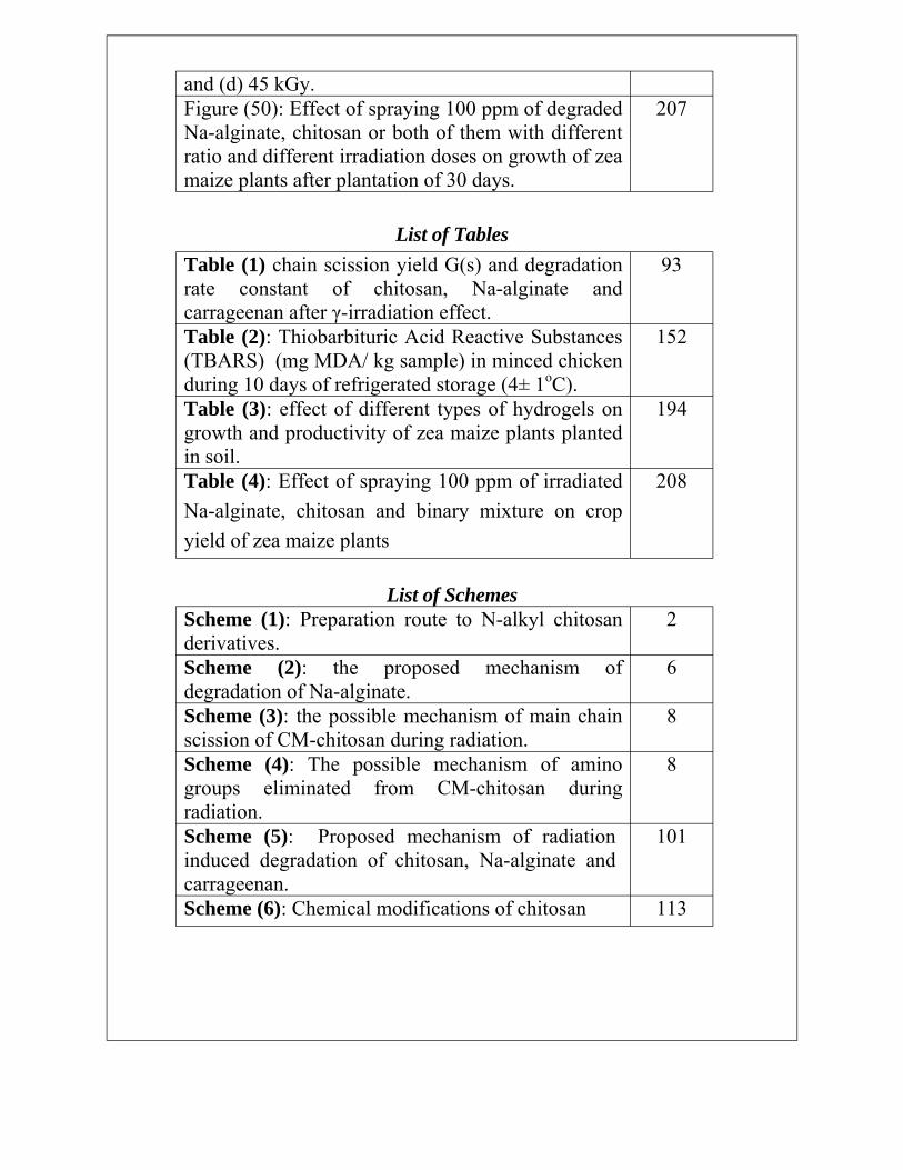
207 and (d) 45 kGy. Figure (50): Effect of spraying 100 ppm of degraded Na-alginate, chitosan or both of them with different ratio and different irradiation doses on growth of zea maize plants after plantation of 30 days.
List of Tables Table (1) chain scission yield G(s) and degradation 93 rate constant of chitosan, Na-alginate and carrageenan after γ-irradiation effect. Table (2): Thiobarbituric Acid Reactive Substances 152 (TBARS) (mg MDA/ kg sample) in minced chicken during 10 days of refrigerated storage (4± 1oC). Table (3): effect of different types of hydrogels on 194 growth and productivity of zea maize plants planted in soil. Table (4): Effect of spraying 100 ppm of irradiated 208 Na-alginate, chitosan and binary mixture on crop yield of zea maize plants
List of Schemes Scheme (1): Preparation route to N-alkyl chitosan 2 derivatives. Scheme (2): the proposed mechanism of degradation of Na-alginate.
6
Scheme (3): the possible mechanism of main chain 8 scission of CM-chitosan during radiation. Scheme (4): The possible mechanism of amino groups eliminated from CM-chitosan during radiation.
8
Scheme (5): Proposed mechanism of radiation 101 induced degradation of chitosan, Na-alginate and carrageenan. Scheme (6): Chemical modifications of chitosan 113

Abbreviations List
Name Abbr. 1,1-diphenyl-2-picrylhydrazyl DPPH Chitosan CS Degree of substitution DS Ethylene diamine tetra acetic acid EDTA Ferric chloride FeCl3
Gamma Irradiation γ-rays Gel Permeation Chromatography GPC Hydrogen peroxide H2O2
Infra Red Spectroscopy FT-IR Kilogray (absorbed dose of radiation) kGy Malonaldehyde MDA Molecular Weight Mwt Na-alginate Alg N-maleoyl-chitosan NMCS N-phthaloyl-chitosan NPhCS Part per million ppm Scanning Electron Microscopy SEM Sulphonated-chitosan SCS The 50 % Inhibition concentration IC50 Thermal Gravimetrical Analysis TGA Thiobarbituric Acid Reactive Substances TBARS Ultra Violet Spectroscopy UV-Vis X - Ray Diffraction XRD

Chapter I Introduction

Chapter I Introduction Chapter I
Introduction
With the development of biotechnological science, the demands on new polymeric materials are increasing rapidly. Natural polymers considered as one of polysaccharide promising materials that are widely used in industrial and agricultural fields. Polysaccharides are composed of long chains of monosaccharide units bound together by glycosidic bonds. The linkage of monosaccharides into chains creates chains of greatly varying length, ranging from chains of just two monosaccharides, which makes the disaccharide to the polysaccharides, that consists of many thousands of the sugars. Definitions of how large a carbohydrate must be to fall into the categories polysaccharides or oligosaccharides vary according to personal opinion.
Natural polymers or their derivatives such as cellulose, starch, chitin/ chitosan, alginate, carrageenan are diverse, abundant, possess unique properties and are now being explored for various knowledge based applications in many fields owning to their unique structure, distinctive properties, safety, non-toxicity, biocompatibility and biodegradability.
The natural polymers can not be synthesized to give various properties such as predictable lot and lot uniformity. But it is easy to modify for improving their properties.
1

Introduction Chapter I
I.1. Improvement of Natural Polymers Properties I.1.1. Chemical Modification
Natural polymers have been chemically and enzymatically modified to impart their physical and chemical properties. In an attempt to improve the water solubility and hemo-compatibility of natural polymers like chitosan, many chemical modifications made to introduce hydrophilic groups to prepare chitosan derivatives with good water solubility, biocompatibility and unique bioactivities using acylation reaction Vanichvattanadecha et al., (2010). Maillard reaction Ying et al., (2011), quaternary reaction Ignatova et al., (2007); Verheul et al., (2008) and carboxymethyl reaction Sreedhar et al., (2007), alkylation reaction Chung et al., (2006) and Ma et al., (2008). An example for alkylation reaction of chitosan was represented in Scheme (1).
Scheme (1): Preparation route to N-alkyl chitosan derivatives Kim et al., (1997).
I.1.2. Radiation Modification of Natural Polymers Different processing technologies have been applied to
transfer natural polymers into the marketable products. Radiation processing offers a clean and additive free method for preparation of value-added novel materials based on
2

Chapter I Introduction renewable natural polymers. The unique advantages of high energy ionizing coupled with the availability of reliable high intensity radiation sources have resulted in ushering the era of radiation processing for a variety of applications, including the areas of health care, food, polymer processing industry and environment. Trials made to develop the applications of radiation processed natural polymers in the emerging areas of biotechnology, pharmaceutical and medical industry. This can serve the twin purpose to utilize the benefits of radiation technology as well as opening new avenues for use of radiation processed natural polymers. To fulfill the demands of specific applications, the natural polymers need to possess different characteristics, for example, while for agricultural applications, radiation processing should lead to the formation of lower molecular products. For the environmental applications demand the formation of crosslinked network structures. To possess wound healing characteristics of natural polymers, incorporation of such as natural polymers with specific biocompatible and wound healing characteristics in synthetic polymers can produce biomaterials.
Generally radiation processing was used early for polymer modification. The irradiation of polymeric materials with ionizing radiation (gamma rays, X-rays, accelerated electrons, ion beams) leads to the formation of very reactive intermediates, free radicals, ions and excited states. These intermediates can follow several reaction paths that result in disproportion, hydrogen abstraction, arrangements and/or the formation of new bonds. The degree of these transformations depends on the structure of the polymer and the conditions of
3

Introduction Chapter I treatment before, during and after irradiation. Thorough control of all of these factors facilitates the modification of polymers by radiation processing. Nowadays, the modification of polymers covers radiation cross-linking, radiation induced polymerization (graft polymerization and curing) and the degradation of polymers.
In fact, the area of radiation processing of natural polymeric material has largely remained unexplored as most of them degraded when exposed to radiation. It has now been realized that radiation processing can also be beneficially utilized either to improve the existing methodologies used for processing natural polymers or to impart value addition to such products by converting them into a more useful form. The results of research work showed that depending on the irradiation conditions, natural polysaccharides (alginate, chitin/ chitosan, carrageenan, carboxylmethylcellulose, etc.) could be either degraded or crosslinked by radiation. This can serve the twin purpose of integrating professionals in the other fields to utilize the benefits of radiation technology as well as opening new avenues for use of radiation processed natural polymers. Also, this paved the way for development of many successful applications; some of them commercialized for use in agriculture, health care, and environmental protection. Crosslinked natural polymers can be used as hydrogel wound dressings, face cleaning cosmetic masks, adsorbents of toxins, and non-bedsore mats, while low molecular weight products show antibiotic, antioxidant and plant growth promoting properties. These successes clearly indicate that radiation processing of natural polymers is an exciting new area where
4
Chapter I Introduction the unique characteristics of these polymeric materials can be exploited for a variety of practical applications.
I.2. Radiation Induced Degradation of Natural Polymers Degradation is a very important reaction in the chemistry
of high molecular weight compounds. It is used for determining the structure of polymeric compounds, and obtaining valuable low molecular weight substances from natural polymers Li et al., (2010). In recent years, much more attention has been directed to radiation modification and degradation of natural polymers such as carrageenan, Na-alginate and chitosan, to obtain low molecular weight polysaccharides or oligosaccharides. Radiation induced changes in polysaccharide generally are attributed to free-radical reactions. Pulse radiolysis technique has been used for investigating radical-induced chain scission reactions in natural polymer like chitosan and the degradation mechanism of chitosan has been discussed Ulanski and Rosiak, (1992).
During irradiation process of natural polymer like Na-alginate in vacuum, radicals are formed at different positions. Radicals at the C2 position mostly changed to the C1 position followed by some radicals at the C1 position converting to C4 position with scission of the glycosidic bond. The proposed mechanism of degradation of Na-alginate was illustrated in Scheme (2). Radicals at C1 (1a) positions are formed by irradiation. This C1 radical (1a) causes main chain scission (2a) and after scission, the radical at the C1 position converts to the C4 position (2b). Hydrogen of C5 is probably abstracted indirectly by .OH radical or surrounding macroradical. As a
5

Introduction Chapter I result, a double bond between C4 and C5 is immediately formed for stabilization (3). The formation of .O radical from the main chain scission (2a) may give an end group by combining with hydrogen (4a) or form of carbonyl group (4b) by rearrangement. Therefore, in the case of irradiation under oxygen, the radical at the C4 position reacted and stabilized under oxygen without forming any double bonds between C4 and C5.
COONaCOONa NaOOC
O ONaOOC
O O Gamma HOHO .
HO O OH OHO OH O OH O Irradiation HO
nn
Na-alginate (1a)
COONa NaOOC
O . O
HO . HO OHo
HO OH O
(2a) (2b)
H. .
RH Rearrangement + H + NaOOC
O
COONa COONa
O O HO OH HO HO
O (3)
O
HO OH OH HO OH
. .+ R + H
(4a) (4b) Scheme (2): the proposed mechanism of degradation of Na-alginate Nagasawa et al., (2000).
6

Chapter I Introduction The degradation of carboxymethyl-chitosan (CM
chitosan) in aqueous solutions gamma-ray radiation affected by many different factors as additives, such as N2, N2O, H2O2, isopropanol, pH of the CM-chitosan solutions and the substituted groups of CM-chitosan .OH radicals are supposed as the main reactive radicals for H-abstraction reactions with CM-chitosan to form CM-chitosan macroradicals, and then fragmentation and/or rearrangement of CM-chitosan macro-radicals to result in degradation of CM-chitosan. The possible reaction pathways of chain breakage of CM-chitosan are expected to follow the general procedure in Schemes (3) and (4). The main mechanisms are H-abstraction reaction, fragmentation and/or rearrangement of radicals at C1, C2, C4, and C5, which is similar to the degradation of chitosan. As Scheme (3) was shown, in the CM-chitosan aqueous solution the radiolysis of water yields reactive species such as .OH and .H, which will cause H-abstraction reaction firstly, and then fragmentation and/or rearrangement of CM-chitosan radicals to result in degradation of CM-chitosan. From Scheme (3), it also can be found that the degradation of CM-chitosan induces the formation of carbonyl groups at the terminal chains of CMchitosan. Simultaneously the formation of hydroxyl groups at C2 and partial amino groups will be eliminated as shown in Scheme (4).
7

Introduction Chapter I
Scheme (3): the possible mechanism of main chain scission of CM-chitosan during radiation Haung et al., (2007 a).
Scheme (4): The possible mechanism of amino groups eliminated from CM-chitosan during radiation Huang et al., (2007 a).
8

Chapter I Introduction
I.3. Applications of Natural Polymers The properties of natural polymers depend on the size,
shape, structure, and functional groups (nature, position, distribution). The ability to determine these parameters is of paramount importance when elaborating, testing and applying modification techniques, as the radiation technique, aimed at changing the molecular weight of a polysaccharide to adjust it to the range required for a particular application.
I.3.1. Agricultural Applications There is a great demand for agro-chemical residue-free
fresh agricultural products from natural products that act as growth promoters for plants and that control post-harvest pathogenic diseases, giving priority to that enhance the plant productivity, reduce disease incidence and avoid negative and side effects on human health as a result of the excessive application of synthetic agro-chemicals. Degrading the natural bioactive agents by ionizing radiation and then using them as growth promoting substances and pathologists is a novel emerging technology to exploit the genetic potential of crops in terms of growth, yield and quality. Among them chitosan, alginate or carrageenan, a high molecular polymer, nontoxic, bioactive agent has become a useful appreciated compounds due to its bio-fertilizer, promotion of germination and shoot elongation Wanichpongpan et al. (2001), as a growth promoter for faba bean and zea maize palnts El-Sawy et al., (2010) and Abd El-Rehim et al., (2011), stimulator on growth and yield of rice, wheat, maize, black pepper, bean, cabbage, peanut, soybean, tomato, cotton, strawberry Chandrkrachang (2002);
9

Introduction Chapter I Chmielewski et al., (2007); Dzung (2005), growth stimulator in orchid tissue culture Nge et al. (2006), fungicidal effects or elicitation of defense mechanisms in plant tissues Leon and Daryl, (2004); Hu et al., (2004); Aftab et al., (2011); Khan et al.,(2011), stimulation of growth of bifidiobacteria to resist infection of diseases for plants particularly oligochitosan in agriculture as biotic elicitor to enhance defense responses against diseases Akiyama et al. (1992) and suppression of heavy metals stress Kume et al. (2002).
I.3.2. Antiviral Activity Chitosan and its oligosaccharide are reported to suppress
viral infections in various biological systems. One possible explanation is that, cationic charges of amino groups of chitosan and chitosan oligosaccharides may have additional functions to activate the immune and defense systems in plants and animals. The treatment by chitosan had good effect on (i) Inhibitory activity on plant viruses by decreasing the
number of local necroses caused by different mosaic viruses Pospieszny et al., (1991).
(ii) Inhibitory activity on animal viruses by increasing the generation of active oxygen species in mouse models and these reactive radical species lead viral destruction.
(iii) Inhibitory activity on bacteriophages by preventing several phage infections.
I.3.3. Antitumor Activity Biological activities of chitosan and oligosaccharide could
inhibit the growth of tumor cells by exerting immunoenhancing effects. The antitumor activities that observed in chitosan
10

Chapter I Introduction oligosaccharides are dependent on their structural characteristics such as degree of deacetylation and molecular weight. Medium molecular weight oligochitosan ranging from 1.5 to 5.5 kDa could effectively inhibit the growth of Sarcoma 180 solid (S180) or Uterine cervix carcinoma No.14 (U14) tumor in BALB/c mice Jeon and Kim (2002).
I.3.4. Fat Lowering and Hypocholesteromic Effects Chitosan is capable of binding dietary fats and it can prevent their absorption through the gut Kanauchi et al. (1995). Czechowska-Biskup et al., (2005) reported that radiation or sonochemical degradation of chitosan may be useful in improving fat binding properties of chitosan as an active component of dietary food additives.
I.3.5. Immuno-stimulant Effects Immunostimulants are compounds that stimulate non
specific immune system by enhancing the capability of defending activity of phagocytes (macrophages and neutrophils). A number of carbohydrate derivatives including mannan oligosaccharides and chitosan oligosccharides have been reported to possess immunostimulting properties Matsuo and Miyazono (1993). It was reported that both oligomers of chitin and chitosan are effective in enhancing migratory activity of macrophages Okamoto et al., (2003). Chitin and chitosan reduce the migration of macrophages and it suggested that chitin and chitosan might absorb some substances in culture medium that are involved in migration of macrophages Okamoto et al., (2002).
11

Introduction Chapter I
I.3.6. Food Preservation Food preservation is the process of treating and handling
food to stop or slow down spoilage (loss of quality, edibility or nutritional value) and thus allow for longer storage. Preservation usually involves preventing the growth of bacteria, yeasts, fungi, and other micro-organisms (although some methods work by introducing benign bacteria, or fungi to the food), as well as retarding the oxidation of fats which cause rancidity.
I.3.6.1. Antimicrobial Activity Microbial contamination, growth and oxidation of lipids in
foods during processing and storage are the major causes of food borne illnesses and loss of shelf life. Lipid oxidation, leading to rancidity, is one of the major reasons of meat products quality deterioration, mainly because of their increased fat content. Antimicrobial activity of chitosan and its derivatives against several bacterial species has been recognized and is considered as one of the most important properties linked directly to their possible biological applications. The antibacterial activity of these compounds is influenced by a number of factors such as degree of polymerization Park et al., (2004), level of deacetylation Chung et al., (2004), type of microorganism Gerasimenko et al., (2004) and other physico-chemical properties such as the positive charge and molecular weight.
The use of chitosan in the manufacture of burgers for the enhancement of stability and preservation may allow a
12

Chapter I Introduction reduction of the use of synthetic preservatives in meat, whereas improving the cooking properties of burgers that have low oxidative stability and high susceptibility to microbial growth, resulting in a short self-life. Such effects depend on the molecular weight and concentration. The addition of a chitosan oligomer (Mwt 5000 Da, 0.2%) sausages stored at 4oC in refrigerator for 3 weeks and compared to a control has great effect on lowering lipid oxidation in the sausage compared to the control sausage Jo et al., (2001).
The positively charged nature of chitosan and oligosccharides molecules enhances their antibacterial activity and facilitates their binding with bacterial cell wall and leads to the inhibition of bacterial cell growth. This is because positively charged amino group at C-2 position of the glucosamine monomer interacts with negatively charged carboxylic acid group of the macromolecules of bacterial cell surface and form polyelectrolyte complexes Choi et al., (2001) and Kim et al., (2003). This could act as impermeable layer around the cell and suppress the metabolic activity of the bacteria by blocking of nutrient permeation through the cell wall.
I.3.6.2. Packaging Materials These polymers are used in various combinations to
prepare materials with unique properties which efficiency ensure safety and quality of food products from processing and manufacture through handling and storing, finally, to consumer use. The materials used for food packaging today consists of a variety of petroleum-derived plastic polymers, metals, glass.
13

Introduction Chapter I
In recent years, the interest in food packaging with antimicrobial properties increased considerably, due to the fact that these systems are able to control the microbiological decay of perishable food products. The successful promotion and use of biological, renewable materials for production of packaging materials based on renewable materials such as paper and board which are based on cellulose, the most abundant renewable natural polymer. Chitosan is well known for its excellent film-forming property and broad antimicrobial activity novel antimicrobial active packaging technologies to improve the quality and safety and to extend the shelf-life of perishable foods against bacteria and fungi Cagri et al., (2004).
I.4. Antioxidants and their Activity In recent years, antioxidants have attracted a great deal of attention. Antioxidants are classified as compounds capable of delaying, retarding or preventing autooxidation processes caused by reactive oxygen Shahidi and Wanasundara (1992). They act as oxygen scavengers, react or trap with free radicals and chelate catalytic metals and thus retard oxidative deterioration Kanatt et al., (1998). The antioxidants are classified into three types: (1) antioxygen radical, reducing substances and antioxidants such as carotenes and ascorbic acid. (2) antiradicals and primary antioxidants. (3) metal chelators.
Antioxidants are an important group of food additives as health protecting factors. In addition, they have the ability to protect against detrimental change of oxidizable nutrients and
14

Chapter I Introduction extend shelf life of foods. Antioxidants are also widely used as additives in fats and oils and in food processing to prevent or delay spoilage of foods. Nowadays, antioxidants receive remarkable attention due to the ability to preserve foodstuffs by retarding deterioration, rancidity and/or discoloration caused by oxidation of fats and oils in foods result in a consequent decrease in nutritional quality and safety caused by the formation of secondary, potentially toxic, compounds. The use of antioxidants is not restricted to foodstuffs. They can also be used to prevent the degradation of food packaging during processing and storage, improving the end-use application.
Antioxidant activity has been expressed in various ways including the percentage of the reagent used, the oxidation inhibition rate. An easier way to present antioxidant activity of foods would be to reference a common reference standard such as ascorbate, Trolox, vitamin E and butylated hydroxytoluene (BHT). Percentage of inhibition IC50 (the concentration of antioxidant which provides 50% inhibition) are used very frequently as parameters characterizing the antioxidant power.
I.4.1. Radical Scavenging Activities Many biological compounds including carbohydrates,
peptides and some phenolic compounds have been identified as potent radical scavengers. In recent years, polysaccharides have been demonstrated to scavenge free radicals in vitro and to be used as antioxidants for the prevention of oxidative damage in foods Kim and Thomas (2007) and living organisms. The antioxidant activity of polysaccharides depends upon several structural parameters, such as the molecular
15

Introduction Chapter I weight Je et al., (2004) and Wang et al., (2004), type and position of functional groups, (such as hydroxyl, sulfate, amine, carboxyl and phosphate), degree of substitution, type of polysaccharide and glycosidic branching Melo et al., (2002). For these applications, specific molecular weights are required. So, considerable attention has recently been directed to the modification and preparation of low molecular weight fractions or oligosaccharides and identification their antioxidant properties Sun et al., (2009). The lower molecular weight, the higher potential to scavenge different radicals such as 1,1-diphenyl-2-picrylhydrazyl (DPPH•), hydroxyl, superoxide and carbon centered radicals. This may be due to the fact that low molecular weight chitosan have a non-compact structure and more free functional groups that could react with free radicals Ji et al. (2007). The radical scavenging properties of chitosan oligosaccharide are dependent on their degree of deacetylation and molecular weights.
I.4.2. Prooxidant Action of Antioxidants Potent antioxidants can autoxidize and generate reactive substances and thus also act as prooxidants, depending on the systems. The prooxidant activity is a result of the ability to reduce metals such as Fe3+ that react with O2 or H2O2 to form initiators of oxidation. As a general rule, the antioxidants extracted from plants can show prooxidant activity at low concentration and antioxidant activity over certain critical values Yen et al., (1997) and Wanasundara and Shahidi, (1998). Environmental factors, such as climatic growth conditions, growth, ripening stage, temperature and duration of storage and thermal treatment have been related with
16

Chapter I Introduction antioxidant activity due to inactivation of peroxidases (responsible for prooxidant action) Gazzani et al., (1998).
I.4.3. Practical Applications of Antioxidants The antioxidant compounds from residual sources could be used for: (1) Increasing the stability of foods by preventing lipid peroxidation and protecting from oxidative damage. Therefore, become popular as a means of increasing shelf life and to reduce wastage and nutritional losses by inhibiting and delaying oxidation Tsuda et al., (1994). (2) Protecting oxidative damage in living systems by scavenging oxygen radicals. (3) Increasing the oxidation stability of vegetable oils is important for industrial practice, and many antioxidant tests are based on this ability to retard or inhibit the oil rancidity. (4) Improvement in color stability for different species of rock fish was observed in the presence of antioxidant extracts from shrimp shell waste. (5) Preventing loss or improving the stability of pigments from red beet juice in the food industry Han et al., (1998). (6) Inhibiting the warmed-over flavor commonly associated with cooked roast beef which has been reheated Chambers et al., (1988).
I.4.4. Sources of Antioxidant Compounds I.4.4.1. Synthetic Sources Butylated hydroxytoluene (BHT), butylated hydroxyanisole (BHA), t-butylhydroquinone, ascorbic acid and propyl gallate are the most commonly used antioxidants in the food industry. There are some serious problems concerning the toxicity of these compounds Hayashi et al., (1993). The synthetic
17

Introduction Chapter I compounds are suspected of being responsible for liver damage and carcinogenesis Grice (1988).
I.4.4.2. Natural Sources During the past few decades, interest had been developed
to search for effective natural antioxidants from different sources for use in foods or medicinal materials to replace synthetic antioxidants, since they can protect the human body from free radicals and retard the progress of many chronic diseases Kinsella et al., (1993). Sources of natural antioxidants are primarily plant phenolics, that may occur in all parts of the plants such as fruits, vegetables, nuts, seeds, leaves, roots and barks Pratt and Hudson (1990), spices and herbs Ramarathnam et al., (1995).
Plant phenolics are multifunctional and can act as reducing agents (free radical terminators), metal chelators and singlet oxygen quenchers. The most common plant phenolic antioxidants include flavonoid compounds, cinnamic acid derivatives, coumarins, tocopherols and polyfunctional organic acids Hertog et al., (1993). Those natural antioxidants constitute a broad range of compounds including phenolic compounds, nitrogen containing compounds, and carotenoids which have the capacity to protect the human body from radicals and retard the progress of many chronic diseases Velioglu et al., (1998).
Seaweed is considered to be a rich source of antioxidants Cahyana et al., (1992) and Lim et al., (2002). In recent years, algal polysaccharides were reported to be useful candidates in
18

Chapter I Introduction the search for an effective, nontoxic substance and have been demonstrated to play an important role as free radical scavengers in vitro and antioxidants for the prevention of oxidative damage in living organisms Zhang et al., (2003), Zhang et al., (2004), Ruperez et al., (2002) and Xue et al., (2001).
I.4.5. Methods for Determining Antioxidant Activity A variety of tests expressing antioxidant potency or
activity of food components has been suggested. These can be categorized into two groups: assays for radical scavenging ability and assays to inhibit lipid oxidation under accelerated conditions Schwarz et al., (2001). The features of an oxidation are a substrate, an oxidant and an initiator, intermediates and final products. Measurement of any of one of these can be used to assess antioxidant activity Antolovich et al., (2002). The antioxidant activity can and must be evaluated with different tests for different mechanisms. Most of the chemical methods are based on the ability to scavenge different free radicals, but also UV-absorption and chelation ability are responsible for the antioxidant activity in oily systems Chen and Ahn (1998). Tests measuring the scavenging activity with different challengers, such as superoxide radical (•O2
¯), hydroxyl (•OH), nitric oxide (•NO), alkylperoxyl radicals, ABTS•+ (radical cation of 2,2'-azinobis(3-ethylbenzo-thiozoline-6-sulphonate), DPPH (1,1-diphenyl-2-picryl hydrazyl radical) have been developed
19

Introduction Chapter I
I.4.5.1. DPPH• Assay A simple method developed to determine the antioxidant
activity of foods. 1,1-diphenyl-2-picrylhydrazyl (DPPH•) radical scavenging assay is one of the most extensively used for antioxidant assays, for assessment of free radical scavenging potential of an antioxidant molecule and considered as one of the standard and easy colorimetric methods for the evaluation of antioxidant properties of pure compounds. DPPH is a stable radical in solution and appears purple color absorbing at 517 nm in ethanol. This assay is based on the principle that DPPH• on accepting a hydrogen (H) atom from the scavenger molecule i.e. antioxidant, resulting into reduction of DPPH• to DPPH2, the purple color changes to yellow with concomitant decrease in absorbance at 517 nm. The color change is monitored by spectrophotometrically and utilized for the determination of parameters for antioxidant properties compared with reference standards viz., ascorbic acid, butyrated hydroxyl toluene (BHT), gallic acid, butylated hydroxyl anisole (BHA) and trolox.
I.4.5.2. Reducing Power Assay Reducing power assay has also been used to evaluate the ability of natural antioxidants to donate electrons Dorman et al., (2003). The reducing power properties are generally associated with the presence of reducing agents, which have been shown to exert antioxidant action by donating a hydrogen atom through breaking the free radicals chain Gordon (1990). The assay is based on the reducing power of a compound (antioxidant). A potential antioxidant will reduce the ferric ion
20
Chapter I Introduction in Fe3+/ferricyanide complex to the ferrous ion (Fe2+). In this assay, the yellow color of the test solution changes to various shades of green and/or blue color depending upon the reducing power of each sample. This is due to the reduction of the Fe3+/ferricyanide complex to the ferrous Fe2+ form. The absorbance is measured at 700 nm; stronger absorbance indicates increased reducing power.
I.4.5.3. Metal Ion Chelating Assay Metallic cations can be used as catalysts during the
oxidation assays. Fe2+ and Cu2+ ions have been widely used as inducers in different systems. The antioxidant activity depends on the metallic catalyst employed for generating the reactive species and it determines whether the supposed antioxidant could act as prooxidant. Transition metal ions can initiate lipid peroxidation and start a chain reaction, which lead to the deterioration of flavor and taste in food Gordon (1990). Iron can stimulate lipid peroxidation by the Fenton reaction and also accelerate peroxidation by decomposing lipid hydroperoxides into peroxyl and alkoxyl radicals that can themselves abstract hydrogen and perpetuate the chain reaction of lipid peroxidation Halliwell (1991). Since ferrous ions (Fe3+) can be reduced by the antioxidants to a catalytically active ion (Fe2+) that provokes the antioxidant to behave as prooxidant are the most effective prooxidants in the food system. The Fe2+-chelating ability was monitored spectrophotometrically by measuring the absorbance of the ferrous iron-ferrozine complex at 562 nm. The same effect is common to other transition metals.
21

Introduction Chapter I
I.4.5.4. Radical-mediated Lipid Peroxidation Inhibition Assay During lipid oxidation, antioxidants act in various ways,
binding metal ions, scavenging radicals and decomposing peroxides. In food related systems, antioxidant activity means chain-breaking inhibition of lipid peroxidation. Linoleic acid, an unsaturated fatty acid is usually used as a model compound in lipid oxidation and antioxidation related assays in which carbon-centered, peroxyl radicals and hydroperoxides, etc., are involved. This method allows dynamic quantification of conjugated dienes as a result of initial poly unsaturated fatty acids oxidation. The principle of this assay is that during linoleic acid oxidation, the double bonds are converted into conjugated double bonds, which are characterized by a strong UV absorption at 234 nm.
I.4.5.5. Thiobarbituric Acid (TBARS) Method Thiobarbituric acid reactive substance (TBARS) method used for measuring the peroxidation of lipids. During lipid peroxidation, lipid peroxides are formed, with a subsequent formation of peroxyl radicals, followed by a decomposition phase to yield aldehydes such as hexanal, malondialdehyde and 4-hydroxynonenal. This assay is based on the detection of a stable product, which is formed between aldehydes and thiobarbituric acid (TBA) in the aqueous phase. The production of TBARS was measured spectrophotometrically at 535 nm after an incubation period of 30 min at 80oC Buege and Aust (1978).
22

Chapter II Literature Review

Chapter II Literature Review
Chapter II Literature Review
This chapter summarizes research progress on (1) Radiation induced degradation of natural polymers. (2) Chemical modification of natural polymers and their antioxidant activity. (3) The applications of natural polymers as antioxidants for improvement of quality and shelf life of food. (4) The applications of low molecular weights natural polymers as growth promoters in agricultural applications.
II.1. Radiation Induced Degradation of Natural Polymers
Hien et al., (2012) degraded chitosan with deacetylation degree (DD) of 70% and average molecular weight (Mw) 90.5×10³ in dilute lactic acid solution containing H2O2 (1%) by gamma irradiation (1.33 kGy/h) at doses in the range 4-16 kGy. Based on the results of molecular weight measured by gel permeation chromatography (GPC), it was found that there was particularly strong synergy between H2O2 and radiation for degradation at the lower radiation doses studied. Radiation scission yields (Gs) were found to be 2.2 µmol/J and 0.2 µmol/J for 5% chitosan with and without 1% H2O2, respectively. The DD of degraded chitosan measured from IR spectra was almost unchanged by the treatment.
Duy et al., (2011) investigated the synergistic degradation of chitosan solution (3%) by γ-irradiation in the presence of hydrogen peroxide (0.25%, 0.5% and 1%). The efficiency of
23
Literature Review Chapter II degradation process was demonstrated by gel permeation chromatography (GPC) analysis through determination the average molecular weight of degraded chitosan (oligochitosan). Structures of resultant oligochitosan were characterized by Fourier transforming infrared spectra (FT-IR) and X-ray diffraction (XRD). The results showed that oligochitosan with molecular weight from 5000 to 10,000 could be prepared by γ-irradiation of chitosan solution containing a small amount of hydrogen peroxide at low dose less than 10 kGy. There was almost no significant change in the main chain structure of oligochitosan. However, the obtained oligochitosans lost about 10% of amino groups and the formation of carboxyl groups could not be specified by FT-IR spectra. The morphology state of oligochitosan was essentially amorphous, which differs from that of original chitosan. The combined γ-ray/H2O2 method is significantly efficient for scale-up manufacture of oligochitosan.
The effects of gamma irradiation on chitosan were determined in terms of physicochemical and functional properties by Ocloo et al., (2011). Shrimp chitosan was extracted from shell using a chemical process involving demineralization, deproteinization, decolorization and deacetylation. Chitosan (in a solid state) irradiated at dose of 25 kGy. Results showed that, there was no significant difference in the degree of deacetylation of chitosan. Significant differences (P<0.05) were observed in the viscosity and viscosity-average molecular weight of the chistosan samples. Viscosity and molecular weight decreased with the irradiation dose of 25 kGy. Chitosan had low antioxidant
24

Chapter II Literature Review activity compared with BHT. Water binding capacity ranged from 582.40% to 656.75% and fat binding capacity was between 431.0% and 560.55%. Irradiation had a major effect on the viscosity and the viscosity-average molecular weight of chitosan.
N-Succinyl chitosan (N-SC) products with various degrees of substitution were synthesized by a direct reaction between chitosan and succinic anhydride. The susceptibility of the synthesized polymers to degradation upon their exposure to γ-ray radiation was investigated by Vanichvattanadecha et al., (2010). The results were compared with the received chitosan. The size exclusion chromatographic results showed that chitosan and N-SC products in their dilute aqueous solution state were more subservient to degradation by γ-ray radiation than in their solid film state. Increasing the radiation dose resulted in decreasing the molecular weights of the polymers. Structural analyses of the irradiated polymers by FT-IR and UV-vis spectrophotometry indicated the increase in the amount of carbonyl groups with the radiation dose. The formation of the carbonyl groups suggested that the radiolysis of chitosan and N-SC products occurred at the glycosidic linkages. In addition, FT-IR, elemental analysis and proton nuclear magnetic resonance spectroscopy (1H-NMR) results suggested that γ-ray radiation affected both the N-acetyl and N-substituted groups on the polymer chains.
Radiation-induced degradation of sodium alginate (NaAlg) with different G/M ratios was investigated by Sen et al., (2010). (NaAlg) samples were irradiated with gamma rays
25

Literature Review Chapter II in air at ambient temperature in the solid state at low dose rate. Changes in molecular weights were followed by size exclusion chromatography (SEC). Changes in their rheological properties and viscosity values as a function of temperature, shear rate and irradiation dose were also determined. Chain scission yields, G(S), and degradation rates were calculated. It was observed that G/M ratio was an important factor controlling the G(S) and degradation rate of sodium alginate.
Zainol et al., (2009) irradiated chitosan powder with doses of 60Co γ-rays of 10, 25, 50, 100 kGy, respectively. The properties of chitosan powder and chitosan film were examined and compared with unirradiated chitosan. Physical characteristic of the irradiated powder and film was studied using stereo microscope. It was observed that the γ-rays induce noticeable color tone intensity change to the chitosan. Further investigation using FT-IR analysis has confirmed that the chain scission reaction was occurred as a result of γ-rays exposure through the depolymerization mechanisms. The degree of deacetylation (DD) of chitosan measured using FT-IR showed a negligible effect due to the exposure of γ-rays. Further investigation on the viscosity average molecular weight (Mv) showed a reduction of Mv from 577 kD of pure chitosan to 458 kD, 242 kD, 159 kD and 106 kD for 10, 25, 50 and 100 kGy of γ-radiated chitosan, respectively. In addition, the tensile strength and elongation at break showed a similar decreasing trend with increasing dosage of γ-rays.
Yue et al., (2008) degraded chitosan under conditions of continuous ozone gas application and constant ultrasonic
26

Chapter II Literature Review radiation (UR). The existence of a synergetic effect of ozone and ultrasonic radiation on the degradation of chitosan was demonstrated by means of determination of viscosity-average molecular weight. The efficiency of the ozone and ultrasonic radiation treatment compared with acid hydrolysis on degradation of chitosan was investigated. The structure of the degraded chitosan was characterized by FT-IR and 13C-NMR spectral analyses. The whole initial chitosan's monomer structure still existed in the resulting degraded chitosan with different low molecular weight. There was no significant change of the total degree of deacetylation (DD) of degraded chitosan compared with the initial chitosan. The combined O3/UR technique is promisingly suitable for scale-up manufacture of low-molecular-weight chitosan.
Kang et al., (2007) irradiated chitosan by 60Co γ-rays in the presence of hydrogen peroxide with radiation dose from 10 kGy to 100 kGy. The degradation was monitored by gel permeation chromatography (GPC), revealing the existence of a synergetic effect on the degradation. Structures of the degraded products were characterized with FT-IR, UV-Vis, and XRD. The results showed that the crystallinity of chitosan decreased with degradation and the crystalline state of water-soluble chitosan is entirely different from that of water-insoluble chitosan. An elemental analysis method was employed to investigate changes in the element content of chitosan after degradation. Mechanism of chitosan radiation degradation with and without hydrogen peroxide was also discussed.
27

Literature Review Chapter II Huang et al., (2007 b) focused on the radiation effect of γ-
rays on carboxymethylated chitosan (CM-chitosan) in solid state. The changes in molecular weight of CM-chitosan with absorbed dose were monitored by viscosity method. The results indicated that random chain scissions took place under irradiation. Radiation chemical yield (Gd) of CM-chitosan in solid state with N2-saturated was 0.49, which showed CMchitosan has high radiation stability. FT-IR and UV spectra showed that main chain structures of CM-chitosan were retained and some carbonyl/carboxyl groups were formed and partial amino groups were eliminated in high absorbed dose. XRD patterns identified that the degradation of CM-chitosan occurred mostly in amorphous region.
Aqueous solutions of carboxymethylated chitosan (CMchitosan) were irradiated with γ-rays in various conditions by Huang et al., (2007 c). The degradations of CM-chitosan were faster in the presence of nitrous oxide or hydrogen peroxide. The radiation chemical yields of CM-chitosan degradation were found to decrease at lower pH in which the polymer chains tend to coiled conformation. Intrinsic viscosity of CMchitosan decreased faster than that of carboxymethylated chitin. FT-IR and UV spectra showed that main chain structures of CM-chitosan were remained and some carbonyl/ carboxyl groups were formed during the degradation. The results of elemental analysis (EA) indicated the contents of C/N were increased which suggested the elimination of partial amino groups during radiation.
28

Chapter II Literature Review Wasikiewicz et al., (2005) applied three degradation
methods of ultrasonic, ultraviolet and gamma irradiation to Na-alginate and chitosan in aqueous solutions. The changes in molecular weight were monitored by GPC measurements. The results showed that the most effective method for both polymers was gamma radiation method with a yield of scission Gs = 0.55 x 10⁻⁷ mol/J for 1% alginate and Gs = 3.53 x 10⁻⁷
mol/J for 1% chitosan. Based on FT-IR spectra taken before and after degradation, it was revealed that degradation undergoes by the breakage of the glycosidic bonds of polymers. UV spectroscopy showed absorption peak at 265 nm for alginate and two peaks at the range of 250-280 nm for chitosan. UV spectroscopy for ultrasonic is not revealed and any peak suggesting ultrasonic degradation undergoes different mechanism than ultraviolet and gamma degradations, probably mechanical one.
Wang et al., (2005) studied the synergetic degradation of chitosan by hydrogen peroxide under irradiation with ultraviolet light. The existence of a synergetic effect on the degradation was demonstrated by means of viscometry. The structures of the degraded product were characterized by FT-IR analysis and diffuse reflectance spectra (DRS) analysis. The mechanism of the degradation of chitosan was correlated with cleavage of the glycosidic bond.
Three physical methods of chitosan degradation: irradiation in dry state, irradiation in aqueous solution and sonication in aqueous solution were test and compared in terms of yields and side effects by Czechowska-Biskup et al., (2005).
29

Literature Review Chapter II The influence of average molecular weight of chitosan on fat binding ability in vitro has been studied using a biopharmaceutical model of digestive tract. It was found that reduction in molecular weight leads to a significant increase in the amount of fat bound by 1 g of chitosan. Thus, radiation- or sonochemical treatment may be useful in improving fat-binding properties of chitosan as an active component of dietary food additives.
Lee et al., (2003) irradiated alginate in aqueous solution by 60Co γ-rays in the dose range of 10-500 kGy to assess the effect of irradiation on the degradation of alginate. The irradiation-induced changes in the viscosity, molecular weight, color, monomer composition were measured. The molecular weight of raw alginate was reduced from 300000 to 25000 when irradiated at 100 kGy. The degradation rate decreased and the chain breaks per molecule increased with increasing irradiation dose. No appreciable color changes were observed in the samples irradiated up to 100 kGy, but intense browning occurred beyond 200 kGy. The 13C-NMR spectra showed that homopolymeric blocks increased and the M/G ratio decreased with irradiation. Considering both the level of degradation and the color change of alginate, the optimum irradiation dose was found to be 100 kGy.
Radiation depolymerization of chitosan in the solid state to prepare oligomers by γ-irradiation was investigated by Hai et al., (2003). Low molecular weight chitosan or oligo-chitosans were separated from a chitosan depolymerized by gamma radiation using mixtures of methanol-water and acetone as the
30

Chapter II Literature Review solvents. The biological effect of oligo-chitosan in each fraction was evaluated; the preliminary results indicated that the oligo-chitosan with Mw = 2 x 10⁴ inhibited the growth of fungi at 100 ppm and that with Mw = 800 only enhanced the growth of the same typical fungi.
The effect of γ-rays on alginate in the solid and solution state was investigated by Nagasawa et al. (2000). The degradation in solution was remarkably greater than that in the solid. The molecular weight of alginate in 1% (w/v) solution decreased from 6x10⁵ at 0 kGy to 8x10³ at 20 kGy irradiation while, the equivalent degradation in solid required 500 kGy of irradiation. Degradation G-values were 1.9 and 55 for solid and solution, respectively. The free radicals from irradiated water were responsible for the degradation in solution. The degradation was accompanied by a color change to deep brown for highly degraded alginate. Little color change was observed on irradiation in the presence of oxygen. UV spectra showed a distinct absorption peak at 265 nm for colored alginates. The fact that discoloration of colored alginate was caused on exposure to ozone suggests a formation of double bond in the pyranose ring.
II.2. Chemical Modification of Natural Polymers and Their Antioxidant Activity
Sun et al., (2011) prepared N-maleoyl-chitosan oligosaccharide (NMCOS) and N-succinyl-chitosan oligosaccharide (NSCOS) by acylation with maleic anhydride and succinic anhydride, respectively. The structural changes
31

Literature Review Chapter II were confirmed by FT-IR spectroscopy and their substituting degrees were determined as 0.49 by conductometric titration. Their antioxidant activities were evaluated by scavenging
·superoxide anion O2 ·–, hydroxyl radical OH and determination
of reducing power. The 50% inhibition concentrations (IC50) of NMCOS and NSCOS scavenging effect on O2
·– were 2.25 and 3.27 mg/ml, respectively. The IC50 of NMCOS scavenging effect on ·OH was 0.24 mg/ml. The reducing powers of NMCOS and NSCOS at the concentration of 2.40 mg/ml were determined as 0.46 and 0.41, respectively. The results showed that NMCOS has better antioxidant activities, which may be related to the fact that maleoyl has stronger electron-withdrawing effect than succinyl.
The effects of the molecular weight and ratio of guluronic acid (G) to mannuronic acid (M), G/M, of some sodium alginate (Na-Alg) fractions on their antioxidative properties were investigated by Sen (2011). Low molecular weight fractions of sodium alginate (Na-Alg) with various G/M were prepared by gamma radiation-induced degradation. Antioxidant properties of the fractions with various molecular weight and G/M were evaluated by determining the scavenging ability of DPPH radical. 50% inhibition concentrations of the 50 kGy irradiated Na-Alg having molecular weights of 20.5, 17.7, and 16.0 kDa were found to be 11.0, 18.0, and 24.0 mg/ml, respectively, where as the fractions of the same molecular weight with a lower G/M exhibited a better DPPH scavenging activity. The results demonstrated that its molecular weight and G/M were important factors in controlling the antioxidant properties of Na-Alg.
32

Chapter II Literature Review
The antioxidant activities of different molecular weights and grafting ratios of chitosan–caffeic acid derivatives were investigated by Aytekin et al., (2011). The grafting process was achieved using 1-ethyl-3-(3-dimethylaminopropyl) carbodiimide hydrochloride (EDAC) as covalent connector under different conditions such as molecular-weight of chitosan, molar ratio chitosan and caffeic acid, reaction temperature, pH, and reaction time. The inhibition concentrations (IC50) of products were calculated by reduction of the 1,1-diphenyl-2-picrylhydrazyl in the radical-scavenging assay and reduction of the Fe3+/ ferricyanide complex to the ferrous form in reducing power assay. The EDAC showed maximum activity at 3h, pH 5.0 and room temperature conditions, except high-molecular-weight chitosan in pH 2.0. The products were water-soluble in all pH and showed lower viscosity than native chitosan. The highest grafting ratio of caffeic acid was observed at 15% in low molecular weight chitosan. After 5% grafting of caffeic acid into chitosan, the grafting efficiency was increased by decreasing molecular weight of chitosan at the same conditions. Caffeic acid has main role in the antioxidant activity of products. The maximum IC50 of radical-scavenging activity (0.064 mg/ml) was observed at the highest caffeic acid containing derivative. Water-soluble chitosan and caffeic acid derivatives were obtained by this study without activity loss.
Ying et al., (2011) improved water solubility of chitosan by specific attachment of carbohydrates to the 2-amino functions achieved by Maillard reaction or further reductive alkylation of
33

Literature Review Chapter II Schiff bases. The characteristic physicochemical, rheological properties and antioxidant activities of the derivatives were investigated. The results indicated that the solubility of all the chitosan saccharides before and after reducing had been greatly enhanced comparing to the native chitosan. The Schiff base typed chitosan fructose derivative was highest at 13.2 g/L. Schiff base typed chitosan derivatives existed better solubility and more effective scavenging activity against DPPH radical than N-alkylated chitosan derivatives. The degree of substitution (DS) of the chitosan derivatives increased with higher concentration of saccharide, increasing reaction time and temperature. The reduction of viscosity of chitosan derivatives decreased with increasing reaction time and temperature. The results suggest that the water-soluble chitosan derivatives produced through Maillard reaction may be promising commercial additive in cosmetics and food.
Cho et al., (2011) prepared gallic acid-grafted-chitosans (GA-g-chitosans) with four different grafting ratios by a free radical induced grafting reaction in order to improve the antioxidant and water solubility of chitosan. 1H-NMR and thin layer chromatography were employed to verify the synthesis of GA-g-chitosans. The results revealed that GA was grafted onto the chitosan. The antioxidant properties of the GA-g-chitosans were evaluated using several in vitro models. GA-g-chitosan which has the highest GA content showed 92.26% scavenging activity against 1,1-diphenyl-2-picrylhydrazyl and 93.15% hydrogen peroxide scavenging activity at 50µg/mL. GA-g-chitosan was also showed higher reducing power compared to others. All GA-g-chitosans showed improved antioxidant
34

Chapter II Literature Review capacities compared to plain chitosan treated in the same conditions without gallic acid grafting. Furthermore, the GA-g-chitosans also exhibited good cytocompatibility and effectively inhibited the formation of intracellular reactive oxygen species (ROS) in time- and dose-dependent manner in RAW264.7 macrophages.
Yang et al., (2010) prepared three kinds of carboxymethylated polysaccharides (carboxymethyl chitosan (O-CM-chitosan), carboxymethyl hyaluronan (CMHA) and carboxymethyl starch (CMS)). Their antioxidant activities against hydroxyl radicals were assessed. The results showed that O-CM-chitosan, CMHA and CMS had lower scavenging ability on hydroxyl radicals than chitosan. The scavenging ability of three kinds of polysaccharides on hydroxyl radicals was in the order of chitosan > HA > starch. The scavenging ability of carboxymethylated polysaccharides had the same order as related to its corresponding polysaccharides at higher concentrations (0.8 mg/ml). There were not only hydroxyl groups but also amino or acetamide groups in the molecules of chitosan and HA, but only hydroxyl group for starch. It was suggested that the sequence influence the scavenging activity against hydroxyl radicals might be amino group > acetamide group > hydroxyl group.
Zhong et al., (2010) modified chitosan (CS) of two different molecular weights by reacting with methyl hydrazinedithiocarboxylate and methyl phenylhydrazinedithiocarboxylate to give 2-(hydrazine thiosemicarbazone)-chitosan (2-HTCHCS, 2-HTCLCS) and 2-(phenylhydrazine-
35

Literature Review Chapter II thiosemicarbazone)-chitosan (2-PHTCHCS, 2-PHTCLCS). The structure of the derivatives was characterized by FT-IR spectroscopy and elemental analysis. The antioxidant activities of the derivatives were investigated employing various
•established systems, such as hydroxyl radical ( OH), •superoxide anion (O2 ) scavenging, reducing power and
chelating activity. All of the derivatives showed strong scavenging activity on hydroxyl radical than chitosan and ascorbic acid (Vc). IC50 of 2-HTCHCS, 2-HTCLCS, 2PHTCHCS and 2-PHTCLCS was 0.362, 0.263, 0.531 and 0.336mg/mL respectively. The inhibitory activities of the derivatives toward superoxide radical by the PMS-NADH system were strong. The results showed that the superoxide radical scavenging effect of 2-[phenylhydrazine (or hydrazine)-thiosemicarbazone]-chitosan was higher than that of chitosan. The derivatives had obviously reducing power and chelating activity. The data obtained from vitro models clearly establish the antioxidant potency of 2-[phenylhydrazine (or hydrazine)-thiosemicarbazone]-chitosan.
Chitosan glucose conjugates were prepared using Maillard reaction by Kosaraju et al., (2010). Examination of the extent of Maillard reaction and antioxidant properties showed that, the acid soluble chitosan reacted for 30 min at pH 4.9 had the highest extent of reaction as judged by increased absorbance, the highest degree of modification to the amino group as evidenced by Fourier transform infrared and shifts of the endotherms by differential scanning calorimetry, and the highest antioxidant activity by ferric reducing power and oxygen radical absorbance capacity.
36

Chapter II Literature Review
Spizzirri et al., (2010) synthesized catechin–alginate and catechin–inulin conjugates by adopting free radical-induced grafting procedure. The insertion of catechin onto the polymeric backbones was verified by performing FT-IR, DSC and fluorescence analyses and molecular weight distributions were analyzed by GPC. The antioxidant properties of the obtained conjugates were evaluated and compared with that of blank alginate and inulin. The prepared conjugate showed good antioxidant activity and could be very useful in the optimization of food preservation and to help manufacturers in elaboration of new food products and packaging.
Liu et al., (2009) synthesized various quaternized chitosans (QCSs). Their reducing power and antioxidant
•potency against hydroxyl radicals ( OH) and hydrogen peroxide (H2O2) were explored in vitro. The QCSs exhibited markedly antioxidant activity especially N-trichloroethyl-N,N-dimethyl chitosan (TCEDMCS) whose IC50 on hydroxyl radicals was 0.235 mg/mL. The QCSs showed 65–80% scavenging effect on hydrogen peroxide at a dose of 0.5 mg/mL. The antioxidant activity decreased in the order N-trichloroethyl-N,N-dimethyl chitosan (TCEDMCS) > Ntribromoethyl-N,N-dimethyl chitosan (TBEDMCS) > N-ethyl-N, N-dimethyl chitosan (EDMCS) > N-propyl-N,Ndimethyl chitosan (PDMCS) > N-isobutyl-N,N-dimethyl chitosan (IBDMCS) > Chitosan. Furthermore, the order of their •OH and H2O2 scavenging activity was consistent with the electronegativity of different substituted groups of the QCSs. The QCSs showed much stronger antioxidant activity than that
37

Literature Review Chapter II of chitosan which may be due to the positive charge density of the nitrogen atoms in QCSs strengthened by the substituted groups.
Curcio et al., (2009) synthesized gallic acid-chitosan and catechin-chitosan conjugates by adopting a free radical-induced grafting procedure using ascorbic acid/hydrogen peroxide redox pair as radical initiator. The formation of covalent bonds between antioxidant and biopolymer was verified by performing UV, FT-IR, and DSC analyses. The antioxidant properties of chitosan conjugates were compared with that of a blank chitosan treated in the same conditions but in the absence of antioxidant molecules. The good antioxidant activity shown by functionalized materials proved the efficiency of the reaction method.
The effect of different functional groups such as sulfate, amine, and hydroxyl and/or their ionized groups on antioxidant capacities of low-molecular-weight polysaccharides prepared from agar (LMAG), chitosan (LMCH) and starch (LMST) were evaluated by Chen et al., (2009). Ascorbic acid and EDTA were used as positive controls. The in vitro antioxidant capacities of LMAG and LMCH were higher than that of LMST in the DPPH radical, superoxide radical, hydrogen peroxide and nitric oxide radical scavenging and ferrous metal-chelating capacities. The different scavenging capacities may be due to the combined effects of the different sizes of the electron cloud density and the different accessibility between free radical and LMPS.
38

Chapter II Literature Review Sun et al., (2008) prepared three N-carboxymethyl
chitosan oligosaccharides (N-CMCOSs) with different degrees of substitution (NA: 0.28, NB: 0.41, and NC: 0.54, respectively) by the control of the amount of glyoxylic acid in the etherification process of chitosan oligosaccharide (COS). Their antioxidant activities were evaluated by the scavenging of 1,1-diphenyl-2-picrylhrazyl radical (DPPH) radical, superoxide anion and determination of reducing power. The results showed that with increasing the of degree substituting, the scavenging activity of N-CMCOSs against DPPH radical decreased and reducing power increased. As for superoxide anion scavenging, the order is NB > NC > NA. The difference may be related to the different radical scavenging mechanisms and donating effect of substituting carboxymethyl group.
Yen et al., (2008) prepared crab chitosan and its antioxidant properties were studied. Chitosan showed antioxidant activities of 58.3-70.2% at 1 mg/mL and showed reducing powers of 0.32–0.44 at 10 mg/mL. At 0.1 mg/mL, scavenging abilities on hydroxyl radicals were 62.3–77.6%, whereas at 1 mg/mL, chelating abilities on ferrous ions were 82.9–96.5%. IC50 values of antioxidant activity were below 1.5 mg/mL. The results showed that the crab chitosan was good in antioxidant activity, scavenging ability on hydroxyl radicals and chelating abilities on ferrous ions and may be used as a source of antioxidants as a possible food supplement or ingredient in the pharmaceutical industry.
The enhancement of antioxidant activity of irradiated chitosan was investigated by Feng et al., (2008). The structure
39
Literature Review Chapter II of irradiation degraded chitosan was characterized by GPC, FT-IR and 1H-NMR spectroscopy. The molecular weight of chitosan decreased with increasing irradiation dose. Radical mediated lipid peroxidation inhibition, reducing power, superoxide anion radical and hydroxyl radical quenching assays were used for the evaluation of the antioxidant activity of irradiated chitosan. Chitosan (Mwt 2.1 x 10³) which had been irradiated at 20 kGy exhibited high reductive capacity and expressed good inhibition of linoleic acid peroxidation. At a concentrtion of 0.1 mg/mL it could scavenge 74.2% of superoxide radical. At 2.5 mg/mL, scavenging percentage of chitosan irradiated for 0, 2, 10, 20 kGy against hydroxyl radical was 16.6%, 41.1%, 47.1% and 63.8%, respectively. The results show that γ-ray irradiation, especially for 20 kGy, of chitosan gives enough degradation to increase its antioxidant activity as a result of a change in molecular weight.
Xing et al., (2008) investigated the antioxidant potency of high and low molecular weight quaternary chitosan derivatives
·–such as superoxide (O2 ) and hydroxyl (·OH) radicals scavenging, reducing power and iron ion chelating. The obtained results were as follow: firstly, low molecular weight
·–quaternary chitosan had stronger scavenging effect on O2 and ·OH than high molecular weight quaternary chitosan. Secondly, the reducing power of low molecular weight quaternary chitosan was more pronounced than that of high molecular weight quaternary chitosan. Thirdly, ferrous ion chelating potency were showed to increase first and decrease afterwards with increasing concentration for two kinds of quaternary chitosans, namely, they have not concentration-dependence.
40

Chapter II Literature Review However, the scavenging rate and reducing power of high and low molecular weight quaternary chitosans increased with their increasing concentrations, and hence were concentration-dependent.
Guo et al., (2008) prepared quaternized carboxymethyl chitosan (QCMC) derivatives with a degree of quaternization ranging from 34.3% to 59.5%. The antioxidant activity of QCMCs against hydroxyl radicals was assessed. The results indicated that QCMCs have better hydroxyl radicals scavenging activity than that of carboxymethyl chitosan, as a result of the positive charge of the quaternized chitosan.
Zhong et al., (2008) modified chitosan (CS) with two different molecular weights by reacting with 4-hydroxyl-5-chloride-1,3-benzene-disulfo-chloride or 2-hydroxyl-5-chloride-1,3-benzene-disulfo-chloride to give new 2-(4(or 2)-hydroxyl-5-chloride-1,3-benzene-di-sulfanimide)-chitosan (2HCBSAHCS, 2-HCBSALCS, 4-HCBSAHCS, 4-HCBSALCS). The structure of the derivatives was characterized by FT-IR and 13C-NMR spectroscopy. The antioxidant activities of the derivatives were investigated employing various established systems, such as hydroxyl radical (·OH), superoxide anion (O2·–) scavenging, reducing power and chelating activity. All the derivatives showed stronger scavenging activity on hydroxyl radical than chitosan and ascorbic acid, and IC50 of 4HCBSAHCS, 4-HCBSALCS, 2-HCBSAHCS and 2HCBSALCS was 0.334, 0.302, 0.442, 0.346 mg/mL, respectively. The inhibitory activities of the derivatives toward superoxide radical by the PMS-NADH system were strong.
41

Literature Review Chapter II The results showed that the superoxide radical scavenging effect of 2-(4(or 2)-hydroxyl-5-chloride-1,3-benzene-disulfanimide)- chitosan was higher than chitosan. The derivatives had obviously reducing power and slight chelating activity. The results obtained in in vitro models clearly establish the antioxidant potency of 2-(4(or 2)-hydroxyl-5-chloride-1,3-benzene-disulfanimide)-chitosan.
Different molecular weights of water soluble chitosan [WSC1, Mwt = 281x103; WSC2 Mwt = 97.2 x10³; WSC3 Mwt = 17.5 x10³ and WSC4 Mwt =1.7 x10³] were prepared by Feng et al., (2007). The structure of water soluble chitosan was characterized by FT-IR. The pH dependence of water solubility of water soluble chitosan was evaluated from turbidity. Total antioxidant activity, reducing power, superoxide anion radical and hydroxyl radical quenching assay, metal chelating activity, and H2O2 scavenging activity were used for the evaluation of different molecular weight half N-acetylated chitosan in vitro. Low molecular weight water soluble chitosan (WSC4) exhibited high reductive capacity and expressed good inhibition of linoleic acid peroxidation in the linoleic acid model system. WSC4 (0.25mg/ml) scavenged 78.8% of superoxide radical. At 5mg/ml, scavenging percentage of WSC1, WSC2, WSC3, and WSC4 against hydroxyl radical was 49.3%, 66.8%, 77.1%, and 83.7%, respectively. The results indicated that water soluble chitosan is an ideally natural antioxidant and its antioxidant activity depends on its molecular weight.
42

Chapter II Literature Review Chien et al., (2007) studied the effect of chitosans with low
molecular weight (LMWC, Mwt 12kDa), medium molecular weight (MMWC, Mwt 95 kDa) and high molecular weight (HMWC, Mwt 318 kDa) on the antioxidant activity in an aqueous system and in apple juice. Antioxidant activity was determined including DPPH radicals, hydrogen peroxide and superoxide anion radicals, as well as metal ion chelating capacity. LMWC exhibited stronger scavenging activity toward DPPH radicals, superoxide anion radicals and hydrogen peroxide compared to either MMWC or HMWC. At a concentration of 0.8 mg/ml, the LMWC in apple juice exhibited 88.2%, 99.8% and 93.0% scavenging activities toward DPPH radicals, hydrogen peroxide and superoxide anion radicals, respectively. At a concentration of 1.0 mg/ml the LMWC in apple juice exhibited 70.0% ferrous ion chelating activity. The results obtained in vitro models clearly establish the antioxidant potency of DMWCs. These in vitro results suggested that LMWC can increase antioxidant activity in apple juice.
Three new kinds of 1,3,5-thiadiazine-2-thione derivatives of chitosan with two different molecular weights have been prepared by Ji et al. (2007). Their structures were characterized by FT-IR spectroscopy. The substitution degree of derivatives calculated by elemental analyses was 0.47, 0.42, 0.41, 0.38, 0.41 and 0.36, respectively. The results showed that substitution degree of derivatives was higher with lower molecular weight. The antioxidant activity was studied using hydroxyl radical scavenging, superoxide radical scavenging and reducing power. Antioxidant activity of the 1,3,5-
43

Literature Review Chapter II thiadiazine-2-thione derivatives of chitosan were stronger than that of chitosans and antioxidant activity of low molecular weight derivatives were stronger than that of high molecular weight derivatives so it is a potential antioxidant in vitro.
Sun et al., (2007 a and b) prepared chitosan and carboxymethyl chitosans (CMCTSs) oligomers with different molecular weights by oxidative degradation method involving hydrogen peroxide (H2O2) and the combined degradation method using hydrogen peroxide and microwave radiation. Viscosity determination and end group analysis were applied to measure molecular weights of chitosan oligomers. Effects of concentration of H2O2 and degradation time on molecular weights of chitosan oligomers were studied. Both methods were effective to prepare chitosan oligomers from the initial chitosan (8.5×10⁵ Da). The degradation process of chitosan will be accelerated with the aid of microwave and degradation time may be reduced. The antioxidant activity of chitosan oligomers was evaluated as radical scavengers against superoxide anion and hydroxyl radical by application of flow injection chemiluminescence technology. Chitosan oligomers A, B, C and D of molecular weight (2300, 3270, 6120, and 15,250 Da) had different antioxidant activity. Among the four chitosan oligomers, oligomer D (15,250 Da) had the lowest scavenging ability against superoxide anion and hydroxyl radicals. For superoxide anion scavenging, the 50% inhibition concentration (IC50) of other three oligomers A, B, and C were 5.54, 8.11, and 12.15 mg/mL, respectively. And for hydroxyl radical scavenging the values were 0.4, 0.76, and 1.54 mg/mL, respectively. At the concentration range examined, the
44

Chapter II Literature Review maximal inhibiting efficacy of A, B, C, and D were 89, 75, 74, and 41% for superoxide anion, and 71, 65, 51, and 7% for hydroxyl radical. The 50% inhibition concentrations (IC50) of CMCTSs of molecular weights of 1130, 2430 and 4350 Da were 10.36, 17.57, and 23.38 mg/ml, respectively. The above results showed that CMCTSs with lower molecular weight had better superoxide anion scavenging activity. These results indicated that chitosand CMCTSs an oligomers with lower molecular weight had better antioxidant activity.
Zhong et al., (2007) modified chitosan (CS) and chitosan sulfates (CSS) of different molecular weights (Mwt) with 4acetamidobenzene sulfonyl chloride to obtain sulfanilamide derivatives of chitosan and chitosan sulfates. The preparation conditions such as different reaction time, temperature, solvent, and the molar ratio of reaction materials are discussed. Their structures were characterized by FT-IR spectroscopy and elemental analyses. The antioxidant activities of the derivatives were investigated employing various established in vitro systems, such as hydroxyl-radical (·OH), scavenging superoxide anion O2
·– and reducing power. All kinds of the compounds showed stronger scavenging activity on hydroxyl radical than ascorbic acid (Vc). The inhibitory activities of the derivatives toward superoxide radical by the PMS-NADH system were obvious. The results showed that the superoxide radical scavenging effect of sulfanilamide derivatives of chitosan and chitosan sulfates was stronger than that of original CS and CSS. All of the derivatives were efficient in the reducing power. The results indicated that the sulfanilamide
45

Literature Review Chapter II group was grafted on CS and CSS increased the reducing power of them obviously.
Wang et al., (2007) investigated the antioxidant activities of three marine oligosaccharides, alginate oligosaccharides (AOs), chitosan oligosaccharides (COs), and fucoidan oligosaccharides (FOs) in vitro by several antioxidant assays, including hydroxyl radical scavenging, superoxide radical scavenging, erythrocyte hemolysis inhibiting, metal chelating activities, and anti-lipid peroxidation. The results showed that these oligosaccharides exhibited different activities in various assays. AOs had the highest scavenging hydroxyl radical activity than FOs and COs at all the tested amounts. COs had the highest scavenging superoxide radical and inhibiting erythrocyte hemolysis activity than AOs and FOs at all the tested amounts. In the assay of chelating Fe2+, COs and FOs indicated good chelation while AOs hardly had any activity. In the assay of antilipid peroxidation, only COs had significantly high antioxidant activity.
Yuan et al., (2006) investigated the antioxidant activity of κ-carrageenan oligosaccharides and their chemical modification derivatives employing various established in vitro systems, such as reducing power, iron ion chelation, and total antioxidant activity using β-carotene–linoleic acid system. The oversulfated (SD), lowly (LAD), and highly acetylated derivatives (HAD) exhibited antioxidant activity higher than that of carrageenan oligosaccharides. The results indicated that the chemical modification of carrageenan oligosaccharides can enhance their antioxidant activity in vitro.
46

Chapter II Literature Review
Je and Kim (2006) prepared novel water soluble chitin derivative by chemical modification to evaluate its antioxidant activities by free radical scavenging potential using electron spin resonance spin trapping technique. Aminoethyl-chitin (AEC) exhibited free radical scavenging activities against 1,1-diphenyl-2-picrylhydrazyl (DPPH), hydroxyl, superoxide, and peroxyl radicals. AEC quenched DPPH and peroxyl radical over 55% and 59% at 4 mg/mL, and also suppressed superoixde radical over 58% at 2 mg/mL. AEC was more active against hydroxyl radical, and scavenging ratio was 92.2% at 0.12 mg/mL. These results suggested that free amino group in the –CH2CH2NH2 plays an important role in the free radical scavenging activity. In addition, cytotoxic effect of AEC was assessed using human lung fibroblast (MRC-5) cell line, and AEC showed less toxic against MRC-5.
Guo et al., (2006) synthesized N-substituted chitosan and quaternized chitosan and their antioxidant activity against hydroxyl radicals was assessed. Compared with the antioxidant activity of chitosan, the results indicated that the two kinds of chitosan derivatives had different scavenging ability on hydroxyl radicals, which should be related to the form of amido in the two kinds of chitosan derivatives.
Huang et al., (2006) synthesized two chitooligosaccharides (COS) derivatives with different substitution degrees. Scavenging of carbon-centered and DPPH radicals were directly affected by the amount of abstractable hydrogen atoms in COS molecules. The structure activity relationships
47

Literature Review Chapter II revealed that chelation of Fe2+ ions indirectly contributed for their observed hydroxyl radical scavenging activity apart from hydrogen abstraction.
Xing et al., (2005 b) prepared chitosan sulfates and their antioxidant potencies were investigated using various established in vitro systems, such as 1,1-diphenyl-2-picrylhydrazyl (DPPH), superoxide, hydroxyl radicals scavenging, reducing power, iron ion chelating and total antioxidant activity. All kinds of sulfated chitosans showed strong inhibitory activity toward superoxide radicals, strong concentration-dependent inhibition of deoxyribose oxidation and strong efficiency in the reducing power. The results obtained suggested the possibility of such sulfated chitosans to be effectively employed as ingredient in health or functional food, to alleviate oxidative stress.
Xing et al., (2005 c) determined the antioxidant activity of high molecular weight and high sulfate content chitosans (HCTS). The results showed that HCTS could scavenge superoxide and hydroxyl radical. Its IC50 was 0.012 and 3.269 mg/mL, respectively. It had obviously reducing power and slight chelating activity. The results obtained in vitro models clearly establish the antioxidant potency of HCTS. It is potential antioxidant in vitro.
Guo et al., (2005) prepared five kinds of Schiff bases of chitosan and carboxymethyl chitosan (CMCTS) and their antioxidant activity was studied using an established system, such as superoxide and hydroxyl radical scavenging. Obvious
48

Chapter II Literature Review differences between the Schiff bases of chitosan and CMCTS were observed which might be related to contents of the active hydroxyl and amino groups in the molecular chains.
Huang et al., (2005) studied the scavenging and antioxidant activity of hydroxyethyl chitosan sulfate (HCS) against 1,1-diphenyl-2-picrylhydrazyl (DPPH), hydroxyl and carbon-centered radical species. HCS could scavenge DPPH (33.78%, 2.5 mg/mL) and carbon-centered radicals (67.74%, 0.25 mg/mL) effectively. The results showed that chitosan sulfate did not exhibit any scavenging activity against hydroxyl radicals, but increased its generation. Therefore, HCS can be identified as antioxidant that effectively scavenges carbon centered radicals to retard lipid peroxidation.
Lin and Chou (2004) prepared water-soluble disaccharide chitosan derivatives having various degrees of substitution by reductive alkylation of chitosan with lactose, maltose or cellobiose. Antioxidative activities were determined including radical scavenging effect for 1,1-diphenyl-2-picryl-hydrazyl (DPPH) radicals, superoxide anion radicals, hydrogen peroxide and copper ion chelating ability of these chitosan derivatives. It was found that the chitosan derivatives exhibited multiple antioxidative activities that varied with the concentration, DS with disaccharides and the kind of disaccharide present in the derivative molecule. A stronger scavenging effect for superoxide anion radicals, DPPH radicals and H2O2 was noted with the chitosan derivatives having lower DS with disaccharide than those with higher DS. The disaccharide chitosan derivatives were found to show the highest excellent
49

Literature Review Chapter II hydrogen peroxide scavenging activity. At a concentration of 400 µg/ml, all the chitosan derivatives exhibited 60% or greater scavenging activity for hydrogen peroxide.
Xing et al., (2004) used microwave radiation to introduce N-sulfo and O-sulfo groups into chitosan. It was found that microwave heating is a convenient way to obtain a wide range of products of different degrees of substitution and molecular weight only by changing reaction time or/and radiation power. Moreover, microwave radiation accelerated the degradation of sulfated chitosan and the molecular weight of sulfated chitosan was considerably lower than that obtained by traditional heating. There are no differences in the chemical structure of sulfated chitosan obtained by microwave and by conventional technology. FT-IR and 13C-NMR spectral analyses demonstrated that a significantly shorter time is required to obtain a satisfactory degree of substitution and molecular weight by microwave radiation than by conventional technology. The antioxidant activity of low-molecular-weight and high-sulfate-content chitosans (LCTS and HCTS) was determined. The results showed LCTS could scavenge superoxide and hydroxyl radical. Its IC50 is 0.025 and 1.32mg/mL, respectively. It is a potential antioxidant in vitro.
Zhu et al., (2004) prepared a series of N-alkyl or N-aryl chitosan quaternary ammonium salt using 96% deacetylated chitosan. Their scavenging activities against superoxide anion radical were investigated by chemiluminescence. The IC50
values of these compounds range from 280 to 880 µg/ml, which should be attributed to their different substitutes.
50

Chapter II Literature Review
The radical scavenging effects of chitooligosaccharides (COSs) on hydroxyl radical, superoxide radical, alkyl radical, and 1,1-diphenyl-2-picrylhydrazyl (DPPH) radical were investigated by Park et al., (2003). COSs exhibited strong scavenging activity on hydroxyl radicals, superoxide radicals and weak scavenging activity on alkyl radicals and DPPH radicals. The radical scavenging activity of COSs increased with increasing of concentration. The results suggested that the scavenging activity of COSs is dependent on their molecular weights and tested radicals.
The alkali etherification of chitosan with chloroacetic acid followed by graft copolymerization with methacrylic acid and acrylic acid to prepare graft chitosan derivatives was done by Sun et al., (2003). Their antioxidant activity was estimated as superoxide anion scavengers by chemiluminescence techniques. The derivatives with low grafting percentages have a relatively low 50% inhibition concentration (IC50), which could be related to the fact that they have different contents of hydroxyl and amino groups in the polymer chains.
Xie et al., (2001) prepared water soluble chitosan derivatives by graft copolymerization of maleic acid sodium onto hydroxypropyl chitosan and carboxymethylchitosan. Their scavenging activities against hydroxyl radical ·OH were investigated by chemiluminescence technique. They exhibit IC50 values ranging from 246 to 498 µg/mL which may be attributed to their different contents of hydroxyl, amino and different substituting groups.
51
Literature Review Chapter II
II.3. Applications of Natural Polymers for Improvement of Quality and Shelf Life of Foods
Chang et al., (2011) evaluated the antioxidative properties of a chitosan-glucose Maillard reaction product (CG-MRP) and its effect on pork qualities during refrigerated storage. The results showed that the 1,1-dipheny1-2-picrylhydrazy1 (DPPH) radical scavenging activities, ferrous ion chelating abilities, and reducing powers of various CG-MRP solutions were not significantly different. Pork loins soaked in the CG-MRP solutions or deionized water for 10 min and without dipping were stored at 4oC for 7 days. Little influence was observed on color values of the samples. Dipping in CG-MRP tended to retard the increases in volatile basic nitrogen (VBN) and thiobarbituric acid-reactive substances (TBARS) values, and resulted in lower microbial counts during storage. No detrimental influence on the sensory characteristics was found.
Xylan, a hemicellulose extracted from corn cobs, was co-heated with chitosan by Li et al., (2011) to prepare a polysaccharide based food preservative. UV absorbance, browning and fluorescence changes indicated the presence of Maillard reaction between the two reactants. Antioxidant capacities, including 2,2-diphenyl-1-picrylhydrazyl (DPPH) radical scavenging activity and reducing power, showed that the xylan-chitosan conjugates possessed excellent antioxidant activity depending on the heating time while chitosan or xylan alone did not possess any. The antimicrobial activity of the conjugates against Escherichia coli and Staphylococcus aureus
52

Chapter II Literature Review was higher than chitosan. The results indicated that the Maillard reaction conjugate of xylan and chitosan was a promising preservative for various food formulations to enhance microbial safety and extend shelf life.
Sayas-Barberá et al., (2011) added chitosan of high and low molecular weights at 0%, 0.25%, 0.5% and 1% concentrations to a burger model system. The antioxidant activity of chitosan was studied in vitro. Cooking properties were significantly affected by the chitosan. High molecular weight chitosan improved all cooking characteristics compared with control samples. Low molecular weight chitosan increased the shelf life of burgers, enhanced the red color and reduced total viable counts, compared with control and high molecular weight chitosan samples. The antioxidant activity of chitosan was dependent on molecular weight and concentration. The results indicate that high molecular weight chitosan (HMWC) improved all cooking characteristics and antioxidant activity while low molecular weight chitosan extended the red color and reduced total viable counts.
Wu and Mao (2009) added chitosan to kamaboko gels made from grass carp (Ctenopharyngodon idellus) and the effects on preservation quality were determined by biochemical determinations (pH, total volatile basic nitrogen [TVBN], thiobarbituric acid [TBA], peroxide value) and microbiological determinations (total bacterial count) during storage at 4oC. Application of chitosan, at a level of 1% (w/w), exhibited a beneficial effect on inhibiting lipid oxidation and bacterial growth in the gels. The preservative function was related to the
53

Literature Review Chapter II molecular weight of chitosans. Relative low molecular weight chitosan showed a higher antioxidant capacity than high molecular weight chitosan. However, mixture of 300 and 10 kDa chitosans exhibited the highest antibacterial activity. The results provide a possible application of chitosan as a food additive for surimi-based foods.
Gonzalez-Aguilar et al., (2009) aimed to evaluate the effect of chitosan coatings on the overall quality of fresh-cut papaya. Chitosan coatings of low (LMWC), medium (MMWC) and high (HMWC) molecular weights at concentrations of 0.01 and 0.02 gmL−1 were applied to fresh-cut papaya cubes. The treated cubes were stored at 5oC and changes in quality were evaluated. The results showed that MMWC maintained the highest color values and firmness. Chitosan coatings suppressed mesophilic plate count, and the growth of molds and yeast, compared to controls. The MMWC coatings at 0.02 g mL−1 resulted in the highest antimicrobial activity and decreased the activity of the enzymes polygalacturonase and pectin methylesterase, followed by low and high MW chitosan coatings at 0.02 gmL−1. The application of the MMWC treatment at 0.02 gmL−1 could be used to reduce deteriorative processes, maintain quality and increase the shelf life of fresh-cut papaya stored at 5oC.
Kanatt et al., (2008) investigated the addition of chitosan and mint mixture (CM) as a preservative for meat and meat products. Meat is prone to both microbial and oxidative
54

Chapter II Literature Review spoilage and therefore it is desirable to use a preservative with both antioxidant and antimicrobial properties. Mint extract alone had good antioxidant activity but poor antimicrobial activity, while chitosan alone showed poor antioxidant activity with excellent antimicrobial properties. Addition of chitosan to mint extract did not interfere with the antioxidant activity of mint. The IC50 value of 1,1-diphenyl-2-picrylhydrazyl (DPPH) for CM (17.8 µg/ml) was significantly (p<0.05) lower than that for mint extract (23.6 µg/ml). CM efficiently scavenged superoxide and hydroxyl radicals. The antimicrobial activities of CM and chitosan were comparable against the common food spoilage and pathogenic bacteria, the minimum inhibitory concentration being 0.05%. CM was more effective against Gram-positive bacteria. The shelf life of pork cocktail salami as determined by total bacterial count and oxidative rancidity was enhanced in CM-treated samples stored at 0-3oC.
The effect of chitosan (0.5% and 1%) added individually or in combination with nitrites (150 ppm) on microbiological, physicochemical and sensory properties of fresh pork sausages stored at 4oC for 28 days was investigated by Soultos et al., (2008). Chitosan addition resulted in significant (p<0.05) inhibition of microbial growth, while nitrites did not seem to protect sausages from microbial spoilage. A gradual reduction of nitrites was observed till the end of storage, when nitrites were almost depleted in all nitrite containing samples. The rate of lipid oxidation in fresh pork sausages was significantly decreased (p<0.05) by addition of increasing levels of chitosan, while samples containing both chitosan and nitrites showed the lowest malondialdehyde (MDA) values, indicating a
55

Literature Review Chapter II synergistic antioxidative effect. Consequently, the samples containing the combination of nitrites and chitosan at any level deteriorated less rapidly and were judged as more acceptable than all the other samples.
Rao et al., (2008) enhanced the antibacterial spectrum of lysozyme with the use of chitooligosaccharides (COS) produced by radiation treatment of chitosan. Exposure of chitosan solution to gamma irradiation led to formation of oligosaccharides of different molecular weights. COS with molecular weight of 8.3 kDa were found to exhibit highest antioxidant potential in free radical scavenging assay but antibacterial activity decreased with decrease in molecular weight. Combination of COS and lysozyme was more effective against Gram-negative bacteria than when used alone. This clearly indicated the synergistic effect of the two antibacterial agents added together. When tested in meat model system, the combination treatment resulted in complete elimination of Escherichia coli, Pseudomonas fluorescens and Bacillus cereus and reduced the load of Staphylococcus aureus cells in packed inoculum and storage studies. The shelf life of minced meat contained COS lysozyme mixture was extended up to 15 days at chilled temperatures.
The feasibility of chitosan with chicken salt-soluble proteins (SSP) was investigated by Kachanechaia et al., (2008). The parameters studied were molecular weight (MW), percent degree of deacetylation (%DD), and concentration of chitosan, each at three levels (low, medium, and high). The cold-set gels were formed by mixing various preparations of
56

Chapter II Literature Review chitosan with 8%w/v SSP and setting at 4oC for 24h. The physico-chemical properties of the developed gels, i.e. color, rheological characteristics, texture, disulfide content, non-denaturing gel–electrophoresis pattern, and gel structure, were determined. Chitosan can act as cold-set binder when using in the form of alkali-precipitate. The MW, %DD and concentration significantly affected all of the characteristics of cold-set SSP gel. The inclusion of chitosan resulted in improvement of texture (p<0.05), increasing of the disulfide content (p<0.05), and improvement of the rheological characteristics and structure of cold-set SSP gel, while the pattern on non-denaturing gel–electrophoresis remained unchanged. However, the inclusion of chitosan also increased the lightness and paleness (p<0.05). The appropriate characteristics of chitosan giving the best cold-set gel was chitosan with MW of 1.84x10⁵ Da, 94%DD at the concentration of 1.5% by weight.
Kim and Thomas (2007) investigated the antioxidant activity of chitosans of different molecular weights (30, 90 and 120 kDa chitosan) in salmon (Salmo salar). The progress of oxidation was monitored by employing the 2-thiobarbituric acid-reactive substances (TBARS) and 2,2- diphenyl-1-picrylhydrazyl (DPPH) scavenging assays. The results showed that all chitosans exhibited antioxidative activities in salmon. The addition of chitosans to salmon reduced lipid oxidation for seven days of storage. The TBARS values of salmon containing chitosan were significantly lower than those of the control (p<0.01). At 0.2% (w/v) and 0.5% (w/v) concentrations, the TBARS with chitosan addition was
57

Literature Review Chapter II decreased by 75% and 45%, respectively, over 15 days. At 1% concentration, the TBARS value with native chitosan addition was decreased by 32% after 15 days of storage. 90 kDa chitosan showed an increased DPPH free radical-scavenging activity with increasing concentration in the range of 0.2–1% (w/v). The free radical-scavenging activity of the 0.2 mM DPPH solution was saturated by 30 kDa chitosan at a concentration of ≥ 0.7% (w/v), resulting in a strong antioxidant activity of approximately 85%. This was comparable to the DPPH free radical scavenging activity of BHT.
Effects of the molecular weight, type of chitosans and pH of chitosan solution on antibacterial activity against Salmonella Enterica Enteritidis and on internal quality of chitosan-coated eggs were evaluated by Hyun et al., (2007) during 4 wk of storage at 25oC. Two types of chitosans were studied: α-chitosans with 4 different molecular weights (Mw=282, 440, 746, and 1110 kDa) and β-chitosan (Mw=577 kDa). The α-chitosan with 282 kDa exhibited stronger bactericidal effects than did other α- and β-chitosans. The weight loss, Haugh unit, and yolk index values suggested that coating of eggs with α-chitosan with 282 kDa increased the shelf-life of eggs by almost 3 wk at 25oC compared with noncoated eggs. The pH (4.5, 5.0, and 5.5) of the α-chitosan (282 kDa) solution did not affect the internal quality of chitosan-coated eggs. Therefore, coating of eggs with 282 kDa α-chitosan without pH adjustment (initial pH of 4.5) may offer a protective barrier against contamination of S. Enteritidis while simultaneously preserving the internal quality of eggs.
58

Chapter II Literature Review The effect of chitosan coating in fresh-cut mushroom preservation including microbiological, enzyme activities, color characteristics and chemical quality attributes was examined Eissa (2007). Fresh-cut mushrooms were treated with aqueous solution of 0.5, 1 and 2 g chitosan/ 100 mL, placed in polyethylene bags, and then stored at 4oC. Application of chitosan coating delayed discoloration associated with reduced enzyme activities of polyphenoloxidase, peroxidase, catalase, phenylalanine ammonia lyase and laccase, as well as lower total phenolic content. Also, it reduced enzyme activities of cellulase, total amylase and α-amylase. Microbiological development of the fresh-cut mushroom treated with chitosan coating was also inhibited compared to the control. The results showed that increasing the concentration of chitosan coating enhanced the beneficial effects of chitosan on extended shelf life and maintained quality of fresh-cut mushroom.
Rao et al., (2005) prepared chitosan from chitin of a viscosity of 16 cP, molecular weight of 17.54 kDa, and degree of deacetylation (DD) of 74%. The antioxidant activity of chitosan increased upon irradiation without significantly affecting its antimicrobial property. The effect of irradiated chitosan coating in terms of its antimicrobial and antioxidant properties in shelf stable intermediate moisture (IM) meat products immediately after irradiation and during storage was assessed. The products were coated with chitosan and irradiated (4 kGy). No viable bacteria or fungi were detected in chitosan-coated irradiated products. In contrast, IM meat products that were not subjected to gamma radiation showed
59

I
Literature Review Chapter II visible fungal growth within 2 weeks. The chitosan-coated products showed lower thiobarbituric acid- reactive substances (TBARS) than the non-coated samples for up to 4 weeks of storage at ambient temperature. The results indicated the potential use of chitosan coating for the preparation of safe and stable meat products
Kanatt et al., (2004) investigated the novel use of irradiated chitosan as a natural antioxidant for minimizing lipid peroxidation of radiation-processed lamb meat. Antioxidant activity of chitosan isolated from shrimp waste was determined by the beta carotene bleaching assay and 1,1-Diphenyl-2-picrylhydrazyl (DPPH) scavenging activity. Irradiation of chitosan at 25 kGy dose of gamma radiation resulted in a sixfold increase in its antioxidant activity and reducing power as compared to the non-irradiated chitosan as measured by DPPH assay. The suitability of irradiated chitosan for controlling lipid oxidation of radiation-processed meat was also investigated. It was found that the addition of irradiated chitosan to meat before radiation processing was more effective in minimizing lipid perioxidation than unirradiated chitosan as measured by TBA number and carbonyl content. TBA values of irradiated meat containing irradiated chitosan decreased as compared to the corresponding sample without chitosan.
60

Chapter II Literature Review
I.4. Applications of Low Molecular Weight Natural Polymers in Agricultural Fields as Growth Promoters
Abd El-Rehim et al., (2011) controlled the radiation degradation of Na-alginate by the addition of some additives such as potassium per-sulfate (KPS), ammonium per-sulfate (APS), or H2O2 to Na-alginate polymers during irradiation process to enhance, accelerate, reduce the dose required for their degradation process and also to improve the quality of the end use products for the agricultural purposes. The highest degradation rate of Na-alginate was obtained when the APS was used. Molecular weight and structural changes of the degraded alginates were determined. The possible practical use of such degraded Na-alginate as a growth promoter for zea maize plants was investigated. The use of radiation degraded alginate not only increases the productivity of zea maize plants but also improve its quality. The results showed that the end product of irradiated Na-alginate may be benefited in agricultural purposes as growth promoter for some plants.
Idrees et al., (2011) studied the effects of various concentrations of γ-irradiated sodium alginate (ISA) of 20, 40, 60, 80, and 100 ppm on the agricultural performance of Catharanthus roseus L. (Rosea) in terms of growth attributes, photosynthesis, physiological activities, and alkaloid production. The results revealed that ISA applied as leaf-sprays at concentrations from 20 to 100 ppm might improve growth, photosynthesis, physiological activities, and alkaloid production in C. roseus L. significantly. The ISA
61

Literature Review Chapter II concentrations of 80 ppm proved to be the best one compared to other concentrations applied.
Aftab et al., (2011) studied the effect of depolymerized form of sodium alginate produced by γ-irradiation on Artemisia annua L with regard to growth attributes, physiological and biochemical parameters and artemisinin content. The results revealed that the irradiated sodium alginate (ISA) applied as leaf-sprays at a concentration of 20-120 mgL−1 improved the growth attributes, photosynthetic capability, enzyme activities and artemisinin content of the plant significantly. Application of ISA at 80 mgL−1 increased the values of the attributes studied to the maximum extent. The enhancement of leafartemisinin content was ascribed to the ISA-enhanced H2O2
content in the leaves.
Effects of chitosan and chitosan oligomer solutions on biophysical characteristics, growth, development and drought resistance of coffee have been investigated by Dzung et al., (2011). The experiments which involved spraying chitosan and chitosan oligomer onto the leaves of coffee were conducted in a greenhouse and in the field. The concentration of chitosan and chitosan oligomer solution used was 0, 20, 40, 60 and 80 ppm. The results showed that chitosan oligomer enhanced strongly the content of chlorophylls and carotenoid in the leaves of coffee seedlings up to 46.38–73.51% compared to the greenhouse control. Application of chitosan oligomers also increased mineral uptake of coffee and stimulated the growth of coffee seedlings. Spraying chitosan oligomers with concentration of 60 ppm increased the height of the coffee
62

Chapter II Literature Review seedlings up to 33.51%, in the stem diameter up to 30.77% and the leaf in area by up to 60.53%. The application of chitosan oligomer could be a good way of increasing the drought resistance of coffee seedlings, increasing content of total chlorophylls up to 15.36%, enhancing mineral uptake of coffee by 9.49% N; 11.76% P; 0.98% K; 18.75% Mg; 3.77% Ca and decreasing 15.25% the rate of fallen fruits compared to the control contributed to increasing yield and developing sustainable production of coffee in Vietnam.
Sarfaraz et al., (2011) degraded sodium alginate by 60Co gamma rays to evaluate the efficiency of irradiated sodium alginate (ISA) on Foeniculum vulgare Mill. The study aimed to find out the effects of various concentrations of ISA viz. deionised water (control) and UN (un-irradiated), 20, 40, 60, 80, 100 and 120 ppm ISA in order to get the best response of fennel in terms of various attributes. The growth attributes (shoot and root lengths, number of leaves, fresh and dry weights per plant), yield and quality attributes (number of umbels per plant, number of umbellets per umbels, 100-seed weight, seed yield per plant, content and yield of essential oil) and biochemical parameters (total chlorophyll and carotenoids contents, carbonic anhydrase activity, nitrate reductase activity and proline content) were determined at 70 days after sowing. The results obtained by treatment with un-irradiated sodium alginate showed poorest effect and gave equal value to the control for all the studied attributes and did not significant to each other in its effect. ISA 80 ppm concentrations proved to be the best concentration compared to the other foliar concentrations of ISA. The results revealed that ISA applied as
63

Literature Review Chapter II leaf-sprays at concentrations of 20 to 120 ppm, improved growth, yield and quality attributes and biochemical parameters of fennel significantly. However, further investigations are required to comprehend the mechanism and mode of action of alginate-derived oligomers for plant productivity and quality.
El-Sawy et al., (2010) degraded chitosan by γ-rays from a 60Co source in the presence of initiators such as ammonium per-sulfate or hydrogen peroxide. The factors affecting the degradation process such as irradiation dose, type of initiator and its concentrations were studied. The efficiency of degradation methods was verified by viscometric and GPC analysis through determination of the average molecular weight of degraded chitosan. The irradiation degradation in the presence of chemical initiator was much more appropriate from economical point of view because it reduced the irradiation doses required for degradation. Characterization of degraded polymer by FT-IR spectroscopy, UV–vis spectroscopy, XRD, ESR and TGA analysis was investigated. The water-soluble chitosan separated from degraded chitosan prepared at different irradiation doses showed a strong effect on the growth of faba bean plants and can be used in agriculture fields as a growth promoter.
Review article by El Hadrami et al., (2010) mentioned that chitin and chitosan have potential effect in agriculture with regard to control plant diseases. These molecules were shown to display toxicity and inhibit fungal growth and development. Fragments from chitin and chitosan are known to have eliciting activities leading to a variety of defense responses in host
64

Chapter II Literature Review plants in response to microbial infections, including the accumulation of phytoalexins, pathogenrelated (PR) proteins and proteinase inhibitors, lignin synthesis, and callose formation. Based on these and other proprieties that help strengthen host plant defenses, interest has been growing in using them in agricultural systems to reduce the negative impact of diseases on yield and quality of crops. This review recapitulates the properties and uses of chitin, chitosan, and their derivatives, and will focus on their applications and mechanisms of action during plant-pathogen interactions.
Abdel-Mawgoud et al., (2010) conducted two field experiments in the two successive seasons of 2007 and 2008 in Qalubiya governorate, Egypt to investigate the effect of chitosan foliar application on the growth, yield and fruit quality of strawberry plants. Transplants were cultivated in a clay loam soil. Chitosan solution was sprayed three times starting at ten weeks after transplanting with four weeks interval in concentrations of 0 (control), 1, 2, 3, and 4 cm3/l until runoff. Results showed that chitosan application improved plant height, number of leaves, fresh and dry weights of the leaves and yield components (number and weight). The responses were positively related to the applied concentrations with the highest peak recorded with 2 cm3/l then started to decline with higher applied concentrations but still significantly higher than control treatment.
Mollah et al., (2009) studied the effect of growth promotion behavior of irradiated sodium alginate (SA) on vegetable (red amaranth, Amaranthus cruentus L.). They found
65

Literature Review Chapter II that the viscosity of the irradiated SA decreased with increasing the irradiation dose. The average molecular weight was also decreased from 104 to 103 orders. Red amaranth was cultivated in 18 different individual plots and SA solution (150 ppm) was applied on red amaranth after 10 days of seedlings at every 6 days interval. The morphological characters of vegetables were studied randomly in different unit plots. The irradiated SA of 37.5 kGy at 150 ppm solution showed the best performance. Dry matter of red amaranth significantly increased at 37.5 kGy of irradiated alginate treatment which was about 50% higher than that of the untreated samples. The effect of SA on red amaranth was found significant increase; i.e. plant height (17.8%), root length (12.7%), number of leaf (5.4%) and maximum leaf area (2%) compared to that of the control vegetative plant production.
The effects of seed priming with 0.25%, 0.50%, and 0.75% (w/v) chitosan solutions at 15°C on the growth and physiological changes of two zea maize inbred lines, Huang C (chilling-tolerant) and Mo17 (chilling-sensitive) were investigated by Guan et al., (2009). The seed priming with chitosan had no significant effect on germination percentage under low temperature stress, it enhanced germination index, reduced the mean germination time (MGT), and increased shoot height, root length, and shoot and root dry weights in both maize lines. The decline of malondialdehyde (MDA) content and relative permeability of the plasma membrane and the increase of the concentrations of soluble sugars and proline, peroxidase (POD) activity, and catalase (CAT) activity were detected both in the chilling-sensitive and chilling-tolerant
66

Chapter II Literature Review maize seedlings after priming with the three concentrations of chitosan. Huang C was less sensitive to responding to different concentrations of chitosan. Priming with 0.50% chitosan for about 60~64 h seemed to have the best effects. Thus, it suggests that seed priming with chitosan may improve the speed of germination of maize seed and benefit for seedling growth under low temperature stress.
Luan et al., (2009) irradiated alginate of average molecular weight (Mw) of 900 kDa and ratio of M (mannuronate)/G (guluronate) of 1.3 by gamma rays in aqueous solution at doses up to 200 kGy. The irradiation dose was shown to be a function for reducing molecular weight of irradiated alginates. The distribution of oligomer fractions in irradiated products was also investigated by separation using ultrafiltration membranes. The irradiated alginate with Mw approximately 14.2 kDa was found to have a positive influence for growing of barley and soybean. The irradiated oligoalginate fraction with Mw ranging from 1 to 3 kDa displayed the strongest effect on the growth and development of the mentioned plants at low concentration (20 ppm). It is suggested that oligoalginate with Mw in the range 1–3 kDa is a trigger for the growth and development of plants.
The effects of chitosan molecular weights, solvent types, and concentrations of chitosan solution, and seed soaking times on growth and selected quality of sunflower sprouts were investigated by Cho et al., (2008). Among 5 chitosans tested (746, 444, 223, 67, and 28 kDa), 28 kDa chitosan exhibited the highest DPPH radical scavenging activity both at 0.1% and
67

Literature Review Chapter II 1.0% concentrations. Optimal conditions selected for cultivation of sunflower sprouts involved soaking seeds in 0.5% chitosan with 28 kDa (dissolved in 0.5% lactic acid) for 18 h. After cultivation for 6 days at 20oC, sunflower seeds soaked in chitosan solution for 18 h under the optimal conditions yielded sprouts with 12.9% higher total weight and 16.0% higher germination rate, compared with those of seeds soaked in water for 18 h (control). Furthermore, the total amino acid content of the former sprouts (12098 mg/100 g) was slightly higher than that of the latter (12057 mg/100 g). Sprouting of sunflower seeds improved DPPH radical scavenging activity, probably due to the increased total phenolic, melatonin, and total isoflavone contents. Similarly, chitosan treated sprouts exhibited slightly improved DPPH radical scavenging activity, probably due to slightly increased total phenolic and melatonin contents, and moderately increased total isoflavone content compared with those of the control. Chitosan treatment increased the total isoflavone content of sprouts by 11.8%, due mainly to the increased daidzein content, compared with that of the control.
Chmielewski et al. (2007) degraded chitosan to obtain a product applicable as biospecimen in protection and stimulation of the plants growth. Depolymerization of chitosan can be carried out by radiation or chemical degradation combined with irradiation method. The efficiency of these methods was verified by viscometric analysis. The chemical-radiation method was much more appropriate from economical point of view to obtain product with lower crystalline phase content than initial one. Finally preliminary agricultural tests
68

Chapter II Literature Review on spring rape seeds were performed. The results showed that the biggest growth was observed for chitosan (molecular weight 47,000 Da) in concentration of 0.1 g/kg of seeds. The higher concentration did not affect plant’s growth. The average growth over-ground plant parts was about 16–22%, diameter of roots was about 11–13%, and mass of roots was about 51–65% higher in comparison to the control.
Abd El-Rehim (2006) prepared polyacrylamide/ sodium alginate (PAAm/Na-alginate) crosslinked copolymers by using electron beam irradiation. The gel content and the swelling behavior of the obtained copolymers were investigated. The thermal and morphological properties of the prepared copolymers were studied using thermogravimetric analysis (TGA) and scanning electron microscopy (SEM). The thermal stability of PAAm significantly changed when mixed with Na-alginate. The addition of PAAm/Na-alginate copolymer in small quantities to sandy soil increased its ability to retain water. The growth and other responses of the faba bean plant cultivated in a soil treated with PAAm and PAAm/Na-alginate copolymer were investigated. The growth of the bean plant cultivated in a soil containing PAAm/Na-alginate was better than that cultivated in soil treated with PAAm. The most significant difference between the PAAm and its alginate copolymer is that the latter partially undergoes radiolytic and enzymatic degradation to produce oligo-alginate, which acts as a plant growth promoter. The increase in faba bean plant performance by using PAAm/Na-alginate copolymer suggested its possible use in the agriculture field as a soil conditioner,
69

Literature Review Chapter II providing the plant with water as well as oligo-alginate growth promoter
The effect of shrimp and fungal chitosan on the growth and development of orchid plant meristemic tissue in culture was investigated by Nge et al., (2006). The growth of meristem explants into protocorm-like bodies in liquid medium was accelerated up to 15 times in the presence of chitosan oligomer, the optimal concentration being 15 ppm. The 1 kDa shrimp oligomer was slightly more effective compared to 10 kDa shrimp chitosan and four times more active compared to high molecular weight 100 kDa shrimp chitosan. The 10 kDa fungal chitosan was more effective compared with 1 kDa oligomer. The development of orchid protocorm into differentiated orchid tissue with primary shoots and roots was studied on solid agar medium. The optimal effect, the generation of 5–7 plantlets in 12 weeks was observed in the presence of 20 ppm using either 10 kDa fungal or 1 kDa oligomer shrimp chitosan. The data are consistent with preliminary results from field experiments and confirm unequivocally that a minor amount of chitosan has a profound effect on the growth and development of orchid plant tissue.
Relleve et al., (2005) degraded k-, i- and λ- carrageenans by gamma rays in the solid state, gel state or solution at various doses in air at ambient temperature. The molecular weights obtained were in the range of 10.5 x10⁴ to 0.8 x 10⁴ of narrow molecular weight distribution. The chemical structural changes of carrageenans were accompanied by appearance of UV absorbance peak at 260 nm and a characteristic FT-IR band at
70

Chapter II Literature Review 1728 cm-1. Due to radiation-induced desulfation carrageenan oligomer has lower sulfate content. From comparison of radiation degradation yield (Gd), susceptibility to degradation of the three types of carrageenans in aqueous form follows the order of (λ > i > k) which could have been influenced by their conformational state. The oligomer obtained from k-carrageenan with molecular weight of 1.0x10⁴ showed a strong growth promotion effect on potato in tissue culture.
Luan et al., (2005) degraded chitosan of 80% degree of deacetylation and a weight-average molecular mass (Mw) of 48 kDa by γ-rays at doses up to 200 kGy in a 10% (w/v) solution. The Mw of chitosan was reduced from 48 to 9.1 kDa by irradiation. The characteristics of irradiated chitosan were analyzed by using FTIR spectroscopy and an elemental analyzer. The amino group was found to be stable whereas the C-O-C group decreased with increase in the dose. The product of chitosan irradiated at 100 kGy with an Mw of 16 kDa showed the strongest growth promotion effect on plants in vitro. For shoot culture, supplementation with irradiated chitosan increased the fresh biomass of shoot clusters (7.2-17.0%) as well as the shoot multiplication rate (17.9-69.0%) for Chrysanthemum morifolium (florist's chrysanthemum), Limonium latifolium (limonium or sea-lavender), Eustoma grandiflorum (lisianthus, tulip gentian or Texas bluebell) and Fragaria ananassa (modern garden strawberry). The optimum concentrations of irradiated chitosan were found to be approx. 70-100 mg/l for chrysanthemum, 50-100 mg/l for lisianthus and 30-100 mg/l for limonium. For the plantlet culture, the optimum concentrations were found to be approx. 100 mg/l for
71

Literature Review Chapter II chrysanthemum, 30 mg/l for lisianthus, 40 mg/l for limonium and 50 mg/l for strawberry. Supplementation with optimum concentrations of irradiated chitosan resulted in a significant increase in the fresh biomass (68.1% for chrysanthemum, 48.5% for lisianthus, 53.6% for limonium and 26.4% for strawberry), shoot height (19.4% for chrysanthemum, 16.5% for lisianthus, 33.9% for limonium and 25.9% for strawberry) and root length (40.6% for chrysanthemum, 66.9% for lisianthus, 23.4% for limonium and 22.6% for strawberry). In addition, treatment with irradiated chitosan enhanced the activity of chitosanase in treated plants and also improved the survival ratio and growth of the transferred plantlets acclimatized for 10-30 days under green house conditions.
The effect of the treatment of chitosan at various concentrations (0.01%, 0.05%, 0.1%, 0.5%, and 1%) upon sweet basil (Ocimum basilicum L.) before seeding and transplanting was investigated by Kim et al., (2005). The total amount of the phenolic and terpenic compounds increased after the chitosan treatment. Especially, the amounts of rosmarinic acid (RA) and eugenol increased 2.5 times and 2 times, respectively, by 0.1% and 0.5% chitosan treatment. Due to the significant induction of phenolic compounds, especially RA, the corresponding antioxidant activity assayed by the DPPH radical scavenging test increased at least 3.5-fold. Also, the activity of PAL, a key regulatory enzyme for the phenylpropanoid pathway, increased 32 times by 0.5% chitosan solution. Moreover, after the elicitor chitosan treatment, the growth in terms of the weight and height of the sweet basil significantly increased about 17% and 12%,
72

Chapter II Literature Review respectively. The results demonstrated that an elicitor such as chitosan can effectively induce phytochemicals in plants, which might be another alternative and effective means instead of genetic modification.
Luan et al. (2003) degraded alginate in aqueous solution with a molecular weight of 9.04x10⁵ Da by gamma rays to study the effectiveness of degraded alginate as a growth promoter for plants in tissue culture. Alginate irradiated at 75 kGy with an Mw of approx. 1.43x10⁴ Da had the highest positive effect in the growth of flower plants, namely limonium, lisianthus and chrysanthemum. Treatment of plants with irradiated alginate at concentrations of 30-200 mg/l increased the shoot multiplication rate from 17.5 to 40.5% compared with control. In plantlet culture, 100 mg/l irradiated alginate supplementation enhanced shoot height (9.7-23.2%), root length (9.7-39.4%) and fresh biomass (8.1-19.4%) of chrysanthemum, lisianthus and limonium compared with that of the untreated control. The survival ratios of the transferred flower plantlets treated with irradiated alginate were almost the same as the control value under green house conditions. However, better growth was attained for the treated plantlets.
Kume et al. (2002) utilized radiation processing of carbohydrates such as chitosan, sodium alginate, carrageenan, cellulose and pectin for recycling the bio-resources and reducing the environmental pollution. These carbohydrates were easily degraded by irradiation and various kinds of biological activities such as anti-microbial activity, promotion of plant growth, suppression of heavy metal stress,
73

Literature Review Chapter II phytoalexins induction, etc. were induced. Also some carbohydrate derivatives, carboxymethylcellulose and carboxymethyl-starch, could be crosslinked under certain radiation condition and produce the biodegradable hydrogel for medical and agricultural use.
Tham et al., (2001) studied the effect of radiation degraded chitosan on plants stressed with vanadium. The toxicity of vanadium (V) and the effect of chitosan have been investigated on soybean, rice, wheat and barley. The results showed that wheat and barley were sensitive to V than rice and soybean but all seedlings of these plants were damaged at 2.5 µg/ ml V (in VCl3). These damages were reduced by application of radiation-degraded chitosan. The recovery of growth and reduction of V levels in seedlings were obtained by the treatments with 10–100 mg/ml chitosan irradiated at 70– 200 kGy of γ-rays in 1% solution. The reductions of V and Fe contents in plants were due to the ability of chitosan to for chelate complexes with metals in solution. The result of BAS analysis showed that the absorption and transportation of 48V to the leaf from root was suppressed with irradiated chitosan. Chitosan irradiated at suitable doses (100 kGy) is effective as plant growth promoters and heavy metal eliminators in crop production.
Vasyukova et al., (2001) obtained low molecular weight water soluble chitosan (5kDa) after enzymatic hydrolysis of native crab chitosan to display an elicitor activity by inducing the local and systemic resistance of Solanum tuberosum potato and Lycopesicon esculentum tomato to Phytophthora infestans
74

Chapter II Literature Review and nematodes, respectively. Chitosan induced the accumulation of phytoalexins in tissues of host plants; decreased the total content; changed the composition of free sterols producing adverse effects on infesters; activated chitinases, β-glucanases, and lipoxygenases; and stimulated the generation of reactive oxygen species. The activation of protective mechanisms in plant tissues inhibited the growth of taxonomically different pathogens (parasitic fungus Phytophthora infestans and root knot nematode Meloidogyne incognita).
Hien et al., (2000) degraded alginate by γ-rays from a 60Co source in liquid state and in solid state (powder form). The irradiated alginate with a molecular weight less than 104 Da showed a strong effect on the growth-promotion of rice and peanut. Low concentration of degraded alginate from 4% solution irradiated at 100 kGy is effective for the growth-promotion of plants and the suitable concentrations are 50 ppm for rice and 100 ppm for peanut.
Vasyukova et al., (2000) obtained water soluble low molecular weight chitosan (3-10 kDa) by enzymatic degradation of high molecular weight chitosan to be used as elicitors of resistance to late blight in potato.
75

Chapter III
Materials and
Methods
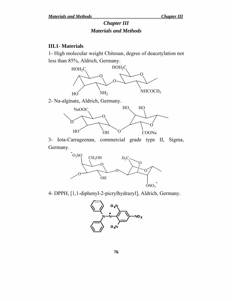
Materials and Methods Chapter III Chapter III
Materials and Methods
III.1- Materials 1- High molecular weight Chitosan, degree of deacetylation not less than 85%, Aldrich, Germany.
O HOH2C
NHCOCH3
O HOH2C
HO NH2
O
2- Na-alginate, Aldrich, Germany.
O
OH
NaOOC
O
HO O O
HOHO
COONa 3- Iota-Carrageenan, commercial grade type II, Sigma, Germany.
-
O O
O3SO
OH
CH2OH
O O
OSO3 -
O H2C
4- DPPH, [1,1-diphenyl-2-picrylhydrazyl], Aldrich, Germany.
76

Chapter III Materials and Methods 5- Ferrozine, [3-(2-pyridyl)-5,6-diphenyl-1,2,4-triazine-4’,4’’-
disulfonic acid sodium salt], Fluka, Germany.
6- Linoleic acid 80%, ABCR GmbH chemicals, Germany. O
HO
7-Ascorbic acid, El-Nasr Company for chemicals, Egypt.
8- Maleic anhydride and phthalic anhydride were purchased from NICE laboratory reagents, India.
9- Chlorosulphonic acid 97%, EDTA, ferric chloride and ferrous chloride were supplied from BDH, India.
10- Potassium ferricyanide, Riedel laboratory reagents, India. 11-Trichloroacetic acid and thiobarbituric acid were supplied
by SUVCHEM laboratory chemicals, India. 12-Chicken breasts were collected from a local poultry
processing plant immediately after evisceration. Samples were transported to the laboratory in an ice box and stored at 4oC for no longer than 2h before use. Samples were separated from the bones and minced in an electrical
77

Materials and Methods Chapter III mincer. Twenty gram samples were sealed in polyethylene bags after the addition of 0.1% of the tested antioxidants and put on the refrigerator (4±1oC) shelf until analysis.
13-Linear polyacrylamide (PAAm), Mwt over 5000000 was supplied by LAB Chemie, India.
14- Hydrogen peroxide (H2O2) 30% Loba chemie, India. 15-Sodium chloride, calcium chloride, ferric chloride,
ammonium nitrate, ammonium phosphate, ammonium sulfate and urea were supplied from El-Nasr Company for chemicals, Egypt.
16-Zea maize seeds from Agrochemical Company for production of seeds and agricultural crops, Egypt.
Other reagents and solvents were of analytical grade.
III.2. Methods and Apparatus III.2.1-Gamma Radiation Source
The samples were irradiated with the 60Co Candian irradiation facility gamma rays at a dose rate ranging from 4- 3.52 kGy/h. The irradiation facility was constructed by the National Center for Radiation Research and Technology (NCRRT), Atomic Energy Authority of Egypt (AEAE), Nasr city, Cairo, Egypt.
III.2.2- Synthesis of Chitosan Derivatives III.2.2.1- Synthesis of N-maleoyl and N-phthaloyl chitosan N-maleoyl-chitosan (NMCS) or N-phthaloyl-chitosan (NPhCS) was synthesized through the reaction between the amine groups of glucosamine units within chitosan backbone and maleic anhydride or phthalic anhydride Yu et al., (2010). Briefly, certain amount of chitosan (0.1mol) was dissolved in
78
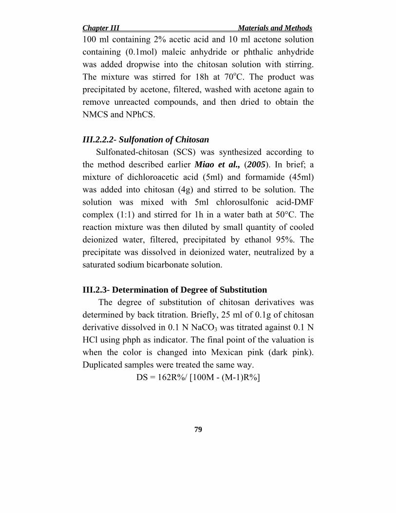
Chapter III Materials and Methods 100 ml containing 2% acetic acid and 10 ml acetone solution containing (0.1mol) maleic anhydride or phthalic anhydride was added dropwise into the chitosan solution with stirring. The mixture was stirred for 18h at 70oC. The product was precipitated by acetone, filtered, washed with acetone again to remove unreacted compounds, and then dried to obtain the NMCS and NPhCS.
III.2.2.2- Sulfonation of Chitosan Sulfonated-chitosan (SCS) was synthesized according to the method described earlier Miao et al., (2005). In brief; a mixture of dichloroacetic acid (5ml) and formamide (45ml) was added into chitosan (4g) and stirred to be solution. The solution was mixed with 5ml chlorosulfonic acid-DMF complex (1:1) and stirred for 1h in a water bath at 50°C. The reaction mixture was then diluted by small quantity of cooled deionized water, filtered, precipitated by ethanol 95%. The precipitate was dissolved in deionized water, neutralized by a saturated sodium bicarbonate solution.
III.2.3- Determination of Degree of Substitution The degree of substitution of chitosan derivatives was determined by back titration. Briefly, 25 ml of 0.1g of chitosan derivative dissolved in 0.1 N NaCO3 was titrated against 0.1 N HCl using phph as indicator. The final point of the valuation is when the color is changed into Mexican pink (dark pink). Duplicated samples were treated the same way.
DS = 162R%/ [100M - (M-1)R%]
79

Materials and Methods Chapter III where 162 is the molecular weight of the anhydrous glucose unit, R is the reaction percent and M is the molecular weight of the substituent.
III.2.4- Radiation Preparation of Low Molecular Weights Natural Polymers
III.2.4.1- For Antioxidant Activity Chitosan (CS) or Na-alginate or carrageenan or N-
maleoyl-chitosan (NMCS) or N-phthaloyl-chitosan (NPhCS) or sulfonated-chitosan (SCS) solutions with different concentrations (mg/ml) were irradiated by γ-rays at different doses of 10, 20 and 30 kGy at dose rate of 3.52 kGy/ h.
Chitosan (CS) or N-maleoyl-chitosan (NMCS) or N-phthaloyl-chitosan (NPhCS) or sulfonated chitosan (SCS) in the solid form were treated by 10% (v/w) H2O2 and mixed well as paste then irradiated by γ-rays at different doses of 25, 50, 75 and 100 kGy at the same dose rate.
III.2.4.2- For Agricultural Hydrogel Chitosan or Na-alginate (solid form) were mixed well with
10% H2O2 (v/w) as a paste, then packaged in polyethylene bags and subjected to γ-rays with different doses at 25, 50, 75 and 100 kGy.
III.2.4.3- For Growth Promotion 5% Na-alginate or 5% chitosan in 1% lactic acid were
prepared then treated by 1% (v/w) H2O2 and mixed well to be irradiated by γ-rays at different doses of 25, 35 and 45 kGy.
80

Chapter III Materials and Methods
III.2.5- Measurements of Antioxidant Activity III.2.5.1- Determination of Scavenging Effect on DPPH Radicals Measurement of free radical scavenging activity on DPPH radicals was determined according to the method described by Yamaguchi et al., (1998). Briefly, 1.5 ml of DPPH solution (0.1mM, in 95% ethanol) was incubated with varying concentrations of chitosan or Na-alginate or carrageenan or chitosan derivatives. The reaction mixture was shaken well and incubated for 15 min at room temperature and the absorbance of the resulting solution was read at 517 nm against a blank (control). Ascorbic acid was used for comparison as antioxidant materials. The radical scavenging effect was measured as a decrease in the absorbance of DPPH and can be calculated using the following equation: Scavenging effect (%) = [1 - (Asamples 517nm/ Acontrol 517 nm)] x 100
III.2.5.2. Determination of Reducing Power The reducing power was determined by the method
described by Yen and Duh (1993). Different concentrations of chitosan or Na-alginate or carrageenan or chitosan derivatives solutions (1ml) were mixed with 0.2M sodium phosphate buffer pH 6.6 (2.5ml) and 1% (w/v) potassium ferricyanide (2.5ml). The mixtures were incubated for 20 min at 50oC. The reaction was terminated by adding 10% (w/v) trichloroacetic acid (2.5ml) to the mixtures, followed by centrifugation for 10 min at 1500 rpm. 2.5ml supernatant was mixed with 2.5ml distilled water and 0.5ml ferric chloride (0.1% w/v) solution and the absorbance was measured at 700 nm. Increasing the
81

Materials and Methods Chapter III absorbance of the reaction mixture indicates the increase in reducing power of the samples. Ascorbic acid was used for comparison as antioxidant materials.
III.2.5.3- Determination of Ferrous Ion-chelating Potential (Metal Chelating Activity) Chelating ability was determined according to the method shown previously Dinis et al., (1994). The reaction mixture contains 0.25ml of chitosan or Na-alginate or carrageenan or chitosan derivatives of different concentrations, 0.5 ml ferrous chloride (2mM), and 0.25 ml Ferrozine (5mM) shaken well, and incubated for 10 min at room temperature. The absorbance of the mixture was measured at 562 nm against blank. EDTA was used for comparison as antioxidant materials. The ability of all samples to chelate ferrous ion was calculated using the following equation: Chelating Effect (%) = [(1 – (A sample 562nm / A control 562nm)] x100
III.2.5.4- Determination of Antioxidant Activity The antioxidant activity was determined by the conjugated
diene method Lingnert et al., (1979). Each chitosan or Na-alginate or carrageenan or chitosan derivatives of different concentrations (100 µl) was mixed with 2ml of 10 mmol/L linoleic acid emulsion in 200 mmol/L sodium phosphate buffer (pH 6.5) in test tubes and placed in darkness at 37oC to accelerate oxidation. After incubation for 15h, 6ml of 600 g/L methanol (60%) in deionized water was added, and the absorbance of the mixture was measured at 234 nm against a blank in a UV-Vis spectrophotometer. Ascorbic acid was used
82

Chapter III Materials and Methods for comparison as antioxidant materials. The antioxidant activity was calculated using the following equation: Antioxidant activity (%) =
[(A control 234nm - A sample 234nm)/ A control 234nm] x 100.
III.2.6- Thiobarbituric Acid Reactive Substances (TBARS) Lipid peroxidation was assessed through the
determination of secondary oxidation products (Malonaldehyde, MDA, CHO-CH2-CHO) formed during 10 days of refrigerated storage (4±1oC), by a spectrophotometric method of Vynche (1970). TBARS was done on days 0, 3 and 10 of refrigerated storage on 3 separate samples of each group. On 0 day, the TBARS was performed only for the control samples.
Triplicate 20g samples (for each group) were homogenized with 100 ml of 7.5% trichloroacetic acid solution (TCA). The homogenate was filtered and 5ml aliquot was transferred to a clean screw capped tube and mixed with 5ml of freshly prepared 0.02M thiobarbituric acid (TBA) solution. The mixture was put in a boiling water bath for 35 minutes until color formation, and then it was left to cool until submitted to conventional spectrophotometry. The absorbance of the developed red color was measured at wavelength 538 nm against blank. The results were then expressed as mg MDA/ Kg sample.
III.2.7- Fungal Toxicity of NMCS
NMCS (2mg/ml) was added to Malt extract agar medium (Malt extracts 20g; glucose 20g and peptone
83

Materials and Methods Chapter III 1g/l) after sterilization, the dissolved NMCS was added right before pouring the media into sterilized plates. After the plates were left to set, 4mm discs were taken from Phanerocheate chrysosporium ATCC 34541 culture, the discs were punched using the broad end of a sterile tip and placed in the center of each plate. Malt extract agar medium was used as a positive growth control. All plates were left to incubate at 30oC, the radial growth was measured every 24h to monitor growth changes in each set. Triplicate plates were used for both the control and sample amended plates. The mycelial growth inhibition was calculated using the following equation:-
The Mycelial Growth Inhibition = (Gc-Gs/Gc) x 100
where Gc is the mycelial growth for Phanerocheate chrysosporium in centimeter (control plates) and Gs is the mycelial growth in sample amended plates. The results obtained are presented as percentage of growth inhibition Murugesan et al., (2009).
III.2.8- Cytotoxicity of NMCS III.2.8.1- Cell Culture
Human colorectal adenocarcinoma cell line (Caco-2) which was purchased from ATCC, USA, was used to evaluate the cytotoxic effect of the tested samples. Cells were routinely cultured in Eagle's Minimum Essential Medium which was supplemented with 20% fetal bovine serum (FBS), 2 mM L-glutamine, containing 100 units/ml penicillin G sodium, 100
84

Chapter III Materials and Methods units/ml streptomycin sulphate, and 250 µg/ml amphotericin B. Cells were maintained at sub-confluency at 37ºC in humidified air containing 5% CO2. For sub-culturing, monolayer cells were harvested after trypsin/EDTA treatment at 37°C. Cells were used when confluence had reached 75%. Tested solid sample was dissolved in dimethyl sulphoxide (DMSO), and then diluted thousand times in the assay. All cell culture material was obtained from Cambrex BioScience (Copenhagen, Denmark). All chemicals were from Sigma/Aldrich, USA, except mentioned. All experiments were repeated three times, unless mentioned.
III.2.8.2- Anti-tumor Activity Cytotoxicity of tested extract was measured against Caco
2 cells using the MTT Cell Viability Assay. MTT (3-[4,5-dimethylthiazole-2-yl]-2,5-diphenyltetrazolium bromide) assay is based on the ability of active mitochondrial dehydrogenase enzyme of living cells to cleave the tetrazolium rings of the yellow MTT and form a dark blue insoluble formazan crystals which is largely impermeable to cell membranes, resulting in its accumulation within healthy cells. Solubilization of the cells results in the liberation of crystals, which are then solubilized. The number of viable cells is directly proportional to the level of soluble formazan dark blue color. The extent of the reduction of MTT was quantified by measuring the absorbance at 570 nm Hansen et al, (1989).
III.2.8.2.1- Reagents preparation MTT solution: 5mg/ml of MTT in 0.9%NaCl.Acidified isoprpanol: 0.04 N HCl in absolute isopropanol.
85

Materials and Methods Chapter III
III.2.8.2.2- Procedure Cells (0.5X10⁵ cells/ well), in serum-free media, were plated in a flat bottom 96-well microplate, and treated with 20µl of serial dilutions of the tested solid sample for 48 h at 37º C, in a humidified 5% CO2 atmosphere. After incubation, media were removed and 40 µl MTT solution / well were added and Incubated for an additional 4 h. MTT crystals were solubilized by adding 180 µl of acidified isopropanol / well and plate was shacked at room temperature, followed by photometric determination of the absorbance at 570 nm using microplate ELISA reader. Triplicate repeats were performed for each concentration and the average was calculated. Data were expressed as the percentage of relative viability compared with the untreated cells compared with the control for the solid sample, with cytotoxicity indicated by <100% relative viability.
Percentage of relative viability = [Abs. of treated cells/ Abs. of control cells)] X 100
Then the half maximal inhibitory concentration (IC50 %) was calculated from the equation of the dose response curve.
III.2.9- Preparation of Hydrogels To obtain PAAm/Alg or PAAm/CS or PAAm/Alg/CS hydrogels, an appropriate weight of dry polyacrylamide (PAAm) and dry degraded Na-alginate or chitosan irradiated at 100 kGy were mixed with 100 ml distilled water to obtain 5% of copolymer concentration. The blend of copolymers left for 3h in a water bath at 60°C until the blend changed to a
86

Chapter III Materials and Methods homogenous paste. Different copolymer compositions were prepared (100/0, 95/5, 90/10, 85/15 and 80/20 of PAAm/ Degraded natural polymer). The blend was put in a polyethylene bag and subjected to γ-rays with different irradiation doses at 5, 10, 20 and 30 kGy. The obtained hydrogels were cut into small pieces and left to dry at room temperature.
III.2.10- Determination of Gel content (%) To extract the soluble part of the hydrogels (uncrosslinked
part), the prepared hydrogels were soaked in water for 24 h at 70oC. Then they were taken out and washed with hot water to remove the soluble part, dried, and weighed. The gel percent in the hydrogel was determined from the equation:
Gel (%) = (Wd / Wi) x 100 where Wd and Wi are the weights of dry hydrogel after and before extraction, respectively.
III.2.11- Water Absorbency Measurement The dried hydrogels of known weights were immersed in distilled water or different cations or different fertilizers or different buffers of different pH's at 25oC until the swelling equilibrium was reached (almost 24h). The hydrogels were removed and then weighed. The experiment was repeated twice for each sample. The following equation was used to determine swelling ratio of hydrogels:
Water Absorbency (g/g) = (Ws – Wg)/ Wg
where Ws and Wg are the weights of wet and dry gel, respectively.
87

Materials and Methods Chapter III
III.2.12- Water Retention Measurement To determine the water retention of the hydrogels, the
swollen gel of known weight were placed in an oven at 25, 35 and 45oC. The gel was weighed at each time interval and the weight loss of water was calculated as follows:
Water Retention (%) = (Wi/ Wt ) x 100 Where Wi and Wt are the weights of retained water inside the hydrogel at initial and time t (h), respectively.
III.2.13- Plantation III.2.13.1- For Agricultural Hydrogels The effect of PAAm/Alg or PAAm/ CS or PAAm/Alg/CS copolymer hydrogels on the growth of zea maize plants was investigated. Zea maize seeds were immersed in water 24h. The swollen seeds were sown 5cm apart from the gel-soil holes or pots. The pots contain proper amount of swollen hydrogels. Water of irrigation remained constant for all replicates in different experiments. The growth rate of the plants is compared with control lines (untreated plant).
III.2.13.2- For Growth Promotion To investigate the synergetic effect of combining of degraded Na-alginate and/or chitosan sprayed on zea maize plants, the concentration of 100 ppm was used. The field was divided into three groups of irradiation treatment of 25, 35 and 45kGy. Each group divided into different separate lines. After plantation of 15, 30, 45 and 60 days, each three lines of plants were sprayed by 100 ppm solution of
(a) Na-alginate irradiated at 25 kGy
88

Chapter III Materials and Methods (b) Chitosan irradiated at 25 kGy (c) Na-alginate/ Chitosan (1/1 v/v) irradiated at 25 kGy (d) Na-alginate/ Chitosan (2/1 v/v) irradiated at 25 kGy (e) Na-alginate/ Chitosan (1/2 v/v) irradiated at 25 kGy
The same method of spraying was repeated for degraded Na-alginate and/or chitosan irradiated at different doses of 35 and 45 kGy. The growth rate of the plants is compared with control lines (untreated plant).
III.2.14- Determination of the Molecular Weights The number average molecular weights of the degraded
polysaccharides were determined by Gel permeation chromatography (GPC) 1100 Agilent instrument. The GPC elution curve was monitored by a refractive index detector of optical unit temperature 25°C, peak width 0.1min and polymer concentration was 0.1 (Wt%). The number average molecular weights were determined from a calibration curve using polyethylene oxide standards of known molecular weights.
III.2.15- FT-IR Spectroscopy The transmittance was carried out in the form of KBr
pellets in the range of 400-4000 cm-1 using infra-red spectrophotometer JASCO FT-IR 6300, Japan.
III.2.16- UV-Vis Spectroscopy UV absorbency was measured by a UV-Vis
spectrophotometer JASCO V-560, Japan, in the range from 190 to 900 nm.
89

Materials and Methods Chapter III
III.2.17- Thermal Gravimetric Analysis (TGA)
The thermal stability of prepared hydrogels was carried out using Perkin Elmar TGA system under nitrogen atmosphere of 10 ml/min was used. The temperature range was from ambient temperature to 600 at flow rate ≈ 10 ml/min.
III.2.18- X-Ray Diffraction (XRD) X-Ray Diffraction patterns were obtained with a XRD
6000 series, Shimadzu apparatus using Ni-filter and Cu-Kα target.
III.2.19- Scanning Electron Microscopy (SEM) The surface topology of the hydrogels was measured with
JEOL JSM-5400 Scanning Electron Microscopy (SEM), Japan. The surfaces of the freeze dried hydrogels were sputter-coated with gold for 3min.
90

Chapter IV
Results and
Discussion

Chapter IV Results and Discussion
Chapter IV Results and Discussion
IV.I. Investigating the Antioxidant Properties of LowMolecular Weight Natural Polymers and Assessing Its Suitability for Food Preservation
The antioxidant activity of polysaccharides depends on several structural parameters, such as the molecular weight, type and position of functional groups, (such as hydroxyl, sulfate, amine, carboxyl and phosphate), type of saccharide, and glycosidic branching.
IV.I.1. Effect of γ-rays on the Molecular Weights of Some Natural Polymers
Radiation-induced effects onto natural polymers can occur through the molecular changes resulting chain crosslinking reaction causing an increase in the molecular weight or chain scission reaction causing a decrease in the molecular weight and substantially changing the polymer material properties. These reactions depends on the chemical nature of the polymer and irradiation conditions i.e., irradiation dose and dose rate Polysaccharides are typical degradable materials under ionizing radiation conditions which undergo degradation reactions through the β-(1–4) glycosidic bond cleavage resulting in the reduction of their average molecular weights Reichmanis et al. (1993). Figure (1) shows the changes in the number average molecular weights of chitosan, Na-alginate and carrageenan in
91

• •
Results and Discussion Chapter IV aqueous solution using GPC after treating with gamma irradiation at different doses. There is a rapid decrease in the number average molecular weights of these polysaccharides. As the irradiation dose increases, the molecular weights of polysaccharides decrease. The molecular weights of chitosan, Na-alginate and carrageenan subjected to 10 kGy, decreased from 1.9x10⁶, 2.4x10⁶ and 4.3x10⁶ (Da) to 3.8x10⁵, 1.1x10⁵
and 2.5x10⁵ (Da), respectively. Meanwhile, with increasing the irradiation dose to 30 kGy, the molecular weights reduced to 9.9x10³, 9.2x10³ and 1.2x10⁴ (Da), respectively.
It is well known that the radiation energy of γ-ray is absorbed mainly by water in dilute aqueous solutions. The radiolysis of water yields reactive species such as (•OH), hydrated electrons (e¯
aq), hydrogen atoms (•H) Hai et al., (2003).
H2O + γ-rays OH, e¯aq, H, H2O2, H2, H+, OH¯
HO• radical is a powerful oxidizing species can react with carbohydrates exceedingly rapidly, abstracting H atom and also, attack the β-(1-4) glycosidic bonds of polysaccharide resulting in reducing their molecular weight very effectively according to the following equations: R-H + •OH R• + H2O R-H + •H R• + H2
R• R1 + R2
where R-H is carbohydrate macromolecules, R. is a macro-radical localized on Cn carbon atom and R1, R2 are fragments of the main chain after scission.
92

Chapter IV Results and Discussion
Mol
ecul
ar W
eigh
t, D
a
107
106
105
104
103
0 5 10 15 20 25 30 35
Irradiation Dose (kGy)
Figure (1): The average molecular weights of (●) chitosan, (○) Na-alginate and (▼) carrageenan in aqueous solution after γ-irradiation at different doses.
93

Results and Discussion Chapter IV
IV.I.2. Determination of Chain Scission Yield and Degradation Rate Constant
The efficiency of radiation-induced events is expressed by the so-called G-value. The G-value is equal to the number of events per 100 eV of energy absorbed has been customarily used to measure radiation–chemical yield. Molecular weight values of polysaccharide under investigation were used for the determination of chain scission yield G(s) and their degradation rate. Supposing that scission is the only mode of action of radiation on polysaccharide, the radiation–chemical yield of degradation (scission) G(s) is determined from the Alexander– Charlesby–Ross equation Charlesby (1960):
1 / MnD = 1/ Mn0 + 1.04 x 10⁻⁷ G(s) D
where the absorbed dose (D) is in kGy and MnD and Mn0 are the number average molecular weights of the polymer before and after irradiation.
The equation given by Jellinek (1955) is modified and used in the determination of degradation rate, where N is the average number of bond cleavages per original polymer molecule, or named the degree of scission, this value can be calculated by
N = (Mn0 / MnD) – 1 where Mn0 and MnD are the Mn at time zero and after irradiation to a certain dose (D), respectively.
94
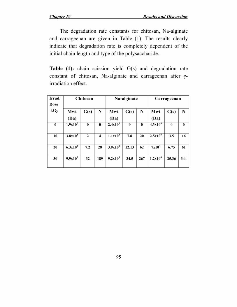
Chapter IV Results and Discussion
The degradation rate constants for chitosan, Na-alginate and carrageenan are given in Table (1). The results clearly indicate that degradation rate is completely dependent of the initial chain length and type of the polysaccharide.
Table (1): chain scission yield G(s) and degradation rate constant of chitosan, Na-alginate and carrageenan after γ-irradiation effect.
Irrad. Chitosan Na-alginate Carrageenan Dose kGy Mwt G(s) N Mwt G(s) N Mwt G(s) N
(Da) (Da) (Da) 0 1.9x10⁶ 0 0 2.4x10⁶ 0 0 4.3x10⁶ 0 0
10 3.8x10⁵ 2 4 1.1x10⁵ 7.8 20 2.5x10⁵ 3.5 16
20 6.3x10⁴ 7.2 28 3.9x10⁴ 12.13 62 7x10⁴ 6.75 61
30 9.9x10³ 32 189 9.2x10³ 34.5 267 1.2x10⁴ 25.36 344
95

Results and Discussion Chapter IV
IV.I.3. Structural Changes of Degraded Natural Polymers
IV.I.3.1. UV-Vis Spectroscopy
Figure (2) shows the UV-Vis spectra of unirradiated and irradiated chitosan, Na-alginate and carrageenan. For the irradiated chitosan Figure (2 A), two absorption peaks at 275 and 315 nm were observed. The intensity of these peaks increased with increasing the irradiation dose. These two peaks may be due to the presence of unsaturated carbonyl and carboxyl groups. The obtained results are consistent with those reported by Ulanski and Rosiak (1992).
For the irradiated Na-alginate and carrageenan Figure (2 B and C), a new absorption band at 275 nm and the peak intensity increase with increasing the irradiation dose. The intensity of such peak increased with increasing the irradiation dose. It can be assigned to carbonyl or hydroxyl group formed after the main chain scission of the polysaccharide as explained by Nagasawa et al., (2000).
96

Chapter IV Results and Discussion
Abs
orba
nce
0.0
0.5
1.0
1.5
2.0
2.5
(A) Chitosan
250 300 350 400 450 500 Wavelength (nm)
(
Abs
orba
nce
0.0
0.5
1.0
1.5
2.0
2.5
Abs
orba
nce
0.0
0.5
1.0
1.5
2.0
B) Na-alginate (C) Carrageenan
250 300 350 400 450 500 250 300 350 400 450 500
Wavelenghth (nm) Wavelenghth (nm)
Figure (2): UV-Vis spectra of (A) chitosan, (B) Na-alginate and (C) carrageenan; (—) unirradiated polysaccharide solution and the irradiated solution at 10 kGy (…..), 20 kGy (---) and 30 kGy (-..-..-).
97

Results and Discussion Chapter IV
IV.I.3.2. FT-IR Spectroscopy Figure (3) shows the FT-IR spectra of unirradiated and irradiated chitosan, Na-alginate and carrageenan. FT-IR spectrum of chitosan Figure (3A, curve a) shows distinctive absorption bands appeared at 3440, 1638, 1598, 1387, 1156 and 1097 cm-1 corresponds to –OH and –NH2 groups, stretching C=O of amide group, N-H bend, NH of amide, asymmetric bridge-O-stretch and skeletal vibration involving the C-O stretch, respectively. FT-IR spectrum of irradiated chitosan Figure (3A, curves b, c and d), it was observed that as the irradiation dose increases, the intensity of the absorption peak at 1602 cm-1 assigned to the N–H bend vibration of –NH2
increases and shift to lower wavenumber. This may be due to the decreasing of inter or intra- molecular hydrogen bonding between the –OH and –NH2 groups. The peak at 1638 cm-1due to C=O group was gradually shifted to 1645 cm-1, while its intensity increased with increasing the γ-ray irradiation dose. Also, the intensity of the peak at 1385 cm-1 assigned to C–O stretch vibration increased and moved toward the higher wavenumber as the irradiation dose increases. The vibrational band at 1100 cm-1 corresponding to the ether bond in the pyranose ring has no significant change indicating that the stability of the β-glycosidic bonds and distribution of glycosidic bonds in the molecular chains of chitosan.
FT-IR spectrum of Na-alginate Figure (3B, curve a), the peaks at 3390, 1620 and 1095-1035 cm-1 are attributed to hydroxyl, C–O–O, C–O–C groups, respectively Sartori et al.
98

Chapter IV Results and Discussion (1997). Peak at 1610 cm-1 for –COONa of Na-alginate was taken as the reference peaks due to the fact that carboxyl groups do not change after degradation. The spectrum of irradiated Na-alginate Figure (3B, curves b, c and d) exhibited most of the characteristic absorption peaks of native alginate but with some differences. For instance, the scission of glycosidic bonds leads to the formation of hydroxyl group, which is manifested as an increase in the ratio of hydroxyl group peak at 3425 cm-1.
FT-IR spectrum of carrageenan Figure (3C, curve a), the absorption band between 855-805 cm-1 indicated that the sulfate group was attached at the C-4 position of galactose. The band at 934 cm-1 was attributed to the presence of 3,6- anhydro-galactose. The vibrational band at 1075 cm-1 that corresponds to the ether bond in the pyranose ring The signal at 1262 cm-1 was attributed to the asymmetric stretching of S=O. The absorption bands at 2941 and 3460 cm-1 due to C-H stretch groups and hydroxyl (O-H) groups, respectively. The spectrum of irradiated carrageenan Figure (3C, curves b, c and d) exhibited most of the characteristic absorption peaks of native carrageenan but there is a new band appeared at 1732 cm-1 due to glycoside bonds cleavage with the change of the structure of reducing end residue and formation of –C=O groups.
From the data obtained by UV-Vis and FT-IR studies, the proposed mechanism of degradation of chitosan, Na-alginate and carrageenan irradiated in the liquid state after irradiation is illustrated in Scheme (5).
99

Results and Discussion Chapter IV
Figure (3): FT-IR spectra of (A) chitosan, (B) Na-alginate and (C) carrageenan; (a) unirradiated polysaccharide and irradiated polysaccharide at (b) 10 kGy, (c) 20 kGy and (d) 30 kGy.
100

Chapter IV Results and Discussion
O
R2
R1
O
HO O O
R1
R2HO
O R1
O
R2HO
n
.
Irradiation Gamma
Polysaccharide
O
HO R2 .O R1
.+ OH, RH
R1 HO R2 O
O .. + HOH + R
OOHO R2 R1
Chitosan, R1 = CH2OH and R2 = NH2
Na-alginate, R1 = COONa and R2 = OH
Carageenan, R1 = CH2OH and R2 = OSO3Na
Scheme (5): Proposed mechanism of radiation induced degradation of chitosan, Na-alginate and carrageenan.
101

Results and Discussion Chapter IV
IV.I.4. Comparative Study on the Antioxidative Properties of Some Natural Polymers Degraded by γ-rays
IV.I.4.1. Scavenging Effect (%) on DPPH Radicals
DPPH is one of the compounds that possessed a proton free radical which decreases significantly on exposure to proton radical scavengers Yamaguchi et al., (1998). Further it is well accepted that the DPPH free radical scavenging by antioxidants is due to their hydrogen-donating ability Chen and Ho (1995).
The DPPH scavenging effect (%) of chitosan, Na-alginate and carrageenan with different concentrations and irradiation doses was measured and the results were depicted in Figure (4). In general DPPH radical scavenging effect (%) of chitosan, Na-alginate and carrageenan increased with increasing the polysaccharide concentration to a certain extent about 1 mg/ml and then it levelled off even with further increase in the concentration. Also, an increase in the DPPH radical scavenging effect (%) was observed by increasing the irradiation dose of polysaccharides. The results revealed that the DPPH radical scavenging effect (%) of the investigated polysaccharides followed the order chitosan > Na-alginate > carrageenan. The DPPH scavenging effect (%) for the unirradiated chitosan, Na-alginate and carrageenan at a concentration of 0.5 mg/ml was 5, 3.2 and 3.1 (%), respectively. Meanwhile, using 30kGy irradiation dose, the DPPH scavenging effect (%) became 60.2, 52.7 and 51.1 (%),
102

Chapter IV Results and Discussion respectively. The DPPH scavenging effect (%) of chitosan irradiated at 30 kGy is nearly the same as that of ascorbic acid which is 62.5 (%).
The inhibition concentration (IC50 %) value is a good parameter to evaluate the antioxidant ability, which means that the antioxidant concentration to reduces to 50%. The IC50 (%) on DPPH radicals was determined to be 0.154, 0.359, and 0.438 mg/ml for chitosan, Na-alginate and carrageenan irradiated at 30 kGy, respectively as compared by that of ascorbic acid 0.14 mg/ml. These results reveled that the irradiation of chitosan enhances its antioxidant activity.
103

Results and Discussion Chapter IV
)
0
10
20
30
40
50
60
70
80
Scav
enin
g E
ffec
t (%
Chitosan
0.0 0.5 1.0 1.5 2.0
Concentration (mg/ml)
)
0
10
20
30
40
50
60
70
80
)
0
10
20
30
40
50
60
70
80
Carragenann
Scav
engi
ng E
ffec
t (%
Scav
engi
ng E
ffec
t (%
Na-alginate
0.0 0.5 1.0 1.5 2.0 0.0 0.5 1.0 1.5 2.0
Concentration (mg/ml) Concentation (mg/ml)
Figure (4): Scavenging effect (%) on DPPH radicals of chitosan, Na-alginate and carrageenan; (●) unirradiated polysaccharides and the irradiated polysaccharides at different doses of (○) 10 kGy, (▼) 20 kGy and (■) 30 kGy. (x) Ascorbic acid as reference.
104

Chapter IV Results and Discussion
IV.I.4.2. Determination of Reducing Power
The reducing capacity of a compound may serve as a significant indicator of its potential antioxidant activity. Figure (5) shows the reducing power of unirradiated and irradiated chitosan, Na-alginate and carrageenan at different doses as a function of concentration comparing with ascorbic acid (positive control). Low molecular weight polysaccharides showed high reducing power, and the reducing power increased with increasing the polysaccharides concentration. From the results obtained, the irradiation of these polysaccharides enhances the degradation process, the lower molecular weights, the higher reducing power obtained. The reducing power of irradiated chitosan is much higher than that of Na-alginate and carrageenan.
At 2 mg/ml polysaccharide concentration, the reducing power of unirradiated chitosan, Na-alginate and carrageenan was 0.3319, 0.3504 and 0.3146, respectively. Meanwhile, using 30 kGy irradiation dose, the reducing power of chitosan, Na-alginate and carrageenan became 1.3574, 1.2380 and 0.9377, respectively. The reducing power of ascorbic acid was 1.48. The role of reducing agents is to exert antioxidant action by breaking the free radicals chain by donating a hydrogen atom Gordon (1990) and Shimada et al., (1992).
105

Alginate
Results and Discussion Chapter IV
0.0
0.2
0.4
0.6
0.8
1.0
1.2
1.4
1.6
1.8
A b s
o rb a
n ce
at 7
00 n
m
Chitosan
0.0 0.5 1.0 1.5 2.0
Concentration (mg/ml)
0.0
0.2
0.4
0.6
0.8
1.0
1.2
1.4
1.6
1.8
Carrageenan
A b s
o rb a
n ce
a t 7
0 0 n
m
Na-alginate A
b so r
b an c
e at
7 00
n m
0.0
0.2
0.4
0.6
0.8
1.0
1.2
1.4
1.6
1.8
0.0 0.5 1.0 1.5 2.0 0.0 0.5 1.0 1.5 2.0
Concentration (mg/ml) Concentration (mg/ml)
Figure (5): Reducing power of chitosan, Na-alginate and carrageenan; (●) unirradiated polysaccharides and the irradiated polysaccharides at different doses of (○) 10 kGy, (▼) 20 kGy and (■) 30 kGy. (x) Ascorbic acid as reference.
106

Chapter IV Results and Discussion
IV.I.4.3. Chelating Effects on Ferrous Ions
Transition metal ions such as ferrous ions can initiate lipid peroxidation and start a chain reaction, which lead to the deterioration of flavor and taste in food Gordon (1990). Since ferrous ions are the most effective prooxidants in the food system, the high ferrous ion-chelating abilities of chitosan would be beneficial if they were formulated into foods Yamaguchi et al., (1988). Ferrozine can quantitatively form complexes with Fe2+. In the presence of chelating agents, the complex formation is disrupted with the result that the red color of the complex is decreased. Measurement of color reduction therefore allows estimation of the chelating activity of the coexisting chelator.
The chelating effect (%) of unirradiated and irradiated chitosan, Na-alginate and carrageenan at different doses as a function of concentration using EDTA as positive control was shown in Figure (6). Low molecular weight polysaccharides showed high chelating ability. The chelating ability increased with increasing the concentration of polysaccharides. The results revealed that the irradiated chitosan had the highest chelating ability. The affinity of chitosan to chelate Fe2+ ion mainly comes from the presence of amino groups, which contain lone electron pairs that help to form chitosan-Fe2+
complexes Guzman et al., (2003).
At 1 mg/ml polysaccharide concentration, the chelating effect (%) of unirradiated chitosan, Na-alginate and
107

Results and Discussion Chapter IV carrageenan was 40, 21.1 and 19.3 (%), respectively, in comparison with that of EDTA which gave 66.4 (%). While, using 30 kGy irradiation dose, the chelating (%) of chitosan, Na-alginate and carrageenan enhanced and became 77.5, 51.1 and 62.4 (%), respectively. The IC50 (%) of the chelating effect (%) was 0.2293, 0.8023 and 0.238 for chitosan irradiated at 20 kGy, Na-alginate irradiated at 30 kGy and carrageenan irradiated at 30 kGy, respectively. The chelating effect (%) of 30 kGy irradiated chitosan was higher than that of Na-alginate and/ or carrageenan.
Chitosan possesses high chelating capacity for various metal ions in an acidic environment and responsible for the removal of metal ions in many industries. The high ferrous ion-chelating abilities of chitosan would be beneficial if they were formulated into foods. This suggests the possibility of using the γ-ray irradiated chitosan as antioxidants to prevent flavor changes caused by lipid peroxidation.
108

Chapter IV Results and Discussion
i(%
)
0
10
20
30
40
50
60
70
80
90 Chitosan
Che
latn
g E
ffec
t
0.0 0.5 1.0 1.5 2.0
Concentration (mg/ml)
i (%
)
0
10
20
30
40
50
60
70
80
90
i (%
)
0
10
20
30
40
50
60
70
80
90
Che
latn
g E
ffec
t
Che
latn
g E
ffec
tCarragenann Na-alginate
0.0 0.5 1.0 1.5 2.0 0.0 0.5 1.0 1.5 2.0
Concentration (mg/ml) Concentration (mg/ml)
Figure (6): Chelating effect (%) on ferrous ions of chitosan, Na-alginate and carrageenan; (●) unirradiated polysaccharides and the irradiated polysaccharides at different doses of (○) 10 kGy, (▼) 20 kGy and (■) 30 kGy. (x) EDTA as reference.
109

Results and Discussion Chapter IV
IV.I.4.4. Determination of Antioxidant Activity
Total antioxidant activity (%) of unirradiated and irradiated chitosan, Na-alginate and carrageenan with different concentrations was measured and the results were depicted in Figure (7). Low molecular weight irradiated chitosan, Na-alginate and carrageenan showed high antioxidant activity (%) and the antioxidant activity increased with increasing the polysaccharide concentration. The results revealed the irradiated chitosan was the highest antioxidant activity (%) rather than Na-alginate and carrageenan. The maximum antioxidant activity of irradiated chitosan, Na-alginate and carrageenan was 33.37, 26.3 and 30.0 (%), respectively using 30 kGy dose and 2mg/ ml polysaccharide concentration as compared by the unirradiated samples value of 5.93, 3.8 and 9.6 (%), respectively.
The high antioxidant activity of chitosan oligosaccharide mainly attributed to the hydroxyl and amino groups. The lower the molecular weights of chitosan oligosaccharide, the higher the antioxidant activity obtained. This may be due to the radiation induced degradation of chitosan that causes decreasing the intra-molecular hydrogen bonds between hydroxyl and amino groups Xie et al., (2001).
110

Chapter IV Results and Discussion
)
0
10
20
30
40
50
Ant
ioxi
dant
Act
ivity
(%
Chitosan
0.0 0.5 1.0 1.5 2.0
Concentration (mg/ml)
0
10
20
30
40
50
0
10
20
30
40
50
Ant
ioxi
dant
Act
ivity
(%)
Ant
ioxi
dant
Act
ivity
(%)
Na-alginate Carragenann
0.0 0.5 1.0 1.5 2.0 0.0 0.5 1.0 1.5 2.0
Concentration (mg/ml) Concentration (mg/ml)
Figure (7): Antioxidant activity (%) of chitosan, Na-alginate and carrageenan; (●) unirradiated polysaccharides and the irradiated polysaccharides at different doses of (○) 10 kGy, (▼) 20 kGy and (■) 30 kGy. (x) Ascorbic acid as reference.
111

Results and Discussion Chapter IV
IV.I.5. Improvement of Antioxidant Activity of Chitosan by Chemical Treatment and Ionizing Radiation
IV.I.5.1. Synthesis and Characterization of Chitosan Derivatives To overcome the limited solubility, due to the presence of primary amino group, chitosan derivatives were prepared by introducing ionic groups (or substituting group) into the chitosan backbone, which are solvated by polar solvents like water through polar-polar interactions and promote solubility of the macromolecule. In the present study, substituted chitosan derivatives were synthesized to produce water soluble derivatives at neutral pH. Scheme (6) describes the chemical modification of chitosan with maleic anhydride, phthalic anhydride and sulfonation using chlorosulfonic acid-DMF complex to get N-maleoyl-chitosan (NMCS), N-phthaloyl-chitosan (NPhCS) and sulfonated-chitosan (SCS), respectively. Chitosan derivatives may be demonstrated to exhibit antioxidant activity that might be more useful than that of native chitosan as an antioxidant agent in a food product. The antioxidant activity of the polymer is governed by many factors among them the degree of substitution that has a direct impact on the secondary structure of the polymeric chain and can also influence the solubility of the polymer in organic or aqueous solvents. Thus, the degree of substitution of chitosan derivatives treated by different reagent was calculated as 0.57, 0.31 and 0.38 for the NMCS, NPhCS and SCS, respectively.
112

Chapter IV Results and Discussion
O
OH
O
HO NH-C=O
O
O
O
n
70 oC
CH=CH-COOH NMCS
O
OH
O
NH2HO
O
OH
O
HO
O
O
O
NH
O
HO
O
n n
70 oC
CS
NPhCS
O
OSO3Na
O
NH2HO
n
3 / oC
3
1) ClSO H / DMF 60
2) NaHCO
SCS
Scheme (6): Chemical modifications of chitosan
113

Results and Discussion Chapter IV
IV.I.5.2. Characterization of Chitosan Derivatives by FT-IR
The structural changes on chitosan after chemical modifications were confirmed using FT-IR. Figure (8) shows the FT-IR spectra of CS, NMCS, NPhCS and SCS. FT-IR spectrum of chitosan (CS) Figure (8, curve a) was previously illustrated as in Figure (3A, curve a).
FT-IR spectrum of NMCS Figure (8, curve b) shows absorption band at 2941 cm-1 (aliphatic C-H groups). The absorption bands at 3440 and 1598 cm-1 (characteristics for amino group) decrease, and the new band at 1719 cm-1 for (C=O) of acid groups appears. Characteristic bands for -NHC=O- and -CH=CH- appears at 1632 and 1547 cm-1, respectively. FT-IR spectrum of NPhCS Figure (8 curve c), shows the absorption bands at 2941and 3074 cm-1 due to C-H stretch of the –CH=CH- groups and C-H stretch of aromatic system, respectively. The new peaks at 1719, 1773 and 1547
-1cm are characteristic for the carbonyl group (C=O) of carboxylic acid, residual anhydride and -CH=CH- groups, respectively. FT-IR spectrum of SCS Figure (8 curve d), the new absorption bands appeared at 817 cm-1 and 1235 cm-1
which correspond to the C-O-S and S=O bond stretching, respectively. The absorption bands at 1700 cm-1 characteristics for C=O groups appears due to some ring opening occurred during the reaction by chlorosulfonic acid.
114

Chapter IV Results and Discussion
Figure (8): FT-IR spectra of (a) CS, (b) NMCS, (c) NPhCS and (d) SCS.
115

Results and Discussion Chapter IV
IV.I.5.3. Characterization of Chitosan Derivatives by XRD Analysis
Figure (9) shows the X-ray diffraction (XRD) patterns of chitosan and its derivatives. The XRD pattern of chitosan Figure (9 curve a) exhibited three characteristic peaks around 2θ = 10.4o, 19.8o and 22.0o Samuels (1981) and Zhang & Neau, (2001) corresponding to the L-2 polymorph of shrimp chitosan (high degree of crystallinity morphology of chitosan) Hasegawa et al., (1992). The reflection fall at 2θ = 10.4o
assigned to the crystal form I and the strongest reflection appeared at 2θ = 19.8o corresponding to crystal form II. This was an indication of high degree of crystallinity of chitosan due to the inter-molecular and extra-molecular hydrogen bonding Samuels (1981).
The XRD pattern of NMCS Figure (9 curve b) have only one broad peak at around 2θ = 20°, which indicates that crystal forms have been destroyed in NMCS and became more amorphous in comparison to the native chitosan. The lower crystallinity of NMCS ascribed to the presence of maleoyl groups, might have hindered the formation of inter- and extra-molecular hydrogen bonds after chemical modification. As a result, the solubility of NMCS is better than that of chitosan. NMCS can be disperse into distilled water and obtain a transparent and stable system.
116

Chapter IV Results and Discussion The XRD pattern of NPhCS Figure (9 curve c), have
characteristic peaks at 2θ = 6.8°, 19.2° and 26.5°. The decrease in crystallinity of NPhCS could be ascribed to the presence of aryl group. This is possible that the steric hindrance of the aryl group obstructed the formation of inter- and intra-molecular hydrogen bonds of the chitosan backbone.
The XRD pattern of SCS Figure (10 curve d), suggesting that chemical modification reduced its capability to form a hydrogen bond. Sulfonation of chitosan proceeds randomly along the chitosan chain in solution, efficiently destroying the regular packing of the original chitosan units. Finally, a totally amorphous sulfonated-chitosan is available.
117

Results and Discussion Chapter IV
I n t e n i s t y
a
b
c
d
5 10 15 20 25 30 35
Theta-2theta (deg)
Figure (9): XRD spectra of (a) CS, (b) NMCS, (c) NPhCS and (d) SCS.
118
40

Chapter IV Results and Discussion
IV.I.5.4. Effect of γ-rays on the Molecular Weights of Chitosan Derivatives
The solubility of chitineous substances is usually associated with the crystallinity and intra- and inter-molecular hydrogen bonding within the polymer chains that plays a major role in the low solubility of these substances. Ionizing radiation can break the interaction between the chitineous substances molecules, decrease their crystallinty (%), decrease their molecular weight and enhance their solubility. These changes are expected to increases the antioxidant activity of chitineous substances. Figure (10) shows the changes in the number average molecular weights of chitosan and its derivatives after treating with gamma irradiation at different doses in aqueous solutions. As the irradiation dose increases, the number average molecular weight of chitosan and its derivatives gradually decreases. The degradation of the backbone mainly occurred in a random fashion and wide variety of lower molecular weights and shorter chain polymers or oligomers may be produced. Using 30 kGy irradiation dose reduces the number average molecular weights of CS, NMCS, NPhCS and SCS from 1.9x10⁶, 1.7x10⁶, 2.7x10⁶ and 6.7x10⁵ to 9.95x10³, 8.9x10³, 1.2x10⁴ and 1.9x10⁴ Da, respectively. Polysaccharides are typical degradable materials under ionizing radiation based on the reduction of molecular weights Leonhardt et al., (1985).
119
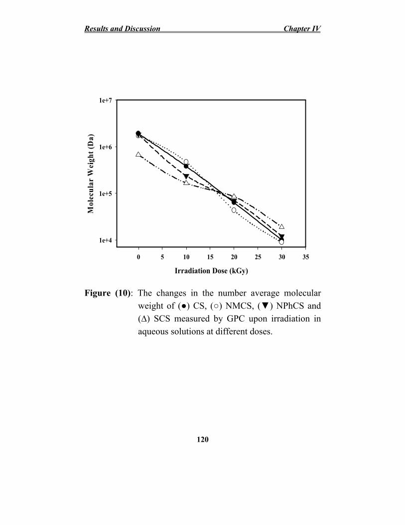
Results and Discussion Chapter IV
Mol
ecul
ar W
eigh
t (D
a)
1e+7
1e+6
1e+5
1e+4
0 5 10 15 20 25 30 35
Irradiation Dose (kGy)
Figure (10): The changes in the number average molecular weight of (●) CS, (○) NMCS, (▼) NPhCS and (∆) SCS measured by GPC upon irradiation in aqueous solutions at different doses.
120

Chapter IV Results and Discussion Figure (11) shows the changes in the number average
molecular weights of chitosan and its derivatives treated by 10% H2O2 (v/w) and irradiated by γ-rays at different doses in the solid form. Hydrogen peroxide as chemical initiator when used in small concentration causes breaking of 1,4-β-D-glucoside bonds and rapid decrease in the molecular weight Tian et al., (2004); El-Sawy et al., (2010) and Abd El-Rehim et al., (2011). It was observed that the addition of initiators to the polysaccharides during the irradiation process accelerates the degradation process. Meanwhile, with increasing the irradiation dose, the decrease in the number average molecular weight of chitosan and its derivatives as well as the increase in the rate of degradation process were observed. Using the 25kGy in presence of H2O2 the number average molecular weights of CS, NMCS, NPhCS and SCS decreased from 1.9x10⁶, 1.7x10⁶, 1.73x10⁶ and 6.7x10⁵ Da to the molecular weights of 7.8x10⁵, 7.3x10⁵, 7.7x10⁵ and 1.53x10⁵ Da, respectively. Meanwhile, with increasing the irradiation dose to 100 kGy, the number average molecular weights of CS, NMCS, NPhCS and SCS became 9.7x10⁴, 4.93x10⁴, 6.95x10⁴
and 4.17x10⁴ Da, respectively.
It is clear that the degradation rate of chitosan derivatives irradiated in solution is much higher than that irradiated in paste like form. The irradiation dose required for degradation of chitosan derivatives is low if compared with that in paste like form. The water radiolysis products like •OH radicals have a great effect on positive degradation of chitosan derivatives.
121

Results and Discussion Chapter IV
M o l
ecu l
a r W
eig h
t (D
a)
1e+7
1e+6
1e+5
1e+4 0 25 50 75 100
Irradiation Dose (kGy)
Figure (11): The changes in the number average molecular weight measured by GPC of (●) Cs, (○) NMCS, (▼) NPhCS and (∆) SCS treated by 10% H2O2
(v/w) and exposure to gamma irradiation in solid form as paste.
122

Chapter IV Results and Discussion
IV.I.5.5. Structural Changes of Irradiated Chitosan Derivatives
IV.I.5.5.1. UV-Vis Spectroscopy
The UV-Vis spectra of irradiated and unirradiated NMCS, NPhCS and SCS in the solution form at different doses of 10, 20 and 30 kGy Figure (12) and in the solid form treated by 10% H2O2 and irradiated as paste at 25, 50, 75 and 100 kGy Figure (13). Spectra of the NMCS, NPhCS and their irradiated solutions show absorption band at 275-280 nm which may be due to formation of unsaturated bond. Also, there is a new absorption peak appeared for the irradiated chitosan derivatives.
For the irradiated NMCS, the peak at 350 nm is due to –COCH=CHCOOH group. For NPhCS absorption peak appeared at 420 nm due to the benzene rings of NPhCS. The intensity of such peaks increased with increasing the irradiation dose due to resonance effect of the aromatic groups. Irradiated SCS spectra showed two absorption peaks at 250 and 290-295 nm. These peaks are due to some formation of unsaturated bonds. The intensity of the peaks increased with increasing the irradiation dose.
For the irradiated chitosan a new absorption band was evident around 280–315 nm which may be caused by the n→σ*
transition for the amino groups of chitosan and may also
123

Results and Discussion Chapter IV assigned to the n→л* transition for the carbonyl or carboxylic groups. The peak intensity increased with increasing the irradiation dose. This peak can be ascribed to carbonyl or hydroxyl groups formed after the main chain scission of chitosan and hydrogen abstraction reaction Ulanski and Rosiak (1992).
The intensity of UV absorbency of chitosan derivatives in the solution was higher than that of chitosan irradiated in paste like form. This is can be attributed to large amount of unsaturated functional groups produced by radiation in the presence of water. The irradiation of the chitosan derivatives in the water increases their degradation rate and enhances their functionality.
124

Chapter IV Results and Discussion
Figure (12): UV-Vis spectra of CS, NMCS, NPhCS and SCS in the solution form; unirradiated (—) and the irradiated at dose of 10 kGy (….), 20 kGy (---) and 30 kGy (-..-..-).
125

Results and Discussion Chapter IV
0
1
2
3
4
5 CS
Abs
orba
nce
0.0
0.1
0.2
0.3
0.4
0.5
Abs
orba
nce
NMCS
250 300 350 400 450 500 250 300 350 400 450 500
Wavelength (nm) Wavelength (nm)
0
1
2
3
4
5
0.0
0.2
0.4
0.6
0.8
1.0
1.2
1.4
)
Abs
orba
nce
Abs
orba
nce
NPhCS SCS
Wavelength (nm340 360 380 400 420 440 460 480 500
Abs
orba
nce
0.00
0.05
0.10
0.15
0.20
0.25
0.30
250 300 350 400 450 500 250 300 350 400 450 500 Wavelength (nm) Wavelength (nm)
Figure (13): UV-Vis spectra of CS, NMCS, NPhCS and SCS treated by 10% H2O2 (v/w), (—) unirradiated polymer and the irradiated polymer in solid form at different doses of 25 kGy (…..), 50 kGy (---), 75 kGy (-..-..) and 100 kGy (– – –).
126

Chapter IV Results and Discussion
IV.I.5.5.1.2. FT-IR Spectroscopy
FT-IR spectroscopy has been shown to be a powerful tool for the study of the physicochemical properties of polysaccharides. Figure (14) shows the FT-IR spectra of unirradiated and irradiated CS, NMCS, NPhCS and SCS in the solution form at different doses of 10, 20 and 30 kGy.
The FT-IR spectra of NMCS in different degradation conditions are shown in Figure (14B, curve a). The strong broad bands in spectrum at around 3400 cm−1 is assigned to the hydrogen-bonded –OH and –NH bands. The bands at 2935 cm−1 and 1598 cm−1 correspond to C–H stretching vibration and N–H bend vibration, respectively Chen and Park (2003). The band at 1380 cm−1 which results from the coupling of C–N axial stretching and N–H angular deformation increases as irradiation dose increases Britto and Campana-Filho (2004). Furthermore bands at 1071 cm−1 (C–O of – COH–) and 886 cm−1 (vibration of ring) were confirmed in spectra. The carboxyl groups (–COOH) of NMCS bands at 1717 cm−1 were noticed and its intensity increases as the irradiation dose increases. Figure (14B, curves b, c and d). The band assigned to the (–CONH–) at 1630 cm−1 appeared and its absorbance intensity increases as the irradiation dose increases Tian et al., (2004). This means that the radiation aids to oxidize the NMCS rather than open the glycoside ring. The same results were obtained for NPhCS and SCS.
127

Results and Discussion Chapter IV
Figure (14): FT-IR spectra of (A) CS, (B) NMCS, (C) NPhCS and (D) SCS; (a) unirradiated and the irradiated in aqueous solutions at different doses of (b) 10 kGy, (c) 20 kGy and (d) 30 kGy.
128

Chapter IV Results and Discussion Figure (15) shows the FTIR spectra of CS, NMCS,
NPhCS and SCS and that irradiated in presence of 10% H2O2
in the solid form as paste at 50 and 100 kGy.
For the irradiated chitosan spectrum Figure (15A, curve b and c), it was observed that as the irradiation dose increases, the intensity of the absorption peak at 1602 cm-1 assigned to the N–H bend vibration of –NH2 increases and shift to low wavenumber at 1598 cm-1. This may be due to the decreasing of inter or intra- molecular hydrogen bonding between the –OH and –NH2 groups. Additionally, another characteristic peak at 1638 cm-1, which could be disturbed by carbonyl groups of chitosan, was gradually shifted to 1651 cm-1, while its intensity increased with an increase in the γ-ray irradiation dose. Also, the intensity of the peak at 1385 cm-1 assigned to C–O stretch vibration increased and moved toward the higher wavenumber as the irradiation dose increases. The vibrational band at 1100 cm-1 that corresponds to the ether bond in the pyranose ring has no significant change, which indicates that, the stability of the β-glycosidic bonds and distribution of glycosidic bonds in the molecular chains of chitosan. In general the changes in spectra confirmed that carbonyl or carboxyl groups were formed, partial amino groups were eliminated and/ or slightly some ring opening occurred during the radiation degradation process of chitosan.
FTIR spectrum of irradiated NMCS Figure (15B curve b and c) in different degradation conditions is the same results as in Figure (14B). For irradiated NPhCS spectrum Figure (15C, curves b and c), the absorption bands at 2941 and 3074 cm-1 are
129

Results and Discussion Chapter IV due to C-H stretch of the -CH=CH- groups and C-H stretch of aromatic system, respectively. Also, the peaks at 1712, 1775 and 1548 cm-1 are characteristic for the carbonyl group (C=O) of amide bond and still present after irradiation. In the spectrum of the irradiated SCS Figure (15D, curves b and c), the absorption bands appearing at 817 cm-1 and 1235 cm-1
which correspond to the C-O-S and S=O bond stretching, respectively were present. The results indicated that the important functional groups were still present after irradiation and that the main polysaccharide chain structure remained during the degradation process.
From the all FT-IR spectra of Figure (14 and 15), it can be concluded that the radiation degrades the chitosan derivatives to lower molecular weights. The radiation degradation of chitosan and its derivatives lead to carbonyl groups formation. Furthermore, the profiles of chitosan derivatives are similar so that it can be concluded that the main polysaccharide chain structure and substituting groups introduced to chitosan were almost remained during degradation process. As mentioned above, the degradation rate of chitosan derivatives in the solution form is higher than that in the paste like form. The amount of functional groups resulted from radiation oxidation reaction of chitosan derivatives is high. Consequently, the peak intensity of such functional groups is higher than that of chitosan derivatives in the paste like form.
130

Chapter IV Results and Discussion
%
T r a n s m i t t a n c e
%
T r a n s m i t t a n c e
(A) a
b
c
(B)
a
b
c
4000 3600 3200 2800 2400 2000 1600 1200 800 400 4000 3500 3000 2500 2000 1500 1000 500
Wavenumbers Wavenumbers
%
T r a n s m i t t a n c e
%
T r a n s m i t t a n c e
a
c
c
a
b b
(C) (D)
4000 3600 3200 2800 2400 2000 1600 1200 800 400 4000 3600 3200 2800 2400 2000 1600 1200 800 400
Wavenumbers Wavenumbers
Figure (15): FT-IR spectra of CS, NMCS, NPhCS and SCS treated by 10% H2O2 (v/w) (a) unirradiated polymers, and irradiated ones in solid form at 50 kGy (b) and 100 kGy (c).
131

Results and Discussion Chapter IV
IV.I.5.6. Evaluation of Antioxidant Activity of Chitosan Derivatives
IV.I.5.6.1. Determination of Scavenging Effect on DPPH Radicals Figure (16) shows the scavenging effect (%) on DPPH radicals of unirradiated and irradiated CS, NMCS, NPhCS and SCS in the aqueous solutions. In general, the DPPH radical scavenging effect (%) increased as the concentration of the chitosan and its derivatives increased to a certain extent about 0.5 mg/ml and then it leveled off with further increase in the concentration. Also, the irradiated chitosan and its derivatives showed increasing the scavenging ability on DPPH radicals if compared with unirradiated ones. As the molecular weight of chitosan and its derivatives decreases, the DPPH scavenging effect (%) increases.
The scavenging effect (%) on DPPH of chitosan and its derivatives follows the order NMCS > NPhCS > SCS > CS. The DPPH scavenging effect (%) of the unirradiated CS, NMCS, NPhCS and SCS, at a concentration of 0.5 mg/ml was 4.95, 28.97, 28.9 and 11.26 (%), respectively. The scavenging effect (%) became 18, 79.2, 39.3 and 35.2 (%), respectively when chitosan and its derivatives exposed to γ-rays at 10 kGy. Meanwhile at 30 kGy, the scavenging effect (%) of CS, NMCS, NPhCS and SCS reaches to 60.2, 82.7, 59.8 and 55.4 (%), respectively.
132
Chapter IV Results and Discussion Figure (17) shows the scavenging effect (%) on DPPH
radicals of CS, NMCS, NPhCS and SCS treated by 10% H2O2
(v/w) in solid form. It was found from Figure (17A) that, the scavenging effect (%) increased with increasing the irradiation dose due to the decrease in the molecular weights of polysaccharide. The NMCS was the highest scavenging effect (%) and the order of the scavenging effect is as followed NMCS > NPhCS > SCS > CS.
At 25 kGy, the scavenging effect (%) on DPPH radicals of CS, NMCS, NPhCS and SCS treated by 10% H2O2 increased to 34.2, 72.67, 60.89 and 57.98 (%), respectively. Meanwhile, the scavenging effect (%) on DPPH radicals of unirradiated ones was 7.9, 32.9, 31.1 and 11.8 (%), respectively. Also, with increasing the irradiation dose up to 50 kGy, the DPPH scavenging effect (%) increased to 38.8, 77.8, 65.9 and 60.4 (%), respectively then leveled off even with further increase in the dose. Figure (17B) showed that the scavenging effect (%) on DPPH radicals increased with increasing the concentration of the irradiated chitosan and its derivatives to 1mg/ml and then leveled off even with further increase in the concentration.
The IC50 (%) on DPPH radicals for CS, NMCS, NPhCS and SCS irradiated in solution form at 30 kGy was 0.184, 0.047, 0.182 and 0.224 for SCS mg/ml, respectively. Meanwhile, the IC50 (%) on DPPH radicals for CS, NMCS, NPhCS and SCS irradiated at 50 kGy in solid form as paste was 0.148, 0.403 and 0.538 (mg/ml) for NMCS, NPhCS and SCS, respectively. The (IC50) of ascorbic acid as antioxidant material (reference) was 0.144 (mg/ml). These results revealed
133

Results and Discussion Chapter IV that irradiated NMCS has a good antioxidant activity if compared with ascorbic acid.
It was reported by Sun et al., (2008) that IC50 of N-carboxymethyl chitosan oligosaccharides (Mwt 5kDa) with degree of substitution 0.54 on DPPH radicals was 0.71 mg/ml. Meanwhile, in the present study, the IC50 (%) on DPPH radicals of chitosan treated with maleic anhydride (NMCS) of Mwt 43 kDa and degree of substitution 0.57 was 0.047 mg/ml.
The N-alkylated disaccharide chitosan derivatives with different degrees of substitution (DS) of 20–30% exhibited the highest DPPH radicals scavenging abilities of 80–95% at 0.1 mg/ml, followed by the derivatives with DS of 40–50% and 60-70%. Apparently, the scavenging ability of chitosan might be reduced after sulfonation or might be enhanced after N-alkylation of disaccharide Lin and Chou (2004). These results reveled that irradiated NMCS has a good antioxidant activity if compared with ascorbic acid.
134

20
30
40
50
60
70
80
90
100
20
30
40
50
60
70
80
90
Chapter IV Results and Discussion
CS 100
Scav
engi
ng E
ffec
t (%
)
NMCS90
Scav
engi
ng E
ffec
t (%
) 80
70
60
50
40
30
20
10 10
0 0 0.0 0.5 1.0 1.5 2.0 0.0 0.5 1.0 1.5 2.0
Concentration (mg/ml) Concentration (mg/ml) 100 100
Scav
engi
ng E
ffec
t (%
) NPhCS SCS90
80
Scav
engi
ng E
ffec
t (%
)
70
60
50
40
30
20
10 10
0 0 0.0 0.5 1.0 1.5 2.0 0.0 0.5 1.0 1.5 2.0
Concentration (mg/ml) Concentration (mg/ml) Figure (16): Scavenging effect (%) on DPPH radicals of CS,
NMCS, NPhCS and SCS irradiated in the solution form at different doses of (●) 0 kGy, (○) 10 kGy, (▼) 20 kGy and (■) 30 kGy. (x) Ascorbic acid.
135

Results and Discussion Chapter IV
90
80
70
60
50
40
30
Scav
engi
ng E
ffec
t (%
)Sc
aven
ging
Eff
ect (
%)
20
10
0
(A)
0 25 50 75 100
Irradiation Dose (kGy)
90
80
70
60
50
40
30
20
10
0
(B)
0.0 0.5 1.0 1.5 2.0 Concentration (mg/ml)
Figure (17): The scavenging effect (%) on DPPH radicals of (●) CS, (○) NMCS, (▼) NPhCS and (∆) SCS treated in solid form by 10% H2O2 (v/w). (A) irradiation at different doses using 1mg/ml concentration and (B) different concentration at 50 kGy irradiation dose. (x) Ascorbic acid.
136

Chapter IV Results and Discussion
IV.I.5.6.2. Determination of Reducing Power
The reducing power of different molecular weights of γ-ray irradiated NMCS, NPhCS and SCS in the solution form with different concentrations as shown in Figure (18). The reducing power of irradiated chitosan and its derivatives increases with increasing the irradiation dose as well as their concentrations. This is due to the decrease in the molecular weight of chitosan and its derivatives. At 0.5 mg/ml concentration, At 10 kGy and 0.5 mg/ml concentration, the reducing power for CS, NMCS, NPhCS and SCS was 0.69, 0.867, 0.78, and 0.68, respectively. Meanwhile, the reducing power of unirradiated CS, NMCS, NPhCS and SCS was 0.27, 0.60, 0.60 and 0.40, respectively. Also, at 30 kGy, the reducing power was 0.98, 1.47, 1.22 and 1.18, respectively. The value of reducing power of ascorbic acid was 1.39 which is closed to those values of 30 kGy irradiated chitosan derivatives. The increase in reducing power upon irradiated chitosan and its derivatives indicates the enhancement of their antioxidant activity property and suggests the possibility of using the γ-ray irradiated chitosan and its derivatives as antioxidants that prevent flavor changes caused by lipid peroxidation.
Figure (19) shows the reducing power of CS, NMCS, NPhCS and SCS treated by 10% H2O2 (v/w). It was observed from Figure (19A) that the reducing power of chitosan and their derivatives increased with increasing the irradiation dose till 50 kGy then leveled off with further increase in the irradiation dose. Also, the NMCS was the highest reducing
137

Results and Discussion Chapter IV power. Fig (19B) shows that the reducing power of γ-ray (50kGy) treated chitosan and its derivatives increased with increasing the polysacharide concentration.
The reducing power of CS, NMCS, NPhCS and SCS irradiated in solution form at 30 kGy and 0.5 mg/ml concentration was 0.98, 1.47, 1.22 and 1.18, respectively. Meanwhile, the reducing power of CS, NMCS, NPhCS and SCS irradiated in solid form at 50 kGy and 1mg/ml concentration was 0.7363, 1.0590, 0.9729 and 0.8797, respectively.
N-maleoyl chitosan oligosaccharide of Mwt 7310 Da and substituting degree of 0.49 was synthesized by Sun et al., (2011), the reducing power of such oligosaccharides at concentration of 2.4 mg/ml was at the absorbance of 0.46. While in the present study the prepared NMCS of Mwt 8900 Da and degree of substitution of 0.57 showed improvement in reducing power at the absorbance of I.47 at low concentration (0.5mg/ml).
138

Chapter IV Results and Discussion
CS 1.8 1.8
NMCS 1.6 1.6
Abs
orba
nce
at 7
00 n
m
Abs
orba
nce
at 7
00 n
m
1.4
1.2
1.0
0.8
0.6
0.4
Abs
orba
nce
at 7
00 n
m
Abs
orba
nce
at 7
00 n
m
1.4
1.2
1.0
0.8
0.6
0.4 0.2 0.2 0.0 0.0
0.0 0.5 1.0 1.5 2.0 0.0 0.5 1.0 1.5 2.0 Concentration (mg/ml) Concentration (mg/ml)
1.8 1.8
1.6 NPhCS
1.6
0.2 0.2
0.0 0.0
SCS
0.0 0.5 1.0 1.5 2.0 0.0 0.5 1.0 1.5 2.0
Concentration (mg/ml) Concentration (mg/ml)
Figure (18): Reducing power of CS, NMCS, NPhCS and SCS irradiated in the solution form at different doses of (●) 0 kGy, (○) 10 kGy, (▼) 20 kGy and (■) 30 kGy. (x) Ascorbic acid.
139
1.4 1.4
1.2 1.2
1.0 1.0
0.8 0.8
0.6 0.6
0.4 0.4

Results and Discussion Chapter IV
1.4
1.2
1.0
0.8
0.6
0.4 A
bsor
banc
e at
700
nm
Abs
orba
nce
at 7
00 n
m
0.2
0.0
(A)
0 25 50 75 100 Irradiation Dose (kGy)
1.6
1.4
1.2
1.0
0.8
0.6
0.4
0.2
0.0
(B)
0.0 0.5 1.0 1.5 2.0
Concentration (mg/ml)
Figure (19): Reducing power of (●) CS, (○) NMCS, (▼) NPhCS and (∆) SCS treated by 10% H2O2 (v/w). (A) irradiation at different doses using 1(mg/ml) concentration and (B) different concentration at 50 kGy irradiation dose. (x) Ascorbic acid.
140

Chapter IV Results and Discussion
IV.I.5.6.3. Determination of Chelating Effects on Ferrous Ions
The ferrous ion-chelating effect of different molecular weights of γ-ray irradiated NMCS, NPhCS and SCS in the solution form with different concentrations was shown in Figure (20). It was found that the chelating ability (%) of chitosan and its derivatives increased with increasing their concentration. Also, with decreasing the molecular weight of chitosan and its derivatives, the chelating ability increased. It is clear that NMCS possesses the highest chelating capacity for Fe2+ ions. The chelating ability of unirradiated CS, NMCS, NPhCS and SCS at 1 mg/ml was 40, 49.9, 45 and 40.2 (%), respectively, while the value of chelating ability at 10kGy of irradiation and 1mg/ml concentration was 53.6, 82.1, 77.7 and 59.4 (%), respectively. With increasing the irradiation dose to 30 kGy, the chelating ability value became 77.5, 89.8, 89.3 and 79.5, respectively. Meanwhile, the value of chelating ability of the EDTA (as a reference) at 1 mg/ml was 66.4%.
Generally metal chelating capacity was significant since it reduced the concentration of the catalyzing transition metal. There are intra-molecular hydrogen bonds, van der Waals and the intermolecular attraction forces in the molecules of chitosan and its derivatives which affect their reactivity towards iron ions. After chemical and radiation modification, some of the intra-molecular hydrogen bonds of chitosan are destroyed and the reactivity is improved. The degree of destruction of these bonds may be affected by the size and type of the groups introduced to chitosan. Also, factors such as affinity for water
141

Results and Discussion Chapter IV and crystallinity were changed and may affect the ion-chelating activity of chitosan derivatives. On the other hand, the free functional groups of low-molecular weight chitosan and its derivatives as NH2, –COOH, –CONH– and –SO3H help to form chitosan-Fe2+ complexes Guzman et al., (2003); Duh et al., (1999) and Xing et al., (2005).
Figure (21) shows the chelating effects of CS, NMCS, NPhCS and SCS treated by 10% H2O2 (v/w). The chelating ability (%) of chitosan and their derivatives enhanced and increased with increasing the irradiation dose (Figure 21A). Also, the chelating effects (%) increased with increasing the concentration of the irradiated chitosan and their derivatives to 1mg/ml and then leveled off even with further increase in the concentration. The chelating effect (%) of unirradiated CS, NMCS, NPhCS and SCS treated by 10% H2O2 was 40.1, 49.9, 45 and 42.8 (%), respectively. At 25kGy, the chelating effect (%) of CS, NMCS, NPhCS and SCS increased to 42.6, 73.6, 70.9 and 68.1 (%), respectively, meanwhile at 50kGy, the chelating effect (%) was 50.1, 81.1, 79.1 and 73.7 (%), respectively and tend to increase to the maximum chelation at 100kGy of 57.2, 88.3, 84.2 and 78.6 (%), respectively. The high ferrous ion chelating abilities of chitosan derivatives would be beneficial if they were formulated into foods.
The IC50 (%) of the chelating ability (%) was 0.299 mg/ ml for CS irradiated in solution form at 20 kGy, 0.098 mg/ml for NMCS irradiated in solution form at 10 kGy, 0.126 mg/ml for NPhCS irradiated in solution form at 20 kGy and 0.098 mg/ml for SCS irradiated in solution form at 30 kGy. While, the IC50
142

Chapter IV Results and Discussion of chelating effect (%) for NMCS, NPhCS and SCS irradiated in solid form at 50 kGy, was 0.09, 0.103 and 0.221 (mg/ml), respectively.
The ferrous ion-chelating ability of chitosan oligosaccharides (Mwt 4.7kDa) is 57.6% at the concentration of 12.8 mg/ml was reported Wang et al., (2007), while in the present study, the chelating ability value of irradiated chitosan (Mwt 10 kDa) was 77.5% at 1mg/ml concentration. This means that the treatment of chitosan by radiation enhance its chelating ability by increasing the functional groups as a results of oxidation reaction. Moreover, the concentration of irradiated chitosan required for ferrous ion chelating ability reduced more than 12 times.
143

Results and Discussion Chapter IV
100
CS100
Che
latin
g E
ffec
t (%
)
NMCS90 90
80 C
hela
ting
Eff
ect (
%) 80
70 70 60 60 50 50 40 40 30 30 20 20 10 10
0 0 0.0 0.5 1.0 1.5 2.0 0.0 0.5 1.0 1.5 2.0
Concentration (mg/ml) Concentration (mg/ml)
100 C
hela
ting
Eff
ect (
%)
100 NPhCS SCS 90
90
Che
latin
g E
ffec
t (%
) 80 80 70 70 60 60 50 50 40 40 30 30 20 20
10 10
0 0 0.0 0.5 1.0 1.5 2.0 0.0 0.5 1.0 1.5 2.0
Concentration (mg/ml) Concentration (mg/ml)
Figure (20): Chelating effect (%) of CS, NMCS, NPhCS and SCS irradiated in the aqueous solution at different doses of (●) 0 kGy, (○) 10 kGy, (▼) 20 kGy and (■) 30 kGy. (x) EDTA.
144

Chapter IV Results and Discussion
100
90
80
70
60
50
40
30
Che
latin
g E
ffec
t (%
)C
hela
ting
Eff
ect (
%)
20
10
0
(A)
0 25 50 75 100
Irradiation Dose (kGy)
100
90
80
70
60
50
40
30
20
10
0
(B)
0.0 0.5 1.0 1.5 2.0
Concentration (mg/ml)
Figure (21): Chelating effect (%) of (●) CS, (○) NMCS, (▼) NPhCS and (∆) SCS treated by 10% H2O2 (v/w). (A) irradiation at different doses using 1(mg/ml) concentration and (B) different concentration at 50 kGy irradiation dose. (x) Ascorbic acid.
145

Results and Discussion Chapter IV
IV.I.5.6.4. Determination of Antioxidant Activity
The total antioxidant values of irradiated CS, NMCS, NPhCS and SCS in the solution form with different concentrations using linoleic acid, as unsaturated fatty acid are shown in Figure (22). The antioxidant activity (%) increased as the concentration of the chitosan and its derivatives increased Also, with increasing the irradiation dose, the antioxidant activity (%) increased and follow the order NMCS > NPhCS > SCS > CS. Comparison study between the chitosan derivatives and ascorbic acid as antioxidants was performed. It was found that the antioxidant activity of 30kGy irradiated chitosan and its derivatives closed to that of ascorbic acid. Introducing new functional groups such as –COOH, –CONH– and –SO3H into chitosan chains, improves its water solubility and enhance chitosan’s antioxidant activity. In addition, ionizing radiation was used to break the interaction between the chitosan molecules and decreases its molecular weight.
Figure (23) shows the antioxidant activity of CS, NMCS, NPhCS and SCS treated by 10% H2O2 (v/w). The antioxidant activity (%) increased as the concentration of the chitosan and its derivatives increased. Also, with increasing the irradiation dose, the antioxidant activity (%) increased.
The antioxidant activity of 2 mg/ml of unirradiated CS, NMCS, NPhCS and SCS was 4.7, 16.1, 12.2 and 11.1 (%), respectively. While, the antioxidant activity irradiated at 30 kGy in solution form was 33.3744, 44.8936, 40.0526 and
146

Chapter IV Results and Discussion 39.4405 (%), respectively. Moreover, the antioxidant activity of CS, NMCS, NPhCS and SCS irradiated at 50 kGy in solid form was 23.2527, 38.4627, 35.7682 and 31.2828 (%), respectively.
The properties of substituting groups may affect the antioxidant activity of chitosan and its derivatives in such way: firstly, the substitution will reduce the amount of active amino and hydroxyl groups in the polymer chains; secondly, the substitution may partly destroy the inter-molecular and intramolecular hydrogen bonds. The charge properties of substituting groups may affect the antioxidant activity of chitosan and its derivatives Guo et al., (2006 and 2008). The property of substituting groups might affect the activity of amino and hydroxyl groups therefore lead to the change of antioxidant activity.
The four methods for determining the antioxidant activity of irradiated chitosan and its derivatives reveled that chitosan derivatives with different molecular weights exhibit antioxidant activity and NMCS possessed the highest scavenging effect on DPPH radicals, reducing power, ferrous ion chelating activity assays and radical mediated lipid peroxidation inhibition, if compared with CS, NPhCS, and SCS.
147

Results and Discussion Chapter IV
ii
)
0
10
20
30
40
50 CS
ii
(%)
0
10
20
30
40
50
Ant
iox
dant
Act
ivty
(%
Ant
oxid
ant A
ctvi
ty
NMCS
0.0 0.5 1.0 1.5 2.0 0.0 0.5 1.0 1.5 2.0
Concentration (mg/ml) Concentration (mg/ml)
50
NPhCS
50
Ant
ioxi
dant
Act
ivity
(%) SCS
Ant
ioxi
dant
Act
ivity
(%)
40 40
3030
2020
1010
0 0
0.0 0.5 1.0 1.5 2.0 0.0 0.5 1.0 1.5 2.0
Concentration (mg/ml) Concentration (mg/ml)
Figure (22): Antioxidant activity of NMCS, NPhCS and SCS irradiated in the solution form at different doses of (●) 0 kGy, (○) 10 kGy, (▼) 20 kGy and (■) 30 kGy. (x) Ascorbic acid.
148

Chapter IV Results and Discussion
45
40
35
30
25
20
15
10A
ntio
xida
nt A
ctiv
ity (%
) A
ntio
xida
nt A
ctiv
ity (%
) 5
0 0 25 50 75 100
(A)
Irradiation Dose (kGy)
45
40
35
30
25
20
15
10
5
0
(B)
0.0 0.5 1.0 1.5 2.0
Concentration (mg/ml)
Figure (23): Antioxidant Activity (%) of (●) CS, (○) NMCS, (▼) NPhCS and (∆) SCS treated by 10% H2O2
(v/w). (A) irradiation at different doses using 1(mg/ml) concentration and (B) different concentration at 50 kGy irradiation dose. (x) Ascorbic acid.
149

Results and Discussion Chapter IV
IV.I.6. γ-Irradiated Chitosan and Its Derivatives as Antioxidants for Minced Chicken
The concentration of malonaldehyde (MDA) formed during 3 and 10 days of refrigerated storage (4±1oC) was represented in Table (1) via the absorbance measured using spectrophotometer. It was clear that there was a dramatic increase in the MDA in control samples during storage (from 0.587 at 0-time to 1.234 mg MDA/ Kg after 10 days).
Effectiveness of irradiated CS and NMCS treatment in reducing the lipid per-oxidation in minced chicken was studied resulting in a highly significant decrease in the MDA concentration compared with the control. The MDA concentration in samples containing 100 kGy irradiated NMCS was (0.262 and 0.454 mg MDA/Kg after 3 and samples containing 100 kGy irradiated CS was (0.333 and 0.550 mg MDA/ Kg after 3 and 10 days of storage. Meanwhile, the MDA concentration in the control samples was (0.835 and 1.234) mg MDA/Kg after 3 and 10 days of storage, respectively. This means that the addition of 100 kGy irradiated CS and NMCS at 0.1 (wt%) concentration to minced chicken resulted in a highly significant decrease in malonaldehyde (MDA) content, (about 50 and 70-80%, respectively), after 10 days storage at 4±1oC compared with the control, suggesting that such natural antioxidants may be an alternative for improving the oxidative deterioration of minced chicken during refrigerated storage.
150

Chapter IV Results and Discussion This finding was similar to that reported by Darmadji and
Izumimoto (1994) that the TBA value (expressed as mg MDA/ Kg) of beef increased sharply in control samples after 10 days of storage at 4oC as compared with those containing chitosan 1%. In beef patties, Dzudie et al., (2004) reported that the increase in TBA values was attributed to the accumulation and formation of MDA throughout storage either chemically or microbiologically which indicated oxidative deterioration of lipid into the end products TBARS during storage. Georgantelis et al. (2007) reported that the MDA contents of the beef burger control reached a maximum value of 4814.7 µg/ kg on the 180th day of frozen storage, while samples containing chitosan did not exceed the threshold values until the end of the storage time.
IV.I.7. Toxicity of NMCS Figure (24A) represents the percentage of fungal inhibition
zone during 144h as calculated from the fungal radial growth around the media containing NMCS. The result showed that in the first day, the initial fungal inhibition zone reached to 42%. The inhibition zone decreases and disappeared after 144h. The decrease in inhibition zone reflects an adaptation of the fungus to the polymer. Figure (24B) showed the effect of NMCS on the proliferation of Caco-2 cells after 48h of incubation. It was found that the treatment of Caco-2 cells with NMCS did not show strong proliferative effect at small concentrations or cytotoxic effects at higher ones regarding and their IC50 was 253.7 and 643.5 µg/ml, respectively.
151

Results and Discussion Chapter IV
Table (2): Thiobarbituric Acid Reactive Substances (TBARS) (mg MDA/ kg sample) in minced chicken during 10 days of refrigerated storage (4± 1oC).
Groups Storage (days) 0-time 3 days 10 days
Control 0.587± 0.082
0.835± 0.084 A 1.234± 0.138 A
CS 0 kGy 0.460± 0.076 B 0.705± 0.056 BC CS 50 kGy 0.359± 0.072 BC 0.847± 0.035 B CS 100 kGy 0.333± 0.040 BC 0.550± 0.033 CD
NMCS 0 kGy
0.420± 0.039 BC 0.606± 0.03 CD
NMCS 50 kGy
0.376± 0.039 BC 0.511± 0.011 D
NMCS 100 kGy
0.262±0.031C 0.454± 0.031 D
Means in the same column for the same storage day with a different letter differ significantly (p<0.05)
152

Chapter IV Results and Discussion
G r o
w th
In hi
bitio
n (%
)
100
80 (A)
60
40
20
0 0 24 48 72 96 120 144 168
Time (h)
(B)
Figure (24): (A) Effect of NMCS on percentage of inhibition
radial growth of mycelial and (B) Cytotoxic effect of different concentration of NMCS using MTT assay
153

Results and Discussion Chapter IV
IV.II. Radiation Induced Crosslinking of PAAm Incorporated Low Molecular Weights Natural Polymers for Possible Use in the Agricultural Applications
There is a strong need for new plant growth media with increased water and nutrient holding capacity. Polymer hydrogels are a commonly available product for ensuring adequate hydration as they have the ability to absorb large quantities of water. Among these hydrogels polyacrylamide (PAAm) which is one of the most popular polymer, having also been used to reduce water runoff and increase infiltration rates in field agricultural applications. There are different methods to improve water holding capacity of polyacrylamide among them; adjusting the density of its crosslinking network structure. Radiation technique is promising for preparation of hydrogels because a polymer in aqueous solution or in the water swollen state readily undergoes crosslinking on irradiation to yield a gel-like material. Irradiation dose can control the crosslinking yield of polymer. Addition of few amount of natural degraded polymer during the irradiation processing of PAAm can also reduce the hydrogel network structural density and influences its water holding capacity. Moreover, the low molecular weight natural polymer like alginate or chitosan incorporated in PAAm hydrogel help in promoting the plant growth when such hydrogels used as water supplied soil conditioner.
In this study, the crosslinked copolymers hydrogels of γ-rays degraded Na-alginate or chitosan or both of them with
154

Chapter IV Results and Discussion PAAm were prepared using the ionizing radiation. The preparation conditions of the PAAm/Alg, PAAm/CS and PAAm/Alg/CS hydrogels, such as the effect of the irradiation dose and the copolymer composition on the matrix gel content and water absorbency will be studied.
IV.II.1. Effect of Copolymer Compositions and Irradiation Dose on Gel Content (%) of Prepared Hydrogels
Ionizing radiation can be used to obtain permanent hydrogels crosslinked by covalent chemical bonds. The crosslinking extent for the copolymer blends depends on the degree of compatibility as well as physical and chemical intermolecular interaction between the blend components. There are many factors affecting the copolymer crosslinking, including copolymer compositions and irradiation dose.
The effect of copolymer compositions and irradiation dose on gel content (%) of PAAm/Alg was investigated in Figure (25). For all exposure doses investigated here, the gel content reached its maximum value at copolymer composition of PAAm/Alg (95/5). Thereafter, any increase in Na-alginate content leads to a decrease in gel content. Also at the same PAAm/Alg composition, as the irradiation dose increases the gel content increases. The gel content of (PAAm/Alg; 95/5) was 76.7, 78.3, 88.3 and 93.7 (%) at irradiation dose of 5, 10, 20 and 30 kGy, respectively. Meanwhile, the gel content (%) of PAAm was 62, 70, 77.7 and 78.5 (%) at the same corresponding irradiation dose.
155

• •
Results and Discussion Chapter IV It is well known that polysaccharides, such as Na-alginate, undergo chain scission reaction by radiation through cleavage the β-(1-4) glycosidic bonds. Meanwhile, PAAm undergoes crosslinking when exposed to the radiation. Therefore, the presence of Na-alginate may affect the overall polymer crosslinking process and, consequently, influence the properties of the obtained copolymers hydrogel. At lower irradiation doses, the crosslinking reaction competes actively with the facile chain degradation reaction. The crosslinking reaction occurred due to the presence of hydroxyl radicals from water as a result of an indirect effect of radiation. These radicals subsequently abstract hydrogen atoms from the anhydro-glucopyranose rings, creating macro radicals on the polymeric (PAAm) backbone.
Figure (26) shows the effect of PAAm/CS compositions and irradiation dose on gel content (%) of prepared PAAm/CS copolymer. The gel content decrease as the irradiation dose and the amount of CS in the copolymer feed solution increases. But the gel content of PAAm/ CS hydrogel was lower than that of PAAm/Alg hydrogel at the same conditions. The gel content was 63.3, 68.4, 63.4 and 57.6 (%) at copolymer composition (PAAm/CS; 95/5) and irradiation dose of 5, 10, 20 and 30kGy, respectively. The presence of lactic acid in feed solution play
•important effect on the OH radicals produced from radiolysis of water. When an aqueous solution of PAAm is exposed to radiation, OH and H radicals and hydrated electrons are produced, as major part of the energy that is absorbed by the water solvent as shown in the following equation:
156

Chapter IV Results and Discussion •H2O + γ-rays → H• , OH, e¯aq, H2, H2O2, H+, ¯OH, H3O+
•OH radicals are mostly responsible for crosslinking of PAAm including polysaccharides Kumar et al., (2005). In the presence of lactic acid (used for chitosan solvation for PAAm/CS hydrogels) the medium of the feed solution became slightly acid (pH 4) and the appearance of less reactive
•oxidizing species O• instead of more reactive OH is expected due to the following reaction:
•OH + ¯OH O•¯ + H2O Therefore, the yield of gel content in PAAm hydrogel containing CS is reduced and became lower than that containing Na-alginate.
Figure (27) shows the effect of copolymer compositions and irradiation dose on gel content (%) of PAAm/Alg/CS. At low irradiation doses (5-20kGy), the same behavior was obtained when Alg/CS mixture was used instead of Na-alginate. By increasing the doses, the CS natural polymer tends to degrade and the crosslinking process rate is decreased. The same behavior was obtained as PAAm/Alg. The PAAm gel content in the presence of both Alg/CS is lower than that formed in the presence of Alg and higher than that formed in the presence of CS. The gel content was 67.5, 72, 77.3 and 74 (%) at copolymer composition (PAAm/Alg/CS; 95/2.5/2.5) and irradiation dose of 5, 10, 20 and 30 kGy, respectively. It can be concluded that the gel content (%) at the same condition of PAAm/Alg › PAAm/Alg/CS › PAAm/CS hydrogel as shown in Figure (28).
157

Results and Discussion Chapter IV
Gel
C o n
ten t
(%)
100
90
80
70
60
50
40
5 kGy 10 kGy 20 kGy 30 kGy
100/0 95/5 90/10 85/15 80/20 PAAm/Alg
Copolymer Composition (%)
Figure (25): Effect of copolymer compositions and irradiation dose on gel content (%) of PAAm/Alg hydrogel prepared with copolymer concentration (5wt%).
158

Chapter IV Results and Discussion
G el
C on
tent
(%)
100
90
80
70
60
50
40
5 kGy 10 kGy 20 kGy 30 kGy
100/0 95/5 90/10 85/15 80/20 PAAm/CS
Copolymer Composition (%)
Figure (26): Effect of copolymer compositions and irradiation dose on gel content (%) of PAAm/CS hydrogel prepared with copolymer concentration (5wt%).
159

Results and Discussion Chapter IV
G el
C o n
t e n t
(%)
100
90
80
70
60
50
40
5 kGy 10 kGy 20 kGy 30 kGy
100/0/0 95/2.5/2.5 90/5/5 85/7.5/7.5 80/10/10 PAAm/Alg/CS
Copolymer Compositions (%)
Figure (27): Effect of copolymer compositions and irradiation dose on gel content (%) of PAAm/Alg/CS hydrogel prepared with copolymer concentration (5wt%).
160

Chapter IV Results and Discussion
G el
C on
tent
(%)
80
75
70
65
60
55
50100/0 95/5 90/10 85/15 80/20 PAAm/
/PAAm/Alg PAAm CS PAAm/Alg/CS
Polysaccharide Copolymer Composition (%)
Figure (28): Effect of copolymer compositions and irradiation dose on gel content (%) of prepared hydrogels.
161

Results and Discussion Chapter IV
IV.II.2. Effect of Copolymer Compositions and Irradiation Dose on Water Absorbency of Prepared Hydrogels
The basic feature of super-absorbent hydrogel is its ability to absorb and hold large amounts of liquid hundreds of times greater than its own weight without dissolving in it. The swelling properties of the investigated crosslinked hydrogels are mainly related to the elasticity of the network and the presence of hydrophilic functional groups and/or the polarized charges.
The Effect of copolymer compositions and irradiation dose on water absorbency (g/g) of PAAm/Alg hydrogel prepared with copolymer concentration (5wt%) was shown in Figure (29). It was observed that the water absorbency of PAAm gel increases with irradiation dose up to 10 kGy, thereafter, any increase in irradiation dose leads to decreases in water absorbency. The water absorbency of PAAm/Alg hydrogel increases with increasing the content of Na-alginate in the copolymer compositions to reach the maximum value at copolymer composition of PAAm/Alg (95/5 wt/wt). Thereafter, any increase in Na-alginate content, the decrease in water absorbency was obtained. At copolymer composition (PAAm/Alg; 95/5) and irradiation dose of 5, 10, 20 and 30 kGy, the water absorbency was 449, 578, 350 and 326 (g/g), respectively compared with water absorbency (g/g) of PAAm hydrogel without blending with Na-alginate at the same doses which gave water absorbency 412, 390, 255 and 237 (g/g), respectively.
162

Chapter IV Results and Discussion
The maximum water absorbency of PAAm/Alg was observed at 10 kGy and copolymer composition of PAAm/Alg (95/5). The improvement of the copolymer water absorbency with the increase of the Na-alginate content can be mainly attributed to the ionic character of Na-alginate. The mixing of Na-alginate with PAAm enhances the swelling behavior of the latter, due to differences in the expansion of the copolymer network and its affinity magnitude for water which leads to an increase in water absorbency. On the other hand, the crosslinking of the copolymer prepared at low irradiation dose has a slight effect on the copolymer chain mobility; as a result, its affinity to swell in water is high. However, for those prepared at high irradiation dose, the dense crosslinking, which is generally found to occur in the amorphous region, retards the chain mobility of the copolymer. Also, the existence of crosslinking causes the copolymer chains to undergo a lower relaxation, which narrows the mesh size of the free volumes and, subsequently, prevents a greater number of penetrated water molecules to swell the polymer.
Figure (30) shows the effect of copolymer compositions and irradiation dose on water absorbency (g/g) of PAAm/CS hydrogel. It was observed that the water absorbency of PAAm/ CS hydrogel of different copolymer compositions decreases with increasing the irradiation dose. Also, the water absorbency decreases with increasing the content of chitosan. The water absorbency (g/g) of PAAm/CS hydrogel was lower than that of PAAm/Alg hydrogel at the same conditions. This indicates the degradation of chitosan under the formation of crosslinked
163

Results and Discussion Chapter IV hydrogel was higher than that of Na-alginate. The water absorbency (g/g) of PAAm/CS hydrogel was 371, 348, 278 and 268 (g/g), respectively with copolymer compositions (PAAm/chitosan; 95/5) and irradiation dose of 5, 10, 20 and 30kGy, respectively.
Figure (31) shows the effect of copolymer compositions and irradiation dose on water absorbency (g/g) of PAAm/Alg/CS. It was observed that the water absorbency (g/g) is enhanced by the presence of Na-alginate in PAAm/Alg/CS hydrogel rather than PAAm/CS. Also, the same behavior was obtained as PAAm/CS. The water absorbency (g/g) was 397, 378, 313 and 292 (g/g) at copolymer compositions (PAAm/Alg/CS; 95/2.5/2.5) and irradiation dose of 5, 10, 20 and 30 kGy, respectively. It can be concluded that the water absorbency (g/g) of PAAm/Alg › PAAm/Alg/CS › PAAm/CS hydrogel.
Figure (32) shows the photographs of dry and swollen PAAm and PAAm/Alg hydrogels. The lack of ionic character in the PAAm polymers structure greatly reduces their ability to absorb water. The water absorbency was found to increase by increasing the Na-alginate content in the copolymer.
.
164

Chapter IV Results and Discussion
W a t
er A
b so r
b en c
y (g
/g)
600
550
500
450
400
350
300
250
200
5 kGy 10 kGy 20 kGy 30 kGy
100/0 95/5 90/10 85/15 80/20 PAAm/Alg
Copolymer Compositions (%)
Figure (29): Effect of copolymer compositions and irradiation dose on water absorbency (g/g) of PAAm/Alg hydrogel prepared with copolymer concentration (5wt%).
165

Results and Discussion Chapter IV
Swel
ling
rat
io (g
/g)
450
400
350
300
250
200
150
100
50100/0 95/5 90/10 85/15 80/20 PAAm/CS
Copolymer Compositions (%)
5 kGy 10 kGy 20 kGy 30 kGy
Figure (30): Effect of copolymer compositions and irradiation dose on water absorbency (g/g) of PAAm/CS hydrogel prepared with copolymer concentration (5wt%).
166

Chapter IV Results and Discussion
Sw el
ling
ratio
(g/g
) 450
400
350
300
250
200
150
5 kGy 10 kGy 20 kGy 30 kGy
100/0 95/2.5/2.5 90/5/5 85/7.5/7.5 80/10/10 PAAm/Alg/ CS
Copolymer Compositions (%)
Figure (31): Effect of copolymer compositions and irradiation dose on water absorbency (g/g) of PAAm/Alg/CS hydrogel prepared with copolymer concentration (5wt%).
167

Results and Discussion Chapter IV
PAAm PAAm/Alg
Figure (32): photographs of dry and swollen hydrogels.
168

Chapter IV Results and Discussion
IV.II.3. Surface Morphology of Swollen Hydrogels
The pore structure at the surface of the prepared hydrogels was thought to be important for their fast swelling property. To investigate the effect of crosslinking density formed during irradiation process on the pore structure of the prepared copolymer hydrogels, the surface morphology of swollen hydrogel samples dehydrated by freeze dryer technique was examined by SEM as shown in Figure (33).
The micromorphology of the prepared hydrogel as well as its pore diameter mainly depends on those factors affecting the swelling properties of hydrogels such as the sample dehydration methods, the type of copolymer, and its crosslinking density. In other words, the copolymer compositions and the irradiation doses seriously affect the copolymer crosslinking degree and micromorphology of the polymer. It is clear that the crosslinking density formed during the irradiation process seriously affects the pore structure of the prepared superabsorbent hydrogel.
The morphology of freeze dried PAAm hydrogels and its copolymers Figure (33 a-d) showed highly porous of network structure where they haves a great affinity to swell in water. The morphology of PAAm and its copolymers showed a homogeneous, deeply high porous with honeycomb-like structure and seemed to indicate a higher accessibility of water to the amorphous regions of the copolymers. These results may be due to the differences in the expansion of the copolymer
169

Results and Discussion Chapter IV network and its affinity magnitude for water and high crosslinking density randomly aggregated and exhibited a granular structure of narrow pore size except the morphology of freeze dried PAAm/CS hydrogels Figure (33 c) showed a relatively wide pore structure due to the lower crosslinking density formed.
170
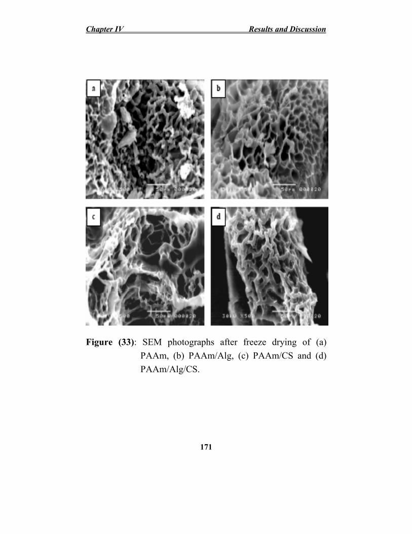
Chapter IV Results and Discussion
Figure (33): SEM photographs after freeze drying of (a) PAAm, (b) PAAm/Alg, (c) PAAm/CS and (d) PAAm/Alg/CS.
171

Results and Discussion Chapter IV
IV.II.4. Effect of Various Types of Cations on Water Absorbency of Prepared Hydrogels
It is important to investigate swelling behaviors of the prepared hydrogels in various salt solutions in order to expand of their applications especially for agriculture. The effect of different cations on water absorbency of the prepared hydrogels using mono-, di- and trivalent cations with the same anions at different ionic strength was investigated. The effect of ionic strength of various types of cation solutions on water absorbency (g/g) of PAAm/Alg, PAAm/CS and PAAm/Alg/CS hydrogels, respectively was shown in Figures (34, 35 and 36).
The absorbing ability of anionic hydrogels in various salt solutions is significantly decreased comparing to the absorbing values in distilled water. The presence of ions in the swelling medium has a profound effect on the swelling behavior of the hydrogels. In this work, the salts added, NaCl, CaCl2 and FeCl3, were found to exert a ‘salting out’ effect. The water absorbency decreases with increasing the ionic strength of cation solution. Also, under a given ionic strength, the water absorbency decreases with increasing ionic valence of the salt, resulting low absorbency in trivalent ions (Fe3+) and relatively high absorbency in monovalent ions (Na+) solutions.
At the concentration of 10-4 M for Fe3+, Ca2+ and Na+
cation solutions, the water absorbency of PAAm/Alg hydrogel reduces to about 63, 39 and 16 (%) of water capacity. In case of PAAm/CS, water absorbency reduces to about 52, 31 and 14
172

Chapter IV Results and Discussion (%) respectively. Meanwhile, PAAm/Alg/CS water absorbency reduces to about 59, 35 and 15 (%), respectively.
This sensitivity of salt has been attributed to the reduction of osmotic pressure difference between the gel network and the external solution Abd El-Rehim et al., (2006); Hoffman (2002); Tyliszczak et al., (2009); Zhang et al., (2006) and Chen et al., (2005). In additional, in the presence of cations, they shield the anion and prevent efficient electrostatic anion-anion repulsion. This well known undesired absorbing loss is often referred to as charge screening effect. In the case of salt solutions with multivalent cations, ionic crosslinking at the surface of particle cause an appreciable decrease in absorbing capacity Pourjavadi and Ghasemzadeh (2007).
The swelling behavior can be principally attributed to the dissociation degree of electrostatic attraction of PAAm with Na-alginate and/or chitosan, when the PAAm/Alg was swollen in various ion solutions. Alkali metal ions (Na+) can freely move all over the entire network. Alkaline earth metal ions (Ca2+) bind strongly with double carboxlyate groups, and its mobility through the polymer network become limited. However, PAAm/natural polymer network exhibits strong chelating ability with heavy metal ions (Fe3+), and as a result, the ion mobility is restricted and the water absorbencies for these superabsorbents decrease as the increase in concentrations of external saline solutions.
173

Results and Discussion Chapter IV
W at
er A
bsor
benc
y (g
/g)
600
500
400
300
200
100
00 10-4 10-3 10-2 10-1
2 FeCl3
NaCl CaCl
100
Concentration (M)
Figure (34): Effect of various types of cation solutions on water absorbency (g/g) of PAAm/Alg hydrogel prepared with copolymer concentration (5wt%), copolymer compositions (95/5) and 10 kGy irradiation dose.
174

Chapter IV Results and Discussion
W at
er A
bsor
benc
y (g
/g)
350
300
250
200
150
100
50
00 10-4 10-3 10-2 10-1
100
2 3
NaCl CaClFeCl
Concentration (M)
Figure (35): Effect of various types of cation solutions on water absorbency (g/g) of PAAm/CS hydrogel prepared with copolymer concentration (5wt%), copolymer compositions (95/5) and 10 kGy irradiation dose.
175

Results and Discussion Chapter IV
Wat
er A
bsor
benc
y (g
/g)
400
350
300
250
200
150
100
50
00 10-4 10-3 10-2 10-1 100
2 3
NaCl CaClFeCl
Concentration (M)
Figure (36): Effect of various types of cation solutions on
water absorbency (g/g) of PAAm/Alg/CS
hydrogel prepared with copolymer concentration (5wt%), copolymer compositions (95/5) and 10
kGy irradiation dose.
176

Chapter IV Results and Discussion
IV.II.5. Effect of Various Types of Fertilizers on Water Absorbency of Prepared Hydrogels
Fertilizer and water are important factors in agriculture, so it is very important to improve the utilization of water resources and fertilizer nutrients. Nitrogen salts, phosphorous and potassium (NPK), which are added to plant as fertilizers, are the main elements for plant nutrition and have a significant role in plant growth. Figures (37, 38 and 39) show the effect of ionic strength of various types of fertilizers solutions (nutrient solutions containing nitrogen) on water absorbency of PAAm/Alg, PAAm/CS and PAAm/Alg/CS hydrogels, respectively. It was observed that the water absorbency decreases with increasing the concentration of fertilizer. An increase in concentration of fertilizer leads to gel shrinkage, which is affected by the type of added salts. This is due to at high salt concentrations; an additional electrostatic swelling should be completely screened Khokhlov et al., (1993). This causes the swelling force to decrease. The hydrogel absorbency in different types of nitrogen sources are in the order; Urea > ammonium nitrate > ammonium phosphate > ammonium sulfate. Urea is a nonionic fertilizer causes low reduction in hydrogel water absorbency. However, the ammonium sulfate or nitrate is an ionizable fertilizer salt that causes high reduction in the swelling of the hydrogel and may bind with the hydrogel. The presence of ionic groups during agrochemical’s release in water prevent water molecules from diffusing into the hydrogels, thus decreasing the swelling capacity Jacobs et al., (2005) and Abd El-Rehim et al., (2006).
177

Results and Discussion Chapter IV
600
550
500
450
400
350
300
250
200
150
100
50
0
W a t
er A
b so r
b en c
y (g
/g)
0 10-4 10-3 10-2 10-1
iUrea
ii
Ammonium N trate
Ammon um Phosphate Ammon um Sulphate
100
Concentration (M)
Figure (37): Effect of various types of fertilizers solutions (nutrient solutions containing nitrogen) on water absorbency (g/g) of PAAm/Alg hydrogel prepared with copolymer concentration (5wt%), copolymer compositions (95/5) and 10 kGy irradiation dose.
178

Chapter IV Results and Discussion
W a t
er A
b so r
b en c
y (g
/g)
350
300
250
200
150
100
50
00 10-4 10-3 10-2 10-1 100
Ammonium Nitrate Urea Ammonium Phosphate Ammonium Sulphate
Concentration (M)
Figure (38): Effect of various types of fertilizers solutions (nutrient solutions containing nitrogen) on water absorbency (g/g) of PAAm/CS hydrogel prepared with copolymer concentration (5wt%), copolymer compositions (95/5) and 10 kGy irradiation dose.
179

Results and Discussion Chapter IV
400W
a ter
A b s
o rb e
n cy
(g/g
) 350
300
250
200
150
100
50
00 10-4 10-3 10-2 10-1 100
i iUrea Ammon um N trate
Ammonium Phosphate Ammonium Sulphate
Concentration (M)
Figure (39): Effect of various types of fertilizers solutions
(nutrient solutions containing nitrogen) on water absorbency (g/g) of PAAm/Alg/CS hydrogel
prepared with copolymer concentration (5wt%), copolymer compositions (95/2.5/2.5) and 10
kGy irradiation dose.
180

Chapter IV Results and Discussion
IV.II.6. Effect of pH on Water Absorbency
The agricultural hydrogels may be used in different types of soil, it is important to study the effect of different pH media on the hydrogel water absorbency. The swelling of the hydrogels, which may be used as soil conditioners, is affected by the soil pH, which differs from one area to another. The effect of pH on swelling behavior of the PAAm, PAAm/Alg, PAAm/CS and PAAm/Alg/CS copolymer hydrogels are shown in Figure (40).
PAAm/γ-rays degraded Na-alginate and/or chitosan copolymer hydrogel shows swelling behavior is pH dependent. The hydrogel absorbency decreases rapidly at lower pH 1-3. For all copolymer hydrogels, by increasing the pH, the swelling increases to be fully swollen at pH 6; thereafter, it tends to slightly decrease at higher pH, while it is relatively collapsed at lower pH’s. The results can be explained according to the following: at low pH solution, most of the carboxylate groups in the Na-alginate are converted to carboxylic acid groups in an acidic environment, and then the repulsion between polymeric chains decreases, which leads to the decrease of water absorbency. The ratio of the non-ionized –COOH groups to the ionized acid ones (-COO-) increases. This increases the likelihood of the formation of inter- and intra-molecular hydrogen bonds; as a result, the hydrophilicity of the copolymer decreases. At higher pH of the external solution, the repulsion between the carboxylate groups, which
181

Results and Discussion Chapter IV are responsible for the higher swelling of the polymer, becomes predominant. The decrease in copolymer swelling at higher pH may be due to the screening effect of counter ions on the Na-alginate polyanion chains Lee and Wu (1996). The increase in copolymer swelling ratio at high pH in comparison with PAAm is due to the ionic character of the prepared copolymer, which greatly responds to external stimuli, such as the pH of the surrounding medium.
182

300
Chapter IV Results and Discussion
Wat
er A
bsor
benc
y (g
/g) 250
200
150
100
50
0
0 1 2 3 4 5 6 7 8 9 10 11 12 13
PAAm
PAAm/PAAm/Alg
CS PAAm/Alg/CS
pH
Figure (40): Effect of pH on water absorbency of hydrogels. Hydrogels prepared with copolymer concentration; (5wt%), copolymer compositions of (95/5), (95/5), (95/2.5/2.5) PAAm/Alg, PAAm/CS, PAAm/Alg/CS and irradiation dose; 10 kGy.
183

Results and Discussion Chapter IV
IV.II.7. Water Retention Capability
The retention of moisture or water in the soil is a fundamental process upon which all plantations depend. The large pore spaces in any soils prevent water retention, dry out easily, and leach precious nutrients past plant roots. The soil holds water in two ways: as a film coating on soil particles, and in the pore space between particles. Soil porosity depends on soil texture and structure. Water can be held tighter in small pores than in large pores. The small pores allow the soil to hold more water by capillary forces. For this reason, clay soil with many small pores can hold more water than sand soils. The addition of organic materials generally produces a sandy soil with increased water holding capacity, largely as a result of its influence on soil aggregation and associated increased pore space as well as improved the balance between small and large water pores in the soil itself. Superabsorbent polymers markedly increase water-holding ability in the soils by absorbing and retaining large amount of water. Moreover, the hydrogels occupy some space between the sand grains, bind them together, and increase the number of small pore space between sand particles.
Water retention capability of swollen hydrogels as a function of time at room temperatures was investigated in Figure (41). It was observed that the presence of hydrogel based PAAm improves their water retention capability and decreases the water evaporation. At room temperature and after
184

Chapter IV Results and Discussion one day, the water retention ratio of PAAm/Alg, PAAm/CS, PAAm/Alg/CS and PAAm was 86, 61.4, 71.7 and 38 %, respectively. While, after 4 days hydrogel based PAAm had given off much water and so the water retention ratio became 63, 31, 37 and 3 %, respectively. The water retention of hydrogels follows the order; PAAm/Alg > PAAm/Alg/CS > PAAm/CS > PAAm.
The ability of hydrogel to retain water depends on the amount of free and bounded absorbed water. In fact any hydrogel possess the ability to retain bounded water rather than free water. The amount of bound water absorbed by hydrogel depends on the amount and kind of functional groups. As the hydrogel functional groups like COONa and OH groups increases the absorbed bounded water increase. The amount of functional groups for PAAm increases by adding COONa and OH rich natural polymer like alginate and chitosan which aids in absorb and bound with large amount of water molecules. Therefore, the water retention of PAAm/natural polymer is higher than that of PAAm.
Figure (42) also shows water retention of swollen hydrogels as a function of time at different temperature. As the temperature increases, the deswelling rate of gel imbibed water increases and results in decreasing hydrogels water retention.
The superabsorbent PAAm/polysaccharide could absorb and store a large quantity of the water, and allow the water absorbed in it to be slowly released with the decrease of the
185

Results and Discussion Chapter IV soil moisture. The swollen PAAm/polysaccharide was just like an additional water reservoir for the plant-soil system. Consequently, it prolonged irrigation cycles, reduced irrigation frequencies, and strengthened the ability of plants to fight against drought.
From this study, it could be inferred that PAAm/polysaccharide hydrogels had good water-retention capacity in soil, moisture preservation capacity and that with the use of water can be saved and managed so that they can be effectively for agriculture and horticulture applications, especially for saving water in dry and desert regions and for accelerating plant growth. The reason was that the PAAm/polysaccharide hydrogels could absorb and store a large quantity of the water in soil, and allow the water absorbed in it to be slowly released with the decrease of the soil moisture. Simultaneously, nutrition could also be released slowly with the water. Therefore, the swollen PAAm/polysaccharide hydrogels was just like an additional nutrient reservoir for the plant–soil system, and thus could increase the utilization efficiency of water and fertilizer at the same time.
186

Chapter IV Results and Discussion
100
90
80
70
60
50
40
30
20
10
0
i)
/Alg /
Wat
er R
eten
ton
(%
PAAm PAAmPAAm CS PAAm/Alg/CS
0 1 2 3 4 5 6 7
Time (day)
Figure (41): Water retention of swollen hydrogels as a function of time at room temperature. Hydrogels prepared with copolymer concentration; (5wt%), copolymer compositions of (95/5), (95/5), (95/2.5/2.5) PAAm/Alg, PAAm/CS, PAAm/Alg/CS and irradiation dose of 10 kGy.
187
8

Results and Discussion Chapter IV
)
0
10
20
30
40
50
60
70
80
90
100
25 oC
35 oC
45 oC )
0
10
20
30
40
50
60
70
80
90 25 oC
35 oC
45 oC
Wat
er R
eten
tion
(%
PAAm/Alg
Wat
er R
eten
tion
(%
100
PAAm
0 1 2 3 4 5 6 7 8 0 1 2 3 4 5 6 7
Time (day) Time (day)
(%)
0
10
20
30
40
50
60
70
80
90
100 25 oC
35 oC
45 oC )
0
10
20
30
40
50
60
70
80
90
100 25 oC
35 oC
45 oC
PAAm/CS
Wat
er R
eten
tion
Wat
er R
eten
tion
(%
PAAm/Alg/CS
0 1 2 3 4 5 6 7 8 0 1 2 3 4 5 6 7
Time (day) Time (day)
Figure (42): Water retention of swollen hydrogels as a function of time at different temperatures.
188
8
8

Chapter IV Results and Discussion
IV.II.8. Thermal Stability
The thermal stability of prepared copolymer hydrogels was shown in Figure (43). It is obvious that there is a significant change in the thermal stability of PAAm when blended with different types of polysaccharides. The weight loss of PAAm hydrogel occurs in two stages, the first one starts below 150°C and is assigned to loss of moisture and the second stage starts at 420oC corresponds to the main chain decomposition. Meanwhile, the weight loss of polysaccharide-based PAAm hydrogel occurs in multi-stages. There is no difference between the thermal stability and weight loss percent of PAAm blending with different types of polysaccharides. The thermal stability of crosslinked PAAm is higher than that of PAAm/polysaccharide. This is due to the low thermal stability of polysaccharides.
189

Results and Discussion Chapter IV
Figure (43): TG curves as a function of temperature of prepared copolymer hydrogels.
190

Chapter IV Results and Discussion
IV.II.9. Applications of Copolymer Hydrogels in Agricultural Purposes
The test field evaluation of the copolymer based PAAm for its possible uses in agriculture as a soil conditioner supplies the plant with water and growth regulator was performed using zea maize as a model plant. The plant response towards the soil conditioner can be detected through the change in its height, leaf width, and total plant dry weight. As shown in Figure (44A) and table (3) which describe plant length (cm), leaf width (cm), ear length (cm), ear radius (cm), number of rows/ ear, 100 grain wt (g), ear wt (g) and grain yield/ acre (ardab) of zea maize plants planted in soil containing different types of hydrogels, the growth of zea maize plant cultivated in the soil containing PAAm/Alg hydrogels are greater than those cultivated in gel-free soil (control). The presence of hydrogels mixed with the soil improved the plant length and leaf width. The plant response cultivated in soil mixed with PAAm/CS is the lowest one if compared with that planted in soil containing PAAm/Alg/Cs and PAAm/Alg. The response of plant towards different hydrogels used followed the order PAAm/Alg > PAAm/Alg/CS > PAAm/CS. The Grain yield/ acre for the plant cultivated in soil mixed with PAAm/Alg increased by 50% if compared with that of control one. Zea maize plant planted in soil mixed with PAAm/Alg hydrogels gives plant length (cm), leaf width (cm), ear length (cm), ear radius (cm), No. of rows/ ear, 100 grain wt (g), ear wt (g) and grain yield/ acre (ardab) of 302, 13, 28.2, 9.2, 14, 39.39, 345 and 36.88,
191

Results and Discussion Chapter IV respectively if compared by control plants which give 240, 9, 24.5, 7, 12, 28.05, 225 and 24.05, respectively.
The hydrogel particles around the maize roots absorbed large quantities of water; thus, soil water retention improves, and the available water for the plant increases. Moreover, oligo-saccharide fragments like chitosan and alginate which had successfully acted as a plant growth promoter, and also as an enhancer and accelerator for the metabolic activities, photosynthetic capability, enzyme activities of the plants Abd El-Rehim et al., (2004). The oligosaccharide like chitosan and alginate had successfully acted as a plant growth promoter, and also as an enhancer and accelerator for the metabolic activities, photosynthetic capability, enzyme activities of the plants, and sustained release from PAAm. As a result, growth promotion and a positive effect on quality and quantity of zea maize plant production are noticed as shown in Figure (44B). In fact, the hydrogel in that case supplies the plant with water and growth regulator in the same time Abd El-Rehim et al., (2004); El-Sawy et al., (2010); Abd El-Rehim et al., (2011).
192

Chapter IV Results and Discussion
Figure (44): (A) Effect of prepared copolymer hydrogels on growth of zea maize plant planted in soil and (B) Growth of zea maize cob yield size. (a) untreated (control), (b) PAAm/Alg, (c) PAAm/CS and (d) PAAm/Alg/CS.
193

Results and Discussion Chapter IV
Table (3): effect of different types of hydrogels on growth and productivity of zea maize plants planted in soil.
Plant length (cm)
Leaf width (cm)
Ear length (cm)
Ear radius (cm)
No. of rows/ ear
100 grain wt (g)
Ear wt (g)
Grain yield/ acre
(ardab)
Control 240 9 24.5 7 12 28.05 225 24.05
PAAm/ Alg
302 13 28.2 9.2 14 39.39 345 36.88
PAAm/ CS
282 10.5 24.5 7.5 12 34.31 255 27.26
PAAm/ Alg/CS
297 11.4 27 8.7 14 37.65 295 31.5
194

• •
Chapter IV Results and Discussion
IV.III. Preparation of Low Molecular Weight Natural Polymers by γ-Radiation and Their Growth Promoting Effect on Zea Maize Plants
IV.III.1. Synergistic Effect of Combining Ionizing Radiation and Oxidizing Agents on Degradation of Polysaccharides
Studying the effect of ionizing radiation and oxidizing agents as hydrogen peroxide on the degradation process of Na-alginate and chitosan was investigated and shown in Figure (45). In this study, the concentration of 5% Na-alginate or chitosan solution was selected. It was found that the molecular weight of the Na-alginate or chitosan decreased with increasing the irradiation dose. After irradiation the resultant oligochitosan solution is directly used as a biotic elicitor for plants.
It is obvious from Figure (45) that combining both ionizing radiation and oxidizing agents (H2O2) enhances and accelerates the rate of polymer scission, dramatically decreases the molecular weight and reduces the dose required for degradation of Na-alginate or chitosan. The reason is due to the formation of hydroxyl radicals (•OH) through the radiolysis of water and H2O2 as illustrated by Woods and Pikaev (1994):
H2O + γ-rays OH, e¯aq, H, H2O2, H2, H+, OH¯
H2O2 2 •OH Furthermore, during the irradiation, e¯
aq and •H can react with H2O2 as follows:
¯ •OH + OH¯ e aq + H2O2
195

Results and Discussion Chapter IV •H• + H2O2 H2O + OH
Qin et al., (2002) reported that hydrogen peroxide is substantially more acidic than water and formed perhydroxyl anion which is instable. The decrease in stability of H2O2 is caused by the instability of the HOO¯. γ-rays increase the decomposition of H2O2. The perhydroxyl anion reacts with H2O2 to form the highly reactive hydroxyl radical (HO•) H2O2 H+ + HOO¯
H2O2 + HOO¯ HO• + O2¯ + H2O
In general hydroxyl radical reacts with carbohydrates exceedingly rapidly, abstracting a C-bonded H atom according to the general equation: R-H + HO• R• + H2O These radicals then undergo further reactions before ending up as products as follows:
R. (C1 , C6) F1 . + F2 (scission)
where R-H polysaccharide macromolecules, R• (Cn) and F. 1, F2
are fragments of the main chain after scission.
196

Chapter IV Results and Discussion
Figure (45): The average molecular weights of chitosan and Na-alginate 5% solution using GPC after γ-irradiation at different doses; (●) chitosan + γ-rays, (○) chitosan + 1wt% H2O2 + γ-rays (▼) Na-alginate + γ-rays and (∆) Na-alginate + 1wt% H2O2 + γ-rays.
197

Results and Discussion Chapter IV
IV.III.2. Characterization of Irradiated Natural Polymers
IV.III.2.1. UV-Vis Spectroscopy
Figure (46) shows the UV-Vis spectra of unirradiated and irradiated Na-alginate at different doses. There is a new absorption band around 275-280 nm and the band intensity increases with increasing the irradiation dose. This can be assigned to unsaturated bonds of Na-alginate formed after main chain scission and/or hydrogen abstraction reaction occurred during irradiation Nagasawa et al., (2000). The aqueous solution Na-alginate pale yellowish color that changed to brown by radiation confirmed the formation of the unsaturated bonds. As the exposure dose increases, the brown color intensity changes to deep one.
Figure (47) shows the UV-Vis spectra of unirradiated and irradiated chitosan. For the irradiated chitosan, two absorption peaks at 275 and 315 nm were observed. The intensity of these peaks increased with increasing the irradiation dose. These two peaks may be due to the presence of unsaturated carbonyl and carboxyl groups. The obtained results are consistent with those reported by Ulanski and Rosiak (1992).
198

Chapter IV Results and Discussion
A b s
o rb a
n ce
2.5
2.0
1.5
1.0
0.5
0.0
225 250 275 300 325 350 375 400
l lk lll
Na-a g BNa-a g 25 kGy Na-a g 35 kGy Na-a g 45 kGy
Wavelength (nm)
Figure (46): UV-Vis spectra of Na-alginate (—) unirradiated and the irradiated 5wt% Na-alginate solution in presence of 1wt% H2O2 at (…..) 25, (---) 35 and (-..-) 45 kGy.
199
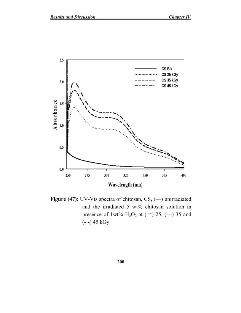
Results and Discussion Chapter IV
Ab s
o r b a
n c e
2.5
2.0
1.5
1.0
0.5
0.0
250 275 300 325 350 375 400
CS Blk CS 25 kGy CS 35 kGy CS 45 kGy
Wavelength (nm)
Figure (47): UV-Vis spectra of chitosan, CS, (—) unirradiated and the irradiated 5 wt% chitosan solution in presence of 1wt% H2O2 at (…..) 25, (---) 35 and (-..-) 45 kGy.
200

Chapter IV Results and Discussion
IV.III.2.2. FT-IR spectroscopy
Figure (48) shows the FT-IR spectra of unirradiated and irradiated Na-alginate and chitosan solutions in presence of 1wt% H2O2 at different irradiation doses of 25, 35 and 45 kGy. Comparing with the FT-IR spectrum of Na-alginate or chitosan the spectra of resultant oligo-alginate or oligo-chitosan exhibited most of the bands as the original one, indicating that the main polysaccharide structure of the resulting oligomers still remained.
In the spectrum of native Na-alginate in Figure (48, curve a), the peaks at 3390 and 1095-1035 cm-1 are attributed to hydroxyl and C–O–C groups, respectively. The asymmetric and symmetric stretching of carboxylate vibrations appeared at 1620 and 1420 cm−1, respectively Leal et al., (2008). The spectrum of γ-rays/oxidized Na-alginate Figure (48 curves b, c and d) exhibited most of the characteristic absorption peaks of native Na-alginate but with some differences. For instance, the bands at 1620 cm-1 for carboxylate groups, at 3340 for OH groups and at 1035 cm-1 and 1095 for (C–O stretching) which became broader and shift to another wavenumbers. The spectrum also showed the new bands appeared at 1725 cm-1; indicating that the formation of carboxyl groups suggesting that ionizing radiation treatment under extreme conditions broke the glucosidic bonds with the change of the structure of reducing end residue and formation of –C=O groups. Also, the scission of glycosidic bonds leads to the formation of hydroxyl
201

Results and Discussion Chapter IV group, which is manifested as an increase in the ratio of hydroxyl group peak at 3425 cm-1.
FT-IR spectrum of chitosan Figure (49 curve a) shows distinctive absorption bands appear at: 3440 cm-1 (–OH and – NH2), 2875-2920 cm-1 (C-H stretching), 1638 cm-1 (the stretching of amide C=O), 1598 (-NH bend), 1387 cm-1 (amide –NH), 1156 cm-1 and 1097 cm-1 corresponds to the ether bond of saccharine ring. The spectrum of γ-rays/oxidized chitosan Figure (49 curves b, c and d) exhibited most of the characteristic absorption peaks of native chitosan but with some differences. For instance, the increase in the ratio of hydroxyl groups at 3445 cm-1due to the decrease in the inter-and intra-molecular hydrogen bonding between –OH and –NH2
groups of chitosan resulting from the scission of glycosidic bonds leads to the formation of hydroxyl group, which is manifested as an increase in the ratio of hydroxyl group. The characteristic peaks at 1638 cm-1 (–C=O) and 1385 cm-1 (C–O stretch vibration) were shifted to higher wavenumber and its intensity increased with increasing the irradiation dose which manifested the formation of carbonyl or carboxyl after degradation. In general the changes in spectra confirmed that carbonyl or carboxyl groups were formed, partial amino groups were eliminated and/ or slightly some ring opening occurred during the radiation degradation process of chitosan.
202

Chapter IV Results and Discussion
%
T r a n s m i t t a n c e
a
d
c
b
4000 3500 3000 2500 2000 1500 1000 500
Wavenumbers
Figure (48): FT-IR spectra of Na-alginate (a) unirradiated and the irradiated 5wt% Na-alginate solution in presence of 1wt% H2O2 at (b) 25, (c) 35 and (d) 45 kGy.
203

Results and Discussion Chapter IV
%
T r a n s m i t t a n c e
d
c
a
b
4000 3500 3000 2500 2000 1500 1000 500
Wavenumbers
Figure (49): FT-IR spectra of chitosan (a) unirradiated and the irradiated 5wt% chitosan solution in presence of 1wt% H2O2 at (b) 25, (c) 35 and (d) 45 kGy.
204

Chapter IV Results and Discussion
IV.III.3. Applications of Degraded Na-alginate and Chitosan in Agricultural Purposes as Growth Promoters
The main objective of the research was to elaborate the method of degradation of natural polysaccharides to obtain a product applicable as biospecimen in protection and stimulation of the plants growth. The aim of the agricultural tests was to determine the impact of oligo-alginate and/or oligo-chitosan on growth of the zea maize plants. The effects of different molecular weights Na-alginate, chitosan and binary mixture with different ratios on the growth of zea maize plant were investigated as shown in Figure (50). The test field results in Figure (50) showed that the treatment by spraying zea maize plants (100 ppm) with the 25, 35 and 45 kGy irradiated Na-alginate or chitosan or both of them at different ratios enhances the plant growth performance.
The results presented in Figure (50) and illustrated in Table (4) showed that the highest growth was observed for Na-alginate and chitosan irradiated at 45 kGy with an average molecular weight of 1.5x10⁴ and 9.4x10³ Da, respectively. Also, the spraying of plants with binary mixture of degraded Na-alginate/chitosan has a promising effect on the growth especially the ratio of (1/2). The binary mixture Na-alginate/chitosan with ratio of (1/2) at 45kGy gives plant length (cm), paper width (cm), ear length (cm), 100 grain wt (g), ear wt (g) and grain yield/acre (ardab) 313, 12.2, 27, 40.54, 393
205

Results and Discussion Chapter IV and 36.1, respectively if compared with that of control one which gives 266, 9.8, 25, 22.35, 25 and 23.45, respectively.
Chitosan has a positive impact on plants growth. It was reported that irradiated chitosan solution is effective as plant growth enhancer. Chitosan enhances the vegetative growth in terms of the average values of stem length, number of growing leaves, including leaf width and length. The use of irradiated Na-alginate improved the quality of maize plant. The grain size, grain weight, total protein and total oil (%) increased if compared with the control one. The alginate oligomers generated by de-polymerization of Na-alginates have been reported to stimulate the plant growth, seed germination and shoot elongation in plants Hien et al., (2000). They act as signal molecules that regulate plant growth and development as well as the defense reactions in plants by regulating gene expression. The results suggest that alginate derived oligosaccharide probably applied as leaf-sprays improved the growth attributes, enhanced the acceleration of the metabolic activities, photosynthetic capability and enzyme activities Hu et al., (2004); Aftab et al., (2011); Khan et al., (2011); Li et al., 2010).
206

Chapter IV Results and Discussion
Figure (50): Effect of spraying 100 ppm of degraded Na-alginate, chitosan and both of them with different ratio and different irradiation doses on growth of zea maize plants after plantation of 30 days.
207

Results and Discussion Chapter IV
Table (4): Effect of spraying 100 ppm of irradiated Na-alginate, chitosan and binary mixture on crop yield of zea maize plants
. Dose Plant
length, (cm)
Paper width, (cm)
Ear length, (cm)
100 grain wt (g)
Ear wt (g)
Grain Yield/ acre (ardab)
0kGy Control 266 9.8 25 22.35 256 23. 45
Alg 263 10.3 26 24.73 278 25.47
CS 268 10.5 25.8 23.25 270 24.74
25kGy Alg/CS(1/1) 274 10.7 26.4 25.19 288 26.39
Alg/CS(2/1) 281 12.1 26.8 25.35 292 26.75
Alg/CS(1/2) 283 12.2 27.5 26.15 302 27.67
Alg 291 10.7 29 32.17 337 30.88
CS 298 11.3 28 30.22 323 29.59
35kGy Alg/CS(1/1) 287 12.1 28 28.7 334 30.6
Alg/CS(2/1) 291 12.3 28.5 32.95 349 31.98
Alg/CS(1/2) 308 12.6 29.5 33.93 359 32.89
Alg 307 10.6 29.2 38.61 377 34.54
CS 314 11.8 28 36.27 360 32.98
45kGy Alg/CS(1/1) 309 12.3 27.6 34.47 358 32.8
Alg/CS(2/1) 318 12.3 30.3 39.5 379 34.73
Alg/CS(1/2) 313 12.2 27 40.54 393 36.1
208
Summary and
Conclusion

Summary and Conclusion
Summary and Conclusion
The present study divided into three parts. First part focused on investigating the antioxidant properties of low molecular weight natural polymers such as chitosan, Na-alginate and carrageenan degraded and assessing their suitability for food preservation. Second part focused on radiation induced crosslinking of PAAm incorporated low molecular weights of Na-alginate or chitosan and/or both of them for possible use in the agricultural applications. Third part focused also on using such degraded natural polymers in the area of agriculture as plant growth promoters. The results obtained can be summarized as follows:
I. Investigating the Antioxidant Properties of Low Molecular Weight Natural Polymers and Assessing their Suitability for Food Preservation
1. Chitosan was modified with maleic anhydride, phthalic anhydride and chlorosulfonic acid-DMF complex to get N-maleoyl-chitosan (NMCS), N-phthaloyl-chitosan (NPhCS) and sulfonated-chitosan (SCS), respectively.
2. The chemical modification of chitosan derivatives was confirmed using FT-IR. For NMCS new bands appeared at 1719, 1632 and 1547 cm-1 characteristic for (C=O) of acid groups, –NHC=O– and CH=CH-, respectively. For NPhCS new bands appeared at 2941, 3074, 1719, 1773 and 1547
-1cm are characteristic for C-H stretch of the -CH=CH-
209

-1
Summary and Conclusion groups, C-H stretch of aromatic system, carbonyl group (C=O) of carboxylic acid, residual anhydride and unsaturated double bond -CH=CH- groups, respectively. For SCS new bands appeared at 817 and 1235 cmcorrespondng to the C-O-S and S=O bond stretching, respectively.
3. The XRD analysis of chitosan and its derivatives was investigated. It was found that the crystallinty decreased by chemical modifications.
4. The effect of γ-rays on the molecular weights of some natural polymers such as chitosan, Na-alginate, carrageenan and chitosan derivatives was investigated in aqueous solutions (irradiated at 10-30 kGy) and in solid form (irradiated at 25-100 kGy). There is a rapid decrease in the number average molecular weights with increasing the irradiation dose. The dose required for the polymer degradation in the solution form was lower than that in the solid form.
5. Structural changes of unirradiated and irradiated chitosan, Na-alginate, carrageenan and chitosan derivatives were investigated by UV-Vis and FT-IR spectroscopy. The results revealed that during radiation degradation process there are some unsaturated groups formed and the main polysaccharide chain structure was almost remained.
6. Evaluation of antioxidant activity of unirradiated and irradiated chitosan, Na-alginate, carrageenan and chitosan
210
Summary and Conclusion derivatives by using scavenging effect on DPPH radicals, reducing power, the ferrous ion chelating activity and radical mediated lipid peroxidation inhibition assays were investigated.
7. The results revealed that the irradiation process enhances the antioxidant activity of polysaccharides. The lower the molecular weight oligosaccharides, the higher antioxidant activity obtained. The antioxidant activity of irradiated chitosan was higher than that of Na-alginate and carrageenan. Low molecular weights chitosan derivatives were more pronounced than irradiated chitosan and the antioxidant activity (%) follows the order NMCS > NPhCS > SCS > CS. It was found that the IC50 (%) on DPPH radicals of irradiated chitosan at 30 kGy, irradiated NMCS at 30 kGy and ascorbic acid was 0.154, 0.47 and 0.144 mg/ml, respectively.
8. Effectiveness of irradiated CS and NMCS in reducing the lipid peroxidation in minced chicken was studied. Using100 kGy irradiated CS and NMCS to minced chicken at 0.1% concentration resulted in a highly significant decrease about 50% and 70% in malonaldehyde (MDA) content, respectively, after 10 days of storage at 4±1oC compared with the control. The results suggested the possible use of such polysaccharides as natural antioxidants to improve the oxidative deterioration of minced chicken during refrigerated storage.
211

Summary and Conclusion 9. The toxicity of NMCS on inhibition growth of fungus
(Mycelial) with time was investigated. The results showed that the fungus has a capability for adaptation with the NMCS polymer. MTT assay showed no cytotoxic effects of NMCS on cells at small concentrations or higher ones
II. Radiation Induced Crosslinking of PAAm Incorporated Low Molecular Weights Natural Polymers for Possible Use in the Agricultural Applications
Preparation of crosslinked PAAm hydrogel incorporated with degraded Na-alginate, chitosan and/or both of them was carried out using the ionizing radiation at different copolymer compositions and the results as follows:
10.The effect of copolymer compositions and irradiation dose on gel content (%) of PAAm/Alg, PAAm/CS and PAAm/Alg/CS was investigated. The gel content (%) of PAAm/Alg > PAAm/Alg/CS > PAAm/CS hydrogels.
11.The Effect of copolymer compositions and irradiation dose on water absorbency (g/g) of PAAm/Alg, PAAm/CS and PAAm/Alg/CS was investigated. It was found that the water absorbency of PAAm, PAAm/Alg, PAAm/CS and PAAm/Alg/CS was 390, 578, 348 and 378 (g/g), respectively, at copolymer concentration (5wt%), copolymer compositions (95/5) and irradiation dose of 10 kGy.
212

Summary and Conclusion 12.The effect of ionic strength of various types of cations (such
as Na+, Ca2+, Fe3+), various types of fertilizers (such as ammonium nitrate, urea, ammonium phospahte and ammonium sulfate) and pH on water absorbency (g/g) of prepared hydrogels was studied. It was found that the water absorbency decreases with increasing the ionic strength of cation solution or concentration of fertilizer. Also, under a given ionic strength, water absorbency decreases with the increase in ionic valence of the salt. The swelling of the hydrogels is affected by pH.
13.Water retention capability of swollen hydrogels as a function of time at room temperature and different temperatures was investigated. It was observed that the presence of polysaccharides with PAAm hydrogel improves hydrogel water retention capability and decreases its water loss. Also, with increasing the temperature, the deswelling rate of gel imbibed water increases and results in decreasing hydrogels water retention
14. The thermal stability and surface morphology of prepared copolymer hydrogels were investigated. The thermal stability of crosslinked PAAm is higher than that of PAAm/polysaccharide due to the thermal degradation of polysaccharide which accelerates the thermal degradation of PAAm. It was found from SEM that the crosslinking density formed during the irradiation process affects the pore structure of the prepared hydrogels.
213

Summary and Conclusion 15.Applications of copolymer hydrogels in agricultural fields
was performed using zea maize as a model plant. The results showed that the presence of hydrogels had growth promotion effect on quality and quantity of zea maize plants due to the presence of oligo-alginate and/or oligo-chitosan. The productivity of maize plant in presence of PAAm/Alg hydrogels was the highest compared with the control resulting in an increase about 50% in production yield.
III. Development of Applications of Low Molecular Weight Polysaccharides in the Area of Agriculture as Plant Growth Promoter 16.The effect of γ-irradiation on degradation of 5% Na-alginate
and chitosan in the presence of H2O2 at different doses as a function of molecular weight changes was investigated. It is observed that the addition of H2O2 during the irradiation process accelerates the degradation process.
17.UV-Vis and FT-IR spectra of irradiated Na-alginate and chitosan were measured. The results revealed that there are some unsaturated groups formed and the main polysaccharide chain structure was almost remained.
18.The effects of spraying 100 ppm of different molecular weights of Na-alginate or chitosan or both of them with different ratios on the growth of zea maize plant were investigated. It was found that the test field results showed that the treatment of zea maize plants with oligo-alginate or oligo-chitosan or binary mixture results in enhancement of the plant growth performance. The increase in plant
214

Summary and Conclusion performance by using degraded Na-alginate and/or chitosan suggested its possible use in agriculture purposes as growth promoters for plants.
From the above mentioned results, it could be concluded that the irradiation process enhanced the antioxidant activity of polysaccharides. The lower molecular weight of polysaccharides, the higher the antioxidant activity obtained. The modification of chitosan by chemical treatment and ionizing radiation improved the antioxidant activity rather than chitosan itself. The addition of 100 kGy irradiated CS and NMCS to minced chicken resulted in a highly significant decrease in malonaldehyde (MDA) content suggesting their possible use as natural antioxidants for improving the oxidative deterioration of minced chicken during refrigerated storage.
The preparation of PAAm containing low molecular weight natural polymers had growth promotion effect on quality and quantity of zea maize plants when added to zea maize seeds and the productivity of zea maize plants was the highest in presence of PAAm/Alg hydrogel. The increase in plant performance by spraying degraded Na-alginate and chitosan to zea maize plants suggested their possible use in agriculture purposes as growth promoters for plants.
215
References

. Reference
References
1. Abd El-Rehim H. A., El-Sawy N. M., Farag I. A., Elbarbary A. M., Carbohyd. Polym., 86, 1439-1444 (2011).
2. Abd El-Rehim H. A., Hegazy E. A., Abd El-Mohdy H. L., J. Appl. Polym. Sci., 93, 1360-1371 (2004).
3. Abd El-Rehim H. A., Hegazy E. A., Abd El-Mohdy H. L., J. Appl. Polym. Sci., 101, 3955-3962 (2006).
4. Abd El-Rehim H. A., J. Appl. Polym. Sci., 101, 35723580, (2006).
5. Abdel-Mawgoud A. M. R, Tantawy A. S., El-Nemr M. A., Eur. J. Sci. Res., 39, 161-168 (2010).
6. Aftab T., Khan M. M. A., Idrees, M. Naeem, M., Moinuddin, Hashmi M. N., Varshney L., Rad. Phys. Chem., 80, 833-836 (2011).
7. Akiyama H., Endo T., Nakakita R., Murata K., Yonemoto Y., Okayama K., Biosci. Biotech. Biochem., 56, 335-356 (1992).
8. Antolovich M., Prenzler P. D., Patsalides E., McDonald S., Robards K., Analyst, 127, 183-198 (2002).
9. Aytekin A. O., Morimura S., Kida K., J. Biosci. Bioeng., 111, 212-216 (2011).
10. Britto D. D., Campana-Filho S. P., Polym. Degrad. Stab. 84, 353-361 (2004).
11. Buege J. D., Aust S., Methods Enzymol. 30, 303-310 (1978).
12. Cagri A., Ustunol Z., Ryser E., J. Food Prot., 67, 833848 (2004).
216

.Reference
13. Cahyana A. H., Shuto Y., Kinoshita Y., Biosci., Biotechnol., Agrochem., 56, 1533-1535 (1992).
14. Chambers L. J., Jimbin M., McDonald R., Antioxidant composition. EP0267630 (1988).
15. Chandrkrachang S., The application of chitin and chitosan in agriculture in Thailand. In K. Suchiva, S. Chandrkrachang, P. Methacanon, &M.G. Peter V (Eds.), Advances in chitin science, 458-462 (2002).
16. Chang H. L., Chen Y. C., Tan F. J., Food Chem., 124, 589-595 (2011).
17. Charlesby A., Atomic Radiation and Polymers, Pergamon Press, New York, (1960).
18. Chen S. K., Tsai M. L., Huang J. R., Chen R. H., J. Agric. Food Chem., 57, 2699-2704 (2009).
19. Chen X. G., Park H. J., Carbohyd. Polym., 53, 355-359 (2003).
20. Chen X., Ahn D. U., J. Am. Oil Chemists' Society, 75, 1717-1721 (1998).
21. Chen Z. B., Liu M. Z., Ma S. M., React. Funct. Polym., 62, 85-92 (2005).
22. Chen, C. W.; Ho, C. T., J. Food Lipids, 2, 35-46 (1995). 23. Chien P.-J., Sheu F., Huang W.-T. , Su M.-S., Food
Chem., 102, 1192-1198 (2007). 24. Chmielewski A. G., Migdal W., Swietoslawski J.,
Swietoslawski J., Jakubaszek U., Tarnowski J., Rad. Phys. Chem., 76, 1840-1842 (2007).
25. Cho M. H., No H. K., Prinyawiwatkul W., J. Food Sci., 73, 70-77 (2008).
217

. Reference
26. Cho Y.-s, Kim S.-k, Ahn C.-b, Je J.-y, Carbohyd. Polym., 83 1617-1622 (2011).
27. Choi B. K., Kim K. Y., Yoo Y. J., Oh S. J., Choi J. H., Kim C. Y., Int. J. Antimicrob. Agents, 18, 553-557 (2001).
28. Chung Y. C., Su Y. P., Chen C. C., Jia G., Wang H. L., Wu J. C., Acta Pharmacologica Sinica, 25, 932-936 (2004).
29. Chung Y. C., Tsai, C. F., Li, C. F., Fisheries Sci., 72, 1096-1103 (2006).
30. Curcio M., Puoci F., IEMMA F., Parisi O. I, Cirillo G., Spizzirri U. G., Picci N., J. Agric. Food Chem., 57, 5933-5938 (2009).
31. Czechowska-Biskup R., Rokita B., Ulanski P., Rosiak J. M., Nucl. Inst. Meth. Phys. Res. B, 236, 383-390 (2005).
32. Darmdji P., Izumimoto M., Meat Sci., 38, 243-254 (1994).
33. Dorman H. J. D., Kosar M., Kahlos K., Holm Y., Hiltunen R., J. Agric. Food Chem., 51, 4563-4569 (2003).
34. Duh D., Tu Y. Y., Yen G. C., Lebensm. -Wiss. Technol. 32, 269-277 (1999).
35. Duy N. N., Phu D. V., Anh N. T., Hien N. Q., Rad. Phys. Chem., 80, 848-853 (2011).
36. Dzudie T., Kouebou C. P., Essia-Ngang J. J., Mbofung C. M. F., J. Food Eng., 65, 67-72 (2004).
37. Dzung N. A., J. Chitin Chitosan, 10, 109-113 (2005). 38. Dzung N. A., Khanh V. T. P., Dzung T. T., Carbohyd.
Polym., 84, 751-755 (2011).
218

.Reference
39. Eissa H. A. A., J. Food Quality, 30, 623–645 (2007). 40. El Hadrami A., Adam L. R., El Hadrami I. and Daayf
F., Marine Drugs, 8, 968-987 (2010). 41. El-Sawy N. M., Abd El-Rehim H. A. , Elbarbary A. M.,
Hegazy E. A., Carbohyd. Polym., 79, 555-562 (2010). 42. Feng T., Du Y., Li J., Wei Y., Yao P. Eur. Food. Res.
Technol., 225, 133-138 (2007). 43. Feng T., Du Y., Li J., Hu Y., Kennedy J. F., Carbohyd.
Polym., 73, 126-132 (2008). 44. Gazzani G., Papetti A., Massolini G., Daglia M., J.
Agricult. Food Chem., 46, 4118-4122 (1998). 45. Georgantelis D., Blekas G., Katikou P. Ambrosiadis I.,
Fletouris D. J., Meat Sci., 75, 256-264 (2007). 46. Gerasimenko D. V., Avdienko I. D., Bannikova G. E.,
Zueva O. Y., Varlamov V. P., Appl. Biochem. Microbiolo., 40, 253-257 (2004).
47. Gonzalez-Aguilar G. A., Valenzuela-Soto E., Lizardi-Mendoza J., Goycoolea F., Martiınez-Tellez M. A., Villegas-Ochoa M. A., Monroy-Garcia I. N., Ayala-Zavala J. F., J. Sci. Food Agric., 89, 15-23 (2009).
48. Gordon M. H., The mechanism of antioxidant action in introduction, in: B.J.F. Hudson (Eds.), Food antioxidants: Elsevier Applied Science, New York, 1-18 (1990).
49. Grice H. C., Food Chem. Toxicolo., 26, 717-723 (1988). 50. Guan Y-J, Hu J., Wang X-J, Shao C-X, J. Zhejiang
University Sci., 10, 427-433 (2009). 51. Guo Z. Y., Xing R. E., Liu, S., Zhong Z. M., Li P. C.,
Carbohyd. Polym., 73, 173-177 (2008).
219

. Reference
52. Guo Z., Liu H., Chen X., Ji X. and Li P., Bioorg. Medic. Chem. Lett., 16, 6348-6350 (2006).
53. Guo Z., Xing R., Liu S., Yu H., Wang P., Li C. and Li P., Bioorg. Medic. Chem. Lett., 15, 4600-4603 (2005).
54. Guzman J, Saucedo I, Revilla J, Navarro R, Guibal E., Int. J. Biolog. Macromol., 33, 57-65 (2003).
55. Hai L., Diep T. B., Nagasawa N., Yoshii F., Kume T. Nucl. Inst. Meth. Phys. Res. B, 208, 466-470 (2003).
56. Halliwell B., Am. J. Med., 91, 814-822 (1991). 57. Han D., Kim, S. J., Kim, S. H., Kim, D. M., J. Food
Sci., 63, 69-72 (1998). 58. Hansen M.B., Nielsen S.E., Berg K. J. Immunol.
Methods, 119, 203-210 (1989). 59. Hasegawa M., Isogai A., Onabe F., Usuda M., Atalla R.
H., J. Appl. Polym. Sci., 45, 1873-1879 (1992). 60. Hayashi Y., Morimoto K., Miyata N., Sato H., Toxicolo.
Indust. Health, 9, 243-249 (1993). 61. Hertog M. G. L., Feskens E. J. M., Hollman P. C. H.,
Katan M. B., Kromhout D., Lancet, 342, 1007-1011 (1993).
62. Hien N. Q., Nagasawa N., Tham L. X., Yoshii F., Dang V. H., Mitomo H., Makuuchi K., Kume T., Rad. Phys. Chem., 59, 97-101 (2000).
63. Hien N. Q., Phu D. V., Duy N. N., Lan N. T. K., Carbohyd. Polyme., 87, 935-938 (2012).
64. Hoffman A. S., Adv. Drug Deliv. Rev., 43, 3-12 (2002). 65. Hu P., Jiang X., Hwang H., Liu S., Guan H., J. Appl.
Physiolo., 16, 73-76 (2004).
220

.Reference
66. Huang L., Zhai M., Peng J., Li J., Wei G., Carbohyd. Polym., 67, 305-312 (2007 a).
67. Huang L., Peng J., Zhai_M., Li J., Wei G., Radiat. Phys. Chem., 76, 1679-1683 (2007 b).
68. Huang L., Zhai M., Peng J., Li J., Wei G., Carbohyd. Polym., 68, 109-115 (2007 c).
69. Huang R., Mendis E., Kim S.-k, Int. J. Biolog. Macromole., 36, 120-127 (2005).
70. Huang R., Rajapakse N., Kim S.-K., Carbohyd. Polym., 63, 122-129 (2006).
71. Hyun K. S., No H. K., and Prinyawiwatkul W., J. Food Sci., 72, 44-48 (2007).
72. Idrees M., Naeem M., Alam M., Aftab T., Hashmi N., Khan M. M. A., Moinuddin, Varshney L., Agric. Sci. China, , 10(8): 1213-1221 (2011).
73. Ignatova M., Manolova N., Rasgkov L., Eur. Polym. J., 43, 1112-1122 (2007).
74. Jacobs D. F., Salifu K. F., Seifert J. R., Forest Ecol. Manag., 214, 28-39 (2005).
75. Je J. Y., Park P. J., Kim S. K., Food Chem. Toxicolo., 42, 381-387 (2004).
76. Je J-Y and Kim S-K, Bioorg. Medic. Chem. Lett., 16, 1884-1887 (2006).
77. Jellinek H. H. G., Degradation of Vinyl Polymers, Wiley and Sons, New York (1955).
78. Jeon Y. J., Kim S. K., J. Microbiolo. Biotechn., 12, 503-507 (2002).
221

. Reference
79. Ji X., Zhong Z., Chen X., Xing R., Liu S., Wang L. and Li P., Bioorg. Medic. Chem. Lett., 17, 4275-4279 (2007).
80. Jo C., Lee J.W., Lee K.H., Byun M.W., Meat Sci., 59, 369-375 (2001).
81. Kachanechaia T., Jantawata P., Pichyangkura R., Food Hydrocoll., 22, 74-83 (2008
82. Kanatt S. R., Paul P., D’Souza S. F., Thomas P., J. Food Sci., 63, 198-200 (1998).
83. Kanatt S. R., Chander R., Sharma A., Int. J. Food Sci. Techn., 39, 997-1003 (2004).
84. Kanatt S. R., Chander R., Sharma A., Food Chem., 107, 845-852 (2008).
85. Kanauchi O., Deuchi K., Imasato Y., Shizukuishi M., Kobayashi E., Biosci. Biotechn. Biochem., 59, 786-790 (1995).
86. Kang B., Dai Y.-d, Zhang H.-q, Chen D., Polym. Degrad. Stab., 92, 359-362 (2007).
87. Khan Z. H., Khan M. M. A., Aftab T., Idrees M., Naeem M., Front. Agric. China, 5, 122-127 (2011).
88. Khokhlov A. R., Starodubtzev S. G., Vasilevskaya V. V., Adv. Polym. Sci., 109, 123- (1993).
89. Kim C. H., Choi J. W., Chun H. J., Choi K. S., Polym. Bullet. 38, 387-393 (1997).
90. Kim H-J, Chen F., Wang X., Rajapakse N. C., J. Agric. Food Chem., 53, 3696-3701 (2005).
91. Kim J. Y., Lee J. K., Lee T. S., Park W. H., Int. J. Biolo. Macromol., 32, 23-27 (2003).
222

.Reference
92. Kim K. W., Thomas R. L., Food Chem., 101, 308-313 (2007).
93. Kinsella J. E., Frankel E., German B., Kanner J., Food Technolo., 4, 85-89 (1993).
94. Kosaraju S. L., Weerakkody R., Augustin M. A., J. Agric. Food Chem., 58, 12449-12455 (2010).
95. Kumar M., Varshney L., Francis S., Radiat. Phys. Chem., 73, 21-27 (2005).
96. Kume T., Nagasawa N., Yoshii F., Rad. Phys. Chem., 63, 625-627 (2002).
97. Lee D. W., Choi W., Byun, M. W., Park H. J., Yong-Man Yu Y.-M, Lee C. M., J. Agric. Food Chem. 51, 4819-4823 (2003).
98. Lee W.-F., Wu R.-J., J. Appl. Polym. Sci., 62, 10991114 (1996).
99. Leon A. T., Daryl C., Postharvest Biolo. Technolo. 32, 1-13 (2004).
100. Leonhardt J., Arnold G., Baer M., Langguth H., Gey M., Hgbert S., Rad. Phys. Chem., 25, 899-904 (1985).
101. Li X., Shi X., Wang M., Du Y., Food Chem., 126, 520525 (2011).
102. Li X., Xu A., Xie, H, Yu W., Xie, W., Ma X., Carbohyd. Polym., 79, 660-664 (2010).
103. Lim S. N., Cheung P. C. K., Ooi V. E. C., Ang P. O., J. Agric. Food Chem., 50, 3862-3866 (2002).
104. Lin H.-Y., Chou C.-C., Food Res. Int., 37, 883-889 (2004).
105. Lingnert H., Vallentin K., Eriksson C. E., J. Food Process. Preserv., 3, 87-104 (1979).
223

. Reference
106. Liu J., Sun H., Dong F., Xue Q., Wang G., Qin S., Guo Z., Carbohyd. Polym., 78, 439-443 (2009).
107. Luan L. Q., Nagasawa N., Ha V. T. T., Hien N. Q., Nakanishi T. M., Rad. Phys. Chem., 78, 796-799 (2009).
108. Luan L.Q., Nguyen Q. H., Nagasawa N., Kume T., Yoshii F., Nakanishi T. M., Biotechnol. Appl. Biochem., 38, 283-288 (2003).
109. Luan le Q., Ha V.T., Nagasawa N., Kume T., Yoshii F., Nakanishi T.M. , Biotechnol. Appl. Biochem., 41, 49-57 (2005).
110. Ma G., Yang D. Z., Zhou Y. S., Xiao M., Kennedy J. F., Nie J., Carbohyd. Polym., 74, 121-126 (2008).
111. Matsuo K., Miyazono I., Nippon Suisan Gakkaishi, 59, 1377-1379 (1993).
112. Melo M. R. S., Feitosa J. P. A., Freitas A. L. P., de Paula, R. C. M., Carbohyd. Polym., 49, 491-498 (2002).
113. Miao J., Chen G. H., Gao C. J., Desalination, 181, 173183 (2005).
114. Mollah M. Z. I., Khan M. A., Khan R. A., Rad. Phys. Chem., 78, 61-64 (2009).
115. Murugesan K., Yang I. H., Kim Y. M., Jeon J. R., Chang Y. S., Appl. Microbiolo. Biotechn., 82, 341-350 (2009).
116. Nagasawa N., Mitomo H., Yoshii F., Kume T., Polym. Deg. Stab. 69, 279-285. (2000).
117. Nge K.L., New N., Chandrkrachang S., Stevens W. F., Plant Sci., 170, 1185-1190 (2006).
224

.Reference
118. Ocloo F. C. K., Quayson E. T., Adu-Gyamfi A., Quarcoo E.A., Asare D., Serfor-Armah Y., Woode B.K., Rad. Phys. Chem., 80, 837-841 (2011).
119. Okamoto Y., Inoue A., Miyatake K., Ogihara K., Shigemasa Y., Minami S., Macromol. Biosci., 3, 587590 (2003).
120. Okamoto Y., Kawakami K., Miyatake K., Morimoto M., Shigemasa Y., Minami S., Carbohyd. Polym., 49, 249252 (2002).
121. Park P. J. Je J. Y., Byun H. G., Moon S. H., Kim S. K. J. Microbiol. Biotechn., 14, 317-323 (2004).
122. Park P.-J., Je J.-Y., Kim S.-K., J. Agric. Food Chem., 51, 4624-4627 (2003).
123. Pospieszny H., Chirkov S., Atabekov J., Plant Sci., 79, 63-68 (1991).
124. Pourjavadi A., Ghasemzadeh H., Polym. Eng. Sci., 47, 1388-1395 (2007).
125. Pratt D. E., Hudson B. J. F. Natural antioxidants not exploited commercially. In B. J. F. Hudson (Ed.), Food Antioxidants, 171-192. Amsterdam: Elsevier (1990).
126. Ramarathnam N., Osawa T., Ochi H., Kawakishi S., Trends Food Sci. Techn., 6, 75-82 (1995).
127. Rao M. S., Chander R., Sharma A., J. Food Sci., 70, 325-331 (2005).
128. Rao M.S., Chander R., Sharma A., LWT - Food Sci. Tech., 41, 1995-2001 (2008).
129. Reichmanis E., Frank C., O’Donnell J., Hill D., O’Donnell J, Washington, DC: Am. Chem. Society, 2 (1993).
225
. Reference
130. Relleve L., Nagasawa N., Luan L.Q., Yagi T., Aranilla C., Abad L., Kume T., Yoshii F., De la Rosa A., Polym. Degrad. Stab., 87, 405-410 (2005).
131. Ruperez P., Ahrazem O., Leal J. A., J. Agric. Food Chem., 50, 840-845 (2002).
132. Samuels R. J., J. Polym. Sci. Polym. Phys. Ed., 19 10811105 (1981).
133. Sarfaraz A, Naeem M., Nasir S., Idrees M., Aftab T., Hashmi N., Masroor M., Khan A., Moinuddin and Varshney L., J. Medic. Plants Res., 5, 15-21, (2011).
134. Sartori C., Finch S. D., Ralph B., Polym., 38, 43-51 (1997).
135. Sashiwa H., and Shigemasa Y., Carbohyd. Polym. 39, 127-138 (1999).
136. Sayas-Barberá E., Quesada J., Sánchez-Zapata E., Viuda-Martos M., Fernández-López F., Pérez-Alvarez J.A., Sendra E., Meat Sci., 88, 740-749 (2011).
137. Schwarz K., Bertelsen G., Nissen L. R., Gardner P.T., Eur. Food Res. Technolo., 212, 319-328 (2001).
138. Sen M., Appl. Rad. Isotopes, 69, 126-129 (2011). 139. Sen M., Rendevski S., Kavaklı P. A., Sepehrianazara A.,
Rad. Phys. Chem., 79, 279-282 (2010). 140. Shahidi F., Wanasundara P. K. J. P. D., Phenolic
Antioxidants, 32, 67- 103 (1992).
141. Shimada K., Fujikawa K., Yahara K., Nakamura T., J. Agric. Food Chem., 40, 945-948 (1992).
142. Soultos N., Tzikas Z., Abrahim A., Georgantelis D., Ambrosiadis I., Meat Sci., 80, 1150-1156 (2008).
226

.Reference
143. Spizzirri U. G., Parisi O. I., Iemma F., Cirillo G., Puoci F., Curcio M., Picci N., Carbohyd. Polym., 79, 333-340 (2010).
144. Sreedhar B., Aparna Y., Sairam M., Hebalkar N., J. Appl. Polym. Sci., 105, 928-934 (2007).
145. Sun L., Wang C., Shi Q., Ma C., Int. J. Biolog. Macromol., 45, 42-47 (2009).
146. Sun T., Xie W., Xu P., Macromole. Biosci., 3, 320-323, (2003).
147. Sun T., Yao Q., Zhou D., Mao F., Bioorg. Medic. Chem. Lett., 18, 5774-5776 (2008).
148. Sun T., Zhou D., Xie J., Mao F., Eur. Food Res. Technol. 225, 451-456 (2007 a).
149. Sun T., Zhou D., Mao F., Zhu Y., Eur. Polym. J., 43, 652-656 (2007 b).
150. Sun T., Zhu Y., Xie J., Yin X., Bioorg. Medic. Chem. Lett., 21, 798-800 (2011).
151. Tham L. X., Nagasawa N., Matsuhashi S., Ishioka N. S., Ito T., Kume T., Rad. Phy. Chem., 61, 171-175 (2001).
152. Tian F., Liu Y., Hu K., Zaho B., Carbohyd.Polym., 57, 31-37 (2004).
153. Tsuda T., Ohshima K., Kawakishi S., Osawa T., J. Agricult. Food Chem., 42, 248-251 (1994).
154. Tyliszczak B., Polaczek J., Pielichowski K., Polish J. Environ. Stud., 3, 475-479 (2009).
155. Ulanski P., Rosiak J. M. Rad. Phys. Chem., 39, 53-57. (1992).
156. Vanichvattanadecha C. , Supaphol P., Nagasawa N. , Tamada M., Tokura S., Furuike T., Tamura H.,
227

. Reference
Rujiravanit R., Polym. Degrad. Stab., 95, 234-244 (2010).
157. Vasyukova N. I., Chalenko G. I., Gerasimova N. G., Perekhod E. A., Ozeretskovskaya O. L., Irina A. V., Varlamov V. P., Albulov A. I., Appl. Biochem. Microbiol., 36, 372-376 (2000).
158. Vasyukova N. I., Zinov’eva S. V., Il’inskaya L. I., Perekhod E. A., Chalenko G. I., Gerasimova N. G., Il’ina A. V., Varlamov V. P., and Ozeretskovskaya O. L., Appl. Biochem. Microbiol., 37, 1, 103-109 (2001).
159. Velioglu Y. S., Mazza G., Gao L., Oomah B. D., J. Agric. Food Chem., 46, 4113-4117 (1998).
160. Verheul R. J., Amidi M., Wal S., Riet E., Jiskoot W., Hennink W., Biomat., 29, 3642-3649 (2008).
161. Vyncke W. C., Fette Seifen Anstrichmittel, 72, 10841087 (1970).
162. Wanasundara U. N., Shahidi F., Food Chem., 63, 335342 (1998).
163. Wang J., Jiang X., Mou H., Guan H., J. Appl. Phycolo., 16, 333-340 (2004).
164. Wang P., Jiang X., Jiang Y., Hu X., Mou H., Li M., Guan H., Nat. Prod. Res., 21, 646-654 (2007).
165. Wang S.-M, Huang Q.-Z, Wang Q.-S., Carbohyd. Res., 340, 1143-1147 (2005).
166. Wanichpongpan P., Suriyachan K., Chandrkrachang S., in: T. Uragami, K. Kurita, T. Fukamizo (Eds.), Chitin and Chitosan in Life Science, Yamaguchi, 198-201 (2001).
228

.Reference
167. Wasikiewicz J. M., Yoshii F., Nagasawa N, Wach R. A., Mitomo H., Rad. Phys. Chem., 73, 287-295 (2005).
168. Wu T., Mao L., J. Food Process. Preserv., 33, 218-230 (2009).
169. Xie W. M., Xu P. X., Liu Q., Bioorg. Medic. Chem. Lett., 11, 1699-1701 (2001).
170. Xing R., Liu S., Guo Z., Yu H., Wang P., Li C., Bioorg. Med. Chem., 13, 1573-1577 (2005 a).
171. Xing R., Liu S., Guo Z., Yu H., Zhong Z., Ji X., Li P., Eur. J. Medic. Chem., 43, 336-340 (2008).
172. Xing R., Liu S., Yu H., Guo Z., Li., Li P., Carbohyd. Polym., 61, 148-154 (2005 c).
173. Xing R., Liu S., Yu H., Zhang Q., Li Z. and Li P., Carbohyd. Res., 339, 2515-2519 (2004).
174. Xing R., Yu H., Liu S., Zhang W., Zhang Q., Li Z., Li P., Bioorg. Medic. Chem., 13, 1387-1392 (2005 b).
175. Xue C. H., Fang Y., Lin H., Chen L., Li Z. J., Deng D., Lu C. X., J. Appl. Phycol., 13, 67-70 (2001).
176. Yamaguchi R., Tatsumi, M. A., Kato, K., Yoshimitsu U., . Agric. Biolog. Chem., 52, 849-850 (1988).
177. Yamaguchi T., Takamura H., Matoba T., Terao J., Biosci. Biotechn. Biochem., 62, 1201-1204 (1998).
178. Yang S., Guo Z., Miao F., Xue Q., Qin S., Carbohyd. Polym., 82, 1043-1045 (2010).
179. Yen G. C., Duh P. D., J. Am. Oil Chem. Soc., 70, 383386 (1993).
180. Yen G.-C., Chen H.-Y., Peng H.-H., J. Agricult. Food Chem., 45, 30-34 (1997).
229

. Reference
181. Yen M.-T, Yang J.-H, Mau J.-L, Carbohyd. Polym., 74, 840-844 (2008).
182. Ying G-q, Xiong W-y, Wang H., Sun Y., Liu H-z, Carbohyd, Polym., 83, 1787-1796 (2011).
183. Yu Y., Li Y., Liu L., Zhu C., Xu Y., J. Polym. Res., 18, 283-291 (2010).
184. Yuan H., Song J., Zhang W., Li X., Li N. and Gao X., Bioorg. Medic. Chem. Lett., 16, 1329-1334 (2006).
185. Yue W., Yao P., Wei Y., Mo H., Polym. Degrad. Stab., 93, 1814-1821 (2008).
186. Zainol I., Akil H M., Mastor A., Mat. Sci. Eng. C, 29, 292-297 (2009).
187. Zhang H., Neau S. H., Biomat., 22, 1653-1658 (2001). 188. Zhang J., Liu R., Li A.and Wang A. , Polym. Adv.
Technol., 17, 12-19 (2006). 189. Zhang Q. B., Li N., Liu X. G., Zhao Z. Q., Li Z. E., Xu
Z. H. Carbohyd. Res., 339, 105-111 (2004). 190. Zhang Q. B., Yu, P. Z., Li Z. E., Zhang H., Xu Z. H., Li
P. C., J. Appl. Phycol.,, 15, 305-310 (2003). 191. Zhong Z., Ji X., Xing R., Liu S., Guo Z., Chen X. and Li
P., Bioorg. Medic. Chem., 15, 3775-3782 (2007). 192. Zhong Z., Xing R., Liu S., Wang L., Cai S., Li P., Eur.
J. Medic. Chem., 43, 2171-2177 (2008). 193. Zhong Z., Zhong Z., Xing R., Li P., Mo G. L., Int. J.
Biolog. Macromol., 47, 93-97 (2010). 194. Zhu X. Y., Wu J. M., Jia Z. S., Chinese Chem. Lett., 15,
808-810 (2004).
230

العربي صلملخ ا


العربي ا لملخصالعربـــى الملخص
ع ة ترج تخدامأهمي واداس سدةللأالم الم ى ادرتهلق آ ىاظالحف عل عل ضادةةجتانلا ة ضار واد ة الم نالغذائي رال م لا ات تغين سدة ع والزيالأآ وتدهون
وادصلاحية ةالم د و . الغذائي أنثبت قي ف
ة ة الأطعم سدة القابل د لك ذ وآللأآ دةمتمدي ض درات بع دة الكربوهي سكرياتعدي نال تخدا يمك واد آ ها ما س ضادة م مسد الأعن الناتج الضررمن للوقاية للاآسدة ة في ة آ دالأطعم ك ويعتم ىذل وععل نالوظيفيةومجموعاتهالجزيئيووزنهالسكرياتعديد البوليمير.ذلك ه ذهرآزت لالة ى الرس عاع ة دراسعل اميتأثيرالإش ؤ الج تحكم و ينالم ي ال سيرف ضتك بعة رات ي البوليم لالطبيعي وزان مث ات و الكيت صوديوم ألجين ان و ال للحصول الكاراجينى وليم عل ةأوزانذات رات يب غيرةجزيئي ددة (Oligosaccharides)ص متع
ات مجموع الوليميرات ي الب مالت تكت
ة تخدامالوظيفي عاع باس الإش.م ة وت ضاد الالخواصدراس سدة للأ ة م آ ه ذ ه ل
شتقات لكيتا لمع بالإ ذلك و اع ش خال آضاد ال اص و سدة ل ة م لأآ سيرها
ن المحضرة ق ع ائيطري ةوالتحويرالكيمي عاعب المعالج وزان الإشسدةمضادةطبيعيةآموادإستخدامها لإمكانية ظللأآ ةلحف ديلاالأغذي وادعنب الم.المعروفة الكيميائية
الة تناولت آما ضاالرس وزان استخدامأي ات و الكيت صوديوم ألجين م التي ال تم آماللنباتاتنموآمنظماتالزراعيالمجال فيالجامي اع شع بالإ تكسيرها طت خل
ق عنأآريلاميدالبولي معإشعاعياالمعالجةالبوليميرات تلك رابططري شبكيالت العاع ؤين بالإش ضيرهيدروجيلاتلتحالم . ة الزراعي ت المجالا فيللتربة آمحسنات دامها خ لاست هب والإحتفاظ
ا درة له ة الق ىالعالي صاصعل اءالمإمتن تلخيص ويمك
التاليةالنقاطفي الدراسة:
ة : أولا واص دراس ضادة ال الخ بعضسدةللأآ م ة رات يوليم لبا ل سرة المالطبيعي كالأغذيةلحفظ متها ئملا مدى وتقييمالجامي اع شع بالإ
1.شتقات بعضتحضير تم وزان ام ل لكيت ل مث و مالوي زانالكيتN-maleoyl- ] chitosan [(NMCS) ل و وزانالكيتفاثالوي
[ sulfonated-chitosan] (SCS) N-phthaloyl-chitosan]
[ (NPhCS) عنالمسلفن والكيتوزان

Cالعربيالملخصق ائيويرالتح طري تخداموزانللكيتالكيمي دباس امض أنهيدري كح و الماليد امض أنهيدري ك الفح سلفنة ثالي تخداموال امضباس الفونالكلورح ى كي وس عل
ا م آم لت شتقات توصيفو تحلي وزان ام عةالأشطيف باستخدام لكيت والي ال . ت)السينية شعةالأ حيود طيف و(FT-IR) الحمراء تحتXRD. (
م .2 ة ت عاع أتدراس امي ثيرالإش ىالج وزانعل ات و الكيت صوديوم ألجين و ال الكاراجينعاعية إش
ان ( 10-30 )
شتقات و وزانلكيت ا مو راي آيل يج ة وف الالحال
يالمحضرة ة ف سائلةالحال دال جرع عنصلبة
( 25-100 )ة وقبفالمعالج و راي آيل ا .ج م آم ت
ات سيد أآ
الهيH2O2 د عاعيةجرع عن إش اتدروجينع ر تتب يالتغي ةالأوزان ف ذهلالجزيئي وليما ه تخدام رات ي لب ل چ ال جهازباس ي
ذ الكروم د.(GPC)المنف د وق صهنأنوج يحنق وزن اف اداكاتوجرافي للالجزيئيالوزنوأن رات يالبوليملهذه الجزيئي ادة يق الإشعاعيةالجرعةبزي .السائلةالحالةفيللإشعاعالمعرضةالبوليميرات وخاصة
3.م ة ت ردراس يالتغي شكلف ائيالال وزان بن ات للكيت صوديوم وألجين و البالإشعاع المعالجة والغير عالجة الم المحضرة لكيتوزان ا مشتقات و الكاراجينانسجيةالفوقالأشعة فطي باستخدام ةبنف )والمرئيUV-Visالأشعة ف طي و ( ت راء تح رتالحم ائج أظه شكل أن النت ائيال ذه البن د .FT-IR)( له وقر لمتكسيرهاتم التي الطبيعية رات ي البوليم ديتغي ةبع بالإشعاع معالجةالعملي
شكلتأثر عدم لك ذ آ و زالحلقي ال دراتالممي للكربوهي )Glycosidic ring( المتكونةالوظيفية مجموعات النسبة وزيادة.
م .4 ة ت ضاد ال ص وا خ الدراس سدة ل ة م وزان ل لأآ ات و لكيت صوديوم ألجين و الشتقات و الكاراجين لكيت ا م وزانان
أثير ى ه ت شقوق اعل مرآب لالحرة لة الم عاعيا عالج ة والغيرمإش ن عالج ث م حي•DPPH اس و وة قي زال ق شاطية و الإخت ن
حامضباستخدامالدهونأآسدةفوقنشاطية قياسو الحديد اتأيونمع ترابط الك و اللينول ة ي ادة والمقارن سدةبم ةمؤآ ل تجاري امضمث كوربيك ح د .الأس وقائج أظه ةأنالنت عاعالتك عملي لبالإش البهلمث وليمذهسيررت
رات ي البوليممع ادتزدالنشاطية تلكأن حيث لأآسدة ل دة مضا الالنشاطية تحسنة رات ي الطبيعي
النتائج أظهرت آما .(Oligosaccharides)الصغيرةالجزيئية الأوزان ذات

العربي ا لملخصواص أن سدةضادةالمالخ الجوزانللكيتللأآ عاعيا المع رضل أف إش من وأآب
والكاراجين ال ألجين انصوديومات سدة ل دةضا شتقات لأآ وزان لكيت ا لم
الج عاعيا المع إش.د ك أن ووج شاطية تل النة الم م ال عاعيا عالج رإش ن أآب الكيتم
الج عاعيا المع د و .إش ر ظأ ق ائج ت ه أنالنتوزانN-maleoyl-chitosan
(NMCS) ام خاصيةعلى هتأثيرحيث من فضل الأ هو الحرة شقوق ل لالإلته• . الحديد اتأيونمعالترابط نشاطية والإختزال قوة و DPPH لمرآب
لمرآبالحرة لشقوقلالإلتهام صية خاعلى المؤثرأن وجد
(IC50) ط المثب ترآيز الDPPH ل لكيتوزانامن لكل وزان ومالوي ]الكيتN-maleoyl-chitosan[
(NMCS) الج ة المع عاعيةبجرع 30إشو وحجراي آيل0.144 ( و 0.047 و 0.154 هى
ا.
كوربيك مض الأسملي/ ملجم(يالتوال على
5.أثيردراسة تم ةت وزانإستخدام وإمكاني شتقاتالكيت الج ه وم ظإشعاعيا المع لحفالتبريددرجةعند أيام10 و 3لمدةالتخزين عندالدجاجلحم مفروم) درجة 4ة د .) مئوي وزانإضافةأن وجد وق ل N-maleoyl-chitosan وأالكيت مالوي
لحيث منفعالتأثير له % 0‚1 بترآيزإشعاعيا المعالج كيتوزان ال سبة تقلي نوين دات تك دالمالونالدالألدهي ن ةالناتج هي سدة ع دهون ال أآ
.معالج الغيرالدجاجلحم مفرومب مقارنةالتوالي لى ع % 70 و % 50 منسبة راوحبن تت
6.لسميةتأثير دراسة ب وزان ال مالوي ]كيتN-maleoyl-chitosan) [NMCS( يطفي نخفاض ا ناك ه أن النتائج أظهرت فقد الفطرياتنمومعدل على وتثب نم
. ريللبوليم لفطر ا ئمة لموايدل مماالوقت بمرور الفطريات
ا ثا ؤينبالإشعاعشبكيالرابط الت : ني وليالم دللب عأآريلامي وليمرات بعضم البالزراعيةالمجالاتفيلاستحدامهاالصغيرةالجزيئيةالأوزانذات الطبيعية
م دروجيلاتضيربعضتح ت نالمحضرةالهي وليم دالب ويأآريلامي المحتى صوديوم الات ألجين عل
ؤين وين الم دروجيلهي لتكوزانالكيت وأ
بكياشرابط اآلاأو ا هم تخدام مع عاع باس امي الإش الج
همت درةل ةالق ىالعالي صاصعل اءالمإمتيلي ما النتائج أظهرت و رة ذالنباتنموعلىتأثيرهامدى ودراسةبه والإحتفاظ:

Cالعربيالملخص7.ةب ت دراس
الشبكي الترابط)الجيل تكويني الترآيب أثيرالمحتوى (
ذلك الإتأثير وآ ة سبة الجرع ن ى عل عاعية شالمحضرة نسبة . للهيدروجيلات ان وين وجد تك
بزيادة تزداد حتى الجيل الإشعاعية أن آما .آيلوجراي 30 الجرعة سبة وجد نالجيل آال تكوين هي المحضرة :يالت للهيدروجيلات
PAAm/Alg > PAAm/Alg/CS > PAAm/CS
تأثيرالمحتوى عند .8 ي دراسة ذلك الترآيب درة تأثير وآ ق ى عل الإشعاعية الجرعةالماء المحضرة إمتصاص درة . للهيدروجيلات ق ان وجد د اء فق الم إمتصاص
ى 378و 348 و 578 و 390 ه(ن م ل و PAAm لك م( م/ ج جPAAm/Alg و PAAm/CS و PAAm/Alg/CS التوالي . على
د .9 ين بع صاص تعي إمت درة م ق ي ف ضرة المح دروجيلات لاح الهي أم ل حاليل محالي ي ف ضا وأي شحنة ال ة مختلف ات صدر الكاتيون آم ة المختلف مدة الأس
(Na+) ا يليهاتللعناص للنب ضرورية ال سية الرئي د. ر صاصية وج الإمت درة الق أن
شحنة ال ة أحادي ات الكاتيون أملاح ة حال ي ف ى أعل للهيدروجيلاتCa2+) ال ة ثنائيFe3+ شحنة )
دروجيلات للهي صاصية ل الإمتآال و النيتروجين :يالت هي
ال ة ثلاثي م شحنة ث )ا ). د آم درةأي وج الق أن ضاعل ة المحتوي ة المختلف مدة الأس ل ى محالي
Urea > ammonium nitrate > ammonium phosphate > ammonium sulphate.
10.ةب دروجينيت دراس الهي الأس أثيرأعل أن الهيدروجيني الماء صاصلإمت قيمة ى وجد الأس عند آانت
ى صاصية عل الإمت درة دروجيلات الق .للهي(6)في أي
11.
شب يي الوسط ف ن يمك ما وأقل القلوي الوسط في متوسطة آانت بينما المتعادل هالحامضي . الوسط
ينب درة تعي ات ق درج أثير ت ت تح اء للم ضرة المح دروجيلات الهي اظ احتفالمختلفة أن فقد . الحرارة المعا الطبيعي اتريالبوليم ترابط وجد ولي لجة ة الب مع
سن يح د اء أآريلامي بالم اظ الإحتف ة ذلكو درج اظ آ احتف ة درج ل تقبالهيدروجيل المحيط الوسط حرارة درجة بزيادة للماء . الهيدروجيلات

العربي ا لملخصم .12 دروجيلات إضافة ت يالمحضرة الهي ذرةيطالمحالوسط ف ذرةاتنباتلب ال
سبةو نباتالإنسبةأنالنتائج هرت ظأ قد و . ات نبات النمو معدل ومتابعة اج ن إنتصول زداد المح تخدامت دروجيلاتباس ضرةالهي ةالمح ةالمختلف بالمقارنات ر بالنبات ةالغي دروجيلمعامل رابالهي ودنظ اتلوج صوديوم ألجين وأ الوزان ةالأوزانذات الكيت ضغيرةالجزيئي لال دروجيلداخ يالهي ل الت تعمةفي أآبر الذرة إنبات نسبةأن النتائج أظهرت آما . للنباتاتنموآمنظمات حالزداد حيث PAAm/Algالهيدروجيل استخدام دل ي اج مع ذرةمحصولإنت ال50%بنسبة.
تخدام : اثل ثا قو اس وليمرات اتطبي ة لب الطبيعيللنباتاتنموآمنظماتالزراعي المجال
13.
ي م الت سيرهات عاعتك ي بالإش ف
ىالجامي الإشعاع ثير أ ت ةدراس تم سيرعل صوديوم ألجيناتتك وزان و ال الكيتز ي%5 بترآي لالحف بعضوج ق د و H2O2. مث وحظ ق وجأنل ودازاتودةلتصبحالإشعاعيةالجرعةمنتقللالتكسيروبالتاليعملية سهل ي الحفاز عملي
. ة مقبولتكلفةذات اقتصادية
14.م ة ت واصدراس الكيت و ال ألجينخ وزانصوديوماتسجيةالفوقالأشعة فطي باستخدام بالإشعاع بنف
عمالر و الج الج الغي المع(UV-Vis)الأشعة ف طي و
راء تحت (الحم(FTIR . ائج أظهرت شكل أن النت ائيال ذه البن وليم له رات يالبديتغير لمتكسيرهاتم التي الطبيعية ةبع عدم لك ذ وآ بالإشعاع معالجةالعملي
سبة ادة وزي(Glycosidic ring) للكربوهيدراتالمميزالحلقيالشكل تأثر ن .المتكونةالوظيفية مجموعات ال
صوديوم ألجيناتمنالصغيرةالجزيئية الأوزان وتطبيق استخدام دراسة تم .15 الةبنسببينهماالمخلوطأو الكيتوزان وأ بالإشعاعالتكسيرمن والناتجةمختلف
د اتالجرع عن ذرة د . ال ه فوج رش أن ب
عاعية وجراي 45 و 35 و 25الإش ى آيل مع عل100زء ن ج ونم الملي
دل(100 ppm)
و ات بننمن ذه م ه
اتسطح على ة المعالج البوليمرات تجالنب ه ين أثيرعن الت يسفع طل ىفق علو ات نم نالنب ضاولك ىأي دلعل اجمع صولإنت االمح د مم ةيؤآ إمكاني
. المختلفةللنباتاتنموآمنظمات استخدامهما
Cالعربيالملخص
عستن الإش ة المعالج أن ة الدراس ك تل ن م للكربوهي خلص اعيةتح سكريات أن ال ث شاطية حي الن ك ت تل
عدي دةدراتخواص المسن سدةها للأآ عضادة م زداد
ذات الصغيرة البوليميرات الجزيئية أن .الأوزان سدةل دةمضاال النشاطية ووجد لأآالمحضرةا لمشتقات الجو لكيتوزان المع وزان الكيت ن م أفضل الإشعاعيا المعالجةال .إشعاعيا أن المحضرة كيتوزان ووجد فعالشعاعياإ والمعالجة ومشتقاته تأثير لهظ ال لحف م لح روم دجاج مف
مئوية ).درجةلم زين التخ د دأي 10 و 3دة عن التبري ة درج د عن (4ام
افة ضرة إض المح دروجيلات تخدام الهي ؤين باس الم امي الج عاع ن الإش مى عل وي المحت د أآريلامي ات البولي صوديوم ألجين ا الك وأ ال مع ا آلاهم أو وزان يت
ذرة لب هيط ل ذرة ال ات نبات ضغيرة ال ة الجزيئي الأوزان المح ذات الوسط ي أثيرت فادة فعال زي ى دل عل ات مع النبات و ادة نم المحصول وزي اج إنت سبة 100 وبرش . ن
المليون من 100)جزء ppmالأوزان وأ الصوديوم ألجينات من( ذات وزان الكيتضغيرة ال ة س الجزيئي ى النب عل ت طح
النبين هات عن فعتج ولالأثير نم ى عل ط فق يس
أي ن ولك معات ى عل إنتضا تخدامهمادل اس ة إمكاني د يؤآ ا مم صول المح اجللنباتات نمو .المختلفة آمنظمات

وأبنائي وزوجتي مى أو بى أ إلى اهداء
إلى فقكم يو وأن الجزاء سن أح يجزيكم أن لكم االله أدعوا
عز االله رضوان إلى بها للتقرب الخالصة الأعمال احسن
والرأى والعافية الصحة بوافر لكم االله وأدعوا ومحبته وجل
ىات حي آل منى م فلك والأخرة والدنيا ين الد في السديد
ىات وتحي

شمـــس عين جامعـــةـوم ـ ـــــــــــالعلـ آليـــة اء ــــــــــ كيمي الم قســـ
تقديــــر و شـكــــــر
سادة الىوالتقديرالشكربخالص اتقدم اءوالوفالحب بكل ى المشرفونال علونأناالله وأسالالعمل اذهلإنجازوتعبجهدمن لوه ذ ب لما الرسالة ك ذ يك في لصحةبوافرلكماالله وأدعواحسناتهم ميزان ةال رأىوالعافي سديدوال يال دينف ال .وتحياتى ىحيات آلمنى م فلكوالأخرة والدنيا
شمسعين جامعة-العلوم آلية-العضويةالكيمياء أستاذعبدالعزيزسليمانأحمد السيد / د . أ- 1
لبحوثالقومي المرآز-الإشعاعآيمياء أستاذزيا حجعبدالعزيزحمدأ السيد / د . أ- 2الذريةالطاقة هيئة-الإشعاع تكنولوجيا و
لبحوثالقومي المرآز-الإشعاعآيمياء أستاذالصاويمحمد نعيم / د . أ- 3الذريةالطاقة هيئة-الإشعاع تكنولوجيا و
لبحوث يالقوم المرآز-الإشعاعآيمياء أستاذرحيم لاعبداحمد حسن / د . أ- 4الذريةالطاقة هيئة-الإشعاع تكنولوجيا

شمـــسعين جامعـــة ـوم ـــــــــــالعلــ آليـــة اء ــــــــــ كيمي ال قســـم
رفاعيأحمدالبربريمحمد أحمد : الب ــــــالط اسم
المرآز بالبوليميراتلكيمياءالإشعاعيةالبحوث قسم : العملمكان
الذريةالطاقة هيئة- الإشعاع وتكنولوجيالبحوث القومي
لكيمياء افي العلوم فىالفلسفةفي دآتوراة : العلميةالدرجة
الكيميــــــاء : م ــــــــــــــــ القس
العضوية الكيمياء : الفرعيالتخصص
العلـــــــوم : ة ــــــالكلي اسم
شمــس عين : ة ــــــــــــ الجامع
2012: منحال سنة

شمـــسعين جامعـــة
ـوم ـ ـــــــــــالعلـ آليـــةاء ــــــ ـ ــــ كيمي ال قســـم
لكيمياء افي العلوم فىالفلسفة في الدآتـــــوراة رسالة
الدرجة لكيمياء افي العلوم فىالفلسفةفي دآتوراة : اسمالطالب رفاعيأحمدالبربريمحمد مد أح : اسم
الرسالة الممكنةوتطبيقاتهاالطبيعيةالبوليميراتلبعضالإشعاعي التحوير : عنوان
:الإشراف لجنةشمسعين جامعة-العلوم آلية-العضويةالكيمياء أستاذعبدالعزيزسليمانأحمد السيد / د . أ-1
تاذ ا أس عاع ءآيمي ز-الإش ومي المرآ وثالق لبح الذريةالطاقة هيئة-الإشعاع تكنولوجيا وزيا حجعبدالعزيزحمدأ السيد / د . أ-2
تاذ اء أس عاعآيمي ز-الإش ومي المرآ وثالق لبح الذريةالطاقة هيئة-الإشعاع تكنولوجيا والصاويمحمد نعيم / د . أ-3
تاذ اء أس عاعآيمي ز-الإش ومي المرآ وثالق لبح الذريةالطاقة هيئة-الإشعاع ولوجيا تكن ورحيم لاعبداحمد حسن / د . أ-4
:والمناقشةالحكم لجنةذرية لالطاقةا هيئة- يةلإشعاعا كيمياء ال أستاذعليفؤاد هشام-1
حوث بللالقوميبالمرآز البوليميراتآيمياء أستاذالغفارعبدأحمد محمود-2
شمسعين جامعة-العلوم آلية-العضويةالكيمياء أستاذعبدالعزيزسليمانأحمد السيد / د . أ-3
تاذ اء أس عاعآيمي ز-الإش ومي المرآ وثالق اولبح الذريةالطاقة هيئة- الإشعاعحجازيعبدالعزيزأحمد السيد / د . أ-4 تكنولوجي
العليــــــــــــا الدراســـــــات
الإجـــــــازة بتاريخ :ختم الرســـالة : أجيزت
الكلية مجلس الجامعة موافقة مجلس موافقة

شمـــسينع جامعـــةـوم ـ ـــــــــــالعلـ آليـــة اء ــــــــــ كيمي ال قســـم
ةا الدآتـــــــــــــــور رسالة
الدرجة لكيمياء افي العلوم فىالفلسفةفي راة دآتو : اسم
الطالب رفاعيأحمدالبربريمحمد أحمد : اسم
الرسالة "الممكنةوتطبيقاتهاالطبيعيةالبوليميراتلبعضالإشعاعييرتحو لا ": عنوان
:الإشراف لجنةشمسعين عة جام-العلوم آلية-العضويةالكيمياء أستاذعبدالعزيزسليمانأحمد السيد / د . أ-1
لبحوثالقومي المرآز-الإشعاعآيمياء أستاذزيا حجعبدالعزيزحمدأ السيد / د . أ-2الذريةالطاقة هيئة-الإشعاع تكنولوجيا و
لبحوثالقومي المرآز-الإشعاعآيمياء أستاذالصاويمحمد نعيم / د . أ-3الذريةالطاقة هيئة-الإشعاع تكنولوجيا و
لبحوثالقومي المرآز بالإشعاعآيمياء أستاذحيمرلا عبداحمد حسن / د . أ-4الذريةالطاقة هيئة-الإشعاع تكنولوجيا و
العليــــــــــــا الدراســـــــات
الإجـــــــازة بتاريخ :ختم الرســـالة : أجيزت
الكلية مجلس الجامعة موافقة مجلس موافقة

س ــــشمعين جامعة ـوم ـــــــــــالعل ـه ــ آلي
اء ـــــــــ كيمي ال قســـم
"الممكنةوتطبيقاتهاالطبيعيةالبوليميراتبعضلالإشعاعي التحوير "
العـــــــلوم ــــــة آليإلى ـةمقدم رسـالةشمــــــــسعين جامعــــــــــة
ةا الدآتـــــــوردرجــــــــةعلي للحصول اء ـــــ الكيميفيالعلومفي الفلسفة في
اشراف تحت حجازي عبدالعزيز أحمد السيد / د. أ-2 عبدالعزيز سليمان أحمد السيد / د. أ-1
لبحوثالقومي المركز بالإشعاعكيمياء أستاذالعضويةالكيمياء اذ أستالذريةالطاقة هيئة-لإشعاعا وتكنولوجياشمسعين جامعة-العلوم لية ك
عبدالرحيمأحمد حسن / د. أ- 4الصاويمحمد نعيم / د. أ-3 لبحوثالقومي المركز بالإشعاعكيمياء أستاذ لبحوثالقومي المركز ب الإشعاعكيمياء أستاذالذريةالطاقة هيئة-الإشعاع تكنولوجيا والذريةقةاطلا هيئة-الإشعاع كنولوجيا تو
من ه ـــ مقدمرفاعيأحمدالبربريمحمد أحمد
)2002(الكيمياءفيالعلوم بكالوريوس )2008(الكيمياءفي العلومفي ماجيستير
(2012)