ra and cardiovascular morbidity
TRANSCRIPT
8/3/2019 Ra and Cardiovascular Morbidity
http://slidepdf.com/reader/full/ra-and-cardiovascular-morbidity 1/9
No. 65 Bones and Joints
Rheumatoid ArthritisAutoimmunity as a Consequence of Premature Aging
Cornelia M. WeyandJörg J. GoronzyMayo Clinic, Rochester, Minn.
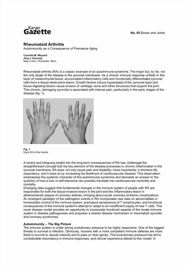
Rheumatoid arthritis (RA) is a classic example of an autoimmune syndrome. The major but, by far, notthe only target of the disease is the synovial membrane. As a chronic immune response unfolds in thislayer of mesenchymal tissue, accumulated inflammatory cells and functionally differentiated synovialcells form a tissue-destructive lesion. Growth factors induce hyperplasia of the synovial layer andtissue-digesting factors cause erosion of cartilage, bone and other structures that support the joint.This chronic, damaging synovitis is associated with intense pain, particularly in the early stages of the
disease (fig. 1).
Fig. 1.Early RA of the hands.
A recent and intriguing insight into the long-term consequences of RA has challenged thestraightforward concept that the key element of the disease processes is chronic inflammation in thesynovial membrane. RA does not only cause pain and disability; more importantly, it shortens lifeexpectancy, and it does so by increasing the likelihood of cardiovascular disease. This observationemphasizes the systemic character of this autoimmune syndrome and demands an answer to thequestion of how a loss in self-tolerance can possibly translate into cardiovascular morbidity andmortality.Emerging data suggest that fundamental changes in the immune system of people with RA areresponsible for both the tissue-invasive lesion in the joint and the inflammatory lesion inatherosclerotic plaque of coronary arteries, bringing about acute coronary ischemic complications.An emergent paradigm of the pathogenic events in RA incorporates new data on abnormalities inhomeostatic control of the immune system, premature senescence of T lymphocytes, and functionalconsequences of the immune system's attempt to adapt to an insufficient supply of new T cells. Thisnovel disease model provides an opportunity to incorporate functional aspects of the innate immunesystem in disease pathogenesis and proposes a shared disease mechanism in rheumatoid synovitisand coronary syndromes.
Autoimmunity – The Big PictureThe immune system is under strong evolutionary pressure to be highly responsive. One of the biggestthreats to survival is infection. Obviously, humans with a more competent immune defense are morelikely to survive to sexual maturity and to pass on their genes. This evolutionary pressure has led toconsiderable redundancy in immune responses, and clinical experience attests to this model. In

8/3/2019 Ra and Cardiovascular Morbidity
http://slidepdf.com/reader/full/ra-and-cardiovascular-morbidity 2/9
patients transplanted with allografts, immune responses can be therapeutically suppressed to such adegree that the totally foreign tissue is not rejected. The ability to immunosuppress an individual for aperiod of years with relative safety emphasizes the surplus of immunocompetence. Yet, the survivaladvantage imposed by an extremely reactive immune system is jeopardized if that system turnsagainst the host and causes self-destruction. Thus, evolutionary pressures selecting for ahyperreactive immune system must be combined with similar pressures optimizing self-tolerance. A
philosophical view of immunity, defense and maintenance of self-integrity may predict that a clusteringof forces to achieve these goals would be most efficient. It remains a strong possibility that efficiencyin host defense and protection of self are counteracting forces with tolerance mechanismssuppressing immunity to a required threshold.Given its complexity and its drive for superb efficiency, the immune system is prone to failure.Unintended recognition of self is the most frequent deficiency of the system. Surprisingly,autoimmunity is not a feature of a young immune system, when the immune network functions at itsprime. Instead, the risk of developing autoimmune disease, including RA, increases with age. Ingeneral, RA manifests in hosts who have passed the apex of their reproductive years and in whomevolutionary pressures towards prompt immune responsiveness are declining. Aging of the immunesystem should be associated with loss of function, and the likelihood of developing RA shouldprogressively decrease. Yet, the opposite is the case; the incidence of RA increases as individualsage. The lesson from this observation is that a dwindling of immunity, as it occurs with advancing age,
does not reduce the risk of autoimmunity. Instead, deteriorating function of the complex immunesystem appears to provide ideal conditions for a breakdown of self-tolerance.The traditional paradigm interprets RA as an aberrant response of the adaptive immune system to an,as-yet-unidentified, arthritogenic antigen, consistent with the view that autoimmunity is a result of overreacting. It has been proposed that T lymphocytes specific for such an arthritogenic antigeninduce a memory response, which is relatively resistant to immunosuppressive therapy. Tissuedestruction has been understood as the sequela of a persistent antigen-driven T- and B-cell response.This model ignores that the risk for RA is inversely related to the functionality of the adaptive immunesystem throughout a lifetime. Recent data provide a foundation from which to view the pathogenesisof RA in a different perspective, namely that of a relative immunoincompetence of the adaptiveimmune system in affected people. We propose that accelerated immunosenescence is the primaryrisk factor for autoimmunity and that several of the proinflammatory characteristics of the immunesystem in RA are facilitated by natural killer (NK) T cells. These cells emerge as a consequence of
insufficient T-cell production and function by bridging the adaptive and innate immune systems.
Premature Immunosenescence in RAGuided by the concept that established T-cell responses are a key factor in the pathogenesis of RA,therapeutic interventions have targeted T lymphocytes. Specifically, experimental trials have exploredwhether depletion of T cells can induce a long-lasting benefit and open a window of opportunity for regenerating T cells that no longer follow a disease-mediating pattern of recognizing arthritogenicantigen. In multicenter trials, people with RA received humanized antibodies to the CAMPATH-1antigen, which is expressed on T and B cells, or to CD4, which is expressed on T-cell subsets.Antibody treatment led to a prompt depletion of circulating T cells. A substantial proportion of treatedpatients had improvement of clinical symptoms, yet disease activity returned while peripheral T-cellnumbers remained suppressed. Indeed, one of the major observations of these trials was thatperipheral T-cell lymphopenia persisted for years. The repertoire of surviving and regenerating T cells
was severely contracted with monoclonal T-cell populations dominating the emergent T-cell pool. Apossible conclusion from these observations was that people with RA have a fundamental defect inthe de novo generation of T cells.Using technology developed in the last few years, we can now estimate the in vivo production of newT cells by the thymus. The thymus is of maximal size during puberty and then undergoes involution.Thymic output can be semiquantified from the amount of episomal DNA in T lymphocytes, which isproduced during T-cell receptor rearrangement. Quantification of such T-cell receptor excision circles(TRECs) in healthy normal donors has demonstrated that TREC-containing T cells decreasesubstantially between the ages of 20 and 60 years. In 60-year-old individuals, TREC concentrationsare reduced to about 5% of those of 20-year-olds, suggesting that involution of the thymus isassociated with a dramatic reduction in the output of newly generated T lymphocytes. Following age60, the vast majority of normal donors have minimal thymic activity, at least under physiologicconditions, as estimated by the presence of TREC-containing T lymphocytes in the peripheral blood
compartment. TREC quantification suggests that people with RA lose thymic function early in life andsuffer from insufficient T-cell production (fig. 2). When compared with age-matched controls, people
8/3/2019 Ra and Cardiovascular Morbidity
http://slidepdf.com/reader/full/ra-and-cardiovascular-morbidity 3/9
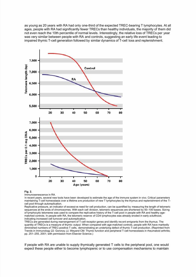
as young as 20 years with RA had only one-third of the expected TREC-bearing T lymphocytes. At allages, people with RA had significantly fewer TRECs than healthy individuals, the majority of them didnot even reach the 10th percentile of normal levels. Interestingly, the relative loss of TRECs per year was very similar between people with RA and controls, suggesting an early life event leading toimpaired thymic T-cell generation followed by similar dynamics of T-cell loss and replenishment.
Fig. 2.
Immunosenescence in RA.In recent years, several new tools have been developed to estimate the age of the immune system in vivo. Critical parametersmaintaining T-cell homeostasis over a lifetime are production of new T lymphocytes by the thymus and replenishment of the T-cell pool through autoreplication.Replicative pressure, an indicator of excessive need for cell production, can be quantified by measuring the length of telomericsequences at the ends of chromosomes. With each cell division, telomeric sequences are shortened by 50–100 bases. Sizingof lymphocytic telomeres was used to compare the replicative history of the T-cell pool in people with RA and healthy age-matched controls. In people with RA, the telomeric reserve of CD4 lymphocytes was already eroded in early adulthood,indicating increased cell turnover and autoreplication.TRECs are generated during rearrangement of T-cell receptor genes and identify recent emigrants from the thymus. Thequantity of TRECs is a measure of thymic output. When compared with age-matched controls, people with RA have markedlydiminished numbers of TREC-positive T cells, demonstrating an underlying defect of thymic T-cell production. (Reprinted fromTrends in Immunology 22: Goronzy JJ, Weyand CM: Thymic function and peripheral T-cell homeostasis in rheumatoid arthritis;pp. 251–255, 2001; with permission from Elsevier Science.)
If people with RA are unable to supply thymically generated T cells to the peripheral pool, one wouldexpect these people either to become lymphopenic or to use compensation mechanisms to maintain

8/3/2019 Ra and Cardiovascular Morbidity
http://slidepdf.com/reader/full/ra-and-cardiovascular-morbidity 4/9
normal T-cell numbers. Lymphopenia occurs in people with Felty’s syndrome. In this subset of people,one finds elevated rheumatoid factor titers and a high likelihood of extra-articular complications.However, in most people with RA, circulating T-cell numbers remain in the normal range. Onemechanism of compensation could be that people with RA increase T-cell turnover by drivingavailable T cells into proliferation. Utilization of this compensation mechanism by people with RA issupported by data on premature erosion of telomeres in CD4 T cells. Telomeric ends of chromosomes
are shortened by about 50 bases with each cell cycle. Thus, the length of telomeric sequences canprovide an estimate of the proliferative history of lymphocyte populations. In healthy people, telomeresof CD4 T cells are progressively shortened with advancing age. Telomeric sizes are maintainedbetween the ages of 20 and 40 years. Accelerated telomeric loss then leads to significant shortening,and a plateau is reached by age 65. The dynamics of telomeric size reflects the need for thelymphocyte pool to be replenished by replication and, therefore, parallels the age-dependentdecrease in thymic T-cell production. In people with RA, telomeres are almost completely eroded by25–30 years of age, indicating that people with RA deplete their telomeric reserve much earlier in life.Premature erosion of lymphocyte telomeres is consistent with the model of accelerated lymphocyteproliferation to compensate for an insufficient influx of thymic emigrants. Additional support for ahistory of excessive autoreplication of CD4 T cells in people with RA has come from studies of thereplicative potential in in vitro systems. CD4 T cells derived from people with RA attain a significantlylower clonal size when driven to proliferate in vitro. Because somatic cells have a limited potential to
divide, excessive autoproliferation to fill the peripheral T-cell pool should restrict the capacity of a T-cell clone to expand in response to antigen. Age-inappropriate telomeric loss and insufficient clonalexpansion in response to T-cell receptor triggering are already present within the naive T-cell subset,making it unlikely that these defects are caused by the disease process itself. Also, telomeric erosionin people with RA is independent of disease duration and does not increase with persistent disease,suggesting that impaired thymic production and compensatory T-cell proliferation are not secondaryevents induced by the chronic inflammatory disease process.
Immunosenescent T Cells – Picking up Bad HabitsThe shift of thymic failure to younger age in people affected by RA should produce prematureimmunosenescence. Because most people with RA do not present with frank lymphopenia, they mustsubstitute with T cells that have been generated independently of the thymus. It is now recognizedthat T cells in the periphery are under constant turnover and represent a dynamic cell population.
Indeed, in models of lymphopenia induced in mice, naive T cells are driven into homeostaticproliferation. Under physiologic conditions (e.g. age-related reduction in T-cell regeneration),homeostasis-driven T-cell development produces functional and self-tolerant T cells. Overexertion of this mechanism has the potential to favor the emergence of functionally distinct T cells that requirerelatively little stimulation to respond. It follows that hosts with increased self-replication of T cells mayassemble a repertoire of T cells with pathogenic potential. It goes without saying that even minor changes in the balance between the influx of novel T cells and the replacement of T cells byautoreplicated clonotypes could have a profound impact on adaptive immune responses. To completethe picture, an additional scenario warrants consideration. If homeostasis-driven T-cell proliferation isinsufficient to compensate for the decline in thymic function, T cells derived from extrathymic sourcesmay fill the void. Again, the composition of the peripheral T-cell compartment in terms of functionalcapabilities and responsiveness to antigen could change dramatically.Possible consequences of increased autoreplication instead of de novo T-cell generation have been
studied in people with RA. The experiments combined T-cell receptor gene amplification andsequence-specific hybridization to estimate T-cell diversity. Oligonucleotide probes specific for arbitrarily selected T-cell receptor sequences were generated to quantify the frequencies of individualclonotypes in the donor. As expected, individual T-cell clonotypes are explicitly infrequent in normalindividuals. More than 50% of T-cell receptor beta-chains can be detected only once, even bysampling as many as 50 million T cells. In people with RA, most T-cell receptor sequences arerepeatedly found, and median frequencies of individual T cells are tenfold higher than in age-matchedcontrol donors. This contraction in T-cell diversity affects not only memory T cells but also naive Tcells. The loss of T-cell receptor diversity reemphasizes that people with RA have replicated T cells tosecure the maintenance of sufficient numbers and to avoid lymphopenia.Contraction of T-cell diversity disobeys one of the fundamental principles of the immune system. Toensure T-cell reactivity to an unlimited spectrum of antigens, the T-cell pool is filled with cellsexpressing a clonally distributed receptor. Clonal expansion of CD4 T cells in people with RA is
common and leads to the outgrowth of large clonal populations. Expanded CD4 clonotypes can beisolated from patients and, thus, have been carefully investigated for their functional characteristics
8/3/2019 Ra and Cardiovascular Morbidity
http://slidepdf.com/reader/full/ra-and-cardiovascular-morbidity 5/9
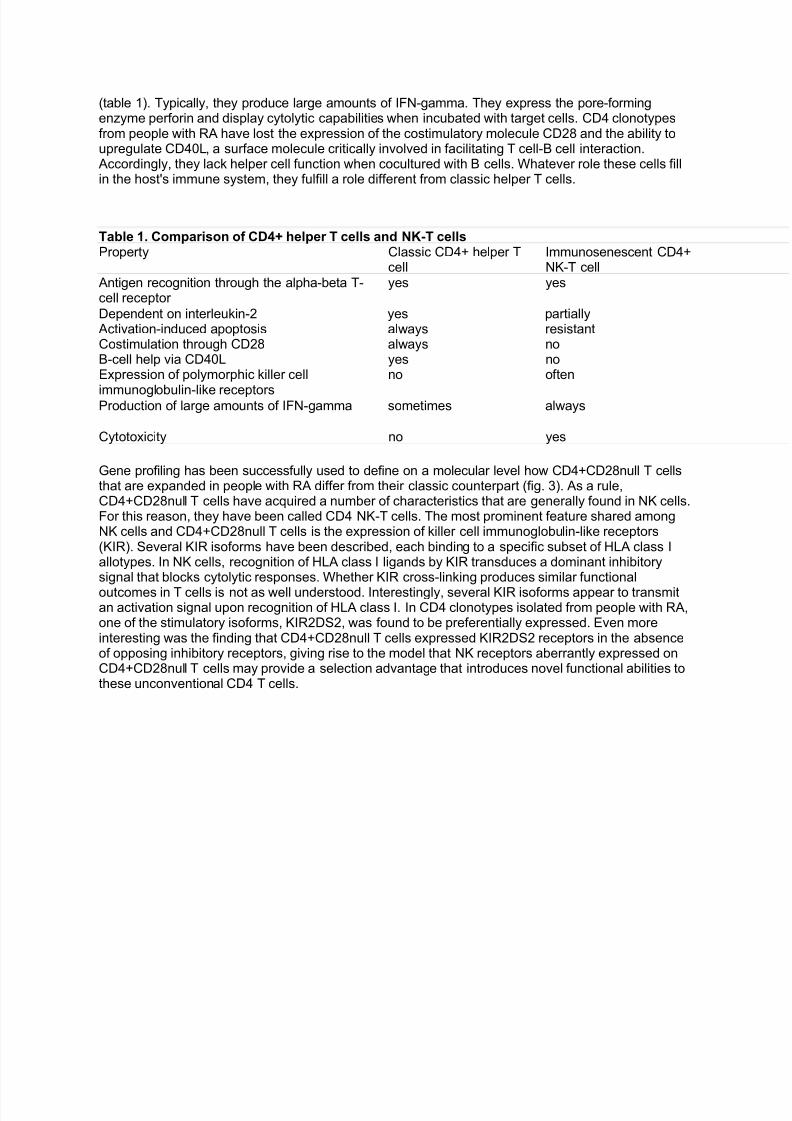
(table 1). Typically, they produce large amounts of IFN-gamma. They express the pore-formingenzyme perforin and display cytolytic capabilities when incubated with target cells. CD4 clonotypesfrom people with RA have lost the expression of the costimulatory molecule CD28 and the ability toupregulate CD40L, a surface molecule critically involved in facilitating T cell-B cell interaction.Accordingly, they lack helper cell function when cocultured with B cells. Whatever role these cells fillin the host's immune system, they fulfill a role different from classic helper T cells.
Table 1. Comparison of CD4+ helper T cells and NK-T cellsProperty Classic CD4+ helper T
cellImmunosenescent CD4+NK-T cell
Antigen recognition through the alpha-beta T-cell receptor
yes yes
Dependent on interleukin-2 yes partiallyActivation-induced apoptosis always resistantCostimulation through CD28 always noB-cell help via CD40L yes noExpression of polymorphic killer cell
immunoglobulin-like receptors
no often
Production of large amounts of IFN-gamma sometimes always
Cytotoxicity no yes
Gene profiling has been successfully used to define on a molecular level how CD4+CD28null T cellsthat are expanded in people with RA differ from their classic counterpart (fig. 3). As a rule,CD4+CD28null T cells have acquired a number of characteristics that are generally found in NK cells.For this reason, they have been called CD4 NK-T cells. The most prominent feature shared amongNK cells and CD4+CD28null T cells is the expression of killer cell immunoglobulin-like receptors(KIR). Several KIR isoforms have been described, each binding to a specific subset of HLA class Iallotypes. In NK cells, recognition of HLA class I ligands by KIR transduces a dominant inhibitorysignal that blocks cytolytic responses. Whether KIR cross-linking produces similar functional
outcomes in T cells is not as well understood. Interestingly, several KIR isoforms appear to transmitan activation signal upon recognition of HLA class I. In CD4 clonotypes isolated from people with RA,one of the stimulatory isoforms, KIR2DS2, was found to be preferentially expressed. Even moreinteresting was the finding that CD4+CD28null T cells expressed KIR2DS2 receptors in the absenceof opposing inhibitory receptors, giving rise to the model that NK receptors aberrantly expressed onCD4+CD28null T cells may provide a selection advantage that introduces novel functional abilities tothese unconventional CD4 T cells.

8/3/2019 Ra and Cardiovascular Morbidity
http://slidepdf.com/reader/full/ra-and-cardiovascular-morbidity 6/9
Fig. 3.Phenotypic and functional properties of senescent CD4 T cells that accumulate in people with RA.Classic helper T cells (left) are equipped with receptors that facilitate communication with other cells, inducing cell activation
and providing T-cell help. The cell surface profile of CD4 NK-T cells (right) is dramatically altered, imposing novel functionalcapabilities on these unconventional T lymphocytes. First, NK-T cells have lost the CD28 molecule, a receptor regulating T-cellreactivity, expansion, and apoptosis. Second, NK-T cells have gained the potential to destroy contacting cells. And third, NK-Tcells have acquired a series HLA class I-specific receptors (KIR) and other receptors (CD161) that are typical in the innateimmune system.
Taken together, in people with RA, a significant proportion of the T-cell compartment is occupied byimmunosenescent CD4 T cells with an altered phenotype and functional profile (fig. 4). The major shiftinvolves the loss of the costimulatory molecules, CD28 and CD40L, and a gain of receptors typicallyfound on cells of the innate immune system. The acquisition of cytotoxic machinery also indicatesadaptation to innate responses. CD4+CD28null KIR+ T cells combine recognition structures from NKand T cells, equipping them with the ability to mediate effector functions of both cell lineages. Thefunctional profile of these T cells strongly supports a direct involvement in disease mechanisms. The
questions to be answered center on their precise contribution to the breakdown of self-tolerance, theinduction and maintenance of chronic inflammation, and the targeting of the disease to the joint.

8/3/2019 Ra and Cardiovascular Morbidity
http://slidepdf.com/reader/full/ra-and-cardiovascular-morbidity 7/9
– – – – – – – – – – – – – – – – – – – – – – – – – – – – – – – – – – – – – – – – – – – – – – – – – –
Fig. 4.The emerging disease model for RA.Insufficient influx of newly generated T cells from the thymus forces the immune system to use compensation mechanisms tomaintain homeostasis. Excessive autoreplication of T cells leads to senescence of the T-cel l pool and the emergence of genetically, phenotypically, and functionally altered CD4 T cells. These senescent CD4 T cells, which have escaped from
tolerance mechanisms, contribute to inflammatory lesions in blood vessels and synovial membranes.
Vascular Injury in RA – From the Joint to the HeartWhile aware of the shortened life expectancy of people with RA, the rheumatology community hasonly recently focused attention on the possible causes of premature death. The model of acceleratedimmunosenescence would predict that manifestations of immunodeficiency, e.g. increasedsusceptibility to infections, could play a role. Side effects induced by therapies could also account for shortened lifespan. However, population-based studies have drawn attention to an increased rate of cardiovascular complications in RA. The recognition of heightened risk for ischemic heart disease inpeople with chronic inflammatory diseases has almost coincided with increasing awareness of inflammatory pathways in atherosclerosis and acute coronary syndromes. A fundamental paradigmshift in the understanding of how atherosclerotic plaque leads to vaso-occlusion and cardiac ischemia
has occurred over the last decade. The old paradigm proposed that incremental growth of atherosclerotic plaque would eventually cause mechanical obstruction. The new paradigmaccommodates data from several distinct directions and focuses on the process of plaque rupture,giving rise to superimposed atherothrombosis. Disruption of atherosclerotic plaque is closely linked tothe presence and functional activity of immune cells in coronary plaque.The population of CD4 T cells infiltrating into unstable coronary plaque includes CD4+CD28null Tcells. These T cells are overrepresented in the blood, undergo clonal expansion, invade theatherosclerotic plaque, and can be isolated from ruptured plaque in cases of fatal myocardialinfarction. We have carefully studied CD4+CD28null T cells in people with unstable angina and havefound them to be identical to the unconventional CD4 T cells in RA. Their functional contribution inunstable angina extends beyond their participation in plaque inflammation. By producing largeamounts of IFN-gamma, CD4+CD28null T-cell clonotypes hyperstimulate macrophages and maintainactivation of the innate immune system.
The emergence of immunosenescent CD4 T cells in RA and unstable angina suggests shareddisease mechanisms in both syndromes (fig. 5). Support for the model comes from a recent studyidentifying KIR2DS2, a receptor expressed on CD4+CD28null T cells, as a disease-risk gene in RA.

8/3/2019 Ra and Cardiovascular Morbidity
http://slidepdf.com/reader/full/ra-and-cardiovascular-morbidity 8/9
Specifically, people with RA who had the KIR2DS2 genotype had a multifold higher risk of progressingto rheumatoid vasculitis. Rheumatoid vasculitis is a frank inflammatory vasculopathy, feared for itsserious clinical complications and known to represent the most severe manifestation of RA. Shareddisease manifestations in RA and in the inflammation of atherosclerotic plaque provide an explanationfor the increased cardiovascular risk of people with RA and also define coronary artery disease as an
immune-mediated disorder. The future lies in exploring how immunosenescent CD4 T cells, andpossibly other immune pathways, participate in vascular wall injury. Immune-dependent tissue injurycould easily cause rupture of atherosclerotic plaque. Could it also be a key mechanism in rheumatoidsynovitis?
Rheumatoid arthritis Acute coronary syndromes
Immune-mediated tissue injury
Immune lesions in the vascular wall
Excessive production of proinflammatory cytokines
Expansion of unconventional CD4 T cells
Inflammation of the synovialmembrane
Autoantibody production
Abnormalities of lipid metabolism
Fig. 5.Pathogenic pathways in rheumatoid arthritis and acute coronary syndromes.Several pathways of immune activation and selection are shared in RA and acute coronary syndromes, providing anexplanation for the increased risk of cardiovascular morbidity in people with RA. Mechanisms shared in these two inflammatorysyndromes must be complemented by additional abnormalities to bias the disease process towards rheumatoid inflammation or inflammation-induced instability of atherosclerotic plaque.
Conclusions and ModelUnconventional CD4 T cells with a proinflammatory functional profile participate in the two major manifestations of rheumatoid disease, rheumatoid synovitis and rheumatoid vascular injury. Theseunconventional CD4 T cells are distinctly infrequent in healthy individuals, but they occupyconsiderable space in the T-cell pool of people with RA. Their expansion is closely related to age-inappropriate failure in T-cell production and appears to be the result of excessive replicative stressimposed by homeostatic mechanisms attempting to maintain cell numbers. The rheumatoid immunesystem is biased towards hyperreactivity, despite failing adaptive immune pathways. Becausemechanisms of immune responsiveness as well as self-tolerance should deteriorate in senescentsystems, even slight inequalities in the sensitivity to age should have profound consequences. Thus,understanding precisely what goes wrong in people with RA comes with the great promise to facilitateunderstanding of how the aging immune system could be turned into a wiser immune system.
AcknowledgementThe authors acknowledge James W. Fulbright for illustrations and assistance with manuscript preparation.
Cornelia M. Weyand and Jörg J. Goronzy are Professors of Medicine and Immunology at Mayo Medical School andGraduate School, Rochester, Minn. Their research program has focused on the immunopathology of rheumatoid arthritis andvasculitis, including studies on acute complications of coronary artery disease. They are the editors of the book RheumatoidArthritis (Current Directions in Autoimmunity, Vol. 3), published by Karger in 2001.
8/3/2019 Ra and Cardiovascular Morbidity
http://slidepdf.com/reader/full/ra-and-cardiovascular-morbidity 9/9
Selected Reading
Goronzy JJ, Weyand CM: T cell homeostasis and autoreactivity in rheumatoid arthritis; in Goronzy JJ,Weyand CM (eds): Rheumatoid Arthritis. Curr Dir Autoimmun. Basel, Karger, 2000, vol 3, pp 112–132.
Goronzy JJ, Weyand CM: Thymic function and peripheral T-cell homeostasis in rheumatoid arthritis.Trends Immunol 2001;22:251–255.
Koetz K, Bryl E, Spickschen K, O'Fallon WM, Goronzy JJ, Weyand CM: T cell homeostasis in patientswith rheumatoid arthritis. Proc Natl Acad Sci USA 2000; 97:9203–9208.
Mackall CL, Hakim FT, Gress RE: Restoration of T-cell homeostasis after T-cell depletion. SeminImmunol 1997;9:339–346.
Weyand CM, Goronzy JJ, Liuzzo G, Kopecky SL, Holmes DR, Frye RL: T-cell immunity in acutecoronary syndromes. Mayo Clin Proc 2001;76:1011–20.
Weyand CM, Klimiuk PA, Goronzy JJ: Heterogeneity of rheumatoid arthritis: From phenotypes togenotypes. Springer Semin Immunopathol 1998;20:5–22.
Yen JH, Moore BE, Scholl D, Schaid DJ, Weyand CM, Goronzy JJ: MHC class-I recognizingreceptors are disease-risk genes in rheumatoid arthritis. J Exp Med 2001;193:1159–1167.
Close Window