r common benign and malignant neoplasms the skinsmj.sma.org.sg/4901/4901te1.pdf · tips from the...
TRANSCRIPT

Tips From The Experts Singapore MedJ2008,49(1):6
CME Article r Common benign and malignant neoplasms of the skin Shim T W H, Naidu S, Lim J, Lim T C
Skin cancer is the seventh most common cancer among
Singaporean males and the eighth commonest cancer among
Singaporean females. Skin cancer is becoming more
common among Singaporeans. From the period of 1968 to
2002, the average percentage increase in the age standardised
rates of skin cancer was 1.27 in males and 1.55 in females.'
BENIGN SKIN LESIONS
Seborrhoeic keratosis and inverted follicular
keratosis
Aetiology
The aetiology of seborrhoeic keratosis (Fig. 1) is unknown.
Patients with great numbers of lesions may have a positive
family history which may reflect a genetic propensity. Its
occurrence may also be related to sun exposure.(23)
Clinical presentation
They commonly occur after the age of 30 years, and can
rgV4,72.1
Fig. 1 Photograph shows seborrhoeic keratosis and inverted
follicular keratosis.
Skin tumours frequently present as a "lump" or "mole" on
the unsuspecting patient. Differentiating and diagnosing
these lesions could also prove to be a daunting challenge
to the clinician. This article provides a brief overview of
the common, or clinically important, skin tumours
encountered in clinical practice.
occur on any part of the body except mucous membranes.
They begin as flat and sharply -demarcated brown macules.
With progression, they develop a polypoidal and an uneven
surface with a characteristic stuck -on appearance.
Follicular prominence is also a characteristic feature
presenting as pale follicular plugs within a darker lesion
or dark (black or brown) plugs within a pale lesion.
Multiple eruptive seborrhoeic keratoses (the sign of Leser-
Trelat) is associated with multiple malignancies, including
carcinomas of the colon, stomach and breast as well as
lymphoma, leukaemias and melanoma.(4)
Histology
This lesion consists of uniform basaloid cells with
interspersed keratin -filled horn cysts. Melanocytes are
also present and melanin production contributes to darker
lesions. Other clinicopathological variants of seborrhoeic
keratosis include reticulated seborrhoeic keratosis, clonal
seborrhoeic keratosis, irritated seborrhoeic keratosis,
stucco keratosis melanoacanthoma and dermatosis
papulosa nigra. Inverted follicular keratosis is believed
to be an inflammatory variant of seborrhoeic keratosis.
It is characterised by an endophytic process within the
epithelium of a pilosebaceous follicle.(5)
Treatment
Treatment is instituted when the lesions are functionally
and cosmetically disturbing. Options include cryotherapy,
electrodesiccation and laser therapy with Q -switched ruby
laser. Surgical excision and histological evaluation are
recommended for lesions that are atypical, and when
malignancy is suspected.
Department of General Surgery, Section of Plastic and Reconstructive Surgery, Tan Tock Seng Hospital, 11 Jalan Tan Tock Seng, Singapore 308433
Shim TWH, MBBS, MRCSE Registrar
Department of Surgery, Division of Plastic, Reconstructive and Aesthetic Surgery, National University Hospital, 5 Lower Kent Ridge Road, Singapore 119074
Naidu S, MBBS, MMed, MRCSE Registrar
Lim J, MMed, FRCSE, FRCS, Senior Consultant
Lim TC, MBBS, FRCSE, FAMS Senior Consultant and Head
Correspondence to: Dr Thiam-Chye Lim Tel: (65) 6772 4240 Fax: (65) 6777 8427 Email:surlimtc@nus. edu.sg

Singapore M ed J 2008; 49 (1) : 7
Sebaceous hyper plasia
Aetiology
In newborns, sebaceous hyperplasia results from maternal androgens causing overgrowth of the sebaceous glands.(s)
In adults, its aetiology is related to decreased androgen levels resulting in decreased cell turnover of the gland,
prolonged ultraviolet radiation exposure and direct causative effect of cyclosporine.(6
Clinical presentation
Sebaceous hyperplasia occurs in individuals beyond the middle age. It presents as elevated soft and yellowish
nodules with a central umbilication at the site of the ductal opening. In Fordyce's disease, similar lesions occur on
the vermillion border of lips and oral mucosa. Sebaceous hyperplasia also commonly occurs in newborns.(7'8) These
are characterised by tiny macules or papules at the opening of each pilosebaceous follicle over the nose and cheeks.
Sebaceous hyperplasia is also seen in solid organ transplant patients, these are thought to be induced by cyclosporine.(6
Histology
Lobules of mature sebaceous glands surround a dilated sebaceous duct which opens into the epidermis or mucosal
surface.(9)
Treatment
Laser therapy, electrodesiccation and topical bichloracetic acid can be used to treat this condition. Oral isotretinoin
is also effective for diffuse multiple lesions. Neonatal sebaceous hyperplasia needs no treatment and will resolve
by 4-6 months.
Syringoma
Clinical presentation
Syringomas are benign tumours of the eccrine ducts. It occurs more commonly in females than in males, with onset
usually at puberty or the third and fourth decade of life. It commonly involves the eyelids, axillae, umbilicus and
pubic area. It is characterised by skin coloured or yellowish firm papules 1-3 mm in size. Eruptive syringoma
(eruptive hidradenoma of Darier and Jaquet) is a rare variant. Large numbers of lesions occur on the neck, chest and
abdomen, which may remain or disappear.(10)
Histology
The lesions consist of small cystic ducts and are lined by solid epithelial strands embedded within a fibrous stroma.
Some of the epithelial cells possess a comma -like tail assuming a tadpole appearance. The ducts may connect with
dilated cysts of intraepidermal ducts but do not connect with the secretory segment of glands.(5,I1)
Treatment
These lesions can be treated with excision, electrodesiccation and curettage, dermabrasion and CO2 laser resurfacing.
X ant homa
Aetiology
It is caused by lipid (LDL) deposition under the skin or
other sites, such as tendons or gastrointestinal tract.
Possible mechanisms include capillary leakage of LDL,
increased acetylated LDL or oxidised LDL uptake into
macrophages and increased local tissue lipogenesis.(12)
Clinical presentation
They present as soft, flat yellowish plaques with sharply
defined margins (Fig. 2). They can occur on any part of
k
1 } "7,'3+_o r.
Fig. 2 Photograph shows a xanthelasma, a xanthoma of the
eyelid.
Singapore M ed J 2008; 49 (1) : 8
the body, such as elbows, knees, hands, feet, buttocks or eyelids (xanthelasma). They may be a sign of an underlying
disorder, such as hyperlipidaemia, primary biliary cirrhosis, diabetes mellitus or familial hypercholesterolaemia.
Histology
They consist of xanthoma cells, foamy histiocytes laden with intracellular lipids (mainly cholesterol) in the upper
dermis.(12
Treatment
Excision, laser ablation, chemical cauterisation and electrodesiccation can be used, in addition to treating the
underlying lipid disorder. The patient must be warned of recurrence.(13)
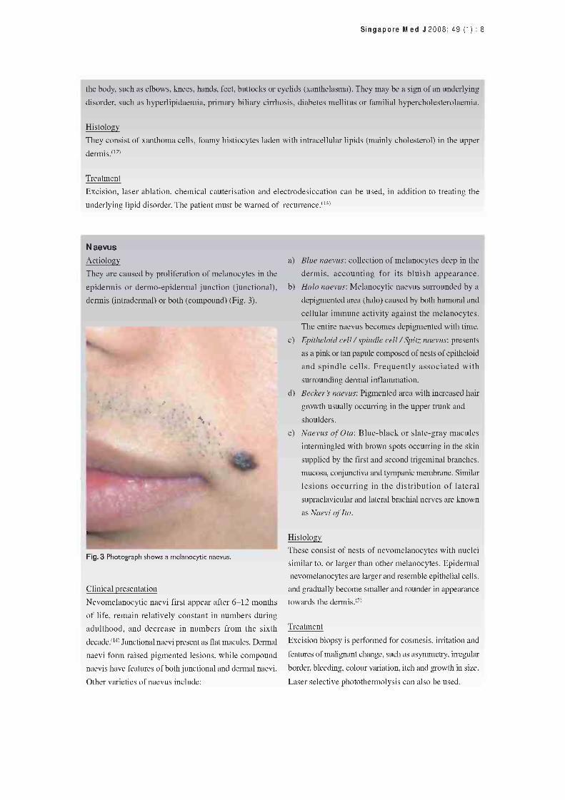
N aevus Aetiology
They are caused by proliferation of melanocytes in the
epidermis or dermo-epidermal junction (junctional),
dermis (intradermal) or both (compound) (Fig. 3).
Fig. 3 Photograph shows a melanocytic naevus.
Clinical presentation
Nevomelanocytic naevi first appear after 6-12 months
of life, remain relatively constant in numbers during
adulthood, and decrease in numbers from the sixth
decade.(14) Junctional naevi present as flat macules. Dermal
naevi form raised pigmented lesions, while compound
naevis have features of both junctional and dermal naevi.
Other varieties of naevus include:
a)
b)
c)
d)
e)
Blue naevus: collection of melanocytes deep in the
dermis, accounting for its bluish appearance.
Halo naevus: Melanocytic naevus surrounded by a
depigmented area (halo) caused by both humoral and
cellular immune activity against the melanocytes.
The entire naevus becomes depigmented with time.
Epitheloid cell / spindle cell / Spitz naevus: presents
as a pink or tan papule composed of nests of epitheloid
and spindle cells. Frequently associated with
surrounding dermal inflammation.
Becker's naevus: Pigmented area with increased hair
growth usually occurring in the upper trunk and
shoulders.
Naevus of Ota: Blue -black or slate -gray macules
intermingled with brown spots occurring in the skin
supplied by the first and second trigeminal branches,
mucosa, conjunctiva and tympanic membrane. Similar
lesions occurring in the distribution of lateral
supraclavicular and lateral brachial nerves are known
as Naevi of Ito.
Histology
These consist of nests of nevomelanocytes with nuclei
similar to, or larger than other melanocytes. Epidermal
nevomelanocytes are larger and resemble epithelial cells,
and gradually become smaller and rounder in appearance
towards the dermis.(s)
Treatment
Excision biopsy is performed for cosmesis, irritation and
features of malignant change, such as asymmetry, irregular
border, bleeding, colour variation, itch and growth in size.
Laser selective photothermolysis can also be used.

Singapore M ed J 2008; 49 (1) : 9
Cutaneous horn Treatment
Clinical presentation Shave biopsy, including the base, is both diagnostic and
It usually occurs in the elderly, and presents as a conical, curative.
dense, hyperkeratotic nodule due to unusual cohesiveness
of keratinised material. It may be white or yellowish and
straight, curved or twisted (Fig. 4). It may result from
solar keratoses, seborrhoiec keratoses, filiform warts, t i
trichilemmal keratoses, keratoacanthomas and basal cell
epitheliomas. There is a need to exclude premalignant
and malignant lesions in cutaneous horns.(is) Lesions with
a low height -to -base ratio is more likely to be malignant.('6
Histology
Areas of hyperkeratosis and parakeratosis are seen. A
granular layer may be present under the hyperkeratotic
areas with variable degree of acanthosis. Fig. 4 Photograph shows a cutaneous horn on the forehead.
Epidermoid cyst
Aetiology
This lesion occurs as a result of proliferation of surface epidermal cells within the dermis. Epidermoid cysts
(Figs. 5 a-b) result from occlusion of the pilosebaceous follicle, implantation of epidermal cells into the dermis
following penetrating injury, or trapping of epidermal cells along embryonal fusion planes.
Clinical presentation
Presents as a dome -shaped lesion, tethered to the overlying skin, and is freely mobile over the underlying structures.
The central punctum represents the obstructed orifice of the pilosebaceous follicle. The lesion is filled with keratinaceous
material.
Histology
These cysts are lined with stratified squamous epithelium and are filled with keratinous material arranged in multiple
layers, and sometimes contain melanin and calcified material.
Treatment
Excision biopsy. Infected cysts should be treated with a course of antibiotics, failing which drainage of the abscess
is needed. Excision is then carried out after the infection and inflammation have subsided.
5a -77;:1`r "
5b
Fig. 5a Photograph shows a cyst at the postauricular surface. Fig. 5b Photograph shows a cyst at the right check.

Singapore M ed J 2008; 49 (1) : 10
Pyogenic granuloma
Aetiology unknown
Clinical presentation
Benign vascular lesion of the skin or mucosa (Fig. 6). It
presents as a red nodule or papule which develops rapidly
over a period of weeks, and is prone to bleeding and
ulceration. May be associated with drug ingestiono7)
(systemic retinoids, Indinavir) orpregnancy(18) (in second
and third trimesters).
Histology
Numerous capillaries and venules arranged in radial
pattern within an oedematous stroma containing variable
amounts of inflammatory infiltrates. A regressing lesion
shows extensive fibrosis.
Treatment
Excision biopsy, shave biopsy and electrocautery,
cryotherapy, laser therapy.(19)
H aemangioma
Aetiology
This lesion may represent a hamartomatous proliferation
of endothelial cells (Fig. 7).
Clinical presentation
Its onset is usually after birth. Development is divided
into proliferative (rapid growth for 3- 9) months, up to
18 months) and involutive phase (over 2-6 years). It
presents as an erythematous macule or ecchymotic patch
with irregular borders occasionally. Large lesions may
be associated with ptosis, obstruction of vision, high
output cardiac failure, thrombocytopenia or haemolytic
anaemia. 50% of the cases will regress by five years and
70% will regress by seven years of age.(2°)
Histology
Proliferation of endothelial cells and pericytes with
formation of vascular spaces. The involution phase is
characterised by progressive fibrosis and disappearance
of blood vessels.
Fig. 6 Photograph shows a pyogenic granuloma at the upper
lip.
Treatment
Intralesional steroid injections can be given for small
haemangiomas. Large life -threatening haemangiomas are
treated with systemic glucocorticoids and interferon alfa -
2a and 2b. Surgical excision is performed after regression
has ceased, or when complications such as bleeding and
ulceration arise. 2°)
Fig. 7 Photograph shows haemangioma at the right upper
eyelid.

Singapore M ed J 2008; 49 (1) : 11
PREMALIGNANT SKIN LESIONS
Congenital naevus
Aetiology
They develop probably between 40 days of gestation and
six months in utero. Genetic mechanisms may account
for familial aggregation.
Clinical presentation
These lesions are present at birth. They are characterised
by pigmented lesions with regular margins, smooth or
lobular surfaces (Fig. 8) and occasionally have long coarse
hair. The risk of melanoma development is proportional
to the size, especially if it involves over 5% of body
surface, or > 20 cm in adolescents (large/giant congenital
naevus). The risk of malignant change ranges from 5%
to 40%.(21)
Histology
Similar to acquired naevus, but frequently involves the
lower dermis and more likely to involve the dermal
appendages and neurovascular structures.
Treatment
Regular follow-up and prophylactic excision, preferably
before onset of puberty. 21,22
Actinic keratosis
Aetiology
Actinic keratosis (Fig. 9) occurs in areas exposed to
sunlight. UV -B radiation induces thymidine dimer
formation in DNA and RNA, as well as p53 gene mutations
and telomerase alterations.(23)
Clinical presentation
These lesions are more common in fair -skinned and blue-
eyed individuals with chronic sun exposure and in
immunosuppressed individuals. They occur in sun -exposed
areas, presenting as rough, scaly papules and plaques.
The risk of evolving to squamous cell carcinoma is
13%-20% over ten years.(232)
Histology
Atypical, pleiomorphic keratinocytes occur in the basal
cell layer which may extend to the granular and cornified
layers with hyperkeratosis and parakeratosis.
Fig. 8 Photograph shows a giant hairy naevus.
Treatment
Cryosurgery, topical fluorouracil, curettage, topical
tretinoin, imiquimod 5% (interferon inducer) and
photodynamic therapy with 5-aminolevulinic acid.
Reduced sun exposure and regular use of sunblock prevent
its development.(23)
'.4
Fig. 9 Photograph shows actinic keratosis over the sealp.

Singapore Med J 2008; 49 (1) : 12
Keratoacant homa
Aetiology
The tumour arises from hair follicles, and is caused by
exposure to sun and carcinogens, such as pitch and tar.
Other aetiological factors, such as trauma, human
papilloma virus, genetic and immunosuppression, have
been implicated.(2s)
Clinical presentation
This lesion presents as a bud -shaped or dome -shaped
lesions with a central keratinous crater (Figs. 1Oa-b).
Rapid growth occurs within a period of six weeks, followed
by a period of involution over 4-6 months, leaving a
pitted scar.(252) Progression to squamous cell carcinoma
is rare.(26)
Histology
It is composed of singularly well -differentiated squamous
epithelium with little pleiomorphism and anaplasia with
masses of keratin. Pseudocarcinomatous infiltration does
not extend beyond the level of hair follicles and cutaneous
glands.
Treatment
Surgical excision, intralesional 5-fluorouracil, bleomycin,
steroids and methotrexate or systemic isotretinoin. Low -
dose irradiation can also be employed.(26)
Sebaceous naevus of ..bdassohn
Aetiology
This may arise from pluripotential primary epithelial germ
cells. Mutations in these cells may give rise to hamartomas
with multiple cell lines.
Clinical presentation
Presents as a yellow -brown lesion with a linear
configuration (Figs. lla-c). Its development may be
related to hormonal fluctuation-raised appearance at
birth then flattened during childhood and raised again
during puberty. 5%-7% may develop into BCC. May
also develop into many other benign and malignant
tumours.
Histology
Papillomatous hyperplasia is present in the epidermis
with numerous sebaceous glands in the dermis. Apocrine Treatment
glands, small hair follicles and buds of basaloid cells are Surgical excision before puberty, after which the risk of
also present. malignancy increases.
10a
1111114;
10b
Fig. 10 Photographs show (a) umbilicated keratocanthoma at
the cheek; and (b) keratocanthoma at the forehead.
11a
1
1
G - 1 º o ° _.;# á
I . .,.-,
Le .ç 6
S
6% 4° ' Fig. 11 Photograph shows (a) sebaceous naevus at scalp for case I.

Singapore Med J 2008; 49 (1) : 13
11b
r, 17^
.
r !'y _,6 I
f
11c
Fig. 11 Photographs show sebaceous naevus (b) at scalp for case 2; and (c) at trunk for case 3.
MALIGNANT SKIN LESIONS
Squamous cell carcinoma
Aetiology
Ultraviolet irradiation from sun exposure, infrared irradiation or X-ray; chemical carcinogens such as arsenic,
hydrocarbons, tar and pesticides; viral agents such as human papilloma virus; chronic wounds such as Marjolin's
ulcer and chronic scars; and impaired immunity either from immunosuppression or immunodeficiency.(27,28)
Clinical presentation
It is the second commonest skin cancer in Singapore.(') Invasive squamous cell carcinoma (SCC) can arise de novo
or from precursor lesions, such as actinic keratosis, Bowen's disease, erythroplasia of Queyrat, leukoplakia and
intraepidermal epithelioma. Depending on the site of
occurrence, it can present as a raised, pink -coloured 12a
keratotic papule or plaque; an area of induration with
erosion and ulceration (oral SCC); moist red plaques with
subsequent induration and ulceration (anogenital SCC);
white plaque (intraoral leukoplakia); or exophytic o ` i+;
fungating lesions (verruca variant of SCC). As these
lesions progress, they become locally invasive and
destructive, fixed to underlying and adjacent structures
and metastasise to distant sites (Figs. 12a -c).
- T
12b 12c
Fig. 12 Photographs show (a) SCC at scalp; (b) Bowen's disease with SCC; and (c) SCC at the upper lip.

Singapore Med J 2008; 49 (1) : 14
Histology
There is proliferation of atypical keratinocytes in architectural disarray. Hyperkeratosis, parakeratosis and acanthosis
is present. The atypical keratinocytes display pleiomorphism, hyperchromatic nuclei and mitoses. Keratinisation also
occurs, forming horn pearls composed of concentric layers of squamous cells and keratin. Histological variants of
SCC include adenoid, spindle cell, clear cell, papillary and signet ring SCC.(5)
Treatment
Surgical excision with a 4-6 mm margin, cryotherapy, electrodesiccation and curettage and Mohs micrographic
surgery can be performed with excellent cure rates. 27 Other treatment modalities include laser ablation, photodynamic
therapy, local immunotherapy (Imiquimod) and local chemotherapy (5 -FU). Prevention by decreased exposure to
UV radiation should be advised. Chemoprevention with oral retinoic acid agents are being evaluated.(29)
Basal cell carcinoma Aetiology
Basal cell carcinoma (BCC) arises from the pluripotential cells of the basal cell layer of the epidermis and follicular
structures. UV -induced mutation of the p53 gene and tumour suppressor genes in chromosome 9 (Gorlin syndrome,
naevoid basal cell carcinoma) and mutations of the patched (PTCH) gene in the patched hedgehog pathway have
been implicated. Arsenic exposure and immunosuppression also increases the risk of BCC.(5,30)
Clinical presentation
BCC (Figs. 13 a-c) is the commonest skin cancer in Singapore with the commonest subtype being nodular BCC.
It is characterised by a dome -shaped papule with telangiectasia and pearly -white border associated with a central
crusted or ulcerated area. Other clinical variants of BCC include:
a) Superficial BCC: Erythematous scaly plaque with a raised border and occasional central ulceration.
b) Pigmented BCC: Presence of brown and black pigment within the BCC.
c) Cystic BCC: presents as blue -gray cystic nodules.
d) Micronodular BCC: Clinically similar to nodular BCC but associated with subclinical extension through the
dermis which may be widespread.
e) Morpheaform/sclerosing BCC: Presents as a whitish plaque with ill-defined margins and associated with aggressive
growth.
13a ° 13b
. 13c ' __ _-
Fig. 13 Photographs show (a) BCC with rodent ulcer; (b) pigmented BCC; and (c) nodular BCC.

Singapore M ed J 2008; 49 (1) : 15
Histology
The cells have large, oval hyperchromatic nuclei with scanty cytoplasm. The peripheral cells are arranged in a
palisading pattern. Tumour masses are surrounded by mucinous stroma upon which tumour growth is dependent.
Chronic inflammatory infiltrates are also commonly seen around the tumour. High risk of recurrence is seen in the
infiltrative tumour edge, tumour cells forming strands (morpheaform pattern), poor or absent peripheral palisading
and marked nuclear pleiomorphism.
Treatment
Surgical excision with a 4 -mm margin,(31) Mohs micrographic surgery, cryotherapy and electrodesiccation with
curettage. Topical-FU, Imiquimod, radiotherapy and intralesional interferon alfa -2b,02'33) have been used to treat
superficial BCC.
Malignant melanoma Aetiology
Risk factors include family history, multiple benign or atypical naevi, previous melanoma, immunosuppression, sun
sensitivity and UV irradiation. The progression of a benign naevus to malignant melanoma occurs in a stepwise
fashion. N -Ras (retrovirus-associated DNA sequences), BRAF (regulation of a-foetoprotein) gene mutations and
abnormal activation of MAPK (mitogen-activated protein kinase) result in melanocyte hyperplasia. Mutations of
CDKN2A (cyclin-dependent kinase inhibitor 2A), resulting in inactivation of tumour suppressor genes p16 and p19
and PTEN (phosphatase and tensin homologue), would then result in cytologic atypia and formation of dysplastic
naevus. Increased PKB (protein kinase B) activity and increased cyclin D1 expression results in uncontrolled
hyperplasia, clonal proliferation and decreased differentiation, precipitating in a radial growth phase. E-cadherin
loss, with increased N cadherin and aV (33 integrin expression results in a vertical growth phase. Finally, the absence
of TRPM1 (melastatin 1) results in metastatic spread.(34)
Clinical presentation
Melanoma (Figs. 14 a-c) is rare in Singapore, and can be classified into several types as follows:
a) Superficial spreading melanoma: Most common type. Presents as a deeply pigmented macule or slightly raised
plaque with colour variegation.
b) Lentigo maligna melanoma: Least common. Occurs in the older age group. Presents as tan, brown or black flat
lesions with convoluted borders and prominent notching.
c) Acral lentiginous melanoma: More common in darker pigmented individuals. Occur in the sole, palm or beneath
the nail plate. Presents as tan, brown or black flat lesions or nodules/papules. Hutchinson's sign (pigmentation
of the posterior nail fold) in subungal melanoma is an ominous sign.
d) Nodular melanoma: Commonly arises de novo or from pre-existing naevi. Presents as a dark blue -black or bluish -
red nodule or papule or even as a polypoid lesion with a stalk.
e) Amelanotic melanoma: Melanomas which lack pigment altogether.
Histology
a) Superficial spreading melanoma: Malignant melanocytes expand in multiple layers within the epidermis, and
superficial papillary body of the dermis, in the radial growth phase. In the vertical growth phase, they extend
into the reticular dermis and beyond.
b) Lentigo maligna melanoma: Consists of atypical melanocytes initially lined in a single layer along the basal
layer above the basement membrane, and subsequently invades the dermis and deeper tissues.
c) Acral lentiginous melanoma: Macular areas consist of large melanocytes with large atypical nuclei and elongated
dendrites which extend to the granular layer. In the papular/nodular areas, the melanocytes assume a spindle
shape and extend into the dermis.
d) Nodular melanoma: It arises in the dermo-epidermal junction and invasion of the dermis may also occur
with invasion of the epidermis. Consists of large epitheloid cells, spindle cells, small cells or combinations of
these.

Singapore M ed J 2008; 49 (1) : 16
Treatment
Patients should be staged. Stages I and II denote local cutaneous disease, stage III disease involves regional nodes,
while stage IV disease is associated with distant metasases. The depth of invasion can be classified according to the
Breslow or Clark levels. The treatment of melanoma is surgical excision with a margin of 0.5-2 cm, depending on
the thickness of the tumour. Sentinel lymph node biopsy aid in prognostication, but both elective lymph node or
sentinel lymph node dissection do not confer survival advantage. Lymph node dissection can be performed in recurrent
disease to improve local palliative control. Interferon a-2ß is the only FDA approved adjuvant therapy in patients
with high risk of disseminated disease. It increases disease -free survival but not overall survival.(35)
14a 14b
}
14c
Fig. 14 Photographs show (a) superficial spreading melanoma; (b) acral lentiginous melanoma at the left sole; and (c) nodular
melanoma.
CONCLUSION
It is sometimes difficult to differentiate benign from malignant skin tumours. Clinical features suggestive of malignancy
include rapid growth in size, change in colour, presence of satellite nodules, asymmetry, contact bleeding and itch.
Malignant skin cancers are also more common among fair -skinned individuals and hence, among the Chinese race
in Singapore.(36) A high index of suspicion must be maintained, especially in fair -skinned patients. Suspicious lesions
should be biopsied to obtain definitive histological diagnosis. As malignant lesions frequently resemble one another
rather closely, histological analysis may be the only means of reaching a definitive diagnosis.

Singapore Med J 2008; 49 (1) : 17
REFEREN CES 1. Seow A, Koh WP, Chia KS, et al. Trends in Cancer Incidence in
Singapore: 1968-2002. Singapore Cancer Registry Report no. 6, 2004.
2. Kwon OS, Hwang EJ, Bae JH, et al. Seborrheic keratosis in the Korean
males: causative role of sunlight. Photodermatol Photoimmunol Photomed 2003; 19:73-80.
3. Yeatman JM, Kilkenny M, Marks R. The prevalence of seborrhoeic
keratoses in an Australian population: does exposure to sunlight play
a part in their frequency? Br J Dermatol. 1997; 137:411-4.
4. Schwartz RA. Sign of Leser -Trélat. J Am Acad Dermatol 1996;
35:88-95.
5. Freedberg IM, Eisen AZ, Wolff K, et al. Fitzpatrick's Dermatology in
General Medicine. 5th ed. New York: McGraw-Hill.
6. Pang SM, Chau YP. Cyclosporin-induced sebaceous hyperplasia in
renal transplant patients. Ann Acad Med Singapore 2005; 34:391-3.
7. Moosavi Z, Hosseini T. One-year survey of cutaneous lesions in 1000
consecutive Iranian newborns. Pediatr Dermatol 2006; 23:61-3.
8. Rivers JK, Frederiksen PC, Dibdin C. A prevalence survey of dermatoses
in the Australian neonate. J Am Acad Dermatol 1990; 23:77-81.
9. Elder DE, Elenitsas R, Johnson BL Jr, Murphy GF. Lever's Histopathology of the Skin. 9th ed. Philadelphia: Lippincott Williams
and Wilkins, 2004.
10. Nguyen DB, Patterson JW, Wilson BB. Syringoma of the moustache
area. J Am Acad Dermatol 2003; 49:337-9.
11. Lee JH, Chang JY, Lee KH. Syringoma: a clinicopathologic and
immunohistologic study and results of treatment. Yonsei Med J 2007;
48:35-40.
12. Russo GG. Hyperlipidemias. Clin Dermatol 1996; 14:367-74.
13. Rohrich RJ, Janis JE, Pownell PH. Xanthelasma palpebrarum: a review
and current management principles. Plast Reconstr Surg 2002;
110:1310-4.
14. MacKie RM, English J, Aitchison TC, Fitzsimons CP, Wilson P. The
number and distribution of benign pigmented moles (melanocytic naevi) in a healthy British population. Br J Dermatol 1985;
13:167-74.
15. Menda -Gutiérrez E, Gutiérrez -Díaz E, Redondo -Marcos I, Ricoy JR,
García -Torre JP. Cutaneous horns of the eyelid: a clinicopathological
study of 48 cases. J Cutan Pathol 2004; 31:539-43.
16. Yu RC, Pryce DW, Macfarlane AW, Stewart TW. A histopathological
study of 643 cutaneous horns. Br J Dermatol 1991; 124:449-52.
17. Segaert S, Van Cutsem E. Clinical signs, pathophysiology and
management of skin toxicity during therapy with epidermal growth
factor receptor inhibitors. Ann Oncol 2005; 16:1425-33.
18. Kanda N, Watanabe S. Regulatory roles of sex hormones in cutaneous
biology and immunology. J Dermatol Sci 2005; 38:1-7.
19. Ghodsi SZ, Raziei M, Taheri A, et al. Comparison of cryotherapy and
curettage for the treatment of pyogenic granuloma: a randomized trial.
Br J Dermatol 2006; 154:671-5.
20. Sundine MJ, Wirth GA. Hemangiomas: an overview. Clin Pediatr (Phila). 2007; 46:206-21.
21. Tannous ZS, Mihm MC Jr, Sober AJ, Duncan LM. Congenital melanocytic nevi: clinical and histopathologic features, risk of melanoma, and clinical management. J Am Acad Dermatol 2005;
52:197-203.
22. Zaal LH, Mooi WJ, Sillevis Smitt JH, van der Horst CM. Classification
of congenital melanocytic naevi and malignant transformation: a review
of the literature. Br J Plast Surg 2004; 57:707-19.
23. Fu W, Cockerell C J. The actinic (solar) keratosis : a 21st -century
perspective. Arch Dermatol 2003; 139:66-70.
24. Butani AK, Arbesfeld DM, Schwartz RA. Premalignant and early
squamous cell carcinoma. Clin Plast Surg 2005; 32:223-35.
25. Karaa A, Khachemoune A. Keratoacanthoma: a tumor in search of a
classification. Int J Dermatol 2007; 46:671-8.
26. Schwartz RA. Keratoacanthoma: a cinico -pathologic enigma. Dermatol
Surg 2004; 30:326-33.
27. Rudolph R, Zelac DE. Squamous cell carcinoma of the skin. Plast
Reconstr Surg 2004; 114:82e -94e.
28. Pfister H. Chapter 8: Human papillomavirus and skin cancer. J Natl
Cancer Inst Monogr 2003; 31:52-6.
29. Wright TI, Spencer JM, Flowers FP. Chemoprevention of nonmelanoma
skin cancer. J Am Acad Dermatol 2006; 54:933-46.
30. Gloster HM Jr, Neal K. Skin cancer in skin of color. J Am Acad
Dermatol 2006; 55:741-60.
31. Walker P, Hill D. Surgical treatment of basal cell carcinomas using
standard postoperative histological assessment. Australas J Dermatol
2006; 47:1-12.
32. Rubin AI, Chen EH, Ratner D. Basal -cell carcinoma. N Engl J Med
2005; 353:2262-9.
33. Ceilley RI, Del Rosso JQ. Current modalities and new advances in the
treatment of basal cell carcinoma. Int J Dermatol 2006; 45:489-98.
34. MillerAJ, Mihm MC Jr. Mechanisms of Disease: Melanoma. N Engl
J Med 2006; 355:51-65.
35. Markovic SN, Erickson LA, Rao RD, et al. Malignant melanoma in
the 21st century, part 2: staging, prognosis, and treatment. Mayo Clin
Proc 2007; 82:490-513.
36. Koh D, Wang H, Lee J, et al. Basal cell carcinoma, squamous cell
carcinoma and melanoma of the skin: analysis of the Singapore Cancer
Registry data 1968-97. Br J Dermatol 2003; 148:1161-6.

Singapore M ed J 2008; 49 (1) : 18
SINGAPORE MEDICAL COUNCIL CATEGORY 3B CME PROGRAMME Multiple Choice Questions (Code SMJ 200801 A)
Question 1. The following are features suggestive of skin malignancy:
(a) Rapid growth in size.
(b) Well -demarcated and regular margins.
(c) Presence of satellite nodules.
True False
(d) Atypical site of occurrence on the body.
Question 2. Indicate whether the following statements are true or false:
(a) Skin cancers are more common among the Malay and Indian races.
(b) Malignant melanoma is common in Singapore.
(c) The most common skin cancer among Singaporeans is squamous cell carcinoma.
(d) The incidence rate of skin cancers among Singaporeans is increasing gradually.
Question 3. The following are subtypes of basal cell carcinoma:
(a) Superficial spreading.
(b) Nodular and micronodular.
(c) Epitheloid.
(d) Morpheaform.
Question 4. The following skin tumours are caused by sunlight -induced damage:
(a) Bowen's disease.
(b) Seborrhoiec keratoses.
(c) Marjolin's ulcer.
(d) Basal cell carcinoma.
Question 5. Indicate whether the following statements are true or false:
(a) Xanthomas are caused by LDL deposition in tissues.
(b) Haemangiomas grow rapidly from birth up to 18 months, and subsequently involute over
a period of 2-6 years.
(c) Cutaneous horns are never associated with any malignant potential.
(d) The risk of malignant change of a naevus is higher when it involves more than 5% of the
body surface or is larger than 20 cm.
Doctor's particulars:
Name in full.
MCR number: Specialty
Email address.
SUBMISSION INSTRUCTIONS: (1) Log on at the SMJ website: http://www.sma.org.sg/cme/smj and select the appropriate set of questions. (2) Select your answers and provide your name, email address and MCR number. Click on "Submit answers" to submit.
RESULTS: (1) Answers will be published in the SMJ March 2008 issue. (2) The MCR numbers of successful candidates will be posted online at www.sma.org.sg/cme/smj by 15 March 2008. (3) All online submissions will receive an automatic email acknowlegment. (4) Passing mark is 60%. No mark will be deducted for incorrect answers. (5) The SMJ editorial office will submit the list of successful candidates to the Singapore Medical Council.
Deadline for submission: (January 2008 SMJ 3B CME programme): 12 noon, 25 February 2008.