quick guide – nobelguide radiographic guide guide* – nobelguide™ radiographic guide q teeth...
TRANSCRIPT
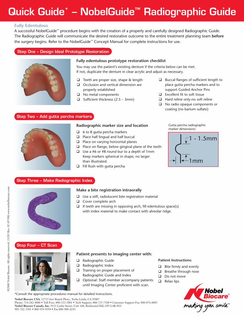
Quick Guide* – NobelGuide™ Radiographic Guide
qTeeth are proper size, shape & length qOcclusion and vertical dimension are properly establishedq No metal components qSufficient thickness (2.5 – 3mm)
qBuccal flanges of sufficient length to place gutta percha markers and to support Guided Anchor PinsqExcellent fit to soft tissueqHard reline only-no soft relineqNo radio opaque components or coating (no barium sulfate)
You may use the patient’s existing denture if the criteria below can be met. If not, duplicate the denture in clear acrylic and adjust as necessary.
Radiographic marker size and location
q6 to 8 gutta percha markers qPlace half lingual and half buccalq Place on varying horizontal planes qPlace on flange, below gingival plane of the teethqUse a #6 or #8 round bur to a depth of 1mm Keep markers spherical in shape, no larger than illustrated.qFill flush with gutta percha
Make a bite registration intraorally
qUse a stiff, radiolucent bite registration material qCover complete arch q If teeth are missing in opposing arch, fill edentulous space(s) with index material to make contact with alveolar ridge.
Patient presents to imaging center with:
qRadiographic Guide qRadiographic Indexq Training on proper placement of Radiographic Guide and Index qOptional: Staff member accompany patients until Imaging Center proficient with scan.
Patient Instructions:
q Bite firmly and evenly qBreathe through noseqDo not moveqRelax lips
Fully edentulous prototype restoration checklist
Fully Edentulous A successful NobelGuide™ procedure begins with the creation of a properly and carefully designed Radiographic Guide. The Radiographic Guide will communicate the desired restorative outcome to the entire treatment planning team before the surgery begins. Refer to the NobelGuide™ Concept Manual for complete instructions for use.
*Consult the appropriate procedures manual for detailed instructions.
Nobel Biocare USA. 22715 Savi Ranch Pkwy., Yorba Linda, CA 92887 Phone: 714-282-4800 • Toll Free: 800-322-5001 • Tech Support: 888-725-7100 • Customer Support Fax: 800-876-8805Nobel Biocare Canada, Inc. 9133 Leslie Street, Unit 100, Richmond Hill, ON L4B 4N1905-762-3501 • 800-939-9394 • Fax 800-900-4243
Step One – Design Ideal Prototype Restoration
Step Two – Add gutta percha markers
Step Four – CT Scan
Step Three – Make Radiographic Index
1mm
1 - 1.5mm
Gutta percha radiographic marker dimensions:
©20
08 N
obel
Bio
care
. All
righ
ts r
eser
ved.
212
05 R
ev. 0
2 (0
7/08
) ww
w.no
belb
ioca
re.c
om

Quick Guide* – NobelGuide™ Radiographic Guide Partially Edentulous A successful NobelGuide™ procedure begins with the creation of a properly and carefully designed Radiographic Guide. The Radiographic Guide will communicate the desired restorative outcome to the entire treatment planning team before the surgery begins. Refer to the NobelGuide™ Concept Manual for complete instructions for use.
qTeeth are proper size, shape & length qOcclusion and vertical dimension are properly establishedq No metal components qSufficient thickness (2.5 – 3mm)q Cover full palate if appropriate qAdd at least 4 inspection windows to ensure proper seating upon insertion
q Do not add acrylic to occlusal/incisal surfaces of areas to be restored qBuccal flanges of sufficient length to place gutta percha markers and support Guided Anchor Pins (if required)qNo radio opaque components or coating (no barium sulfate)
Radiographic marker size and location
q6 to 8 gutta percha markers qPlace half lingual and half buccalq Place on varying horizontal planes qPlace on flange, below gingival plane of the teethq Use a #6 or #8 round bur to a depth of 1mm Keep markers spherical in shape, no larger than illustrated.qFill flush with gutta percha
Make a bite registration on the articulator.
qOpen the incisal pin 3 to 5mm qEnsure no overlap of incisal or posterior occlusionq Use a stiff, radiolucent bite registration material qCover complete archq If teeth are missing in opposing arch, fill edentulous space(s) with index material to make contact with alveolar ridge.
Patient presents to Imaging Center with:
qRadiographic Guide qRadiographic Indexq Training on proper placement of Radiographic Guide and Index qOptional: Staff member accompany patients until Imaging Center proficient with scan.
Patient Instructions:
q Bite firmly and evenly qBreathe through noseqDo not moveqRelax lips
Partially edentulous prototype restoration checklist Take upper & lower impressions and bite registration. Create diagnostic waxup. Reproduce diagnostic waxup of planned teeth in clear acrylic. Use only radiolucent materials.
Step One – Design Ideal Prototype Restoration
Step Two – Add gutta percha markers
Step Three – Make Radiographic Index
Step Four – CT Scan
Daniel K. Marinic, DDS
Daniel K. Marinic, DDS
*Consult the appropriate procedures manual for detailed instructions.
Nobel Biocare USA. 22715 Savi Ranch Pkwy., Yorba Linda, CA 92887 Phone: 714-282-4800 • Toll Free: 800-322-5001 • Tech Support: 888-725-7100 • Customer Support Fax: 800-876-8805Nobel Biocare Canada, Inc. 9133 Leslie Street, Unit 100, Richmond Hill, ON L4B 4N1905-762-3501 • 800-939-9394 • Fax 800-900-4243
©20
08 N
obel
Bio
care
. All
righ
ts r
eser
ved.
212
05 R
ev. 0
2 (0
7/08
) ww
w.no
belb
ioca
re.c
om
1mm
1 - 1.5mm
Gutta percha radiographic marker dimensions: