quantitative explanation of …...instructions for use title quantitative explanation of...
TRANSCRIPT

Instructions for use
Title QUANTITATIVE EXPLANATION OF FRAGMENTATION OF THE SKELETAL BONDS IN MASS SPECTRAOF AMINO ACID ESTERS
Author(s) HIROTA, Kozo
Citation JOURNAL OF THE RESEARCH INSTITUTE FOR CATALYSIS HOKKAIDO UNIVERSITY, 16(1), 259-270
Issue Date 1968
Doc URL http://hdl.handle.net/2115/24860
Type bulletin (article)
File Information 16(1)_P259-270.pdf
Hokkaido University Collection of Scholarly and Academic Papers : HUSCAP

J. Res. Inst. Catalysis, Hokkaido Univ., Vol. 16, No.1, pp. 259 to 270 (1968)
QUANTITATIVE EXPLANATION OF FRAGMENTATION OF THE SKELETAL BONDS IN
MASS SPECTRA OF AMINO ACID ESTERS*)
By
Kozo HIROTA
Department of Chemistry, Faculty of Science, Osaka University,
Toyonaka, Osaka, Japan
(Received December 25, 1967)
Abstract
Mass spectra of ethyl esters of twelve amino acids have been investigated, in order to
confirm the usefulness of the MO method to study the fragmentation of their skeletal bonds.
The result has been shown successful to predict the base peaks of each ester without
ambiguity and their fractions to the total peaks semiquantitatively.
Since the constants characteristic to each functional group have been found transferable
to different compounds, applicability of this method to more complex compounds has been
shown promising, even though its theoretical basis is not established yet.
Introduction
It has been shown that the molecular orbital (MO) theory for mass spectra, in spite of its semi-empirical nature, can explain quantitatively the skeletal fragmentation in mass spectra of such compounds as normaP) and cyclic alkanes,2) ketones,3) amines,4) ethers,5) and esters.6
) Thereby, knowledge of
the constants characteristic to each functional group is required. If they are given, relative abundance of each mass-spectral fragments composed of n carbon numbers (Cn ) has been predicted quantitatively.
It will be a next step for confirming the usefulness of the theory to investigate the transferability of the characteristic constants to any other compound. If it be shown successful, the theory will be promising to be used widely to investigate mass spectra, even though its theoretical basis is not established yet. Amino acid esters are suitable compounds from this viewpoint, because they include several functional groups in their structure, and
* ) The XIV report on the molecular orbital theory for mass spectra (XIII: YAMAMOTO,
ITOH and HIROTA, Nippon Kagaku Zasshi, 89, in prsess (1968)).
259

260
Kozo HIROTA
TABLE 1. Amino Acid Esters
Glycine ester
Alanine ester
a-amino butyric acid ester
p-Amino butyric acid ester
7-Amino butyric acid ester
Norleucine ester
Leucine ester
Isoleucine ester
Asparatic acid ester
Lysine ester
Serine ester
Phenylalanine ester
6 5 4 3 C-C-O-C-C
2 I • III NH2 0
7 (; 5 4 3 C-C-C-O-C-C
2 I III NH2 0
876543 C-C-C-C-O-C-C
2 I • III NH2 0
876543 C-C-C-C-O-C-C
2 I • III NH2 0
876543 C-C-C-C-O-C-C
2 I III NH2 0
10 9 8 7 6 5 4 3 C-C-C-C-C-C-O-C-C
2 I • III NH2 0
C,9 8 7 [, 5 4 3 'C-C-C-C-O-C-C
C~O 2 I III NH2 0
9876543 C-C-C-C-C-O-C-C
10 I 2 I • III CNH2 0
12 11 10 9 8 7 6 5 4 C-C-O-C-C-C-C-O-C-C
113 12 III o NH2 0
1110987654 C-C-C-C-C-C-O-C-C I 3 12 III
NH2 NH20
7 6 5 4 3 CH2-CH-C-O-C-C
8 I 12 • III OH NH2 0
876543 O-C-C-C-O-C-C
12 III NH2 0

Quantitative Explanation of Fragmentation of the Skeletal Bonds in Mass Spectra
most of the characteristic constants have already been determined. Besides, they are interesting from the standpoint of analyzing amino acids.
Mass spectra of ethyl esters of several important amino acids were already measured, and mechanism of their fragmentation was also investigated by BIEMANN et al.7) In the present paper, ethyl esters of glycine, alanine, a-, /3-, r -amino butyric acid (ABA), norleucine, leucine, isoleucine, asparatic acid, lysine, serine and phenylalanine will be studied. The skeletal structure of these twelve esters are shown in Table 1,*) where the numerals attached to each skeletal bond are the bond numbers to be used frequently. Numbering of the C-H and N-H2 bonds is omitted, because their bond-scission will not be discussed.
Theory
In the first report of this research,l) it was assumed that the SClSSlOnprobability of each skeletal bond of the bombarded molecule is given in proportion to the electron density of the highest occupied MO at the bond. This fundamental assumption was modified into a generalized one in a previous report,S) where the electron density of the bombarded molecule was substituted for the charged density of the superexcited species produced from the molecule by electron bombardment. . The former assumption, therefore, can be regarded as a conventional one to calculate the scisson probability of the ions. However, as has been discussed in the previous reports, both results are the same at the present stage of the theory, because the method of calculation remains also the same. This situation is convenient to carry out the prediction of fragmentation of organic compounds, because of simplicity in calculation.
According to this standpoint, mass spectral data will be arranged by collecting the peaks under the same skeletal bond-scission, so that comparison between theory and experiment becomes simplified. This procedure assumes implicitly that the peaks due to hydrogen attachment and detachment can be regarded as much slower secondary processes than those of scission of skeletal bonds. The secondary fragmentations have to be considered to some extent also in the skeletal bond-scission in complex molecules, and of course, these processes have to be included to explain all the mass spectral peaks completely.
The most important point to be noted lies in the second assumption that the C = 0 and C-N bonds are not rupturable, irrespective of the charge density at these bonds. This assumption was adopted already in the cases of ketones, amines and acids, and could explain the experimental result very well. The
*) Vertical dotted lines at the bonds indicate the most rupturable bonds to be explained by Table V.
261

262
Kozo HIROTA
success thus obtained may be explained by the possibility that, due to the calculation method of approximation, a larger part of the charge density at C=O and C-N bonds corresponds to that at the lone pair electrons, loss of which does not effect of the stability of the bonds and produces merely molecular ions. However, since this explanation lacks a firm theoretical basis, this assumption will be regarded as a conventional one to explain the experiment, until the time when it is replaced by a more reasonable one, which will be reported in future.
Calculation
Now, the charge density at the HO orbital is calculated by the approximate method of LCBO,9) making the CH3, CH2, CH and NH2 group as united atoms. Details of the procedure are not described, in order to avoid duplication with the previous reports.3
,4) Instead of it, the procedure will be explained by making a-aminobutyric acid (ABA) ester as an example. The secular equation to determine the eigen-values of the ester IS,
ao-E 0 0 0
o aN-E 0 0
o 0 a,-E jj
jjo
o o
jjo
jjN
o o f30
jjo
o o
f3 a-E f3 0
o jj a-E jj
o jjN
o o o
o o o o o
o o o
o o o
jj a-E jj 0
o jj a-E f3
o 0 f3 a,-E
= O. (1)
The constants a, jj, ao etc. in Eq. (1) denote the coulombic and exchange integrals in the meaning of the present LCBO method. Their definition may
TABLE II. Coulombic and Exchange Integrals
i if-I C=O C-NH2 C-OH C6H5-C C-C C-Cl
i C-C a P Po PN POR pp
i+l C-Cl a Po PN POR pp C=O ao C-NH2 aN C-OH a OR C-C6H 5 Pp

Quantitative Explanation of Fragmentation of the Skeletal Bonds in Mass Spectra
be understood easily by Table II. The remaining at corresponds to a of the terminal CH2-CH3 or CH3-CH bond, and its value is taken to be a + 2{3, according to the unpublished results on normal alkanes.
In Eq. (1), C-O-C bond is taken to be the same as C-C-C bond for the sake of simplicity, considering the successful result when this approximation was adopted to the fragmentation of five methyl esters, CH3COOCnH2n+l.6)
In the present research, it is sufficient only if the expansion coefficients of HO energy level, Ah' can be determined, and it is not the primary aim to evaluate the energy levels, including ionization potentials. Considering the situation, ao, aN, {3o etc. are substituted by Eq. (2) and A by (a-E)I {3.
ao = a+xo{3, aN = a+xN{3, {3o-Yo{3, {3N=YN{3, ( 2 )
Thus, Eq. (1) is transformed into Eq. (3).
A+Xo 0 0 0 Yo Yo 0 0
0 A+XN 0 0 0 YN YN 0
0 0 A+2 1 0 0 0 0
0 0 1 A 1 0 0 0 =0.
Yo 0 0 1 A 1 0 0 ( 3 )
Yo YN 0 0 1 A 1 0
0 YN 0 0 0 1 A 1
0 0 0 0 0 0 1 A+2
Similarly, XO H , x P' YOH and Yp , which can be defined as above become
TABLE III. Comparison of Experimental and Theoretical Scission Probability of Bonds of Propyl Alcohol and Propyl Benzene
Bond Number
Scission probability
Exper. %
Calc. %
Constants used in
calculation
Propyl alcohol 123
HO-C-C-C
13 84 3
(API #425)
20 71 9
Xl = 1, X2=0, x3=2
Yl2 = 1, Y23 = 1
Propyl benzene*) 123
O-C-C-C
8 89 3
(API # 310)
10 83 7
Xl = 0, X2 = -1, X3 = 2
Yl2 = 0.5, YZ3 = 1
*) Presence of the peaks ascribable to rupture of benzene ring is neglected in the comparison, though they amount to ca. 20% of the total peak height.
263
264
Kozo HIROTA
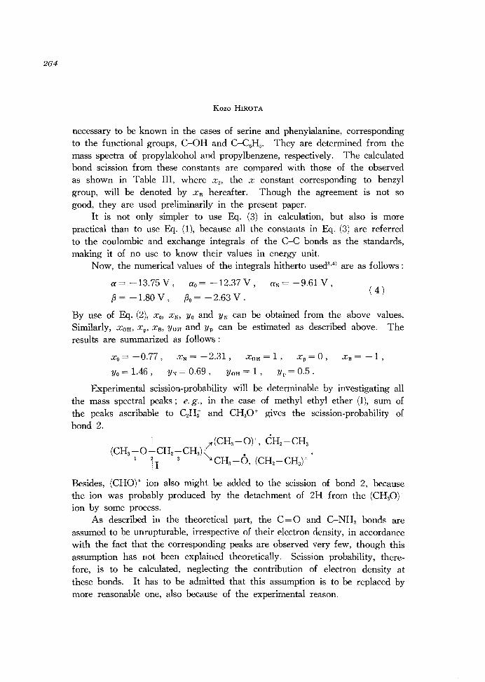
necessary to be known in the cases of serine and phenylalanine, corresponding to the functional groups, C-OH and C-C6H5' They are determined from the mass spectra of propylalcohol and propylbenzene, respectively. The calculated bond scission from these constants are compared with those of the observed as shown in Table III, where X 2 , the x constant corresponding to benzyl group, will be denoted by X B hereafter. Though the agreement is not so good, they are used preliminarily in the present paper.
It is not only simpler to use Eq. (3) in calculation, but also is more practical than to use Eq. (1), because all the constants in Eq. (3) are referred to the coulombic and exchange integrals of the C-C bonds as the standards, making it of no use to know their values in energy unit.
Now, the numerical values of the integrals hitherto used3,4) are as follows:
a= -13.75 V,
~= -1.80 V ,
ao = -12.37 V,
~o = -2.63 V.
aN= -9.61 V, ( 4 )
By use of Eq. (2), X O, XN, Yo and YN can be obtained from the above values. Similarly, XOH, x P' X B, YOH and Yp can be estimated as described above. The results are summarized as follows:
Xo = -0.77,
Yo = 1.46 ,
X N = - 2.31 , XOH = 1 , xp = 0 ,
YN = 0.69 , YOH = 1 , Yp = 0.5 .
Experimental scission-probability will be determinable by investigating all the mass spectral peaks; e. g., in the case of methyl ethyl ether (I), sum of the peaks ascribable to CzH; and CH30+ gives the scission-probability of bond 2.
j l'(CH3 -O),-, CH2-CH3 (CH3-0-CH2-CH3)~. .
1 11 3 CH3-O, (CH2-CH3)+
Besides, (CHO)+ lOn also might be added to the scission of bond 2, because the ion was probably produced by the detachment of 2H from the (CH30)+ ion by some process.
As described in the theoretical part, the C=O and C-NH2 bonds are assumed to be unrupturable, irrespective of their electron density, in accordance with the fact that the corresponding peaks are observed very few, though this assumption has not been explained theoretically. Scission probability, therefore, is to be calculated, neglecting the contribution of electron density at these bonds. It has to be admitted that this assumption is to be replaced by more reasonable one, also because of the experimental reason.

Quantitative Explanation of Fragmentation of the Skeletal Bonds in Mass Spectra
Results and Discussion
In Table IV, calculated energy values of the HO level are shown in ). unit for each ester, and also the squared expansion coefficients, i. e., the
TABLE IV. HO Energy Levels and the Expansion Coefficients over the Bond Orbitals of Amino Acid Esters
Ester Glycine Alanine a-ABA .a-ABA r-ABA lNorleuCine
HO energy level .h (J. unit) 3.33 2.64 2.66 2.59 2.44
I 2.66
1 1.988 10.520 8.131 4.005 1.923 7.508
2 91.006 75.334 79.393 82.038 92.038 79.954 ... ---- ------- ----- ---- .. -_ .. _----
3 0.000 0.002 0.001 0.001 0.002 0.004
4 0.000 0.037 0.025 0.024 0.041 0.027 Electron density
5 0.042 0.218 0.148 0.134 0.201 0.138 of the HO
levela ) % 6 7.009 13.646 10.875 4.533 0.287 10.078
7 0.244 1.363 8.751 1.411 1.837
8 0.063 0.514 3.098 0.372
9
I I I
0.071
10 0.010
Ester Leucine I I I . I Asparatic I so eucme acid Lysine Serine I Phenyl-alanine
HO energy level Ah (A unit) 2.66
I 2.66
I 2.66
I 2.66 2.82
I 2.80
1 7.434 7.083 6.698 7.418 7.984 4.668
2 80.026 80.568 79.760 79.706 79.572 83.214 ---- ------------
3 0.004 0.003 0.780 0.267 0.001 0.001 ...
4 0.026 0.024 0.003 0.004 0.024 0.011
5 0.136 0.127 0.022 0.026 0.144 0.668 Electron density
6 9.988 9.566 0.118 0.136 10.700 6.588 of the HO
levela) % 7 1.897 2.217 9.085 9.965 1.464 5.352
8 0.425 0.237 2.458 1.880 0.109 0.099
9 0.032 0.033 1.058 0.414
10 0.032 0.142 0.014 0.116
11 0.002 0.069
12 0.000
a) Electron density above the dotted line is assumed not to contribute to fragmentation.
265

266
Kozo HIROTA
electron density in %. In the case of phenylalanine, scission of the benzene ring was ignored, because it cannot be calculated by the present approximation.
Number of the most rupturable bond and the fraction of the peaks thus producible to the total peaks can be derived easily from Table IV. The results are described in the corresponding position in each ether in Table V, where the fraction of the electron density is the one when contribution of C=O and NH2 are omitted from the total. Experimental base peaks and bond number relating to their ion formation are shown in Table V for each esters
TABLE V. Highest Peaks in Mass Spectra of Amid Acid Esters and their Number of Most Rupturable Bondsa )
Ester I Glycine i Alanine I a-ABA I .a-ABA I J'-ABA INorleu~ine J
1° Observed base peak 30 44 58
I CNHj C2NHt C3NH~
2° Its numbers of the 6 6 6 I ruptured bond
3° Fraction to the i) 98 81 67 I total peaks % ii)b) - 82 ca. 80
I ,
I I Calculated bond num- 6 6 6 ber of the base peak ,
i I I Fraction of the elec- 99
I 96 87
I tron density %
!
Leucine I Isoleucine I AS~~i~ric I ------------------~-
Ester
1° Observed base peak
2° Its numbers of the ruptured bond
3° Fraction to the i) total peaks % ii)
Calculated bond number of the base peak
Fraction of the electron density %
C
5
86 5NHt2
6
39 5-70
6
80
a) Ionizing voltage: 70 e.v.
86 I 116 C5NHt2 C5NOHto
6 7
48
I
35 68 50-60
6 7
77 71
44 30 86 C2NHt CNHt C5NHt2
7 8 6
67 49 45 68 - 65
7 I 8 6
63 51 80
L · Is· I Phenyl-ysme enne I· a anme
84 60 120 C5NHto C2NOHt CsNHto
7 6 6
43 53
I 43c)
58d ) 59 -
7 6 6
79 85 54
b) The values when the peaks probably produced successively by fragmentation of the base peak ion are taken into account.
c) If the peaks ascribable to the boud-scission of the benzene ring is eliminated from the total, this % increases up to 47'/0.
d) The mle 30 peak was allotted to the secondary ion from the mle 101 peak primarily produced.

Quantitative Explanation of Fragmentation of the Skeletal Bonds in Mass Spectra
from the spectra of BIEMANN et al.7l Though their determination is easy, values of 3° in the table include several points to be mentioned.
On the ambiguous peaks, the proposed assignment of BIEMANN et al. was adopted, but such peaks were not many; e. g., the mle 84 peak from lysine ester was ascribed to the one produced by elimination of NH3 from
+ primarily producible NH2-CH2-(CH2)3-CH-NH2 ion (m/e 101), and the peak was counted into the fragment ion produced by scission of bond 7. However, there remained still several undeterminable peaks, though their contribution was not so large as to make the conclusion obscure. As an exceptional case, height of an unassignable mle 70 peak reaches 30% to the base peak in asparatic acid ester. This peak was explained preliminarily by successive scissions occurring at bonds 7 and 10, and accompanied by loss of a H atom, if the spectrum of 15N labelled ester is considered.
29 ---: 116----::- --- 29 1,1 'I 5
CH3 -CH2 -0 -C-CH2 -CH -C-O -CH2 - CH3 II I: II : o NH2 0
1:.1
------> (CH3-CH2-0-C-CH2-CH)+
------> (C-CH2-CH)+ II I o NH2
: II I o NH2
-H ml e ~ 71 ------>
mle 116
mle 70 ( 5 )
In the table, fractions of the base peak and the one produced by the same bond-scission to the total are described at 3° i) in Table V. Besides, total fractions of such peaks, which are estimated to be produced by successive scission of the above peaks, are described in 3° ii).
Numbers of the most rupturable bond coincide completely well between theory and experiment. However, quantitative prediction of the scission probability of the most rupturable bond is not so good, because its fraction to the total peaks differs by 30----50% from that of calculated electron density in some larger esters, especially in lysine and serine esters. Of course, the situation would be improved if better characteristic constants be selected. The above results, however, are still sufficient, in order to predict the base peaks of each esters, because most of the second highest peaks are less by -half than the base peaks, except phenylalanine ester. In this ester, the highest
267

268
Kozo HIROTA
···102 7 6
C6H5 -CH -CH -C-O -CH2 -CH3 i I II i NH2 0
mle 120
mle 120 peak is produced by the scission of bond 6 as expected by the largest electron density in Table IV, but its contribution to the total peaks reaches only 32% in height. Bond 7 of this ester is also rupturable, as indicated by large electron density at this bond, and actually mle 102 peak appears as the second highest, and total contribution of the scission at bond 7 amounts to 40% in height. Now that such an explanation can be given theoretically, this ester will not become an obstacle in analyzing the ester, but rather offers a support to the theory.
In the above discussion, the most rupturable bond, but not the most producible peak was made the problem in concern. This is inevitable due to the reason that, as widely accepted, when a parent positive ion ruptured into a positive ion and a radical, any general rule on which species becomes charged is not known yet. However, since the base peaks include always more negative group in the case of amino acid esters, their assignments are practically easy, because the fragment containing amino group would become the positive ion. It can be said, therefore, that the base peaks of the amino acid esters are predictable by the present method.
Besides, this result is satisfactory from the viewpoint of the present research to investigate the transferability of the constants characteristic to each functional groups, even though some of them are still of preliminary nature. Investigation in this line has to be carried out also from the reason to consider the scission of c=o bond and rupture of phenyl group. Further, degree of agreement between theory and experiment may increase if better characteristic constants are selected. Especially, substitution of the constants for the c-o bond of ether type for others is desirable, because main cause of quantitative discrepancy in the fraction of the electron density in Table V is ascribable to smaller percentage of theoretical scission probability at the C-O bond. For instance, the mle 29 peak from asparatic acid ester can be assignable to be C2H; ion by comparing the spectrum of 15N labelled ester, indicating the peak to be produced by scission of bond 5 or 11. Contrary to the theoretical expectation, its height reaches half the base peak mle 116. If the height of this peak is omitted, experimental values at 3° i) and 3° ii) in Table V increase upto 42% and 60,....,72%, respectively.

Quantitative Explanation of Fragmentation of the Skeletal Bonds in Mass Spectra
Second, similar substitution is neccessary on the characteristic constants of the C= 0 and C-N bonds, corresponding to the experimental result that they are ruptured, though in small amount. Of course, a more advanced approximation has to be adopted basing on theoretical grounds, in order to answer this requirement.
Further, other amino acid esters having special functional groups, say tyrosine, methionine, proline, etc. will be investigated similarly in the cases when their characteristic constants are known. During the procedure, it has to be taken care that this method is applicable only to evaluate the primary scission of the skeletal bonds. Actually, secondary scission may occur immediately after the primary, and some of them occurs so fast that the peaks produced are included in the mass spectra. One of the important example is the mle 84 peak of lysine ester, as already indicated. It will be a remaining problem in what case such a secondary process has to be considered in the explanation of the peaks in mass spectra.
Conclusion
Though detailed theoretical basis of the MO method was not given, it would be sufficient to conclude that the present theory will be potentially useful as a semi-empirical tool, to study the mass spectra of such complex compounds as amino acids esters, so far as the primary scission of skeletal bonds is concerned.
Acknowledgment
The author wishes to express his sincere thanks to Mr. Y. NIWA and Mr. 1. Fu JIT A of his Department for their help to carry out this research. This paper is dedicated to Professor Juro HORIUTI.
Remarks added in Proof: Consistancy of the parameters peculiar to each
functional group, Eq. (4), was shown on various chain compounds in a paper
published already (K. HIROTA, Nippon Kagaku Zasshi, 89, 327 (1968)).
References
1) K. FUEKI and K. HIROTA, Nippon Kagaku Zasshi, 80, 1202 (1959); 81, 212 (1960).
2) K. HIROTA and Y. NIWA, Tetrahedron Letters, 5757 (1966).
3) K. HIROTA and K. HA TADA, Bull. Chern. Soc. Japan, 38, 599 (1965); Z. Physik.
Chern., N. F., 44, 328 (1965); Sci. Pap. Inst. Phys. Chern. Res., 60, 78 (1966); Kinetika
i Katailiz, 8, 748 (1967).
269
270
Kozo HIROTA
4) K. HIROTA and M. ITOH, Bull. Chern. Soc. Japan, 39, 1406 (1966).
5) K. HIROTA and]. TAKEZAKI, Bull. Chern. Soc. Japan, 41, 76 (1968).
6) T. IsOY A and T. NAGUMO, Ann. Meeting of Chern. Soc. Japan, No.2 E, 114 (April
2, 1967).
7) K. BIEMANN, J. SIEBL and F. GAPP, J. Am. Chern. Soc., 83, 3795 (1961).
8) K. HIROTA and Y. NIW A, ]. Phys. Chern., 2, 5 (1968).
9) R. D. BROWN, J. Chern. Soc., 1953, 2615. Due to the approximation adopted here,
the result is the same, even if the equivalent orbital method is used.lO)
10) G. G. HALL, Proc. Roy. Soc., A 205, 541 (1951).