quantitative estimation of protein in urinefac.ksu.edu.sa/sites/default/files/lab3_5.pdf ·...
TRANSCRIPT
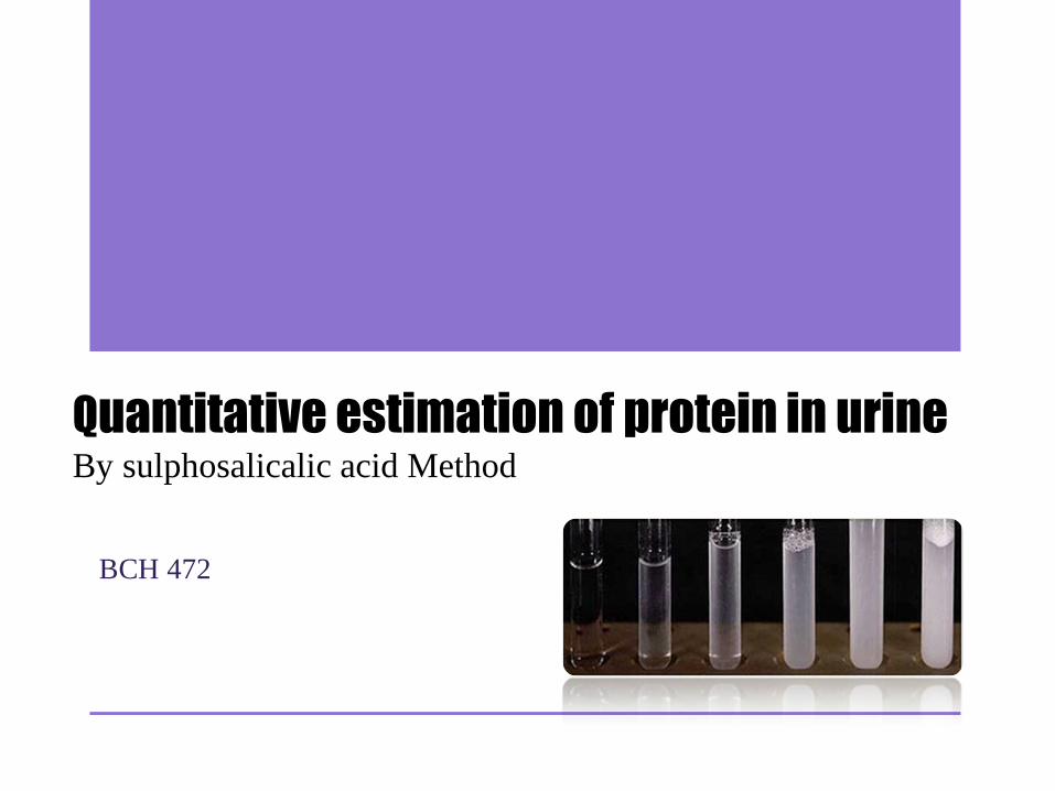
Quantitative estimation of protein in urine By sulphosalicalic acid Method
BCH 472
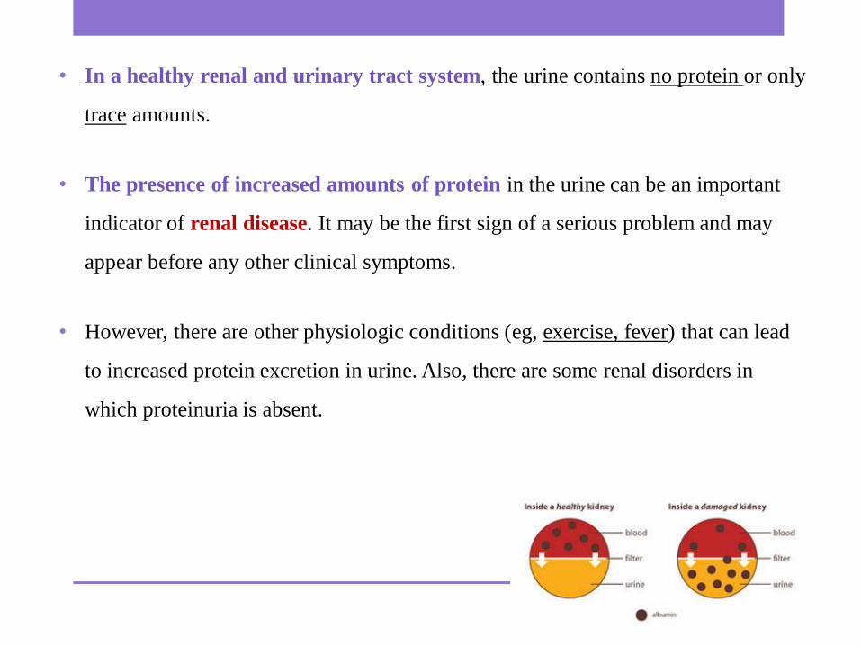
• In a healthy renal and urinary tract system, the urine contains no protein or only
trace amounts.
• The presence of increased amounts of protein in the urine can be an important
indicator of renal disease. It may be the first sign of a serious problem and may
appear before any other clinical symptoms.
• However, there are other physiologic conditions (eg, exercise, fever) that can lead
to increased protein excretion in urine. Also, there are some renal disorders in
which proteinuria is absent.
Proteinuria:
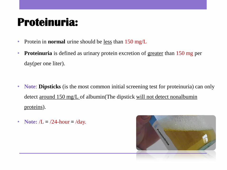
• Protein in normal urine should be less than 150 mg/L
• Proteinuria is defined as urinary protein excretion of greater than 150 mg per
day(per one liter).
• Note: Dipsticks (is the most common initial screening test for proteinuria) can only
detect around 150 mg/L of albumin(The dipstick will not detect nonalbumin
proteins).
• Note: /L = /24-hour = /day.

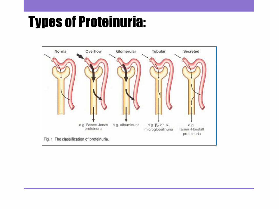
Types of Proteinuria:
Type Cause
Glomerular
proteinuria
• Results from a disruption of the glomerular filtration barrier which
increased filtration of normal plasma protein and because albumin has
the highest concentration in the plasma it is called albuminuria eg.
Malignant hypertention
Tubular
proteinuria
• Defect in the reabsorption eg, Fanconi Syndrom
- low molecular weight protein that is found in urine
Overflow
proteinuria
• Overflow of high plasma
- high concentrations of low molecular weight protein found in urine
eg, Multiple myloma. In multiple myeloma excessive amounts of
immunoglobulin light chains are produced.
Secretory
proteinuria
• Over secretion of certain proteins in the tubules, most notably the over
secretion of Tamm-Horsfall proteins eg, in interstitial nephritis
Types of Proteinuria:
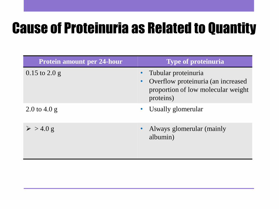
Cause of Proteinuria as Related to Quantity
Protein amount per 24-hour Type of proteinuria
0.15 to 2.0 g
• Tubular proteinuria
• Overflow proteinuria (an increased
proportion of low molecular weight
proteins)
2.0 to 4.0 g
• Usually glomerular
> 4.0 g
• Always glomerular (mainly
albumin)

• The quantitative estimation of the daily excretion of protein is of value to the clinician in
order to give a general idea of the type of renal disease, its severity and to monitor the
results of treatment given.
• The protein content can be determined by numerous methods eg, Biuret, Lowry, Bradford.
• In this lab turbidimetric method will be used.
• Determination of total protein by measurement of protein turbidity produce by mixed with
an anionic organic acid such as sulfosalicylic acid , TCA , or benzethonium chloride.
• Sulphosalicylic acid is used in this experiment to precipitate the protein in a 24 hour
sample of urine. The turbidity is proportional to the concentration of the protein, and may
be measured with a spectrophotometer at 500 nm.

• Practical Part

Sulfosalicylic acid (SSA) test :
• The sulfosalicylic acid (SSA) turbidity test quantitatively screens for proteinuria. The
advantage of this easily performed test is its greater sensitivity for proteins such as
Bence Jones.
• The SSA reaction will detect globulin and Bence-Jones proteins, in addition to
albumin (although it is more sensitive to albumin).

Principle:
• Sulfosalsalyic acid is an anion(-) which neutralizes the protein cations(+) leading
to its precipitation (pH in highly acidic media, the protein will be positively
charged, which is attracted to the acid anions that cause them to precipitate.)
• Then the radiation of a wavelength which is not absorbed by the solution is made
to pass through the suspension and the apparent absorption will be solely because
of the scattering by the particles.
• The transmission decrease with increasing protein concentration.

Method: 1-Set up a series of test tube as follows, label from 1- 7
Tube Protein Stock Solution( 140 mg/dl) 0.85%
Saline Protein concentration mg/dl
1 4.5 1.5
2 3 3
3 2.4 3.6
4 1.5 4.5
5 0.9 5.1
6 0.3 5.7
7( Blank) 0 6
Urine Sample - -
2-Set another 8 test tube labeled 1-7 and pipette in each one Add 8 ml of
sulfosalicylic acid
Tube sulfosalicylic acid
1 8 ml
2 8 ml
3 8 ml
4 8 ml
5 8 ml
6 8 ml
7( Blank) 8 ml
Urine Sample 8 ml
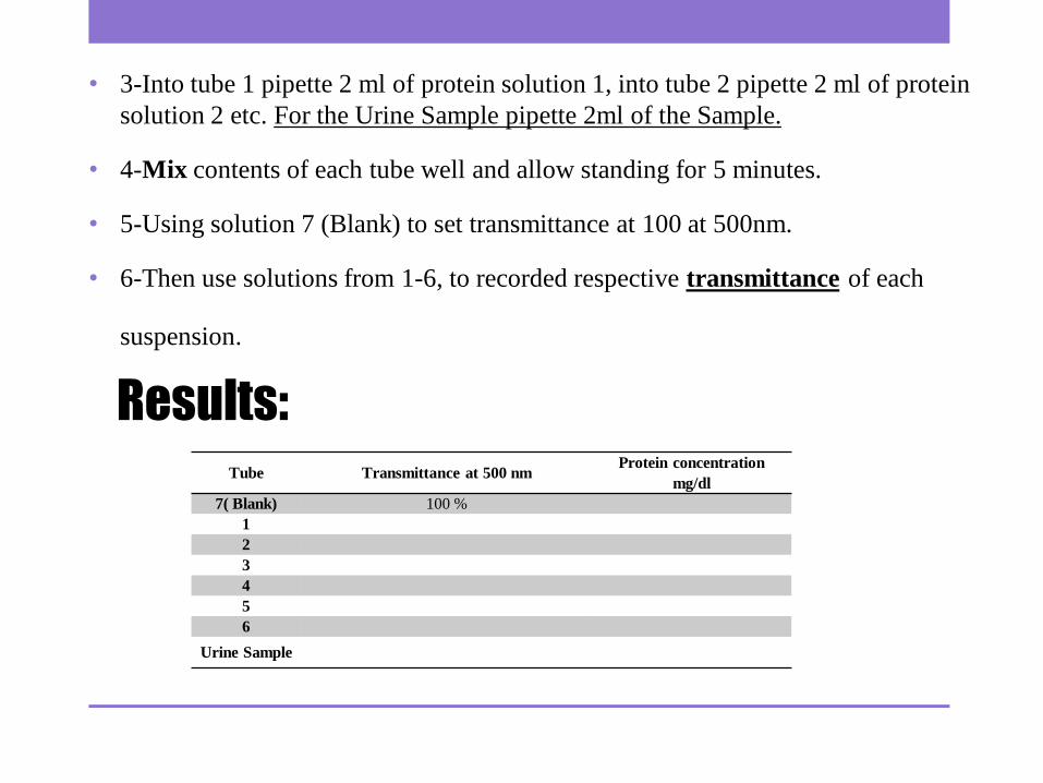
• 3-Into tube 1 pipette 2 ml of protein solution 1, into tube 2 pipette 2 ml of protein
solution 2 etc. For the Urine Sample pipette 2ml of the Sample.
• 4-Mix contents of each tube well and allow standing for 5 minutes.
• 5-Using solution 7 (Blank) to set transmittance at 100 at 500nm.
• 6-Then use solutions from 1-6, to recorded respective transmittance of each
suspension.
Results: Tube Transmittance at 500 nm
Protein concentration
mg/dl
7( Blank) 100 %
1
2
3
4
5
6
Urine Sample

• Plot Transmittance against Protein concentration mg/dl.
• Determine the Protein concentration of Urine Sample from the standard curve.
• Compare the result you got with the normal range of protein execration in 24 h
urine specimen (you must convert the unit to g/L) if you know that the protein
execration in healthy sample (0- less than 0.150g/24 h).
• Comment on the clinical conditions of the patient if it is present.
• Assuming that the 24 hour urine sample for the patient = 1000 ml.

Questions :
Which protein can be used as a marker of Multiple myloma ? And how?
Explain how total protein can be determined by measuring protein turbidity ?
Why the resulting graph is a descending curve ?