quantitative analysis of the speech and lip … · tesi di dottorato di . fernanda vincia...
TRANSCRIPT

UNIVERSITÀ DEGLI STUDI DI MILANO SCUOLA DI DOTTORATO IN SCIENZE MORFOLOGICHE,
FISIOLOGICHE E DELLO SPORT DIPARTIMENTO DI SCIENZE BIOMEDICHE PER LA SALUTE
DOTTORATO DI RICERCA IN SCIENZE MORFOLOGICHE – XXV CICLO
BIO16
QUANTITATIVE ANALYSIS OF THE SPEECH AND LIP MOVEMENTS THROUGH OPTOELECTRONIC
MOTION ANALYSIS AND SURFACE ELECTROMYOGRAPHY
Tesi di Dottorato di FERNANDA VINCIA SIDEQUERSKY
MATRICOLA: R08866
Tutor: Chir.ma prof.ssa Chiarella Sforza Coordinatore: Chir.ma prof.ssa Laura Vizzotto
Anno Accademico 2011/2012

I dedicate this work to my parents who raised me and always supported me in my decisions.
TABLE OF CONTENTS
1 ACKNOWLEDGEMENTS .................................................................................. 5
2 LIST OF ABBREVIATIONS ............................................................................... 6
3 ABSTRACT .......................................................................................................... 7
4 INTRODUCTION ................................................................................................ 8
5 ANATOMY AND FUNCTION .......................................................................... 11
5.1 FACE ......................................................................................................... 11
5.2 FACIAL NERVE ....................................................................................... 12
5.3 MUSCLES OF FACIAL EXPRESSION .................................................. 15
5.3.1 MUSCLES OF THE UPPER FACE ........................................................ 16
5.3.2 MUSCLES OF THE MIDFACE .............................................................. 18
5.3.3 MUSCLES OF THE LOWER FACE AND NECK .................................. 21
5.4 LABIAL STRUCTURE ............................................................................ 22
5.5 MOVEMENTS OF THE LIPS ................................................................. 23
5.6 ANATOMY OF SPEECH ......................................................................... 24
5.6.1 OVERVIEW OF SPEECH PRODUCTION ............................................. 24
5.6.2 ARTICULATION ................................................................................... 24
5.6.3 PRODUCTION OF VOWELS ................................................................ 25
5.6.4 PRODUCTION OF CONSONANTS ....................................................... 25
6 MATERIAL AND METHODS .......................................................................... 27
6.1 CLINICAL EXAMINATION ................................................................... 27
6.1.1 SELF-JUDGEMENT OF SEVERITY ..................................................... 27
6.1.2 OROFACIAL MYOFUNCTIONAL EVALUATION .............................. 28
6.2 SUBJECTS ................................................................................................ 29
6.3 INSTRUMENTATIONS ........................................................................... 30
6.3.1 OPTOELECTRONIC MOTION ANALYZER ........................................ 30
6.3.2 ELECTROMYOGRAPHIC SYSTEM ..................................................... 33
6.3.3 RECORDING PROTOCOL .................................................................... 34
6.3.4 MEASUREMENT PROTOCOL.............................................................. 38
6.3.5 METHOD ERROR .................................................................................. 41

7 KINEMATICS ANALYSIS OF LIP MOTION IN HEALTHY SUBJECTS ... 42
7.1 METHODS ................................................................................................ 42
7.1.1 DATA COLLECTION ............................................................................ 42
7.1.2 STATISTICAL ANALYSIS .................................................................... 43
7.2 RESULTS .................................................................................................. 44
8 ELECTROMYOGRAPHIC ANALYSIS - HEALTHY SUBJECTS................ 56
8.1 METHODS ................................................................................................ 56
8.1.1 DATA COLLECTION ............................................................................ 56
8.1.2 STATISTICAL ANALYSIS .................................................................... 57
8.2 RESULTS .................................................................................................. 58
9 DISCUSSION ...................................................................................................... 61
10 GENERAL CONCLUSIONS ............................................................................. 68
11 REFERENCES ................................................................................................... 69

5
1 ACKNOWLEDGEMENTS The greatest achievements of my life could only be possible because I had
people around me that always contributed in a special way. I would like to thank Prof.ssa Chiarella Sforza that, with all her infinite
experience and knowledge, helped me see beyond her shoulders. Prof.ssa thank you for your patience, your confidence, and for accepting me in a group that contributed to my scientific and intellectual growth. Today you are my source of inspiration and admiration, I am very proud to have you as a teacher.
I would like to thank Profa. Cláudia Maria de Felício, that since the first lessons, made me fascinated by Fonoaudiologia and kept me interested in move forward. Thanks for your overwhelming support, understanding and trust, without you I would not have gotten this far.
Today I can tell everyone that I had not only good teachers, but giants that made me look beyond and beyond.
I would like to express my very great appreciation to all professors, researchers, colleagues and member of my department. I have learned so much from all of you. Especially thanks to Dr. Andrea Mapelli, Dr. Isabella Annoni, Dr. Matteo Zago, Salvatore Sergi (Mimmo). I can say that I grew up not only intellectually, but with you I found the true practice of multidisciplinary teamwork.
Dr. Marcio de Menezes, I would like to thank you for all the help you gave me during my adaptation and your always useful advices.
Special thanks for the friendship of my UNIALA fellows Dr. Andre Botelho and Dr. Michele Bail.
To my friends at USP, thanks for all the support and help, not only financially, but mainly for friendship. It is a pleasure to work and have you as friends. Special thanks to Ana Medeiros, Barbara Machado, Carina Borges, Cláudia Ferreira, Fabíula Favareto, Gislaine Folha, Lúcia Giglio and Mariela Bisson.
Paulo Ferreira, thanks for your practical advices and for helping and supporting me during my coming.
I would like to thank my love, partner and friend Lucas Lamonato for his infinite patience, love, care, presence and dedication over these three years. Lucas, thank you for your understanding and your support.
Adriano Nunes, Fábio Sousa, Ijanete Silva and Natalia Landgraf, thank you for your friendship.
6
2 LIST OF ABBREVIATIONS 2D: bi-dimensional
3D: three-dimensional
ANOVA: analysis of variance
ASYM: asymmetry index for sequence of natural numbers and words
CCD: charge coupled device
EMG: electromyography
NS: not significant
OMES - Orofacial Myofunctional Evaluation with Scores Protocol
RDC: research diagnostic criteria
RMS: root mean square
SD: standard deviation
TMD: temporomandibular disorder
TMJ: temporomandibular joint
7
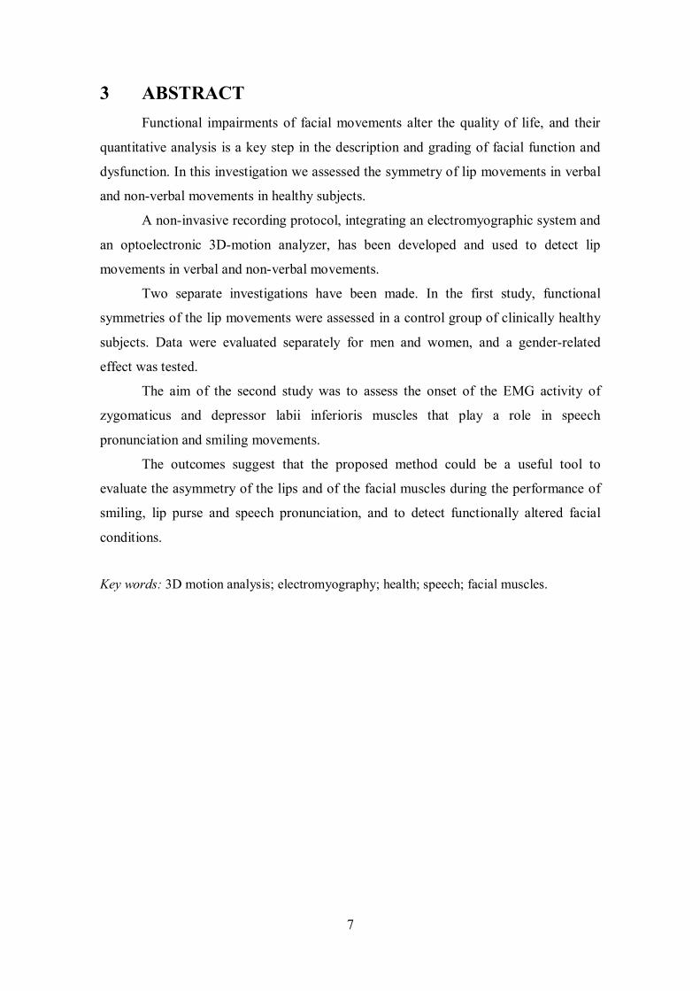
3 ABSTRACT Functional impairments of facial movements alter the quality of life, and their
quantitative analysis is a key step in the description and grading of facial function and
dysfunction. In this investigation we assessed the symmetry of lip movements in verbal
and non-verbal movements in healthy subjects.
A non-invasive recording protocol, integrating an electromyographic system and
an optoelectronic 3D-motion analyzer, has been developed and used to detect lip
movements in verbal and non-verbal movements.
Two separate investigations have been made. In the first study, functional
symmetries of the lip movements were assessed in a control group of clinically healthy
subjects. Data were evaluated separately for men and women, and a gender-related
effect was tested.
The aim of the second study was to assess the onset of the EMG activity of
zygomaticus and depressor labii inferioris muscles that play a role in speech
pronunciation and smiling movements.
The outcomes suggest that the proposed method could be a useful tool to
evaluate the asymmetry of the lips and of the facial muscles during the performance of
smiling, lip purse and speech pronunciation, and to detect functionally altered facial
conditions.
Key words: 3D motion analysis; electromyography; health; speech; facial muscles.

8
4 INTRODUCTION The face characterizes human beings and in particular the mouth and lips plays a
key role in the evaluation and recognition of the craniofacial complex.
The smile is one of the most frequent facial expressions, and is used to transmit
positive emotional state, as well as to serve social functions such as greeting. Like many
facial expressions, the smile can be produced either deliberately by voluntary movement
of the Zygomaticus major muscles or spontaneously (Lapatki et al., 2003; Schmidt et
al., 2006).
During speech pronunciation, the movements of the articulators create temporal
sequences of sounds, characterized by activations of the tongue, jaw, lips, vocal folds
and velum muscles, i.e. movements of the vocal tract articulators, and the resulting
vocal tract shapes correspond to the produced patterns of speech. At the extrinsic level,
acoustic signals, visual and perceptual salience similarly correspond to the produced
patterns of speech (Smith 1992; Green et al., 2000; Bianchini and Andrade, 2006; Van
der Geld et al., 2008; Sawyer et al., 2010; Grimme et al., 2011).
For speech pronunciation the timing is critical, since it can carry relevant
information for the communication process, and generally involves the coordination of
different end-effectors and different movements of the face, and requires a sequence of
well-coordinated orofacial movements (Grimme et al., 2011).
Facial expressions can be altered in various pathologic conditions and
malformations, deriving from central nervous system diseases, neuromuscular and
peripheral nerve paralysis (mostly, facial nerve paralysis), drug administration,
dentofacial deformities and scars, congenital anomalies (Trotman et al., 1998a; Okada,
2001; Wachtman et al., 2001; Mishima et al., 2004; Nooreyazdan et al., 2004; Tarantili
et al., 2005; Tzou et al., 2005; Proff et al., 2006; Agostino et al., 2008; Mehta et al.,
2008; Sawyer et al., 2010; Sforza et al., 2012).
In several medical and dental fields, facial dysfunctions are usually assessed
independently from their origin, and several clinical and instrumental assessments can
be used to grade both spontaneous and instructed movements (Trotman et al., 2000;
Wachtman et al., 2001; Linstrom et al., 2002; Giovanoli et al., 2003; Nooreyazdan et
al., 2004; Proff et al., 2006; Ferrario and Sforza, 2007; Hontanilla and Aubá, 2008;
Mehta et al., 2008; Popat et al., 2008a).

9
Clinical assessments focalize on total and local facial motion, synkinesis and
movement asymmetries (Proff et al., 2006; Ferrario and Sforza, 2007; Reitzen et al.,
2009), whilst quantitative methods can assess both the movements of selected facial
landmarks and their trajectories (Linstrom et al., 2002; Giovanoli et al., 2003; Ferrario
and Sforza, 2007; Hontanilla and Aubá, 2008; Mehta et al., 2008; Popat et al., 2008b;
Sawyer et al., 2010). Clinical evaluations can have a reduced inter-examiners
repeatability, with problems in data sharing among different care takers or research
centers. To overcome these limitations, quantitative methods for the assessment of
facial movements have been proposed (Trotman et al., 2000; Okada, 2001; Kang et al.,
2002; Linstrom et al., 2002; Tzou et al., 2005; Proff et al., 2006).
Nowadays, several three-dimensional motion analyzers allow a non-invasive
quantitative assessment of soft tissue facial movements without interfering with the
subject (Weeden et al., 2001; Coulson et al., 2000; 2002; Giovanoli et al., 2003;
Johnston et al., 2003; Mishima et al., 2004; Nooreyazdan et al., 2004; Proff et al., 2006;
Ferrario and Sforza, 2007; Agostino et al., 2008; Popat et al., 2008a,b; Sforza et al.,
2010b,c; 2012; Verzé et al., 2011a,b).
The detection and quantitative analysis of facial movements is a key step in the
description and grading of facial function and dysfunction, during diagnosis, treatment
and follow-up of their disorders (Trotman et al., 2000; Okada et al., 2001; Coulson et
al., 2002; Kang et al., 2002; Tzou et al., 2005; Proff et al., 2006; Hontanilla and Aubá,
2008; Mehta et al., 2008; Okamoto et al., 2010; Sawyer et al., 2010).
In our laboratory, we developed a method for the non-invasive, three-
dimensional assessment of facial movements using an optoelectronic motion analyser
(Ferrario and Sforza, 2007; Sforza et al., 2010b,c; 2012). This method was found to be
minimally disturbing, reliable, and to accurately detect total and local motion during the
performance of standardized facial animations (Sforza et al., 2010b,c; 2012). It offers a
valuable support for the extraction of numeric values, which are very useful in the
differential diagnosis (Ferrario and Sforza, 2007; Popat et al., 2008a,b; Mapelli et al.,
2009; Sforza et al., 2010b; Verzé et al., 2011a,b).
Patient can be compared to reference values obtained from healthy individuals
according to age, gender and ethnicity (Frey et al., 1999; Tzou et al., 2005; Sforza et al.,
2010b,c). Longitudinal assessments can also be performed during treatment and
rehabilitation.
10
Another useful system in the differential diagnosis process and in the therapeutic
planning is surface electromyography (EMG). EMG can monitor orofacial functions, it
is highly sensitive in measuring muscle performance, monitoring their efficiency, and
comparing different groups of individuals (Ferrario et al., 2000a, 2006a,b, Galo et al.,
2006, 2007; Tartaglia et al 2008; Felício et al., 2009b, 2012).
Among the various assessments of facial function, the detection of symmetry (or
its counterpart, asymmetry) is one of the most important, and even lay observers can
detect facial asymmetries at rest and during mimicry. Considering the well-known
fluctuating asymmetry existing in all subjects (Sforza et al., 2010d), before defining
clinical values for “asymmetry”, it is necessary to define a threshold in healthy
individuals.
Several studies about the quantitative evaluation of the symmetric and
asymmetric facial movements using non-invasive methods like three-dimensional
optoelectronic analysis (Ferrario and Sforza, 2007; Sforza et al. 2010 b,c; 2012) and
EMG (Ferrario et al., 2000a, 2001; Felício et al., 2009b) were performed, but in all
cases only not-verbal animations were analyzed. Indeed, facial and labial movements
during speech production are an integral part of the social life of each person, and their
quantitative analysis is mandatory in several clinical fields.
The assessments of healthy individuals would provide data that can be used as
normality parameters for the comparison with the patients with morphologic and/or
functional problems in the skull-facial area.
In the present investigation, three-dimensional motion analysis and surface EMG
have been applied to analyse both facial kinematics and facial muscle function in a
group of young healthy subjects.
11
5 ANATOMY AND FUNCTION
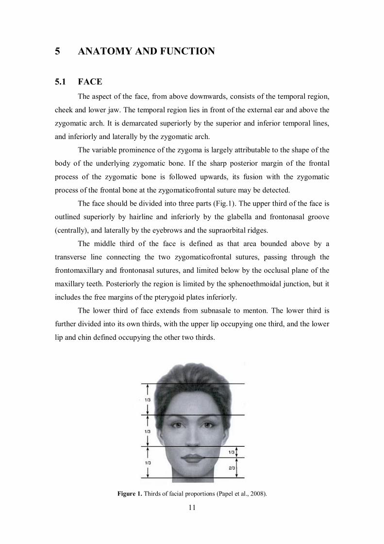
5.1 FACE The aspect of the face, from above downwards, consists of the temporal region,
cheek and lower jaw. The temporal region lies in front of the external ear and above the
zygomatic arch. It is demarcated superiorly by the superior and inferior temporal lines,
and inferiorly and laterally by the zygomatic arch.
The variable prominence of the zygoma is largely attributable to the shape of the
body of the underlying zygomatic bone. If the sharp posterior margin of the frontal
process of the zygomatic bone is followed upwards, its fusion with the zygomatic
process of the frontal bone at the zygomaticofrontal suture may be detected.
The face should be divided into three parts (Fig.1). The upper third of the face is
outlined superiorly by hairline and inferiorly by the glabella and frontonasal groove
(centrally), and laterally by the eyebrows and the supraorbital ridges.
The middle third of the face is defined as that area bounded above by a
transverse line connecting the two zygomaticofrontal sutures, passing through the
frontomaxillary and frontonasal sutures, and limited below by the occlusal plane of the
maxillary teeth. Posteriorly the region is limited by the sphenoethmoidal junction, but it
includes the free margins of the pterygoid plates inferiorly.
The lower third of face extends from subnasale to menton. The lower third is
further divided into its own thirds, with the upper lip occupying one third, and the lower
lip and chin defined occupying the other two thirds.
Figure 1. Thirds of facial proportions (Papel et al., 2008).

12
5.2 FACIAL NERVE The facial nerve emerges from the skull base at the stylomastoid foramen and
almost immediately gives off the nerves to the posterior belly of digastric and
stylohyoid, and the posterior auricular nerve, which supplies the occipital belly of
occipitofrontalis and some of the auricular muscles (Fig. 2).
Figure 2. Distribution of the facial nerve. The branches given off immediately after the nerve exits the
stylomastoid foramen. (Standring et al., 2008, modified).
The nerve next enters the parotid gland high up on its posteromedial surface and
passes forwards and downwards behind the mandibular ramus. Within the substance of
the gland it branches into superior (temporofacial) and inferior (cervicofacial) trunks,
usually just behind and superficial to the retromandibular vein. The trunks branch
further to form a parotid plexus. Five main terminal branches arise from the plexus, they
diverge within the gland and leave by its anteromedial surface, medial to its anterior
margin, to supply the muscles of facial expression (Fig. 3).

13
Figure 3. The branches of the facial nerve on the face. (Rizzolo and Madeira, 2005, modified)
The temporal branch usually divides into anterior and posterior rami soon after
piercing the parotidomasseteric fascia below the zygomatic arch; there is often a middle
(frontal) ramus. Twigs supply intrinsic muscles on the lateral surface of the auricle, and
the anterior and superior auricular muscles, and communicate with the
zygomaticotemporal branch of the maxillary nerve and the auriculotemporal branch of
the mandibular nerve. The more anterior branches supply the frontal belly of
occipitofrontalis, orbicularis oculi and corrugator, and join the supraorbital and lacrimal
branches of the ophthalmic nerve.
Zygomatic branches are generally multiple. They cross the zygomatic bone to
the lateral canthus of the eye and supply orbicularis oculi: they may also supply muscles
innervated by the buccal branch. Twigs communicate with filaments of the lacrimal
nerve and the zygomaticofacial branch of the maxillary nerve.
The buccal branch is usually single. It has a close relationship to the parotid duct
for about 2.5 cm after emerging from the parotid gland, and typically lies below the
duct. Superficial branches run beneath the subcutaneous fat and superficial musculo-
aponeurotic system. Some branches pass deep to procerus and join the infratrochlear
and external nasal nerves. Upper deep branches supply zygomaticus major and levator
14
labii superioris, and form an infraorbital plexus with the superior labial branches of the
infraorbital nerve. They also supply levator anguli oris, zygomaticus minor, levator labii
superioris alaequae nasi and the small nasal muscles: these branches are sometimes
described as lower zygomatic branches. Lower deep branches supply buccinator and
orbicularis oris; they communicate with filaments of the buccal branch of the
mandibular nerve.
There are usually two marginal mandibular branches. They run forwards towards
the angle of the mandible under platysma, then turn upwards across the body of the
mandible to pass under depressor anguli oris. The branches supply risorius and the
muscles of the lower lip and chin, and filaments communicate with the mental nerve.
The cervical branch emerges from the lower part of the parotid gland and runs
anteroinferiorly under platysma to the front of the neck. Typically single, it supplies
platysma and communicates with the transverse cutaneous cervical nerve.
Cutaneous branches of the facial nerve accompany the auricular branch of the
vagus; they are believed to innervate the skin on both auricular aspects, in the conchal
depression and over its eminence.
15
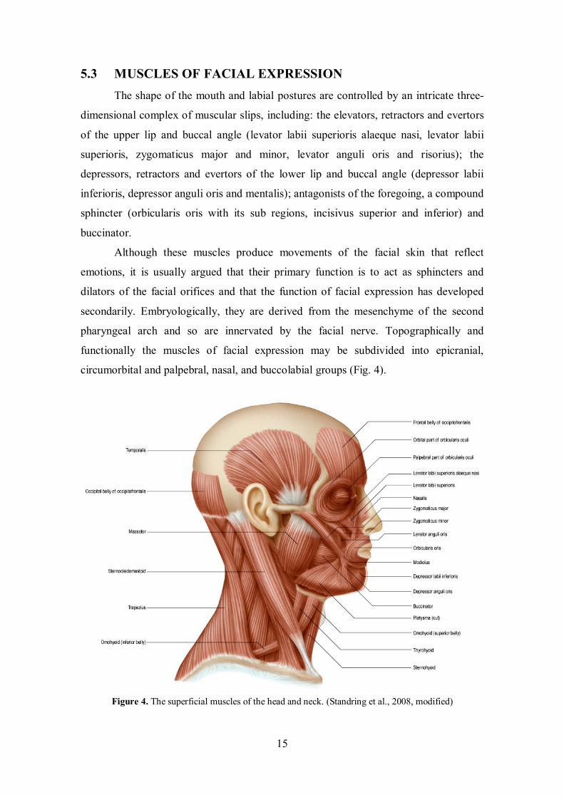
5.3 MUSCLES OF FACIAL EXPRESSION The shape of the mouth and labial postures are controlled by an intricate three-
dimensional complex of muscular slips, including: the elevators, retractors and evertors
of the upper lip and buccal angle (levator labii superioris alaeque nasi, levator labii
superioris, zygomaticus major and minor, levator anguli oris and risorius); the
depressors, retractors and evertors of the lower lip and buccal angle (depressor labii
inferioris, depressor anguli oris and mentalis); antagonists of the foregoing, a compound
sphincter (orbicularis oris with its sub regions, incisivus superior and inferior) and
buccinator.
Although these muscles produce movements of the facial skin that reflect
emotions, it is usually argued that their primary function is to act as sphincters and
dilators of the facial orifices and that the function of facial expression has developed
secondarily. Embryologically, they are derived from the mesenchyme of the second
pharyngeal arch and so are innervated by the facial nerve. Topographically and
functionally the muscles of facial expression may be subdivided into epicranial,
circumorbital and palpebral, nasal, and buccolabial groups (Fig. 4).
Figure 4. The superficial muscles of the head and neck. (Standring et al., 2008, modified)
16
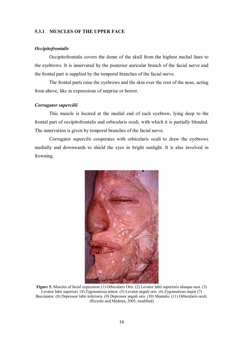
5.3.1 MUSCLES OF THE UPPER FACE
Occipitofrontalis
Occipitofrontalis covers the dome of the skull from the highest nuchal lines to
the eyebrows. It is innervated by the posterior auricular branch of the facial nerve and
the frontal part is supplied by the temporal branches of the facial nerve.
The frontal parts raise the eyebrows and the skin over the root of the nose, acting
from above, like in expressions of surprise or horror.
Corrugator supercilii
This muscle is located at the medial end of each eyebrow, lying deep to the
frontal part of occipitofrontalis and orbicularis oculi, with which it is partially blended.
The innervation is given by temporal branches of the facial nerve.
Corrugator supercilii cooperates with orbicularis oculi to draw the eyebrows
medially and downwards to shield the eyes in bright sunlight. It is also involved in
frowning.
Figure 5. Muscles of facial expression (1) Orbicularis Oris. (2) Levator labii superioris alaeque nasi. (3)
Levator labii superiori. (4) Zygomaticus minor. (5) Levator anguli oris. (6) Zygomaticus major (7) Buccinator. (8) Depressor labii inferioris. (9) Depressor anguli oris. (10) Mentalis. (11) Orbicularis oculi.
(Rizzolo and Madeira, 2005, modified)
17
Procerus
It arises from a fascial aponeurosis attached to the periosteum covering the lower
part of the nasal bone, the perichondrium covering the upper part of the lateral nasal
cartilage and the aponeurosis of the transverse part of nasalis. It is inserted into the
glabellar skin over the lower part of the forehead between the eyebrows.
It is innervated by temporal and lower zygomatic branches from the facial nerve.
Procerus draws down the medial angle of the eyebrow and produces transverse
wrinkles over the bridge of the nose. It is active in frowning and ‘concentration', and
helps to reduce the glare of bright sunlight.
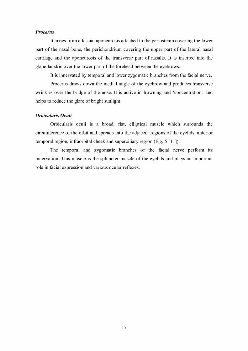
Orbicularis Oculi
Orbicularis oculi is a broad, flat, elliptical muscle which surrounds the
circumference of the orbit and spreads into the adjacent regions of the eyelids, anterior
temporal region, infraorbital cheek and superciliary region (Fig. 5 [11]).
The temporal and zygomatic branches of the facial nerve perform its
innervation. This muscle is the sphincter muscle of the eyelids and plays an important
role in facial expression and various ocular reflexes.

18
5.3.2 MUSCLES OF THE MIDFACE
Nasalis
Nasalis muscle consists of transverse and alar components. The transverse part
(compressor naris) is attached to the maxilla above and lateral to the incisive fossa, and
lateral to the alar part. The alar part (pars alaris or dilator naris posterior) is attached to
the maxilla above the lateral incisor and canine, lateral to the bony attachment of
depressor septi, and medial to the transverse part, with which it partly merges. The
innervation of nasalis muscle is performed by the buccal branch of the facial nerve. It
may also be supplied by the zygomatic branch of the facial nerve.
The transverse part compresses the nasal aperture at the junction of the vestibule
and the nasal cavity. The alar parts draw the alae and posterior part of the columella
downwards and laterally and so assist in widening the nares and in elongating the nose.
They are active immediately before inspiration.
Levator Labii Superioris Alaeque Nasi
It is attached to the upper part of the frontal process of the maxilla, then
descends inferolaterally, dividing into a medial slip attached to the greater alar cartilage
and the skin over it and a lateral slip prolonged inferolaterally to blend with levator labii
superioris and orbicularis oris (Fig. 5 [2]). The innervation is given by zygomatic and
superior buccal branches of the facial nerve. The lateral slip raises and everts the upper
lip and raises, depends and increases the curvature of the nasolabial furrow’s superior
part; the medial slip dilates the nostril and displaces laterally and modifies the curvature
of the inferolaterally convex circumalar furrow.
Levator Labii Superioris
It descends from the inferior orbital margin, being attached to the maxilla and
zygomatic bone above the infra-orbital foramen, and converges into the upper lip
between the lateral slip of elevator labii superioris alaeque nasi and zygomaticus minor
with, more deeply, levator anguli oris (Fig. 5 [3]). Levator labii superioris is innervated
by the zygomatic and buccal branches of the facial nerve, and it elevates and events the
upper lip.

19
Zygomaticus Minor
It is attached to the zygomatic bone behind the zygomaticomaxillary suture,
descends medially into the upper lip; separated superiorly from levator labii superioris
by a narrow triangular interval, but inferiorly blends with this muscle (Fig. 5 [4]). It is
innervated by the zygomatic and buccal branches of the facial nerve, and elevates the
upper lip exposing maxillary teeth and assists in deepening and elevating the nasolabial
furrow. With the other main elevators, it curls the upper lip in smiling and in smugness,
contempt or disdain.
Zygomaticus major
It extends from the zygomatic bone, in front of the zygomaticotemporal suture,
to the modiolus near the buccal angle, blending here with levator anguli oris and
orbicularis oris and also, more deeply, with order modiolar muscles (Fig. 5 [6]). The
innervation is performed by the zygomatic and buccal branches of the facial nerve.
Zygomaticus major retracts and elevates the modiolus and buccal angle, as in laughing.
It is also a fixator of the modiolus.
Levator Anguli Oris
It is attached to the canine fossa below the infra-orbital foramen, whence it
converges and mingles near the buccal angle (at the modiolus) with zygomaticus major,
depressor anguli oris and other muscular bands including orbicularis oris. (Fig. 5 [5]).
Zygomatic and buccal branches of the facial nerve perform its innervation. It raises the
modiolus and buccal angle, incidentally displaying the teeth in smiling, and contributes
to the depth and contour of the nasolabial furrow.
Buccinator
It is attached linearly to the external surfaces of the alveolar processes of maxilla
and mandible, opposite the molar teeth; curving posteromedially around the sites of the
third molar teeth, the upper part crosses the maxillary tuberosity. Inferiorly it attaches at
the junction of the ramus and body of the mandible joint to the posterior end of
mylohyoid line. Posteriorly it insets at the anterior border of pterygomandibular raphe,
which is interposed between it and the superior pharyngeal constrictor (Fig. 5 [7]).
Buccinator muscle is innervated by the buccal branch of the facial nerve. This muscle

20
compresses the cheeks against the teeth, passing food between them in mastication, or
expelling air when the cheeks are distended. Its labial extensions are mentioned below.
Orbicularis Oris
Actually this muscle consists of four substantially independent quadrants (upper,
lower, left and right), each of which contains a larger pars peripheralis and a smaller
pars marginalis (Fig. 6 [A, B, C, D]). This muscle is innervated by the buccal and
mandibular branches of the facial nerve (Fig. 5 [1]).
Figure. 6. (A) Principal sulci, creases and ridges of the face. (B) The disposition of the modiolus and
orbicularis oris pars peripheralis and pars marginalis. (C) Parasagittal section of the upper lip in repose. (D) As C but slightly contracted forming a narrowed profile. (E) Superimposed outlines of C and D.
(Standring et al., 2008, modified).
Risorius
Risorius is a highly variable muscle that ranges from one or more slender
fascicles to a wide, thin superficial fan. The buccal branches of the facial nerve give
itsinnervation. Muscle risorius pulls the corner of the mouth laterally in numerous facial
activities, including grinning and laughing.

21
5.3.3 MUSCLES OF THE LOWER FACE AND NECK
Depressor Labii Inferioris
This quadrilateral muscle isattached to the mandibular oblique line, between
symphysis menti and the mental foramen, ascending medially into the skin and mucosa
of the lower lip, blending and intersecting with its contralateral and with orbicularis
oris. It is continuous below and laterally with platysma (pars labialis); its superficial
part contains some admixed fat but its overlying panniculus adiposus is very thin (Fig. 5
[9]). It is innervated by the mandibular branch of the facial nerve. It depresses the lower
lip laterally in mastication, may assist its eversion and contributes to expressing irony,
sorrow, melancholy, doubt etc.
Depressor Anguli Oris
It ascends from the mandibular mental tubercle and its continuation, the oblique
line, inferolateral to depressor labii inferioris, and then converges into a narrow
fasciculus blending with others muscles at the modiolus near the buccal angle. (Fig. 5
[8]). The buccal and mandibular branches of the facial nerve perform the innervation. It
depresses the modiolus and buccal angle laterally in opening the mouth and in
expressing sadness. During opening of the mouth the buccolabial sulci are stretched,
flattened and become indistinct; the mentolabial sulcus becomes more horizontal and its
central part deepened.
Mentalis
A conical fasciculus lying lateral to the inferior labial frenulum, it is attached in
the mandibular incisive fossa and descends to the mental skin (Fig. 5 [10]). Mentalis is
innervated by the mandibular branch of the facial nerve. It raises the mental tissues,
mentolabial sulcus, (wrinkling the mental skin) and base of the lower lip, aiding its
protrusion / eversion, as in drinking, speech and also expressing doubt or distain.
Platysma
Platysma is described as a muscle of the neck but it is considered here as a
contributor to the orbicularis oris muscle complex; this muscle arises from the fascia
over the upper chest and clavicle and extends over the anterolateral neck to meet in the
midline at the lower chin margin. It has mandibular, labial and modiolar parts.
22
5.4 LABIAL STRUCTURE The almost endless variety of neuromuscular controls of the lips and oral fissure
during speech and non-verbal expressive communication are commonly integrated with
fluctuating patterns in the masticatory, lingopharyngeal, laryngeal, circumnasal,
circumorbital and palpebral, and so intraocular and extraocular muscle groups.
When the face is in repose, the lips are in gentle contact and the teeth
maintaining a narrow interocclusal clearance, describing an approximately hexagonal
area(borders: superior, inferior, paired superolateral and inferolateral).
The superior border is between the attached margin of the lower external nose
and the upper lips and includes the bilateral curved circumalar sulci, the ridge forming
the posterior rim of each nostril; it then continues beneath the junction of the anterior
nasal spine of the maxillae and the mobile part of the nasal septum.
The superolateral boundaries, described above, incline downwards and laterally
from the upper end of the circumalar sulcus to the modiolus and correspond to the so-
called nasolabial sulci (or superior buccolabial sulci).
The inferolateral boundaries extend downwards and medially from the lateral
angles of the hexagon over the modioli to the down-curved lateral ends of the centrally
transverse mentolabial sulcus, the latter forming the inferior boundary. A transverse line
between the external angle (i.e. between the equilibrium ‘resting’ positions of the
modioli) separates the area, and crosses the level of the usually slightly undulant line of
the contact between apposed free ‘red-lip’ surfaces at the closed oral fissure. A
considerable variation (between individuals, sexes and races) in the dimensions and
curvatures of the exposed red-lip surfaces is commonplace.
23
5.5 MOVEMENTS OF THE LIPS The various groups of direct labial tractors may act together or individually, and
their effects may involve a complete labial quadrant, or be restricted to a short segment.
For example, partial contraction of the superior labial tractors can result in localized
elevation of a segment of the upper lip, in a postural expression reminiscent of the
“canine snarl”.
Normally, however, the activity of the tractors is modified by the superimposed
activity of orbicularis oris and the modiolar muscles. The resultant actions range from
delicate adjustments of the tension and profile of the lip margins to large increases of
the oral fissure with eversion of the lips.
Lip protrusion is passive in its initial stages. It may be suppressed by powerful
contraction of the whole of orbicularis oris or enhanced by selective activation of parts
of the direct labial tractors.
However, lip movements must accommodate separation of the teeth brought
about by mandibular depression at the temporomandibular joints. Beyond a certain
range of mouth opening, labial movements are almost completely dominated by
mandibular movements. Thus over the last 2.5–3 cm interincisal distance of wide jaw
separation, strong contraction of orbicularis oris cannot effect lip contact, and instead it
causes full-thickness inflection of upper and lower lips, including the vermilion zone,
towards the oral cavity, wrapping them around the incisal edges, canine cusps and
premolar occlusal surfaces.
Contraction of marginalis is considered to alter the cross-sectional profile of the
free margin of the vermilion zone such that both the gentle bulbous profile of the upper
lip and the smooth posterosuperior convexity of the lower lip change to a narrow,
symmetrical triangular profile. The transformed rims, whose length and tension can be
delicately controlled, have been named labial cords. They are known to be involved in
the production of some consonantal (labial) sounds. A labial cord may also function as a
‘vibrating reed’ in whistling or playing a wind instrument such as the trumpet.

24
5.6 ANATOMY OF SPEECH
5.6.1 OVERVIEW OF SPEECH PRODUCTION
All speech requires an input of energy. For all sounds in Western European
languages, and most sounds in other languages, this energy takes the form of a
pulmonary expiration. This continuous airflow is converted into a vibration within the
larynx by a mechanism called phonation, in which the vocal folds vibrate periodically,
interrupting the column of air as it leaves the lungs and converting it into a series of
discrete puffs of air. Speech sounds that are produced by vocal fold vibration in this
way are said to be voiced. Speech sounds that are produced without vocal fold vibration
are termed unvoiced sounds.
Amplification and modification of the sound occur in the supralaryngeal vocal
tract, narrow at the larynx and broadening out proximally as it passes through the
pharynx, and oral and nasal cavities. This tube acts as a passive amplifier of the sound.
The supralaryngeal vocal tract modifies the basic vibration of the larynx by altering its
geometry, length and calibre: it provides a series of resonators that can dampen or
amplify certain sound frequencies and can transiently interrupt the exhaled air flow and
modify it to produce speech. This process is known as articulation. The range of sounds
that the human vocal tract is capable of producing is very wide, although any one
human language will employ a subset of these sounds to convey meaning.
5.6.2 ARTICULATION
The sound produced by the phonation is not a pure tone because several
harmonics at multiples of the fundamental frequency are also generated. In the human
vocal tract, the fundamental frequency and its harmonics are transmitted to the column
of air which extends from the vocal cords to the exterior, mainly through the mouth.
Part of the airstream can also be diverted through the nasal cavities when the soft palate
is depressed to allow air into the nasopharynx. The supralaryngeal vocal tract acts as a
selective resonator whose length, shape and volume can be varied by the actions of the
muscles of the pharynx, soft palate, fauces, tongue, cheeks and lips; the relative
positions of the upper and lower teeth, which are determined by the degree of opening
and protrusion or retraction of the mandible; and alterations in the tension of the walls
of the column, especially in the pharynx. Thus, the fundamental frequency (pitch) and

25
harmonics produced by the passage of air through the glottis are modified by changes in
the supralaryngeal vocal tract.
During articulation the egressive airstream is given a rapidly changing specific
quality by the articulatory organs, the lips, oral cavity, tongue, teeth, palate, pharynx,
and nasal cavity. In order to analyse the way in which the articulators are used in
different speech sounds, words are broken down into units called phonemes, which are
defined as the minimal sequential contrastive units used in any language.
The human vocal tract can produce many more phonemes than are employed in
any one language. Not all languages have the same phonemes, and within the same
language, the phonemes can vary in different parts of the same country and in other
countries where that language is also spoken. Reproducing phonemes that are not used
in native speech is difficult because such phonemes require unfamiliar positioning of
the speech organs.
5.6.3 PRODUCTION OF VOWELS
All vowel sounds require phonation by vibration of the vocal cords. The sounds
of the different vowels are determined by the shape and size of the mouth, and the
positions of the tongue and lips are the most important variables. The tongue may be
placed high or low (close and open vowels), or further forwards or back (front and back
vowels) and the lips may be rounded or spread.
5.6.4 PRODUCTION OF CONSONANTS
The production of consonants always involves some degree of constriction of the
vocal tract. There are many more consonants than vowels, and, in general, consonants
cannot be combined to produce syllables.
Consonants may be classified of the basis of where the constriction occurs,
termed the place of articulation; the degree or extent of constriction, termed the manner
of articulation; the shape of the constriction, termed the stricture; and whether or not
there is vibration of the vocal folds, when consonants are described as voiced or
unvoiced respectively.
Consonants may also be classified as labial, dental, alveolar, velar, uvular,
pharyngeal or glottic, depending upon whether the point of maximum constriction
occurs at the level of the lips, teeth, bony ridge behind the teeth, palate, uvula or
pharynx (Fig. 7). Different parts of the tongue can be used in combination with the
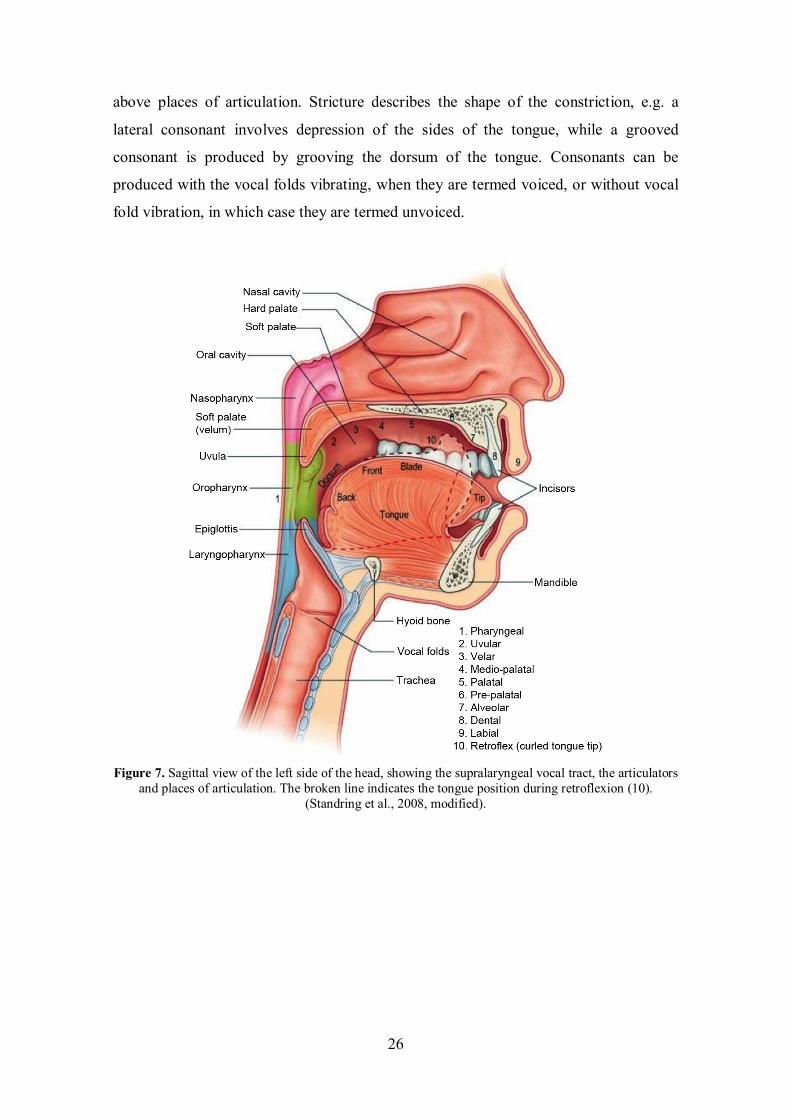
26
above places of articulation. Stricture describes the shape of the constriction, e.g. a
lateral consonant involves depression of the sides of the tongue, while a grooved
consonant is produced by grooving the dorsum of the tongue. Consonants can be
produced with the vocal folds vibrating, when they are termed voiced, or without vocal
fold vibration, in which case they are termed unvoiced.
Figure 7. Sagittal view of the left side of the head, showing the supralaryngeal vocal tract, the articulators
and places of articulation. The broken line indicates the tongue position during retroflexion (10). (Standring et al., 2008, modified).
27
6 MATERIAL AND METHODS
6.1 CLINICAL EXAMINATION Subjects were examined while sitting on a dental chair in a room with
appropriate lighting, by the same examiner, specialist in temporomandibular joint
disorders (TMD) and orofacial pain. Data considered in the present study referred to
tenderness to palpation in the masseter, temporal and supra-hyoid muscles and in the
temporomandibular joint (TMJs). The subjects were asked to grade their pain using a
printed numerical scale from zero (absence of pain) to 10 points (greatest pain possible).
The TMJ region was also palpated during the mandibular movements for the
identification of joint noises, which were confirmed by auscultation. The Research
Diagnostic Criteria for TMD (RDC/TMD - axis I, Dworkin and LeResche, 1992) was
used for classification.
6.1.1 SELF-JUDGEMENT OF SEVERITY
The ProTMDmulti questionnaire was used to determine the perception (presence
and severity) of TMD signs and symptoms by the subjects. The questionnaire is divided
into two parts; the first part asks about the presence of TMD signs and symptoms with a
series of 12 questions requiring a positive or negative reply. In the second part, the
subjects were asked to indicate the severity of nine signs and symptoms present (or no)
according to the situation, i.e. when waking up, during mastication, when speaking and
at rest. Severity was indicated on a printed 11-point numerical scale where zero
corresponded to the complete absence of the symptom, and 10 corresponded to the
highest possible severity. The severity score was the sum of the scores attributed to each
sign and symptom in the four questioned situations. The severity score varies between
zero (absence) and 40 (the highest possible severity) (Felício et al., 2009a).
28
6.1.2 OROFACIAL MYOFUNCTIONAL EVALUATION
The components of the stomatognathic system were evaluated in terms of
mobility according to the Orofacial Myofunctional Evaluation with Scores Protocol
(OMES – Part I) (Felício and Ferreira, 2008) when the healthy subject was asked to
perform the following movements:
• Lips: protrusion, retrusion, lateral to the right and left;
• Tongue: protrusion, retrusion, lateral to the right and left, raising, and
lowering and ability to keep the tongue in stable protrusion for 5 seconds;
• Mandible: protrusion, lowering, raising, lateral to the right and left.
In the analysis, separate movements of each component were considered normal
if precise and without tremors. Dysfunction was considered present when lack of
precision in the movement, tremor, associated movements of other components (e.g.,
lips accompanying the movements of the tongue) or inability to perform the movement
was observed.
According to the OMES Protocol, the examiner attributed scores on a 3 point
scale: 3 = normal; 2 = insufficient ability; 1 = absence of ability or being unable to
perform the task.
To complement the analysis for jaw movements, measurements (in mm),
symmetry/asymmetry during mouth opening and closing, right and left laterality and
protrusion will also be considered. Scores were attributed according to the protocol.

29
6.2 SUBJECTS Twenty healthy young adults (10 men and 10 women) aged 20 to 41 years,
natural speakers of Italian language, participated in the study. They were recruited from
the students and staff attending the Department of Biomedical Sciences for Health,
University of Milan. All subjects had a clinically normal facial function, no previous
facial trauma, paralysis or surgery, no known neurological diseases, and no and current
orthodontic treatment.
To be recruited, the healthy subjects had no to present TMD according to the
Research Diagnostic Criteria for TMD (RDC/TMD, Dworkin and LeResche, 1992), to
the ProTMDmulti protocol (Felício et al, 2009a), and an orofacial myofunctional
evaluation (Felício and Ferreira, 2008), as detailed in Chapter 5.1.

30
6.3 INSTRUMENTATIONS
6.3.1 OPTOELECTRONIC MOTION ANALYZER
Lip movements in verbal and non-verbal activities were recorded using an
optoelectronic three-dimensional motion analyzer, the SMART-E system (BTS S.p.a,
Garbagnate Milanese, Italy).
High-precision infrared sensitive CCD video cameras (Fig. 8) are coupled with
the video processor with up to 120 Hz sampling ratio. The 3D positions of lightweight,
passive and retro-reflective markers are instantly recorded with a spatial accuracy of up
to 0.1 mm.
Figure 8. Detail of a camera.
In brief, stroboscopic infrared light (wavelength, 880 nm) is emitted by an array
of LED (light emitting diodes) mounted around the lens of each camera, and the CCD
sensor detects the reflection from the markers placed on the body.
The process of recognizing passive markers in the 2D video frames is performed
via enhanced blob analysis. The 3D coordinates of each marker are finally computed
based upon the 2D data of at least two cameras. This process, called spatial
triangulation, needs the system to be previously calibrated. Calibration allows the
system to estimate the capture volume, the relative position and orientation of the
cameras (external parameters), their geometric and optical characteristics (internal
parameters).
The BTS SMART system requires two calibration phases. The “static
calibration” sets the position and the orientation of the global reference system: all

31
cameras simultaneously record a still, special reference device (Fig. 9), whose marker
reciprocal distances are known to the system. The “dynamic” calibration exploits the
epipolar constraint between a 3D point and its 2D projections on the sensor of two
cameras: all cameras simultaneously record a rigid bar (Y axis) in motion throughout
the working volume.
Figure 9. Global reference system.
At the end of the metric calibration and correction of optical and electronic
distortions, the system provides the current accuracy level, which will characterize the
following acquisition sessions.
Once a movement has been recorded, special software provides the spatial
configuration of the marker set (Fig. 10).
Figure 10. 3D graphic representation of the marker set at issue.
X
Y
Z

32
The operator has to label the markers of interest in one frame, opening the
corresponding model previously created; afterward, the system should be able to
recognize all moving markers, tracking their pathways.
To record lip movements (Ferrario and Sforza et al., 2007; Sforza et al., 2010b,c;
2012), nine cameras were deployed around a stool (Fig. 11), and calibrated to create a
60 (width) cm x 60 (height) cm x 60 (depth) cm working volume; metric calibration and
correction of optical and electronic distortions are performed before each acquisition
session using a 20-cm wand, with a resulting mean dynamic accuracy of 0.121mm (SD
0.086), corresponding to 0.0158% (Sforza et al., 2010c). A 60 Hz capture rate was used
for all acquisitions.
Figure 11. Setting of the cameras with respect to the subject sat on a stool.

33
6.3.2 ELECTROMYOGRAPHIC SYSTEM
The BTS FREEEMG system (BTS S.p.a, Garbagnate Milanese, Italy) is a
wireless electromyographic device with active probes weighting just 8 grams for signal
acquisition and wireless transmission (Fig. 12). The probes amplify the differential
EMG signals captured by disposable pre-gelled silver/silver chloride bipolar surface
electrodes, digitize them and communicate with a portable receiving unit. The complete
absence of cables allows for quick and comfortable preparation of the subject, without
affecting in any way the motor pattern. This system is easily connectable with the
motion analyzer, permitting the real time recording of synchronized kinematic and
electromyographic data.
Figure 12. Detail of an EMG probe clipped on a pair of electrodes.
(http://www.btsbioengineering.com/BTSBioengineering/Surfaceemg/BTSFREEEMG300/BTS_FREEEMG300.html, last accessed 11 December 2012).
In surface EMG, a bipolar probe measures the voltage difference between two
electrodes, which is the sum of the electrical contributions of the active motor units;
thus it reflects both the muscle membrane properties and the central control strategies.
With small inter-electrode distance with respect to the muscle size, the activity
conducted from adjacent muscles is similar on the two leads, and therefore is partly
rejected (Castroflorio et al., 2008).

34
6.3.3 RECORDING PROTOCOL
For each subject, the recordings took approximately 40 minutes (considering
also the time needed for subject’s preparation). The protocol did not involve dangerous
or painful procedures, and it was preventively approved by the ethics committee of the
Department of Biomedical Sciences for Health, University of Milan.
After the methods and aims of the investigation had been completely described,
written informed consent was obtained from each participant.
Kinematic assessment of speech and lip movements
Subjects sat on a stool inside the working volume and were asked to perform a
series of standardized lip movements and speech pronunciation. During the execution of
the movements, for each camera special software detected the two-dimensional
coordinates of facial landmarks identified by a set of 2-mm round reflective markers.
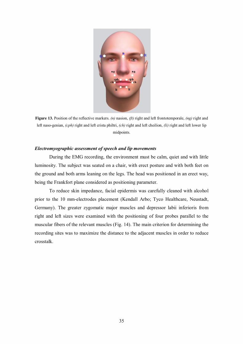
Landmarks were chosen from classic anthropometry (Farkas, et al., 1994): n, nasion; ft,
right and left frontotemporale; ng, right and left naso-genian; cph, right and left crista
philtri; ch, right and left cheilion; li, right and left lower lip midpoints (Figs. 13, 14).
The positions of the markers were carefully controlled to avoid any interference with lip
and speech movements (Trotman et al., 2003; Hontanilla and Aubá, 2008; Sforza et al.,
2010b,c; 2012). Subsequently, all the coordinates were converted to metric data, and a
set of three-dimensional coordinates for each landmark in each frame that constituted
each movement was obtained.
Each animation was explained and shown to the subjects, which practiced before
data acquisition. For each expression, each subject performed ten standardized
maximum facial expression from rest (Wachtman et al., 2001; Hontanilla and Aubá,
2008; Mehta et al., 2008; Sforza et al., 2010b,c), without modifications of the markers
positions.
The healthy subjects performed four standardized non-verbal movements: open
mouth smile, closed mouth smile, spontaneous smile and lip purse; and verbal
movements: natural and random (e.g. o, a, i, u, e) sequence of the five vowel, a
sequence of natural numbers (e.g. 1 to 10) and 29 Italian words (Table 1).
35
Figure 13. Position of the reflective markers. (n) nasion, (ft) right and left frontotemporale, (ng) right and
left naso-genian, (cph) right and left crista philtri, (ch) right and left cheilion, (li) right and left lower lip
midpoints.
Electromyographic assessment of speech and lip movements
During the EMG recording, the environment must be calm, quiet and with little
luminosity. The subject was seated on a chair, with erect posture and with both feet on
the ground and both arms leaning on the legs. The head was positioned in an erect way,
being the Frankfort plane considered as positioning parameter.
To reduce skin impedance, facial epidermis was carefully cleaned with alcohol
prior to the 10 mm-electrodes placement (Kendall Arbo; Tyco Healthcare, Neustadt,
Germany). The greater zygomatic major muscles and depressor labii inferioris from
right and left sizes were examined with the positioning of four probes parallel to the
muscular fibers of the relevant muscles (Fig. 14). The main criterion for determining the
recording sites was to maximize the distance to the adjacent muscles in order to reduce
crosstalk.

36
Figure 14. Position of the reflective markers and the electrodes of the BTS FREEEMG system (BTS
S.p.a, Garbagnate Milanese, Italy).
All the necessary explanations were given previously, as well as the lip
movements were practiced with the examiner prior to test performance.
The subjects were asked to perform 10 consecutive voluntary open and closed-
mouth smiles, and a spontaneous smile, while watching a fun film. The resting time
between the open and closed smiles was about 3 seconds (Fig. 15).
Figure 15. Raw EMG signal of the right zygomatic major muscle during open-mouth smile.
37
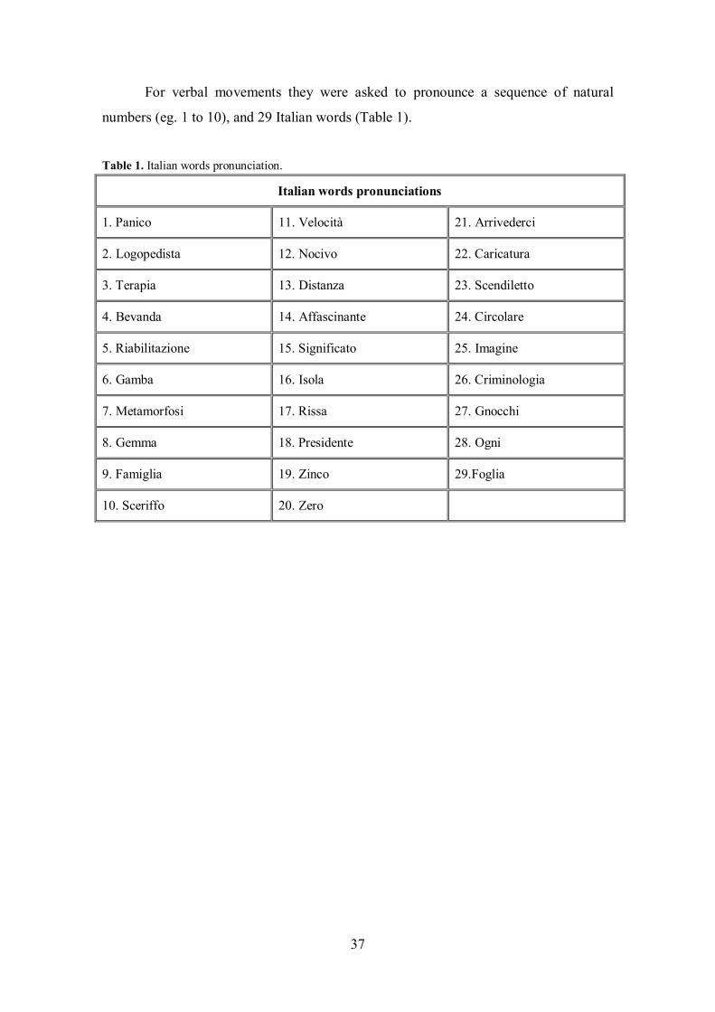
For verbal movements they were asked to pronounce a sequence of natural
numbers (eg. 1 to 10), and 29 Italian words (Table 1).
Table 1. Italian words pronunciation.
Italian words pronunciations
1. Panico 11. Velocità 21. Arrivederci
2. Logopedista 12. Nocivo 22. Caricatura
3. Terapia 13. Distanza 23. Scendiletto
4. Bevanda 14. Affascinante 24. Circolare
5. Riabilitazione 15. Significato 25. Imagine
6. Gamba 16. Isola 26. Criminologia
7. Metamorfosi 17. Rissa 27. Gnocchi
8. Gemma 18. Presidente 28. Ogni
9. Famiglia 19. Zinco 29.Foglia
10. Sceriffo 20. Zero

38
6.3.4 MEASUREMENT PROTOCOL
Both raw EMG signals and marker coordinates constituted the input data for the
protocol calculations, which were implemented on Microsoft Excel and Smart Analyzer.
Descriptive and inferential statistics were evaluated by means of SPSS Statistics.
Kinematic of speech and lip movements
For each subject, head and neck motion was subtracted from the raw facial
movements using the three cranial (reference) markers, so only movements occurring in
the face (activity of mimetic muscles) were further considered. Subsequently, for each
of the 8 facial markers, the three-dimensional movements during both verbal and non-
verbal activities were computed, and the modulus (intensity) of the three-dimensional
vector of maximum displacement from rest was calculated (Ferrario and Sforza, 2007;
Sforza et al., 2010b,c; 2012). For the natural sequence of numbers and words
production, instantaneous displacements was also calculated.
For each animation, the landmark (single or paired) with the largest
displacement from rest was identified. For smile movements, the latero-lateral (right-left
direction) component of the maximum displacement of the analysed landmarks was
computed.
The frontal area of movement (XY planes; unit, mm2) for smiles, lip purse and
vowels is estimated with the perimeter defined by the 6 labial markers (cph, ch and li).
The ∆ area (unit, mm2) was calculated between the areas of maximum expression and
rest position.
To assess differential movements between the two hemi-faces, percentage
indices of asymmetry were computed as:
(right side displacement - left side displacement) / (right side displacement + left side displacement) × 100.
The indices were computed for the total movement, as well as for the single
landmarks. The indices range between -100% (complete left-side prevalence during the
movement) and +100% (complete right-side prevalence) (Linstrom et al., 2002; Sforza
et al., 2010c; 2012).

39
For the kinematic analysis of the markers of number sequences and words
pronunciation, data were low-pass filtered using a Butterworth filter with cutoff
frequency at 8 Hz.
The percentage overlapping coefficient (POC, unit: %) for words and numbers was
calculated as:
POC = [1 – Σ right side displacement i – left side displacement i / Σ right side displacement i + left side displacement i ] × 100
(i - instantaneous)
The index ranges between 0% (no symmetry) and 100% (perfect symmetry).
The total percentage indices of asymmetry for sequence of numbers and words
pronunciation (ASYM, unit %) was computed as:
ASYM = mean (right side displacement i – left side displacement i) / mean (right side displacement i + left side displacement i) × 100
(i - instantaneous)
The ASYM index ranges between -100% (left-side prevalence of pronunciation)
and +100% (right-side prevalence of pronunciation).
Electromyographic signals of speech and lip movements
For each muscle the EMG signal was filtered with a Butterworth "high pass"
filter at the cutoff frequency of 50 Hz. Then we calculated the root mean square (RMS)
signal with a time window of 25 ms, which was “low pass” filtered at 5 Hz.
For each movement, a custom made algorithm allowed the semi-automatic
detection of the beginning of the muscle activity, and the time of latency between the
activities of the four muscles was calculated.
The time difference in the onsets of electrical activity between the right and the
left side of the face is referred to as asymmetry. Asymmetry is related to either the
zygomatic muscles or the depressor labii inferioris (Fig. 17). It is expressed in seconds
(s). The time difference in the onsets of electrical activity between zygomatic and
depressor labii inferioris muscles is referred to as asynchrony. Asynchrony is related to
each side of the face separately, either right or left (Fig. 18). Like asymmetry, it is

40
expressed in seconds (s).
For open-mouth, closed-mouth and spontaneous smiles, the time of activation,
the asymmetry between the right and left zygomatic major or depressor labii inferioris
muscles, and the asynchrony between the zygomatic major and depressor labii inferioris
were analyzed.
For speech pronunciation (natural sequence of numbers, and words), we
calculated only by the asymmetry between the right and left depressor labii inferioris
muscles.
Figure 16. Asymmetry onset time of signal of the (1) right zygomatic major muscle, (2) left zygomatic major muscle, (3) right depressori labii inferioris muscle, and (4) left depressori labii inferioris muscle
during open-mouth smile.
Figure 17. Asynchrony onset time of signal of the (1) right zygomatic major muscle, (2) left zygomatic major muscle, (3) right depressori labii inferioris muscle, and (4) left depressori labii inferioris muscle
during open-mouth smile.
1
2
3
4
1
2
3
4

41
6.3.5 METHOD ERROR
For the optoelectronic three-dimensional motion analyzer, within- and between-
session repeatability was previously assessed in healthy subjects. Within session, the
technical error of the measurement for single landmarks (random error) was, on
average, 0.5 and 3.38 mm, showing good reproducibility. Between sessions, all facial
movements had standard deviations lower than 1mm (Sforza et al., 2010b,c).
For EMG analysis, the repeatability was assessed with hierarchical ANOVA,
showing good reproducibility in the recorded smiles activities.

42
7 KINEMATICS ANALYSIS OF LIP MOTION IN
HEALTHY SUBJECTS
7.1 METHODS
7.1.1 DATA COLLECTION
The kinematic indices calculated for verbal and non-verbal activities are
summarized in table 2.
Table 2. Kinematics indices for verbal and non-verbal activities: (cph) crista philtri, (ch) cheilion, (li) lower lip midpoints.
KINEMATIC INDICES
Asymmetry (%)
(ch, cph, li, total)
Landmarks and Total Mobility
(mm)
Frontal area (mm2)
∆ area (mm2)
POC (%)
ASYM (%)
Open-mouth smile
Closed-mouth smile
Spontaneous smile
Lip purse
Natural sequence vowels
Random sequence vowels
Natural sequence numbers
Italian words

43
7.1.2 STATISTICAL ANALYSIS
For all subjects, ten series of lip movements (open-mouth smile, closed-mouth
smile, spontaneous smile, lip purse), natural and random sequences of vowels
production were averaged, and the mean values of maximum marker displacement for
each movement used for subsequent analysis. For the natural sequence of numbers and
words production, the instantaneous displacements of landmarks was calculated.
Descriptive statistics were obtained for each marker, the total lip movements, the
asymmetry indices, and frontal area, separately for each sex.
The normal distribution of data was checked with the Kolmogorov-Smirnov test.
The largest displacement from rest, total mobility and frontal area were
compared between sexes by Student’s t-Test for unpaired samples in all smiles and lip
purse movements. To assess if the asymmetry indices significantly deviated from the
expected value of 0, Student’s t-tests for paired samples were made.
For natural and random sequences of vowels, the largest displacement from rest
was compared between sexes by Student’s t-Test for unpaired samples, and the total
mobility was compared by three-way factorial analysis of variance (factors: sexes, side,
and vowels pronunciation). For words and sequences of numbers, the POC and
asymmetry indices were compared by two-way factorial analysis of variance (factors:
sex and pronunciation).
The significance level was set at 5% for all statistical analyses (p < 0.05).

44
7.2 RESULTS All data within each subgroup (men, women) were normally distributed
(Kolmogorov–Smirnov tests, p > 0.05), and no significant difference was found
between male and female mean ages (Student’s unpaired t-test; all p-values > 0.05).
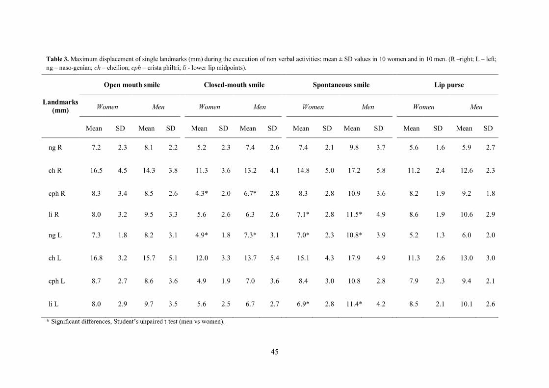
On average, during the execution of the non-verbal activities the cheilion
landmarks had the largest displacements in both sex groups. For open-mouth smile and
lip purse activities, the largest displacement from rest was similar between sex groups
without statistically significant differences (Student’s unpaired t-test; all p-values >
0.05). In contrast, men had significant larger movements than women during the
execution of the closed-mouth smile (right crista philtri and left naso-genian landmarks,
both p = 0.04), and the spontaneous smile (right and left lower lip, and left naso-genian
landmarks, respectively, p = 0.01, p = 0.03 and p = 0.02) (Table 3).
Within open mouth smile, closed-mouth smile and lip purse movements, the
total facial mobility was similar in both sexes, without statistically significant
differences (Student’s unpaired t-test; all p-values > 0.05). In contrast, in spontaneous
smile sex-related differences were found on the left side (p = 0.041; Student’s unpaired
t-test): the male group performed this movement with larger landmark displacements
than the female group (Fig. 18).
45
Table 3. Maximum displacement of single landmarks (mm) during the execution of non verbal activities: mean ± SD values in 10 women and in 10 men. (R –right; L – left; ng – naso-genian; ch – cheilion; cph – crista philtri; li - lower lip midpoints).
Landmarks (mm)
Open mouth smile Closed-mouth smile Spontaneous smile Lip purse
Women Men Women Men Women Men Women Men
Mean SD Mean SD Mean SD Mean SD Mean SD Mean SD Mean SD Mean SD
ng R 7.2 2.3 8.1 2.2 5.2 2.3 7.4 2.6 7.4 2.1 9.8 3.7 5.6 1.6 5.9 2.7
ch R 16.5 4.5 14.3 3.8 11.3 3.6 13.2 4.1 14.8 5.0 17.2 5.8 11.2 2.4 12.6 2.3
cph R 8.3 3.4 8.5 2.6 4.3* 2.0 6.7* 2.8 8.3 2.8 10.9 3.6 8.2 1.9 9.2 1.8
li R 8.0 3.2 9.5 3.3 5.6 2.6 6.3 2.6 7.1* 2.8 11.5* 4.9 8.6 1.9 10.6 2.9
ng L 7.3 1.8 8.2 3.1 4.9* 1.8 7.3* 3.1 7.0* 2.3 10.8* 3.9 5.2 1.3 6.0 2.0
ch L 16.8 3.2 15.7 5.1 12.0 3.3 13.7 5.4 15.1 4.3 17.9 4.9 11.3 2.6 13.0 3.0
cph L 8.7 2.7 8.6 3.6 4.9 1.9 7.0 3.6 8.4 3.0 10.8 2.8 7.9 2.3 9.4 2.1
li L 8.0 2.9 9.7 3.5 5.6 2.5 6.7 2.7 6.9* 2.8 11.4* 4.2 8.5 2.1 10.1 2.6
* Significant differences, Student’s unpaired t-test (men vs women).

46
Figure 18. Total mobility in the four standardized lip movements (mean + 1SD).
* Significant differences on the left side in spontaneous smile, Student’s unpaired t-test (men vs women).
The asymmetry of the upper lip landmarks was significantly different from 0
during closed-mouth smile in women (p = 0.033, Student’s t-Test for paired samples),
in the other movements (open-mouth smile, spontaneous smile and lip purse) no
significant differences were found in asymmetry indices in all landmarks (cph, ch and
li) and in total asymmetry (Fig. 19).
Figure 19. Mouth asymmetry indices for the four symmetric lip movements (mean ± 1SD).
* Significant difference in women during closed-mouth smile, Student’s paired t-test.
0
10
20
30
40
50
60
70 mm
Women
Men
*
-20
-15
-10
-5
0
5
10
15
20
cheilion crista philtri lower lip total
%
Women - open mouth smile Women - closed mouth smile Women - spontaneous smile Women - Lip purse Men - open mouth smile Men - closed mouth smile Men - spontaneous smile Men - Lip purse
Right
Left
*
47
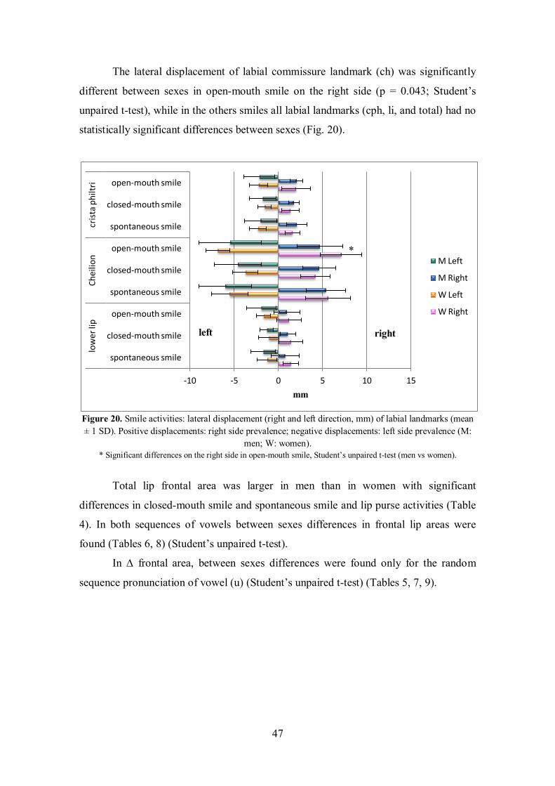
The lateral displacement of labial commissure landmark (ch) was significantly
different between sexes in open-mouth smile on the right side (p = 0.043; Student’s
unpaired t-test), while in the others smiles all labial landmarks (cph, li, and total) had no
statistically significant differences between sexes (Fig. 20).
Figure 20. Smile activities: lateral displacement (right and left direction, mm) of labial landmarks (mean ± 1 SD). Positive displacements: right side prevalence; negative displacements: left side prevalence (M:
men; W: women). * Significant differences on the right side in open-mouth smile, Student’s unpaired t-test (men vs women).
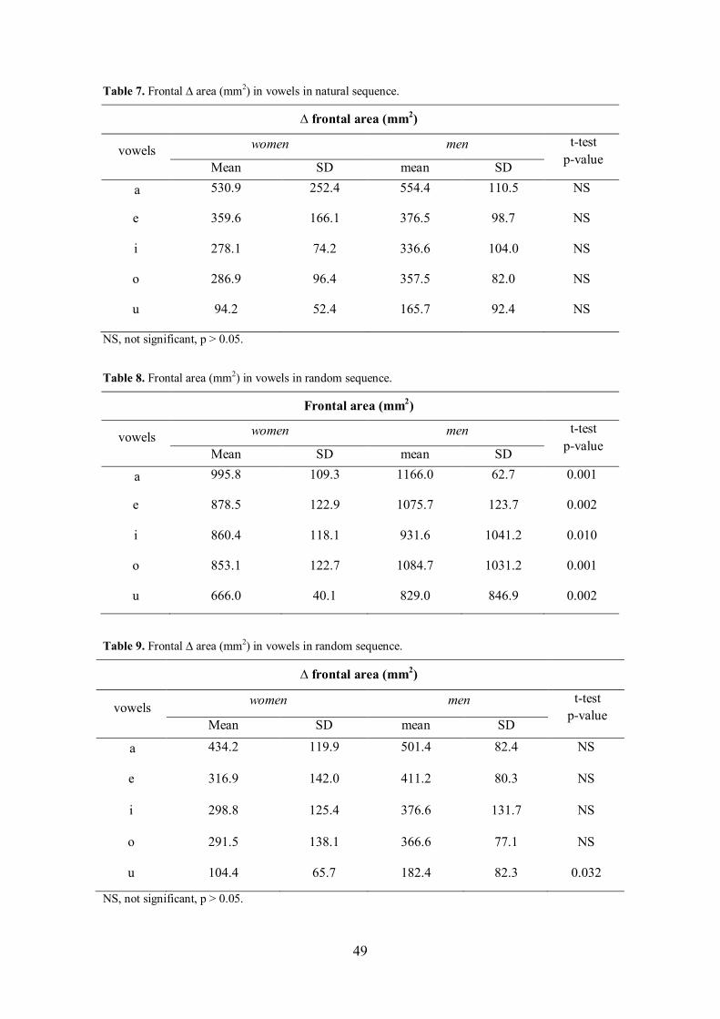
Total lip frontal area was larger in men than in women with significant
differences in closed-mouth smile and spontaneous smile and lip purse activities (Table
4). In both sequences of vowels between sexes differences in frontal lip areas were
found (Tables 6, 8) (Student’s unpaired t-test).
In ∆ frontal area, between sexes differences were found only for the random
sequence pronunciation of vowel (u) (Student’s unpaired t-test) (Tables 5, 7, 9).
-10 -5 0 5 10 15
spontaneous smile
closed-mouth smile
open-mouth smile
spontaneous smile
closed-mouth smile
open-mouth smile
spontaneous smile
closed-mouth smile
open-mouth smile
low
er li
p Ch
eilio
n cr
ista
philt
ri
mm
M Left
M Right
W Left
W Right
right left
*

48
Table 4. Frontal area (mm2) in smiles and lip purse movements.
NS, not significant, p > 0.05.
Table 5. Frontal ∆ area (mm2) in smiles and lip purse movements.
NS, not significant, p > 0.05.
Table 6. Frontal area (mm2) in vowels in natural sequence.
NS, not significant, p > 0.05.
Frontal area (mm2)
women men t-test p-value
mean SD mean SD Open mouth smile 981.3 152.4 1059.9 166.5 NS
Closed mouth smile 540.2 70.0 734.7 144.2 p = 0.002
Spontaneous smile 874.7 228.5 1105.7 193.3 p = 0.0031
Lip purse 655.9 69.0 806.1 102.5 p = 0.001
∆ frontal area (mm2)
women men t-test p-value
mean SD mean SD
Open mouth smile 434.0 155.1 404.3 161.4 NS
Closed mouth smile -12.8 77.9 76.7 149.4 NS
Spontaneous smile 311.5 235.3 442.4 159.9 NS
Lip purse 87.6 57.2 148.8 85.0 NS
Frontal area (mm2)
vowels women men t-test p-value
Mean SD mean SD a 1089.9 233.6 1208.8 130.6 NS
e 918.6 146.7 1030.9 142.6 NS
i 837.1 76.2 991.0 146.9 p = 0.011
o 845.9 97.1 1011.9 107.4 p = 0.002
u 653.2 56.6 820.0 115.9 p = 0.001
49
Table 7. Frontal ∆ area (mm2) in vowels in natural sequence.
NS, not significant, p > 0.05.
Table 8. Frontal area (mm2) in vowels in random sequence.
Table 9. Frontal ∆ area (mm2) in vowels in random sequence.
NS, not significant, p > 0.05.
∆ frontal area (mm2)
vowels women men t-test p-value Mean SD mean SD
a 530.9 252.4 554.4 110.5 NS
e 359.6 166.1 376.5 98.7 NS
i 278.1 74.2 336.6 104.0 NS
o 286.9 96.4 357.5 82.0 NS
u 94.2 52.4 165.7 92.4 NS
Frontal area (mm2)
vowels women men t-test p-value Mean SD mean SD
a 995.8 109.3 1166.0 62.7 0.001
e 878.5 122.9 1075.7 123.7 0.002
i 860.4 118.1 931.6 1041.2 0.010
o 853.1 122.7 1084.7 1031.2 0.001
u 666.0 40.1 829.0 846.9 0.002
∆ frontal area (mm2)
vowels women men t-test p-value
Mean SD mean SD a 434.2 119.9 501.4 82.4 NS
e 316.9 142.0 411.2 80.3 NS
i 298.8 125.4 376.6 131.7 NS
o 291.5 138.1 366.6 77.1 NS
u 104.4 65.7 182.4 82.3 0.032

50
On average, during the execution of the natural and random sequences of
vowels, the lower lip midpoint landmarks had the largest displacements in both sex
groups. Significant sex-related differences of vowels pronunciation were found for the
right cheilion landmarks for natural sequence of vowels (a) (p = 0.043), (e) (p = 0.040),
and for left crista philtri landmark for random sequence of vowel (o) (p = 0.013) (Tables
10, 11).
For both sequences of vowel pronunciation (natural and random), the total
mobility in vowels (o, i, u) was similar, without significant differences (three-way
factorial analysis of variance; factors sex, vowels and sides, all p-values > 0.05). In
contrast, significant side-related differences of vowels pronunciation were found for the
vowels (a) (p = 0.010), and (e) (p = 0.007) (Fig. 21).
Figure 21. Total mobility in the natural and random sequences of vowels production (mean + 1SD).
* Significant side-related differences of vowels (natural and random) pronunciation, three-way ANOVA.
0
10
20
30
40
50
60 mm
Women R
Women L
Men R
Men L
a e i o u
*
*

51
Table 10. Maximum displacement of single landmarks (mm) during the execution of natural sequence vowels: mean ± SD values in 10 women and in 10 men. (R –right; L – left; ng – naso-genian; ch – cheilion; cph – crista philtri; li - lower lip midpoints).
Natural sequence vowels
Landmarks (mm)
a e i o u
Women Men Women Men Women Men Women Men Women Men
Mean SD Mean SD Mean SD Mean SD Mean SD Mean SD Mean SD Mean SD Mean SD Mean SD
ng R 3.6 1.6 3.4 1.3 2.2 1.1 2.2 0.8 1.7 1.4 1.7 0.8 3.0 1.6 3.1 1.1 3.1 1.4 3.3 1.1
ch R 9.7* 2.7 7.6* 1.4 6.8* 2.4 4.8* 1.5 4.2 2.2 3.0 1.2 5.5 2.8 5.0 1.3 6.2 2.4 5.8 2.2
cph R 2.1 1.2 1.6 1.0 2.1 1.4 1.5 1.0 2.1 1.1 1.6 1.0 1.8 1.0 2.5 1.8 4.1 1.5 3.9 1.6
li R 19.7 6.9 17.6 2.2 12.9 5.3 11.4 3.8 8.7 2.3 9.2 3.2 12.4 4.2 12.7 3.2 7.4 1.9 9.0 2.2
ng L 3.8 1.5 3.1 0.9 2.3 1.4 1.9 0.8 1.7 1.3 1.7 0.6 2.7 1.2 3.2 1.4 2.8 1.4 3.1 2.1
ch L 9.3 3.3 7.2 1.5 6.9 3.3 4.4 1.6 4.1 2.2 3.6 1.2 5.4 2.2 6.1 3.0 5.5 2.2 6.2 4.5
cph L 2.0 1.3 1.7 1.4 1.8 1.2 1.5 1.1 1.7 0.9 1.8 1.2 1.4 0.7 2.7 2.2 3.5 1.5 4.2 2.0
li L 19.9 7.9 17.2 1.9 12.7 5.5 12.0 3.8 8.9 3.2 9.1 3.4 12.2 4.3 12.0 2.9 7.7 2.0 8.8 2.6
* Significant differences, Student’s unpaired t-test (men vs women).

52
Table 11. Maximum displacement of single landmarks (mm) during the execution of random sequence vowels: mean ± SD values in 10 women and in 10 men. (R –right; L – left; ng – naso-genian; ch – cheilion; cph – crista philtri; li - lower lip midpoints).
Random sequence vowels
Landmarks (mm)
a e i o u
Women Men Women Men Women Men Women Men Women Men
Mean SD Mean SD Mean SD Mean SD Mean SD Mean SD Mean SD Mean SD Mean SD Mean SD
ng R 3.4 1.4 2.9 1.4 2.0 1.2 2.0 0.6 1.9 1.1 1.9 1.1 3.3 1.1 3.4 1.7 3.0 1.2 2.9 1.3
ch R 7.8 1.8 6.9 1.7 6.7 3.7 5.3 2.0 5.0 3.1 4.1 2.5 5.9 1.7 5.2 2.1 6.1 2.1 5.6 1.8
cph R 2.1 1.5 1.5 0.8 2.6 2.3 1.9 1.4 2.6 2.8 2.4 1.3 1.9 0.7 2.4 0.9 3.3 0.7 3.2 1.9
li R 16.6 4.2 15.8 3.2 11.8 4.3 12.7 3.1 8.9 2.4 10.3 4.6 12.9 3.4 13.4 4.6 8.8 2.4 9.0 2.7
ng L 3.0 0.9 2.6 1.0 2.2 1.0 1.8 0.6 1.7 1.0 1.6 1.1 2.8 0.6 2.9 1.2 2.8 1.0 2.6 0.9
ch L 7.6 2.4 6.4 1.3 6.0 2.9 5.1 2.1 4.4 2.5 4.2 2.5 6.2 1.4 5.0 1.8 6.5 2.0 5.7 2.2
cph L 1.3 0.4 1.8 1.1 1.7 1.1 1.8 1.4 2.0 1.6 2.3 1.2 1.5* 0.9 2.8* 1.3 3.4 1.2 3.3 1.9
li L 16.6 4.3 15.5 3.2 11.5 4.3 12.2 3.2 9.0 2.7 9.9 4.8 13.3 3.3 12.5 4.3 8.5 2.4 8.6 2.1
* Significant differences, Student’s unpaired t-test (men vs women).

53
Mouth asymmetry indices were not significantly different between sexes in both
sequences of vowels (a, i) (Student’s unpaired t-test; p-values < 0.05). On the other
hand, in vowels pronunciation in natural sequence, significant differences were found in
(li) landmark for vowel (e) (p = 0.046) and in (cph) landmark for vowel (u). In the
random sequence vowel pronunciation, a significant difference was found in (cph) and
(li) landmarks for vowel (o) (p = 0.007; p = 0.017) (Tables 12, 13).
For both sequences of vowel pronunciation, some significant differences from 0
were found. For the natural sequence, vowels e (li, p = 0.046) and u (cph, p = 0.039)
were significantly asymmetric in women, and vowels a (total, p = 0.024), e (li, p =
0.021 and total, p = 0.004) and o (li, p = 0.039) in men. For the random sequence, a
significant difference was found in women for vowels a (total, p = 0.006), e (cph, p =
0.031 and total p = 0.025) and o (cph, p = 0.036), and in men for vowels e (li, p = 0.015)
and o (cph, p = 0.020 and li, p = 0.042). Table 12. Mouth asymmetry indices for natural sequence of vowels in women and men (ch – cheilion; cph – crista philtri; li - lower lip midpoints).
* Significant differences, Student’s unpaired t-test (men vs women). # Significant differences, Student’s paired t-test (difference from 0).
% Asymmetry (ch) Asymmetry (cph) Asymmetry (li) Total Asymmetry
women mean SD mean SD mean SD mean SD a 3.5 8.3 4.6 27.9 0.0 2.3 1.1 4.1
e 2.6 10.8 0.9 29.2 -0.8*# 4.1 0.7 4.9
i 0.3 7.9 8.7 26.1 0.1 5.5 1.5 4.5
o -1.7 14.5 10.2 16.6 0.6 3.2 1.3 5.0
u 6.8 10.8 9.7*# 12.9 -1.9 6.3 4.1 6.7
men mean SD mean SD mean SD mean SD a 3.0 5.2 -2.6 23.7 1.1 2.2 1.8# 2.0
e 5.4 9.2 2.9 13.5 2.8*# 3.2 3.4# 2.6
i -9.8 15.8 -3.1 26.4 1.9 5.2 -1.8 6.7
o -5.9 16.5 -0.4 9.0 2.6# 3.6 -0.5 7.0
u 3.1 22.5 -2.5* 7.0 2.1 5.7 1.2 9.4

54
Table 13. Mouth asymmetry indices for random sequence of vowels in women and men (ch – cheilion; cph – crista philtri; li - lower lip midpoints).
* Significant differences, Student’s unpaired t-test (men vs women). # Significant differences, Student’s paired t-test (difference from 0).
On average, the ASYM index of the sequence of numbers and words
pronunciation was similar in both sexes, without significant differences (p > 0.05, two-
way factorial analysis of variance) (Figs. 22, 23).
% Asymmetry (ch) Asymmetry (cph) Asymmetry (li) Total Asymmetry
women Mean SD mean SD mean SD mean SD
a 2.9 7.0 13.4 28.5 0.1 2.0 2.7# 2.3
e 5.6 8.2 19.8# 24.6 1.5 3.9 5.2# 6.2
i 5.6 10.5 5.7 14.3 -0.4 3.7 3.3 6.3
o -3.9 9.4 18.4*# 23.6 -2.1* 4.7 0.1 4.1
u -3.7 7.7 -0.2 11.2 1.5 6.3 0.3 5.4
men Mean SD mean SD mean SD mean SD
a 3.7 7.1 -6.9 23.5 0.9 2.1 1.4 3.0
e 2.5 13.8 4.0 27.2 2.4# 2.6 3.5 6.9
i -0.8 15.2 -5.2 18.4 2.8 5.4 1.9 6.6
o 0.6 9.6 -7.7*# 8.5 3.3*# 4.5 2.0 4.8
u 0.1 9.2 -1.8 10.8 1.9 5.0 1.5 5.3

55
Figure 22. ASYM index in Italian words pronunciation (mean ± 1SD).
Figure 23. ASYM index in numbers pronunciation (mean ±1SD).
On average, the POC index for Italian words and sequence of numbers ranged
between 91 and 96%, with significant differences in speech pronunciation (p = 0.031,
two-way factorial analysis of variance; factors: sexes and speech pronunciation).
-10
-8
-6
-4
-2
0
2
4
6
8
10
12
1 2 3 4 5 6 7 8 9 10 11 12 13 14 15 16 17 18 19 20 21 22 23 24 25 26 27 28 29
%
Italian words pronunciation
Women
Men
right
left
-10
-8
-6
-4
-2
0
2
4
6
8
10
12
1 2 3 4 5 6 7 8 9 10
%
Italian numbers pronunciation
Women
Men
right
left

56
8 ELECTROMYOGRAPHIC ANALYSIS - HEALTHY
SUBJECTS
8.1 METHODS
8.1.1 DATA COLLECTION
EMG indices for verbal and non-verbal activities are summarized in table 14.
Data of 2 men and 2 women were lost for technical reasons in closed-mouth smile. In
spontaneous smile, we analyzed only 15 subjects, because one subject did not perform
the spontaneous smiles and data of other four subjects were lost for technical reasons.
Table 14. EMG indices for verbal and non-verbal activities (r: right, l: left ).
EMG INDICES
Asymmetry (r and l
zygomaticus major)
Asymmetry (r and l depressor labii inferioris)
Asynchrony (r zygomaticus
major and r depressor labii
inferioris)
Asynchrony (l zygomaticus
major and l depressor labii
inferioris)
Open-mouth smile
Closed-mouth smile
Spontaneous smile
Natural numbers
Italian words

57
8.1.2 STATISTICAL ANALYSIS
For all subjects, ten series of open and closed-mouth smiles, spontaneous smile,
natural sequence of numbers and words pronunciation were made. Descriptive statistics
were obtained for electromyographic time onset in open-mouth, closed-mouth and
spontaneous smiles, natural sequence of numbers, and words pronunciation.
The normal distribution of data was checked with the Kolmogorov-Smirnov test.
Both instructed smiles (open and closed-mouth), were compared by 4-way
analyses of variance (factors: sexes; muscles; side; subjects). The spontaneous smile
was compared by two-way analysis of variance (factors muscle, side, and muscle × side
interaction).
The natural sequence of numbers and words pronunciations was compared by
two-way analysis of variance (factors sexes and pronunciation).
The significance level was set at 5% for all statistical analyses (p < 0.05).

58
8.2 RESULTS All data within each subgroup (men, women) were normally distributed
(Kolmogorov–Smirnov tests, p > 0.05), and no significant difference was found
between male and female mean ages (Student’s unpaired t-test; all p-values > 0.05).
Significant interactions were found in muscle × subject in open (p < 0.001) and
closed-mouth smile (p < 0.001; four-way factorial analysis of variance).
Overall, for both smiles the zygomatic muscles tend to anticipate the depressor
labii inferioris muscles in women, while in men the opposite was found (Tables 15, 16). Table 15. Onset timing of muscle activity in closed-mouth smiles.
Table 16. Onset timing of muscle activity in open-mouth smiled.
In spontaneous smiles no differences were found (Table 17, two-way factorial analysis of variance; factors muscle, side and muscle × side interaction: all p-values > 0.05).
Onset time women men
Closed-mouth smile (s) mean SD mean SD right zygomatic major 0.05 0.07 0.06 0.10
left zygomatic major 0.04 0.07 0.05 0.10
right depressor labii inferioris 0.07 0.07 0.06 0.06
left depressor labii inferioris 0.07 0.09 0.05 0.05
Onset time women men
Open-mouth smile (s) mean SD mean SD right zygomatic major 0.06 0.07 0.07 0.07
left zygomatic major 0.07 0.07 0.06 0.07
right depressor labii inferioris 0.06 0.06 0.03 0.06
left depressor labii inferioris 0.07 0.07 0.04 0.05

59
Table 17. Onset timing of muscle activity in spontaneous smiles.
NS, not significant, p > 0.05.
For speech pronunciation, the onset time of activation was calculated only by the
asymmetry between the right and left depressor labii inferioris muscles. No significant
differences were found in both sexes and in speech pronunciation (two-way factorial
analysis of variance; factor sexes, factor pronunciation: all p-values > 0.05).
Figure 24. Asymmetry between right and left depressor labii inferioris muscles in both sexes (Italian
words pronunciation) (mean ±1SD).
-0.25
-0.20
-0.15
-0.10
-0.05
0.00
0.05
0.10
0.15
0.20
0.25
1 2 3 4 5 6 7 8 9 10 11 12 13 14 15 16 17 18 19 20 21 22 23 24 25 26 27 28 29
s
Italian words pronunciation
Women Men
Onset time right left ANOVA 2
ways Spontaneous smile (s) mean SD mean SD
zygomatic major 0.10 0.12 0.13 0.12 NS
depressor labii inferioris 0.09 0.11 0.10 0.11 NS

60
Figure 25. Asymmetry between right and left depressor labii inferioris muscles in both sexes (Italian
numbers pronunciation) (mean ±1SD).
-0.20
-0.15
-0.10
-0.05
0.00
0.05
0.10
0.15
0.20
0.25
1 2 3 4 5 6 7 8 9 10
s
Italian numbers pronunciation
Women Men
61
9 DISCUSSION Emotions play a key role in social communication, and interactions in humans
crucially depend on facial expressions. Spontaneous facial mimicry provides unique
insights into the biological determinants of emotional responses. The neurological
control of a smile consists of a complex process involving many facets, central
pathways of voluntary and evoked smiling (Root and Stephens, 2003; Achaibou et al.,
2008; Rymarczyk et al., 2011).
Facial expressions and speech articulation are produced by the synergistic or co-
operative action of many different facial muscles, involving movements of the mouth
(lower and upper lips) (Root and Stephens, 2003; Holberg et al., 2006; Van der Geld et
al., 2008; Sjögreen et al., 2011).
The smile is characterized by an upward curving of the corners of the mouth,
indicating pleasure, amusement, or derision, a pleasant or favorable disposition or
aspect, which is produced by the contraction of the zygomatic major muscle. Zygomatic
minor muscle (drawing the skin of the lip between the philtrum and the lip corner
obliquely upwards and laterally) and levator Anguli Oris (incidentally displaying the
teeth) also contributes to smiling.
According to Frank and Ekman (1993), each smile, using a combination of
different muscles, conveys different messages, for example the spontaneous smiles are
formed by the contraction of both the zygomatic major and the orbicularis oculi
muscles, while posed smiles involve only the zygomatic major muscle. These different
types of smiles are often distinguishable during social interaction.
Facial movements reflect spontaneous responses of viewers to emotional stimuli,
and the identification of the neural correlates of this phenomenon can supply important
findings about the brain mechanisms of emotion and social cognition. However, little is
known about the neural processes underlying facial mimicry, and in particular whether
emotional face perception and the subsequent emotion reaction are somehow related or
completely independent processes (Achaibou et al., 2008).

62
The instruments
Facial mimicry can be assessed in two ways: by analyzing the effectors of the
movements (that is, by surface or needle EMG of facial muscles) or by investigating the
final result of muscle contraction (that is, by three-dimensional motion analysis of
selected facial landmarks). Both methods have advantages and limitations.
Several instruments have been developed for the non-invasive assessment of
facial movements; in particular, optoelectronic motion analyzers working with passive,
retroreflective markers appear to be the best suitable for data collection in both patients
and healthy subjects. These instruments allow a complete and detailed assessment of
motion in all parts of the face collecting both qualitative and quantitative data (Trotman
et al., 1998a,b; Weeden et al., 2001; Coulson et al., 2002; Johnston et al., 2003;
Mishima et al., 2004; Nooreyazdan et al., 2004; Ferrario and Sforza, 2007; Agostino et
al., 2008; Sforza et al., 2010b,c; 2012; Popat et al., 2010; Verzé et al., 2011a,b).
The optoelectronic instrument used in this study was calibrated with an accuracy
lower than 0.02%. This means that the movement of each 2-mm marker could be
detected within 0.12 mm, a value similar (Hontanilla and Aubá, 2008), or even better
(Trotman et al., 1998a, 2003) than those reported in previous studies.
While motion analysis inform only about the amount and quality of the
movements, EMG can offer more insight into the neuromuscular determinants of the
action. In particular, needle EMG is used mainly in clinics for the detection of the
activity of selected parts of the muscles, and its applications in research are limited by
its invasiveness. In contrast, surface EMG provides non-invasive data that can be easily
obtained also in control subjects without side effects, discomfort or pain. A limitation of
surface EMG is the electrode dimension: small muscles and single muscular bellies or
bundles cannot be investigated by themselves, and all recordings will be contaminated
by cross-talk.
In the current study, great care was taken to ensure the best positioning of the
electrodes, limiting the occurrence of cross-talk. Additionally, surface EMG is
vulnerable to extra-muscular factors that may alter and distort the true electric signal.
One of the principal problems that limit a widespread use of surface EMG in clinics is
the necessary normalization of its recordings, to reduce the ‘biological noise’, and to
allow useful comparisons between different subjects and different studies (DeLuca,
1997; Ferrario et al., 2000a).

63
Normalized data can inform the influence of various conditions on the
neuromuscular activity, avoiding individual variability (anatomical variations,
physiological and psychological status, etc.) and technical variations (muscle cross-talk,
electrode position, etc.), toward the performance of clinically useful longitudinal and
cross-sectional assessments (Ferrario et al., 2009).
Data normalization is a practice currently used for the assessment of other body
muscles (DeLuca, 1997; Hagg et al., 2004). Indeed, to compare EMG recordings in the
amplitude and frequency domains among different subjects it is necessary to relate all
measurements to the electrical muscle activity detected during some standardization
recording, like a maximum voluntary contraction (MVC) (Castroflorio et al., 2005,
2008; Cecílio et al., 2010; Forrester et al., 2010; Santana-Mora et al., 2009). EMG
potentials collected in MVC have been reported to have the best repeatability (Suvinen
et al., 2009; Forrester et al., 2010).
For the jaw elevator muscles, masseter and temporalis muscles, an MVC on
cotton rolls has been reported to have the lowest inter-individual variability (Ferrario et
al., 2006b; Felício et al., 2009b; Forrester et al., 2010), and a method based on this
standardization has been in use in the last 10 years (Ferrario et al., 2000a; Tecco et al.,
2011). Unfortunately, no protocol of this kind has been devised for mimetic muscles
yet, and further investigations on this topic are necessary.
To avoid this limitation, we used only the time-related information provided by
surface EMG, and analysed asymmetry and asynchrony in time and not in the amplitude
domain of the EMG signal. Nonetheless, a large variability of EMG onset timing was
found in this study. Part of the variability may derive from the measurement protocol.
Indeed, EMG data were sampled with a 1 ms temporal resolution (1 kHz data
sampling). Considering both signal noise, and that we used a semiautomatic method for
the detection of the beginning of muscle activity, with some inter-observer variability,
we decided to report data with a 10 ms resolution (0.01 s).

64
The movements
In the current investigation symmetric lip movements involving the lower facial
third were analyzed. Lips movements and a speech pronunciation were performed with
landmarks positioned around the mouth, as previously reported in literature (Holberg et
al., 2006; Trotman et al., 1998a; Weeden et al., 2001; Sforza et al., 2010b,c; 2012). Data
were collected in healthy subjects that performed rest/maximum lip movements
(Weeden et al., 2001; Coulson et al., 2000; 2002; Hontanilla and Aubá, 2008; Sforza et
al., 2010b,c; Sjögreen et al., 2011). Two categories of movements were analyzed:
standardized smiles and lip purse, and speech (verbal) movements (Popat et al.,
2008a,b; 2012; Vimercati et al., 2012; Sidequersky et al., 2012). In a recently study,
Popat et al. (2010) reported better reproducibility for speech movements than for non-
verbal ones. The use of standardized non-verbal movements should therefore always be
coupled with more natural motions that may enhance individual performance (Holberg
et al., 2006; Proff et al., 2006; Sforza et al., 2010b,c; 2012).
In this study the used words are composed by Italian language phonemes in
various positions. Some of the word postures are bilabial speech that were used to give
a good representation of lip movement, that were coupled with sequences of numbers
and vowels, to better analyze the performance of lip movements.
In vowel (a) we observed the largest total mobility, but this movement is
accomplished by both the temporomandibular joints movements and facial muscles.
Previous investigations found that the largest labial movements involved open mouth
animations (Ferrario and Sforza, 2007; Sforza et al., 2010c).
65
Sex differences
In spontaneous smile, a sex difference was found, showing larger displacement
in men than in women. Previous studies reported significantly larger movements in men
than in women (Giovanoli et al., 2003; Tzou et al. 2005; Sforza et al., 2010b), but those
differences were explained by the larger male facial dimensions. Sforza et al. (2010b)
used standardized movements and investigated actual proportional differences
independently from facial dimensions. In the current study, men had a total lip frontal
area larger than women. Similar anthropometric differences were recently reported in
stereophotogrammetric studies (Ferrario et al., 2000b; Sforza et al., 2010a).
During non-verbal activities, the cheilion landmarks had the largest
displacements in both sex groups, paralleling the results reported by Sforza et al.
(2010c) in both young and mid-aged healthy adults of both sexes.
No sex-related differences were found in non-verbal movements, pronunciation
of numbers and words sequence, whereas sex-related differences were found in vowels
(e, o, u). In closed-mouth smile, a significant asymmetry in crista philtri landmark
movements was found in women. In young healthy women, Sforza et al. (2010c) found
a left-side prevalence in mouth asymmetry in the “surprise” animation, and during eye
closing (cheilion and lower lip landmarks).

66
Asymmetry and asynchrony
In accord with the current findings, the movements were larger on the left than
on the right side of the face during the execution of both instructed symmetric
movement (Coulson et al., 2002; Giovanoli et al., 2003; Tzou et al., 2005), and
emotional facial expressions (Borod et al., 1998; Nicholls et al., 2004; Okamoto et al.,
2010), while in others studies the right-side movements were somewhat larger than the
left-side ones in instructed symmetric movement (Ferrario et al., 1994, 1995, Shaner et
al., 2000; Sforza et al., 2010c).
Some investigations explain the asymmetries found for facial expressions by the
different information processing styles of two hemispheres: the right-side cerebral
hemisphere exceeds the left-side one in processing emotional information, and it can
control the left side of the face better (Borod et al., 1998, Nicholls et al., 2004; Okamoto
et al, 2010; Sforza et al., 2010d). The different nervous supplies of the upper facial third
(bilateral central nervous system commands to the facial nerve nuclei) and lower facial
third (only commands to the contralateral nucleus) should also be considered (Urban et
al., 2001; Ercan et al., 2008; Okamoto et al, 2010). Another hypothesis for larger left
side facial movements is the influence of handedness (Coulson et al., 2002).
Some differences were found between the natural and random sequences of
vowels pronunciation, with a somewhat larger asymmetry for random sequences. We
tested both conditions to better describe the movements even in relatively “unusual”
situations. Unfortunately, no previous study reported data on this topic, and further
investigations are necessary.
In literature the influence of gender on hemispheric specialization for emotion
was suggested, and some studies of emotional processing suggested that females show
more lateralization than males (Ladavas et al., 1980 in Borod et al., 1998).
In the current study, no sides-related differences were found in EMG onset
timing in smiles and speech movements. In literature, some studies have broadly
investigated the asymmetry in facial expressions from the zygomatic muscle, and
greater activity on the left than on the right side was reported (Dimberg and Petterson,
2000; Zhou and Hou, 2006). These investigations analyzed the strength of the EMG
signal and not the timing of the onset. When measuring asymmetry and asynchrony in
the strength of muscular action, both measures may be affected by anatomical factors
independent on the strength of muscular contraction (e.g. thickness, mass and elasticity
of the skin).

67
For open-mouth and closed-mouth smiles the zygomaticus major muscles tends
to anticipate the depressor labii inferioris muscles in women, while in men the
asynchrony between these muscles was opposite. The asynchrony differences in EMG
onset timing may result from differences in the innervation and mechanical properties of
upper and lower facial muscles.
The motoneurons pools controlling the muscles of facial expression reside in a
brainstem nucleus called the facial motor nucleus. This nucleus lies in the caudal
pontine tegmentum, and contains a musculotopic arrangement of subnuclei, each
innervating a small group of facial muscles (Root and Stephens, 2003).
The upper facial muscles are bilaterally innervated, while the lower facial
muscles are contralaterally innervated, with the largest group associated with the mouth
(Urban et al., 2001; Ercan et al., 2008; Okamoto et al, 2010). Furthermore, the facial
muscles groups are partially overlapping, and the cheek region represents a particularly
crowded region. The zygomaticus major and depressor labii inferioris muscles lie in
close proximity to a variety of muscle groups, thus being surface EMG recordings of
these muscles groups is susceptible to cross talk (Larsen et al., 2003).
For speech pronunciation, the brain must generate motor commands to control
the activation of many different motoneurons pools, which include those innervating the
muscles of the articulators, the larynx, and the chest wall (Smith, 2006). The physiology
of how the different facial nerve branches activate their target muscles when we smile is
a mechanism to be explored. Within each facial nerve, motoneurons may fire at a
different timing to produce a facial movement, such as a smile.
68
10 GENERAL CONCLUSIONS The three-dimensional movement analysis is an important step in the description
of facial function and morphology. In Speech-Language Therapy, nowadays, one of the
greatest challenges is to quantify the results obtained by rehabilitation processes,
associated to motor patterns and muscle contraction control.
Quantitative, objective and accurate evaluation of lips, facial muscle activity and
speech pronunciation is essential for a better understanding of the normal function and
for grading of facial dysfunction in patients during diagnosis, treatment and follow-up
of their disorders.
Current clinical assessments and medical treatments are increasingly evidence-
based, relying on a widespread diffusion of diagnostic tools and treatment protocols that
should make scientific-based options available to the largest number of health
professionals. In an effort to make diagnosis as objective as possible, a non-invasive
protocol of involving both EMG signals and kinematic data was developed, in order to
develop estimations of expressions and speech in clinically healthy subjects.
The outcomes suggest that the proposed method could be a useful tool to
evaluate the patients with facial lesions. Assessment of these patients would profit from
this quantitative approach, thus reducing the discordance among several clinical
examinations.
The principal limitation of this study is the number of analysed subjects, which
was under the proper sample size to avoid type II errors. Another limitation is the
variability of the electromyography onset timing of the subjects in verbal and not verbal
activities.
Additionally, a concurrent analyses of the EMG and landmark displacements
signals is missing: we limited our present investigation to separate assessments, but a
new data analysis protocol is currently under development, and these data will be re-
analyzed trying to combine the information on the effector (muscular activation) and on
the final effect (landmark displacement).
69
11 REFERENCES
Achaibou A, Pourtois G, Schwartz S, Vuilleumier P. Simultaneous recording of EEG
and facial muscle reactions during spontaneous emotional mimicry. Neuropsychologia.
2008 Mar 7;46(4):1104-13.
Agostino R, Bologna M, Dinapoli L, Gregori B, Fabbrini G, Accornero N, Berardelli A.
Voluntary, spontaneous, and reflex blinking in Parkinson's disease. Mov Disord. 2008
Apr 15;23(5):669-75.
Bianchini EM, de Andrade CR. A model of mandibular movements during speech:
normative pilot study for the Brazilian Portuguese language. Cranio. 2006
Jul;24(3):197-206.
Borod JC, Koff E, Yecker S, Santschi C, Schmidt JM. Facial asymmetry during
emotional expression: gender, valence, and measurement technique. Neuropsychologia.
1998 Nov;36(11):1209-15.
Castroflorio T, Farina D, Bottin A, Piancino MG, Bracco P, Merletti R. Surface EMG
of jaw elevator muscles: effect of electrode location and inter-electrode distance. J Oral
Rehabil. 2005 Jun;32(6):411-7.
Castroflorio T, Bracco P, Farina D. Surface electromyography in the assessment of jaw
elevator muscles. J Oral Rehabil. 2008 Aug;35(8):638-45.
Cecílio FA, Regalo SC, Palinkas M, Issa JP, Siéssere S, Hallak JE, Machado-de-Sousa
JP, Semprini M. Ageing and surface EMG activity patterns of masticatory muscles. J
Oral Rehabil. 2010 Apr;37(4):248-55.
Coulson SE, Croxson GR, Gilleard WL. Quantification of the three-dimensional
displacement of normal facial movement. Ann Otol Rhinol Laryngol. 2000
May;109(5):478-83.
70
Coulson SE, Croxson GR, Gilleard WL. Three-dimensional quantification of the
symmetry of normal facial movement. Otol Neurotol. 2002 Nov;23(6):999-1002.
DeLuca C. The use of surface electromyography in biomechanics. J Appl Biomech,
1997; 13: 135-63.
Dimberg U, Petterson M. Facial reactions to happy and angry facial expressions:
evidence for right hemisphere dominance. Psychophysiology. 2000 Sep;37(5):693-6.
Dworkin SF, LeResche L. Research diagnostic criteria for temporomandibular
disorders: review, criteria, examinations and specifications, critique. J Craniomandib
Disord. 1992 Fall;6(4):301-55. Review.
Ercan, I.; Ozdemir, S.T.; Etoz, A.; Sigirli, D.; Tubbs, R.S.; Loukas, M.; Guney, I. Facial
asymmetry in young healthy subjects evaluated by statistical shape analysis. J. Anat.
2008, 213, 663-669.
Farkas LG, Hreczko TA, Katic MJ. Craniofacial norms in North American Caucasians
from birth (one year) to young adulthood, in: L.G. Farkas (Ed.), Anthropometry of the
Head and Face, Raven Press, New York, 1994; 302-313.
Felício CM, Ferreira CL. Protocol of orofacial myofunctional evaluation with scores.
Int J Pediatr Otorhinolaryngol. 2008 Mar;72(3):367-75.
Felício CM, Melchior M de O, Da Silva MA. Clinical validity of the protocol for multi-
professional centers for the determination of signs and symptoms of temporomandibular
disorders. Part II. Cranio. 2009a Jan;27(1):62-7.
Felício CM, Sidequersky FV, Tartaglia GM, Sforza C. Electromyographic standardized
indices in healthy Brazilian young adults and data reproducibility. J Oral Rehabil.
2009b Aug;36(8):577-83.

71
Felício CM, Ferreira CL, Medeiros AP, Rodrigues Da Silva MA, Tartaglia GM, Sforza
C. Electromyographic indices, orofacial myofunctional status and temporomandibular
disorders severity: A correlation study. J Electromyogr Kinesiol. 2012 Apr;22(2):266-
72.
Ferrario VF, Sforza C, Poggio CE, Tartaglia G. Distance from symmetry: a three-
dimensional evaluation of facial asymmetry. J Oral Maxillofac Surg. 1994
Nov;52(11):1126-32.
Ferrario VF, Sforza C, Miani A Jr, Serrao G. A three-dimensional evaluation of human
facial asymmetry. J Anat. 1995 Feb;186 ( Pt 1):103-10.
Ferrario VF, Sforza C, Colombo A, Ciusa V. An electromyographic investigation of
masticatory muscles symmetry in normo-occlusion subjects. J Oral Rehabil. 2000a
Jan;27(1):33-40.
Ferrario VF, Sforza C, Serrao G. A three-dimensional quantitative analysis of lips in
normal young adults. Cleft Palate Craniofac J. 2000b Jan;37(1):48-54.
Ferrario VF, Sforza C, Schmitz JH, Serrao G. Comparison of unilateral chewing
movements vs. dental guidance through the dental guidance ratio. J Prosthet Dent. 2001
Dec;86(6):586-91.
Ferrario VF, Tartaglia GM, Galletta A, Grassi GP, Sforza C. The influence of occlusion
on jaw and neck muscle activity: a surface EMG study in healthy young adults. J Oral
Rehabil. 2006a May;33(5):341-8.
Ferrario VF, Piancino MG, Dellavia C, Castroflorio T, Sforza C, Bracco P. Quantitative
analysis of the variability of unilateral chewing movements in young adults. Cranio.
2006b Oct;24(4):274-82.
Ferrario VF, Sforza C. Anatomy of emotion: a 3D study of facial mimicry. Eur J
Histochem. 2007;51 Suppl 1:45-52.

72
Ferrario VF, Sforza C, Tartaglia GM. Commentary to Suvinen and Kemppainen (JOR
2007;34:631-44). J Oral Rehabil. 2009 Jan;36(1):9-10.
Forrester SE, Allen SJ, Presswood RG, Toy AC, Pain MT. Neuromuscular function in
healthy occlusion. J Oral Rehabil. 2010 Sep;37(9):663-9.
Frank MG, Ekman P, Friesen WV. Behavioral markers and recognizability of the smile
of enjoyment. J Pers Soc Psychol. 1993 Jan;64(1):83-93.
Frey M, Giovanoli P, Gerber H, Slameczka M, Stüssi E. Three-dimensional video
analysis of facial movements: a new method to assess the quantity and quality of the
smile. Plast Reconstr Surg. 1999 Dec;104(7):2032-9.
Galo R, Vitti M, Santos CM, Hallak JE, Regalo SC. The effect of age on the function of
the masticatory system an electromyographical analysis. Gerodontology. 2006
Sep;23(3):177-82.
Galo R, Vitti M, Mattos Mda G, Regalo SC. Masticatory muscular activation in elderly
individuals during chewing. Gerodontology. 2007 Dec;24(4):244-8.
Giovanoli P, Tzou CH, Ploner M, Frey M. Three-dimensional video-analysis of facial
movements in healthy volunteers. Br J Plast Surg. 2003 Oct;56(7):644-52.
Green JR, Moore CA, Higashikawa M, Steeve RW. The physiologic development of
speech motor control: lip and jaw coordination. J Speech Lang Hear Res. 2000
Feb;43(1):239-55.
Grimme B, Fuchs S, Perrier P, Schöner G. Limb versus speech motor control: a
conceptual review. Motor Control. 2011 Jan;15(1):5-33. Review.
Hagg GM, Melin B, Kadefors R. Applications in ergonomics. In: Merletti R, Parker PA,
editors. Electromyography. Hobiken NJ: John Wiley & Sons; 2004, p. 346-7.

73
Holberg C, Maier C, Steinhäuser S, Rudzki-Janson I. Inter-individual variability of the
facial morphology during conscious smiling. J Orofac Orthop. 2006 Jul;67(4):234-43.
Hontanilla B, Aubá C. Automatic three-dimensional quantitative analysis for evaluation
of facial movement. J Plast Reconstr Aesthet Surg. 2008;61(1):18-30.
Johnston DJ, Millett DT, Ayoub AF, Bock M. Are facial expressions reproducible?
Cleft Palate Craniofac J. 2003 May;40(3):291-6.
Kang TS, Vrabec JT, Giddings N, Terris DJ. Facial nerve grading systems (1985-2002):
beyond the House-Brackmann scale. Otol Neurotol. 2002 Sep;23(5):767-71.
Ladavas E, Umiltà C, Ricci-Bitti PE. Evidence for sex differences in right-hemisphere
dominance for emotions. Neuropsychologia. 1980;18(3):361-6.
Lapatki BG, Stegeman DF, Jonas IE. A surface EMG electrode for the simultaneous
observation of multiple facial muscles. J Neurosci Methods. 2003 Mar; 15;123(2):117-
28.
Larsen JT, Norris CJ, Cacioppo JT. Effects of positive and negative affect on
electromyographic activity over zygomaticus major and corrugator supercilii.
Psychophysiology. 2003 Sep;40(5):776-85.
Linstrom CJ, Silverman CA, Colson D. Facial motion analysis with a video and
computer system after treatment of acoustic neuroma. Otol Neurotol. 2002
Jul;23(4):572-9.
Mapelli A, Galante D, Lovecchio N, Sforza C, Ferrario VF. Translation and rotation
movements of the mandible during mouth opening and closing. Clin Anat. 2009
Apr;22(3):311-8.
Mehta RP, Zhang S, Hadlock TA. Novel 3-D video for quantification of facial
movement. Otolaryngol Head Neck Surg. 2008 Apr;138(4):468-72.

74
Mishima K, Yamada T, Fujiwara K, Sugahara T. Development and clinical usage of a
motion analysis system for the face: preliminary report. Cleft Palate Craniofac J. 2004
Sep;41(5):559-64.
Nicholls ME, Ellis BE, Clement JG, Yoshino M. Detecting hemifacial asymmetries in
emotional expression with three-dimensional computerized image analysis. Proc Biol
Sci. 2004 Apr 7;271(1540):663-8.
Nooreyazdan M, Trotman CA, Faraway JJ. Modeling facial movement: II. A dynamic
analysis of differences caused by orthognathic surgery. J Oral Maxillofac Surg. 2004
Nov;62(11):1380-6 Nov;62(11):1372-9.
Okada E. Three-dimensional facial simulations and measurements: changes of facial
contour and units associated with facial expression. J Craniofac Surg. 2001
Mar;12(2):167-74. Erratum in: J Craniofac Surg 2001 Jul;12(4):405.
Okamoto H, Haraguchi S, Takada K. Laterality of asymmetry in movements of the
corners of the mouth during voluntary smile. Angle Orthod. 2010 Mar;80(2):223-9.
Papel I.D, Frodel JL, Holt GR, Larrabee WF, Nachlas NE, Park SS, Sykes JM, Toriumi
DM. Facial Plastic and Reconstructive Surgery. 3th edition. New York, Thieme Medical
Publishers, 2009.
Popat H, Richmond S, Playle R, Marshall D, Rosin P, Cosker D. Three-dimensional
motion analysis - an exploratory study. Part 1: assessment of facial movement. Orthod
Craniofac Res. 2008a Nov;11(4):216-23.
Popat H, Richmond S, Playle R, Marshall D, Rosin P, Cosker D. Three-dimensional
motion analysis - an exploratory study. Part 2: reproducibility of facial movement.
Orthod Craniofac Res. 2008b Nov;11(4):224-8.
Popat H, Henley E, Richmond S, Benedikt L, Marshall D, Rosin PL. A comparison of
the reproducibility of verbal and nonverbal facial gestures using three-dimensional
motion analysis. Otolaryngol Head Neck Surg. 2010 Jun;142(6):867-72.

75
Proff P, Weingärtner J, Rottner K, Bayerlein T, Schoebel S, Kaduk W, Gedrange T.
Functional 3-D analysis of patients with unilateral cleft of lip, alveolus and palate
(UCLAP) following lip repair. J Craniomaxillofac Surg. 2006 Sep;34 Suppl 2:26-30.
Reitzen SD, Babb JS, Lalwani AK. Significance and reliability of the House-
Brackmann grading system for regional facial nerve function. Otolaryngol Head Neck
Surg. 2009 Feb;140(2):154-8.
Rizzolo JR, Madeira MC. Anatomia facial com fundamentos de anatomia sistêmica
geral. São Paulo, Sarvier, 2004.
Root AA, Stephens JA. Organization of the central control of muscles of facial
expression in man. J Physiol. 2003 May 15;549(Pt 1):289-98.
Rymarczyk K, Biele C, Grabowska A, Majczynski H. EMG activity in response to static
and dynamic facial expressions. Int J Psychophysiol. 2011 Feb;79(2):330-3.
Santana-Mora U, Cudeiro J, Mora-Bermúdez MJ, Rilo-Pousa B, Ferreira-Pinho JC,
Otero-Cepeda JL, Santana-Penín U. Changes in EMG activity during clenching in
chronic pain patients with unilateral temporomandibular disorders. J Electromyogr
Kinesiol. 2009 Dec;19(6):e543-9.
Sawyer AR, See M, Nduka C. Quantitative analysis of normal smile with 3D
stereophotogrammetry--an aid to facial reanimation. J Plast Reconstr Aesthet Surg.
2010 Jan;63(1):65-72.
Schmidt KL, Ambadar Z, Cohn JF, Reed LI. Movement differences between deliberate
and spontaneous facial expressions: zygomaticus major action in smiling. J Nonverbal
Behav. 2006;30(1):37-52.
Sforza C, Grandi G, Binelli M, Dolci C, De Menezes M, Ferrario VF. Age- and sex
related changes in three-dimensional lip morphology. Forensic Sci Int. 2010a Jul
15;200(1-3):182.e1-7.

76
Sforza C, Galante D, Shirai YF, Ferrario VF. A three-dimensional study of facial
mimicry in healthy young adults. J Craniomaxillofac Surg. 2010b Sep;38(6):409-15.
Sforza C, Mapelli A, Galante D, Moriconi S, Ibba TM, Ferraro L, Ferrario VF. The
effect of age and sex on facial mimicry: a three-dimensional study in healthy adults. Int
J Oral Maxillofac Surg. 2010c Oct;39(10):990-9.
Sforza C, Laino A, Grandi G, Pisoni L, Ferrario VF. Three-dimensional facial
asymmetry in attractive and normal people from childhood to young adulthood.
Symmetry. 2010d, 2 (4): 1925-1944.
Sforza C, Frigerio A, Mapelli A, Mandelli F, Sidequersky FV, Colombo V, Ferrario VF,
Biglioli F. Facial movement before and after masseteric-facial nerves anastomosis: a
three-dimensional optoelectronic pilot study. J Craniomaxillofac Surg. 2012
Jul;40(5):473-9.
Shaner DJ, Peterson AE, Beattie OB, Bamforth JS. Assessment of soft tissue facial
asymmetry in medically normal and syndrome-affected individuals by analysis of
landmarks and measurements. Am J Med Genet. 2000 Jul 17;93(2):143-54.
Sidequersky FV, Mapelli A, Felício CM, Ferreira CLP, Annoni I, Sforza C. Facial
movements during verbal and not verbal activities: an optoelectronic 3D study. Ital J
Anat Embryol. 2012;117:180.
Sjögreen L, Lohmander A, Kiliaridis S. Exploring quantitative methods for evaluation
of lip function. J Oral Rehabil. 2011 Jun;38(6):410-22.
Smith A. The control of orofacial movements in speech. Crit Rev Oral Biol Med.
1992;3(3):233-67.
Smith A. Speech motor development: Integrating muscles, movements, and linguistic
units. J Commun Disord. 2006 Sep-Oct;39(5):331-49. Epub 2006 Aug 24. Review.

77
Standring S, Borley NR, Collins P, Crossman AR, Gatzoulis MA, Healy JC, Johnson D,
Mahadevan V, Newell RLM, Wigley C. Gray’s anatomy. The anatomical basis of
clinical practice. 40th edition. Edinburgh, London, Melbourne and New York: Churchill
Livingstone, 2008.
Suvinen TI, Malmberg J, Forster C, Kemppainen P. Postural and dynamic masseter and
anterior temporalis muscle EMG repeatability in serial assessments. J Oral Rehabil.
2009 Nov;36(11):814-20.
Tarantili VV, Halazonetis DJ, Spyropoulos MN. The spontaneous smile in dynamic
motion. Am J Orthod Dentofacial Orthop. 2005 Jul;128(1):8-15.
Tartaglia GM, Moreira Rodrigues da Silva MA, Bottini S, Sforza C, Ferrario VF.
Masticatory muscle activity during maximum voluntary clench in different research
diagnostic criteria for temporomandibular disorders (RDC/TMD) groups. Man Ther.
2008 Oct;13(5):434-40.
Tecco S, Crincoli V, Di Bisceglie B, Caputi S, Festa F. Relation between facial
morphology on lateral skull radiographs and sEMG activity of head, neck, and trunk
muscles in Caucasian adult females. J Electromyogr Kinesiol. 2011 Apr;21(2):298-310.
Trotman CA, Stohler CS, Johnston LE Jr. Measurement of facial soft tissue mobility in
man. Cleft Palate Craniofac J. 1998a Jan;35(1):16-25.
Trotman CA, Faraway JJ, Silvester KT, Greenlee GM, Johnston LE Jr. Sensitivity of a
method for the analysis of facial mobility. I. Vector of displacement. Cleft Palate
Craniofac J. 1998b Mar;35(2):132-41.
Trotman CA, Faraway JJ, Essick GK. Three-dimensional nasolabial displacement
during movement in repaired cleft lip and palate patients. Plast Reconstr Surg. 2000
Apr;105(4):1273-83.
78
Trotman CA, Phillips C, Faraway JJ, Ritter K. Association between subjective and
objective measures of lip form and function: an exploratory analysis. Cleft Palate
Craniofac J. 2003 May;40(3):241-8.
Tzou CH, Giovanoli P, Ploner M, Frey M. Are there ethnic differences of facial
movements between Europeans and Asians? Br J Plast Surg. 2005 Mar;58(2):183-95.
Urban PP, Wicht S, Vucorevic G, Fitzek S, Marx J, Thömke F, Mika-Grüttner A, Fitzek
C, Stoeter P, Hopf HC. The course of corticofacial projections in the human brainstem.
Brain. 2001 Sep;124(Pt 9):1866-76.
Van der Geld P, Oosterveld P, Kuijpers-Jagtman AM. Age-related changes of the dental
aesthetic zone at rest and during spontaneous smiling and speech. Eur J Orthod. 2008
Aug;30(4):366-73.
Verzé L, Nasi A, Quaranta F, Vasino V, Prini V, Ramieri G. Quantification of facial
movements by surface laser scanning. J Craniofac Surg. 2011a Jan;22(1):60-5.
Verzé L, Bianchi FA, Dell'Acqua A, Prini V, Ramieri GA. Facial mobility after
bimaxillary surgery in class III patients: a three-dimensional study. J Craniofac Surg.
2011b Nov;22(6):2304-7.
Vimercati SL, Rigoldi C, Albertini G, Crivellini M, Galli M. Quantitative evaluation of
facial movement and morphology. Ann Otol Rhinol Laryngol. 2012 Apr;121(4):246-52.
Wachtman GS, Cohn JF, VanSwearingen JM, Manders EK. Automated tracking of
facial features in patients with facial neuromuscular dysfunction. Plast Reconstr Surg
2001: 107: 1124-1133.
Weeden JC, Trotman CA, Faraway JJ. Three dimensional analysis of facial movement
in normal adults: influence of sex and facial shape. Angle Orthod. 2001 Apr; 71(2):132-
40. Erratum in: Angle Orthod 2001; 71(6):525.

79
Williams PL, Warwick R, Dyson M, Bannister L. Gray’s anatomy. 37th edition.
Edinburgh, London, Melbourne and New York: Churchill Livingstone, 1989.
Zhou R, Hu S. Study of posed emotion in facial EMG asymmetry. Percept Mot Skills.
2006 Apr;102(2):430-4.

80
WEBSITES
http://www.btsbioengineering.com. Accessed on 10/12/2012