quantitative analysis of snake venoms using soluble polymer
TRANSCRIPT
Quantitative Analysis of Snake Venoms UsingSoluble Polymer-based Isotope Labeling*□S
Jacob A. Galan‡, Minjie Guo‡, Elda E. Sanchez§, Esteban Cantu§,Alexis Rodriguez-Acosta¶, John C. Perez§, and W. Andy Tao‡�
We present the design and synthesis of a new quantitativestrategy termed soluble polymer-based isotope labeling(SoPIL) and its application as a novel and inclusivemethod for the identification and relative quantification ofindividual proteins in complex snake venoms. The SoPILreagent selectively captures and isolates cysteine-con-taining peptides, and the subsequent tagged peptides arereleased and analyzed using nanoflow liquid chromatog-raphy-tandem mass spectrometry. The SoPIL strategywas used to quantify venom proteins from two pairs ofvenomous snakes: Crotalus scutulatus scutulatus type A,C. scutulatus scutulatus type B, Crotalus oreganus helleri,and Bothrops colombiensis. The hemorrhagic, hemolytic,clotting ability, and fibrinogenolytic activities of crudevenoms were measured and correlated with difference inprotein abundance determined by the SoPIL analysis. TheSoPIL approach could provide an efficient and widelyapplicable tool for quantitative proteomics. Molecular &Cellular Proteomics 7:785–799, 2008.
The identification and accurate quantification of proteins inhigh throughput analysis are essential components of pro-teomics strategies for biomarker discovery and studying cel-lular functions and processes (1, 2). Although the technologyto measure mRNA expression is more established, the meas-urement of differential protein expression provides a moredirect, accurate, and versatile way to detect global changes incellular dynamics in health and disease (3). Over the pastseveral years, many quantitative techniques and strategieshave been introduced and thoroughly examined. The tradi-tional and frequently used method to investigate differentialprotein abundances on a large scale between samples fromdifferent sources is the staining of proteins separated bytwo-dimensional (2D)1 PAGE. This method falls short in its
reproducibility and its inability to detect low abundance andhydrophobic proteins (4). Recently label-free approaches pri-marily based on the use of LC and highly accurate massspectrometers have been investigated (5, 6). These methods,however, rely heavily on computational software for datatreatment.
Stable isotopic labeling has remained the most popularmethod for quantitative proteomics. The introduction of stableisotopes has typically been carried out by (i) chemical deri-vatization of proteins or peptides, e.g. via the ICAT (7), iso-baric tagging for relative and absolute quantitation (iTRAQ) (8),and isotope-coded protein labeling (9) methods; (ii) enzymecatalyzed labeling, e.g. proteolysis in heavy water incorpo-rates 18O into the newly generated carboxyl-terminal carboxylgroups (10); and (iii) metabolic labeling methods to incorpo-rate isotopically labeled amino acid residues into proteins(stable isotope labeling using amino acids in cell culture (SI-LAC)) (11). ICAT is probably the best described chemicalapproach. Over the years, both advantages and disadvan-tages of ICAT have been recognized. Several variations andmodifications of ICAT have been attempted to make it morepractical and simpler, such as acid-cleavable ICAT reagents(12) and a solid phase based version (13). The adaptation ofthe solid phase capture and release process is a significantimprovement, and the method obviates extra purificationsteps and has the potential for automation and high through-put experiments. A side-by-side comparison with the ICATmethod demonstrated that the solid phase method for stableisotope tagging of peptides is comparatively simpler, moreefficient, and more sensitive. However, the most notable dis-advantages of the solid phase extraction are the heterogene-ous reaction conditions that can exhibit nonlinear kinetic be-havior, unequal distribution and/or access to the chemicalreaction, and solvation problems. The heterogeneous natureof solid phase reaction for proteomics research presents aserious issue for sample recovery and identification, whichusually deal with a small amount of proteins and peptides inextremely complex mixtures.
We attempt to address this issue by the introduction of a
From the ‡Departments of Biochemistry, Chemistry, MedicinalChemistry, and Molecular Pharmacology, Purdue University, WestLafayette, Indiana 47907, §Natural Toxins Research Center, Collegeof Arts and Science, Texas A&M University-Kingsville, Kingsville,Texas 78363, and ¶Immunochemistry Section, Tropical Medicine In-stitute, Universidad Central de Venezuela, Caracas 1041, Venezuela
Received, July 13, 2007, and in revised form, December 17, 2007Published, MCP Papers in Press, December 18, 2007, DOI
10.1074/mcp.M700321-MCP2001 The abbreviations used are: 2D, two-dimensional; SoPIL, soluble
polymer-based isotope labeling; PAMAM, polyamidoamine; PLA2,phospholipase A2; MHD, minimal hemorrhagic dose (minimal protein
amount that will cause a 10-mm hemorrhagic spot); DMF, N,N-di-methylformamide; TCEP, tris(carboxyethyl)phosphine; ACTH, adre-nocorticotropic hormone; SCX, strong cation exchange; TPP, Trans-Proteomic Pipeline; MT, Mojave toxin; HT, hemorrhagic toxin; CAMtoxin, myotoxin isolated from Crotalus adamanteus venom.
Research
© 2008 by The American Society for Biochemistry and Molecular Biology, Inc. Molecular & Cellular Proteomics 7.4 785This paper is available on line at http://www.mcponline.org
by guest on April 10, 2019
http://ww
w.m
cponline.org/D
ownloaded from

new quantitative strategy and reagents, termed soluble poly-mer-based isotope labeling (SoPIL). The new reagents arebased on soluble nanopolymers such as dendrimers. Den-drimers are a class of hyperbranched synthetic polymers.Built from a series of branches around an inner core, theyprovide products of different generations and offer intriguingapplications (14). Dendrimers are authentic nanoparticles withthe dimensions in the range of 2–10 nm. Dendrimers haveextremely well defined cascade motifs with a number of char-acteristics that make them useful in combinatorial chemistryand biological systems. A number of dendrimer types havebeen used as drug candidates for receptor-ligand interac-tions, drug carriers for conferring biosurvival, and membranepermeability and targeting and have found wide use as carri-ers for vaccine antigens (14). We introduced dendrimers toproteomics research for the first time in protein phosphoryl-ation analysis (15), but the application of dendrimers in quan-titative proteomics, and potentially in vivo proteomics, is stillin its early development (16). New applications for dendrimersopen an exciting area for proteomics research that will pro-vide a simple and effective alternative to currently availablequantitative technologies.
Snake venoms contain complex mixtures of secreted pro-teins belonging to many different classes such as neurotox-ins, � toxins, cardiotoxins, myotoxins, hemorrhagins, and dis-integrins (17). These same proteins that cause tissue damageand trauma when animals or humans are envenomed are alsoof medical and pharmacological importance. A number ofdrugs have been derived from snake venom. For example,ancrod from venom of the Malayan pit viper is a fibrinogeno-lytic enzyme that is used to treat patients after myocardialinfarction (18); Aggrastat (tirofiban), first isolated from thevenom of the saw-scaled viper, has antiplatelet and antican-cer properties (18). Many North American and South Ameri-can rattlesnakes venoms possess an abundant source ofthese medically relevant proteins, and new proteins and theiractivities are being characterized steadily.
One of the most abundant classes of proteins in snakevenom is the hemorrhagic toxins. The snake venom hemor-rhagic toxins are metalloproteinases that are characterizedaccording to structural domains, such as subclasses P-I, P-II,P-III, and P-IV, that bear structural similarity to either metal-loproteinase alone or have domains similar to disintegrins orc-type lectins (19–21). These are zinc-containing metallopro-teases characterized by the presence of a protease domainwith additional disintegrins or c-type lectin domains in someof them. They act by degrading the component proteins of thebasement membrane underlying capillary endothelial cells.The toxins also act on these cells causing lysis, resulting inhemorrhage. Some of these toxins have been found to exertadditional effects such as fibrino(geno)lysis and platelet ag-gregation that facilitate hemorrhage (19).
Another highly abundant class of proteins in snake venomsare the phospholipases A2 (PLA2s), which are a major com-
ponent in Viperidae/Crotalidae venom. PLA2 enzymes aresingle chain polypeptides of around 120 residues or mixturesof two to five complementary polypeptides (22). These isoen-zymes catalyze the hydrolysis of phospholipids into free fattyacids and lysolipids. PLA2s display a wide variety of neuro-toxic, cardiotoxic, myotoxic, hemolytic, convulsive, anticoag-ulant, antiplatelet, edema-inducing, and tissue-damaging ef-fects (23).
The disintegrins are another important class of moleculesthat have an active and conserved (RGD or “RGD-like”) se-quence structural homology that is located around a hairpinloop (24). The RGD-containing disintegrins show differentialbinding toward the cell surface receptors known as integrins.Disintegrins can have other active triad motifs (e.g. KGD, MLD,and WGD) that bind and inhibit integrins to varying degrees andcan play a role in cell-to-matrix or cell-to-cell inhibition (25). Theinhibition of integrins can result in altered signal transductionpathways that influence cell migration or adipogenesis, angio-genesis, and other biological events (26–28).
Disintegrins and other snake venom proteins are found inmany species of venomous snakes; however, their abun-dance in venoms varies from snake to snake. Intraspecies andinterspecies variations in snake venom make a unique biolog-ical source of many different interesting toxins, but thesevariations are also the cause for ineffective antivenoms. Oneof the most striking examples of intraspecies variation inrattlesnake venom occurs in the Mohave rattlesnake, Crotalusscutulatus scutulatus (29–31).
The analysis of venom by proteomics approaches, such asHPLC or 2D gel electrophoresis combined with mass spec-trometry, has increased the identification of proteins in thesnake venom proteome (9, 32–35). Calvete and co-workers(33) analyzed the venoms of the three subspecies of Sistruruscatenatus (S. catenatus catenatus, S. catenatus tergeminus,and S. catenatus edwardsii) and Sistrurus miliarius barbouri byoff-line reverse phase HPLC, amino-terminal sequencing,MALDI-TOF, and CID-MS/MS. They observed a difference inthe venom composition of closely related species that havedifferent diets. Birrell et al. (34) studied the diversity of venomproteins from 18 of these snake species, and the venomprotein components were separated by 2D PAGE and identi-fied using mass spectrometry and de novo peptide sequenc-ing. These two methods have demonstrated the potential forcomparative venom protein analysis. To date, little is knownabout the relative abundance of snake venom proteins exist-ing in different species or in the same species with differentgeo-origins. Utilizing stable isotope labeling would provide amore detailed and comprehensible analysis for snake venomvariation.
The aim of this study was to describe and characterize thenovel quantitative proteomics strategy SoPIL and its applica-tions to examine the venoms from the same species of C.scutulatus scutulatus (types A and B) and to examine thevenoms from two geographically unrelated snakes Crotalus
Quantitative Snake Venom Proteomics
786 Molecular & Cellular Proteomics 7.4
by guest on April 10, 2019
http://ww
w.m
cponline.org/D
ownloaded from
oreganus helleri and Bothrops colombiensis (from North andSouth America, respectively). Cysteine-containing peptideswere efficiently isolated, isotopically labeled, and analyzed bytwo-dimensional microcapillary LC-tandem mass spectrome-try for identification and relative quantification.
EXPERIMENTAL PROCEDURES
Synthesis of the SoPIL Reagents
Unless otherwise noted, chemicals were purchased from Sigma-Aldrich.
Synthesis of (3,5-Dimethoxy-4-phenylaminomethylphenoxy)butyricAcid (2)—4-(4-Formyl-3,5-dimethoxyphenoxy)butyric acid (1) (Nova-biochem) (268 mg, 1.0 mmol) was dissolved in 10 ml DMF and MeOH(1:4, v/v) and mixed with aniline (250 mg, 2.5 mmol) and HOAc (75 �l,1.25 mmol) followed by the addition of NaBH3CN (120 mg, 2mmol).The reaction proceeded at room temperature for 2 h, 2 ml of H2O wasadded followed by evaporation by vacuum, and then the product wasextracted with EtOAc and purified by flash chromatography to giveNMR pure product 4-(3,5-dimethoxy-4-phenylaminomethylphenoxy-)butyric acid (2) (260 mg, 75%). 1H NMR (CDCl3, 300 MHz): � 7.17 (t,J � 7.5 Hz, 2 H), 6.75 (d, J � 7.5 Hz, 2 H), 6.80 (t, J � 7.5 Hz, 1 H),6.10 (s, 2 H), 4.27 (s, 2 H), 4.00 (t, J � 6.0 Hz, 2 H), 3.80 (s, 6 H), 2.58(t, J � 7.2 Hz, 2 H), 2.14–2.04 (m, 2 H).
Synthesis of 4-(4-{[(2-Bromoacetyl)phenylamino]methyl}-3,5-dime-thoxyphenoxy)butyric acid (3)—Bromoacetyl chloride (Alfa Aesar) (50�l, 0.6 mmol) in 0.5 ml of anhydrous tetrahydrofuran and 0.6 ml of 1N NaOH were added dropwise to the tetrahydrofuran solution ofcompound 2 (174 mg, 0.5 mmol) at 0 °C. The reaction was continuedat this temperature for 30 min. Then the reaction mixture was neu-tralized with 1 N HCl at 0 °C. The product was extracted with EtOAcand purified by flash chromatography to obtain NMR pure product4-(4-{[(2-bromoacetyl)phenylamino]methyl}-3,5-dimethoxyphenoxy-)butyric acid (3) (175 mg, 75%). 1H NMR (CDCl3, 300 MHz): � 7.26–7.21 (m, 3 H), 6.98–6.97 (m, 2 H), 5.92 (s, 2 H), 4.97 (s, 2 H), 3.96 (t,J � 6 Hz, 2 H), 3.61 (s, 2 H), 3.59 (s, 6 H), 2.58 (t, J � 7.5 Hz, 2 H),2.11–2.05 (m, 2 H). The heavy isotope form was synthesized in thesame way by using [13C6]aniline.
Synthesis of the SoPIL Reagents—PAMAM dendrimer Generation4.0 (28 mg, 2 �mol) was dissolved in 10 ml of 200 mM MES (pH � 5.8)followed by the addition of 4-pentynoic acid (1.6 mg, 16 �mol, 6eq/dendrimer) in 200 �l of DMF, N-hydroxysuccinic anhydride (20 mg,15 mM), and 1-[3-(dimethylamino)propyl]-3-ethylcarbiimide HCl (200mg, 200 mM). The solution was stirred at room temperature for 12 h.After intensive dialysis in water, the solution was concentrated byultrafiltration to 1 ml and split into halves for further reaction withcompound 3 and the heavy form, 13C6-labeled compound 3, respec-tively. Terminal alkyne-functionalized G4-PAMAM (1 �mol) preparedabove was dissolved in 2 ml of 200 mM MES (pH � 5.8). Thencompound 3, either 12C6- or 13C6-labeled (9.2 mg, 20 �mol), in 2 mlof DMF was added to the above solution followed by the addition ofN-hydroxysuccinic anhydride (10 mg, 15 mM) and 1-[3-(dimethylamin-o)propyl]-3-ethylcarbiimide HCl (100 mg, 100 mM). The reaction wascontinued at room temperature for 12 h in the dark. After intensivedialysis in water, the solution was concentrated by ultrafiltration to600 �l, which was directly used for labeling experiments.
Synthesis of Azide Beads
1-Amino-11-azido-3,6,9-trioxaundecane (4) was prepared as re-ported in the literature (36). Aminopropyl controlled pore glass beads(200 mg, NH2: �400 �mol/g) were mixed with succinic anhydride (80mg, 0.8 mmol) in 400 �l of DMF and 200 �l of pyridine. The reactionwas allowed to proceed at room temperature overnight, and the
beads were extensively washed. 1-Amino-11-azido-3,6,9-trioxaunde-cane (4) (109 mg, 500 �mol) in 500 �l of DMF was added to the beadsfollowed by the addition of 1-hydroxybenzotriazole (63 mg, 500 �mol)and diisopropylcarbodiimide (80 �l, 500 �mol). The reaction wasallowed to proceed at room temperature overnight, and the beadswere extensively washed and dried in a vacuum.
Synthesis of Cys-specific Solid Phase Beads
Aminopropyl controlled pore glass beads (100 mg, NH2: 400�mol/g) were prewashed with anhydrous DMF. 1-Hydroxybenzotria-zole (25 mg, 200 �mol) in 100 �l of DMF, compound 3 (92 mg, 200�mol) in 600 �l of DMF, and diisopropylcarbodiimide (64 �l, 400�mol) were added to the beads successively for overnight reaction.The beads were then washed sequentially with DMF and dichlo-romethane, then dried under reduced pressure, and stored at roomtemperature in the dark.
Yield Determination of Capturing Cysteine-containing Peptidesand Acid Cleavage Reactions Using the SoPIL Reagent and the
Direct Solid Phase Approach
A peptide mixture consisting of 100 pmol of cysteine-containinglaminin B (sequence, CDPGYIGSR) and 20 pmol of non-cysteinecontaining angiotensin (sequence, DRVYIHPF) was used. Peptideswere reduced with 5 mM tris(carboxyethyl)phosphine (TCEP) in 100 �lof 0.1 M Tris (pH 8.0) and 5 mM EDTA for 10 min at room temperature.Next 10 nmol of 12C6 SoPIL reagent or 7 mg of Cys-specific solidphase 13C6 beads were used in parallel to capture peptides in a totalvolume of 100 �l under constant agitation. Aliquots of 1 �l of super-natant were drawn from the reaction mixture for MS analysis beforethe start of reactions and at different time points during the reaction.After 1 h of incubation, the reactions were quenched by the additionof 2 �l of 200 mM DTT for 5 min. For the reaction with the SoPILreagent, 10 mg of azide beads was added in a total volume of 400 �lof solution containing 2.5 mM TCEP, 2.5 mM CuSO4, 0.25 mM tris-(triazolyl)amine for 30 min. Both beads were combined and washedsuccessively by 1 M NaCl, H2O, and 80% acetonitrile twice. Onehundred microliters of 90% TFA in water was added to the beads andincubated for 1 h. The released peptides were collected by filtration,and beads were washed with 80% acetonitrile twice. The elutionswere combined and dried under vacuum for MS analysis. Massspectra were acquired at the MS mode using a MALDI-TOF/TOFmass spectrometer (ABI 4700, Applied Biosystems, Foster City, CA).Measurements were performed with a 200-Hz solid state laser inpositive reflector mode with a 2.5-kV acceleration voltage. For eachMS spectrum, 1000 laser shots in an m/z window of 800–3000 wereaccumulated using Data Explorer version 4.2 (Applied Biosystems)software. Mass calibration was achieved using the Sigma calibrationkit that includes reference peptides angiotensin I, ACTH, bradykinin,and fibrinopeptide. For sample preparation 0.3 �l of aliquots of thesolution was spotted on a target and mixed 1:1 with a matrix con-sisting of 80% acetonitrile in water, 0.1% TFA, and 7 mg/ml �-cyano-4-hydroxycinnamic acid.
Venom Collection
Individual Mohave rattlesnake (C. scutulatus scutulatus) types Aand B (Avid 011-032-076 and 011-064-358) were collected in Culber-son County, Texas and Pinal County, Arizona, respectively. A South-ern Pacific rattlesnake (Avid 011-032-076) was collected from South-ern California, San Bernardino County. These snakes are currentlyhoused at the Natural Toxins Research Center, Texas A&M Universi-ty-Kingsville, Kingsville, TX. The B. colombiensis is currently housedat the Instituto de Medicina Tropical, Universidad Central de Vene-
Quantitative Snake Venom Proteomics
Molecular & Cellular Proteomics 7.4 787
by guest on April 10, 2019
http://ww
w.m
cponline.org/D
ownloaded from

zuela, Caracas, Venezuela. Venom from each snake was extracted intheir corresponding locations by allowing the snakes to bite intoParafilm stretched over a disposable plastic cup. Each venom samplewas centrifuged (500 � g for 10 min), filtered through a 0.45-�m filterunder positive pressure, and frozen at �90 °C until lyophilized.
Venom Preparation
Fifty microliters of a 10 mg/ml snake venom solution was prepared,denatured using 8 M urea, and reduced with 5 mM TCEP for 30 min at37 °C. Urea in the samples was then diluted 4 times with 200 mM Tris,pH 8.5. Twenty micrograms of sequence grade trypsin (Promega) wasadded and incubated at 37 °C for 16 h. The samples were then driedto 100 �l.
Isotopic Labeling and Isolation of Cysteine-containing Peptides
Forty microliters of digested peptide mixture from each samplewas labeled with 30 nmol of SoPIL reagent. The venom peptides fromMohave type A and Southern Pacific rattlesnakes were labeled using12C6 SoPIL, and venom peptides from Mohave type B and mapanarewere labeled with 13C6 SoPIL for 2 h. Then 20 �l of 200 mM DTT wasadded to each mixture to quench the excessive bromoacetyl groupon the dendrimer surface. The two mixtures (Mohave types A and B;Southern Pacific and mapanare) were then mixed together followedby the addition of 130 �l of H2O, 50 �l of 20 mM tris(triazolyl)amine,and 20 �l of 50 mM CuSO4 in a final volume of 400 �l and TCEP,CuSO4, and tris(triazolyl)amine concentration of 2.5, 2.5, and 0.25mM, respectively. The mixtures were then incubated with 5 mg ofazide beads at room temperature for 1 h. The beads were washedwith 50 mM Tris, 1 M NaCl, H2O, and 80% acetonitrile three times,respectively. The beads were incubated with 100 �l of 90% TFA for1 h. The released peptides were collected using filtration, and beadswere washed with 80% acetonitrile twice. The elutions were com-bined and dried with a centrifugal SpeedVac.
Nanoflow LC-MS/MS Analysis
The analysis was performed on an Agilent 1100 nanoflow (AgilentTechnologies, Wilmington, DE) connected to an LTQ linear ion trapmass spectrometer (ThermoFisher, San Jose, CA). The dried peptideswere reconstituted in 16 �l of 0.1% formic acid and introduced on thenanoflow system with two-dimensional liquid chromatography (SCXand C18 reverse phase). The strong cation exchange chromatography(one-dimensional) was performed using an Agilent SCX (Zorbax�, 3.5�m, 35 � 0.3-mm, Agilent Technologies) column. The buffer usedwas 0.1% formic acid with the eluting buffer containing 10 differentmolar concentrations of 20, 40, 60, 80, 100, 150, 200, 300, 500, and1000 mM ammonium acetate. The C18 reverse phase LC (2D) wasperformed using an IntegraFritTM (50-cm � 75-mm) capillary column(New Objectives) packed with 5-�m C18 Magic� beads (MichromBioresources, Inc.; 75-�m inner diameter and 12 cm of bed length).The electrospray ionization emitter tip was generated with a laserpuller (Model P-2000, Sutter Instrument Co.). The mass spectrometerwas operated in the data-dependent mode in which a full-scan MSwas followed by MS/MS scans of the 10 most abundant ions with �2to �3 charge states. The mass exclusion time was 180 s.
Data Analysis
The MS/MS data were converted to a mzXML format using theopen source Trans-Proteomic Pipeline (TPP) software (Version 2.9.4),and the resulting mzXML files were searched against the Swiss ProteinDatabase (Version 43.0 with 146,720 entries) using the SEQUESTTM
algorithm on the SorcererTM integrated data appliances (IDA) server(Software Version 2.5.6; SageN, Inc., San Jose, CA). Peptide mass
tolerance was set at 3.0 amu, and MS/MS tolerance was set internallyby the software with the values varying from 0 up to 1 amu. Searchcriteria included a static modification of cysteine residue of 133 Da(mass of cysteine plus light SoPIL reagent tag) and a variable modi-fication of 6 Da for cysteines (for the heavy SoPIL reagent tag) andmethionine oxidation (16 Da). Searches were performed with semit-ryptic digestion and allowed a maximum of two missed cleavages onthe peptides analyzed from the sequence database. The validation ofprotein identification and quantification were performed with the TPPsoftware. The TPP software includes a peptide probability scoreprogram, PeptideProphet (37), that aids in the assignment of peptideMS spectrum and the ProteinProphet program that assigns andgroups peptides to a unique protein or a protein family if the peptideis shared among several isoforms (38). Because snake genome hasnot been sequenced and snake venom has a high sequence homol-ogy in the cysteine-containing region, a number of peptides belong tomore than a single protein. ProteinProphet (38) partially solved theproblem by grouping all identified peptides and adjusting the corre-sponding probability score. However, based on current knowledge ofsnake venom there are still quite a few ambiguous cases, and there-fore peptides were assigned to multiple protein identifications (sup-plemental Tables S1–S4). The PeptideProphet program uses variousSEQUEST scores (e.g. Xcorr) and a number of other parameters tocalculate a probability score for each identified peptide. Pep-tideProphet allows filtering of large scale data sets with assessmentof predictable sensitivity and false positive rates. A PeptideProphetand ProteinProphet threshold of 0.9 probability score was used for allaccepted SoPIL reagent-labeled peptide identifications and proteinassignment (the estimated false positive rate is 1.1% for a probabilityscore of 0.9). Peptide quantifications were analyzed using ASAPRatiosoftware (39), which reconstructs ion chromatograms from SoPILreagent-labeled peaks and performs automated statistical analysis ofpeptide abundance ratios. The ASAPRatio program reconstructs araw single ion chromatogram by summing all ion intensities within anm/z range covering the first three theoretical isotopic peaks of thepeptide and over the chromatographic elution period of the peptide.It then applies the Savitzky-Golay smooth filtering method to obtain asmoothed chromatogram. The peptide elution peak is identified alongwith the corresponding peak center and peak width from thesmoothed chromatogram (39). The quantification of peptides iden-tified in different SCX elutions was averaged with manual inspec-tion. All quantified peptides were manually inspected and verifiedfor authenticity. Information about PeptideProphet, ProteinProphet,and ASAPRatio programs and other programs in Trans-ProteomicPipeline is available on line.
Hemorrhagic Activity
The method of Omori-Satoh et al. (40) was used to determine theminimal hemorrhagic dose (MHD) for the crude venoms. One milli-gram per milliliter solution was prepared for each snake venom. Eightone-half series dilutions were prepared for the snake venoms (1 to1⁄256), of which 0.1 ml of each dilution was injected intracutaneouslyinto the depilated backs of rabbits. After 24 h, the rabbit was sacri-ficed, and the skin was removed. A caliper was used to measure thehemorrhagic spot diameter on the skin, and the MHD was deter-mined. The MHD is defined as the amount of venom protein thatcauses a 10-mm hemorrhagic spot.
Sonoclot Analyzer Profiles
A glass bead-activated test (gbACT� kit obtained from Sienco,Inc.) was used to monitor the effect of venoms on human blood coag-ulation on a Sonoclot� Coagulation and Platelet Function Analyzer(Sienco, Inc.) according to Sanchez et al. (31). Data acquisition and
Quantitative Snake Venom Proteomics
788 Molecular & Cellular Proteomics 7.4
by guest on April 10, 2019
http://ww
w.m
cponline.org/D
ownloaded from

analysis were performed with Signature Viewer software (Sienco, Inc.).
Removal of Serine Proteases from Crude Venoms
A HiTrap benzamidine FF (fast flow) column was used to removethe serine proteases from the crude venoms. One-milliliter (5 mg/ml)venom samples in 0.05 M Tris-HCl, 0.5 M NaCl, pH 7.5 were added toa 1-ml HiTrap column using a 1-ml luer lock syringe. The serineproteinase-free venom was washed out using 0.05 M Tris-HCl, 0.5 M
NaCl, pH 7.5. The serine proteinases were eluted off with 0.01 M HCl,0.5 M NaCl, pH 2.0 in 1-ml aliquots in Eppendorf tubes containing 70�l of 1 M Tris-HCl, pH 9.0.
Fibrinogenolytic Activity
Fibrinogenolytic activity of crude and serine protease-free snakevenom (0.03 mg/ml) was determined using 12% Tris-glycine gels ona PowerPac BasicTM (Bio-Rad). Twenty microliters of fibrinogen so-lution at 2.5 mg/ml in 0.05 M Tris-HCl, pH 7.5, containing 0.5 M NaClwith 10 �l of the venom samples was incubated at 37 °C for 2 and24 h. A total of 12 �l of the fibrinogen/venom mixture was reducedwith 6 �l of Tris-glycine SDS sample buffer (2�) and 2 �l of NuPagesample reducing agent (10�) and boiled for 3 min. The gels werestained with SimplyBlueTM SafeStain (Invitrogen). The proteolytic ac-tivity was determined by the disappearance of the �, �, and � chainsof the fibrinogen.
Hemolytic Activity
The minimal hemolytic activity of the crude venoms was deter-mined as described by Habermann and Hardt (41) with modifications.Briefly 0.3 ml of packed human erythrocytes were washed five timeswith saline solution, and 0.3 ml of a large, fresh egg yolk diluted 1:4with saline solution and 0.25 ml of a 0.01 M CaCl2 solution were addedto 25 ml of 0.8% agarose dissolved in PBS solution (pH 8.1). Themixture was poured into plastic Petri dishes (135 � 80 mm) andallowed to solidify. After the mixture was solidified, 12 3-mm-diameterwells were prepared and filled with 10 �l of crude venom solution atvarious concentrations. The plates were incubated at 37 °C for 5 h,and the diameters of the hemolytic haloes were measured. Salinesolution and PBS were used as controls. The minimal hemolytic dosewas defined as the amount of venom protein that causes a 10-mmhalo spot.
RESULTS AND DISCUSSION
Rationale for the Use of Soluble Nanopolymer-based Re-agents for Quantitative Proteomics—We attempt to addresstwo concerns in existing proteomics research with the SoPILreagents. The first one is the efficiency and consistency ofsample preparation in proteomics research. Sample prepara-tion in proteomics includes the isolation and labeling of aclass of proteins/peptides from complex mixtures. Currentmethods either include extra purification steps that could leadto severe sample loss or solid phase extraction that is usuallylimited by heterogeneous reaction and nonlinear kinetics. Weaddress this issue by building the function groups for reac-tion, isotopic labeling, and isolation on a soluble nanopoly-mer. The soluble polymer, PAMAM dendrimer G4, was func-tionalized with the reactive group-bromoacetyl group for site-specific, stable isotopic labeling of cysteine-containingpeptides, [12C6]aniline or [13C6]aniline as the isotope tag, andpentynyl group as the “handle” for the isolation of polymer-
bound peptides through the highly efficient click chemistry(Scheme 1). The design was based on the concept that spe-cific capture of targeted proteins/peptides, the rate-limitingstep, is more efficient when carried out in the solution phasethan on the solid phase, and in the second step, samplestagged with nanopolymers are isolated on a solid phase bychoosing a highly efficient bioconjugation reaction between apair of bioorthogonal groups on the soluble polymer and onthe solid phase (Scheme 2). The high ratio of the reactivegroups to the handle groups in a homogenous reaction en-ables us to tag the samples without extra purification steps toremove excessive reagents that are normally required forsmall chemical reagents such as ICAT reagents.
Soluble nanopolymers such as dendrimers also provideanother unique feature for proteomics research. Dendrimerscan effectively permeate into living cells and have becomeone important class of molecules for drug and gene delivery.Although this feature was not explored in this study, SoPILcould address the second concern that in vitro proteomicspreparations can only, at best, approximate the functionalstate of proteins in the living cell or organism by achievingproteomics in living cells or in vivo.
Characterization and Application of the SoPIL Method toStandard Peptide and Protein Mixtures—We have investi-gated several types of biologically inert coupling partnersfunctionalized on the PAMAM dendrimer and on the solidphase separately that would react at practical rates at sub-millimolar concentrations at high yield. We chose to use clickchemistry, Cu(I)-catalyzed azide-alkyne cycloaddition, be-cause of its biocompatibility and high reaction efficiency. The
SCHEME 2. Modular composition of the SoPIL strategy.
SCHEME 1. Synthesis of SoPIL reagents. NHS, N-hydroxysuccinicanhydride; EDC, 1-[3-(dimethylamino)propyl]-3-ethylcarbiimide HCl;aq., aqueous.
Quantitative Snake Venom Proteomics
Molecular & Cellular Proteomics 7.4 789
by guest on April 10, 2019
http://ww
w.m
cponline.org/D
ownloaded from
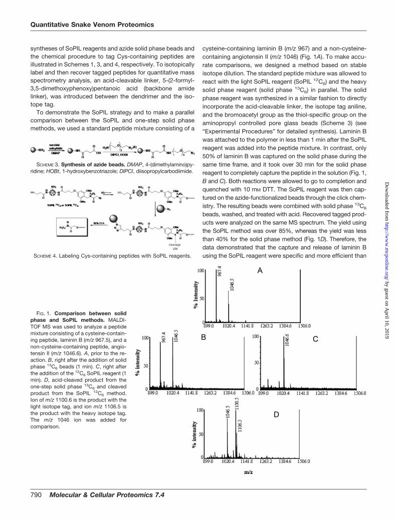
syntheses of SoPIL reagents and azide solid phase beads andthe chemical procedure to tag Cys-containing peptides areillustrated in Schemes 1, 3, and 4, respectively. To isotopicallylabel and then recover tagged peptides for quantitative massspectrometry analysis, an acid-cleavable linker, 5-(2-formyl-3,5-dimethoxyphenoxy)pentanoic acid (backbone amidelinker), was introduced between the dendrimer and the iso-tope tag.
To demonstrate the SoPIL strategy and to make a parallelcomparison between the SoPIL and one-step solid phasemethods, we used a standard peptide mixture consisting of a
cysteine-containing laminin B (m/z 967) and a non-cysteine-containing angiotensin II (m/z 1046) (Fig. 1A). To make accu-rate comparisons, we designed a method based on stableisotope dilution. The standard peptide mixture was allowed toreact with the light SoPIL reagent (SoPIL 12C6) and the heavysolid phase reagent (solid phase 13C6) in parallel. The solidphase reagent was synthesized in a similar fashion to directlyincorporate the acid-cleavable linker, the isotope tag aniline,and the bromoacetyl group as the thiol-specific group on theaminopropyl controlled pore glass beads (Scheme 3) (see“Experimental Procedures” for detailed synthesis). Laminin Bwas attached to the polymer in less than 1 min after the SoPILreagent was added into the peptide mixture. In contrast, only50% of laminin B was captured on the solid phase during thesame time frame, and it took over 30 min for the solid phasereagent to completely capture the peptide in the solution (Fig. 1,B and C). Both reactions were allowed to go to completion andquenched with 10 mM DTT. The SoPIL reagent was then cap-tured on the azide-functionalized beads through the click chem-istry. The resulting beads were combined with solid phase 13C6
beads, washed, and treated with acid. Recovered tagged prod-ucts were analyzed on the same MS spectrum. The yield usingthe SoPIL method was over 85%, whereas the yield was lessthan 40% for the solid phase method (Fig. 1D). Therefore, thedata demonstrated that the capture and release of laminin Busing the SoPIL reagent were specific and more efficient than
FIG. 1. Comparison between solidphase and SoPIL methods. MALDI-TOF MS was used to analyze a peptidemixture consisting of a cysteine-contain-ing peptide, laminin B (m/z 967.5), and anon-cysteine-containing peptide, angio-tensin II (m/z 1046.6). A, prior to the re-action. B, right after the addition of solidphase 13C6 beads (1 min). C, right afterthe addition of the 12C6 SoPIL reagent (1min). D, acid-cleaved product from theone-step solid phase 13C6 and cleavedproduct from the SoPIL 12C6 method.Ion of m/z 1100.6 is the product with thelight isotope tag, and ion m/z 1106.5 isthe product with the heavy isotope tag.The m/z 1046 ion was added forcomparison.
SCHEME 3. Synthesis of azide beads. DMAP, 4-(dimethylamino)py-ridine; HOBt, 1-hydroxybenzotriazole; DIPCI, diisopropylcarbodiimide.
SCHEME 4. Labeling Cys-containing peptides with SoPIL reagents.
Quantitative Snake Venom Proteomics
790 Molecular & Cellular Proteomics 7.4
by guest on April 10, 2019
http://ww
w.m
cponline.org/D
ownloaded from
the one-step solid phase isotopic labeling reagent.To illustrate the quantitative nature of the SoPIL method, a
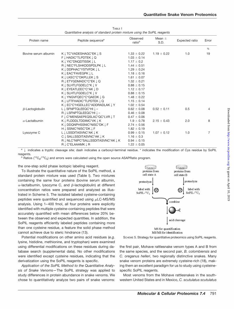
standard protein mixture was used (Table I). Two mixturescontaining the same four proteins (bovine serum albumin,�-lactalbumin, lysozyme C, and �-lactoglobulin) at differentconcentration ratios were prepared and analyzed as illus-trated in Scheme 5. The isolated labeled cysteine-containingpeptides were quantified and sequenced using �LC-MS/MSanalysis. Using 1–400 fmol, all four proteins were explicitlyidentified with multiple cysteine-containing peptides that wereaccurately quantified with mean differences below 20% be-tween the observed and expected quantities. In addition, theSoPIL reagents efficiently labeled peptides containing morethan one cysteine residue, a feature the solid phase methodcannot achieve due to steric hindrance (13).
Potential modifications on other amino acid residues (e.g.lysine, histidine, methionine, and tryptophan) were examinedusing differential modifications on these residues during da-tabase search (supplemental data). No other modificationswere identified except cysteine residues, indicating that thederivatization using the SoPIL reagents is specific.
Application of the SoPIL Method to the Quantitative Analy-sis of Snake Venoms—The SoPIL strategy was applied tostudy differences in protein abundance in snake venoms. Wechose to quantitatively analyze two pairs of snake venoms:
the first pair, Mohave rattlesnake venom types A and B fromthe same species, and the second pair, B. colombiensis andC. oreganus helleri, two regionally distinctive snakes. Manysnake venom proteins are extremely cysteine-rich (18), mak-ing them an excellent paradigm for us to study using cysteine-specific SoPIL reagents.
Most venoms from the Mohave rattlesnakes in the south-western United States and in Mexico, C. scutulatus scutulatus
SCHEME 5. Strategy for quantitative proteomics using SoPIL reagents.
TABLE IQuantitative analysis of standard protein mixture using the SoPIL reagents
Protein name Peptide sequencea Observedratiob
Mean �S.D.
Expected ratio Error
%
Bovine serum albumin K2TC*VADESHAGC*EK2S 1.33 � 0.22 1.19 � 0.22 1.0 19F2HADIC*TLPDTEK2Q 1.03 � 0.14K2YIC*DNQDTISSK2L 1.17 � 0.2R2NEC*FLSHKDDSPDLPK2L 1.44 � 0.51K2DDPHAC*YSTVFDK2L 1.29 � 0.24K2EAC*FAVEGPK2L 1.18 � 0.16K2LKEC*C*DKPLLEK2S 1.61 � 0.07R2ETYGDMADC*C*EK2Q 1.32 � 0.21K2SLHTLFGDELC*K2V 0.88 � 0.15K2EYEATLEEC*C*AK2D 1.12 � 0.17K2SLHTLFGDELC*K2V 0.88 � 0.15K2YNGVFQEC*C*QAEDK2G 1.48 � 0.22K2LFTFHADIC*TLPDTEK2Q 1.15 � 0.14K2EC*C*HGDLLEC*ADDRADLAK2Y 1.02 � 0.54
�-Lactoglobulin L2SFNPTQLEEQC*HI2– 0.62 � 0.08 0.52 � 0.11 0.5 4R2LSFNPTQLEEQC*HI2– 0.46 � 0.08F2C*MENSAEPEQSLAC*QC*LVR2T 0.47 � 0.06
�-Lactalbumin K2FLDDDLTDDIMC*VK2K 1.9 � 0.78 2.15 � 0.43 2.0 8K2DDQNPHSSNIC*NISC*DK2F 2.74 � 0.56H2SSNIC*NISC*DK2F 1.82 � 0.19
Lysozyme C L2LSSDITASVNC*AK2K 0.99 � 0.15 1.07 � 0.12 1.0 7C2SALLSSDITASVNC*AK2K 1.16 � 0.3R2NLC*NIPC*SALLSSDITASVNC*AK2K 0.94 � 0.15R2C*ELAAAMK2R 1.22 � 0.05
a2 indicates a tryptic cleavage site; dash indicates a carboxyl-terminal residue. * indicates the modification of Cys residue by SoPILreagents.
b Ratios (12C6/13C6) and errors were calculated using the open source ASAPRatio program.
Quantitative Snake Venom Proteomics
Molecular & Cellular Proteomics 7.4 791
by guest on April 10, 2019
http://ww
w.m
cponline.org/D
ownloaded from
type A venom, are characterized with Mojave toxins and donot induce severe hemorrhage in animals. In contrast, C.scutulatus scutulatus type B, found in a narrow range ofArizona, lacks Mojave toxins and has hemorrhagic activity.The same amounts of these two characteristically differentsnake venoms, C. scutulatus scutulatus types A and B, werelabeled by light and heavy SoPIL reagents, respectively. Theywere combined and processed as described in Scheme 4.The analysis identified and quantified over 100 unique pep-tides representing over 30 venom proteins in the Swiss Pro-tein Database (See Table II for a partial list; a full list is insupplemental Tables S1 and S2.). The relatively low number ofidentified proteins was due to unsequenced snake genomeand wide dynamic range in venom proteins. Snake venom is
dominated by a number of highly abundant proteins. Ouranalysis also revealed peptide redundancy that could be dueto the high sequence homology conserved in the cysteine-containing region unique to snake venom. The distribution ofidentified proteins is illustrated in Fig. 2A. Approximately 18%of identified proteins were classified as homologs. Consistentwith previous reports on the Mohave rattlesnake, the quanti-tative measurements indicated that several classes of cys-teine-rich proteins dominantly exist in venom A but not invenom B, such as Mojave toxins. The Mohave rattlesnake isan example of extreme intraspecies variation in venom char-acterization (42). Mohave rattlesnake venom is one of themost toxic snake venoms found in the United States. Thelethal dose killing 50% of a mouse population (LD50) ranges
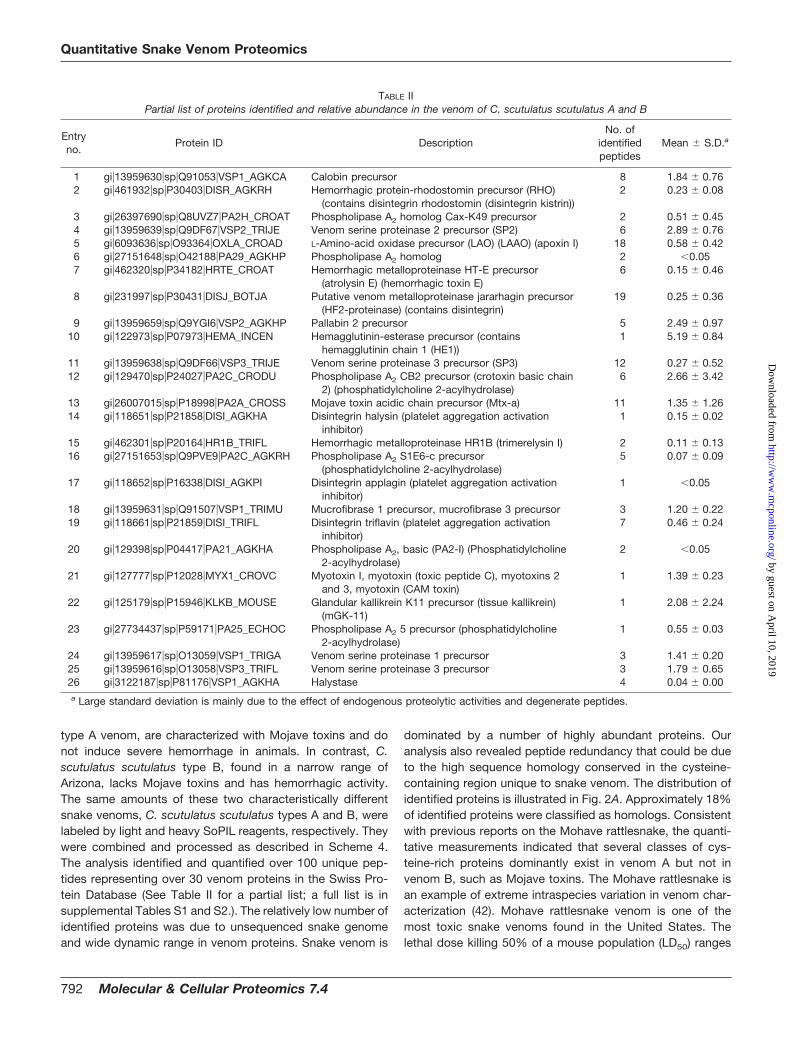
TABLE IIPartial list of proteins identified and relative abundance in the venom of C. scutulatus scutulatus A and B
Entryno.
Protein ID DescriptionNo. of
identifiedpeptides
Mean � S.D.a
1 gi�13959630�sp�Q91053�VSP1_AGKCA Calobin precursor 8 1.84 � 0.762 gi�461932�sp�P30403�DISR_AGKRH Hemorrhagic protein-rhodostomin precursor (RHO)
(contains disintegrin rhodostomin (disintegrin kistrin))2 0.23 � 0.08
3 gi�26397690�sp�Q8UVZ7�PA2H_CROAT Phospholipase A2 homolog Cax-K49 precursor 2 0.51 � 0.454 gi�13959639�sp�Q9DF67�VSP2_TRIJE Venom serine proteinase 2 precursor (SP2) 6 2.89 � 0.765 gi�6093636�sp�O93364�OXLA_CROAD L-Amino-acid oxidase precursor (LAO) (LAAO) (apoxin I) 18 0.58 � 0.426 gi�27151648�sp�O42188�PA29_AGKHP Phospholipase A2 homolog 2 �0.057 gi�462320�sp�P34182�HRTE_CROAT Hemorrhagic metalloproteinase HT-E precursor
(atrolysin E) (hemorrhagic toxin E)6 0.15 � 0.46
8 gi�231997�sp�P30431�DISJ_BOTJA Putative venom metalloproteinase jararhagin precursor(HF2-proteinase) (contains disintegrin)
19 0.25 � 0.36
9 gi�13959659�sp�Q9YGI6�VSP2_AGKHP Pallabin 2 precursor 5 2.49 � 0.9710 gi�122973�sp�P07973�HEMA_INCEN Hemagglutinin-esterase precursor (contains
hemagglutinin chain 1 (HE1))1 5.19 � 0.84
11 gi�13959638�sp�Q9DF66�VSP3_TRIJE Venom serine proteinase 3 precursor (SP3) 12 0.27 � 0.5212 gi�129470�sp�P24027�PA2C_CRODU Phospholipase A2 CB2 precursor (crotoxin basic chain
2) (phosphatidylcholine 2-acylhydrolase)6 2.66 � 3.42
13 gi�26007015�sp�P18998�PA2A_CROSS Mojave toxin acidic chain precursor (Mtx-a) 11 1.35 � 1.2614 gi�118651�sp�P21858�DISI_AGKHA Disintegrin halysin (platelet aggregation activation
inhibitor)1 0.15 � 0.02
15 gi�462301�sp�P20164�HR1B_TRIFL Hemorrhagic metalloproteinase HR1B (trimerelysin I) 2 0.11 � 0.1316 gi�27151653�sp�Q9PVE9�PA2C_AGKRH Phospholipase A2 S1E6-c precursor
(phosphatidylcholine 2-acylhydrolase)5 0.07 � 0.09
17 gi�118652�sp�P16338�DISI_AGKPI Disintegrin applagin (platelet aggregation activationinhibitor)
1 �0.05
18 gi�13959631�sp�Q91507�VSP1_TRIMU Mucrofibrase 1 precursor, mucrofibrase 3 precursor 3 1.20 � 0.2219 gi�118661�sp�P21859�DISI_TRIFL Disintegrin triflavin (platelet aggregation activation
inhibitor)7 0.46 � 0.24
20 gi�129398�sp�P04417�PA21_AGKHA Phospholipase A2, basic (PA2-I) (Phosphatidylcholine2-acylhydrolase)
2 �0.05
21 gi�127777�sp�P12028�MYX1_CROVC Myotoxin I, myotoxin (toxic peptide C), myotoxins 2and 3, myotoxin (CAM toxin)
1 1.39 � 0.23
22 gi�125179�sp�P15946�KLKB_MOUSE Glandular kallikrein K11 precursor (tissue kallikrein)(mGK-11)
1 2.08 � 2.24
23 gi�27734437�sp�P59171�PA25_ECHOC Phospholipase A2 5 precursor (phosphatidylcholine2-acylhydrolase)
1 0.55 � 0.03
24 gi�13959617�sp�O13059�VSP1_TRIGA Venom serine proteinase 1 precursor 3 1.41 � 0.2025 gi�13959616�sp�O13058�VSP3_TRIFL Venom serine proteinase 3 precursor 3 1.79 � 0.6526 gi�3122187�sp�P81176�VSP1_AGKHA Halystase 4 0.04 � 0.00a Large standard deviation is mainly due to the effect of endogenous proteolytic activities and degenerate peptides.
Quantitative Snake Venom Proteomics
792 Molecular & Cellular Proteomics 7.4
by guest on April 10, 2019
http://ww
w.m
cponline.org/D
ownloaded from
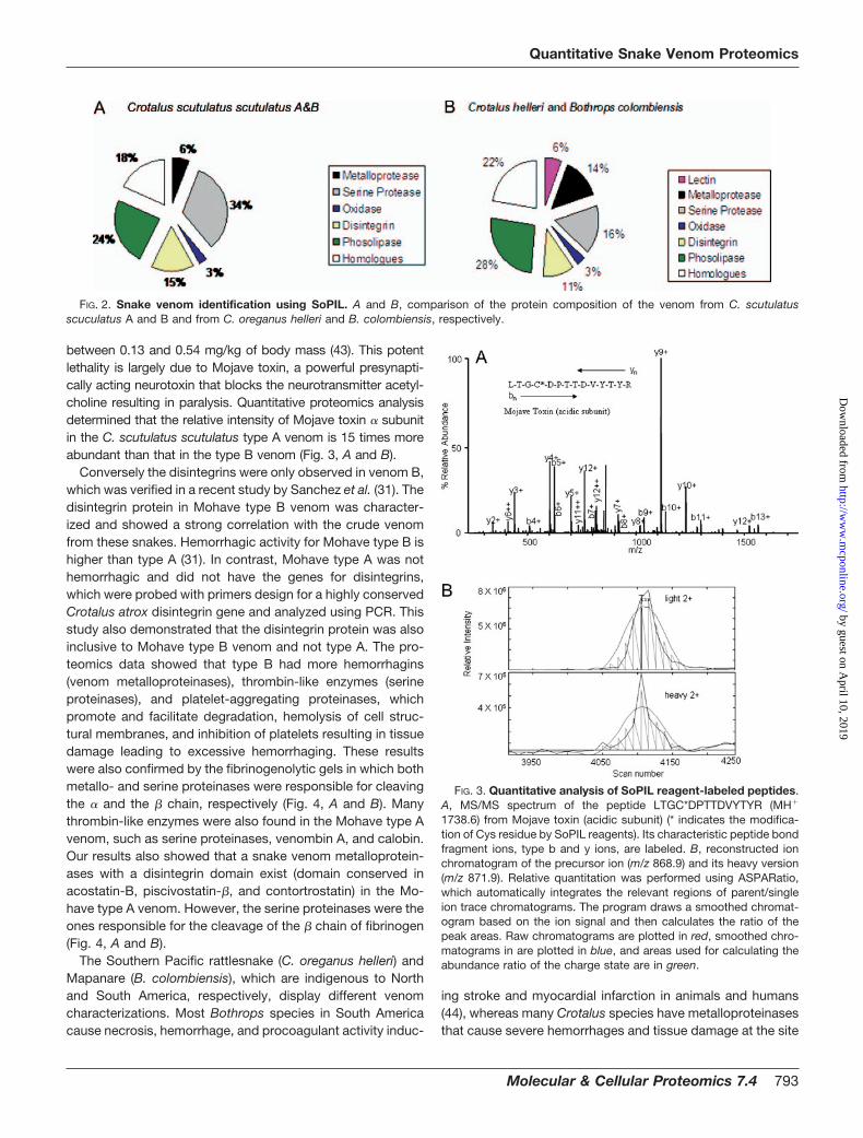
between 0.13 and 0.54 mg/kg of body mass (43). This potentlethality is largely due to Mojave toxin, a powerful presynapti-cally acting neurotoxin that blocks the neurotransmitter acetyl-choline resulting in paralysis. Quantitative proteomics analysisdetermined that the relative intensity of Mojave toxin � subunitin the C. scutulatus scutulatus type A venom is 15 times moreabundant than that in the type B venom (Fig. 3, A and B).
Conversely the disintegrins were only observed in venom B,which was verified in a recent study by Sanchez et al. (31). Thedisintegrin protein in Mohave type B venom was character-ized and showed a strong correlation with the crude venomfrom these snakes. Hemorrhagic activity for Mohave type B ishigher than type A (31). In contrast, Mohave type A was nothemorrhagic and did not have the genes for disintegrins,which were probed with primers design for a highly conservedCrotalus atrox disintegrin gene and analyzed using PCR. Thisstudy also demonstrated that the disintegrin protein was alsoinclusive to Mohave type B venom and not type A. The pro-teomics data showed that type B had more hemorrhagins(venom metalloproteinases), thrombin-like enzymes (serineproteinases), and platelet-aggregating proteinases, whichpromote and facilitate degradation, hemolysis of cell struc-tural membranes, and inhibition of platelets resulting in tissuedamage leading to excessive hemorrhaging. These resultswere also confirmed by the fibrinogenolytic gels in which bothmetallo- and serine proteinases were responsible for cleavingthe � and the � chain, respectively (Fig. 4, A and B). Manythrombin-like enzymes were also found in the Mohave type Avenom, such as serine proteinases, venombin A, and calobin.Our results also showed that a snake venom metalloprotein-ases with a disintegrin domain exist (domain conserved inacostatin-B, piscivostatin-�, and contortrostatin) in the Mo-have type A venom. However, the serine proteinases were theones responsible for the cleavage of the � chain of fibrinogen(Fig. 4, A and B).
The Southern Pacific rattlesnake (C. oreganus helleri) andMapanare (B. colombiensis), which are indigenous to Northand South America, respectively, display different venomcharacterizations. Most Bothrops species in South Americacause necrosis, hemorrhage, and procoagulant activity induc-
ing stroke and myocardial infarction in animals and humans(44), whereas many Crotalus species have metalloproteinasesthat cause severe hemorrhages and tissue damage at the site
FIG. 3. Quantitative analysis of SoPIL reagent-labeled peptides.A, MS/MS spectrum of the peptide LTGC*DPTTDVYTYR (MH�
1738.6) from Mojave toxin (acidic subunit) (* indicates the modifica-tion of Cys residue by SoPIL reagents). Its characteristic peptide bondfragment ions, type b and y ions, are labeled. B, reconstructed ionchromatogram of the precursor ion (m/z 868.9) and its heavy version(m/z 871.9). Relative quantitation was performed using ASPARatio,which automatically integrates the relevant regions of parent/singleion trace chromatograms. The program draws a smoothed chromat-ogram based on the ion signal and then calculates the ratio of thepeak areas. Raw chromatograms are plotted in red, smoothed chro-matograms in are plotted in blue, and areas used for calculating theabundance ratio of the charge state are in green.
FIG. 2. Snake venom identification using SoPIL. A and B, comparison of the protein composition of the venom from C. scutulatusscuculatus A and B and from C. oreganus helleri and B. colombiensis, respectively.
Quantitative Snake Venom Proteomics
Molecular & Cellular Proteomics 7.4 793
by guest on April 10, 2019
http://ww
w.m
cponline.org/D
ownloaded from
of envenomation (45, 46). The same amounts of snake ven-oms of B. colombiensis and C. oreganus helleri were alsolabeled by light and heavy SoPIL reagents, respectively; com-bined; and processed as described in Scheme 4. This secondset for SoPIL analysis identified and quantified a similar num-ber of peptides and proteins in the Swiss Protein Database(Table III and supplemental Tables S3 and S4). The distribu-tion of identified proteins is illustrated in Fig 2B. Similar to thefirst Mohave rattlesnake analysis, 22% of identified proteinswere classified as homologs.
Myotoxins are typically small proteins and peptides in thesnake venom that upon envenomation can induce irreversibledamage to skeletal muscle fibers (myonecrosis) (47). They areabundant and widespread in South American venomoussnakes but can also be found in the venoms from otherspecies, including the timber rattlesnake (Crotalus horridus)found in North America (48). In the analysis, myotoxins I, II,and III were more abundant in B. colombiensis venom than inthe C. oreganus helleri venom. In contrast, CAM toxin, amyotoxin, was found in C. oreganus helleri venom. In addition,phospholipase A2 � (phosphatidylcholine 2-acylhydrolase)was also present in the venom of C. oreganus helleri. Theseresults are consistent with the previous study in which SouthAmerican Bothrops species exhibit increased procoagulantand myotoxic activity compared with many North AmericanCrotalus species (49). Neurotoxic PLA2 myotoxins can also bepresent in a number of viperid/crotalid species, such as cro-toxin, a neurotoxin found in the venom of Crotalus durissusterrificus from South America (50).
The Southern Pacific rattlesnake, C. oreganus helleri, hasmyotoxic, neurotoxic, and hemorrhagic components in itsvenom (51, 52). French et al. (53) reported that Mojave toxin(MT) has been detected in five of 25 C. oreganus helleri usinganti-MT antibodies and was confirmed using nucleotide se-quence analysis. All of the positive venom samples for MTwere collected from Mt. San Jacinto in Riverside County,California. In our study, no MT was found in the venom of theSouthern Pacific venom. Hemorrhagic metalloproteinaseHT-E precursors (atrolysins E, D, and C) were the most abun-dant proteins relative to the mapanare venom in addition toadamalysin II (proteinase II). The minimal hemorrhagic dose(minimal protein amount that will cause a 10-mm hemorrhagicspot) for the Southern Pacific venom has been reported to be2.25 �g (54).
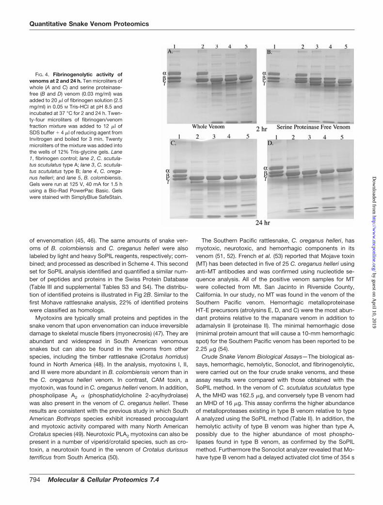
Crude Snake Venom Biological Assays—The biological as-says, hemorrhagic, hemolytic, Sonoclot, and fibrinogenolytic,were carried out on the four crude snake venoms, and theseassay results were compared with those obtained with theSoPIL method. In the venom of C. scutulatus scutulatus typeA, the MHD was 162.5 �g, and conversely type B venom hadan MHD of 16 �g. This assay confirms the higher abundanceof metalloproteases existing in type B venom relative to typeA analyzed using the SoPIL method (Table II). In addition, thehemolytic activity of type B venom was higher than type A,possibly due to the higher abundance of most phospho-lipases found in type B venom, as confirmed by the SoPILmethod. Furthermore the Sonoclot analyzer revealed that Mo-have type B venom had a delayed activated clot time of 354 s
FIG. 4. Fibrinogenolytic activity ofvenoms at 2 and 24 h. Ten microliters ofwhole (A and C) and serine proteinase-free (B and D) venom (0.03 mg/ml) wasadded to 20 �l of fibrinogen solution (2.5mg/ml) in 0.05 M Tris-HCl at pH 8.5 andincubated at 37 °C for 2 and 24 h. Twen-ty-four microliters of fibrinogen/venomfraction mixture was added to 12 �l ofSDS buffer � 4 �l of reducing agent fromInvitrogen and boiled for 3 min. Twentymicroliters of the mixture was added intothe wells of 12% Tris-glycine gels. Lane1, fibrinogen control; lane 2, C. scutula-tus scutulatus type A; lane 3, C. scutula-tus scutulatus type B; lane 4, C. orega-nus helleri; and lane 5, B. colombiensis.Gels were run at 125 V, 40 mA for 1.5 husing a Bio-Rad PowerPac Basic. Gelswere stained with SimplyBlue SafeStain.
Quantitative Snake Venom Proteomics
794 Molecular & Cellular Proteomics 7.4
by guest on April 10, 2019
http://ww
w.m
cponline.org/D
ownloaded from

with a low clot rate of 6.2 clot signals/min, whereas Mohavetype A venom displayed a normal activated clot time of 144 swith a low clot rate of 10 clot signals/min (Fig 5A and Table IV).These results are in accordance with the SoPIL method inwhich more metalloproteinases were found in type B venomthan in type A. Furthermore the � chain of fibrinogen wascleaved by metalloproteinases found in type B venom but notin type A venom (Fig. 4, A and B). Metalloproteinases inhibitblood coagulation (23), and they can be �- and or �-fibrino-genases depending on their specificity to cleave the � or �
chain of fibrinogen (55). Serine proteinases are other types ofproteins found in snake venom that affect platelet aggrega-tion, blood coagulation, fibrinolysis, the complement system,
blood pressure, and the nervous system (23, 56–62). Serineproteinases were found in slightly higher abundance in Mo-have type A venom according to the SoPIL method (Table II).According to the results in the fibrinogenolytic assay, bothtype A and B venoms contained serine proteinases thatcleaved the � chains (Fig. 4, A and B); this was evident whenthe serine proteinases were removed from these venoms andno cleavage of � chain was observed (Fig. 4B). However,there is further evidence that both metallo- and serine pro-teinases in both type A and type B venoms are acting comple-mentarily on the � chains. For instance, whole venom in 2 hwas able to cleave � chain, but when serine proteinases wereremoved the � chains remained, suggesting that serine pro-
TABLE IIIPartial list of proteins identified and relative abundance in the venom of C. oreganus helleri and B. colombiensis
Entryno.
Protein ID DescriptionNo. of
identifiedpeptides
Mean � S.D.a
1 gi�34922459�sp�P83519�LECG_BOTJR Galactose-specific lectin (BJcuL) 2 0.14 � 0.042 gi�6093636�sp�O93364�OXLA_CROAD L-Amino-acid oxidase precursor (LAO) (LAAO)
(apoxin I)17 3.61 � 2.42
3 gi�1171971�sp�P45881�PA21_BOTJR Phospholipase A2 precursor (phosphatidylcholine2-acylhydrolase) (BJUPLA2)
7 0.13 � 0.21
4 gi�27151652�sp�O42192�PA28_AGKHP Phospholipase A2 A� (phosphatidylcholine 2-acylhydrolase)
4 0.40 � 0.58
5 gi�13959633�sp�Q91509�VSP3_TRIMU Mucrofibrase 3 precursor 2 1.45 � 0.756 gi�584725�sp�P34179�ADAM_CROAD Adamalysin II (proteinase II) 8 2.18 � 2.007 gi�231997�sp�P30431�DISJ_BOTJA Putative venom metalloproteinase jararhagin
precursor (HF2 proteinase) (contains disintegrin)21 2.53 � 4.56
8 gi�13959659�sp�Q9YGI6�VSP2_AGKHP Pallabin 2 precursor 4 0.24 � 0.879 gi�27734229�sp�P81509�CHBB_CROHO CHH-B � subunit 6 8.39 � 12.2
10 gi�118651�sp�P21858�DISI_AGKHA Disintegrin halysin (platelet aggregation activationinhibitor)
1 1.37 � 0.15
11 gi�13959630�sp�Q91053�VSP1_AGKCA Calobin precursor 6 0.75 � 0.4512 gi�6093643�sp�P81824�VSP1_BOTJA Platelet-aggregating proteinase PA-BJ 3 0.05 � 0.0213 gi�13959639�sp�Q9DF67�VSP2_TRIJE Venom serine proteinase 2 precursor (SP2) 4 10.9 � 9.5214 gi�118655�sp�P18618�DISI_BOTAT Disintegrin batroxostatin (platelet aggregation
activation inhibitor)3 3.06 � 2.00
15 gi�1171973�sp�P24605�PA22_BOTAS Phospholipase A2 homolog 2 (myotoxin II) 20 0.07 � 0.1216 gi�129400�sp�P20474�PA21_BOTAS Phospholipase A2 (myotoxin I)
(phosphatidylcholine 2-acylhydrolase)9 0.12 � 0.17
17 gi�127786�sp�P24330�MYX_CROAD Myotoxin (CAM toxin) 4 0.44 � 1.0918 gi�1708301�sp�P15167�HRTD_CROAT Hemorrhagic metalloproteinase HT-D and HT-C
precursor (atrolysins D and C) (hemorrhagictoxins C and D)
11 20
19 gi�462320�sp�P34182�HRTE_CROAT Hemorrhagic metalloproteinase HT-E precursor(atrolysin E) (hemorrhagic toxin E)
5 10.9 � 7.28
20 gi�462318�sp�P20897�HRT2_CRORU Hemorrhagic metalloproteinase HT-2 (ruberlysin)(hemorrhagic toxin II)
16 15.0 � 31.1
21 gi�461512�sp�P09872�VSP1_AGKCO Ancrod (venombin A) (protein C activator) (ACC-C) 3 1.47 � 0.5022 gi�129507�sp�P00623�PA2_CROAD Phospholipase A2 � (phosphatidylcholine 2-
acylhydrolase)5 1.51 � 0.45
23 gi�118660�sp�P17349�DISI_TRIEL Disintegrin elegantin (platelet aggregationactivation inhibitor)
7 3.40 � 1.98
24 gi�3914258�sp�P81243�PA21_BOTJA Phospholipase A2 (phosphatidylcholine2-acylhydrolase) (BJ-PLA2)
7 �0.05
25 gi�17433168�sp�Q9PVE3�PA23_BOTAS Phospholipase A2 homolog 3 precursor (myotoxinIII) (M1-3-3)
9 �0.05
a Large standard deviation is mainly due to the effect of endogenous proteolytic activities and degenerate peptides.
Quantitative Snake Venom Proteomics
Molecular & Cellular Proteomics 7.4 795
by guest on April 10, 2019
http://ww
w.m
cponline.org/D
ownloaded from

teinases were solely responsible for the � chain cleavage.However, in 24 h, � chains were completely cleaved with bothcrude and serine proteinase-free venom, and it is obvious thatmetalloproteinases or other enzymes were also responsiblefor the � chain cleavage in the serine proteinase-free venombut required a longer time for the cleavage to occur. Type Avenom contains serine proteinase that cleaves the � chain butalso required more time to do so (Fig. 4, C and D). Theseserine proteinases are thrombin-like enzymes, which makeblood unclottable, and it is safe to state that type A venomcontains different serine proteinases affecting either � or �
chain because it is unlikely that the same serine proteinasescould be affecting both chains (63, 64). A combination of bothmetallo- and serine proteinases exist in a higher abundance intype B venom than in type A venom (Table II), and thus, thecombination of both these proteinases, in particularly the
effect of � chain cleavage by metalloproteinases, could verywell be the result of the delayed clotting time of blood treatedwith type B venom (Fig. 5A).
The venoms of B. colombiensis and C. oreganus hellerishow very distinct coagulant activities from that of the C.scutulatus scutulatus venoms (Fig. 5). Both venoms have in-creased procoagulant activity (Fig. 5), but the mechanism ofclotting may be due to different venom proteolytic enzymesaffecting different factors of the hemostasis pathway (65). B.colombiensis venom cleaves the � and � chains of fibrinogen;C. oreganus helleri also cleaves the � chain and partiallycleaves the � chain. The cleavage of the � and � chains by B.colombiensis venom is due to metalloproteinases becausewhen the serine proteinases were removed from the venomcleavage of both � and � chains still occurred in both 2- and24-h incubation periods (Fig. 4, A, B, C, and D). Again cleav-
TABLE IVProteolytic and Sonoclot activities of venoms C. scutulatus scutulatus A and B, C. oreganus helleri, and B. colombiensis
Venoms MHDa MHemDb Sonoclot ACT,CR, PFc
Fibrinogenolyticd 2 h/24 h
Crude venomSerine
proteinase-freevenom
�g �g
C. scutulatus scutulatus Ae 162.5 9.7 144, 10, 2.2 �/�,� �/�C. scutulatus scutulatus Be 16 4.8 354, 6.2, 2.5 �,�/�,� �/�,�C. oreganus helleri 6 4.8 �26, 51, 1 �,�/�,� �/�,�B. colombiensis 25 2.4 �26, 51, 1 �,�/�,� �,�/�,�
a The amount of protein that will cause a 10-mm hemorrhagic spot.b Minimal phospholipase A dose: the amount of protein that will cause a 10-mm halo.c ACT, activated clot time (s): time in which clot starts forming; CR, clot rate (clot signals/min); PF, platelet function. Normal control range
values are: ACT: 128–213; CR: 15–26; PF: 3–5.d �,�, cleavage of � and � chain; �, cleavage of � chain; �, no cleavage.e C. scutulatus scutulatus A: neurotoxic venom; C. scutulatus scutulatus B: hemorrhagic venom.
FIG. 5. Sonoclot graph analysis of venoms on whole human blood. A glass bead-activated test (gbACT� kit obtained from Sienco, Inc.)was used to monitor activated clot time, clot rate, and platelet function on a Sonoclot Coagulation and Platelet Function Analyzer (Sienco, Inc.).A, solid line, normal human blood control (no venom added); short dashed line, C. scutulatus scutulatus B; long dashed lines, C. scutulatusscutulatus A. B, solid line, normal human blood control; short dashed line, C. oreganus helleri; long dashed line, B. colombiensis.
Quantitative Snake Venom Proteomics
796 Molecular & Cellular Proteomics 7.4
by guest on April 10, 2019
http://ww
w.m
cponline.org/D
ownloaded from

age of the � chain by C. oreganus helleri venom may be due toboth metallo- and serine proteinases acting synergistically on it.The hemolytic activity was higher in B. colombiensis than C.oreganus helleri venom; however, C. oreganus helleri venomwas more hemorrhagic. The hemolytic activity is due to phos-pholipases, whereas hemorrhaging is due to metallo- and serineproteinases; by SoPIL analysis phospholipases and metallopro-teinases were found in higher abundance in B. colombiensisand C. oreganus helleri venoms, respectively (Table III).
Protease Activity in Snake Venoms—This study also ob-served for the first time extensive cleavage of venom proteinsin all four snake venoms. Tables V and VI illustrate the randomprotease cleavage of the metalloproteinase jararhagin andmetalloproteinase HT-D from the C. scutulatus scutulatus andB. colombiensis/C. oreganus helleri analysis, respectively. Amajority of peptides have only one tryptic end (22 of 28 and 13of 17 in Tables V and VI, respectively), and the cleavage wasfound on every amino acid residue in some peptides. Suchextensive cleavage was most likely the result of a combinationof activities by multiple proteases in the snake venom. Pro-teases and other enzymes in snake venom have multiplepurposes such as increasing the prey’s uptake of toxins and
the digestion of preys. Such endogenous protease activities inall four venoms could alter the average protein abundance,resulting in a relatively large error in any existing measurementmethod, including the proteomics method.
It is not clear when venom proteases were activated. Theactivation might occur during the procedure in which the ven-oms were collected. After lyophilization and denaturing, theproteases lost their activities. However, it is possible that theproteases were reactivated during the tryptic digestion period,resulting in extensive cleavages of venom proteins. In addition,it is known that trypsin may result in nonspecific cleavages evenif sequence grade trypsin was used for better specificity (66).
Conclusion—This study presents the first quantitative pro-teomics analysis of snake venom from several species basedon stable isotope labeling. The new SoPIL reagents that se-lectively label and isolate cysteine-containing peptides pro-vide a powerful analytical tool to screen snake venoms thatare cysteine-rich for many diverse classes of proteins. Thisnew methodology has the advantage of homogeneity of thesolution reaction, high efficiency of click chemistry, and con-venience of the solid phase capture/release process. Thequantitative proteomics study helps us understand geograph-ical and environmental variations in snake venoms; this iscrucial in developing better therapeutic agents for treatingsnakebites. In addition, quantitative proteomics can also beused for discovering new therapeutic molecules present insnake venom. The conclusions of this study support the the-ory that there are characteristic differences in the venomswithin the same species and from different geographical lo-cations (31). It is evident that the SoPIL method can be anefficient tool for snake venom research and for a much widerapplication of quantitative proteomics.
TABLE VPartial list of protease cleavage in the venom of C. scutulatus scutu-latus A and B for putative venom metalloproteinase jararhagin precur-
sor (HF2 proteinase) (contains disintegrin)
Peptide sequencea
P2 VEDHCYYHGR2 IG2 TPENCQNECCDAATCK2 LN2 CQNECCDAATCK2K2 LKSGSQCGHGDCCEQCK2 FK2 SGSQCGHGDCCEQCK2 FG2 SQCGHGDCCEQCK2 FC2 GHGDCCEQCK2 FG2 HGDCCEQCK2 FC2 RASMSECDPAEHCTGQSSECPADVFHK2 NR2 ASMSECDPAEHCTGQSSECPADVFHK2 NR2 ASMSECDPAEHCTGQSSECPADVFH2 KA2 SMSECDPAEHCTGQSSECPADVFHK2 NM2 SECDPAEHCTGQSSECPADVFHK2 NC2 DPAEHCTGQSSECPADVFHK2 ND2 PAEHCTGQSSECPADVFHK2 NP2 AEHCTGQSSECPADVFHK2 NA2 EHCTGQSSECPADVFHK2 NE2 HCTGQSSECPADVFHK2 NT2 GQSSECPADVFHK2 NK2 NGQPCLDNYGY2 CK2 NGQPCLDNYGYCYNGN2 CK2 NGQPCLDNYGYCY2 NQ2 KGNYYGYCR2 KK2 GNYYGYCR2 KK2 IPCAPEDVK2 CK2 DNSPGQNNPCK2 MK2 VCSNGHCVDVATA2 YK2 VCSNGHCVDVATAY2 –
a2 indicates a tryptic cleavage site; dash indicates a carboxyl-terminal residue.
TABLE VIPartial list of protease cleavage in the venom of B. colombiensis andC. oreganus helleri for hemorrhagic metalloproteinase HT-D and HT-C
precursor (atrolysins D and C) (hemorrhagic toxins C and D)
Peptide sequencea
G2 KITTNPSVEDHCYYR2 GK2 ITTNPSVEDHCYYR2 GI2 TTNPSVEDHCYYR2 GR2 GRIENDADSTASISACNGLK2 GR2 YIELVVVADHR2 VK2 SHDNAQLLTAIELDEE2 TP2 INLLMGVTMAHELGHNLGMEHDGKDCLR2 GL2 MGVTMAHELGHNLGMEHDGKDCLR2 GM2 GVTMAHELGHNLGMEHDGKDCLR2 GG2 VTMAHELGHNLGMEHDGKDCLR2 GT2 MAHELGHNLGMEHDGKDCLR2 GA2 HELGHNLGMEHDGKDCLR2 GL2 GHNLGMEHDGKDCLR2 GN2 LGMEHDGKDCLR2 GG2 MEHDGKDCLR2 GR2 GASLCIMR2 GY2 KPQCILNKPLR2 I
a2 indicates a tryptic cleavage site.
Quantitative Snake Venom Proteomics
Molecular & Cellular Proteomics 7.4 797
by guest on April 10, 2019
http://ww
w.m
cponline.org/D
ownloaded from
Acknowledgments—We thank Professor M. G. Finn at the ScrippsResearch Institute for the supply of ligand for the click chemistry andfor helpful suggestions. We acknowledge the use of software in theInstitute for Systems Biology developed using federal funds from theNHLBI, National Institutes of Health, under Contract N01-HV-28179.We thank Luis Fernando Navarrete for venom extractions done at theInstituto de Medicina Tropical, Universidad Central de Venezuela.
* This work was supported in part by Purdue University, the Amer-ican Society for Mass Spectrometry, and a National Science Foun-dation CAREER development award (to W. A. T.). This work was alsosupported by grants to the Natural Toxins Research Center at TexasA&M University-Kingsville (National Institutes of Health (NIH)/NationalCenter for Research Resources Grant 1 P40 RR018300-01, NIH/Research Infrastructure in Minority Institutions Grant 5 PMD000216–02, and NIH/Support of Continuous Research Excellence Grant 5 S06GM008107-29) and Fondo Nacional de Ciencia, Tecnologia e Inno-vacion Grant G-2005000400. The costs of publication of this articlewere defrayed in part by the payment of page charges. This articlemust therefore be hereby marked “advertisement” in accordance with18 U.S.C. Section 1734 solely to indicate this fact.
□S The on-line version of this article (available at http://www.mcponline.org) contains supplemental material.
� To whom correspondence should be addressed. Tel.: 765-494-9605; E-mail: [email protected].
REFERENCES
1. Aebersold, R. (2003) Quantitative proteome analysis: methods and appli-cations. J. Infect. Dis. 187, Suppl. 2, S315–S320
2. Ong, S. E., Foster, L. J., and Mann, M. (2003) Mass spectrometric-basedapproaches in quantitative proteomics. Methods 29, 124–130
3. Duggan, D. J., Bittner, M., Chen, Y., Meltzer, P., and Trent, J. M. (1999)Expression profiling using cDNA microarrays. Nat. Genet. 21, (suppl.)10–14
4. Gygi, S. P., Corthals, G. L., Zhang, Y., Rochon, Y., and Aebersold, R. (2000)Evaluation of two-dimensional gel electrophoresis-based proteome anal-ysis technology. Proc. Natl. Acad. Sci. U. S. A. 97, 9390–9395
5. Old, W. M., Meyer-Arendt, K., Aveline-Wolf, L., Pierce, K. G., Mendoza, A.,Sevinsky, J. R., Resing, K. A., and Ahn, N. G. (2005) Comparison oflabel-free methods for quantifying human proteins by shotgun proteom-ics. Mol. Cell. Proteomics 4, 1487–1502
6. Li, X. J., Yi, E. C., Kemp, C. J., Zhang, H., and Aebersold, R. (2005) Asoftware suite for the generation and comparison of peptide arrays fromsets of data collected by liquid chromatography-mass spectrometry.Mol. Cell. Proteomics 4, 1328–1340
7. Gygi, S. P., Rist, B., Gerber, S. A., Turecek, F., Gelb, M. H., and Aebersold,R. (1999) Quantitative analysis of complex protein mixtures using iso-tope-coded affinity tags. Nat. Biotechnol. 17, 994–999
8. Ross, P. L., Huang, Y. N., Marchese, J. N., Williamson, B., Parker, K.,Hattan, S., Khainovski, N., Pillai, S., Dey, S., Daniels, S., Purkayastha, S.,Juhasz, P., Martin, S., Bartlet-Jones, M., He, F., Jacobson, A., andPappin, D. J. (2004) Multiplexed protein quantitation in Saccharomycescerevisiae using amine-reactive isobaric tagging reagents. Mol. Cell.Proteomics 3, 1154–1169
9. Schmidt, A., Kellermann, J., and Lottspeich, F. (2005) A novel strategy forquantitative proteomics using isotope-coded protein labels. Proteomics5, 4–15
10. Stewart, I. I., Thomson, T., and Figeys, D. (2001) 18O labeling: a tool forproteomics. Rapid Commun. Mass Spectrom. 15, 2456–2465
11. Blagoev, B., Kratchmarova, I., Ong, S. E., Nielsen, M., Foster, L. J., andMann, M. (2003) A proteomics strategy to elucidate functional protein-protein interactions applied to EGF signaling. Nat. Biotechnol. 21,315–318
12. Li, J., Steen, H., and Gygi, S. P. (2003) Protein profiling with cleavableisotope-coded affinity tag (cICAT) reagents: the yeast salinity stressresponse. Mol. Cell. Proteomics 2, 1198–1204
13. Zhou, H., Ranish, J. A., Watts, J. D., and Aebersold, R. (2002) Quantitativeproteome analysis by solid-phase isotope tagging and mass spectrom-
etry. Nat. Biotechnol. 20, 512–51514. van Heerbeek, R., Kamer, P. C., van Leeuwen, P. W., and Reek, J. N. (2002)
Dendrimers as support for recoverable catalysts and reagents. Chem.Rev. 102, 3717–3756
15. Tao, W. A., Wollscheid, B., O’Brien, R., Eng, J., Li, X., Bodenmiller, B.,Watts, J., Hood, L., and Aebersold, R. (2005) Quantitative phosphopro-teome analysis using a dendrimer conjugation chemistry and mass spec-trometry. Nat. Methods 2, 591–598
16. Guo, M., Galan, J., and Tao, W. A. (2007) A novel quantitative proteomicsreagent based on soluble nanopolymers. Chem. Commun. (Camb.) 12,1251–1253
17. Farsky, S. H., Antunes, E., and Mello, S. B. (2005) Pro and antiinflammatoryproperties of toxins from animal venoms. Curr. Drug Targets Inflamm.Allergy 4, 401–411
18. Marsh, N., and Williams, V. (2005) Practical applications of snake venomtoxins in haemostasis. Toxicon 45, 1171–1181
19. Bjarnason, J. B., and Fox, J. W. (1994) Hemorrhagic metalloproteinasesfrom snake venoms. Pharmacol. Ther. 62, 325–372
20. Barrett, A. J., and McDonald, J. K. (1986) Nomenclature: protease, protein-ase and peptidase. Biochem. J. 237, 935
21. Wolfsberg, T. G., and White, J. M. (1996) ADAMs in fertilization and devel-opment. Dev. Biol. 180, 389–401
22. Harris, J. B. (1991) Phospholipases in snake venoms and their effects onnerve and muscle, in Snake Venoms (Harvey, A. L., ed) pp. 91–129,Pergamon Press, Oxford
23. Kini, R. M. (2005) Serine proteases affecting blood coagulation and fibrin-olysis from snake venoms. Pathophysiol. Haemost. Thromb. 34,200–204
24. McLane, M. A., Sanchez, E. E., Wong, A., Paquette-Straub, C., and Perez,J. C. (2004) Disintegrins. Curr. Drug Targets Cardiovasc. Haematol. Dis-ord. 4, 327–355
25. Ruoslahti, E. (1996) RGD and other recognition sequences for integrins.Annu. Rev. Cell Dev. Biol. 12, 697–715
26. Ritter, M. R., Zhou, Q., and Markland, F. S., Jr. (2000) Contortrostatin, asnake venom disintegrin, induces �v�3-mediated tyrosine phosphoryla-tion of CAS and FAK in tumor cells. J. Cell. Biochem. 79, 28–37
27. Lin, Y. T., Tang, C. H., Chuang, W. J., Wang, S. M., Huang, T. F., and Fu,W. M. (2005) Inhibition of adipogenesis by RGD-dependent disintegrin.Biochem. Pharmacol. 70, 1469–1478
28. Zhou, Q., Nakada, M. T., Arnold, C., Shieh, K. Y., and Markland, F. S., Jr.(1999) Contortrostatin, a dimeric disintegrin from Agkistrodon contortrixcontortrix, inhibits angiogenesis. Angiogenesis 3, 259–269
29. Glenn, J. L., Straight, R. C., Wolfe, M. C., and Hardy, D. L. (1983) Geo-graphical variation in Crotalus scutulatus scutulatus (Mojave rattlesnake)venom properties. Toxicon 21, 119–130
30. Glenn, J. L., and Straight, R. C. (1989) Intergradation of two different venompopulations of the Mojave rattlesnake (Crotalus scutulatus scutulatus) inArizona. Toxicon 27, 411–418
31. Sanchez, E. E., Galan, J. A., Powell, R. L., Reyes, S. R., Soto, J. G., Russell,W. K., Russell, D. H., and Perez, J. C. (2005) Disintegrin, hemorrhagic,and proteolytic activities of Mohave rattlesnake, Crotalus scutulatusscutulatus venoms lacking Mojave toxin. Comp. Biochem. Physiol. CToxicol. Pharmacol. 141, 124–132
32. Fox, J. W., Ma, L., Nelson, K., Sherman, N. E., and Serrano, S. M. (2006)Comparison of indirect and direct approaches using ion-trap and Fouriertransform ion cyclotron resonance mass spectrometry for exploring vi-perid venom proteomes. Toxicon 47, 700–714
33. Sanz, L., Gibbs, H. L., Mackessy, S. P., and Calvete, J. J. (2006) Venomproteomes of closely related Sistrurus rattlesnakes with divergent diets.J. Proteome Res. 5, 2098–2112
34. Birrell, G. W., Earl, S. T., Wallis, T. P., Masci, P. P., de Jersey, J., Gorman,J. J., and Lavin, M. F. (2007) The diversity of bioactive proteins inAustralian snake venoms. Mol. Cell. Proteomics 6, 973–986
35. Bandeira, N., Clauser, K. R., and Pevzner, P. A. (2007) Shotgun proteinsequencing: assembly of peptide tandem mass spectra from mixtures ofmodified proteins. Mol. Cell. Proteomics 6, 1123–1134
36. Schwabacher, A. W., Lane, J. W., Schiesher, M. W., Leigh, K. M., andJohnson, C. W. (1998) Desymmetrization reactions: efficient preparationof unsymmetrically substituted linker molecules. J. Org. Chem. 63,1727–1729
37. Keller, A., Nesvizhskii, A. I., Kolker, E., and Aebersold, R. (2002) Empirical
Quantitative Snake Venom Proteomics
798 Molecular & Cellular Proteomics 7.4
by guest on April 10, 2019
http://ww
w.m
cponline.org/D
ownloaded from
statistical model to estimate the accuracy of peptide identifications madeby MS/MS and database search. Anal. Chem. 74, 5383–5392
38. Nesvizhskii, A. I., Keller, A., Kolker, E., and Aebersold, R. (2003) A statisticalmodel for identifying proteins by tandem mass spectrometry. Anal.Chem. 75, 4646–4658
39. Li, X., Zhang, H., Ranish, J. A., and Aebersold, R. (2003) Automatedstatistical analysis of protein abundance ratios from data generated bystable-isotope dilution and tandem mass spectrometry. Anal. Chem. 75,6648–6657
40. Omori-Satoh, T., Sadahiro, S., Ohsaka, A., and Murata, R. (1972) Purifica-tion and characterization of an antihemorrhagic factor in the serum ofTrimeresurus flavoviridis, a crotalid. Biochim. Biophys. Acta 285,414–426
41. Habermann, E., and Hardt, K. L. (1972) A sensitive and specific plate test forthe quantitation of phospholipases. Anal. Biochem. 50, 163–173
42. Glenn, J. L., and Straight, R. (1978) Mojave rattlesnake Crotalus scutulatusscutulatus venom: variation in toxicity with geographical origin. Toxicon16, 81–84
43. Glenn, J. L., and Straight, R. C. (1982) The rattlesnakes and their venomyield and lethal toxicity, in Rattlesnake Venom: Their Actions and Treat-ment (Tu, A. T., ed) p. 110, Marcel Dekker, Inc., New York
44. Mosquera, A., Idrovo, L. A., Tafur, A., and Del Brutto, O. H. (2003) Strokefollowing Bothrops spp. snakebite. Neurology 60, 1577–1580
45. Salazar, A. M., Rodriguez-Acosta, A., Giron, M. E., Aguilar, I., and Guerrero,B. (2007) A comparative analysis of the clotting and fibrinolytic activitiesof the snake venom (Bothrops atrox) (serpentes:viperidae) from differentgeographical areas in Venezuela. Thromb. Res. 20, 95–104
46. Rodrıguez-Acosta A., Uzcategui, W., Azuaje, R., Aguilar, I., and Giron, M. E.(2000) A clinical and epidemiological analysis of accidental bites bysnakes of the genus Bothrops in Venezuela. Rev. Cubana Med. Trop. 52,90–94
47. Gutierrez, J. M., and Lomonte, B. (1995) Phospholipase A2 myotoxins fromBothrops snake venoms. Toxicon 33, 1405–1424
48. Bober, M. A., Glenn, J. L., Straight, R. C., and Ownby, C. L. (1988) Detec-tion of myotoxin �-like proteins in various snake venoms. Toxicon 26,665–673
49. Gopalakrishnakone, P., Dempster, D. W., Hawgood, B. J., and Elder, H. Y.(1984) Cellular and mitochondrial changes induced in the structure ofmurine skeletal muscle by crotoxin, a neurotoxic phospholipase A2complex. Toxicon 22, 85–98
50. Gralen, N., and Svedberg, T. (1938) The molecular weight of crotoxin.
Biochem. J. 32, 1375–137751. Metsch, R. B., Dray, A., and Russell, F. E. (1984) Effects of the venom of the
Southern Pacific rattlesnake, Crotalus viridis helleri, and its fractions onstriated and smooth muscle. Proc. West. Pharmacol. Soc. 27, 395–398
52. Bush, S. P., and Siedenburg, E. (1999) Neurotoxicity associated with sus-pected southern Pacific rattlesnake (Crotalus viridis helleri) envenoma-tion. Wilderness Environ. Med. 10, 247–249
53. French, W. J., Hayes, W. K., Bush, S. P., Cardwell, M. D., Bader, J. O., andRael, E. D. (2004) Mojave toxin in venom of Crotalus helleri (SouthernPacific Rattlesnake): molecular and geographic characterization. Toxicon44, 781–791
54. Sanchez, E. E., Galan, J. A., Perez, J. C., Rodriguez-Acosta, A., Chase,P. B., and Perez, J. C. (2003) The efficacy of two antivenoms against thevenom of North American snakes. Toxicon 41, 357–365
55. Ouyang, C., and Teng, C. M. (1976) Fibrinogenolytic enzymes of Trimeresu-rus mucrosquamatus venom. Biochim. Biophys. Acta 420, 298–308
56. Meier, J., and Stocker, K. (1991) Effects of snake venoms on hemostasis.Crit. Rev. Toxicol. 21, 171–182
57. Braud, S., Bon, C., and Wisner, A. (2000) Snake venom proteins acting onhemostasis. Biochimie (Paris) 82, 851–859
58. Kini, R. M. (2004) Platelet aggregation and exogenous factors from animalsources. Curr. Drug. Targets Cardiovasc. Haematol. Disord. 4, 301–325
59. Markland, F. S. (1998) Snake venoms and the hemostatic system. Toxicon36, 1749–1800
60. Kornalik, F. (1991) The Influence of Snake Venom Proteins on Blood Co-agulation, Pergamon Press, New York
61. Kini, R. M., Rao, V. S., and Joseph, J. S. (2002) Procoagulant proteins fromsnake venoms. Haemostasis 31, 218–224
62. Joseph, J. S., and Kini, R. M. (2004) Snake venom prothrombin activatorssimilar to blood coagulation factor Xa. Curr. Drug Targets Cardiovasc.Haematol. Disord. 4, 397–416
63. Aronson, D. L. (1976) Comparison of the actions of thrombin and thethrombin-like venom enzymes ancrod and batroxobin. Thromb. Haemo-stasis 36, 9–13
64. Bell, W. R., Jr. (1997) Defibrinogenating enzymes. Drugs 54, Suppl. 3,18–31
65. Kini, R. M., Joseph, J. S., and Rao, V. S. (2002) Prothrombin Activators fromSnake Venoms, John Wiley, Chichester, UK
66. Picotti, P., Aebersold, R., and Domon, B. (2007) The implications of pro-teolytic background for shotgun proteomics. Mol. Cell. Proteomics 6,1589–1598
Quantitative Snake Venom Proteomics
Molecular & Cellular Proteomics 7.4 799
by guest on April 10, 2019
http://ww
w.m
cponline.org/D
ownloaded from