quality assessments of untreated and washed quinoa ... assessments of untreated and washed quinoa...
TRANSCRIPT

Quality assessments of untreated and washed Quinoa (Chenopodium
quinoa) seeds based on histological and foaming capacity investigations
L.W.D. van Raamsdonk1, V. Pinckaers
1, J. Ossenkoppele
1, R. Houben
2, M. Lotgering
2, and M. J. Groot
1
1RIKILT - Institute of Food Safety, Wageningen University & Research centre, P.O. Box 230, 6700 AE Wageningen, the
Netherlands 2Customs Laboratory, P.O. BOX 58933, 1040 ED Amsterdam. The Netherlands
Quinoa seed has a high nutritional value, but has a coating of bitter-tasting saponins, making it unpalatable. Therefore the
seeds are usually processed in order to remove the naturally occurring saponins from the seeds. To investigate the impact
of processing, untreated and washed seeds of the white and brown types of quinoa were investigated histologically and by
foaming capacity evaluations. Reference samples of known origin and treatment were investigated as well as unknown
samples. The results revealed a relationship between the presence of saponin containing papillose cells at the outermost
layer of the seed hull in the histological sections and the foaming capacity of the seeds. After washing, the papillose cells
were severely damaged or completely removed and virtually no foam formation was observed. This investigation indicated
that washing resulted in an effective removal of the saponin layer, leading to quality improvement of the seeds intended
for human and animal consumption. The same features were observed for the unknown samples. These results imply that
the treatment of the investigated samples was based on washing. The determination of the type of treatment applied
provided useful information for the correct tax classification for Custom purposes.
Keywords: quality assessment, histology, microscopy, quinoa
Introduction
Quinoa (Chenopodium quinoa Willd.) is a crop originating from South America, and is grown for its edible seeds,
which have an excellent nutritional value. It provides a balanced source of all essential amino acids, and a range of
other nutritious compounds, such as fatty acids. Furthermore, it is a rich source of phosphorus, magnesium and iron [1,
2]. Quinoa is frequently considered a cereal, but it is in fact a dicotyledon belonging to the family of Chenopodiaceae
(goosefoot), the seed structures of which differ from those of the well-known cereals. The seeds can be used as a
replacement of cereals and have an application in certain diets, because they do not contain gluten [3].
There are three types of quinoa: white(coloured white/beige/reddish), brown and black. Seeds of quinoa consist of an
embryo with a radix and two cotyledons, a central perisperm containing food reserves, and an outer structure for
chemical and physical protection [4]. So far seeds of one type of quinoa only has been studied histologically [4], but the
type of quinoa investigated was not mentioned. Although their study provides useful information for the specific
histology of quinoa seeds, it is known that large differences between species or other groups can exist with respect to
their seed structure, even for closely related taxa [5].
The plant produces saponins in the outer seed hull as defence against bird predation. These saponins are a drawback
for the food and feed application of quinoa, since saponins possess a bitter taste and exhibit toxic effects [6, 7]. The
outer layers of the seed hull, which contain the saponins, can be removed from the seeds by washing or mechanical
means, The type and extent of processing determine the quality and safety of the food/feed end product. Moreover, the
type of treatment of quinoa is the distinguishing feature for classification of the seeds in tariff groups in the framework
of customs regulations. Washing of cereal grains, in contrast to other treatments, is considered exclusively as a cleaning
procedure. Therefore, washed cereals are classified in a tariff group with a low tax rate.
Based on the mentioned circumstances, it is important to establish the type as well as the extent of the processing
prior to use. There are several challenges with respect to the background and trade quality of the quinoa seeds on the
market: a) quinoa is not a cereal in a botanic sense, and b) the type of treatment is not known. Mechanical removal of
the seed hull or polishing as applied to real cereals, is not likely to be applied in the same way to quinoa seeds, because
quinoa lacks the firm outer seed hulls as present in cereals [cf. 8].
The aim of the current study was to explore the possibility of histological examinations and foaming capacity
measurements for determining the type and the intensity of treatment of quinoa seeds. A total of four seed batches were
included in the study, the batches consisted of two reference materials with known background and two imported lots,
belonging to two colour types.
Microscopy: Science, Technology, Applications and Education A. Méndez-Vilas and J. Díaz (Eds.)
©FORMATEX 2010 1033
______________________________________________

Material and Methods
Materials
Four batches were provided by the Dutch Customs Laboratory. The batches 1 and 2 were from known origin and
processing status, batches 3 and 4 were imported parties from unknown origin and treatment status. From each batch
one sample was kept in original state and not further processed (A samples) and another sample was laboratory washed
(B samples). The details of the batches are indicated by a number/letter combination 1A to 4B, and are listed in Table 1.
Table 1 Overview of quinoa seed samples. Each sample is indicated by a number. The letter A refers to the original
sample, the letter B to the washed equivalent.
Sample code Description Seed colour
1A Original sample from Bolivia. Untreated. White
1B As sample 1A, laboratory washed. White
2A Original sample from Bolivia. Untreated. White and red
2B As sample 2A, laboratory washed. White
3A Imported party, sample collected by the Customs Authority. White
3B As sample 3A, laboratory washed. White
4A Imported party, sample collected by the Customs Authority. Brown
4B As sample 4A, laboratory washed. Brown
Samples 1B, 2B, 3B and 4B were laboratory washed with tap water at ambient temperature, until foam was no longer
formed, some of the samples are illustrated in figures 1 and 2.
a) b)
Fig. 1. a) Sample 1A: original quinoa sample from Bolivia untreated, b) Sample 4A: imported quinoa sample with unknown
processing history. Sample codes are explained in Table 1.
a) b)
Fig. 2. a) Sample 2A: original quinoa sample from Bolivia, untreated, b) Sample 2B: laboratory washed equivalent of sample 2A.
Sample codes are explained in Table 1.
Microscopy: Science, Technology, Applications and Education A. Méndez-Vilas and J. Díaz (Eds.)
1034 ©FORMATEX 2010
______________________________________________

Methods
Histological examinations
Seeds from all samples were fixed in 4 % buffered formaldehyde and routinely processed to paraffin sections. Samples
were cut into sections of 5 µm thick with a standard microtome for producing histological slides. Selected slices of
different orientations were mounted on microscopic slides, paraffin was removed in a sequence of solutions with xylol,
alcohol and water, and stained with one of the following staining procedures, according to [9, 10]:
- HE (haematoxylin and eosin stain): solutions: 2 g haematoxylin (CI 75290), 100 g aluminium ammonium
sulphate and 0.4 g sodium iodate were dissolved overnight in 200 ml distilled water, after being completely
dissolved, 100 g chloral hydrate and 2 g citric acid was added; 5 g eosin yellow (CI 45380) was dissolved in
479 ml distilled water, 521 ml ethanol 96% and 1 ml acetic acid 100% was added. Cut slices are stained with
haematoxylin for 3 min, rinsed in cold tap water (10 min), and stained in eosin for 20 sec.
- Lugol staining (iodine-potassium iodine): solution: 2 g potassium iodide was dissolved in 100 ml water, 1 g
iodine was added while frequently shaking. Cut slices were stained with lugol until a sufficient staining was
reached. Stained slides were processed in a sequence of solutions with water, alcohol and xylol before the final
embedding. Two series of slides based on two different seeds were prepared for each of the eight samples.
Seed coats were dissected and examined laterally in non-permanent slides. All slides were examined using
Olympus microscopes of type BX40F and BX60F, and images were made with the Soft Imaging Solutions SC20
camera with a magnification of 200x, unless otherwise stated.
Foaming capacity determinations
Foaming experiments were carried out by soaking 15 seeds in approximately 30 ml of water with subsequent shaking
for several minutes. Under these conditions, saponins from the out layer of quinoa seeds tend to form a foam layer on
the water.
In order to exclude that saponins are present in other layers than the PA cells, additionally 15 seeds of samples 4A
and 4B were dissected and crushed before applying the soaking and shaking experiment.
Results
Normal histology
Seed anatomy: The outer structure of the seeds consists of several layers, which are indicated in the images by the
following abbreviations:
PA: papillose cells;
PD: protoderm
PSt: pericarp stretched cells;
SC: seed coat, consisting of a one cell layer with thickened secondary cell walls (exotesta), and a small layer of
flattened cells (endotegmen);
ES: endosperm, only visible clearly in the area of the Radix.
PA and PSt together form the pericarp. Abbreviations are chosen to be compatible with the nomenclature of [4].
The embryo (E) has a circular shape around the central perisperm body (PS). Depending on the orientation, the
embryo is visible as two parts at both sides of the perisperm (radial view), or as a halter shape (tangential view; figure
3a). The presence of starch in the perisperm is demonstrated by the black staining after lugol (figure 3b).
a) b)
Fig. 3. a) Histological view of a radial and tangential section of a quinoa seed of sample 4A (HE staining, 40 X). E: embryo, PS:
perisperm. b) Histological section of quinoa seed (Lugol staining, 60 x), starch stains black.
Microscopy: Science, Technology, Applications and Education A. Méndez-Vilas and J. Díaz (Eds.)
©FORMATEX 2010 1035
______________________________________________
Reference samples
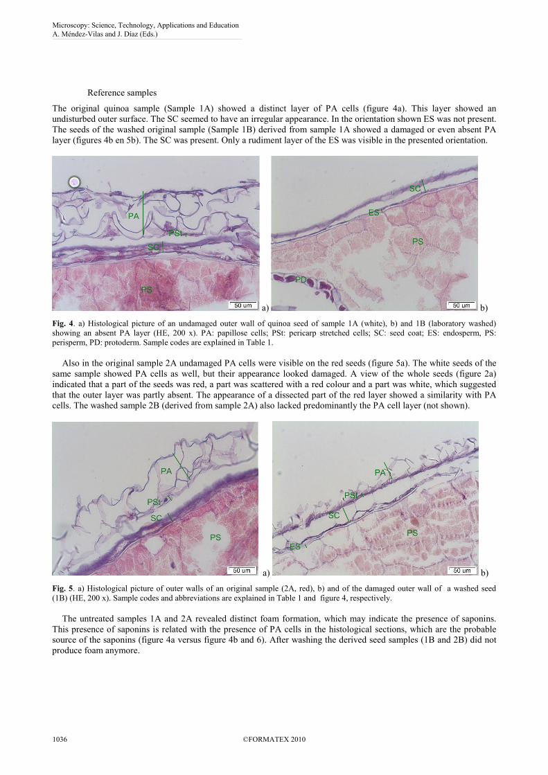
The original quinoa sample (Sample 1A) showed a distinct layer of PA cells (figure 4a). This layer showed an
undisturbed outer surface. The SC seemed to have an irregular appearance. In the orientation shown ES was not present.
The seeds of the washed original sample (Sample 1B) derived from sample 1A showed a damaged or even absent PA
layer (figures 4b en 5b). The SC was present. Only a rudiment layer of the ES was visible in the presented orientation.
a) b)
Fig. 4. a) Histological picture of an undamaged outer wall of quinoa seed of sample 1A (white), b) and 1B (laboratory washed)
showing an absent PA layer (HE, 200 x). PA: papillose cells; PSt: pericarp stretched cells; SC: seed coat; ES: endosperm, PS:
perisperm, PD: protoderm. Sample codes are explained in Table 1.
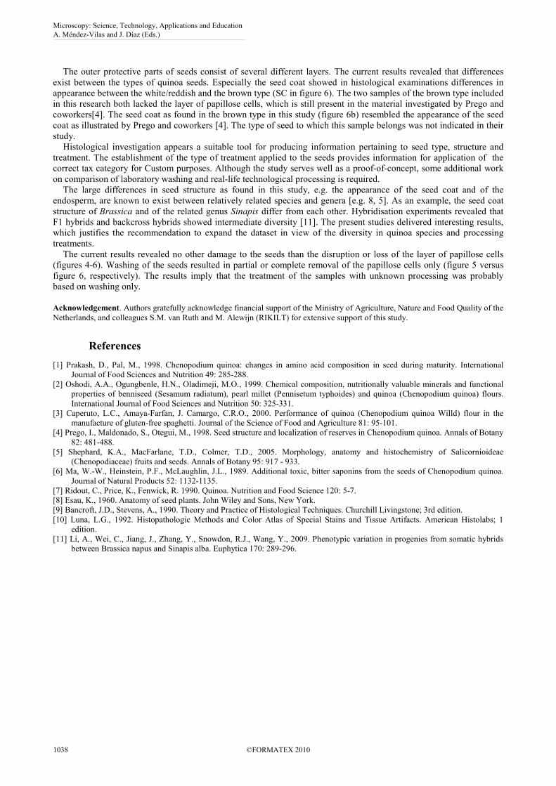
Also in the original sample 2A undamaged PA cells were visible on the red seeds (figure 5a). The white seeds of the
same sample showed PA cells as well, but their appearance looked damaged. A view of the whole seeds (figure 2a)
indicated that a part of the seeds was red, a part was scattered with a red colour and a part was white, which suggested
that the outer layer was partly absent. The appearance of a dissected part of the red layer showed a similarity with PA
cells. The washed sample 2B (derived from sample 2A) also lacked predominantly the PA cell layer (not shown).
a) b)
Fig. 5. a) Histological picture of outer walls of an original sample (2A, red), b) and of the damaged outer wall of a washed seed
(1B) (HE, 200 x). Sample codes and abbreviations are explained in Table 1 and figure 4, respectively.
The untreated samples 1A and 2A revealed distinct foam formation, which may indicate the presence of saponins.
This presence of saponins is related with the presence of PA cells in the histological sections, which are the probable
source of the saponins (figure 4a versus figure 4b and 6). After washing the derived seed samples (1B and 2B) did not
produce foam anymore.
Microscopy: Science, Technology, Applications and Education A. Méndez-Vilas and J. Díaz (Eds.)
1036 ©FORMATEX 2010
______________________________________________

a) b)
Fig. 6. a) Histological picture of outer walls with the papillose cells completely removed of sample 3B (washed in laboratory), b) and
of sample 4A (Brown, sample of imported party), (HE, 200 x). Sample codes and abbreviations are explained in Table 1 and figure
4, respectively.
Imported seed parties
The white seeds taken from the imported party (3A) and the related washed sample 3B (derived from sample 3A) both
lacked the layer with PA cells (figure 6a). The brown seeds of samples 4A and 4B also showed no PA layer (figure 6b),
which suggested that samples 3A and 4A were treated prior to import into the Netherlands. The microscopic image of
this sample after laboratory washing (4B) was similar to that of the original sample (4A). The seed coat (SC) was
obvious from the a thickened secondary cell wall and the endosperm (ES) was clearly present (figure 6b). None of the
original seeds from the imported parties and their washed counterparts produced foam (table 2).
Additional soaking and shaking of crushed seeds of samples 4A and 4B showed no substantial amount of foam on
the surface of the solution. A considerable amount of starch was dissolved from the perisperm of the crushed seeds, but
the dissolved starch did not cause any foam on the surface of the solution.
The results of the washing and soaking experiments and the histological observations are summarized in table 2.
Table 2 Histological observations and foam capacity measurement data of original and laboratory washed quinoa seed
samples
Sample Treatment Histology: presence of
PA cells (histologal
results)
Foaming capacity
Sample 1A Untreated Present Considerable foaming
Sample 1B Washed Damaged or absent No foaming
Sample 2A Untreated Present, some damaged Considerable foaming
Sample 2B Washed Nearly absent No foaming
Sample 3A Unknown Absent No foaming
Sample 3B Washed Absent No foaming
Sample 4A Unknown Absent No foaming ; No foaming after
crushing
Sample 4B Washed Absent No foaming ; No foaming after
crushing
Discussion and conclusions
The results of both the histological examinations and the foaming capacity experiments showed a relationship between
the histological observed presence of papillose (PA) cells as outer layer of the seed hull and the production of foam on
top of the water solution. The results are summarised in Table 2. This relationship indicates that the saponins, which are
likely to cause the foam, are predominantly present in the PA cells [cf. 7]. The effect of laboratory washing of the seeds
with a PA cell layer ranged from complete removal of the PA cell layer of some seeds, to damage to the PA cell layer in
other seeds.
Laboratory washed seeds revealed no PA cell layer (samples 1B and 2B). The similar appearance of the imported
parties (samples 3A and 4A) indicates that these parties were probably washed before export from their producing
country (figure 6). The additional laboratory washing had therefore (samples 3B and 4B) no additional effect.
Microscopy: Science, Technology, Applications and Education A. Méndez-Vilas and J. Díaz (Eds.)
©FORMATEX 2010 1037
______________________________________________
The outer protective parts of seeds consist of several different layers. The current results revealed that differences
exist between the types of quinoa seeds. Especially the seed coat showed in histological examinations differences in
appearance between the white/reddish and the brown type (SC in figure 6). The two samples of the brown type included
in this research both lacked the layer of papillose cells, which is still present in the material investigated by Prego and
coworkers[4]. The seed coat as found in the brown type in this study (figure 6b) resembled the appearance of the seed
coat as illustrated by Prego and coworkers [4]. The type of seed to which this sample belongs was not indicated in their
study.
Histological investigation appears a suitable tool for producing information pertaining to seed type, structure and
treatment. The establishment of the type of treatment applied to the seeds provides information for application of the
correct tax category for Custom purposes. Although the study serves well as a proof-of-concept, some additional work
on comparison of laboratory washing and real-life technological processing is required.
The large differences in seed structure as found in this study, e.g. the appearance of the seed coat and of the
endosperm, are known to exist between relatively related species and genera [e.g. 8, 5]. As an example, the seed coat
structure of Brassica and of the related genus Sinapis differ from each other. Hybridisation experiments revealed that
F1 hybrids and backcross hybrids showed intermediate diversity [11]. The present studies delivered interesting results,
which justifies the recommendation to expand the dataset in view of the diversity in quinoa species and processing
treatments.
The current results revealed no other damage to the seeds than the disruption or loss of the layer of papillose cells
(figures 4-6). Washing of the seeds resulted in partial or complete removal of the papillose cells only (figure 5 versus
figure 6, respectively). The results imply that the treatment of the samples with unknown processing was probably
based on washing only.
Acknowledgement. Authors gratefully acknowledge financial support of the Ministry of Agriculture, Nature and Food Quality of the
Netherlands, and colleagues S.M. van Ruth and M. Alewijn (RIKILT) for extensive support of this study.
References
[1] Prakash, D., Pal, M., 1998. Chenopodium quinoa: changes in amino acid composition in seed during maturity. International
Journal of Food Sciences and Nutrition 49: 285-288.
[2] Oshodi, A.A., Ogungbenle, H.N., Oladimeji, M.O., 1999. Chemical composition, nutritionally valuable minerals and functional
properties of benniseed (Sesamum radiatum), pearl millet (Pennisetum typhoides) and quinoa (Chenopodium quinoa) flours.
International Journal of Food Sciences and Nutrition 50: 325-331.
[3] Caperuto, L.C., Amaya-Farfan, J. Camargo, C.R.O., 2000. Performance of quinoa (Chenopodium quinoa Willd) flour in the
manufacture of gluten-free spaghetti. Journal of the Science of Food and Agriculture 81: 95-101.
[4] Prego, I., Maldonado, S., Otegui, M., 1998. Seed structure and localization of reserves in Chenopodium quinoa. Annals of Botany
82: 481-488.
[5] Shephard, K.A., MacFarlane, T.D., Colmer, T.D., 2005. Morphology, anatomy and histochemistry of Salicornioideae
(Chenopodiaceae) fruits and seeds. Annals of Botany 95: 917 - 933.
[6] Ma, W.-W., Heinstein, P.F., McLaughlin, J.L., 1989. Additional toxic, bitter saponins from the seeds of Chenopodium quinoa.
Journal of Natural Products 52: 1132-1135.
[7] Ridout, C., Price, K., Fenwick, R. 1990. Quinoa. Nutrition and Food Science 120: 5-7.
[8] Esau, K., 1960. Anatomy of seed plants. John Wiley and Sons, New York.
[9] Bancroft, J.D., Stevens, A., 1990. Theory and Practice of Histological Techniques. Churchill Livingstone; 3rd edition.
[10] Luna, L.G., 1992. Histopathologic Methods and Color Atlas of Special Stains and Tissue Artifacts. American Histolabs; 1
edition.
[11] Li, A., Wei, C., Jiang, J., Zhang, Y., Snowdon, R.J., Wang, Y., 2009. Phenotypic variation in progenies from somatic hybrids
between Brassica napus and Sinapis alba. Euphytica 170: 289-296.
Microscopy: Science, Technology, Applications and Education A. Méndez-Vilas and J. Díaz (Eds.)
1038 ©FORMATEX 2010
______________________________________________