qgurph &kdswhu &ulvwlqd$uhldv … · 2 servers through scopus, isi, cochra ne and pubmed...
TRANSCRIPT
Chapter 3
Oral Health in Down Syndrome
Cristina Areias, Benedita Sampaio–Maia,Viviana Macho, Ana Norton, Paula Macedo andDavid Casimiro de Andrade
Additional information is available at the end of the chapter
http://dx.doi.org/10.5772/60652
Abstract
Oral health in Down Syndrome (DS) individuals has some peculiar aspects that mustbe considered in the follow up of these patients. In this chapter, we will focus on theoral and maxillofacial morphological alteration, the most prevalent oral pathologiesas well as preventive measures and strategies for pathologies management in thispopulation. Also, future research on oral health of DS will be discussed.
Keywords: Down syndrome, caries, periodontitis, saliva, microbiological and bio‐chemical parameters, maxillary treatment, otorhinolaryngology, airway obstruc‐tion, pacifier-shaped device
1. Introduction
The skeletal and soft tissue features associated to DS individuals may contribute to increaseddrooling, angular cheilitis, dry mouth, and an increased prevalence and severity of fissuredtongue and lower lips.[1-4] Bruxism (tooth-grinding) is a behavioural manifestation displayedby some DS individuals that may further contribute to alterations in tooth morphology andmineralization.[5-7]
Also, DS individuals have a significantly higher prevalence of some oral diseases, includingperiodontal disease, which develops at early age and is rapidly progressive as well as oralcandidosis. [8-10] On the other hand, a lower prevalence of dental caries appears to be acharacteristic of DS populations, although there are some controversial results.[11-15] The
© 2015 The Author(s). Licensee InTech. This chapter is distributed under the terms of the Creative CommonsAttribution License (http://creativecommons.org/licenses/by/3.0), which permits unrestricted use, distribution,and reproduction in any medium, provided the original work is properly cited.

alteration in oral microbiology and biochemistry of DS population will be discussed in thelight of these infectious diseases.[11]
However, the oral health problems experienced in people with DS are still not fully defined,so future studies are suggested.
2. Materials and methods
Bibliographic research undertaken through the MEDLINE/PubMed, Science Direct and B-onsearch engine as well as through the library archives of Porto University’s School of DentalMedicine between 1988 and 2014 limited to articles published in English, French, Spanish andPortuguese. In total, 63 bibliographic references were obtained according to the followingfactors of inclusion: articles released by Porto University’s School of Dental Medicine’s serversthrough SCOPUS, ISI, Cochrane and PUBMED database, articles with titles referred in thekeywords mentioned above.
3. Results and discussion
3.1. Maxillofacial and oral morphological features
Patients with DS have a high prevalence of specific otorhinolaryngologic pathology, includingrecurrent sinusitis and chronic nasal obstruction, as a consequence of hypotonia and cranio‐facial malformations (Figure 1.).[16-20]
The base of the skull, the frontal bone and the paranasal sinus are significantly small, leadingto a decrease in the size of the sella turcica. There’s a flattening of the cranial base as a resultof vertical hypoplasia of the structures of the skull (Figure 1.).[2, 9, 17, 21]
Figure 1. In DS, the respiratory dysfunction is among the pathologies that cause most worries and imply serious dys‐function in the individual development, learning ability and sleep capacity, as well as having large family repercus‐sion.
Health Problems in Down Syndrome46

2
servers through SCOPUS, ISI, Cochrane and PUBMED database, articles with titles referred in the keywords mentioned above.
Results and Discussion1. Maxillofacial and oral morphological featuresPatients with DS have a high prevalence of specific otorhinolaryngologic pathology, including recurrent sinusitis and chronic nasal obstruction, as a consequence of hypotonia and craniofacial malformations (Figure 1.).[16-20]
The base of the skull, the frontal bone and the paranasal sinus are significantly small, leading to a decrease in the size of the sella turcica. There’s a flattening of the cranial base as a result of vertical hypoplasia of the structures of the skull (Figure 1.).[2, 9, 17, 21]
Figure 1. In DS, the respiratory dysfunction is among the pathologies that cause most worries and imply serious dysfunction in the individual development, learning ability and sleep capacity, as well as having large family repercussion.
Figure 2. Dry skin and lips with fissures.
These problems, together with their short Eustachian tubes, predispose them to chronic media otitis with effusion and conductive hearing loss, which interferes with their language acquisition. They are also more susceptible to recurrent infections, particularly of the upper airway.[16-20]
Sleep apnea is diagnosed in more than 50% of the patients and may adversely affect behaviour, growth and neurodevelopment.[16, 20]
Another common abnormality is the dysfunction of the thyroid gland. Individuals with DS tend to present hypothyroidism and it is related to an underdevelopment of the bones and the teeth and to a
Figure 2. Dry skin and lips with fissures.
These problems, together with their short Eustachian tubes, predispose them to chronic mediaotitis with effusion and conductive hearing loss, which interferes with their language acquis‐ition. They are also more susceptible to recurrent infections, particularly of the upper airway.[16-20]
Sleep apnea is diagnosed in more than 50% of the patients and may adversely affect behaviour,growth and neurodevelopment.[16, 20]
Another common abnormality is the dysfunction of the thyroid gland. Individuals with DStend to present hypothyroidism and it is related to an underdevelopment of the bones and theteeth and to a delayed tooth eruption.[1, 2] The atlanto-axial joint, which is responsible forpromoting communication between the first and the second vertebrae, is unstable in about20% of the individuals.[1, 2] This defect may cause spinal cord compression during suddenmovements of flexion and extension and, therefore, the dentist must be very careful whilehandling their necks.[1, 2]
When compared to the general population, these children have up to 20 times higher risk ofdeveloping leukaemia.[1, 2] The dentist should be alert to the presence of persistent lesionsand spontaneous gingival bleeding as it may be an early sign of leukaemia.[3]
Figure 3. Some facial features of DS, to notice the hypotonia of facial muscles and tongue, with open mouth andtongue protrusion. Hypotonic lips, incomplete lip closure.
Oral Health in Down Syndromehttp://dx.doi.org/10.5772/60652
47

Mental health and behavioural problems, including attention deficit disorder, hyperactivity,obsessive-compulsive disorder and depression are common among individuals with DS.[9,21, 22] Most of them also develop Alzheimer’s disease around the fourth or fifth decade of life.[1, 2] This degenerative disease is related to an over-expression of β-amyloid precursor protein(βAPP), which is the expression of one of the triplicated genes in DS.[1, 2, 9] The most commoncraniofacial features observed in children with DS are small nose, low nasal bridge, narrow,short, deep and high palate, bifid uvula, hypertrophy of the tonsils, underdeveloped jaw, cleftlip, incomplete lip closure, hypotonic lips, fissured tongue, inaccurate and slow tonguemovement, anterior open bite, posterior crossbite and reductions in the maxillary arch andchanges in temporary and permanent tooth eruption (Figure 2., Figure 3., Figure 4., Figure5.).[6, 7, 9, 16, 17, 20, 23, 24]
3
delayed tooth eruption.[1, 2] The atlanto-axial joint, which is responsible for promoting communication between the first and the second vertebrae, is unstable in about 20% of the individuals.[1, 2] This defect may cause spinal cord compression during sudden movements of flexion and extension and, therefore, the dentist must be very careful while handling their necks.[1, 2]
When compared to the general population, these children have up to 20 times higher risk of developing leukaemia.[1, 2] The dentist should be alert to the presence of persistent lesions and spontaneous gingival bleeding as it may be an early sign of leukaemia.[3]
Mental health and behavioural problems, including attention deficit disorder, hyperactivity, obsessive-compulsive disorder and depression are common among individuals with DS.[9, 21, 22] Most of them also develop Alzheimer’s disease around the fourth or fifth decade of life.[1, 2] This degenerative disease is related to an over-expression of β-amyloid precursor protein (βAPP), which is the expression of one of the triplicated genes in DS.[1, 2, 9] The most common craniofacial features observed in children with DS are small nose, low nasal bridge, narrow, short, deep and high palate, bifid uvula, hypertrophy of the tonsils, underdeveloped jaw, cleft lip, incomplete lip closure, hypotonic lips, fissured tongue, inaccurate and slow tongue movement, anterior open bite, posterior crossbite and reductions in the maxillary arch and changes in temporary and permanent tooth eruption (Figure 2., Figure 3., Figure 4., Figure5.).[6, 7, 9, 16, 17, 20, 23, 24]
Figure 3. Some facial features of DS, to notice the hypotonia of facial muscles and tongue, with open mouth and tongue protrusion. Hypotonic lips, incomplete lip closure.
Figure 4. The scrotal tongue, sometimes with tongue diastasis, is often found in this syndrome.
Figure 4. The scrotal tongue, sometimes with tongue diastasis, is often found in this syndrome.
Figure 5. Lateral or anterior open bite is often found and tongue interposition worsens and maintains open bite.
This hypotonicity is associated with ligament laxity, easily visible throughout the body. Itinduces hyper-flexible joints, which can compromise the periodontal ligaments.[20] Excess of
Health Problems in Down Syndrome48

saliva on the labial commissure is also related to the muscle hypotonicity and can lead toirritation, cracking (angular cheilitis), aphthous ulcers and infectious conditions like candi‐diasis.[3, 24]
According to [Oredugba (25)], 51% of class I dental malocclusion and 47% of class III among43 individuals with DS and only 5% of class III in the control group (individuals withoutDS), concluding that class III has a higher incidence in DS individuals than in the generalpopulation.
[Musich (26)] and [Soares K (27)] have concluded that class III is more frequent in DS individ‐uals. Anatomically, the facial mid-third is underdeveloped but the mandible follows normaldevelopment (pseudo-progeny). This midface dysplasia also contributes to the narrow maxilla(Figure 5., Figure 6.).[7, 11, 18, 20]
Mandible measurements are not significantly different from normal subjects. However, atransverse expansion may occur due to lingual pressure.[20, 28] This intermaxillary discrep‐ancy prevents the optimal intercuspal position to occur, which is needed to stabilize themandible and the hyoid bone during mastication and swallowing.[16, 20]
Figure 6. Natural position of the tongue, resting against the palate, creating a favourable force vector to maxillarygrowth; the protruded tongue and its low position, no longer exercises the vector of expansive force on the maxilla; onthe other hand, the maintenance of the open mouth increases the compression.
Anterior crossbite is primarily attributed to the anteroposterior deficiency of the maxillary archdevelopment, resulting in a crossbite of the mandible, projecting the jawbone arch towards thefront of the maxilla (Figure 5., Figure 6.).[5, 6, 26-29]
Dental anomalies are very common, both in the primary and permanent teeth and occur withan incidence five times greater in DS individuals than in general population (Figure 7.).[7, 23]
Anomalies in the number (fewer), size (smaller) and morphology (crowns may also be short,small and conical) and the timing of their development (late dentition) are constant featuresof this syndrome.[7, 23] In the primary dentition, the most commonly absent teeth are lateralincisors, while in the permanent dentition, third molars, second premolars and lateral incisors,in this sequence, are the most frequently missing teeth (Figure 7.). [3, 7, 20]
Patients with DS have complete tooth mineralization, delayed tooth eruption (six to eighteenmonths) changes in the sequence of eruption (mainly of the temporary teeth), high incidence
Oral Health in Down Syndromehttp://dx.doi.org/10.5772/60652
49

of impacted teeth (incisors and canines) and teeth agenesis. Microdontia, enamel hypoplasia,hypodontia of deciduous teeth and oligodontia are the most common dental anomalies.Structural abnormalities include taurodontia, peg-shaped teeth, fusion and germination.Canines are the most affected regarding shape and size.[3, 11, 20, 29, 30]
Figure 7. Delayed tooth eruption, compression and crowded teeth, microdontia, enamel hypoplasia, hypodontia of de‐ciduous teeth and oligodontia.
3.2. Oral pathology
3.2.1. Caries
A majority of published studies have reported that people with DS have lower caries ratesthan people without DS, although several studies found that people with and without DS sharethe same caries rates, and some reported higher caries rates in those with DS.[7, 13, 14, 31] Thedifferences herein described may be related to the control group used in each study: non-related healthy individuals, healthy siblings or other cognitive impaired individuals. The mostcommonly used indicator of caries experience is an index comprising disease and treatmentmarkers, the DMFT (Decayed, Missing and Filled Teeth).[11, 12, 14] This index should beanalysed together with factors such as a diet, frequency of snacking, social status, oral healthin close relatives, dental awareness and past dental history. In studies of Areias et al. [11, 13,14], the controls were sibling-matched, closest in age, in order to reduce the bias of factors likediet, social status and familiar oral health. In relation to DS, a more accurate assessment ofcaries experience risk is likely to be obtained by also examining the specific morphology.[32-34] The literature attributes the low prevalence of caries in individuals with DS to factorssuch as: eruptive pattern (teeth erupt later and so they are exposed to caries’ etiological factorsfor less time); high prevalence of bruxism (flatter occlusal surfaces facilitate self-cleaning andoral hygiene, eliminating food debris that could be adhered to the sulcus and serve as asubstrate for oral bacteria)[35]; dental morphology (microdontic teeth and diastema allow anearly detection of caries with a simple clinical examination and without a radiologicalexamination); salivary composition and differences in the composition of the microbiota
Health Problems in Down Syndrome50

(saliva buffer capacity of the individuals with DS appears to be higher when compared togeneral population); visit the dentist early in life (these children have several health problemsand their parents seem to be easily warned of the oral risk factors).[11, 13, 14, 36, 37]
Saliva plays a crucial role in the defense against periopathogenic and cariogenic bacteria in theoral cavity and the equilibrium between demineralisation and remineralisation of enamel anddentin.[3, 12, 38] Consequently, the protective effects of salivary constituents, salivary flowrates and the salivary buffering capacity are essential.[36, 39-43] It is agreed almost universallythat the salivary flow rate is significantly reduced in individuals with Down syndrome.[13, 36,39-43] Also, Siqueira et al. [39] studied the whole unstimulated and stimulated saliva of peoplewith Down syndrome for 2–5 years and found that salivary buffering capacity of theseindividuals is increased compared with healthy individuals of the same age. Regardingcariogenic microorganisms, it was reported that in adults and in children with trisomy 21, thelower caries rates was associated with lower levels of Streptococcus mutans in saliva.[13, 44]Besides the microbial factors, various salivary components are connected with the prevalenceof caries.[45] The reduced saliva flow in DS individuals may be related to the existence ofchanges in the secretory function of the salivary glands of individuals with trisomy 21 and/orhypotonic muscle.[35, 36, 39-43, 45] Regarding the pH of the saliva of individuals with trisomy21, there are some studies in which the values are higher [43] than ordinary people, whileothers have observed similar[11] or lower values.[39] There are several factors that couldinfluence the results described in the literature, such as the analyses method (as used by eachresearcher), age of individuals, geographic location, food habits and time of collection. Thebuffer capacity of saliva is the ability to prevent changes in the pH of the environment (i.e. thebuffer system is the major determinant of salivary pH). [36, 40, 43, 46] Salivary amylase is animportant enzyme in the oral cavity. These authors showed low enzyme activity in individualswith trisomy.[40, 41, 46] Areias C. et al. [36] found in respect to α-amylases, the absolute salivaryconcentration as well as salivary secretion rate was similar between DS and sibling controls.IgA is the predominant immunoglobulin in saliva and which is produced by plasma cells ofthe salivary glands.[47] The IgA prevents microbial adherence, which can also justify reducingthe prevalence of caries in children with Down syndrome.[45] A decrease in the levels of IgAin children with trisomy 21 (although not statistically significant) is explained by the onset ofa state of immunodeficiency. [36] Other studies have shown differences concerning the IgA(higher in the group with Down syndrome).[40, 47] Siqueira et al. [40] showed that individualswith trisomy 21 have a greater concentration of protein in saliva, a fact that may be related tothe low flow of saliva. Other ions analysed as zinc, magnesium, phosphorus and calciumshowed no statistically significant differences between the group with Down syndrome andcontrol group.[36]
3.2.2. Periodontal disease
The Gingivitis and Periodontitis are the two main subgroups of periodontal diseases affectinga high percentage of the world population and is therefore a serious public health problem.[8]Dental practitioners are challenged by the high incidence of early-onset aggressive periodontaldisease in DS; these patients have higher levels of periodontal pathogens and periodontitis-
Oral Health in Down Syndromehttp://dx.doi.org/10.5772/60652
51

associated interproximal bone loss. The complex anatomy, physiology, immunology andmicrobiology underscore the need for further investigation in specific areas related to dentaltreatment of these patients.[8, 48, 49] Gingivitis and periodontal disease start early in life, andthe severity of these diseases increase with age. The prevalence of periodontal disease inadolescents with DS is 30% to 40%. Consequently, a large number of young people with DSlose their permanent anterior teeth in their early teens. In individuals in their thirties, theincidence of periodontal disease rises up to nearly 100%.[8] Cichon et al. [8]suggested that severeperiodontal destruction that occurs in individuals with DS is compatible with aggressiveperiodontitis. Thus, periodontal disease is the most significant oral health problem in peoplewith DS. The increased incidence of periodontal disease can be explained by the muscularhypotonicity and its consequences, dentoalveolar joint laxity, lack of understanding of theneeds of oral hygiene, impaired dexterity, compromised immune system, low T cells countand leukocyte dysfunction. [31, 47, 48, 50, 51] Nutritional deficiencies can have an impact onperiodontal health. Many studies have shown that there are a lot of nutrients that can have anegative impact on periodontal disease, but its wanted some vitamins, metals, antioxidantsand proteins. [8, 49] By the time, only the deficiency in vitamin C and calcium and hyperlipi‐demia demonstrated significant results of increase risk in progression of the periodontaldisease.[8, 49] Periodontal disease is induced by a complex microbiota, such as Tannerellaforsythia and Treponema denticola (together called the red complex), which triggers intenseinflammatory reaction. DS individuals demonstrate a high prevalence of periodontal diseasecompared with those who are otherwise chromosomally normal (euploids). [1, 49] Clinicalparameters after non-surgical mechanical periodontal treatment were similar in diseased andhealthy sites, independent of the genetic background.[10, 49] Diseased sites of DS and controlpatients harboured similar levels of P. gingivalis and T. forsythia at baseline, but significantlyhigher levels of T. denticola were found in DS patients. Increased levels of P. gingivalis at healthysites were found in DS individuals. Non-surgical periodontal therapy decreased the levels ofred complex microorganisms and improved the tested clinical parameters of diseased sites inboth groups. However, the levels of red complex bacteria were higher in diseased sites of DSpatients after the periodontal treatment.[10, 49] Although the mechanical periodontal treat‐ment seemed to be effective in DS subjects over a short-term period, the red complex bacterialevels did not decrease significantly in diseased sites, as occurred in controls. Therefore, forDS patients, it seems that the conventional non-surgical periodontal therapy should beimproved by utilising adjuvant to reduce the presence of periodontal pathogens.[10, 49]
3.2.3. Bruxism
Bruxism is quite common in this population, initiating at very young age, and often persistingthroughout life. [5] The increased frequency of bruxism in DS population is associated tochronic anxiety, underdeveloped nervous system, malocclusion and TMJ dysfunction due tohypotonicity, hyper flexibility and laxity of the supporting ligaments. Initially, it creates anerosion of the pits and fissures of the occlusal surfaces (that become smoother), enabling self-cleaning with tongue and facilitating oral hygiene. [5, 11, 14] On the other hand, it can lead to
Health Problems in Down Syndrome52

an overloading of the supporting tissues and subsequent teeth fractures. These patients shouldbe monitored through a regular program to allow an early diagnosis of the problems relatedto bruxism. [5, 14] In cases where bruxism is diagnosed, it is necessary to reposition the jawand to decrease teeth grinding. Unfortunately, patients with a severe bruxism are the oneswith more neurological problems and the treatment may not be successful.[5, 52]
3.3. Preventive and therapeutic intervention
This section intends to guide clinicians regarding the most important preventive meas‐ures in this population and also suggest the best approaches to improve the most com‐mon oral pathologies in DS individuals.[48] In this regard, oral hygiene and habits will bediscussed and specific methods such as adapted DS pacifiers and rapid maxillary expan‐sion will be suggested. According to Areias C. et al., [11, 14] children with trisomy 21 startvisiting the dentist before his brothers, probably due to increased parental concern. Thismay also explain the lower rate of DMF index found, considering that parents are firstalerted to the need for effective oral health services.[11, 14] An appointment with the dentistregularly is important at all ages, but is essential in childhood and adolescence. In thestudy of Macho V. et al., [6, 53, 54], the prevalence of occlusal anomalies found in mixeddentition was higher in the DS group than in their siblings. To improve the oral health ofpeople with DS, health programs must incorporate intervention methods to control oralhygiene, to make Fluor and sealants application and to prevent and treat malocclusions asearly as possible.[6, 53, 54] Therefore, there’s a need to do a complete radiographicexamination to identify hypodontia and other anomalies; occupational therapy to strength‐en orofacial musculature; early mixed dentition orthodontic examination to screen forhabits; and airway assessment, including consideration of tonsillectomy, palate expansionand tongue crib appliances (Figure 7., Figure 8., Figure 9. and Figure 10.). [16, 20, 24, 28,55] Patients with DS should not be excluded from the general population with regard todental care.[30, 56] From the ethical perspective, dentists must accept the responsibility andcommitment to contribute with their knowledge to improve the quality of life of thesepeople. Though children with DS can be excellent orthodontic patients, orthodonticprognosis may be poor because of their learning disability, parafunctional habits and severeperiodontal disease (Figure 7., Figure 8., Figure 9. and Figure 10.). [16, 20, 24, 28, 55] Thepossible treatments of DS include prevention, interception or correction of the anomalies.We may find several actions during the different phases of growth: in early ages, and inthe absence of teeth; in the temporary phase of teething; in the mixed phase of teething;in the definite phase of teething, up to the adult age. Orthodontic, orthopaedic and surgicalapproaches are possibilities of treatment. Complementary and peculiar approaches mayinclude diminution of the size of the tongue, alteration from its position and increase ofthe space for the tongue – increase of the maxilla. To increase the maxilla, we may usetechniques that produce first the bone growth or we modify the position of the musclesto orient the bone growth. These techniques are complementary and may be used indifferent ages. Each DS person is unique and it is fundamental that we always make anindividualised study (Figure 8.).
Oral Health in Down Syndromehttp://dx.doi.org/10.5772/60652
53

9
care.[30, 56] From the ethical perspective, dentists must accept the responsibility and commitment to contribute with their knowledge to improve the quality of life of these people. Though children with DS can be excellent orthodontic patients, orthodontic prognosis may be poor because of their learning disability, parafunctional habits and severe periodontal disease (Figure 7., Figure 8., Figure 9. and Figure 10.). [16, 20, 24, 28, 55] The possible treatments of DS include prevention, interception or correction of the anomalies. We may find several actions during the different phases of growth: in early ages, and in the absence of teeth; in the temporary phase of teething; in the mixed phase of teething; in the definite phase of teething, up to the adult age. Orthodontic, orthopaedic and surgical approaches are possibilities of treatment. Complementary and peculiar approaches may include diminution of the size of the tongue, alteration from its position and increase of the space for the tongue – increase of the maxilla. To increase the maxilla, we may use techniques that produce first the bone growth or we modify the position of the muscles to orient the bone growth. These techniques are complementary and may be used in different ages. Each DS person is unique and it is fundamental that we always make an individualised study (Figure 8.).
Figure 8. Baby mouth with Down syndrome.
Palatal platesTo improve the suction, drooling and the chewing and secondarily to help the development of the language, Castillo-Morales [57-61] developed a plate that consisted of an acrylic plate with many strategically placed accessories that stimulate different areas of the tongue, cheeks and lips, awakening reflexes that changed positions of different muscle groups (Figure 9.). The improvement of the health, face and the language favours the integration of the child with DS among the family and in society.[55] These plates have many benefits like give better respiratory characteristics, decrease in respiratory infections, improvement in sleep disturbance and improvement of bruxism. With the lingual re-establishment, it permits a better pronunciation of words and benefits aesthetically; secondly, it can even change the face of DS patients. The plate may be used for extended and repeated periods of the day, which contrasts with other stimulation methods (Figure10.).[57-61]In selected cases, a palatal plate is an excellent complement to traditional speech therapy and the most dominant factor for succeeding is motivation. [57-61]
After 12 months of therapy, the mean duration of the factor "closed mouth” was significantly longer (p < 0.001) and "inactive protrusion of the tongue” significantly shorter (p < 0.001) in the test group than in the control group. The results indicate that in children with Down syndrome, palatal plate therapy may be a valuable complement to a training program for improving orofacial muscle function. It may be used as a prophylactic measure of the tongue protrusion.
Figure 8. Baby mouth with Down syndrome.
3.4. Palatal plates
To improve the suction, drooling and the chewing and secondarily to help the developmentof the language, Castillo-Morales [57-61] developed a plate that consisted of an acrylic platewith many strategically placed accessories that stimulate different areas of the tongue, cheeksand lips, awakening reflexes that changed positions of different muscle groups (Figure 9.). Theimprovement of the health, face and the language favours the integration of the child with DSamong the family and in society.[55] These plates have many benefits like give better respira‐tory characteristics, decrease in respiratory infections, improvement in sleep disturbance andimprovement of bruxism. With the lingual re-establishment, it permits a better pronunciationof words and benefits aesthetically; secondly, it can even change the face of DS patients. Theplate may be used for extended and repeated periods of the day, which contrasts with otherstimulation methods (Figure10.).[57-61]In selected cases, a palatal plate is an excellentcomplement to traditional speech therapy and the most dominant factor for succeeding ismotivation. [57-61]
After 12 months of therapy, the mean duration of the factor "closed mouth” was significantlylonger (p < 0.001) and "inactive protrusion of the tongue” significantly shorter (p < 0.001) inthe test group than in the control group. The results indicate that in children with Downsyndrome, palatal plate therapy may be a valuable complement to a training program forimproving orofacial muscle function. It may be used as a prophylactic measure of the tongueprotrusion.
Undesirable effects after the insertion of the plate can occur; for example, development of activeavoidance of the plate with the tongue, which results in more pronounced tongue protrusion,no clinical reaction, immediate habituation to the plate, hyper salivation or more pronouncedtongue protrusion after the suspension of the treatment, even if for a short period.[57-62] Allthese reactions were rare, but required fitting a new plate, or suspension of the treatment.
Due to the presence of a protruding tongue and a muscular hypotonicity, these children haveoral–motor problems (seen during swallowing, chewing and sucking) and are mouth breathers(exhibit open bite). The use of an adapted pacifier could prevent these problems (Figure 9).[3,7, 11, 20]
Health Problems in Down Syndrome54

Figure 9. Reflex of Weiffenbach and repositioning of the tongue by the palatal plate.
When teeth are erupting, the plate does not adhere to the palate. So, they take off the plateeasily, which leads to stopping the usage of the plate for a period until the child is about 3years old. When the device is omitted for a period of time in the early stages of treatment, itwill result in a return to the pathological condition. It is important to notice that people thatalready use the plates on their patients, don’t refer major problems. Do not put plates if thereis not a strong motivation by the parents. The motivation of the parents is fundamental.[57-62]
Figure 10. Examples of traditional palatine plates with tongue stimulat (a) or (in pink or in blue), and lip stimulatior(b)
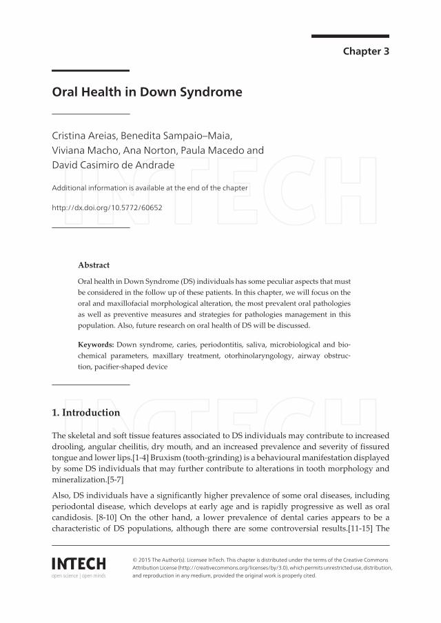
Due to fear of plate swallowing and child choking, the plate developed by Castillo-Moralesdid not allow a prolonged use and required adult supervision [58]. Thereafter, Andrade C. etal [20, 63] developed a modified, pacifier-shaped device, that provide greater security,prolonged usage time (even at night), less concern by caregivers, and better acceptance bysociety. The results of its use show an improvement in aesthetics with less tongue protrusionand higher time of closed mouth when compared to the traditional Castillo-Morales plate(Figure 11., Figure 12., Figure 13.).[12, 20, 58, 63]
3.5. Fixed appliances
We can place fixed appliances in preschool age, if the child is cooperative. Problems likeanterior or posterior crossbites should be corrected as soon as they are found. Fixed applianceshave better results because we can have a greater control of their use, which does not happenwith removable appliances, which can either be used or not. We may expand the maxilla togain more space to the tongue and in DS we prefer to do it expanding from the apical base, to
Oral Health in Down Syndromehttp://dx.doi.org/10.5772/60652
55

obtain an orthopaedic effect. At certain ages, the effects of maxillary disjunction may be morefavourable, which has to do with the overall growth of the jaws and the individual himself(Figure 13., Figure 14.). [20, 28]
Advantages of the device
• Fixed appliance
• Easy to clean
• Acts in a short time
Figure 11. An alteration of the traditional plate now like a pacifier-shaped device.
Figure 12. Pacifier-shaped device; the results of its use show an improvement in aesthetics with less tongue protrusion and higher time of closed mouth when compared to the traditional Castillo-Morales plate.
Fixed appliances
We can place fixed appliances in preschool age, if the child is cooperative. Problems like anterior or posterior crossbites should be corrected as soon as they are found. Fixed appliances have better results because we can have a greater control of their use, which does not happen with removable appliances, which can either be used or not. We may expand the maxilla to gain more space to the tongue and in DS we prefer to do it expanding from the apical base, to obtain an orthopaedic effect. At certain ages, the effects of maxillary disjunction may be more favourable, which has to do with the overall growth of the jaws and the individual himself (Figure 13. , Figure 14). [20, 28]
Figure 11. An alteration of the traditional plate now like a pacifier-shaped device.
Health Problems in Down Syndrome56
• Does not need intense cooperation from parents or from the children.
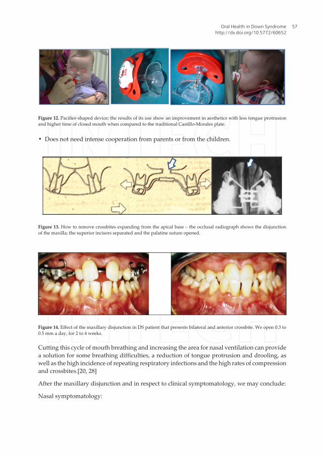
Figure 13. How to remove crossbites expanding from the apical base – the occlusal radiograph shows the disjunctionof the maxilla; the superior incisors separated and the palatine suture opened.
Figure 14. Effect of the maxillary disjunction in DS patient that presents bilateral and anterior crossbite. We open 0.3 to0.5 mm a day, for 2 to 4 weeks.
Cutting this cycle of mouth breathing and increasing the area for nasal ventilation can providea solution for some breathing difficulties, a reduction of tongue protrusion and drooling, aswell as the high incidence of repeating respiratory infections and the high rates of compressionand crossbites.[20, 28]
After the maxillary disjunction and in respect to clinical symptomatology, we may conclude:
Nasal symptomatology:
12
Figure 11. An alteration of the traditional plate now like a pacifier-shaped device.
Figure 12. Pacifier-shaped device; the results of its use show an improvement in aesthetics with less tongue protrusion and higher time of closed mouth when compared to the traditional Castillo-Morales plate.
Fixed appliances We can place fixed appliances in preschool age, if the child is cooperative. Problems like anterior or posterior crossbites should be corrected as soon as they are found. Fixed appliances have better results because we can have a greater control of their use, which does not happen with removable appliances, which can either be used or not. We may expand the maxilla to gain more space to the tongue and in DS we prefer to do it expanding from the apical base, to obtain an orthopaedic effect. At certain ages, the effects of maxillary disjunction may be more favourable, which has to do with the overall growth of the jaws and the individual himself (Figure 13. , Figure 14). [20, 28]
- Advantages of the device – Fixed appliance
– Easy to clean
Figure 12. Pacifier-shaped device; the results of its use show an improvement in aesthetics with less tongue protrusionand higher time of closed mouth when compared to the traditional Castillo-Morales plate.
Oral Health in Down Syndromehttp://dx.doi.org/10.5772/60652
57

• Global improvement of cases and controls, but in that the cases show up, markedly, a minorincidence of rhinorrhea: "went the first summer in that my son had the nose completelywithout secretions".
Otologic symptomatology:
• Smaller evidence in the resolution from the serous otitis, that persisted in the cases and inthe controls.
Rapid maxillary expansion produced a significant augmentation of nasal volume in childrenwho had been treated (p<0.05) compared to the control group; these results were stable throughthe period of retention.
The rapid maxillary expansion (disjunction) in infants with Down syndrome:
• Diminishes the number of infections of the superior airways.
• Improves nasal permeability.
• Improves several parameters analysed by speech therapists.
• Tongue Mobility
• Articulation
• Intelligibility
Figure 15. Patient with Down syndrome that present a bilateral crossbite (photographs extra and intra - orals, modelsand X rays).
Health Problems in Down Syndrome58

Rapid maxillary expansion (RME) increased space for the tongue in the oral cavity (Figure15., Figure 16., Figure 17.). This in turn results in a reduction of tongue protrusion and drooling.These aspects, in addition to the enlargement of the maxilla, often lead to an aesthetic im‐provement noted by the parents of the RME children. By placing the tongue in its normalposition, the speech is improved, and so the aesthetics and self-confidence of the individual,facilitating his integration in society. This procedure may be carried out concomitantly withother surgical procedures for upper airway obstruction, sleep apnea and chronic otitis mediawith effusion. Rapid maxillary expansions give these patients a better airway, if used (Figure15., Figure 16., Figure 17.). It is important to make exercise routines and teach these patientsto use this new airway, increasing attention and activity, and leading to a better generaldevelopment, better general health and less hours of work lost by the family. The indicationsshould be accurate. The effect is similar to the infants from the general population. The use ofthis device should be included in the suggestion to medical and connected Associations ofparents of Down’s syndrome children.
Figure 16. In less than one month, they obtain an increase from the apical base sufficient for eliminating the bilateralcrossbite and the compression of the maxilla; this helps to facilitate the nasal ventilation.
We should not be afraid to use facial masks or other orthodontic devices because these patientsare very supportive, and we should encourage all dentists to treat these patients.
We cannot forget that all cases must have an appropriate fixed orthodontic contention, in mosttimes for life.The orthodontic retention phase minimises unwanted dental movements andmaintains the corrections, and in these cases, most of the times, a fixed contention is necessarybecause hypotonia and deleterious habits are very dangerous for the stability of the correction(Figure 18., Figure 19., Figure 20., Figure 21.). [20, 28]
Oral Health in Down Syndromehttp://dx.doi.org/10.5772/60652
59
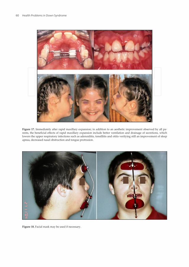
Figure 17. Immediately after rapid maxillary expansion; in addition to an aesthetic improvement observed by all pa‐rents, the beneficial effects of rapid maxillary expansion include better ventilation and drainage of secretions, whichlowers the upper respiratory infections such as adenoiditis, tonsillitis and otitis verifying still an improvement of sleepapnea, decreased nasal obstruction and tongue protrusion.
Figure 18. Facial mask may be used if necessary.
Health Problems in Down Syndrome60

Figure 19. Facial mask immediately after disjunction.
Figure 20. DS patients cooperate with the dentist and we should not be afraid to treat these patients.
4. Conclusion
Patients with Down syndrome present peculiar orofacial features that, when not corrected,may interfere with their physical, psychological and social development.
Children with this syndrome have a high risk of developing malocclusion and periodontalproblems, and these should be the main concerns in their treatment needs. When planning thedental treatment of patients with Down syndrome, dental practitioners should alwaysconsider their general health, in order to achieve a holistic and interdisciplinary approach.Nevertheless, there is a need to improve the oral health services available to individuals withDS, to further investigate the interrelations between all their health problems, and to providea higher level of information to parents of DS.
Oral Health in Down Syndromehttp://dx.doi.org/10.5772/60652
61

Acknowledgements
The authors supported this investigation.
Author details
Cristina Areias1*, Benedita Sampaio–Maia2, Viviana Macho1, Ana Norton1, Paula Macedo1 andDavid Casimiro de Andrade1
*Address all correspondence to: [email protected]
1 Faculty of Dental Medicine of Porto University, Porto, Portugal
2 Faculty of Dental Medicine, INEB - Instituto de Engenharia Biomédica and Instituto de In‐vestigação e Inovação em Saúde, Universidade do Porto, Portugal
Figure 21. The crossbite may be present in primary dentition. If we have cooperation, we must correct the temporaryteeth and not wait for the permanent teeth to get the treatment.
Health Problems in Down Syndrome62

The authors declare that they have no conflict of interest.Cristina Areias, Benedita Sampaio-Maia: responsible for the conception and design.Cristina Areias, Benedita Sampaio-Maia, Viviana Macho and Ana Norton were responsiblefor the data collection and manuscript redaction.Benedita Sampaio-Maia and Paula Macedo were responsible for the critical revision of itscontents.David Casimiro de Andrade was responsible for graphics and photos and the critical revi‐sion of its intellectual contents and final approval of the version to be published.All authors declare that written informed consent was obtained from the patient (or otherapproved parties) for publication of this research paper.
References
[1] Klug WS, Cummings MR, Spencer CA. Concepts of Genetics. Pearson Education,Upper Saddle River; 2006.
[2] Lewis R. Human Genetics Concepts and Applications, 7th ed. McGraw-Hill, NewYork; 2007.
[3] Shore S, Lightfoot T, Ansell P. Oral disease in children with Down syndrome: Causesand prevention. Community practitioner: The journal of the Community Practition‐ers’ & Health Visitors’ Association. 2010;83(2):18–21.
[4] Mouradian WE. The face of a child: Children’s oral health and dental education.Journal of dental education. 2001;65(9):821–31.
[5] Macho V, Andrade D, Areias C, Norton A, Coelho A, Macedo P. Prevalência de hábi‐tos orais deletérios e de anomalias oclusais numa população dos 3 aos 13 anos. Revport estomatol med dent cir maxilofac. 2012;53(3):143–7.
[6] Macho V, Seabra M, Pinto A, Soares D, Andrade A. Alterações craniofacial e particu‐laridades orais na Trissomia 21. Acta Pediatr Port. 2008;39(5):190–4.
[7] Macho V. Caracterização de uma população pediátrica com Trissomia 21: FMD-UP;2007.
[8] Cichon P, Crawford L, Grimm WD. Early-onset periodontitis associated with Down’ssyndrome—Clinical interventional study. Annals of periodontology/the AmericanAcademy of Periodontology. 1998;3(1):370–80.
[9] Moreira Lília MA. A síndrome de Down e sua patogénese: Considerações sobre o de‐terminismo genético. Rev Bras Psiquiatr. 2000;22(2):96–9.
[10] Martinez-Martinez RE, Loyola-Rodriguez JP, Bonilla-Garro SE, Patino-Marin N,Haubek D, Amano A, et al. Characterization of periodontal biofilm in Down syn‐
Oral Health in Down Syndromehttp://dx.doi.org/10.5772/60652
63

drome patients: A comparative study. The journal of clinical pediatric dentistry.2013;37(3):289–95.
[11] Areias C. Efeito da composição da saliva na prevalência cárie dentária em criançascom trissomia 21: Universidade do Porto Faculdade de Medicina Dentária; 2011.
[12] Vicente VA. Relação entre a prevalência da doença cárie e risco microbiológico. CiencOdontol Bras. 2008;11(2):44–8.
[13] Areias C, Sampaio-Maia B, Pereira Mde L, Azevedo A, Melo P, Andrade C, et al. Re‐duced salivary flow and colonization by mutans streptococci in children with Downsyndrome. Clinics. 2012;67(9):1007–11.
[14] Areias CM, Sampaio-Maia B, Guimaraes H, Melo P, Andrade D. Caries in Portu‐guese children with Down syndrome. Clinics. 2011;66(7):1183–6.
[15] Holst D, Schuller AA, Aleksejuniene J, Eriksen HM. Caries in populations—A theo‐retical, causal approach. European journal of oral sciences. 2001;109(3):143–8.
[16] Moura CP. Trissomia 21-Perspectiva Otorrinolaringológica. Arquivos de Medicina.2004;18, 1/2: 61 - 65.
[17] Jacobs I, Gray RF, Todd N. Upper airway obstruction in children with Down syn‐drome. Arch Otolaryngol Head Neck Surg. 1996;122:945–50.
[18] Moura CP, Andrade D, Cunha LM, Tavares MJ, Cunha MJ, Barros H, Pueschel SM,Vaz P, Clemente MP. Down syndrome: Otolaryngological effects of rapid maxillaryexpansion. The journal of laryngology & otology. 2008;12(122):1318–24.
[19] de Moura CP, Andrade D, Cunha LM, Tavares MJ, Cunha MJ, Vaz P, et al. Downsyndrome: Otolaryngological effects of rapid maxillary expansion. The journal of lar‐yngology and otology. 2008;122(12):1318–24.
[20] Andrade DJ. Trissomia 21 – Estudo Dento-Maxilo-Facial FMD-UP; 2000.
[21] Fernandes A, Mourato P, Xavier M, Andrade C, Fernandes C, Palha M. Caracteriza‐ção da evolução somática de crianças portuguesas com trissomia 21 – resultados pre‐liminares. Acta Pediatr Port. 1998;29:261–8.
[22] Lewis CW, Grossman DC, Domoto PK, Deyo RA. The role of the pediatrician in theoral health of children: A national survey. Pediatrics. 2000;106(6):E84.
[23] Acerbi AG, de Freitas C, de Magalhaes MH. Prevalence of numeric anomalies in thepermanent dentition of patients with Down syndrome. Special care in dentistry: Offi‐cial publication of the American Association of Hospital Dentists, the Academy ofDentistry for the Handicapped, and the American Society for Geriatric Dentistry.2001;21(2):75–8.
Health Problems in Down Syndrome64

[24] Allison PJ, Hennequin M. The oral assessment in Down syndrome questionnaire(OADS): Development of an instrument to evaluate oral health problems in individu‐als with Down syndrome. Community dental health. 2000;17(3):172–9.
[25] Oredugba FA. Oral health condition and treatment needs of a group of Nigerian in‐dividuals with Down syndrome. Down’s syndrome, research and practice: The jour‐nal of the Sarah Duffen Centre/University of Portsmouth. 2007;12(1):72–6.
[26] Musich DR. Orthodontic intervention and patients with Down syndrome. The angleorthodontist. 2006;76(4):734–5.
[27] Soares K, Mendes R, Junior R, Rosa L, Costa K. Prevalência de maloclusão em porta‐dores de Síndrome de Down na cidade de Teresina-PI. Revista Gaúcha de Odontolo‐gia. 2009;57:187–91.
[28] de Moura CP, Vales F, Andrade D, Cunha LM, Barros H, Pueschel SM, et al. Rapidmaxillary expansion and nasal patency in children with Down syndrome. Rhinology.2005;43(2):138–42.
[29] Macho V, Coelho A, Areias C, Macedo P, Andrade D. Craniofacial features and spe‐cific oral characteristics of Down syndrome children. Oral health and dental manage‐ment. 2014;13(2):408–11.
[30] Allison PJ, Hennequin M, Faulks D. Dental care access among individuals withDown syndrome in France. Special care in dentistry: Official publication of theAmerican Association of Hospital Dentists, the Academy of Dentistry for the Handi‐capped, and the American Society for Geriatric Dentistry. 2000;20(1):28–34.
[31] Law V, Seow WK, Townsend G. Factors influencing oral colonization of mutansstreptococci in young children. Australian dental journal. 2007;52(2):93–100; quiz 59.
[32] Fung K, Allison PJ. A comparison of caries rates in non-institutionalized individualswith and without Down syndrome. Special care in dentistry: Official publication ofthe American Association of Hospital Dentists, the Academy of Dentistry for theHandicapped, and the American Society for Geriatric Dentistry. 2005;25(6):302–10.
[33] Fung K, Lawrence H, Allison P. A paired analysis of correlates of dental restorativecare in siblings with and without Down syndrome. Special care in dentistry: Officialpublication of the American Association of Hospital Dentists, the Academy of Den‐tistry for the Handicapped, and the American Society for Geriatric Dentistry.2008;28(3):85–91.
[34] Gizani S, Papaioannou W, Haffajee AD, Kavvadia K, Quirynen M, Papagiannoulis L.Distribution of selected cariogenic bacteria in five different intra-oral habitats inyoung children. International journal of paediatric dentistry/the British PaedodonticSociety [and] the International Association of Dentistry for Children. 2009;19(3):193–200.
Oral Health in Down Syndromehttp://dx.doi.org/10.5772/60652
65

[35] Tenuta LM, Ricomini Filho AP, Del Bel Cury AA, Cury JA. Effect of sucrose on theselection of mutans streptococci and lactobacilli in dental biofilm formedin situ. Ca‐ries research. 2006;40(6):546–9.
[36] Areias C, Sampaio-Maia B, Macho V, Leal I, Melo P, de Andrade C. Does the chemis‐try in the saliva of Down syndrome children explain their low caries prevalence? Eu‐ropean journal of paediatric dentistry: Official journal of European Academy ofPaediatric Dentistry. 2013;14(1):23–6.
[37] Catarina Barbosa JM, Milagre V, Andrade D, Areias C. Inhalation conscious sedationwith nitrous oxide/oxygen in pediatric dentistry. MEDICALEXPRESS. 2014 Jun;1(3):102-104
[38] Schaff-Blass E, Rozier RG, Chattopadhyay A, Quinonez R, Vann WF, Jr. Effectivenessof an educational intervention in oral health for pediatric residents. Ambulatory pe‐diatrics: The official journal of the Ambulatory Pediatric Association. 2006;6(3):157–64.
[39] Siqueira WL, Bermejo PR, Mustacchi Z, Nicolau J. Buffer capacity, pH, and flow ratein saliva of children aged 2-60 months with Down syndrome. Clinical oral investiga‐tions. 2005;9(1):26–9.
[40] Siqueira WL, de Oliveira E, Mustacchi Z, Nicolau J. Electrolyte concentrations in sali‐va of children aged 6–10 years with Down syndrome. Oral surgery, oral medicine,oral pathology, oral radiology, and endodontics. 2004;98(1):76–9.
[41] Siqueira WL, Nicolau J. Stimulated whole saliva components in children with Downsyndrome. Special care in dentistry: Official publication of the American Associationof Hospital Dentists, the Academy of Dentistry for the Handicapped, and the Ameri‐can Society for Geriatric Dentistry. 2002;22(6):226–30.
[42] Siqueira WL, Siqueira MF, Mustacchi Z, de Oliveira E, Nicolau J. Salivary parametersin infants aged 12 to 60 months with Down syndrome. Special care in dentistry: Offi‐cial publication of the American Association of Hospital Dentists, the Academy ofDentistry for the Handicapped, and the American Society for Geriatric Dentistry.2007;27(5):202–5.
[43] Vijayaprasad KE, Ravichandra KS, Vasa AA, Suzan S. Relation of salivary calcium,phosphorus and alkaline phosphatase with the incidence of dental caries in children.Journal of the Indian Society of Pedodontics and Preventive Dentistry. 2010;28(3):156–61.
[44] Lenander-Lumikari M, Loimaranta V. Saliva and dental caries. Advances in dentalresearch. 2000;14:40–7.
[45] Davidovich E, Aframian DJ, Shapira J, Peretz B. A comparison of the sialochemistry,oral pH, and oral health status of Down syndrome children to healthy children. In‐
Health Problems in Down Syndrome66

ternational journal of paediatric dentistry/the British Paedodontic Society [and] theInternational Association of Dentistry for Children. 2010;20(4):235–41.
[46] Kavanagh DA, Svehla G. Variation of salivary calcium, phosphate and buffering ca‐pacity in adolescents. Archives of oral biology. 1998;43(12):1023–7.
[47] Lee SR, Kwon HK, Song KB, Choi YH. Dental caries and salivary immunoglobulin Ain Down syndrome children. Journal of paediatrics and child health. 2004;40(9–10):530–3.
[48] Cristina Areias VM, Coelho A, Pereira L, Andrade D, Pérez-Mongiovi D, Sampaio-Maia B. Enfoque clínico de niños con síndrome de Down en el consultorio dental.Avances en odontoestomatologia. 2014;(6):307-313.
[49] Tanaka MH, Rodrigues TO, Finoti LS, Teixeira SR, Mayer MP, Scarel-Caminaga RM,et al. The effect of conventional mechanical periodontal treatment on red complexmicroorganisms and clinical parameters in Down syndrome periodontitis patients: Apilot study. European journal of clinical microbiology & infectious diseases: Officialpublication of the European Society of Clinical Microbiology. 2015 Mar;34(3):601-8.doi: 10.1007/s10096-014-2268-7. Epub 2014 Nov 4.
[50] Cornejo LS, Brunotto M, Hilas E. Salivary factors associated to the prevalence and in‐crease of dental caries in rural schoolchildren. Revista de saude publica. 2008;42(1):19–25.
[51] Hennequin M, Allison PJ, Veyrune JL. Prevalence of oral health problems in a groupof individuals with Down syndrome in France. Dev Med Child Neurol. 2000;42(10):691–8.
[52] Martín M, Vasquez E, Diz P, Figueiral H, Vasconcelos L, Figueiredo J, Andrade D.Terapia de Estimulação Orofacial: Princípios e Considerações. Revista de Saúde OralRevista de Saúde Oral. 1996;1:51–4.
[53] Macho V, Palha M, Macedo AP, Ribeiro O, Andrade C. Comparative study betweendental caries prevalence of Down syndrome children and their siblings. Special carein dentistry: Official publication of the American Association of Hospital Dentists,the Academy of Dentistry for the Handicapped, and the American Society for Geriat‐ric Dentistry. 2013;33(1):2–7.
[54] Macho V, Seabra M, Areias C, Ribeiro O, Andrade C. Comparação dos cuidados desaúde oral numa população com trissomia 21 e seus irmãos. Comparação dos cuida‐dos de saúde oral numa população com trissomia 21 e seus irmãos. JADA. 2008;8(5):2-14.
[55] Fischer-Brandies H, Limbrock GJ, Tragner-Born J. La Thérapie Fonctionnelle Sensori‐motrice des Troubles Fonctionnels Oro-Faciaux Chez les Enfants avec I.M.C., d’aprèsCastillo-Morales (Infirmité motrice cérébrale). L’orthodontie Française. 1988;59 Pt2:449-58.
Oral Health in Down Syndromehttp://dx.doi.org/10.5772/60652
67

[56] Chung MH, Kaste LM, Koerber A, Fadavi S, Punwani I. Dental and medical stu‐dents’ knowledge and opinions of infant oral health. Journal of dental education.2006;70(5):511–7.
[57] Carlstedt K, Dahllöf G, Nilsson B, Modeer T. Effect of palatal plate therapy in chil‐dren with Down syndrome. A 1-year study. Acta Odontol Scand. 1996;54(2):122–5.
[58] Castillo-Morales R, Avalle C, Schmid R. Possibilità di Trattamento della PatologiaOrofaciale nella Sindrome di Down con la Placa di Regolazione Motoria. PediatriaPreventiva e Sociale. 1984;34:1–4.
[59] Limbrock GJ, Castillo-Morales R, Hoyer H, Stover B, Onufer C. Castillo-Morales ap‐proach to orofacial pathology in Down syndrome. International journal of orofacialmyology. 1993 Nov;19:30-7.
[60] Limbrock GJ, Fischer-Brandies H, Avalle C. Castillo-Morales’ orofacial therapy:Treatment of 67 children with Down syndrome. Developmental medicine and childneurology. 1991;33:296–303.
[61] Limbrock GJ, Hoyer H, Scheying H. Regulation therapy by Castillo-Morales in chil‐dren with Down syndrome: Primary and secondary orofacial pathology. Journal ofdentistry for children. 1990 Nov-Dec;57(6):437-41.
[62] Hoyer H, Limbrock GJ. Orofacial regulation therapy in children with Down syn‐drome, using the methods and appliances of Castillo-Morales. Journal of dentistryfor children. 1990;Nov–Dec:442.
[63] Andrade C, Tavares P, Rebelo P, Palha M, Tavares M. Placa modificada para trata‐mento de hipotonia oro-muscular em crianças com idade compreendida entre os 2meses e os 2 anos. Ortodontia. 1998;3(2):111–7.
Health Problems in Down Syndrome68