putting the brakes on respiratory distress and failure
TRANSCRIPT

1
Putting the Brakes on Respiratory Distress and Failure
Anne Dabrow Woods, DNP, RN, CRNP, ANP-BC, AGACNP-BC
Chief Nurse
Wolters Kluwer
Philadelphia, PA
Acute Care Nurse Practitioner
Critical Care Service, Penn Medicine, Chester County Hospital
West Chester, PA
Adjunct Faculty
Drexel University, College of Nursing & Health Sciences
Philadelphia, PA Copyright Anne Dabrow Woods; all rights reserved
Disclaimer
• I have nothing to disclose.
Objectives
At the end of the session on the common causes of acute respiratory distress and failure (pulmonary embolism, COPD exacerbation, asthma exacerbation and pulmonary edema); the learner will be able to:
• Identify the etiology and clinical presentation of these disorders.
• Identify the assessment and diagnostic techniques for these disorders.
• Develop a multidisciplinary treatment plan for patients with these acute disorders.

2
Recognizing respiratory distress
Do you recognize this situation?
Do you recognize this situation?

3
Is it respiratory failure?
Acute respiratory failure…
• Type 1 – hypoxemic respiratory failure
• Problem is oxygen!
• PaO2 < 60 mm Hg with normal PaCO2
• Type 2 – hypercarbic respiratory failure
• Problem is carbon dioxide!
• PaCO2 > 50 mm Hg
Identify the causes…

4
It’s all about the pump…
It’s all about the circulation…
It’s all about gas exchange…

5
Arterial blood gases • pH
• Normal 7.35-7.45 • Below 7.35 – acidosis
• Above 7.45 - alkalosis
• PaCO2 • Normal 35-45 mm Hg
• Above 45 means – hypoventilation causing CO2 retention
• Below 35 means – hyperventilation, blowing off CO2
• PaO2 • Normal 80-100 mm Hg
• HCO3 • Normal 22-26 mEq/L ( metabolic compensation by the kidneys)
• High level – kidneys are increasing HCO3 in blood for alkalosis
• Low level – kidneys are decreasing HCO3 in blood for acidosis
• SaO2 – Normal is > 95% (doesn’t always correlate to the SpO2)
ABG interpretation • Step 1: Analyze the pH
• pH < 7.35 = acidosis • pH > 7.45 = alkalosis
• Step 2: Analyze the PaCO2 • PaCO2 > 45 = acidosis • PaCO2 < 35 = alkalosis
• Step 3: Analyze the HCO3 • HCO3 < 22 = acidosis • HCO3 > 26 = alkalosis
• Step 4: Match the PaCO2 or HCO3 with pH • pH < 7.35, PaCO2 > 45 and HCO3 normal = respiratory acidosis (pulmonary issue)
• Hypoventilation, respiratory infection, COPD, Asthma, pulmonary edema, central nervous system or spinal cord injury • Treat by increasing ventilation rate, tidal volume
• pH < 7.35, HCO3 < 22 and PaCO2 normal = metabolic acidosis (kidneys trying to buffer) • Renal failure, DKA, lactic acidosis, sepsis, drugs – ethylene glycol
• pH > 7.45, PaCO2 < 35 and HCO3 normal = respiratory alkalosis (pulmonary issue) • Hyperventilation, pain, anxiety, early stages of pneumonia or PE, excessive mechanical
ventilation • Treat by decreasing ventilation rate
• pH > 7.45, HCO3 > 26 and PaCO2 normal = metabolic alkalosis (kidneys trying to buffer) • Diuretics, steroids, excessive vomiting, dehydration, Cushings, liver failure, hypokalemia
Pulmonary edema

6
What is heart failure?
• HF is a complex clinical syndrome that results from any structural or functional impairment of ventricular filling or ejection of blood (HFSA, 2013)
• HF = pump issue
• HF types
• preserved ejection fraction (> 50%)
• reduced ejection fraction (< 40%)
• Borderline ejection fraction (41-49%)
• Characteristics
• Elevated cardiac filling pressures
• Inadequate peripheral oxygen delivery
Class versus Stage
• NYHA Classification
• I – no symptoms
• II - Slight limitation; symptoms with ordinary activity
• III: marked limitation; symptoms with less than ordinary activity
• IV – severe limitation; symptoms at rest
• AHA Stages
• A – at risk but no structural changes
• B – structural changes; no symptoms
• C – structural changes with prior or current symptoms
• D – refractory HF requiring specialized interventions
Etiology: CO = HR x SV
Stroke volume
Preload
Contractility
Afterload
Heart rate

7
CO = HR x SV
• Effects of heart rate
• Tachycardia
• Decreased time for ventricular filling leads to hypotension
• Decreased myocardial perfusion
• Bradycardia
• Inadequate volume lads to hypotension
• Determinants of Stroke Volume
• Preload – venous return to the heart; affected by stretch and volume – CVP pressure
• Afterload – force the LV has to overcome to eject its contents – constriction of the aorta
• Contractility – ability to pump/contract
Pathophysiology
Renin-Angiotensin
System
Aldosterone System
Sympathetic Nervous System
Sympathetic Nervous System
Low blood volume or pressure recognized by baroreceptors (aorta, carotids)
Baroreceptors alert the brain
Brain signals the adrenals to produce epinephrine and norepinephrine

8
Sympathetic Nervous System
• Epinephrine and norepinephrine actions
• Beta-1 receptors - increased heart rate
• Beta-2 receptors - bronchodilation, vasodilation
• Alpha receptors – vasoconstriction and increase venous return
Renin-Angiotensin-Aldosterone System
Low blood volume through the kidneys
Kidneys produce renin
Renin converts angiotensinogen to angiotensin I
Angiotensin I converted to Angiotensin II in the presence of angiotensin converting enzyme (ACE)
RAAS
Angiotensin II activates receptors on blood vessels (angiotensin II receptors)
Vasoconstriction, increased BP, increased venous return to the heart

9
RAAS
• Aldosterone released from the adrenals in response to high angiotensin II and norepinephrine levels
aldosterone targets the kidneys
tubules retain water and sodium
increased blood volume and BP
Endothelial remodeling = Poor heart function/failure
Endothelial Remodeling
of the Heart
RAAS Action
Sympathetic Action
Aldosterone Action
Atherosclerosis, low Nitric Oxide
levels
Risk factors for HF
• CAD
• DM
• HTN
• Valve disease
• Alcohol
• Congenital heart defect
• Thyroid disease
• Age
• Obesity
• Reduced vital capacity
• Smoking
• LVH
• Sleep apnea

10
Pulmonary edema clinical presentation
• Tachypnea
• Dyspnea on exertion
• Acute shortness of breath/dyspnea
• Crackles
• Wheezes
• Frothy sputum
• Increase work of breathing
• Tachycardia
• Diaphoresis
• Chest pressure/pain
• Edema
Diagnostic studies
• 12 lead ECG
• Stat Chest X-Ray
• ABG
• BNP
• Cardiac enzymes – troponin and CPK, stat and serial enzymes
Pulmonary edema

11
Treatment
• High fowlers position
• Supplemental oxygen – keep SpO2 > 93%; PaO2 > 60 mm Hg
• Bipap (Inspiration/expiration pressure)
• Decrease afterload – vasodilator (Nitroglycerin), ACE inhibitor
• Decrease preload – loop diuretic (furosemide or bumetanide)
• Morphine
• Increase strength of contraction – positive inotropes
• Relieve cardiac ischemia
• Monitor intake/output
Pneumonia
Definition of pneumonia
• Acute, febrile inflammatory disorder of the lungs associated with cough and exertional dyspnea
• Infiltrate on chest x-ray
• Appearance on CXR may lag 24 to 48 hours behind clinical presentation
• Leukocytosis – elevated WBCs

12
Types of pneumonia
• Community-acquired pneumonia
• Hospital-acquired pneumonia
• Healthcare-associated pneumonia
• Ventilator-associated pneumonia
• Other ways to look at pneumonias
• Organism
• Bacterial
• Viral
• Fungal
• Mode of entry
• Aspiration
Comparing organisms • Gram –
• Cocci – neisseria, moraxella
• Bacilli • Aerobic – vibrio,
enterobacter, acinetobacter, ecoli, klebsiella, haemophilus, proteus, salmonella, shigella, pseudomonas, acinobacter
• Anerobic – bacteroides, prevotella, fusobacterium
• Gram + • Cocci
• Aerobic • clusters (staph)
• chains/pairs (strep), enterococcus
• Anerobic – peptococcus
• Bacilli • Aerobic – lactobacillus,
gardenella, cornybacter, listeria
• Anerobic – actinomyces, clostridium
Atypical bacteria
• Colorless – do not color with gram staining
• Responsible for 20 to 30% of CAP
• The big 3 atypicals
• Mycoplasma pneumoniae
• Chlamydia pneumoniae
• Legionella pneumonphilia

13
Let’s look at the alphabet soup of pneumonia…
Community-acquired pneumonia (CAP) • Acquired in the community; most common type of pneumonia
• 4 to 5 million cases per year • 25% of cases require hospitalization • Inhospital mortality 10-12% (mild cases not admitted - < 1%)
• If patient gets admitted to the hospital and develops pneumonia within 48 hours – CAP • Cause: defense mechanism failure
• Cough reflex • Mucociliary clearance system • Immune response
• Organisms • Bacteria
• Strep pneumoniae (most common in adults) – gm + • Haemophilus influenza – gm - • Klebsiella pneumoniae – gm -
• Atypical • Chlamydia pneumoniae • Mycoplasma pneumoniae • Mycobacterium tuberculosis
• Viruses • Respiratory syncytial virus • Adenovirus • Rhinovirus
Hospital-acquired pneumonia (HAP)
• Occurs 48 hours after admission to the hospital and it wasn’t incubating at time of admission
• We gave it to the patient!
• Most common organisms
• Staphylococcus aureus – gram +
• Streptococcus pneumoniae – gram +
• Haemophilus influenzae – gram -

14
Ventilator-associated pneumonia (VAP)
• Occurs 48 to 72 hours post intubation
• Risk increases with poor oral care
• Most common organism
• Pseudomonas aeruginosa – gram -
Healthcare-associated pneumonia (HCAP)
• Definition
• Patient was in hospital or a healthcare setting for 2 or more days within 90 days of infection and develop pneumonia
• Long-term care facilities
• Assisted living
• Rehabilitation
• Nursing home
• IV therapy including antibiotics
• Chemotherapy – within 30 days of current infection
• Wound care – within 30 days of current infection
• Hemodialysis clinic
HCAP organisms
• More similar to HAP than CAP
• Staphylococcus aureus – gram +
• Pseudomonas aeruginosa – gram -
• Less likely but possible
• Streptococcus pneumoniae – gram +
• Haemophilus influenzae – gram -
• MRSA gram +

15
Pneumonia categorized by risk factors
• Aspiration pneumonia or pneumonitis
• R upper and R middle lobe most commonly affected
• Obstruction of the airway
• Tumor
• Secretions
• Inhalation injury
• Hypersensitivity pneumonia
• Near drowning
Comorbidities that increase mortality…
• COPD
• Heart Failure
• Diabetes
• Chronic liver disease
• Chronic kidney disease
• Very old
• Very young
Patient presentation
• “Typical pneumonia”
• Fever
• Chills or Rigors
• Leukocytosis (increased WBCs)
• Cough
• Sputum production
• Increased fremitus
• CXR
• Usually involves one lung and one lobe

16
Patient presentation
• “Atypical pneumonia”
• Fever or low temperature
• Leukocytosis – may be absent or have left shift on CBC (presence of bands)
• Dry cough
• Sore throat
• Headache
• Excessive sweating
• Soreness in chest or with cough
• CXR
• More diffuse pattern on CXR
• May involve more than one lung and multiple lobes
Diagnostics
• Chest X-ray
• Sputum culture
• Blood cultures
• CBC with diff
• Chemistry
• PT/INR – for those on warfarin
• ABG – if worried about acute respiratory failure
RML pneumonia

17
LLL pneumonia
Multilobar pneumonia
To admit or not to admit…
• CURB-65 (predicts mortality)
• Confusion - 1 point
• Uremia (BUN > 19) – 1 point
• Respiratory Rate (> 30/min) – 1 point
• Blood pressure (SBP< 90 or DBP < 60) – 1 point
• Age (> 65 years) – 1 point
• Action
• 0 to 1: treat as outpatient
• 2: consider short stay in hospital or watch closely as outpatient
• 3-5: requires hospitalization

18
Pneumonia severity scale (PSI)
Step 1: Stratify to Risk Class I vs. Risk Classes II-V
Presence of:
Over 50 years of age Yes/No
Altered mental status Yes/No
Pulse ≥125/minute Yes/No
Respiratory rate >30/minute Yes/No
Systolic blood pressure
<90 mm Hg Yes/No
Temperature <35°C or ≥40°C Yes/No
History of:
Neoplastic disease Yes/No
Congestive heart failure Yes/No
Cerebrovascular disease Yes/No
Renal disease Yes/No
Liver disease Yes/No
If any "Yes", then proceed to
Step 2
If all "No" then assign to Risk
Class I
Step 2: Stratify to Risk Class II vs III vs IV vs V
Demographics Points Assigned
If Male +Age (yr)
If Female +Age (yr) - 10
Nursing home
resident +10
Comorbidity
Neoplastic
disease +30
Liver disease +20
Congestive
heart failure +10
Cerebrovascula
r disease +10
Renal disease +10
Physical Exam Findings
Altered mental
status +20
Pulse
≥125/minute +10
Respiratory rate
>30/minute +20
Systolic blood
pressure <90 mm
Hg +20
Temperature
<35°C or ≥40°C +15

19
Lab and Radiographic Findings
Arterial pH <7.35 +30
Blood urea nitrogen
≥30 mg/dl (9 mmol/liter) +20
Sodium <130 mmol/liter +20
Glucose ≥250 mg/dl
(14 mmol/liter) +10
Hematocrit <30% +10
Partial pressure of arterial O2
<60mmHg +10
Pleural effusion +10
∑ <70 = Risk Class II
∑ 71-90 = Risk Class III
∑ 91-130 = Risk Class IV
∑ >130 = Risk Class V
Treatment for pneumonia
• Right drug for the right bug – antibiotics, antivirals, antifungals
• Hydration
• NSS and LR for volume replacement
• Supplemental oxygen – keep SpO2 > 93%
• Nasal cannula
• HFNC
• Bipap
• Ventilator
• Fever management – fever helpful; don’t treat unless over 101.5 or symptomatic
• Acetaminophen – antipyretic and analgesic
• NSAIDS – antipyretic, analgesic, anti-inflammatory
• Bronchoscopy
• Supportive care
A word about …
• Antihistamines • Works for allergies not pneumonia
• Decongestants • Works for rhinitis and nasal congestion
• Cough suppressants • Only use at night to sleep
• If cough is productive – do not use
• Expectorants • Liquefies secretions
• Use if has nasal congestion or need to loosen secretions

20
Right drug: CAP treatment
• CAP (no comorbid conditions) – macrolide (Azithromycin)
• CAP with risk factors – respiratory fluorquinolone – (Levaquin)
• CAP inpatient (not ICU) –
• respiratory fluoroquinolone (Levaquin) OR
• Macrolide (Azithromycin) + beta-lactam antibiotic (amoxicillin/clavulanate; Augmentin)
• CAP requiring ICU
• respiratory fluoroquinolone (Levaquin) OR
• Macrolide (Azyithromycin) + antipseudomonal coverage (piperacillin/tazobactam; Zosyn)
Right drug: HAP treatment
• Low risk of multiple drug-resistant pathogens; use one of the following
• Ceftriaxone
• Moxifloxacin
• Levofloxacin
• Ampicillin/sulbactam
• Pipercillin/tazobactam
Right drug: HAP/VAP: high risk of multi-drug resistance
• Chose one agent from each category
• Antipseudomonal coverage
• Cefipime
• Piperacillin/tazobactam (Zosyn)
• PCN allergic patients: aztreonam
• A second antipseudomonal coverage
• Levoflaxacin
• Coverage for MRSA
• Vancomycin IV (dosed based on renal function to achieve trough of 15-20 mcg/ml)
• Linezolid

21
COPD exacerbation
Definition of COPD
• A preventable, progressive disease of the lungs caused by airflow limitation that is not fully reversible
• Cause – smoking!
• Chronic bronchitis
• Emphysema
Inflammation
Small airway remodeling and
alveolar destruction
Airflow limitation
The pathophysiology behind COPD

22
Chronic bronchitis versus emphysema
The picture of COPD
Stages of COPD and treatment
• I. Mild – Reduce risk factors, influenza vaccine, SABA if needed
• II. Moderate – SABA + anticholinergic + LABA + Rehab
• III. Severe – SABA + LABA + ICS (for repeated exacerbations
• IV. Very Severe – SABA + LABA + ICS + O2 + consider surgical treatment
• Understanding the acronyms
• SABA – short acting beta-agonist (albuterol)
• LABA – long acting beta-agonist [(salmeterol (Serevent)]
• Anticholinergic – ipratropium (atrovent) or tiotropium (Spiriva)
• ICS – inhaled corticosteroid (beclomethasone, budesonide, fluticasone, triamcinolone)

23
What defines a COPD exacerbation? • Increased sputum production
• Increased sputum purulence
• Increased dyspnea
Treatment
• Oxygen – keep SpO2 88-92% • Hydration • Noninvasive ventilation
• HFNC • Bipap
• Albuterol – episodic symptoms • Duonebs – albuterol + atrovent every 4 to 6 hours (then switch to long
acting once exacerbation under control)- conflicting evidence • Antibiotics or antivirals if has underlying bacterial/viral infection • Flutter valve • Chest percussion • Steroids
• Oral 40-60 mg prednisolone x 5 days (taper if used over 7 days) • For impending respiratory failure
• IV: methylprednisolone 60 mg 1 to 4 times per day up to 240 mg/day; 5-14 days (taper if used over 7 days)
• Ventilatory support
Asthma exacerbation

24
Etiology – reversible inflammation and bronchoconstriction
Clinical presentation
• Breathlessness
• Tachypnea
• Use of accessory muscles
• Wheezing
• Pulses paradoxus
• Decline in lung function – FEV1 or PEF
• A silent chest is a bad sign!!!!

25
Diagnostic studies
• Functional status – Peak flow meter
• Good response is PEF> 80% personal best
• Incomplete response is PEF 50-79% personal best
• Poor response is PEF < 50% personal best
• Check the ABG to assess for acute respiratory failure
Treatment • Oxygen
• Keep SpO2 > 90% (95% in pregnant women) or a PaO2 > 60 mm Hg
• Beta 2-agonist (SABA) • Repetitive albuterol (every 20 – 30 minutes) or continuous therapy
• Addition of anticholinergic treatment can decrease hospital readmissions (duonebs)
• Systemic beta 2 – agonists
• Epinephrine – 1:1,000; 0.3 – 0.5 gm every 20 min for 3 doses SC
• Steroids
• Prednisone 40 mg daily or 1-2 mg/kg in divided doses (max 60 mg/day) for 5-10 days
• Higher dose prednisone has weaker evidence to support use
• Taper if using for more than 7 days
• Antihistamines for allergy/anaphylaxis
• Diphenhydramine
• H2 blockers – ranitidine
Asthma with impending respiratory failure
• Impending respiratory failure
• Signs/Symptoms: Inability to speak, a silent chest, retractions, altered mental status, PaCO2 > 42 mm Hg
• Treatment to prevent and treat acute respiratory failure
• Intubation – avoid barotrauma
• Keep TV at 6 ml/kg
• Keep SpO2 > 90% (95% in pregnant women) or a PaO2 > 60 mm Hg
• IV magnesium – 2 grams over 20 minutes in adults; 25-75mg /kg in children (max of 2 grams)
• Continuous beta 2 agonists
• IV steroids
26
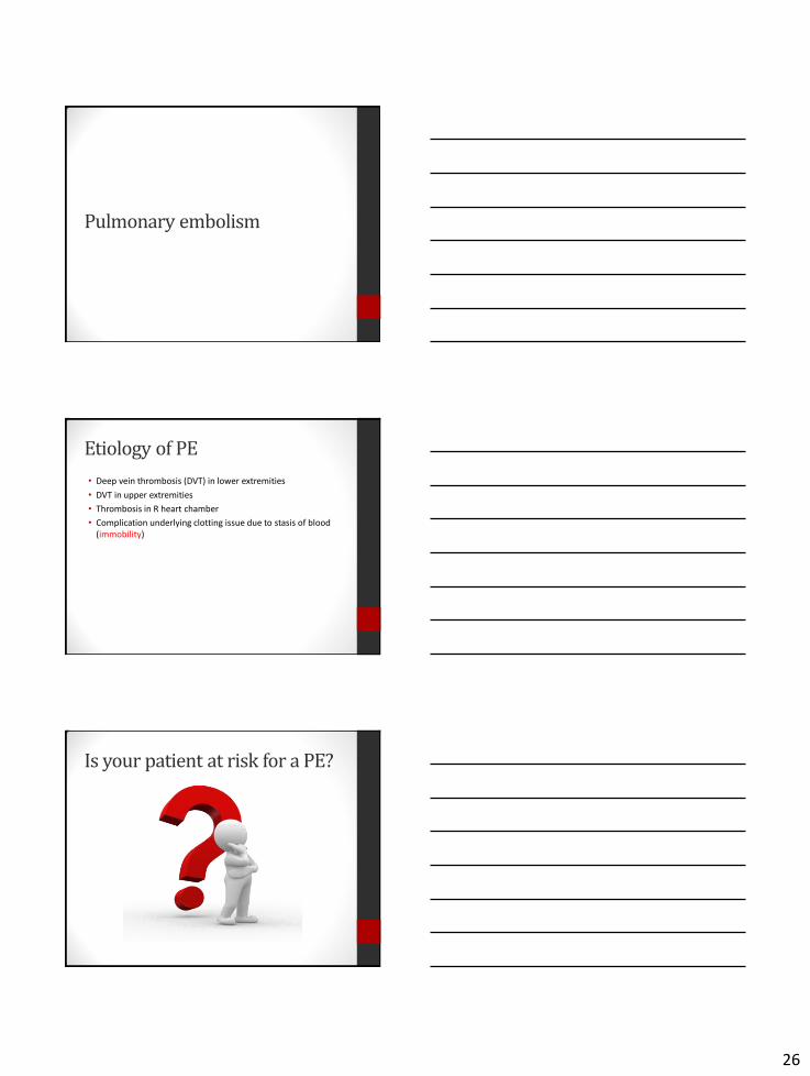
Pulmonary embolism
Etiology of PE
• Deep vein thrombosis (DVT) in lower extremities
• DVT in upper extremities
• Thrombosis in R heart chamber
• Complication underlying clotting issue due to stasis of blood (immobility)
Is your patient at risk for a PE?

27
Immobility
Surgery
Economy class syndrome

28
Hormone based birth control
The evidence on SCDs and TEDs…
Clinical presentation • Pleuritic chest pain • Shortness of breath • Hypoxia • Atypical symptoms
• Seizures • Syncope • Abdominal pain • Fever • Productive cough and
wheezing • Decreased level of
consciousness • Anew onset afib • Hemoptysis • delirium
• Tachypnea • Crackles • Accentuated second heart
sound • Tachycardia • Fever > 37.8 C • Diaphoresis • S3 or S4 heart sound • Signs of DVT • Lower extremity edema • cyanosis

29
Diagnostic studies
• Computerized tomography angiography (CTA) – gold standard
• V/Q scan
• ECG – nonspecific ST-T wave changes
• Echo – enlarged RV • TEE best for central or saddle embolism
• US lower extremities
• Labs • D-Dimer – only shows there is a clot; if negative tells you no PE
• ABG
• CBC with WBC
• Serial troponin levels
• BNP
• If no evident cause: hypercoagulability workup
Treatment
• Anticoagulation • LMWH preferred over UFH infusion
• Decrease dose of LMWH or avoid in patients with elevated Cr or low GFR
• Warfarin bridge – stop heparin once INR is over 2.0 for at least 24 hours but no sooner than 5 days from when warfarin started
• Patient should be on warfarin at least 3 months
• In cancer patients, LMWH is preferred over warfarin
• Repeat PE will require long-term therapy
• Consider thrombotic agents – TPA • Associated with a SBP < 90 or if patient has low bleeding risk
• Surgical options • Placement of vena cava filter
• Removal of embolism
Acute respiratory failure

30
Etiology of Acute Respiratory Failure
• Failure of oxygenation or carbon dioxide elimination • Reduced lung capacity and increased ventilation/perfusion mismatch • Reduced chest wall compliance and diaphragmatic and intercostal
muscle strength • Reduced clearance of airway secretions • Altered responsiveness to hypoxemia and hypercarbia
• Types • Type 1 – hypoxemic respiratory failure
• PaO2 < 60 mm Hg with normal PaCO2 • Fluid fills or collapses the alveoli
• Type 2 – hypercarbic respiratory failure • PaCO2 > 50 mm Hg • PaO2 is often decreased
• Acute versus chronic • Acute – occurs over minutes to hours • Chronic – occurs over days – usually see renal compensation
Understanding the causes… • Type 1 – hypoxemia (PaO2 < 60 mm Hg)
• Cardiogenic cause • Pulmonary edema
• Noncardiogenic cause • Pneumonia
• Pulmonary hemorrhage
• Pulmonary embolism
• Type 2 – Hypercarbia (PaCO2 > 50 mm Hg) • Hypoventilation secondary to
• Drug overdose
• Neuromuscular disease
• Hypercarbia secondary to • Asthma
• COPD
• Pulmonary embolism
Acute Lung Injury versus Acute Respiratory Distress Syndrome

31
Acute Lung Injury (ALI) versus Acute Respiratory Distress Syndrome (ARDS)
• Definition
• Mild (ALI) – PaO2/FiO2 ratio 200-300
• Moderate ARDS – PaO2/FiO2 ratio 100-200
• Severe ARDS – PaO2/FiO2 ratio < or equal to 100
• Mortality rate - 40-45%
• Complications
• 2/3 of survivors have impairment of pulmonary function
• Barotrauma – secondary to pressure
• Volutrauma – secondary to volume of air used to inflate lungs
• Bacterial infections
• Delirium
• Goal: Prevent cellular ischemia and death while correcting the cause
Causes of ALI/ARDS
• SIRS – systemic inflammatory response syndrome • Bacteremia • Pancreatitis • Massive trauma • Shock • Pneumonia – including aspiration • Transfusion related lung injury (TRALI)
• Cytokine mediated response • Inflammation • May occur after single blood product but more common with
multiple blood products
• Transfusion related circulatory overload (TACO) • No inflammation • Signs of volume overload • Elevated BNP
Three phases of ARDS
• Exudative phase • 2 to 4 days post acute lung injury, up to 7 days
• Capillary leaking causes alveolar flooding • Atelectasis
• Inflammation
• No high dose steroids – does not improve outcomes
• Fibroproliferative/proliferative phase • Connective tissue proliferation in response to initial injury
• Steroids maybe helpful in the first 7-14 days of ARDS – improves survivability; steroids may breakdown collagen and inhibit fibrosis
• Resolution and Recovery • 6 to 12 months of recovery

32
Clinical presentation
• Tachypnea
• Dyspnea - Breathlessness
• Crackles
• Cyanosis
• Tachycardia
• Anxiety
• Confusion
• Somnolence
• CXR shows alveolar flooding!
Diagnostic studies
• ABG
• CXR
• Sometimes helpful to get ECG
• Diagnostic tests to determine cause and to monitor clinical improvement
• CBC with diff
• PTT, PT/INR
• Fibrinogen, FDP
• Chem 20 – includes LFTs
• UA
• Blood, sputum, urine cultures
Treatment
• Treat the cause!
• Hypoxemia is major threat to organ dysfunction!
• Oxygen – keep SaO2 > 90%
• Permissive hypercapnia (PaCO2 60-70) with pH of 7.2-7.25
• Bipap
• Vent support • Goal is to increase PaO2 and decrease PaCO2
• Prevent barotrauma – keep TV around 6 ml/kg
• Maintain minimum of 5 cm peep
• Avoid dopamine – constricts the pulmonary capillary beds
• Transfuse as needed

33
Case study 1
• JG is a 36 year old male with a history of asthma who is admitted with LLL pneumonia. He is started on piperacillin/tazobactam and develops acute shortness of breath and wheezing. You give him a neb treatment and it doesn’t improve his breathing.
• What’s going on here?
• How should this be treated?
Case study 2
• WB is a 74 year old male who was admitted for a L knee replacement. He has a history of atrial fibrillation and had been on warfarin which was stopped a week ago. His orthopaedic surgeon did not put him on anticoagulants postoperatively. The patient develops acute shortness of breath, tachypnea, and has an SpO2 of 84%. You notice his L leg is more swollen than his R leg.
• What’s going on here?
• How should this be treated?
Case study 3
• AG is a 70 year old man who has a history of NYHA stage II HF. His wife brings him in an entire container of soup because he doesn’t like hospital food. Two hours later he develops shortness of breath, tachypnea, tachycardia, crackles in all fields and is diaphoretic.
• What’s going on here?
• How should this be treated?

34
Case study 4
• SB is a 65 year old smoker who has a history of COPD and comes to the ED with increasing shortness of breath and more copious sputum. She states she has been around her grandchildren who have had a cold. SB has a temp of 102 F.
• What’s going on here?
• How should this be treated?
Questions?
References • Barkley, T., & Myers, C. (2015). Practice Considerations for Adult-Gerontology Acute Care Nurse Practitioners, Vol.
1. Barkley & Associates: West Hollywood, CA. • Burnham, E., Janssen, W., Riches, D., Moss, M., Downey, G. (2014). The fibroproliferative response in acute
respiratory syndrome: Mechanisms and clinical significance. European Respiratory Journal, 43(1): 276-285. • Camargo, C., Rachelefsky, G., & Schatz, M. (2009). Managing asthma exacerbations in the emergency department.
Proceedings American Thoracic Society; (6), 357-366. • DeCramer, M., & Vestibo, J. (2014). Global initiative for Chronic Obstructive Pulmonary Disease. • File, T. (2016). Treatment of communicty-acquired pneumonia in adults who require hospitalization. UptoDate. • Kaynar, A., & Pinsky, M. (2015). Respiratory failure; Medscape; updated April 1, 2015. • Jaff, M., & McMurtry, S. (2011). Management of massive and submassive pulmonary embolism, ileofemoral deep
vein thrombosis, and chronic thromboemobolic pulmonary hypertension. Circulation, (123), 1788-1830. • Mandell, L., & Wunderlink R. (2007). Infectious Disease Society of America/American Thoracic Society Consensus
guidelines on the management of community-acquired pneumonia in adults. Clinical Infectious Disease. Suppl. 2:s27.
• NAEPP (2016). National Asthma Education and Prevention Guideline. • Papadarkis, M. & McPhee, S. (2015). Current Medical Diagnosis & Treatment. McGraw Hill: New York, NY. • Stoller, J. (2016). Managing COPD Exacerbations. UptoDate. Accessed March 24,2016. • Yancy, C. et al. (2013). ACCF/AHA Guideline for the management of heart failure. Circulation, 128; e240-327. • Wunderlink, R. (2014). Clinical Practice. Community-acquired pneumonia. New England Journal of Medicine;
370:543.