purification and biochemical characteriza- tion of ... · luis gabriel brieba de castro national...
TRANSCRIPT
Purification and Biochemical Characteriza-tion of Bacterial
DEAD-box Proteins
Varinia López-Ramírez Department of Genetic Engineering
Cinvestav Irapuato, México
Luis Gabriel Brieba de Castro National Laboratory of Genomics for Biodiversity
Cinvestav Irapuato, México
Gabriela Olmedo Álvarez Department of Genetic Engineering
Cinvestav Irapuato, México

1 Introduction
DEAD-box proteins are a large family of RNA helicases that disrupt secondary structures of RNA in an ATP-dependent manner. This protein family has a broad distribution in all domains of life. In bacteria, they have been associated with several processes involving RNA molecules, such as ribosomal biogene-sis and assembly, transcription, translation, mRNA decay, and RNA export. In bacteria, the number of genes coding for members of this protein family are highly variable, ranging from one to twelve, while in some endosymbonts, none can be identified. Nevertheless, for the large number of DEAD-box proteins in bacteria, the biochemical description has been focused on members of Escherichia coli and some mem-bers of Bacillus subtilis. Therefore, in this chapter we present a revised compendium of the efforts on the description of this protein family at the molecular and biochemical level in different bacterial species, particularly those that involve the expression and purification of RNA helicases.
2 General Structure of DEAD-box Proteins
DEAD-box proteins are mainly composed of two domains (DEAD and Helicase-C), which have fourteen RNA and ATP binding motifs (Figure 1). In bacteria, some members possess a third domain, DbpA, which is located in the C-terminal region. The DbpA domain has been related to the recognition of hair-pin 92 of the 23S ribosomal subunit. Given the importance of the function of this third domain, it was expected that all bacteria would possess at least one member with a DbpA domain; however, it has been shown that a great number of bacteria lack this type of RNA helicase (López-Ramírez, Alcaraz, Moreno-Hagelsieb, & Olmedo-Álvarez, 2011).
The average size of members of this protein family in bacteria is 499 amino acids, and most DEAD-box proteins have an isoelectric point (pI) between 8 and 10, which is a common characteristic found in nucleic acids binding proteins (Adhikari, Manthena, Sajwan, Kota, & Roy, 2010). Given their preference to interact with RNA and aid in folding/unfolding, DEAD-box proteins are also described as chaperones of RNA (Rajkowitsch et al., 2007).
2.1 Architecture of Bacterial DEAD-box Proteins
The broad range of processes that DEAD-box proteins are involved in suggested that diverse architec-tures would be found; however in bacteria there are just two basic types of architecture: those with ca-nonical DEAD and Helicase_C domains, and those with an additional RNA binding domain (RBD) DbpA (Figure 1). The length of the C-terminal end varies between 30 and 766 amino acids (López-Ramírez, Alcaraz, Moreno-Hagelsieb, & Olmedo-Álvarez, 2011), and the fact that no conserved domains are observed in this part of the protein does not necessarily mean that it does not have a relevant function. If fact, the pI of the C-terminal end is in the range of 10.6 and 11.37, and is probably critical for binding to different complexes (see below) as well as to the RNA substrate. The nature of the biochemical inter-action between RNA molecules and DEAD-box proteins is responsible for substrate recognition and subsequent function (Fairman-Williams, Guenther, & Jankowsky, 2010).

Figure 1: General Structure and Architecture of Bacterial DEAD-box proteins. a. The canoni-cal DEAD and Helicase_C domains are preferentially located in the N-terminal region; b. The DbpA domain participates in the recognition of hairpin 92 of the 23S ribosomal subunit and is present in some members of the DEAD-box protein family in bacteria; c. Fourteen RNA and ATP binding motifs are distributed throughout the main domains. The size of each amino acid represents its frequency at each motif. Adapted from López-Ramírez et al., 2011.
2.2 Molecular Studies on Bacterial DEAD-box Proteins: Interchangeable Factors in Different Complexes
The study of DEAD-box proteins in bacteria has focused on establishing their participation in RNA me-tabolism through in vitro and in vivo evaluation. The DEAD-box proteins that are best characterized in bacteria are the five from E. coli: DbpA, DeaD/CsdA, SrmB, RhlB, and RhlE. All of these proteins be-long to the DEAD-box family and the mechanism of action and level of involvement in RNA metabolism has been elucidated for each member (Table 1). Any one of the coding genes for DEAD-box proteins can be deleted and only the deletion of srmB or csdA produces a cold-sensitive phenotype. Even combina-tions of double mutations are viable (ΔsrmB ΔcsdA, ΔcsdA ΔrhlE, ΔcsdA ΔdbpA, ΔdbpA ΔrhlE, ΔrhlE ΔsrmB, and ΔsrmB ΔdbpA) (Iost & Dreyfus, 2006). Moreover, four of the five DEAD-box proteins in E. coli are involved in ribosomal processing, either by participating in their biogenesis (DbpA) or assembly (RhlE, SrmB, and DeaD/CsdA). The RhlE protein associates with ribosomal subunit 70S and compen-sates for individual defects in DeaD/CsdA and SrmB mutants grown at 16 °C and 20 °C (Jain, 2008). Both of these proteins participate in the assembly of the 50S ribosomal subunit at different levels: SrmB acts at early stages, while DeaD/CsdA is recruited at later stages of processing and it is mainly active at low temperatures. In particular, it has been observed that SrmB associates with ribosomal proteins L4 and L24, and that it has a preference for the 5' end of the 23S rRNA (Trubetskoy, Proux, Allemand, Dreyfus, & Iost, 2009). The orthologues of RhlE and SrmB in Pseudoalteromnas haloplantis TAC125 and in Colwellia psychreythraea 34H exhibit optimal double stranded RNA unwinding activity at tem-peratures below 25 °C, but not ATPase activity at these temperatures (Cartier, Lorieux, Allemand, Drey-fus, & Bizebard, 2010).

Table 1: DEAD-box proteins in Escherichia coli.
Protein DeaD/CsdA of E. coli has been found to interact in vitro with RNase E, both at 37 °C and at 15 °C, suggesting that the protein complex involved in mRNA decay is adaptable to different growing conditions in E. coli (Charollais, Dreyfus, & Iost, 2004; Charollais, Pflieger, Vinh, Dreyfus, & Iost, 2003; Prud'homme-Genereux et al., 2004). There is also evidence that RNase R, in addition to exhibiting RNase activity, possesses RNA helicase activity, suggesting that in the absence of the DeaD/CsdA pro-tein, it can be replaced by RNase R (Awano et al., 2010). Moreover, RhlB is involved in mRNA degrada-tion and interacts with proteins in the degradosome protein complex, specifically RNase E, and this inter-action helps to accelerate mRNA degradation (Ikeda et al., 2011). Moreover, RhlB also helps PNPase to relax mRNA secondary structures during mRNA degradation (Py, Higgins, Krisch, & Carpousis, 1996). In Pseudomonas syringe Lz4W, RhlB can be replaced by RhlE and interacts with RNAse E and R (Puru-sharth et al., 2005), whereas in E. coli, RhlB can be replaced by either DeaD/CsdA or RhlE (Khemici, Toesca, Poljak, Vanzo, & Carpousis, 2004; Prud'homme-Genereux et al., 2004).
Although most DEAD-box proteins are not substrate-specific, the DbpA protein in E. coli has preference for hairpin 92 of the 23S ribosomal subunit (Tsu & Uhlenbeck, 1998). The affinity given by the DbpA recognition domain for such a structure is higher than that of its ortholog in B. subtilis (DeaD/YxiN) (Kossen, Karginov, & Uhlenbeck, 2002; Kossen & Uhlenbeck, 1999). On the other hand, B. subtilis also contains four DEAD-box proteins: YfmL, DeaD/YxiN, CshA, and CshB (Table 2). How-ever, only DeaD/YxiN has been biochemically characterized, and it has been reported to participate in 23S ribosomal subunit biogenesis, acting in a specific way towards the hairpin 92 structure in a similar manner as DbpA of E. coli (Kossen & Uhlenbeck, 1999).
In the case of the cshA and cshB genes, it has been reported that expression increases when B. sub-tilis is incubated at 15 °C (Beckering, Steil, Weber, Volker, & Marahiel, 2002). In particular, mutation of the cshA gene affects the growth of B. subtilis in the exponential phase when the bacterium is grown at 22 °C. In addition, the directionality of the activity of CshA is not specific, because it can separate double strands of RNA without blunt ends in either the 5' or 3' direction (Ando & Nakamura, 2006). Further-more, a simple mutation of cshA or cshB does not affect growth of the bacteria when subjected to cold shock, but at least one of the two genes is required for the development of the bacteria in this condition.

Table 2: DEAD-box proteins in Bacillus subtilis.
The interaction of CshB with CspB was also demonstrated through analysis of the transfer of en-ergy of fluorescent resonance (FRET) during active transcription (Hunger, Beckering, Wiegeshoff, Graumann, & Marahiel, 2006). On the other hand, Lehnik-Habrink et al. (2010) showed that CshA, CshB, and DeaD/YxiN proteins can interact with RNase Y, which is a main component of the degradosome in B. subtilis, and that the union is established between the C-terminus of DEAD-box pro-teins and the RNase.
To date, there have been no reports on the role of protein YfmL, which is the smallest DEAD-box protein in B. subtilis. However, it has been possible to verify that it does not form oligomers and does not interact with the rest of the DEAD-box proteins present in B. subtilis or with the polynucleotide phos-phorylase (PnpA) (Lehnik-Habrink et al., 2010).
2.3 DEAD-box Proteins Improve Adaptability in Bacterial Species
DEAD-box proteins have also been associated with adaptation of bacterial species to different growing conditions. For example, in B. cereus ATCC 14579, its 5 RNA helicases (CshA, CshB, CshC, CshD, and CshE) exhibit increased expression when the bacteria is grown at low temperature (12 °C), but only mu-tations in CshA, CshB, and CshC reduce bacterial growth at 30 °C, reflecting the differential need of these protein family members (Pandiani et al., 2010). Another example of this type of regulation occurs in the cyanobacteria Anabaena sp. PCC st. 7120, where an increase in the expression of the RNA hel-icase CrhC was observed when the bacterium was grown at 15 °C (D. Chamot & Owttrim, 2000). A mu-tation in the DEAD-box CsdA coding gene in Yersinia pseudotuberculosis results in growth defects at 3 °C (Palonen et al., 2012), while in Listeria monocytogenes EGD-e, three of the four DEAD-box protein mutants (Δlmo0866, Δlmo1722, and Δlmo142) have growth defects at 3 °C, and Δlmo1722 and Δlmo142 are non-motile (Markkula, Lindstrom, Johansson, Bjorkroth, & Korkeala, 2012). Another example of the participation of DEAD-box proteins in the adaptability of bacteria to different stress and growth condi-tions is in Clostridium perfringens str. 13R, where an increase in gene expression of a DEAD-box protein was associated with the regulation of oxidative stress (Briolat & Reysset, 2002).
3 Purification Strategies of Bacterial DEAD-box Proteins
Interest in identifying the biochemical properties and the interactions of bacterial DEAD-box proteins with their substrates has motivated researchers to purify some members of this family, and therefore

several strategies have been developed for protein purification. Different types of expression vectors have been used, as well as different bacterial strains. Purification conditions are quite variable as well, includ-ing temperature, salt concentration, and pH range. In some cases, the final protein concentration level varies between 20 and 35 mg/L (Talavera & de la Cruz, 2005). Table 3 lists the different strategies that have been used by different research groups to date.
Enzyme Strain Expression vector
Induction/ growth conditions Reference
DeaD/YxiN B. subtilis
E. coli Rosetta pETM30 IPTG (0.2 M) A
600 nm = 0.5 for
4 h at 37 °C
Karow et al., 2007. Theissen et al., 2008. Aregger & Klostermeier, 2009.
CshA B. subtilis
E. coli M15 pQE60 IPTG (2 mM) 5 h at 37 °C
Ando & Nakamura, 2006.
SrmB and RhlE P. haloplanktis TAC125 C. psychrerythraea 34H
E. coli BL21 (DE3)
pPROEX-HTa
IPTG (0.5 mM) 12-16 °C Overnight
Cartier et al., 2010.
DbpA E. coli
E. coli BL21(DE3) LysS
pET11a IPTG (1 mM) OD
560 nm = 0.4
30 °C for 5 h
Böddeker et al., 1997
DbpA E. coli
E. coli BL21 (DE3) pLys-S
Not mentioned
Not mentioned Talavera & de la Cruz, 2005.
DeaD, SrmB RhlE E. coli
E. coli BL21 E. coli BL21 Star (DE3)
pPROEXHT pET-15b
IPTG (0.5-1 mM) OD
600 of 0.5-1.0
28 °C or 37 °C for 2-4 h
Bizebard & Ferlenghi, 2004.
Table 3: Different expression vectors and E. coli strains used for bacterial DEAD-box purifi-cation.
E. coli is the most often used expression host for bacterial DEAD-box proteins, and therefore the differences between approaches lie with the expression vectors utilized. Most expression systems rely on the translational fusion of histidine tags that allow for the use of immobilized metal affinity chromatog-raphy (IMAC) as part of the purification scheme. When expressing a protein region or domain, the choice of the N- or C-terminal end is an important consideration, because even small differences in length can dramatically influence both solubility and expression (Graslund et al., 2008). For bacterial DEAD-box proteins, the average gene size ranges from 1070 to 3444 bp, and therefore it is possible to clone and express the full length gene of any member. In general, different buffers have also been used in the vari-ous bacterial DEAD-box purification strategies during purification. Table 4 lists some of the reported purification strategies and the buffers employed. The main difference found among these methods is in the salt composition and pH value (6.8-8), despite the average pI being ~9 for these proteins. Notably, only purification of the CshA required the use of urea.

Enzyme Purification Buffer Purif. Strategy
Protein Char-act.
Temp (°C) Reference
DeaD/YxiN B. subtilis
Buffer for cell disruption (50 mm Tris⁄ HCl, pH 7.5, 500 mm NaCl, 2 mm2-mercaptoethanol) and for sonication (20 mm Tris⁄ HCl, pH 7, 500 mm NaCl, 1 mm EDTA) or (20 mm Tris⁄ HCl, pH 8.5, 500 mm NaCl, 1 mm EDTA)
Glutatione affinity, IMAC
Photometrically at 280 nm
4 °C Karow et al., 2007. Theissen et al., 2008.
CshA B. subtilis
Buffer for pellet resuspension (100 mM NaH
2PO
4, 10 mM
Tris-HCl pH 8.0, 8 M Urea). Buffer for resin protein removal (100 mM NaH
2PO
4, 10 mM
Tris-HCl pH 6.3, 8 M Urea). Buffer for protein storage (20 mM Tris-HCl, pH 7.5, 0.5 M NaCl, 50% v/v glycerol, 2 mM DTT, 1 mM EDTA, 0.1 mM phenylmethylsulphonyl flouride (PMSF) with decreasing concen-tration of Urea.
- SDS-PAGE Room Temp.
Ando & Nakamura, 2007.
SrmB, RhlE P. haloplanktis TAC125 C. psychrerythraea 34H DeaD/CsdA, SrmB, RhlE E. coli
20 mM Sodium/Potassium Phosphate buffer pH 7.4, 0.5 M NaCl,) supplemented with 10 mM imidazole, 1 mg/mL pep-statin.
Ion exchange chrom.
Photometrically at 280 nm and SDS-PAGE
Not mentioned
Cartier et al., 2010. Bizebard & Ferlenghi, 2004.
DbpA E. coli
20 mM Mops, pH 6.8 250 mM NaCl 1 mM DTT, protease inhibitor mixture.
Ion ex-change chrom. and gel filtration
SDS-PAGE Not men-tioned
Henn et al., 2001. Talavera & de la Cruz, 2005
Table 4: Purification strategies used during bacterial DEAD-box proteins purification.
4 Biochemical Characterization of Bacterial DEAD-box Proteins
The different functional activities that bacterial DEAD-box proteins exhibit have been measured, includ-ing structural modifications, such as RNA binding, RNA unwinding/annealing, and ATPase activity. These have been detected by assorted biochemical and biophysical methods, including FRET, electro-phoretic mobility shift essay (EMSA), high-field electron-nuclear double resonance spectroscopy (EN-DOR), atomic force microscopy (AFM), small-angle x-ray scattering (SAS), and fluorescence titrations.
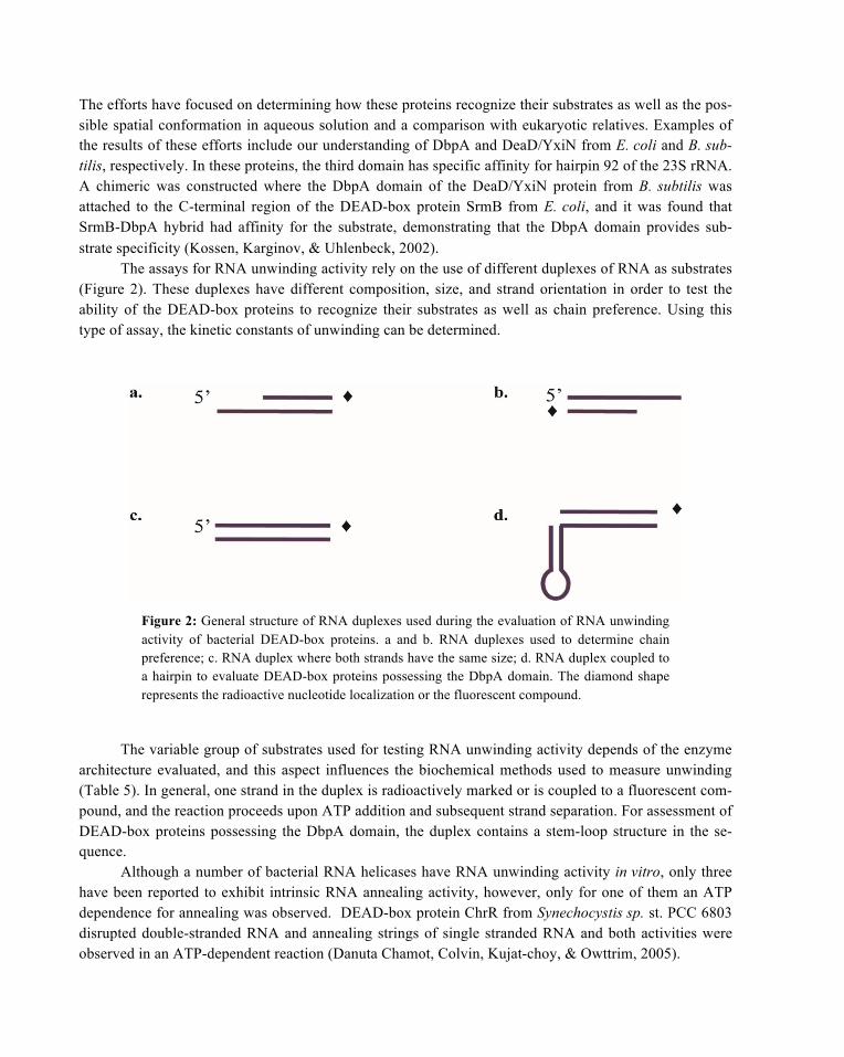
The efforts have focused on determining how these proteins recognize their substrates as well as the pos-sible spatial conformation in aqueous solution and a comparison with eukaryotic relatives. Examples of the results of these efforts include our understanding of DbpA and DeaD/YxiN from E. coli and B. sub-tilis, respectively. In these proteins, the third domain has specific affinity for hairpin 92 of the 23S rRNA. A chimeric was constructed where the DbpA domain of the DeaD/YxiN protein from B. subtilis was attached to the C-terminal region of the DEAD-box protein SrmB from E. coli, and it was found that SrmB-DbpA hybrid had affinity for the substrate, demonstrating that the DbpA domain provides sub-strate specificity (Kossen, Karginov, & Uhlenbeck, 2002).
The assays for RNA unwinding activity rely on the use of different duplexes of RNA as substrates (Figure 2). These duplexes have different composition, size, and strand orientation in order to test the ability of the DEAD-box proteins to recognize their substrates as well as chain preference. Using this type of assay, the kinetic constants of unwinding can be determined.
Figure 2: General structure of RNA duplexes used during the evaluation of RNA unwinding activity of bacterial DEAD-box proteins. a and b. RNA duplexes used to determine chain preference; c. RNA duplex where both strands have the same size; d. RNA duplex coupled to a hairpin to evaluate DEAD-box proteins possessing the DbpA domain. The diamond shape represents the radioactive nucleotide localization or the fluorescent compound.
The variable group of substrates used for testing RNA unwinding activity depends of the enzyme architecture evaluated, and this aspect influences the biochemical methods used to measure unwinding (Table 5). In general, one strand in the duplex is radioactively marked or is coupled to a fluorescent com-pound, and the reaction proceeds upon ATP addition and subsequent strand separation. For assessment of DEAD-box proteins possessing the DbpA domain, the duplex contains a stem-loop structure in the se-quence.
Although a number of bacterial RNA helicases have RNA unwinding activity in vitro, only three have been reported to exhibit intrinsic RNA annealing activity, however, only for one of them an ATP dependence for annealing was observed. DEAD-box protein ChrR from Synechocystis sp. st. PCC 6803 disrupted double-stranded RNA and annealing strings of single stranded RNA and both activities were observed in an ATP-dependent reaction (Danuta Chamot, Colvin, Kujat-choy, & Owttrim, 2005).

Enz
yme
Rea
ctio
n B
uffe
r E
nzym
e co
nc.
RN
A c
onc.
D
irec
tiona
lity
AT
P (m
M)
Tem
p.
(°C
) T
ime
(min
) R
efer
ence
(M
etho
d)
Dea
D/Y
xiN
B.
sub
tilis
50
mM
HEP
ES, p
H 7
.2, 1
50 m
M
KC
l, 5
% g
lyce
rol,
0.1
mM
DTT
, 0.
1 m
g/m
l BSA
, 5 m
M M
gCl 2
10 µ
M
5 µM
15
4-m
er o
f 2,
481
– 2,
634
of
23S
of rR
NA
1 25
30
Th
eiss
en e
t al.,
20
08).
(FR
ET a
nd
EMSA
) (A
regg
er &
K
lost
erm
eier
, 200
9).
(FR
ET a
nd E
MSA
) D
eaD
/Yxi
N
B. s
ubtil
is
50 m
M H
epes
⁄ NaO
H, p
H 7
.5, 1
50
mM
KC
l, 5
mM
MgC
l 2, 1
00 m
M
DTT
, 5%
(v/v
) gly
cero
l, 0.
1 m
g/m
L B
SA
10 µ
M
25 µ
M
50 µ
M
32⁄9
-mer
min
imal
su
bstra
te d
eriv
ed
from
the
154-
mer
23
S rR
NA
1 25
30
(K
arow
et a
l., 2
007)
. (E
MSA
)
Csh
A
B. s
ubtil
is
20 m
M T
ris-H
Cl p
H 7
.5, 1
00 m
M
NaC
l, 3
mM
MgC
l 2, 2
mM
DTT
, 10
0 m
g/m
L B
SA.
100
pmol
3
pmol
5’
- 3’
3’- 5
’ 1
35
60
(And
o an
d
Nak
amur
a, 2
006)
. (E
MSA
) Sr
mB
, Rhl
E
P. h
alop
lank
tis
TAC
125
C
. psy
chre
ryth
raea
34
H
10 m
M H
epes
-Na
(pH
7.5
), 60
m
M N
aCl,
55 m
M K
Cl,
5% (v
/v)
glyc
erol
0.6
–
0.8
mM
20
nM
5’
- 3’
A
TP/M
g 2
1-37
10
(C
artie
r et a
l. 20
10).
(FR
ET)
Csd
A, S
rmB
, Rhl
E
E. c
oli
100
mM
KC
l, 20
mM
Hep
es p
H
7.5,
10%
(v/v
) gly
cero
l.
0.05
–
0.5
mM
10
nM
5’
-3’
3’-5
’ 2
25
60
(Biz
ebar
d &
Fe
rleng
hi, 2
004)
. (E
MSA
) D
bpA
E.
col
i 20
mM
Tris
-HC
l, pH
7.5
, 20
mM
N
H4C
l, 2
mM
Mg 2
Cl,
1 m
M D
TT
0.2
–
2.5
mM
20
pm
ol
5’-3
’ 2
37
20
(Hen
n et
al.,
200
1).
(AFM
) D
bpA
E.
col
i N
ot m
entio
ned
~0.0
75 –
1.
5 m
M
Equi
mol
ar
ratio
Si
ngle
and
dou
ble
rRN
A fr
om 2
531
– 25
63 o
f 23S
Equi
mol
ar
ratio
--
- 3-
24 h
(K
amin
ker e
t al.,
20
11) (
END
OR
)
!T
able
5: R
NA
unw
indi
ng re
actio
n co
nditi
ons o
f bac
teria
l DEA
D-b
ox p
rote
ins.

The variable experimental conditions used for RNA unwinding experiments result in differences in biochemical constant values such as kcat, kd and KMapp even for the same DEAD-box protein evaluated. Sometimes the results are not comparable with their eukaryotic or viral DEAD-box relatives (Table 6). Generally, the helicase assays are performed with an enzyme excess over substrate to allow single turno-ver reaction observation.
Enzyme kcat (s-1)
ATP-driven KMapp
kd
(nM) Reference (Method)
DeaD/YxiN (wt) B. subtilis
1.48(±0.22)
0.012(±0.01)
156 (±74) nM
-----
----
----
(Theissen et al., 2008) (FRET and EMSA) (Aregger & Klostermeier, 2009) (FRET and EMSA)
CshA (wt) B. subtilis
--- 225 µM ---- Ando and Nakamura, 2006 (EMSA)
DbpA (wt) E. coli
----
5.3 (± 0.2) 0.16 ± 0.05/mina
11 nM
65 (± 14) µM
19.8 ± 4.5 nM
0.44
----
22
(Polach & Uhlenbeck, 2002). (EMSA) (Henn, Cao, Hackney, & De La Cruz, 2008). (Fluorescence spectroscopy) (Diges & Uhlenbeck, 2001). (EMSA)
SrmB-Ec* (wt) SrmB-Ph (wt) RlhE-Ec (wt) RlhE-Cp (wt) RhlE-Ph (wt)
0.03 ± 0.003b
0.06 ± 0.01b
0.02 ± 0.005 0.6 ± 0.03 0.7 ± 0.2
0.2 ± 0.04 0.3 ± 0.15
(for ATP (mM), 10° C)
(Cartier et al. 2010) (FRET)
Hera-Tt* (wt) ---- 360 µM (for ATP at 60° C)
---- (Morlang, Weglohner, & Franceschi, 1999) (TLC: Thin Layer Chromatography)
NS3 helicase (wt) (Dengue virus)
2.2 ± 0.1 31 ± 3 µM (for ATP) ---- (Gebhard, Kaufman, & Gamarnik, 2012) (EMSA)
Ded1(wt) Saccharomyces cerevisiae
280 ± 30 min-1 0.37±0.03 mM ---- (Banroques, Doere, Dreyfus, Linder, & Tanner, 2010) (EMSA)
*Ec: Escherichia coli, Cp: C. psychrerythraea 34H, Ph: P. haloplanktis TAC125, Tt: Thermus thermophilus a Vmax b kunw (min-1), 10 bp, 10° C
Table 6. Kinetic constants of DEAD-box proteins.
4.1 Protein-Protein Interaction of DEAD-box Proteins
The C-terminal region of bacterial DEAD-box proteins, interacts with other proteins to direct the proteins to a specific metabolic process. For example, DEAD-box proteins RhlB from E. coli and from V. augus-tum S14 interact with the C-terminal end of RNase E, specifically with amino acid residues 714-758, and

with PNPase (Erce, Low, & Wilkins, 2010; Worrall et al., 2008). On the other hand, RhlB can interact with itself in vitro in the presence of Ca2+ and ATP to form filamentous structures more than 10 µm long and 25 ± 1.8 nm wide. This capability most likely allows for displacement of the RNA degradosome in vivo throughout the cell (Taghbalout & Yang, 2010).
Recently, the description of an RNA degradosome-like complex in B. subtilis provided insight into the components of the RNA degradosome in Gram positive bacteria. It was found that DEAD-box pro-tein CshA is able to interact with RNases J1, J2, and Y, the glucolytic enzymes enolase and 6-phospho-fructo kinase, and the exonuclease PNPase in B. subtilis. In Staphylococcus aureus, CshA also interacts with RNase RnpA (Lehnik-Habrink et al., 2010; Roux, DeMuth, & Dunman, 2011). In addition, CshB from B. subtilis has been shown to interact with a cold shock protein when bacteria are grown at a low temperature (Hunger, Beckering, Wiegeshoff, Graumann, & Marahiel, 2006).
5 Protein Expression and Purification of DEAD-box Proteins from B. subtilis.
Despite the similarity in architecture and the fact that DEAD-box proteins in bacteria are composed of the same DEAD and HelicC domains, the protein expression results obtained for each can vary widely. For instance, we had different results for the expression of the four genes (yfmL, deaD/yxiN, cshA and cshB) encoding the DEAD-box proteins of B. subtilis using pCOLDI (Qing et al., 2004) under the same conditions. The overall expression yields of the purified proteins where 13 mg/L for Yfml, 0.55 mg/L for DeaD/YxiN, 0.62 mg/L for CshA and 2.79 mg/L for CshB. All recombinant proteins, except CshA where purified by IMAC and ionic chromatography, while CshA was purified from inclusion bodies and refold-ed. The purification protocol for YfmL consisted of transformation of the pCOLDI-YfmL vector into electrocompetent E. coli BL21-Star strain and overnight growth on LB-agar plates. A single colony was used to inoculate a 200 mL LB culture supplemented with 100 µg/ml of carbeniciline. The overnight culture was used to inoculate 1 L of LB medium. Bacteria were grown in 2 L flask with vigorous shaking until the cultures reached an OD600 of 0.6. The cultures were incubated at 4°C for 30 min, induced by adding IPTG to a final concentration of 1 mM and grown for 21 h at 16 °C. The cells were harvested by centrifugation and resuspended in 40 ml of lysis buffer (50 mM potassium phosphate pH 7.0, 200 mM NaCl, and 1 mM PMSF) and lysed by sonication. The lysate was centrifuged at 17,000 rpm in a JA-20 rotor (Beckman®) for 30 min at 4 °C. The soluble fraction was filtrated with a 0.45 µm sterile filter and purified by Immobilized Metal Affinity Chromatography (IMAC) using a 1ml pre-packed column Hi Trap FF 1 mL column (GE Healthcare). The first wash consisted of 25 mL of lysis buffer and the second wash of 25 mL of lysis buffer supplemented with 10 mM imidazol. The protein was eluted with 2 mL of 50 mM potassium phosphate pH 7.0, 200 mM NaCl, 500 mM imidazol. The eluate was dialyzed in 50 mM potassium phosphate pH 7.0, 1mM DTT, 200 mM NaCl and 2 mM EDTA. To further purify YfmL, the dialyzed protein was loaded into a Hi Trap SP HP 1 mL column (GE Healthcare) eluted with a NaCl gradient (10 to 1000 mM). YfmL eluted between 200 to 300 mM of NaCl. The fractions were dialyzed overnight in 50 mM potassium phosphate pH 7.0, 1 mM DTT, 200 mM NaCl, 2 mM EDTA, 50% glyc-erol and stored at -20°C. As shown in Figure 3 and 4, Coomassie-blue stained SDS-gels indicated differ-ences in yield purification levels of these DEAD-box proteins of B. subtilis, where protein YfmL, the smallest DEAD-box protein in this microorganism, had the highest yield purification level (13 mg/L). We are in the process of evaluating its ability to unwind RNA duplexes or to anneal single stranded RNA.

Figure 3: Purification steps of YfmL. Proteins were resolved on 10% SDS-PAGE after cell lysis. Lanes (1) without induction; (2) induction with 0.5 M IPTG; (3) soluble fraction; (4) pellet; (5) flow-through; (6) first wash with 200 mM NaCl; (7) second wash with 10 mM im-idazole; (8) protein eluted with 500 mM Imidazole; (9) Molecular weight marker 250 kDa Precision Plus Protein Standards (Bio-Rad®).
Figure 4: Protein yield level of the B. subtilis DEAD-box proteins. The expression vector used was pCOLDI (Qing et al., 2004) in the BL21 Start E. coli strain. Lanes: (1) Prestained Protein Ladder (Fermentas®); (2) YfmL (~42 kDa), (3) DeaD (~54 kDa); (4) CshB (~50 kDa); and (5) CshA (~57 kDa).
6 Crystallization of Bacterial DEAD-box Proteins
Not all DEAD-box proteins have been cloned and expressed as full length proteins. For example, only some regions of these genes are cloned and expressed when the goal is to crystallize the protein, as was the case of the HelicC and the DbpA domains of DeaD/YxiN from B. subtilis and the DEAD-box protein from Geobacillus stearothermophilus, where the DeaD domain was cloned, expressed, and crystallized separately (Carmel & Matthews, 2004; Caruthers, Hu, & McKay, 2006; Wang et al., 2006). Additionally, in order to determine the stability of the amino acid residues in RNA helicases from microorganisms that grow at a high temperature, the DEAD-box protein Hera from the thermophilic bacteria Thermus ther-

mophilus was crystallized (Rudolph, Heissmann, Wittmann, & Klostermeier, 2006). Table 7 lists the different strategies that have been used by different research groups for cloning bacterial DEAD box proteins for crystallization.
Species/Protein Portion crystallized Resolution (Å) Reference
B. subtilis (DeaD/YxiN) Helicase domain 1.95 Caruthers et al., 2006. B. subtilis (DeaD/YxiN) DbpA domain 1.7 Wang et al., 2006. S. aureus Mu 50 (RnpA) N-terminal (DeaD domain) 2.60 Lee et al., 2010.
G. stearothermophilus (DeaD) N-terminal (DeaD domain) 1.85 Carmel & Mattews, 2004. T. thermophilus (Hera) N-terminal (DeaD domain) 1.66 Rudolph et al., 2006.
Table 7: Regions crystallized from bacterial DEAD-box proteins.
Finally, there are numerous members the DEAD-box protein family in some bacterial species within the Shewanella, Colwellia, and Photobacterium genera (López-Ramírez, Alcaraz, Moreno-Hagelsieb, & Olmedo-Álvarez, 2011). However, the different metabolic processes in which these pro-teins are involved as well as the different mechanisms of action are largely unexplored. Therefore, the different biophysics methods currently available for testing their functional activities will promote a new research area for the scrutiny of this protein family at the molecular and biochemical levels in order to understand their substrate recognition, RNA unwinding ability, functional redundancy, protein-protein interaction and differential expression under variable growth conditions. In this chapter, we summarized molecular and biochemical information obtained from the study of some members of bacterial DEAD-box proteins. The differences found in the biochemical parameters of some members, such as KM and kcat, hints at the variability of functions that these proteins may have. However, a challenge also remains in the identification of DEAD-box protein interactors and future experiments should aim at evaluating bio-chemical parameters of the RNA helicases in a complex.
References
Adhikari, S., Manthena, P. V., Sajwan, K., Kota, K. K., & Roy, R. (2010). A unified method for purification of basic pro-teins. Anal Biochem, 400(2), 203-206.
Ando, Y., & Nakamura, K. (2006). Bacillus subtilis DEAD protein YdbR possesses ATPase, RNA binding, and RNA unwinding activities. Biosci Biotechnol Biochem, 70(7), 1606-1615.
Awano, N., Rajagopal, V., Arbing, M., Patel, S., Hunt, J., Inouye, M., et al. (2010). Escherichia coli RNase R has dual activities, helicase and RNase. J Bacteriol, 192(5), 1344-1352.
Banroques, J., Doere, M., Dreyfus, M., Linder, P., & Tanner, N. K. (2010). Motif III in superfamily 2 "helicases" helps convert the binding energy of ATP into a high-affinity RNA binding site in the yeast DEAD-box protein Ded1. J Mol Biol, 396(4), 949-966.
Beckering, C. L., Steil, L., Weber, M. H., Volker, U., & Marahiel, M. A. (2002). Genomewide transcriptional analysis of the cold shock response in Bacillus subtilis. J Bacteriol, 184(22), 6395-6402.

Briolat, V., & Reysset, G. (2002). Identification of the Clostridium perfringens genes involved in the adaptive response to oxidative stress. J Bacteriol, 184(9), 2333-2343.
Carmel, A. B., & Matthews, B. W. (2004). Crystal structure of the BstDEAD N-terminal domain: a novel DEAD protein from Bacillus stearothermophilus. Rna, 10(1), 66-74.
Cartier, G., Lorieux, F., Allemand, F., Dreyfus, M., & Bizebard, T. (2010). Cold adaptation in DEAD-box proteins. Bio-chemistry, 49(12), 2636-2646.
Caruthers, J. M., Hu, Y., & McKay, D. B. (2006). Structure of the second domain of the Bacillus subtilis DEAD-box RNA helicase YxiN. Acta Crystallogr Sect F Struct Biol Cryst Commun, 62(Pt 12), 1191-1195.
Chamot, D., Colvin, K. R., Kujat-choy, S. L., & Owttrim, G. W. (2005). RNA Structural Rearrangement via Unwinding and Annealing by the Cyanobacterial RNA Helicase, CrhR*. Biochemistry, 280, 2036 -2044.
Chamot, D., & Owttrim, G. W. (2000). Regulation of cold shock-induced RNA helicase gene expression in the Cyanobac-terium Anabaena sp. strain PCC 7120. J Bacteriol, 182(5), 1251-1256.
Charollais, J., Dreyfus, M., & Iost, I. (2004). CsdA, a cold-shock RNA helicase from Escherichia coli, is involved in the biogenesis of 50S ribosomal subunit. Nucleic Acids Res, 32(9), 2751-2759.
Charollais, J., Pflieger, D., Vinh, J., Dreyfus, M., & Iost, I. (2003). The DEAD-box RNA helicase SrmB is involved in the assembly of 50S ribosomal subunits in Escherichia coli. Mol Microbiol, 48(5), 1253-1265.
Diges, C. M., & Uhlenbeck, O. C. (2001). Escherichia coli DbpA is an RNA helicase that requires hairpin 92 of 23S rRNA. Embo J, 20(19), 5503-5512.
Erce, M. A., Low, J. K., & Wilkins, M. R. (2010). Analysis of the RNA degradosome complex in Vibrio angustum S14. Febs J, 277(24), 5161-5173.
Fairman-Williams, M. E., Guenther, U. P., & Jankowsky, E. (2010). SF1 and SF2 helicases: family matters. Curr Opin Struct Biol, 20(3), 313-324.
Gebhard, L. G., Kaufman, S. B., & Gamarnik, A. V. (2012). Novel ATP-Independent RNA Annealing Activity of the Dengue Virus NS3 Helicase. PLoS ONE, 7(4), e36244.
Graslund, S., Nordlund, P., Weigelt, J., Hallberg, B. M., Bray, J., Gileadi, O., et al. (2008). Protein production and purifi-cation. Nat Methods, 5(2), 135-146.
Henn, A., Cao, W., Hackney, D. D., & De La Cruz, E. M. (2008). The ATPase cycle mechanism of the DEAD-box rRNA helicase, DbpA. J Mol Biol, 377(1), 193-205.
Hunger, K., Beckering, C. L., Wiegeshoff, F., Graumann, P. L., & Marahiel, M. A. (2006). Cold-induced putative DEAD box RNA helicases CshA and CshB are essential for cold adaptation and interact with cold shock protein B in Bacil-lus subtilis. J Bacteriol, 188(1), 240-248.
Iost, I., & Dreyfus, M. (2006). DEAD-box RNA helicases in Escherichia coli. Nucleic Acids Res, 34(15), 4189-4197.
Jain, C. (2008). The E. coli RhlE RNA helicase regulates the function of related RNA helicases during ribosome assembly. Rna, 14(2), 381-389.
Kaminker, I., Sushenko, A., Potapov, A., Daube, S., Akabayov, B., Sagi, I., et al. (2011). Probing conformational varia-tions at the ATPase site of the RNA helicase DbpA by high-field electron-nuclear double resonance spectroscopy. J Am Chem Soc, 133(39), 15514-15523.
Khemici, V., Toesca, I., Poljak, L., Vanzo, N. F., & Carpousis, A. J. (2004). The RNase E of Escherichia coli has at least two binding sites for DEAD-box RNA helicases: functional replacement of RhlB by RhlE. Mol Microbiol, 54(5), 1422-1430.

Kossen, K., Karginov, F. V., & Uhlenbeck, O. C. (2002). The carboxy-terminal domain of the DExDH protein YxiN is sufficient to confer specificity for 23S rRNA. J Mol Biol, 324(4), 625-636.
Kossen, K., & Uhlenbeck, O. C. (1999). Cloning and biochemical characterization of Bacillus subtilis YxiN, a DEAD protein specifically activated by 23S rRNA: delineation of a novel sub-family of bacterial DEAD proteins. Nucleic Acids Res, 27(19), 3811-3820.
Lehnik-Habrink, M., Pfortner, H., Rempeters, L., Pietack, N., Herzberg, C., & Stulke, J. (2010). The RNA degradosome in Bacillus subtilis: identification of CshA as the major RNA helicase in the multiprotein complex. Mol Microbiol, 77(4), 958-971.
López-Ramírez, V., Alcaraz, L. D., Moreno-Hagelsieb, G., & Olmedo-Álvarez, G. (2011). Phylogenetic distribution and evolutionary history of bacterial DEAD-Box proteins. J Mol Evol, 72(4), 413-431.
Markkula, A., Lindstrom, M., Johansson, P., Bjorkroth, J., & Korkeala, H. (2012). Role of four putative DEAD-box RNA helicase genes in growth of Listeria monocytogenes EGD-e under heat, pH, osmotic, ethanol, and oxidative stresses. Appl Environ Microbiol.
Morlang, S., Weglohner, W., & Franceschi, F. (1999). Hera from Thermus thermophilus: the first thermostable DEAD-box helicase with an RNase P protein motif. J Mol Biol, 294(3), 795-805.
Palonen, E., Lindstrom, M., Somervuo, P., Johansson, P., Bjorkroth, J., & Korkeala, H. (2012). Requirement for RNA helicase CsdA for growth of Yersinia pseudotuberculosis IP32953 at low temperatures. Appl Environ Microbiol, 78(4), 1298-1301.
Pandiani, F., Brillard, J., Bornard, I., Michaud, C., Chamot, S., Nguyen-the, C., et al. (2010). Differential involvement of the five RNA helicases in adaptation of Bacillus cereus ATCC 14579 to low growth temperatures. Appl Environ Mi-crobiol, 76(19), 6692-6697.
Polach, K. J., & Uhlenbeck, O. C. (2002). Cooperative binding of ATP and RNA substrates to the DEAD/H protein DbpA. Biochemistry, 41(11), 3693-3702.
Prud'homme-Genereux, A., Beran, R. K., Iost, I., Ramey, C. S., Mackie, G. A., & Simons, R. W. (2004). Physical and functional interactions among RNase E, polynucleotide phosphorylase and the cold-shock protein, CsdA: evidence for a 'cold shock degradosome'. Mol Microbiol, 54(5), 1409-1421.
Purusharth, R. I., Klein, F., Sulthana, S., Jager, S., Jagannadham, M. V., Evguenieva-Hackenberg, E., et al. (2005). Exori-bonuclease R interacts with endoribonuclease E and an RNA helicase in the psychrotrophic bacterium Pseudomonas syringae Lz4W. J Biol Chem, 280(15), 14572-14578.
Py, B., Higgins, C. F., Krisch, H. M., & Carpousis, A. J. (1996). A DEAD-box RNA helicase in the Escherichia coli RNA degradosome. Nature, 381(6578), 169-172.
Qing, G., Ma, L. C., Khorchid, A., Swapna, G. V., Mal, T. K., Takayama, M. M., et al. (2004). Cold-shock induced high-yield protein production in Escherichia coli. Nat Biotechnol, 22(7), 877-882.
Rajkowitsch, L., Chen, D., Stampfl, S., Semrad, K., Waldsich, C., Mayer, O., et al. (2007). RNA chaperones, RNA anneal-ers and RNA helicases. RNA Biol, 4(3), 118-130.
Roux, C. M., DeMuth, J. P., & Dunman, P. M. (2011). Characterization of components of the Staphylococcus aureus mRNA degradosome holoenzyme-like complex. J Bacteriol, 193(19), 5520-5526.
Rudolph, M. G., Heissmann, R., Wittmann, J. G., & Klostermeier, D. (2006). Crystal structure and nucleotide binding of the Thermus thermophilus RNA helicase Hera N-terminal domain. J Mol Biol, 361(4), 731-743.
Taghbalout, A., & Yang, Q. (2010). Self-assembly of the bacterial cytoskeleton-associated RNA helicase B protein into polymeric filamentous structures. J Bacteriol, 192(12), 3222-3226.

Trubetskoy, D., Proux, F., Allemand, F., Dreyfus, M., & Iost, I. (2009). SrmB, a DEAD-box helicase involved in Esche-richia coli ribosome assembly, is specifically targeted to 23S rRNA in vivo. Nucleic Acids Res, 37(19), 6540-6549.
Tsu, C. A., & Uhlenbeck, O. C. (1998). Kinetic analysis of the RNA-dependent adenosinetriphosphatase activity of DbpA, an Escherichia coli DEAD protein specific for 23S ribosomal RNA. Biochemistry, 37(48), 16989-16996.
Wang, S., Hu, Y., Overgaard, M. T., Karginov, F. V., Uhlenbeck, O. C., & McKay, D. B. (2006). The domain of the Bacil-lus subtilis DEAD-box helicase YxiN that is responsible for specific binding of 23S rRNA has an RNA recognition motif fold. Rna, 12(6), 959-967.
Worrall, J. A., Gorna, M., Crump, N. T., Phillips, L. G., Tuck, A. C., Price, A. J., et al. (2008). Reconstitution and analysis of the multienzyme Escherichia coli RNA degradosome. J Mol Biol, 382(4), 870-883.