pulmonary vascular sclerosis …heart.bmj.com/content/heartjnl/8/2/76.full.pdf · pulmonary...
TRANSCRIPT

PULMONARY VASCULAR SCLEROSIS WITH RIGHTVENTRICULAR FAILURE
BY
A. M. BARRETT AND LESLIE COLE
From Addenbrooke's Hospital and the Department of Pathology, University of Cambridge
Received December 19, 1945
The following is an account of a patient with pulmonary vascular sclerosis who died ofright ventricular failure within four months of the appearance of the first symptom of illhealth.A navvy charge hand, aged 34, was admitted to Addenbrooke's Hospital on November 6, 1942.
In August 1942 he noticed that some of his teeth were veiy loose and tended to bleed easily, and shortlyafter this that he was breathless on exertion. Until then he had always been fit with no cough orbreathlessness. In early October he went to the dentist, and twelve teeth were extracted under generalanesthesia. Following these extractions breathlessness on exertion gradually increased so that whenhe was admitted to hospital he could walk up six steps only with difficulty. A week before admissionhe had also noticed slight swelling of his left leg, occasional palpitation, and some upper abdominalpain and flatulence after food. His appetite remained good and his weight constant. His work hadnot been physically very strenuous but his hours had been long-from eight in the morning sometimesuntil midnight. As this was seven miles from home he used to cycle to and fro. He often worked aseven-day week.
In the past he had had no serious illnesses, no rheumatism, nor rheumatic fever, nor other conditionthat might predispose to cardiac trouble. He had been accustomed to smoke up to 50 cigarettes aday and drank a moderate amount of beer but no spirits. His family history was healthy and did notsuggest any tendency to arteriosclerosis or cardiac disease. His mother and father and brothers andsisters were all alive and well.
On admission he was very breathless .and slightly cyanosed. The pulse was regular-rate 100.Blood pressure 140/90, respiratory rate 20, temperature normal. A teleradiogram showed that theheait was enlarged (15*3 cm. in the transverse diameter) with marked bulging of the pulmonaryartery (Fig. 1). The cardiogram showed right axis deviation with inversion ofT II and T III (Fig. 2). Theheart sounds were poor in quality with gallop rhythm and the pulmonary second sound was accen-tuated. There were no murmurs, clubbing of the fingers, venous pulsation, nor cedema, and noevidence of arteriosclerosis in the radial, brachial, or retinal arteries. The respiratory movementswere good and there were no signs of chronic bronchitis or emphysema; a few crepitations wereaudible at the right base.
The liver appeared congested, the edge being palpable two inches below the costal margin. Therewas no abdominal tenderness and no evidence of ascites. The remaining teeth appeared healthy andthe gums had healed well following extraction. The urine contained a trace of albumin, but wasotherwise normal.
Blood count showed 5*6 million red corpuscles with hemoglobin 112 per cent and 12,800 leucocyteswith a normal differential count. Blood culture was sterile and the Wasserman reaction of the bloodwas negative. Blood urea 37 mg. per 100 c.c. Blood sedimentation rate on two occasions 1 and4 mm. per hour respectively.
Progress. In spite of treatment by rest and full digitilization, progress was steadily downhill fromthe day of admission until death on December 2, 1942. During this period there was practically nochange in either the pulse rate or the respiratory rate. Breathlessness and distress on the slightestexertion became progressively more marked and cyanosis steadily increased.
POST-MORTEM EXAMINATIONCongestion of the lungs, the lower lobes being tough and slightly nodular in consistency and the
cut surface of the anterior borders yellowish-grey in colour, with many small rusty-red patches. Con-siderable emphysema of anterior borders only, without noticeable distention of the lung or formationof bulli. One small fragment ofante-mortem thrombus in a small pulmonary artery in the right lowerlobe, the area supplied by this vessel being hyperaemic but not infarcted. Unusually conspicuousblood vessels everywhere on the cut surfaces of the lungs. Conspicuous atheroma ofmain pulmonaryartery and all its larger branches. Pulmonary veins normal. Only slight atheroma of aorta and itsbranches, almost none of coronary arteries. Slight excess (about 50 c.c.) of clear yellow fluid in
76
on 13 August 2018 by guest. P
rotected by copyright.http://heart.bm
j.com/
Br H
eart J: first published as 10.1136/hrt.8.2.76 on 1 April 1946. D
ownloaded from

FIG. 1I.-Radiogram showing large pulmonary artery.
I .1
I "I~~~~~~~~~~~~~~~~I~~~
FIG. 2.-Cardiogram showing right axis deviation.
.4
-:k . ... ..f
t4.1'.. -I,-
on 13 August 2018 by guest. P
rotected by copyright.http://heart.bm
j.com/
Br H
eart J: first published as 10.1136/hrt.8.2.76 on 1 April 1946. D
ownloaded from

A. M. BARRETT AND L. COLEpericardium. Great dilatation of right side of heart, the cavity of both auricle and ventricle beingabout double the normal size. Considerable hypertrophy of right ventricle, its wall being stiff like apiece of leather. No hypertrophy or dilatation of left auricle or ventricle. Thickness of rightventricle 0O8 cm. Thickness of left ventricle 1-4 cm. The degree of hypertrophy was considerablymore than is represented by these figures because ofthe great dilatation ofthe right ventricle. Brownishred, rather opaque myocardium; with frequent small greyish patches (0 1-0-2 cm. diameter) in thewall of left ventricle suggesting slight fibrosis; heart valves normal. Ante-mortem thrombus in rightauricular appendage and also adhering to the wall of the right auricle at the site of the foramen ovale,which was patent (02 cm. diameter) but valvular. No clot projecting through the foramen into the leftauricle nor any ante-mortem.thrombus anywhere in the left side of the heart. Considerably enlargedhyperemic and anthrocotic bronchial lymph glands, probably acutely inflamed, at hilum of left lungand at bifurcation of trachea. No abnormality of other lymph glands. Dilated stomach. Con-gestion and mucous catarrh of intestines. Chronic venous congestion of liver with oedematousconnective tissue around the normal gall-bladder.
Small effusions of clear yellow fluid in each pleural cavity and in peritoneum (about 115 c.c. ineach). Moderately enlarged firm " chronic heart failure " spleen. Congestion and cloudy swellingofkidneys. Two recent infarcts and an adenoma (0 4 cm. diameter) in left kidney. Bladder, prostate,and urethra normal, also testes and epididymis. Suprarenals, pancreas, thyroid, and pituitary normal;thymus for the most part replaced by fat. Normal brain. Middle ears clear. Pyorrhoea aroundbases of remaining teeth; teeth otherwise in fair condition; gums healed at sites of absent teeth.No clubbing of fingers; no oedema. Considerable cyanosis. Well-developed, well-nourished man.
Weight of Organs. Heart 455 g., left lung 610 g., right lung 710 g., liver 2080 g., spleen 280 g.,right kidney 130 g., left kidney 155 g.
MICROSCOPICAL EXAMINATIONSThe Lungs. Six sections from different parts of the lungs show extensive disintegration of the
alveolar walls, although only the anterior borders of the lungs are frankly emphysematous. In someareas-usually where alveoli are intact-capillaries are greatly engorged: elsewhere the tatteredalveolar walls appear almost bloodless (Fig. 3). The contrast between the sharply defined patches ofcapillary engorgement and the surrounding zones in which it is difficult to distinguish any capillaries
s~~~~~~~~~~~
,45.f~~~~~l 450
t..~~~ ~ ~ ~ ~ ~ ~ 77'..,
16-~ ~ ~ ~ -
V# A14L.
J I% .dj4 ;
W.6
FIG. 3.-At the top, a small muscular artery with conspicuous intimal thickening; arteriole with greatlythickened wall in centre; area of capillary engorgement with preservation of alveolar pattern at bottomright; area of alveolar disintegration with few visible capillaries at bottom left.Goldner's modification of Masson's stain. Magnification x 88.
78
on 13 August 2018 by guest. P
rotected by copyright.http://heart.bm
j.com/
Br H
eart J: first published as 10.1136/hrt.8.2.76 on 1 April 1946. D
ownloaded from
PULMONARY VASCULAR SCLEROSIS
I..
*1~
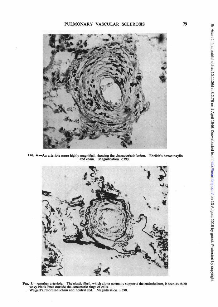
FIG. 4.-An arteriole more highly magnified, showing the characteristic lesion. Ehrlich's haematoxylinand eosin. Magnification x 390.
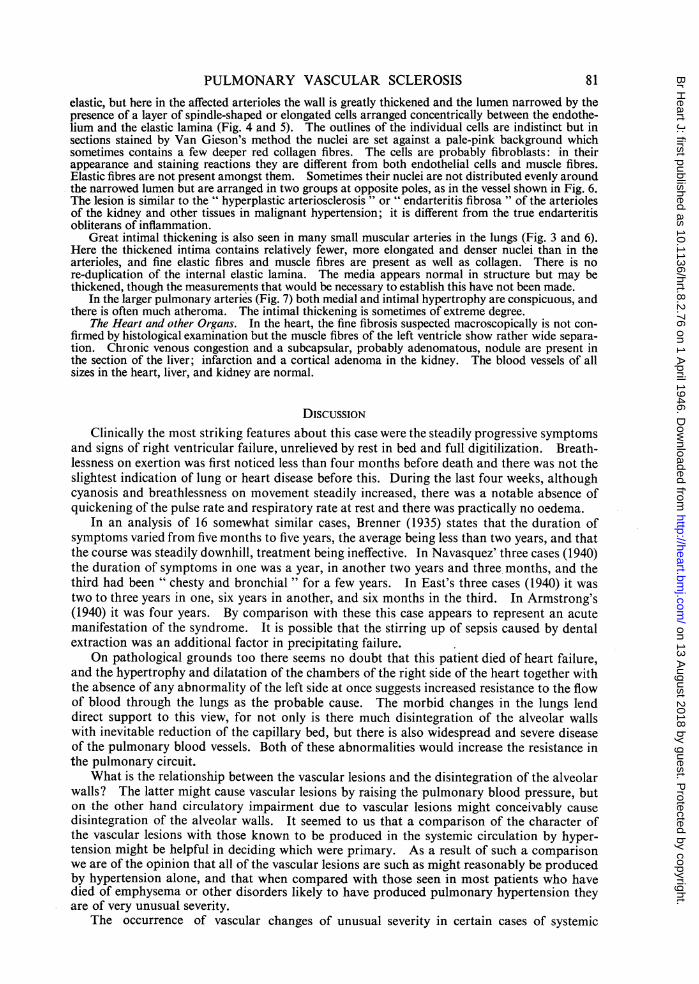
FIG. 5.-Another arteriole. The elastic fibril, which alone normally supports the endothelium, is seen as thickwavy black lines outside the concentric rings of cells.Weigert's resorcin-fuchsin and neutral red. Magnification x 390.
79
on 13 August 2018 by guest. P
rotected by copyright.http://heart.bm
j.com/
Br H
eart J: first published as 10.1136/hrt.8.2.76 on 1 April 1946. D
ownloaded from

A. M. BARRETT AND L. COLE
FIG. 6.-A small muscular artery. The internal and external elastic laminxe which bound the media are bestseen on the left side. Weigert's resorcin-fuchsin and neutral red. Magnification x 88.
A RI,
-~~~~~~~4A
( , < ., ' RJ/~~~~~~M
;~~~~~~~~~~~.~~ ~~4
FIG. 7.-A pulmonary artery. Weigert's resorcin-fuchsin and neutral red. Magnification x 88.
at all, is very striking. Small hemorrhages are present here and there, and many macrophages containhaemosiderin.
Conspicuous abnormalities are present in pulmonary blood vessels of all sizes but are perhapsmost notable in the arterioles and smaller muscular arteries. According to Brenner (1935) the wallof the pulmonary arterioles normally consists of endothelium supported only by a spiral- strand of
80
on 13 August 2018 by guest. P
rotected by copyright.http://heart.bm
j.com/
Br H
eart J: first published as 10.1136/hrt.8.2.76 on 1 April 1946. D
ownloaded from
PULMONARY VASCULAR SCLEROSIS
elastic, but here in the affected arterioles the wall is greatly thickened and the lumen narrowed by thepresence of a layer of spindle-shaped or elongated cells arranged concentrically between the endothe-lium and the elastic lamina (Fig. 4 and 5). The outlines of the individual cells are indistinct but insections stained by Van Gieson's method the nuclei are set against a pale-pink background whichsometimes contains a few deeper red collagen fibres. The cells are probably fibroblasts: in theirappearance and staining reactions they are different from both endothelial cells and muscle fibres.Elastic fibres are not present amongst them. Sometimes their nuclei are not distributed evenly aroundthe narrowed lumen but are arranged in two groups at opposite poles, as in the vessel shown in Fig. 6.The lesion is similar to the " hyperplastic arteriosclerosis " or " endarteritis fibrosa " of the arteriolesof the kidney and other tissues in malignant hypertension; it is different from the true endarteritisobliterans of inflammation.
Great intimal thickening is also seen in many small muscular arteries in the lungs (Fig. 3 and 6).Here the thickened intima contains relatively fewer, more elongated and denser nuclei than in thearterioles, and fine elastic fibres and muscle fibres are present as well as collagen. There is nore-duplication of the internal elastic lamina. The media appears normal in structure but may bethickened, though the measurements that would be necessary to establish this have not been made.
In the larger pulmonary arteries (Fig. 7) both medial and intimal hypertrophy are conspicuous, andthere is often much atheroma. The intimal thickening is sometimes of extreme degree.
The Heart and other Organs. In the heart, the fine fibrosis suspected macroscopically is not con-firmed by histological examination but the muscle fibres of the left ventricle show rather wide separa-tion. Chronic venous congestion and a subcapsular, probably adenomatous, nodule are present inthe section of the liver; infarction and a cortical adenoma in the kidney. The blood vessels of allsizes in the heart, liver, and kidney are normal.
DISCUSSIONClinically the most striking features about this case were the steadily progressive symptoms
and signs of right ventricular failure, unrelieved by rest in bed and full digitilization. Breath-lessness on exertion was first noticed less than four months before death and there was not theslightest indication of lung or heart disease before this. During the last four weeks, althoughcyanosis and breathlessness on movement steadily increased, there was a notable absence ofquickening of the pulse rate and respiratory rate at rest and there was practically no oedema.
In an analysis of 16 somewhat similar cases, Brenner (1935) states that the duration ofsymptoms varied from five months to five years, the average being less than two years, and thatthe course was steadily downhill, treatment being ineffective. In Navasquez' three cases (1940)the duration of symptoms in one was a year, in another two years and three months, and thethird had been " chesty and bronchial " for a few years. In East's three cases (1940) it wastwo to three years in one, six years in another, and six months in the third. In Armstrong's(1940) it was four years. By comparison with these this case appears to represent an acutemanifestation of the syndrome. It is possible that the stirring up of sepsis caused by dentalextraction was an additional factor in precipitating failure.
On pathological grounds too there seems no doubt that this patient died of heart failure,and the hypertrophy and dilatation of the chambers of the right side of the heart together withthe absence of any abnormality of the left side at once suggests increased resistance to the flowof blood through the lungs as the probable cause. The morbid changes in the lungs lenddirect support to this view, for not only is there much disintegration of the alveolar wallswith inevitable reduction of the capillary bed, but there is also widespread and severe diseaseof the pulmonary blood vessels. Both of these abnormalities would increase the resistance inthe pulmonary circuit.
What is the relationship between the vascular lesions and the disintegration of the alveolarwalls? The latter might cause vascular lesions by raising the pulmonary blood pressure, buton the other hand circulatory impairment due to vascular lesions might conceivably causedisintegration of the alveolar walls. It seemed to us that a comparison of the character ofthe vascular lesions with those known to be produced in the systemic circulation by hyper-tension might be helpful in deciding which were primary. As a result of such a comparisonwe are of the opinion that all of the vascular lesions are such as might reasonably be producedby hypertension alone, and that when compared with those seen in most patients who havedied of emphysema or other disorders likely to have produced pulmonary hypertension theyare of very unusual severity.
The occurrence of vascular changes of unusual severity in certain cases of systemic
81
on 13 August 2018 by guest. P
rotected by copyright.http://heart.bm
j.com/
Br H
eart J: first published as 10.1136/hrt.8.2.76 on 1 April 1946. D
ownloaded from

A. M. BARRETT AND L. COLEhypertension is now generally recognized, such patients being said to be suffering frommalignant hypertension. In a study of the structural changes in the lungs in mitral stenosis,Barker and Weiss (1936) have presented evidence of an analogous condition in the pulmonarycircuit-" pulmonary hypertension with malignant sclerosis "-in which the arterioles in thelungs show hyperplastic arteriolosclerosis and, rarely, necrotising arteriolitis quite similar to thelesions seen in the systemic circulation in malignant hypertension. Though we have not foundacute necrotic lesions in the blood vessels of the lungs of our patient, -the resemblance of thearteriolar lesions to the hyperplastic arteriosclerosis has already been noted. It seemspossible that both on clinical and pathological grounds this is a case of malignant pulmonaryhypertension; that a rise of pulmonary blood pressure initiated the malignant sclerosis of thepulmonary arterioles which then exaggerated the pulmonary hypertension and set up a viciouscircle. A vicious circle of this kind is probably the essential feature of malignant as distinctfrom benign hypertension and it explains the rapid worsening and early death of the patient.
If the vascular lesions were the result of pulmonary hypertension it is necessary to enquirehow such hypertension arose. There is no satisfactory answer to this question. Undoubtedlythe capillary bed was considerably reduced by disintegration of the alveolar walls, but it isimpossible to do more than speculate as to whether this was sufficient to account for thehypertension and the origin of the alveolar disintegration remains a mystery. The mostobvious explanation, that the patient was suffering from chronic emphysema, appears to usuntenable. There was no clinical evidence of emphysema; no long history of cough or otherchest trouble and none of the physical signs of emphysema, while post-mortem the existenceof emphysema was not at first noticed, so slight was it. It cannot be denied that emphysemawas present in the anterior borders of the lungs but this emphysema was atrophic in type, beingmerely part of the general disintegration of the lung, and there was no distension of the lungnor any formation of bullae. Thus the condition of the lungs was quite different-from thatseen in typical chronic emphysema. Furthermore Parkinson and Hoyle (1937) have shownthat any gross degree of right ventricular enlargement is uncommon even in severe cases ofchronic hypertrophic emphysema. In their series of 80 cases enlargement could only bedemonstrated radiologically in 18 and in only 4 of these was it great. The case we aredescribing showed no clinical emphysema.
SUMMARY
A patient is described who died of right ventricular failure within four months of the firstappearance of symptoms. There was no previous history of heart, lung, or other disease.Progress was steadily downhill without any response to treatment.
Post-mortem, there was macroscopically conspicuous atheroma of the main pulmonaryarteries and all its larger branches with great dilatation of the right side of the heart and hyper-trophy of the right ventricle, the left side of the heart being normal. Emphysema was onlyvisible along the anterior borders of the lungs and there was no distension of the lungs orformation of bullae.
Microscopically, there was conspicuous medial and intimal hypertrophy in the largerpulmonary arteries with much atheroma, and- great intimal thickening in many of the smallmuscular arteries.' Changes similar to hyperplastic arteriosclerosis of malignant hypertensionwere seen in the pulmonary arterioles. There was widespread disintegration of the alveolarwalls without hypertrophic emphysema.
The significance of these changes is discussed.
We wish to thank Dr. Ff. Roberts for the X-ray examination, and Mr. H. P. Hudson for thephotonuicrographs.
REFERENCES
Armstrong, T. G. (1940). Brit. Heart J., 2, 201.Brenner, 0. (1935). Arch. intern. Med., 56, 211, 457, 724, 976, 1189.East, T. (1940). Brit. Heart J., 2, 189.Navasquez, S. de, Forbes, J. R., and Holing, H. E. (1940). Brit. Heart J., 2, 177.Parker, F. and Weiss, S. (1936). Amer. J. Path., 12, 573.Parkinson, J. and Hoyle, C. (1937). Quart. J. Med. New Series, 6, 59.
82
on 13 August 2018 by guest. P
rotected by copyright.http://heart.bm
j.com/
Br H
eart J: first published as 10.1136/hrt.8.2.76 on 1 April 1946. D
ownloaded from