pulmonary hydatid disease - thorax · pulmonary hydatid disease whom chest radiographs were made...
TRANSCRIPT
Thorax (1972), 27, 365.
Pulmonary hydatid diseaseB. T. le ROUX
The Thoracic Unit, Wentworth Hospital, and the Department of Surgery, University of Natal
Radiographic evidence is presented in confirmation of the contention that uncomplicated isolatedpulmonary hydatid cysts are seldom completely circular and can be confidently recognized ashydatid cysts radiographically and fluoroscopically.
Pulmonary hydatid disease is a particular radio-logical diagnostic problem when the hydatid issmall, single, and symptomless, so that distinctionmust be made from the numerous other causes ofisolated, peripheral pulmonary lesions. The tech-nique of delivery of a pulmonary hydatid cyst,different from the management of other singleperipheral lesions, makes preoperative recognitionof hydatid disease important. Uncomplicated pul-monary hydatid cysts are seldom completelycircular because they are so soft that they aredented and disfigured by adjacent structures-onthis peculiarity of shape there is unanimity ofopinion; that their outline is blurred in one ormore places by areas of pneumonitis, haemorrhageor infarction (Barrett and Thomas, 1944), whosecomprehensive paper carries the salutary and
appropriate admonition cave canem as sub-title,is questionable-sharp definition, without adjacentpulmonary reaction, is typical of the solitary,small uncomplicated hydatid cyst (Shanks andKerley, 1962). Close proximity to a pleural surfaceis common (Brown, 1958) but by no meansinvariable.Hydatid disease is endemic in the Natal mid-
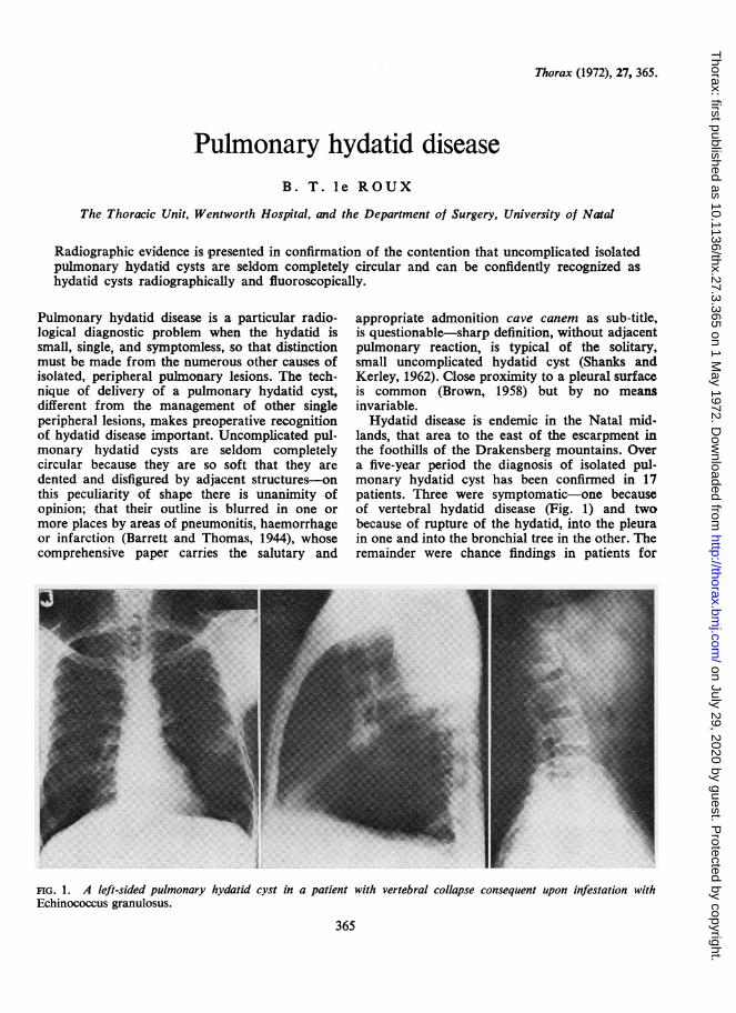
lands, that area to the east of the escarpment inthe foothills of the Drakensberg mountains. Overa five-year period the diagnosis of isolated pul-monary hydatid cyst has been confirmed in 17patients. Three were symptomatic-one becauseof vertebral hydatid disease (Fig. 1) and twobecause of rupture of the hydatid, into the pleurain one and into the bronchial tree in the other. Theremainder were chance findings in patients for
FIG. 1. A left-sided pulmonary hydatid cyst in a patient with vertebral collapse consequent upon infestation withEchinococcus granulosus.
365
on July 29, 2020 by guest. Protected by copyright.
http://thorax.bmj.com
/T
horax: first published as 10.1136/thx.27.3.365 on 1 May 1972. D
ownloaded from
B. T. le Roux
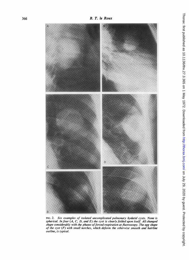
FIG. 2. Six examples of isolated uncomplicated pulmonary hydatid cysts. None isspherical. In four (A, C, D, and E) the cyst is clearly folded upon itself. All changedshape considerably with the phases offorced respiration at fluoroscopy. The egg shapeof the cyst (F) with small notches, which deform the otherwise smooth and hairlineoutline, is typical.
366
on July 29, 2020 by guest. Protected by copyright.
http://thorax.bmj.com
/T
horax: first published as 10.1136/thx.27.3.365 on 1 May 1972. D
ownloaded from
Pulmonary hydatid disease
whom chest radiographs were made for routinepurposes, and in all, the diagnosis of pulmonaryhydatid disease was confidently made on the basisof the dented, often folded, shape of the lesions.None was spherical. Most were the size of a hen'segg. Radiographs of 6 of 14 surgically managedpatients are shown (Fig. 2). Confirmatory evidenceof the softness of the intrapulmonary lesion wasobtained at fluoroscopy, when the cysts were seento change shape over a wide range during fullrespiratory excursion.Management in 14 patients was by incision of
the adventitious capsule and delivery of the cystby inflation of the lungs. Two cysts rupturedduring delivery, with contamination of the pleuralspace. The patients whose pleural spaces were socontaminated have been observed over four years,
and neither has evidence of a recurrence ofhydatid disease. All 14 patients are well.During the same period isolated uncomplicated
pulmonary hydatid disease was diagnosed on radio-graphic and fluoroscopic evidence in eight othersymptomless patients who declined surgical man-agement. These patients continue to attend forregular surveillance and it is hoped, from them,to obtain radiographic evidence of the naturalhistory of untreated pulmonary hydatid cysts.
REFERENCESBarrett, N. R., and Thomas, D. (1944). Pulmonary hydatid
disease. Brit. J. Tuberc., 38, 39.Brown, C. J. Officer (1958). Surgical pathology of hydatid
cysts of the lung. Postgrad. med. J., 34, 195.Shanks, S. C., and Kerley, P. (1962). A Textbook of X-ray
Diagnosis, 3rd ed., Vol. 2. Lewis, London.
2C
367
on July 29, 2020 by guest. Protected by copyright.
http://thorax.bmj.com
/T
horax: first published as 10.1136/thx.27.3.365 on 1 May 1972. D
ownloaded from