pseudomyxoma peritonei: diagnosis and management
TRANSCRIPT

21Pseudomyxoma Peritonei: Diagnosis and ManagementJKAU: Med. Sci., Vol. 13 No. 1, pp: 21-32 (2006 A.D. / 1427 A.H.)
21
Pseudomyxoma Peritonei: Diagnosis and Management
Khalid H. Sait*, MBCHB FRCS(C)
Department of Obstetrics and Gynecology, Faculty of MedicineKing Abdulaziz University, Jeddah, Saudi Arabia
Abstract. Pseudomyxoma peritonei is a rare disease characterized bya large amount of mucinous ascites with peritoneal and omentalimplants. The etiology of the disease remains unclear. Differenthistological categories have been described: the benign, malignant,and the intermediate forms. It is commonly diagnosed incidentally atlaparotomy. Most investigators agree that radical surgical debulkingand appendectomy are the cornerstone of treatment, but the optimalmanagement of the disease remains controversial. The role ofintraoperative and intraperitoneal chemotherapy has been evaluatedby a number of authors. The clinical outcomes vary widely betweenthe different histological types and treatment modalities.
Keywords: Pseudonyxoma peritonei, Intraperitoneal chemotherapy.
Introduction
Pseudomyxoma peritonei (PMP) is a poorly understood disease. It ischaracterized by the presence of massive amounts of mucinous ascites withperitoneal and omental implants, that over time fill the peritoneal cavity. It wasfirst described by Werth in 1884 as massive amounts of mucinous ascites foundin association with benign ovarian neoplasm[1].
There is considerable debate regarding the appropriate classification and typeof tumor to be included in this syndrome. The main controversy relates to theinclusion or exclusion of the more �malignant� forms of the disease and to the
*To whom all correspondence & reprint requests: P.O. Box 80215, Jeddah 21589 Saudi Arabia.Accepted for publication: 03 November 2004. Received: 15 March 2004.

K.H. Sait22
site of origin of the tumor[2] with advances in immunocytochemistry and geneticanalysis, progress has been made in more clearly categorizing this entity.
Incidence
PMP is a rare disease that is more common in women than men.Seventy-five percent of patients with PMP are female. The median age is53-years-old (range 33- to 79-years-old). The estimated incidence is 2 of every10,000 laparotomies performed[3].
Pathogenesis
The pathogenesis of PMP is obscure. Werth postulated that the peritonealsurfaces undergo a peculiar reaction in response to the jelly-like materialreleased from a cyst when it ruptures[1]. Osborn hypothesized that histologicalbenign mucinous epithelium released by the rupture of benign cysts implants onthe peritoneal surfaces and proliferates, causing PMP[4]. They postulated theexistence of a tumorigenic stimulus causing multifocal neoplasms or metaplasiaof the peritoneal surfaces. The historic experiments by Shanks seemed tosupport the idea that PMP is caused by some type of irritating agent ortumorigenic stimulus[1]. In addition the presence of mucinous pathology in boththe ovary and appendix, a common finding at the time of initial laparotomy forPMP, is consistent with widespread mucinous change in the peritoneal cavity.
Histology
Histological diagnosis describes two main diagnostic categories: the benignform-disseminated peritoneal adenomucinosis (DPAM), and a malignant form-peritoneal mucinous carcinomatosis (PMCA)[2]. A third category has also beendescribed; an intermediate form which is called peritoneal mucinouscarcinomatosis with intermediate or discordant features (PMCA-I/D). It hascytological features of both DPAM and PMCA. This classification was firstproposed by Ronnett et al. and is important as it has prognostic significance[2,5,6].As expected patients with the peritoneal adenomucinosis form have asignificantly more favorable prognosis than patients with peritoneal mucinouscarcinomatosis. Histological features of DPAM include peritoneal lesionscomposed of abundant extracellular mucin containing scant simple to focallyproliferate mucinous epithelium with little cytologic atypia or mitotic activity; itmay or may not be associated with an appendiceal mucinous adenoma[2]. Casesclassified as PMCA are characterized by peritoneal lesions composed of moreabundant mucinous epithelium with the architectural and cytologic features ofcarcinoma[2]. In some cases an associated mucinous adenocarcinoma may be
23Pseudomyxoma Peritonei: Diagnosis and Management
found. In a clinicopathologic analysis of 109 cases, about 60 percent wereclassified as DPAM-consistent with an origin from an appendiceal mucinousadenoma. About 28 percent were classified as PMCA-consistent with originfrom an appendiceal or intestinal mucinous adenocarcinoma. Approximately 13percent of the cases with intermediate features were derived fromwell-differentiated appendiceal or intestinal mucinous adenocarcinomas[2].
Origin
There is continued debate regarding the site of origin of PMP. It is mostcommonly associated with mucinous tumors of the appendix or the ovary. Womenwith PMP may have both, appendiceal and ovarian mucinous tumors. The ovariantumors in most of these cases are secondary to appendiceal tumors[7-9], but it maybe difficult to determine the histogenetic origin of these tumors. PMP has beenreported rarely in association with mucinous carcinomas of other organs,including the bile ducts, stomach, pancreas colon, fallopian tube, uterine corpus,urachus, urinary bladder, breast, and lung[3]. Despite the increasing researchregarding the origin of PMP using flow cytometry, genetic analysis, andimmunopathologic analysis, conflicting results are still reported. Over all itseems the vast majority of PMP cases have been shown to result from primarymucinous adenomas of the appendix[7, 8].
Clinical Presentation
PMP is commonly diagnosed incidentally at laparotomy, which is usuallyperformed for other indications such as suspected appendicitis or ovariancarcinoma. There is broad spectrum of clinical presentations which reflects thediffuse nature of the disease within the peritoneal cavity as well as the widerange of tumor histopathological features. Patients with PMP usually havemarked abdominal distension - out of proportion to the sign and symptoms ofabdominal malignancies. Abdominal or palpable ovarian masses and hernias areother clinical manifestations are commonly seen. Nausea, vomiting, fatigue, andurinary tract symptoms have also been reported[10]. The range of the presentingsymptoms is wide and dependant on the anatomic site of the disease.Abdominal pain, weight loss, and gastrointestinal malfunction occur relativelylate in the course of this disease. The bowel eventually becomes so encased inmucin and mucinous globules that it can not function, and ultimately patientsdie of bowel obstruction. Rare presenting problems include cholecystitis,infertility investigation, postmenopausal bleeding, abnormal Papanicolaousmear, pelvic pain, pelvic masses, surgery for fibrosis, laparoscopy for tuballigation, abdominal aortic aneurysm repair, deep vein thrombosis, rectalbleeding, nephrectomy, and anemia[11]. A case of intestinal obstruction has also
K.H. Sait24
been reported[12]. A rare presentation of an inguinal hernia with splenicmetastasis was reported[13]. Extra-peritoneal spread of PMP is infrequent.There are few reports in the medical literature of pleural extension of mucinoustumor in patients with PMP syndrome; these are associated with a poorprognosis[14]. Pulmonary parenchymal metastasis, treated with thoracotomy andexcision of metastatic nodules[15].
Diagnosis
The diagnosis of PMP is often difficult; it usually has an insidiouspresentation. Ultrasonography is frequently performed as the initial diagnosticprocedure. Typical findings are non-mobile, echogenic ascites with multiplesemisolid masses and scalloping of the hepatic and splenic margins[16].Furthermore, an ultrasound may be helpful in diagnostic and therapeuticparacentesis, as mucin is difficult to aspirate.
Computer tomography (CT) of the abdomen and pelvis is the most widelyapplied technology and has been used with great success in the diagnosis of thePMP syndrome. CT findings are often highly suggestive of PMP and sometimesthese are pathognomonic[17]. The most common finding is a large volume ofmucinous ascites, which has the density of fat, and displaces the small boweland the normal mesenteric fat. Other characteristic findings are omentalthickenings, multiseptate lesions, scalloping of organs, and curvilinearcalcifications[16-18].
The role of magnetic resonance imaging (MRI) in the diagnosis of PMP is notclear. There is no data to suggest that MRI is better than CT. It has been reportedthat MRI may be particularly sensitive in detecting visceral invasion, but this isbased on a single study of three patients[18]. At this stage, this mode remainsinvestigational. Sugarbaker et al. have used CT as a tool for differentiatingbetween peritoneal adenomatosis and peritoneal carcinomatosis[19]. Plainradiography and contrast studies are not particularly useful in the diagnosis ofPMP. Sometimes large mucinous abdominal masses may present withdisplacement of the bowel centrally or with obliteration of the border of thepsoas muscle[20]; rarely, punctuate or curvilinear calcifications may be noted.Double contrast techniques may demonstrate the lack of bowel lumeninvolvement that is characteristic of the non-invasive deposits of PMP[3]. Thegross appearance is characteristic. The abdomen is filling with several liters ofthick gelatinous mucoid material and the bowel is encased with mucin. Mucinglobules are found within the omentum, studding the peritoneal surfaces, andfloating throughout the cavity. Perhaps, the most consistent observation duringsurgical exploration of the patient with PMP is the complete or nearly complete

25Pseudomyxoma Peritonei: Diagnosis and Management
absence of mucinous tumor on intestinal surfaces with the exception in theantrum of the stomach and pylorus, the ileocecal valve region, and therectosigmoid colon within pelvis. All three of these sites are fixed to theretroperitoinum which are not actively and freely moving as a result ofperistaltic activity. Apparently, the continuous peristaltic activity of the smallbowel prevents mucinous tumor implantation on these surfaces. To a lesserextent, peristalsis inhibits the adenomatous epithelial cells from settling onto theperitoneal surfaces of the stomach and colon. Loculate mucinous cysts arepresent in the ovaries and/or appendix. Histological diagnosis is required andremains the "golden standard". This can be achieved by paracentesis,laparoscopy, or laparotomy.
Treatment
There is currently no accepted standard treatment for PMP. The treatment ofPMP traditionally involved repeated operative procedures over several yearsuntil there were no further surgical options. Management options variedconsiderably and often reflected the extent of disease, histological subtype, andgeneral conditions of the patient and surgical preference of the treating doctor -options included: observation, surgery, laparoscopic intervention, chemotherapy,radiotherapy, immunotherapy, and mucolytic agents. All of these were utilized inthe management of the disease.
Surgery
Surgical debulking and appendectomy are widely regarded as the mainstay oftreatment of PMP syndrome. In treatment of the PMP, limited debulking andevacuation of mucinous ascites provide for short-term palliation. This is incontrast to a high likelihood of prolonged remission or cure with completecytoreduction and perioperative intraperitoneal chemotherapy[21].
Sugarbaker Technique
The combination of intra-abdominal 5-Fluorouracil (5-FU) and cytoreductionsurgery was reported by Sugarbaker[21] from the Washington Cancer Institute,Washington Hospital Center, in Washington, D.C. for patients with extensivemucinous tumor of gastrointestinal origin. Because of the limited penetration oftissue with intraperitoneal chemotherapy, result is largely dependent on thevolume of residual tumor; a series of peritoneotomy procedures weredeveloped. These involved stripping the partial peritoneum and ressectingstructure at fixed sites that contain visceral peritoneum to accomplish a
K.H. Sait26
complete cytoreduction. Essential to the performance of peritoneotomyprocedures is dissection and tumor evaporation by laser mode or electrosurgeryusing a ball � Tip electrosurgical hand piece in a cutting mode (Table 1). Withthis extensive cytoreductive approach combined with early intraperitonealchemotherapy, the mortality rate was 2.7%Æ The main morbidity includedpancreatitis (7.1%), fistula formation (4.7 %), and anastomotic leaks weresimilar to general surgical techniques at 2.4%. Although, these data areimpressive, there was no control group. It is unlikely that randomized trials inthis setting will be performed.
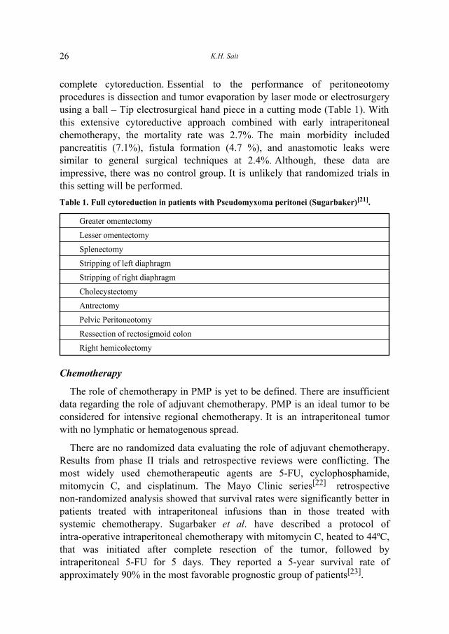
Table 1. Full cytoreduction in patients with Pseudomyxoma peritonei (Sugarbaker)[21].
Greater omentectomy
Lesser omentectomy
Splenectomy
Stripping of left diaphragm
Stripping of right diaphragm
Cholecystectomy
Antrectomy
Pelvic Peritoneotomy
Ressection of rectosigmoid colon
Right hemicolectomy
Chemotherapy
The role of chemotherapy in PMP is yet to be defined. There are insufficientdata regarding the role of adjuvant chemotherapy. PMP is an ideal tumor to beconsidered for intensive regional chemotherapy. It is an intraperitoneal tumorwith no lymphatic or hematogenous spread.
There are no randomized data evaluating the role of adjuvant chemotherapy.Results from phase II trials and retrospective reviews were conflicting. Themost widely used chemotherapeutic agents are 5-FU, cyclophosphamide,mitomycin C, and cisplatinum. The Mayo Clinic series[22] retrospectivenon-randomized analysis showed that survival rates were significantly better inpatients treated with intraperitoneal infusions than in those treated withsystemic chemotherapy. Sugarbaker et al. have described a protocol ofintra-operative intraperitoneal chemotherapy with mitomycin C, heated to 44ºC,that was initiated after complete resection of the tumor, followed byintraperitoneal 5-FU for 5 days. They reported a 5-year survival rate ofapproximately 90% in the most favorable prognostic group of patients[23].
27Pseudomyxoma Peritonei: Diagnosis and Management
Intraperitoneal Chemotherapy
The first attempt to use intraperitoneal chemotherapy in PMP patients wasreported in 1969 by Long et al.[24]. The extremely high and sustained levels ofcytotoxic chemotherapeutic agents can be achieved in the abdomen the rationalefor intraperitoneal therapy is that:
1. Direct infusion of the drugs into the peritoneal cavity permits delivery ofhigh concentrations of drug to abdominal and pelvic surfaces where thetumor is located without producing toxic systemic levels.
2. The water-soluble mucus can not metabolize chemotherapeutic agent inthe peritoneal cavity.
3. PMP superficially invades intra abdominal organs and rarely metastasis,allowing maximum exposure to the drug for long period.
A number of intraperitoneum chemotherapy regimens have been used, but itis extremely difficult to evaluate the results of these, because of not only thesmall numbers of patient in varies series, but also the protracted length of thenatural history of this condition. A major problem in the past withintraperitoneal chemotherapy delivery involved a non-uniform drugdistribution, resulting from intestinal adhesion from tissue surfaced closed offby sutures, and from pooling of intraperitoneal fluid at dependent sites.Another problem with intra-abdominal drug, delivery involved the minimalpenetration of tissue by chemotherapy. Chemotherapy enters tumor nodules bysimple diffusion and penetrated only a few cell layers. The use of chemotherapyin the operating room after complete dissection of an adhesive process andbefore bowel anastomosis was completed and with the abdominal incisionwidely open has been described[25]. These innovative strategies for the use ofintraoperative chemotherapy were reported first by the Japanese and thenfurther developed in United States[23]. Chemotherapy treatment in the operatingroom has been well tolerated. If it is used, the addition of intraperitonealhyperthermia can be used. Heat alone has an anticancer effect and it alsosynergizes the cytotoxicity of chemotherapy; in addition, the penetration ofdrug into tissue is augmented by heat. During the hyperthermal perfusion,careful monitoring of body temperature and intra-abdominal temperature mustbe maintained. Temperature at the infusion is approximately 44ºC and theintra-abdominal temperature is approximately 41ºC. The core temperatureremains below 39.5ºC. Heated intraoperative intraperitoneum chemotherapy islimited to certain institutions where facilities are available using this techniqueto meet the standard for the patient care and staff safety.
In patients with PMP, immediate postoperative intraperitoneal chemotherapywith 5-FU is used. The standardized orders for early postoperativeintraperitoneal 5-FU are followed for postoperative days 1 through 5. In the 6
K.H. Sait28
hours after the intraperitoneal 5-FU installation, the patient's bed remains flat ortilted with the head slightly down. The patient's position is changed everyhalf-hour from the extreme right- to the extreme left-side to promote maximalgravity distribution of intraperitoneal chemotherapy. The use of chemotherapyafter complete removal of cancer maximizes the complete eradication of themalignancy because only minute foci of disease need treatment. Furtherevaluations of intraperitoneum chemotherapy with 5-FU were conducted atNational Cancer Institute in Bethesda, Maryland, USA.
Other Treatment Modalities
Green et al., first used a dextrose solution to �loosen� the peritonealmucinous deposits[26]. The authors found that intraoperative irrigation orpercutaneous lavage with dextrose and water will expedite the removal ofmucus and prevent re-accumulation, although, the exact mechanism for thelatter has not been determined. The use of 10% dextrose has been reported to beassociated with hyperglycemia.
Intraperitoneal radioisotopes and external radiotherapy have also been used,but experience with these agents is limited[27]. Fukuma et al. have describedanecdotal cases of successful treatment of PMP by immunotherapy with astreptococcal preparation, OK-432, as adjuvant treatment after surgery[28].
Prognosis
With such a wide spectrum of disease ranging from a benign to a malignantform and wide variations in treatment modalities, it is not surprising thatprognosis has been difficult to accurately define. This is further complicated bythe fact that the estimation of recurrence rates was based on different series withsmall numbers of patients and different inclusion criteria and differenttherapeutic modalities.
A number of prognostic features have been identified by Sugarbaker et al.[23].These are histopathologic types with the extent of cytoreduction, and previoussurgical interventions. They reviewed the prognostic factors for 120 patientswith PMP of colonic or appendix primary with low grade of tumor biologywhere an attempt made to clear the abdomen by tumor resection andperitoneotomy procedures.
The patients were treated with repeated abdominal lavage immediately aftersurgery was completed. Also, all patients were treated post operatively withintraperitoneal chemotherapy. 46 patients out of 120 patients failed treatment,even though no distance disease was present. Of these 46 patients, 37 (80%)had persisting disease on the peritoneal surfaces. In 10 patients who had

29Pseudomyxoma Peritonei: Diagnosis and Management
adequate aggressive cytoreduction procedure and in whom the disease recurredat a later time were considered to have medical treatment failure.
Patients with complete cytoreduction and adenomucinosis had a 5-yearsurvival rate of 86%, and patients with intermediate histological features had a5-year survival rate of 50%. Patients with incomplete cytoreduction had 5-yearsurvival rate of 20% and 0% at 10 years. These authors concluded thatimprovements in the cytoreductive approach await the development ofsurgically technologies to increase the total clearance of cancer from theabdominal cavity and chemotherapy treatment that is complete enough tosustain control of small-volume residual disease on all peritoneal surfaces[21].
Gough et al.[27] at the Mayo Clinic reported a large number of patients (65patients) with PMP in single institution over a 26-years-interval. Minimaldebulking surgeries were performed in a majority of the patients. 10% hadoptimal cytoreduction surgery. Thirteen percent (13%) of the patients weregiven systemic chemotherapy, in 27% intrapertonium chemotherapy was givenand 18% received intrapertonium radioisotope, 28% receive radiation treatment.No patients received any mucolytic agent. Median follow-up was 12-years (9-25.6-years). 76% of patients had recurrence, the majority of whom treated withreoperation followed by adjuvant therapy. The median survival was 5-9 years(range 0.3-25.6 years).
The estimated 1-, 2-, 5- and 10-years survival rates were 98%, 86%, 53%,and 32%, respectively. They found that systemic chemotherapy associated withshorter survival rate and they concluded that aggressive surgical debulkingfollowed by intraperitoneal radioisotope or chemotherapy should be consideredin patients with PMP[27]. Wertheim et al. reported 23 cases of PMP withmedian follow-up 2.5 years (range 3 months to 31-years-old) in which allpatients underwent surgical minimal cytoreduction. Eleven patients receivedpost operative therapy � 9 recurred. Twelve patients received no therapy � only3 recurred. They concluded that adjuvant therapy has not been shown to bebeneficial[29].
Recurrence
PMP is associated with high recurrence rate. According to the data from theMayo Clinic series based on their study of 56 cases of PMP, 76% of patientsdeveloped recurrence and 50% of recurrences occurred within 2.5 years[22].Most of the patients had carcinoma of the appendix (52%) or ovary (34%).According to their data, 5- and 10-year survival rates were 53% and 32%,respectively, and the median survival was 5.9 years. Another study by Witkampet al., report a 5-year survival of 27% for peritoneal carcinomatosis of

K.H. Sait30
colorectal origin and of 86% for cases of PMP after complete cytoreduction andintraperitoneal chemohyperthermia[30].
Conclusions
PMP is a rare and complex disease with a broad spectrum of presentation andoutcomes. Definitive data regarding the optimal approach to managing thisdisease are not available due to the paucity of randomized trials. Unfortunately,it is unlikely that these trials will be performed due to the rarity of this disease.This highlights the importance of developing groups dealing with these raretumors to allow formal data collection and to provide some guidance as tooptimal therapeutic options in managing these patients.
References
[1] Shanks HGI. Pseudomyxoma peritonei. J Obstet Gynecol Br Common W 1961; 68:212-224.
[2] Ronnett BM, Zahn CM, Kurman RJ, Kass ME, Sugarbaker PH, Shmookler BM.Disseminated peritoneal adenomucinosis and peritoneal mucinous carcinomatosis. Aclinicopathologic analysis of 109 cases with emphasis on distinguishing pathologic features,site of origin, prognosis, and relationship to "pseudomyxoma peritonei". Am J Surg Pathol1995; 19(12): 1390-1408.
[3] Sherer DM, Abulafia O, Eliakim R. Pseudomyxoma peritonei: a review of currentliterature. Gynecol Obstet Invest 2001; 51(2): 73-80.
[4] Osborn LC. Pseudomyxoma peritonei. Report of seven cases. Gynecol Oncol 1973; 1:195-202.
[5] Yan H, Pestieau SR, Shmookler BM, Sugarbaker PH. Histopathologic analysis in 46patients with pseudomyxoma peritonei syndrome: failure versus success with a second-lookoperation. Mod Pathol 2001; 14(3): 164-171.
[6] Jackson SL, Fleming RA, Loggie BW, Geisinger KR. Gelatinous ascites: a cytohistologicstudy of pseudomyxoma peritonei in 67 patients. Mod Pathol 2001; 14(7): 664-671.
[7] Young RH, Gilks CB, Scully RE. Mucinous tumors of the appendix associated withmucinous tumors of the ovary and pseudomyxmona peritonei. A clinicopathologicalanalysis of 22 cases with supporting an origin in the appendix. Am J Surg Pathol 1991; 15(5): 415-29.
[8] Prayson RA, Hart WR, Petras RE. Pseudomyxoma peritonei. A clinicopathologic studyof 19 cases with emphasis on site of origin and nature of associated ovarian tumors. Am JSurg Pathol 1994; 18(6): 591-603.
[9] Ronnett BM, Kurman RJ, Zahn CM, Shmookler BM, Jablonski KA, Kass ME,Sugarbaker PH. Pseudomyxoma peritonei in women: a clinicopathologic analysis of 30cases with emphasis on site of origin, prognosis, and relationship to ovarian mucinoustumors of low malignant potential. Hum Pathol 1995; 26(5): 509-524.
[10] Hinson FL, Ambrose NS. Pseudomyxoma peritonei. Br J Surg 1998; 85(10): 1332-1339.[11] Esquivel J, Sugarbaker PH. Clinical presentation of the Pseudomyxoma peritonei
syndrome. Br J Surg 2000; (10): 1414-1418.[12] Nawaz A, Karakurum A, Weltman D. Pseudomyxoma peritonei manifesting as intestinal
obstruction. South Med J 2000; 93(9): 891-893.

31Pseudomyxoma Peritonei: Diagnosis and Management
[13] Shimoyama S, Kuramoto S, Kawahara M, Yamasaki K, Endo H, Murakami T,Kaminishi M. A rare case of pseudomyxoma peritonei presenting an unusual inguinalhernia and splenic metastasis. J Gastroenterol Hepatol 2001; 16(7): 825-829.
[14] Pestieau SR, Esquivel J, Sugarbaker PH. Pleural extension of mucinous tumor in patientswith pseudomyxoma peritonei syndrome. Ann Surg Oncol 2000; 7(3): 199-203.
[15] Mortman KD, Sugarbaker PA, Shmookler BM, DeGuzman VC, Soberman MS.Pulmonary metastases in pseudomyxoma peritonei syndrome. Ann Thorac Surg 1997 64(5):434-436.
[16] Seshul MB, Coulam CM. Pseudomyxoma peritonei: computed tomography andsonography. AJR Am J Roentgenol 1981; 136(4): 803-806.
[17] Matsumi H, Kozuma S, Osuga Y, Yano T, Yoshikawa H, Tsutsumi O, Taketani Y.Ultrasound imaging of pseudomyxoma peritonei with numerous vesicles in ascitic fluid.Ultrasound Obstet Gynecol 1999; 13(5): 378-379.
[18] Low RN, Barone RM, Lacey C, Sigeti JS, Alzate GD, Sebrechts CP. Peritoneal tumor:MR imaging with dilute oral barium and intravenous gadolinium-containing contrast agentscompared with unenhanced MR imaging and CT. Radiology 1997; 204(2): 513-520.
[19] Sugarbaker PH, Ronnett BM, Archer A, Averbach AM, Bland R, Chang D, DaltonRR, Ettinghausen SE, Jacquet P, Jelinek J, Koslowe P, Kurman RJ, Shmookler B,Stephens AD, Steves MA, Stuart OA, White S, Zahn CM, Zoetmulder FA.Pseudomyxoma peritonei syndrome. Adv Surg 1996; 30: 233-280.
[20] Walenskey RP, Venbrux AC, Prescott CA, Osterman FA Jr. Pseudomyxoma peritonei.AJR Am J Roentgenol 1996; 167(2): 471-474.
[21] Sugarbaker PH. Cytoreductive surgery and peri-operative intraperitoneal chemotherapy asa curative approach to pseudomyxoma peritonei syndrome. Eur J Surg Oncol 2001; 27(3):239-243.
[22] Smith JW, Kemeny N, Caldwell C, Banner P, Sigurdson E, Huvos A. Pseudomyxomaperitonei of appendiceal origin. The Memorial Sloan-Kettering Cancer Center experience.Cancer 1992; 70(2): 396-401.
[23] Sugarbaker PH, Cunliffe W, Belliveau, de Bruijn E, Graves T. Rational forperioperative intraperitoneal chemotherapy as surgical adjuvant for gastrointestinalmalignancy. Reg Cancer Treat 1988; 1: 66-79.
[24] Long RT, Spratt JS Jr, Dowling E. Pseudomyxoma peritonei. New concepts in themanagement with a report of seventeen patients. Am J Surg 1969; 117(2): 162-169.
[25] Elias DM, Ouellet JF. Intraperitoneal chemohyperthermia: rationale, technique,indications, and results. Surg Oncol Clin N Am 2001; 10(4): 915-933, xi.
[26] Green N, Gancedo H, Smith R, Bernett G. Pseudomyxoma peritonei-nonoperativemanagement and biochemical findings. A case report. Cancer 1975; 36(5): 1834-1837.
[27] Gough DB, Donohue JH, Schutt AJ, Gonchoroff N, Goellner JR, Wilson TO, NaessensJM, O'Brien PC, van Heerden JA. Pseudomyxoma pertonei. Long-term patient survivalwith an aggressive regional approach. Ann Surg 1994; 219(2): 112-119.
[28] Fukuma K, Matsuura K, Shibata S, Nakahara K, Fujisaki S, Maeyama M.Pseudomyxoma peritonei: effect of chronic continuous immunotherapy with a streptococcalpreparation, OK-432 after surgery. Acta Obstet Gynecol Scand 1986; 65(2): 133-137.
[29] Wertheim I, Fleischhacher D, McLachlin CM, Rice LW, Berkowitz RS, Goff BA.Pseudomyxoma peritonei: a review of 23 cases Obstet Gynecol 1994; 84(1): 17-21.
[30] Witkamp AJ, de Bree E, Kaag MM, van Slooten GW, van Coevorden F, ZoetmulderFA. Extensive surgical cytoreduction and intraoperative hyperthermic intraperitonealchemotherapy in patients with pseudomyxoma peritonei. Br J Surg 2001; 88(3): 458-463.

K.H. Sait32
ÃöF�«Ë hO�A��« : Êu��d��« w� »�UJ�« w�U<« Â�u�«
XO� 5�� b�U�e�eF�«b�� pK*« WF�U� , VD�« WOK� , bO�u��«Ë ¡U�M�« ÷«d�√ r��
W��uF��« WO�dF�« WJKL*« − �b����
e?O?L?�� , ��U� ÷d?� Êu?��d?��« w� »�UJ�« w�U?<« Â�u�« ÆhK�?��*«Êu�?�d��« w� U?�«�e�«Ë sÚ�� Ó� qJ� vK� ◊U?<« s� �dO?�� W?OL?� �u�u�Æ W?�{«Ë d?O� �«e�U?� ÷d*« »U�?�√ Æ©wL?�A�« ¡U?F�_« ¡UD�® »Úd?Ò��«Ëw� ÆYO��Ë bOL� : 5�u� v�≈ ÷d*« nMB� WHK�<« WO�O�M�« �UJ�_«`�??� W?OKL??� w� W?�b??B�« o�d� s� ÷d?*« «c� hO?�??A� sJ1 lzU??A�«Â«�Ëú� Í�e?'« �U??B?�?�?�ô« Ê√ vK?� Êu?I?H?�� 5?�?�U?��« rEF??� ÆsD��«sJ� , Ãö?F?�« w� ©qC?�_« W?I�d?D�«® W�Ë«e�« d?�?� u?� W��Ëb�« �bz«e�«Ëw� �b�UI�« XLO� Æ ·ö� l{u?� �«e�ô ÷dLK� q��_« ÃöF�«Ë dO�b��«Æ5��U��« s� �b� q�� s� Êu��d?��« q�«�Ë WOKLF�« ¡UM�√ ÍËULOJ�« ÃöF�«YO??� s� W?HK?�?<« ◊U/_« 5� d??O?�??� qJA� nK?�?�� W�d�d??��« ZzU??�M�«
Æ ÃöF�« �d�Ë w{d*« `�dA��«