pseudoexfoliation syndrome
TRANSCRIPT

PseudoexfoliationSyndrome
Presenter: Dr. Gloria George Moderator: Dr. Ajay R. Kamath

Introduction…
▪ Pseudoexfoliation syndrome (PXF) or exfoliation syndrome is the most common identifiable cause of open angle glaucoma
▪ When eye with PXF develops secondary open-angle glaucoma pseudoexfoliation glaucoma (PXG)
▪ Systemic disorder with important eye manifestations- open and closed-angle glaucoma, cataract with zonular instability.
▪ Also associated with increased systemic risk of cardiovascular disorders

Epidemiology and Genetics…
▪ More common in older age groups- late 60’s and early 70’s
▪ U/L or B/L- most U/L cases become B/L over 20 years
▪ 40% of patients with PXF develop PXG
▪ More common in women, but men at higher risk for glaucoma
▪ Geographically- Scandinavian countries, Europe, UK, Middle East and South East Asia
▪ High risk conferred by mutations in the LOXL1 gene at locus 15q22, coding for elastic fibre components of the ECM

Etiology and Pathogenesis…
▪ Grey-white fibrillary extracellular material composed of a protein core surrounded by GAG’s is produced by abnormal BM of ageing epithelial cells in the trabeculum, equatorial lens capsule, iris and ciliary body
▪ Deposited on the anterior lens capsule, zonules, ciliary body, iris, trabeculum, anterior vitreous face and conjunctiva

▪ Inherited microfibrillopathy ultrastructuralappearance of random 10 to 12 nm fibrils, arranged in a fibrillogranular matrix or coiled as spirals
▪ The material behaves like a sticky “Christmas tree” type of protein, aggregating a large number of elastic tissue and basement membrane proteins
▪ Polymorphisms in the coding of LOXL1 gene
▪ LOXL1 enzyme- essential for formation of elastin fibres

Current concept in the pathogenesis of PXS

Ocular and Systemic Sources
▪ Many cell types- lens capsule epithelium, iris epithelium, vascular endothelium, corneal endothelium, and Schlemm's canal endothelium, conjunctiva
▪ Other extrabulbar sites- extraocular muscles, orbital septa, posterior ciliary arteries, vortex veins, and central retinal vessels
▪ PEX material demonstrated in lung, heart, liver, gallbladder, skin, kidney, and cerebral meninges
▪ Associated with a number of vascular disorders, hearing loss and Alzheimer disease.

Clinical and Pathological changes…
▪ Corneal changes (pseudoexfoliationendotheliopathy)
Flakes of pseudoexfoliationmaterial and pigment accumulation on the corneal endothelium (diffuse or as a vertical spindle similar to the Krukenberg spindle)
Specular microscopy of the corneal endothelium -significantly lower than normal cell density and changes in cell size and shape.

▪ Ultrastructural studies have revealed clumps of pseudoexfoliationmaterial adhering to the corneal endothelium and incorporated into the posterior Descemet's membrane.

▪ Iris changes (pseudoexfoliationiridopathy)
Pseudoexfoliation material seen as white flecks on the pupillary margin of the iris, with loss of pigment at the pupillary ruff

Iris transillumination typically reveals a moth-eaten pattern near the pupillary sphincter or diffuse mid-peripheral transillumination defects.

Light and scanning electron microscopy demonstrate pseudoexfoliation material on the posterior surface of the iris
Fluorescein angiographic studies of the iris have revealed hypoperfusion, peripupillary leakage, and neovascularization
Vascular abnormalities or abnormal extracellular matrix production causes iris hypoxia atrophy of the iris pigment epithelium, stroma, and muscle cells
Blood aqueous barrier defect, pseudo-uveitis
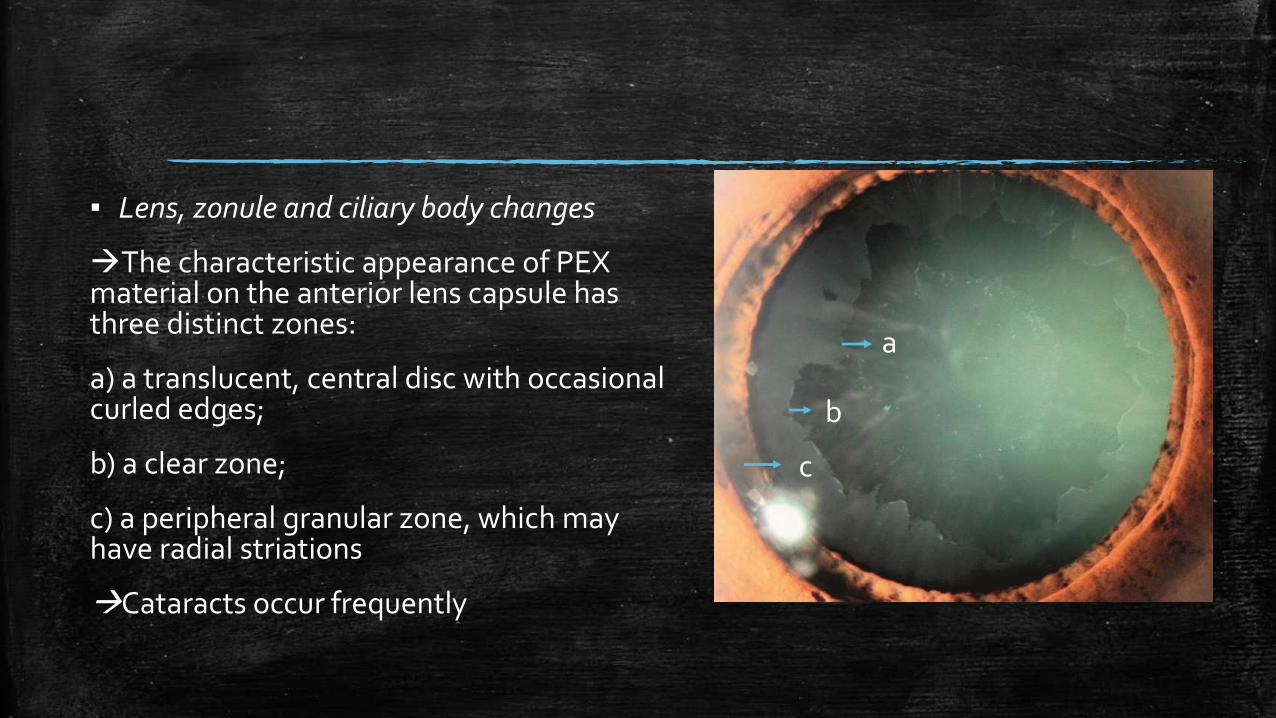
▪ Lens, zonule and ciliary body changes
The characteristic appearance of PEX material on the anterior lens capsule has three distinct zones:
a) a translucent, central disc with occasional curled edges;
b) a clear zone;
c) a peripheral granular zone, which may have radial striations
Cataracts occur frequently
a
b
c

A precapsular film has been noted on the anterior lens capsule of older individuals, which has a ground-glass appearance
It has been suggested that the precapsular layer may be a precursor of the PXS

Pseudoexfoliation material may be detected earliest on the ciliary processes and zonules
Involvement of the zonules can lead to lens subluxation and phacodonesis .
PEX aggregates at the origin and anchorage of the zonules and invades the zonularlamellae, creating areas of weakness
Proteolytic enzymes facilitate zonulardisintegration.

Gonioscopy…
▪ Trabecular hyperpigmentation-most marked inferiorly, patchy distribution
▪ Scalloped band of pigment running on to or anterior to Schwalbe line Sampaolesi line
▪ PXF deposits in the trabeculum can give rise to a ‘dandruff-like’ appearance.
▪ Narrow angles present in some casesincreased risk of angle closure, probably due to zonular laxity.

Ultrasound biomicroscopic findings…
▪ Useful tool to look for presence of exfoliative material on zonules or peripheral lens capsule, zonular weakness and breakage esp. when pupil cannot be easily dilated
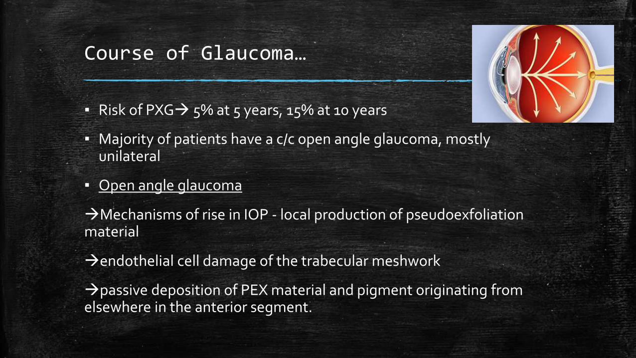
Course of Glaucoma…
▪ Risk of PXG 5% at 5 years, 15% at 10 years
▪ Majority of patients have a c/c open angle glaucoma, mostly unilateral
▪ Open angle glaucoma
Mechanisms of rise in IOP - local production of pseudoexfoliationmaterial
endothelial cell damage of the trabecular meshwork
passive deposition of PEX material and pigment originating from elsewhere in the anterior segment.

Active PEX production within the trabecular meshwork, Schlemm's canal, and collector channels, as well as passive deposition of PEX material within intertrabecular spaces
swelling of the juxtacanalicularmeshwork and gradual narrowing of Schlemm's canal

IOP runs higher, fluctuates, more difficult to control than POAG
Higher prevalence of glaucomatous optic neuropathy, NRR damage more diffuse
Immunoelectron microscopic studies of the lamina cribrosa have shown elastosis - abnormal regulation of elastin synthesis or degradation, in the optic nerve head of patients with PXS

▪ Acute increases in IOP
PXS with open angles may present with acute glaucoma mimicking angle-closure glaucoma
▪ Angle Closure Glaucoma- rare
zonular weakness, causing anterior movement of the lens
lens thickening from cataract formation
increased adhesiveness of the iris to the lens due to PEX material, sphincter muscle degeneration
iris rigidity from hypoxia

Differential Diagnosis
Capsular Delamination or true exfoliation
Primary Amyloidosis
Pigment Dispersion Syndrome

Management…
▪ Challenging to manage due to IOP fluctuation- set lower target pressure
▪ Medical treatment
Same as for POAG
Excellent response to prostaglandin analogues
high incidence of late failure
▪ Laser trabeculoplasty
particularly effective, possibly because of trabecular hyperpigmentation.
argon laser trabeculoplasty
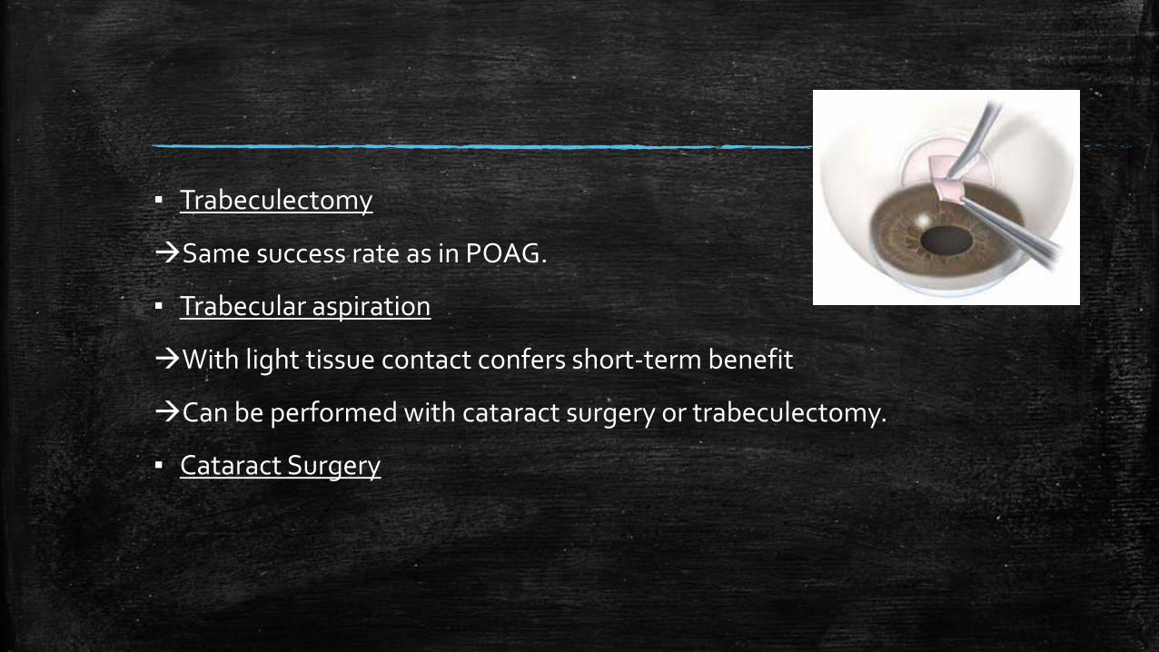
▪ Trabeculectomy
Same success rate as in POAG.
▪ Trabecular aspiration
With light tissue contact confers short-term benefit
Can be performed with cataract surgery or trabeculectomy.
▪ Cataract Surgery

Prognosis…
▪ Worse than in POAG
▪ the IOP is significantly elevated and exhibits greater fluctuation.
▪ Patients with PXF to be reviewed every 6 months
Patient with unilateral PXG and PXF in the fellow eye - high risk (50% in 5 years) of developing PXG in fellow eye.
Patient with unilateral PXG with normal fellow eye has low risk

Management of Cataract…
▪ Frequently indicated
▪ Higher than average risk of zonular and capsular breaks
▪ Poor pupillary dilatation, synechiae b/w iris epithelium and peripheral anterior lens capsule
▪ Preoperativelylook for phacodonesis, asymmetric AC depth, endothelial compromise, UBM may be helpful
▪ Intraoperativelyminimize zonular stress during nucleus manipulation and cortex removal
▪ Large capsulorrhexisminimizes zonular stress, prevents capsular phimosis
▪ During hydrodissection tap center of nucleus to decompress fluid pressure on weak posterior capsule
▪ Small pupil iris hooks, minisphincterotomies, sector iridectomy
▪ Capsular Tension Ring to stabilize the capular bag
▪ Early postoperative period fibrinoid iritis may be present; can be reduced by using heparin surface modified IOLs

References…
Becker-Shaffer's Diagnosis and Therapy of the
Glaucomas, 8th Edition
Shields' Textbook of Glaucoma 6th Edition
Kanski's Clinical Ophthalmology: A Systematic
Approach, 8th Edition
American Academy of Ophthalmology-Glaucoma
