prr.hec.gov.pkprr.hec.gov.pk/jspui/bitstream/123456789/9092/1/adil-thesis- corrected.pdfplagiarism...
TRANSCRIPT

PHYTOCHEMICAL INVESTIGATION ON THE CHEMICAL
CONSTITUENTS OF CARALLUMA FLAVA
Thesis Submitted
for
The Partial Fulfillment of the Degree of
DOCTOR OF PHILOSOPHY IN CHEMISTRY
By
MUHAMMAD ADIL RAEES
DEPARTMENT OF CHEMISTRY
Federal Urdu University of Arts, Science & Technology
Gulshan-e-Iqbal Campus, Karachi-75300, Pakistan
(2017)

PHYTOCHEMICAL INVESTIGATION ON THE CHEMICAL
CONSTITUENTS OF CARALLUMA FLAVA
By
MUHAMMAD ADIL RAEES
DEPARTMENT OF CHEMISTRY
Federal Urdu University of Arts, Science & Technology
Gulshan-e-Iqbal Campus, Karachi-75300, Pakistan
(2017)

Plagiarism Undertaking
I solemnly declare that research work presented in the thesis titled
“PHYTOCHEMICAL INVESTIGATION ON THE CHEMICAL CONSTITUENTS
OF CARALLUMA FLAVA” is solely my research work with no significant
contribution from any other person. Small contribution/help wherever taken has been
duly acknowledged and that complete thesis has been written by me.
I understand the zero tolerance policy of the HEC and Federal Urdu University of Arts,
Science & Technology towards plagiarism. Therefore I as an Author of the above titled
thesis declare that no portion of my thesis has been plagiarized and any material used
as reference is properly referred/cited.
I undertake that if I am found guilty of any formal plagiarism in the above titled thesis
even after award of PhD degree, the University reserves the rights to withdraw/revoke
my PhD degree and that HEC and the University has the right to publish my name on
the HEC/University Website on which names of students are placed who submitted
plagiarized thesis.
Name of Student: Muhammad Adil Raees
Signature:

Author’s Declaration
I, MUHAMMAD ADIL RAEES hereby state that my PhD thesis titled
“PHYTOCHEMICAL INVESTIGATION ON THE CHEMICAL CONSTITUENTS
OF CARALLUMA FLAVA” is my own work and has not been submitted previously
by me for taking any degree from Federal Urdu University of Arts, Science &
Technology Or anywhere else in the country/world.
At any time if my statement is found to be incorrect even after my Graduate the
university has the right to withdraw my PhD degree.
Name of Student: Muhammad Adil Raees
Signature:

Certificate of Thesis Evaluation
It is certified that the thesis entitled “PHYTOCHEMICAL INVESTIGATION ON
THE CHEMICAL CONSTITUENTS OF CARALLUMA FLAVA” has been
submitted by MUHAMMAD ADIL RAEES to the Graduate Research Management
Committee (GRMC) for reward of the degree of DOCTOR OF PHILOSOPHY (Ph.D)
in CHEMISTRY and has been written under my supervision and is thereby approved.
__________________________ ____________________________
DR. TALAT MAHMOOD PROF. AHMED AL-HARRASI
(Research Supervisor), Chairperson Co-Supervisor
Department of Chemistry University of Nizwa, Oman
FUUAST, Karachi Campus
__________________________
DR. HIDAYAT HUSSAIN
Co-Supervisor
University of Nizwa, Oman
DEPARTMENT OF CHEMISTRY
Federal Urdu University of Arts, Science & Technology
Gulshan-e-Iqbal Campus, Karachi-75300, Pakistan
(2017)

Thesis Approved
By
____________________________
DR. TALAT MAHMOOD
Supervisor and Chairperson
Department of Chemistry, FUUAST
___________________________
PROF. DR. RUBINA MUSHTAQ
Dean, Faculty of Science & Technology, FUUAST
__________________________
EXTERNAL EXAMINER

DEDICATED TO MY LOVING
PARENTS
Raees Ahmed
Shahida Raees
&
YOUNGER ROTHERS
Muhammad Faisal Raees
Muhammad Bilal Raees
Without their commitment and support
this work could not be completed on time.
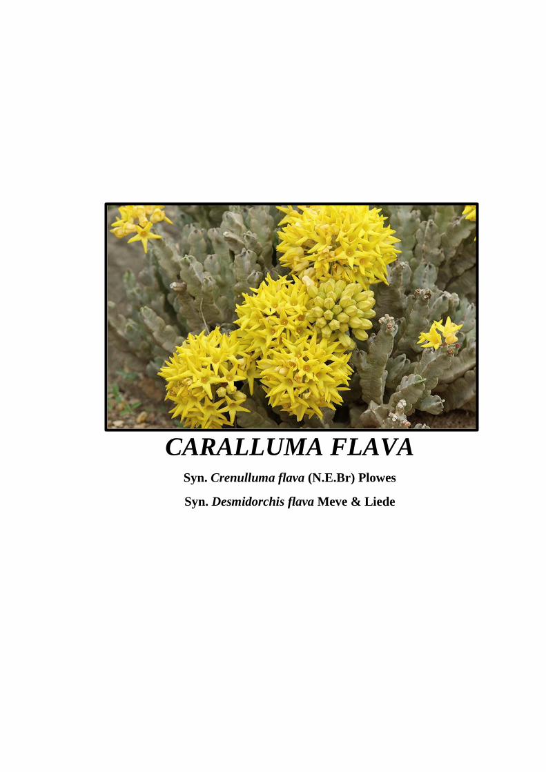
CARALLUMA FLAVA Syn. Crenulluma flava (N.E.Br) Plowes
Syn. Desmidorchis flava Meve & Liede
i
CONTENTS
ACKNOWLEDGEMENT ……………………………………… iv
ABSTRACT ……………………………………………………... vi
KHULASA ………………………………………………………. vii
1. INTRODUCTION …..………………………………………. 1
1.1 Caralluma Flava …………………………………………………….. 6
1.2 Pregnane Glycosides ………………………………………………... 7
1.3 Literature Survey ……………………………………………………. 9
2. PRESENT WORK …………………………………………. 20
3. EXPERIMENTAL ……………………….…………………. 27
3.1 General Note ………………………………………………………… 28
3.2 Extraction and Isolation …………………………………………….. 29
3.2.1 Characterization of Desmiflavaside A (1) …………………… 36
3.2.2 Characterization of Desmiflavaside B (2) …………………… 39
3.2.3 Characterization of Desmiflavaside C (3) …………………… 42
3.2.4 Characterization of Desmiflavaside D (4) …………………… 45
3.2.5 Characterization of Nizwaside (5) …………………………… 48
3.2.6 Characterization of Desflavaside A (6) ……………………… 51
3.2.7 Characterization of Desflavaside B (7) ………………………. 55
3.2.8 Characterization of Desflavaside C (8) ………………………. 59
3.2.9 Characterization of Desflavaside D (9) ……………………... 63

ii
3.3 Biological Activities ………………………………………………… 66
3.3.1 Anticancer Activity …………………………………………. 66
3.3.2 Enzyme Inhibition Activity …………………………………. 73
3.3.3 Antioxidant Activity ………………………………………… 76
3.4 Molecular Docking Studies …………………………………………. 79
4. RESULTS AND DISCUSSION................................................ 87
4.1 New Compounds from Caralluma flava ……………………………. 88
4.1.1 Desmiflavaside A (1) ………………………………………... 88
4.1.2 Desmiflavaside B (2) ………………………………………… 92
4.1.3 Desmiflavaside C (3) ………………………………………… 94
4.1.4 Desmiflavaside D (4) ………………………………………... 97
4.1.5 Nizwaside (5) ………………………………………………... 99
4.1.6 Desflavaside A (6) …………………………………………… 102
4.1.7 Desflavaside B (7) …………………………………………… 106
4.1.8 Desflavaside C (8) …………………………………………… 108
4.1.9 Desflavaside D (9) …………………………………………... 111
4.2 Biological Activities ………………………………………………… 113
4.2.1 Anticancer Activity ………………………………………….. 113
4.2.3 Enzyme Inhibition Activity ………………………………….. 114
4.2.4 Antioxidant Activity ………………………………………… 115
4.3 Molecular Docking Studies ………………………………………….. 116
5. CONCLUSIONS ……………………………………………… 118
6. REFERENCES ………………………………………..……… 120

iii
7. GLOSSARY …………………………………………………... 126
8. APPENDIX ……………………………………………………. 129
9. PUBLICATIONS ……………………………………………... 152
iv
ACKNOWLEDGMENT
First of all, I bow my head to the Omnipresent, Omnipotent and Omniscient Al-Mighty
ALLAH, whose clemency resulted into my success. I would like to pay homage to the
most respectful and sacred personality of the world, Holy Prophet, Hazrat Muhammad
(S.A.W.W). The research work present in this thesis is the result of the many people’s
contributions.
First and foremost, I express my sincere gratitude to the most co-operative supervisor
and chairperson Dr. Talat Mahmood for her constructive supervision, valuable
suggestions, great encouragement, indefinite motivation and scientific support
throughout the research. Her encouraging attitude always raised my morale. I believe
that she has not only guided me in my academics but also in some other matters of life.
I wish to extend my thanks to Dr. Aneela Wahab, Dr. Iffat Mahmood and Dr. Atya
Hassan for their precious time and outstanding suggestions for this thesis writing. They
also contributed greatly in the NMR spectral interpretation and characterization.
My sincere and boundless thanks to Prof. Dr. M. Iqbal Choudhary and Head of NMR
Section, Dr. Atia-tul-Wahab for my training in NMR spectroscopy and operation of
NMR spectrometers at University of Karachi.
I was fortunate enough to perform a part of my research work at University of Nizwa,
the Sultanate of Oman, while doing my job there as NMR Spectroscopist in the NMR
lab, for which I am grateful to Prof. Dr. Ahmed Sulaiman Al-Harrasi for permitting me
to pursue this work in his laboratories along with my job and gave me the opportunity
to become a part of an enthusiastic and ambitious internationally diverse group with
multidisciplinary competences. I believe that I had a profit there from the expertise of
the team as a whole, as well as from each team member individually, which includes
Dr. Hidayat Hussain, Dr. Javid Hussain, Dr. Liaqat Ali, Dr. Najeeb-Ur-Rehman, Dr.
Thomas Dzeha, Dr. Ali Elyassi, Dr. Faruck Lukman-Ul-Hakkim, Dr. Ghulam Abbas,
Mohammad Abdullah Al-Broumi and Ahmed Al-Ghafri. Some very special and joyful
thanks are extended to the Director of Technical Staff, Dr. Obaid Yusuf Khan for the
effective management of NMR and Mass spectroscopy sections as well as his utmost
help and moral support.
v
I cannot forget the cooperation and help that I received from the local Omanis for the
plant collection. The guidance of the local Omani vegetable sellers helped a lot to search
the plant on the high mountains of Oman and I am highly grateful to all of them. I
acknowledge the wonderful and timely jobs carried out by my collaborators Dr. Gilani
at University of Nizwa in Oman for helping me in the plant identification; Dr. Hussain
Yar Khan for carrying out the bioassays for my sample very efficiently.
My gratitude extends to all those who have contributed to my education. I owe very
much to my lab fellows Syed Waseem Ahmed, Syed Wali Shah and Syed Sajjad Haider,
who has always helped me during my studies and proven to be outstanding companion
and good friends. Thank you all for your friendship especially Shah Rizwan Ashrafi for
being there for me whenever I need your help.
At the end, I express my deepest and heartiest gratitude to all my family members,
specially my parents, who are always so loving and caring. They taught me to be dutiful
and serve the nation. They are so wonderful and cooperative that no words can describe.
I could never been able to start and continue this work to the end without their support
and trust. I am proud to have such a loving and enlightened family.

vi
ABSTRACT
The research work embodied in this thesis deals with the isolation of chemical
constituents from the Omani plant named Caralluma flava (N.E.Br) Meve & Liede
along with the spectral characterization, biological activities and computational studies.
Crude crystals (CR) obtained from the squeezed sap of succulent C. flava showed
significant anticancer activity against MDA-MB-231 breast cancer cells and provided
nine new pregnane glycosides namely Desmiflavaside A (1), Desmiflavaside B (2),
Desmiflavaside C (3), Desmiflavaside D (4), Nizwaside (5), Desflavaside A (6),
Desflavaside B (7), Desflavaside C (8) and Desflavaside D (9). The structures of these
isolates were elucidated through spectral analysis using UV, IR, HRSEIMS, NMR and
by comparing with literature reports.
Treatment of MDA-MB-231 breast cancer and SKOV-3 ovarian cancer cells with
compounds 3-5 demonstrated a prominent reduction in the viability of both types of
cancer cells. Furthermore, 3-5 were also tested for their effect on normal breast
epithelial cells (MCF-10-2A) which displayed no significant cytotoxicity on normal
cells. The molecular docking studies of 3-5 revealed that these molecules have a good
potential to bind with the target protein tyrosine phosphatase.
The methanolic extract (ME) of squeezed residual plant and its different fractions also
showed good anticancer activity against MDA-MB-231 cells. Different fractions of
ME, CR and compounds (1-5) were additionally evaluated for enzyme inhibition
(urease, acetylcholinesterase and α-glucosidase) and DPPH antioxidant activities which
displayed no remarkable results.
The research work embodied in this dissertation has resulted four publications as
mentioned in the end of the thesis.

vii

1
1. INTRODUCTION

2
The Sultanate of Oman, despite its arid nature, is endowed with a variety of medicinal
plants (Hussain et al., 2014). It has almost 1204 terrestrial plants and many of them are
used by herbalists in traditional medicine (Alhakmani et al., 2014; Ghazanfar, 1992).
In common with other Gulf countries, several traditional systems of medicinal
treatment are used in Oman. In addition to the use of plants as medicine (Al Tadawee
bil A’ashiab), cupping (Al Hajamah), bone setting (Al Tajbeer) and cauterization
(Wasm, Qai) are also practised. In any of these, specific medicinal plants are used as a
part of the treatment (Ghazanfar, 1994).
Until recently, the only type of cure available to the large section of people in Oman
was traditional medicine. In the past two decades, with the establishment of clinics and
hospitals, traditional medicine (which comprises healing by using plants) has become
less popular but is still used. Many minor ailments such as headaches, stomach upsets,
coughs, colds and fevers are often treated at home with herbal remedies (Ghazanfar and
Al-Sabahi, 1993).
In Sultanate, the traditional knowledge concerning plants and their uses is largely
untapped. In northern and central Oman, there are no educational or formal training
institutes for teaching traditional medicine and the method of cure. Neither is
knowledge documented. Instead, these traditional medicinal knowledge skills are
passed orally from one generation to another or by elders and ancestors through
apprenticeship and once forgotten may not be practised again. Literate healers consult
the available classical work on herbal and traditional medicines for their practice and
still the oral way is highly preferred for transferring and sharing traditional knowledge
(Lupton et al., 2012; Ghazanfar, 1994; Ghazanfar and Al-Al-Sabahi, 1993).
The Sultanate of Oman is a home to large number of succulent and xerophytic
Caralluma species. In Sultanate, the genus is represented by approximately thirteen
species viz., C. flava N.E.Br., C. aucheriana (Decne.) N.E. Br., C. arabica N.E. Br., C.
penicillata (Deflers) N.E.Br., C. quadrangula (Forssk.) N.E.Br., C. hexagona
Lavranos, C. meintjesiana Lavranos, C. dodsoniana Lavranos, C. luntii N. E. Br., C.
adenensis (Deflers) A. Berger, C. edulis (Edgew.) Benth. ex Hook.f., C. tuberculate
N.E.Br., C. awdeliana (Deflers) Berger (Walter and Gillett, 1998; Albers and Meve,
2002; Ghazanfar, 1994; Bruyns et al., 2010; Patzelt, 2015). The scientific studies of the
genus has been carried out in Pakistan, India, Saudi Arabia, Spain, Italy and Nigeria
3
(Fig. 1) but almost no evidences of scientific examination of Caralluma and validation
of its traditional therapeutic uses in the Sultanate of Oman has been found. Out of
Sultanate, numerous in-vitro and very few in-vivo biological investigations of
Caralluma have been conducted on crude extracts and its isolates. Mostly, antidiabetic
studies of Caralluma species are reported but very few species are examined for their
anticancer potential (Fig. 2) (Adnan et al., 2014).
Cancer is reported to be the major public health issue globally and its incidences are
rising across the globe (Burney et al., 2014; Al-Lawati et al., 1999). Among Omani
females, breast cancer was the leading malignancy of the total cancer cases between
1998 to 2007 and continued to be the leading by May, 2014 (Fig. 3 and 4) (Al-Madouj
et al., 2011; WHO, 2014). One out of five Omani women is diagnosed with breast
cancer in her lifetime and a preventive strategy for cancer has not been developed yet
(Renganathan et al., 2014; WHO, 2010).
Taking in to account the facts stated above, the work was undertaken on Omani
medicinal plant for the present doctoral dissertation entitled “Phytochemical
investigation on the chemical constituents of Caralluma flava”. The introduction
provides a brief description of C. flava, structural features of pregnane glycosides, a
review of isolated pregnane glycosides from the genus and their pharmacological
significance. This is followed by a brief discussion of present work containing the
structures of nine new pregnane glycosides isolated for the first time from C. flava.
Further, the breast cancer activity of these pure constituents along with molecular
docking studies are also included.

4
Figure 1: Scientific studies of Caralluma species in different countries (Adnan et al.,
2014).
Figure 2: Pharmacological activities of Caralluma species (Adnan et al., 2014).

5
Figure 3: Cancer incidences of Omani females (WHO, 2014)
Figure 4: Cancer mortality profile of Omani females (WHO, 2014)
Breast
18%
Lymphomas,
multiple
myeloma
10%Leukaemia
10%Colorectum
8%
Stomach
6%
Other
48%
Females Cancer Mortality
400 Deaths
0
50
100
150
200
Breast Thyroid Colorectum Leukaemia
195
53 51 42
Nu
mb
er o
f ca
ses
Cancer

6
1.1 Caralluma Flava
Caralluma flava, commonly known as نبتة الضجع in Arabic language, belongs to the
family Apocynaceae and has various synonyms viz., Desmidorchis flava, Desmidorchis
flavus and Crenulluma flava. It is a regional endemic plant, most often grows on the
Omani-Yemeni border (Mahra-Dhofar) where it extends into the UAE. It grows much
rare in central and northern Oman. C. flava can be found in rocky limestone areas, dry
riverbeds and coastal hills along rocky watercourses. Its succulent Stems are gray in
colour, bluntly square in shape and usually grow straight in clusters. (Patzelt, 2015;
Grulich V, 2015; Formisano, 2009; Mosti, 2004; Albers and Meve, 2002).
This cactus like plant is edible. Its bright yellow flowers and seed pods are eaten after
the rain and the sap filled juicy stems are collected by local people for food. Due to its
extreme bitterness, addition of lemon and spices are preferred before eating. Fresh plant
as well as its dried powdered forms are available in the vegetable and local markets
(Patzelt, 2015; Raees et al., 2016). The plant has been used for generations in traditional
Omani society due to its medicinal properties. Referenced to the reasons mentioned
earlier, scanty information regarding its medicinal uses have been documented only in
Arabic text as given below (سلطنۃ عمان دیوان البالط السطالنی(.
للحروق: یؤخذ نبات الضجججع الطرو ویسججحى يتي ینججمر ناعما ویونججر ثو نهن ی •
ه مكان الحرق.
ا یضاف إلمه • لممون وملح.للسكر: یهرس ویؤكل غضًّ
للطحال والكب : نفس الطریهة السا هة. •
لإلمساك: یؤكل غضا قبل الطوام. •
للغازات: یشر عنمر الضجع و الطوام ق ر كو . •
لضغط ال م: یُونر النبات ویُشر ق ر كو شاو مرتم فن الموم ون إضافة شنء. •
Translation:-
• For burns: Crush the fresh stems until it becomes soft, squeeze it with cloth and then
apply on the burns.
• Diabetes: Eat juicy stems with salt and lemon.
• Spleen and liver: The same method as above.
• Constipation: Eat before taking meal.
• Abdominal gas: Drink a cup of its juice after food.
• Blood pressure: Drink its juice twice a day without adding anything.

7
The phytochemical investigation of C. flava has not been performed yet and no
chemical constituent has been reported so far except the only available data on the
chemical composition of its floral scent volatiles (Jürgens et al., 2006). However,
several other Caralluma species have been investigated and numerous potent
compounds have been isolated, characterized and reported. On reviewing the literature
undertaken on the phytochemistry of genus Caralluma, it could be observed that
pregnane glycosides are one of its major constituents.
1.2 Pregnanes Glycosides
The C-21 steroidal saponins having the usual per-hydro-1,2-cyclopentanophenanthrene
ring system with a two carbon chain at C-17 and β-oriented Me groups at C-10 and C-
13 are known as pregnanes. Most frequently, pregnane derivatives bear a hydroxyl
group at C-14 which possess β-configuration. The configuration at C-5 is α except for
molecules containing a C-5 double bond. Pregnanes have a β-oriented C-3 hydroxyl
group (Adnan et al., 2014). In 1989, Deepak et al. reported some characteristic features
of pregnanes which are given below.
1- Presence of Double bond at C-5.
2- Fusion of rings B and C is always trans.
3- Fusion of rings C and D is cis when a hydroxyl group is present at C-14 and trans
when H is present at C-14.
4- Additional hydroxyl groups at 5α, 6β, 7α, 8β, 11α, 12α or 12β, 14β, 15α, 16α, 17α
or 17β, 20 and 21 which may be partially esterified.
5- Presence of carbonyl group at positons C-l, C-12, C-15 and C-20.
OH
HOH
H
H
AB
C D1
2
3
4
5
6
7
8
9
10
11
12
13
14
15
16
17
18
19
20
21
Basic skeleton of pregnane (Adnan et al., 2014; Deepak et al., 1989)

8
In pregnane glycosides, the glycoside is attached to an alcoholic OH group of the
aglycone portion, most frequently at C-3, and is usually found as a linear saccharide
chain rather than a branched. The most common and successful employed method of
preparative isolation of pregnane glycosides is column chromatography (Deepak et al.,
1997; Al-Massarani and El-Shafae, 2011). Several types of sugar units have been
detected in pregnane glycosides. The most common are:

9
1.3 Literature Survey
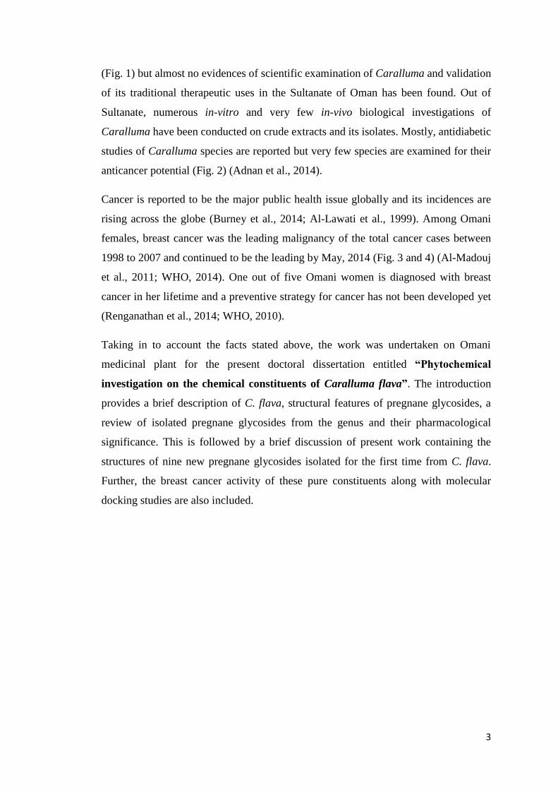
In 1988, four pregnane glycosides viz., boucerosides A-I (10), B-I (11), A-II (14) and
B-II (15) were reported from CHCl3 soluble fraction of the MeOH extract obtained
from dried aerial parts of C. aucheriana. Compounds 10 and 11 contained molecular
formula C62H88O21 whereas 14 and 15 possessed C62H90O21. Preparative HPLC was
used for the purification of these compounds. Later in 1990, two more pregnane
glycosides viz., boucerosides CNC (12) and CNO (13) having molecular formula
C56H80O16 were reported from the same fraction. Pregnane glycosides 10 and 11 had
the similar pregnane skeleton but differed from each another in the sugar moiety, as this
difference was also observed in 14 and 15. A couple of benzoyl groups at position C-
12 and C-20 of the pregnane were present in all of these six molecules (Hayashi et al.,
1988; Ahmad and Basha, 2007; Tanaka et al., 1990).
Lee-Juian et al. (1994) reported pregnane glycosides viz., carumbelloside I (16) and II
(17) isolated from n-BuOH and EtOAc fractions of EtOH extract of fresh whole C.
umbellate plant, respectively. Silica gel column chromatography was used for the
isolation. The structural characterization of these compounds were achieved using
different NMR techniques. TOCSY experiments were reported to be very useful to
discover the correlations among protons of each sugar units (Lee-Juian et al., 1994)
In 2001 and 2002, Abdel-Sattar and his co-workers published two reports on seven
pregnane glycosides namely penicillosides A-G (18-24) isolated from the EtOH extract
of C. penicillata. The plant was collected from Saudi Arabia. All seven compounds (18-
24) had the benzoyl groups present in them at differrent positions and the glycoside
chain was attached at C-3 of the pregnane part. Only compound 18 was found to be a
disaccharide whereas the remaining (19-24) were trisaccharides containing glucose,
digitalose, cymarose, allomerose and thevetose units (Abdel-Sattar et al., 2002, 2001).
10

11

12

13
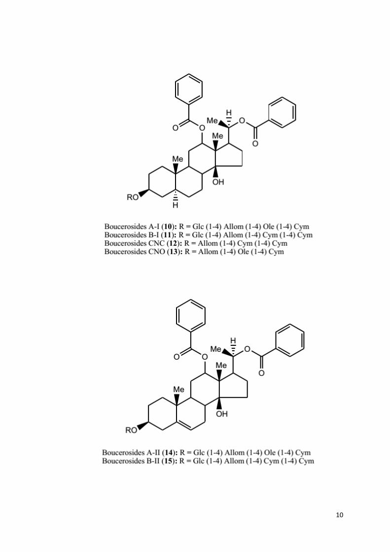
Abdel-Sattar et al. (2007) carried out phytochemical investigation on C. russeliana and
reported three pregnane glycosides viz., russeliosides E-G (25-27) isolated from CHCl3
soluble fraction of the EtOH extract, via chromatography over silica gel column and
RP-18 column on HPLC. All of these three isolates had the similar aglycone portion
carrying benzoyl group at C-12 but differed from each other on the basis of sugar
moiety (Abdel-Sattar et al., 2007).
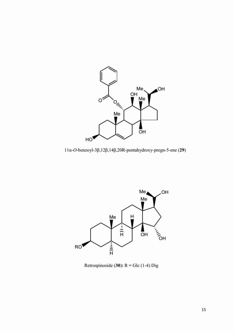
Two pregnanes characterized as 12β-O-benzoyl-3β,11α,14β‚(20R)-pentahydroxy-
pregn-5-ene (28) and 11α-O-benzoyl-3β,12β,14β,(20R)-pentahydroxy-pregn-5-ene
(29) were isolated from the fresh whole C. pauciflora plant. The phytochemical
investigation was carried out by Reddy et al. (2011). No glycoside was found to be
attached in these molecules (Reddy et al., 2011).
Elsebai et al. (2015) reported that the MeOH extract of C. retrospiciens yielded
pregnane glycoside viz., retrospinoside (30), C34H58O13, m.p. 208-210 oC, [α]D -2.3 (c,
2.2, MeOH), after fractionation and repeated chromatographic separation on silica gel
columns. The disaccharide chain attached at C-3 of the pregnane was found to be
comprised of glucose and digitalose units. As the molecule contained many hydroxyl
groups, therefore, in order to obtain better quality of spectra for assignments and
additional information, 30 was acetylated with acetic anhydride-pyridine.
Spectroscopic measurements including 1D and 2D NMR, and HRMS were performed
on the acetylated product (Elsebai and Mohamed, 2015).
Some of the isolated pregnane glycosides from various Caralluma species are listed in
Table 1.
14
15
16
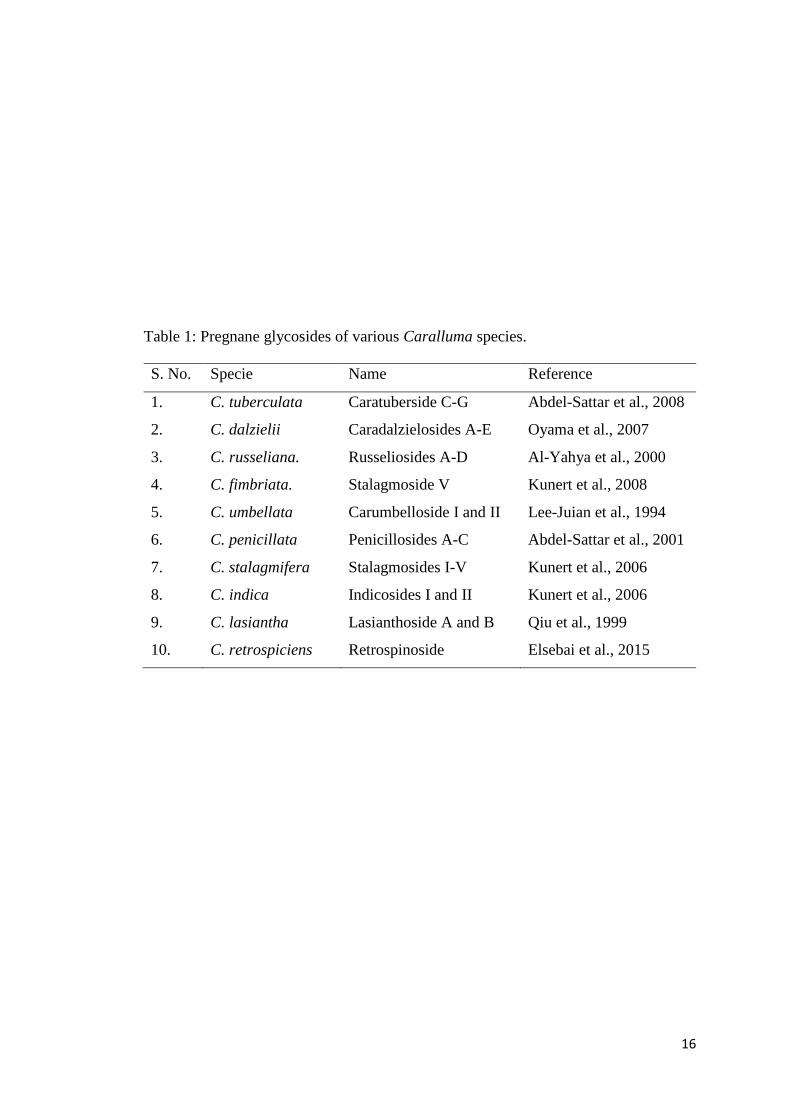
Table 1: Pregnane glycosides of various Caralluma species.
S. No. Specie Name Reference
1. C. tuberculata Caratuberside C-G Abdel-Sattar et al., 2008
2. C. dalzielii Caradalzielosides A-E Oyama et al., 2007
3. C. russeliana. Russeliosides A-D Al-Yahya et al., 2000
4. C. fimbriata. Stalagmoside V Kunert et al., 2008
5. C. umbellata Carumbelloside I and II Lee-Juian et al., 1994
6. C. penicillata Penicillosides A-C Abdel-Sattar et al., 2001
7. C. stalagmifera Stalagmosides I-V Kunert et al., 2006
8. C. indica Indicosides I and II Kunert et al., 2006
9. C. lasiantha Lasianthoside A and B Qiu et al., 1999
10. C. retrospiciens Retrospinoside Elsebai et al., 2015

17
No pharmacological activity has been performed on C. flava except the antioxidant
activity of its aqueous EtOH extract. The investigation showed weaker antioxidant
activity in the DPPH and promising activity in phosphomolybdenum assay (Marwah et
al. 2007). However, extracts of various Caralluma species along with their isolated
pregnane glycosides have been investigated for their pharmacological potential.
Different fractions of the EtOH extract of C. tuberculata were tested on the growth and
viability of various types of cancer cells by Waheed et al. (2011) and his co-workers.
Its EtOAc fraction was reported to be the most anti-proliferative against MDA-MB-468
(46% ± 2.8%) and MCF-7 (94% ± 4.0%) breast cancer cell lines. Pregnane glycoside
viz., 12-O-benzoyl-20-O-acetyl-3β‚12β,14β,20β-tetrahydroxy-pregnan-3-yl-O-β-D-
glucopyranosyl-(1→4)-β-D-glucopyranosyl-(1→4)-3-methoxy-β-D-ribopyranoside
(31) isolated from this active fraction showed moderate micromolar cytotoxicity against
both breast cancer cells (IC50: MDA-MB-468 = 25-50 μM; MCF-7 = 6.25-12.5 μM)
(Waheed et al., 2011).
In 2013, Abdallah et al. and his co-workers tested C. quadrangular MeOH extract and
its various fractions for their cytotoxic activity against MCF-7 breast cancer cells. The
phytochemical investigation of the bioactive chloroform fraction yielded four pregnane
glycosides (32-35) having benzoyl groups attached at C-12 and C-20 of the pregnane
skeleton. The cytotoxicity of these isolates were tested against the same cancer cell line
using doxorubicin as positive control. Pregnane glycoside 32 (IC50 = 4.8 μM), 33 (IC50
= 2.0 μM) and 35 (IC50 = 8.0 μM) showed promising cytotoxic activities. Whereas, 34
was reported to be inactive (Abdallah et al., 2013).
Recently (2015), the MeOH extract of six indigenous folk medicinal plants (Arnebia
decumbens, Caralluma sinaica, Fagonia tenuifolia, Lavandula pubescens, Sonchus
oleraceus, Verbesina encelioides) growing wild in Saudi Arabia were tested for their
anticancer activity using three cancer cell lines (breast, lung and central nervous
system). Among these plants extracts tested, the most potent was C. sinaica which
showed strong anticancer activity against MCF-7, SF-268 and NCI-H460 cancer cell
lines with IC50 = 0.60, 2.01 and 0.02 μg/L, respectively (Albalawi et al., 2015).

18
OH
RO
Me
MeOO
O
OMe
Me
Pregnane Glycoside (31): R = Glc (1-4) Glc (1-4) 3-Methoxy-ribopyranose

19

20
2. PRESENT WORK

21
In the present study, organic extract (CH2Cl2:MeOH, 1:1) of squeezed sap from fresh
C. flava stems afforded shiny yellow crude crystals (CR) whereas the squeezed residual
plant was dried and powdered to extract with methanol (ME) which was solvent
partitioned to finally obtain various fractions (n-hexane: HX; dichloromethane: DM;
ethyl-acetate: EA; n-butanol: BU and aqueous: AQ). Crystals (CR), ME and its
fractions were first investigated for their effect on breast cancer cells (MDA-MB-231).
Interestingly, CR appeared to be more effective than ME and its fractions.
Crude crystals (CR) were subjected to column chromatography using gradients of
CH2Cl2:MeOH which provided nine new pregnane glycosides namely desmiflavaside
A-D (1-4), nizwaside (5) and desflavaside A-D (6-9). The structures of these isolates
(1-9) were established through various spectroscopic techniques including 1D (1H-1H
TOCSY, NOE) and 2D NMR (NOESY, 1H-1H COSY, HMBC), UV, IR and mass
spectrometry such as ESIMS and HRESIMS.
The effects of purified compounds 3-5 against MDA-MB-231 breast cancer cells and
SKOV-3 ovarian cancer cells in culture were examined. All tested compounds showed
significant anticancer activities. Compounds 3-5 were also tested for their cytotoxic
effect on normal breast epithelial (MCF-10-2A) cells and results illustrated that 3 was
slightly cytotoxic towards normal cells whereas 4 and 5 showed no major cytotoxic
effects and their cytotoxicity was selective for the cancer cells only.
Molecular docking studies of compounds 3-5 illustrated that all these three ligand
molecules have a good potential to accurately interact with the target protein tyrosine
phosphatase. Compound 4 demonstrated a comparatively promising docking energy as
compared to 3 and 5.
Furthermore, urease, acetylcholinesterase and α-glucosidase enzyme inhibition and
DPPH antioxidant activities of CR, isolated compounds (1-5) and different fractions of
ME were determined. The results showed no remarkable activity.
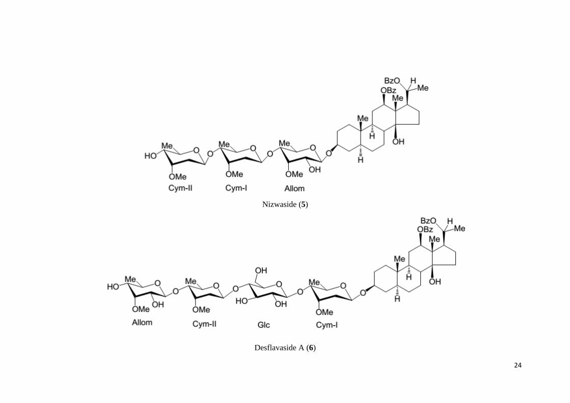
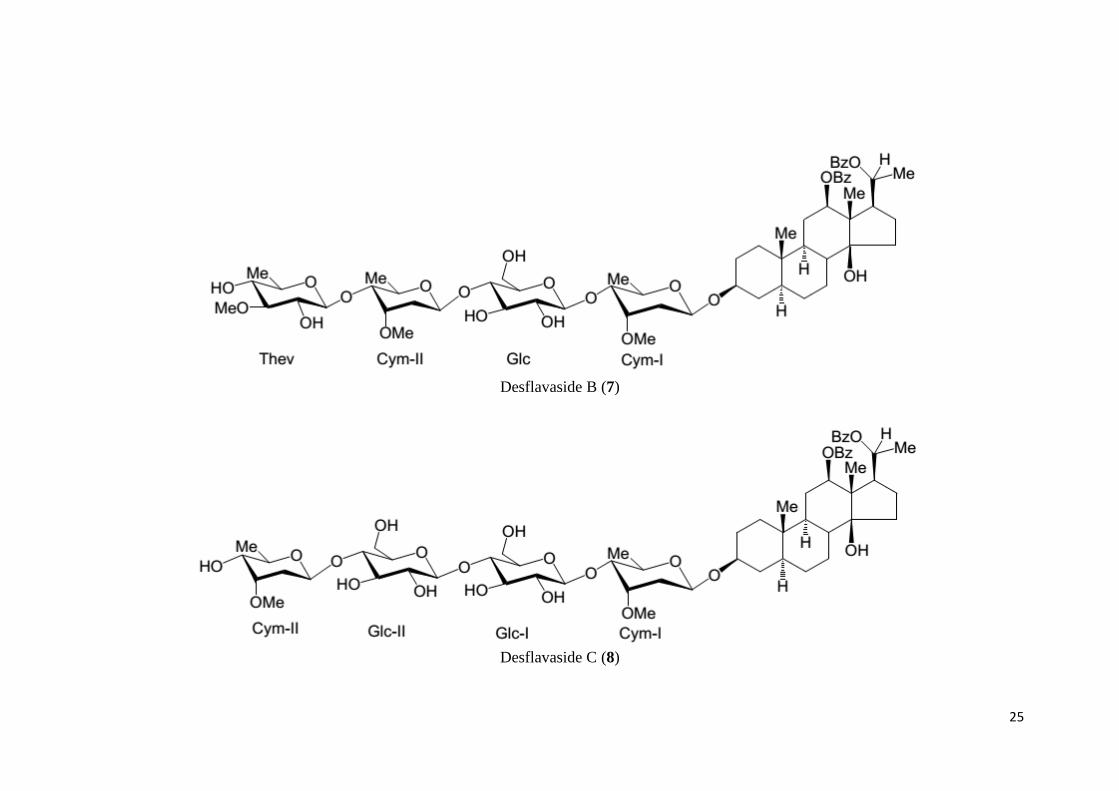
The structures of nine new pregnane glycosides isolated from CR are shown on the
following pages.

22
Structures of New Pregnane Glycosides
Desmiflavaside A (1)
Desmiflavaside B (2)

23
Desmiflavaside C (3)
Desmiflavaside D (4)
24
Nizwaside (5)
Desflavaside A (6)
25
Desflavaside B (7)
Desflavaside C (8)

26
Desflavaside D (9)

27
3. EXPERIMENTAL

28
3.1 General Note
Fresh whole C. flava plants were collected in December 2013 from the Al-Hajar
(Arabic: جبال الحجر means "Stone Mountains") mountainous region of Sultanate of
Oman including Jabal-Akhdar (Arabic: الجبل األخضر means "Green Mountains") and
Jabal-Shams (Arabic: جبل شمس, means "The Mountain of Sun") and were identified by
Dr. Gilani (plant taxonomist). A voucher specimen (BSBHPR-012/2013) was
submitted in the Herbarium of the University of Nizwa, Sultanate of Oman.
The IR and UV spectra were measured on ATR-Tensor 37 and Shimadzu UV-240
spectrophotometers, respectively. Optical rotations of the molecules were
recorded on KRUSS-P-P3000 polarimeter. The ESIMS and HRESIMS spectrum were
measured on Waters Quattro Premier XE Mass and Agilent 6529B Q-TOF Mass
spectrometers, respectively. The 1H-NMR was recorded on Bruker spectrometer
operating with cryoprobe prodigy at 600 MHz, while 13Carbon NMR was
obtained at 150 MHz. The chemical shifts and coupling constant values are
reported in part per million (ppm) and in hertz (Hz), respectively.
Silica gel PF254 (Merck) was used for column chromatography (CC) and for TLC pre-
coated aluminum sheet. Plates of TLC were visualized in the UV-light at 254 and 366
nm and spray of ceric sulfate reagent was used as locating agent.
All the experimental work along with spectroscopic techniques were performed
at the University of Nizwa (UoN). The anticancer activities were performed by Dr.
Husain Yar Khan at UoN. Enzyme inhibition and antioxidant activities were performed
by Dr. Ghulam Abbas.

29
3.2 Extraction and Isolation
Drops of sap exudate oozed out from fresh broken stems of succulent C. flava (30 kg)
were collected by gentle squeezing the juicy stems into a small beaker until it was filled
with 40 mL of sap (Scheme I). It was then extracted with a mixture of CH2Cl2-MeOH
(1:1) by slowly adding the solvent that turned the sap into white milky precipitates
which were left to settle down. The supernatant liquid which dissolved the major
portion of the sap was successively filtered with the help of whatman filter paper and
the filtrate was left for 24 hours in fume hood for drying. After the evaporation of
solvent, the dried beaker was found to have yellow shiny crude crystals (CR; 1.0 g)
(Fig. 5).
A B C
Figure 5: (A) Sap exudate of C. flava (B) Sap precipitates (C) Crude crystals
The crystals (CR) were subjected to CC (n-hexane-CH2Cl2; CH2Cl2; CH2Cl2-MeOH in
order of increasing polarity) which ultimately furnished 10 fractions (Frcn-1 to Frcn-
10) on combining the eluates on the basis of TLC (Scheme II). Frcn-7 obtained on
elution with CH2Cl2-MeOH (8.5:1.5) showed two major spots on TLC which on
separation through preparative TLC (two times; EtOAc-MeOH, 4.0:6.0) gave
Desmiflavaside A (1; 5.0 mg) and Desmiflavaside B (2; 7.0 mg). Frcn-3 on elution with
CH2Cl2-MeOH (9.5:0.5) yielded Desmiflavaside C (3; 6.7 mg). Frcn-1 on elution with
CH2Cl2-MeOH (9.6:0.4) yielded Desmiflavaside D (4; 6.2 mg). Frcn-2 on elution with
CH2Cl2-MeOH (9.6:0.4) provided Nizwaside (5; 12.0 mg). Frcn-4 obtained on elution

30
with CH2Cl2-MeOH (9.2:0.8) was further subjected to CC (CH2Cl2; CH2Cl2-MeOH in
order of increasing polarity) which furnished 5 fractions (Frcn-4-I to Frcn-4-V) on
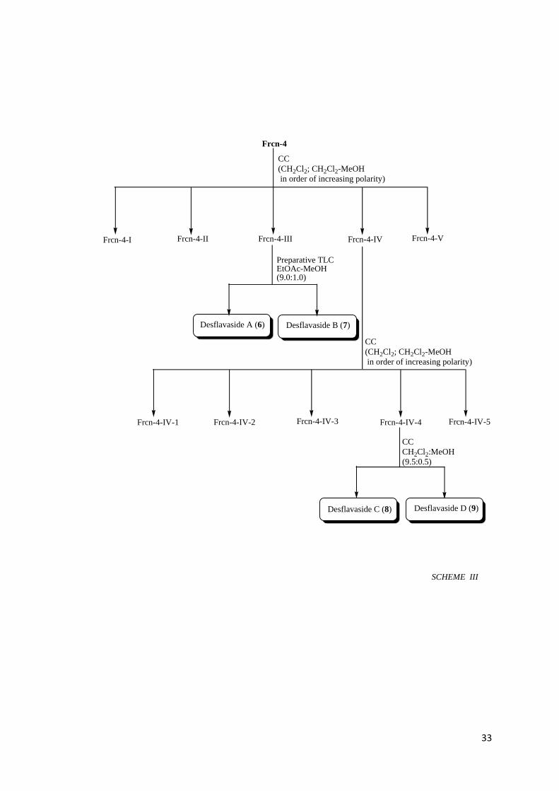
combining the eluates on the basis of TLC (Scheme III). Frcn-4-III on elution with
CH2Cl2-MeOH (9.6:0.4) afforded two compounds Desflavaside A (6; 5.0 mg) and
Desflavaside B (7; 5.2 mg) after separation through preparative TLC (two times;
EtOAc-MeOH; 9.0:1.0). Frcn-4-IV obtained on elution with CH2Cl2-MeOH (9.5:0.5)
was further subjected to CC (CH2Cl2; CH2Cl2-MeOH in order of increasing polarity)
which resulted 5 fractions (Frcn-4-IV-1 to Frcn-4-IV-5) on combining the eluates on
the basis of TLC (Scheme III). Frcn-4-IV-4 obtained on elution with CH2Cl2-MeOH
(9.5:0.5) afforded two compounds Desflavaside C (8; 4.0 mg) and Desflavaside D (9;
4.8 mg). The rest of the fractions containing several compounds in the minor quantities
were not pursued further in the present working.
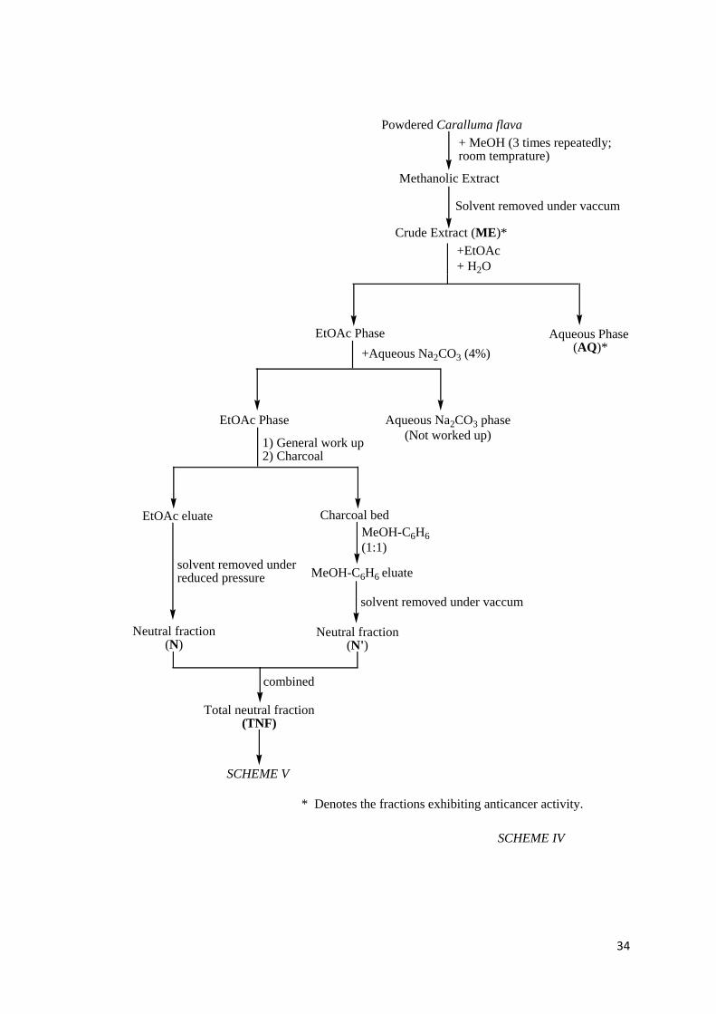
Powdered (2.0 kg) of air dried squeezed residual plant material (= remaining herbage
after squeezing out sap) was repeatedly (thrice) extracted with methanol at normal room
temperature. The syrupy concentrate (ME; 170 g), obtained on solvent removal from
the combined extracts under reduced pressure, was partitioned between EtOAc and H2O
(AQ; 35 g) (Scheme IV). The EtOAc phase was treated with Na2CO3 which separated
the acidic form from the neutral fraction. The EtOAc layer carrying the neutral fraction
was washed with water (Na2SO4) and treated with activated charcoal. It was then
filtered and its filtrate was successfully solvent freed under reduced pressure giving the
neutral fraction (N; 100 g). The charcoal bed was repeatedly eluted with MeOH-C6H6
(1:1) which on usual work-up gave another part of neutral fraction (N'; 20 g). Both N
and N' were mixed together after comparison of their TLC (silica gel PF254; CH2Cl2-
MeOH; 9.5:0.5). The total neutral fraction (TNF; 120 g) thus obtained was partitioned
into hexane soluble (HX; 30 g) and hexane insoluble portions (Scheme V). The hexane
insoluble fraction was again partitioned into DCM soluble (DM; 40 g) and DCM
insoluble portions. The DCM insoluble fraction was again partitioned into EtOAc
soluble (EA; 30 g) and EtOAc insoluble portions. The EtOAc insoluble fraction was
further partitioned into n-BuOH soluble (BU; 18 g) and BuOH insoluble portions. The
darkish BuOH insoluble fraction was very minor in quantity with several spots on TLC,
therefore neglected.

31
SCHEME IV
Extraction and Isolation
Fresh Caralluma flava plant
Stems squeezing by hands
Squeezed sap Squeezed residual plant material
+ CH2Cl2-MeOH
(1:1)
Drying and grindingPrecipitation
Powdered materialFiltration
Residue Filtrate
Neglected Crude crystals(CR)*
Drying at room temperature
Whatman filter paper
SCHEME I
* Denotes the sample exhibiting anticancer activity.
SCHEME II

32
SCHEME II
* Denotes the compounds exhibiting anticancer activity.
CR
CC
(n-hexane-CH2Cl2; CH2Cl2; CH2Cl2-MeOH
in order of increasing polarity)
Frcn-5
Preparative TLCEtOAc-MeOH(4.0:6.0)
Desmiflavaside A (1) Desmiflavaside B (2)*
Frcn-7Frcn-6Frcn-4Frcn-3Frcn-2Frcn-1 Frcn-8 Frcn-9 Frcn-10
Desmiflavaside D (4)*
Nizwaside (5)*
Desmiflavaside C (3)*
CC
CH2Cl2-MeOH
(9.6:0.4)
CC
CH2Cl2-MeOH
(9.6:0.4)
CC
CH2Cl2-MeOH
(9.5:0.5)
SCHEME III
33
SCHEME III
Desflavaside C (8) Desflavaside D (9)
CC
CH2Cl2:MeOH
(9.5:0.5)
Frcn-4
CC
(CH2Cl2; CH2Cl2-MeOH
in order of increasing polarity)
Frcn-4-I Frcn-4-II Frcn-4-VFrcn-4-IVFrcn-4-III
Preparative TLCEtOAc-MeOH(9.0:1.0)
Desflavaside A (6) Desflavaside B (7)
CC
(CH2Cl2; CH2Cl2-MeOH
in order of increasing polarity)
Frcn-4-IV-1 Frcn-4-IV-4Frcn-4-IV-3Frcn-4-IV-2 Frcn-4-IV-5
34
* Denotes the fractions exhibiting anticancer activity.
+ MeOH (3 times repeatedly;room temprature)
Powdered Caralluma flava
Methanolic Extract
Solvent removed under vaccum
Crude Extract (ME)*
+EtOAc
+ H2O
EtOAc Phase Aqueous Phase (AQ)*
+Aqueous Na2CO3 (4%)
EtOAc Phase
1) General work up2) Charcoal
EtOAc eluate Charcoal bed
solvent removed underreduced pressure
MeOH-C6H6
(1:1)
MeOH-C6H6 eluate
solvent removed under vaccum
combined
Total neutral fraction(TNF)
Aqueous Na2CO3 phase
(Not worked up)
Neutral fraction(N')
Neutral fraction(N)
SCHEME IV
SCHEME V

35
SCHEME V
+ n-Hexane
n-Hexanesoluble fraction
(HX)*
n-Hexane insoluble fraction
+ Dichloromethane (DCM)
DCM soluble fraction
(DM)*
DCM insoluble fraction
+EtOAc
EtOAc insoluble fraction
EtOAcsoluble fraction
(EA)*
+ n-BuOH
n- BuOHinsoluble fraction
(neglected)
n- BuOHsoluble fraction
(BU)*
Total neutral fraction(TNF)
* Denotes the fractions exhibiting anticancer activity.
36
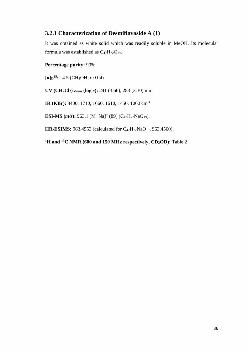
3.2.1 Characterization of Desmiflavaside A (1)
It was obtained as white solid which was readily soluble in MeOH. Its molecular
formula was established as C47H72O19.
Percentage purity: 90%
[α]D25: –4.5 (CH3OH, c 0.04)
UV (CH2Cl2) λmax (log ε): 241 (3.66), 283 (3.30) nm
IR (KBr): 3400, 1710, 1660, 1610, 1450, 1060 cm-1
ESI-MS (m/z): 963.1 [M+Na]+ (89) (C47H72NaO19).
HR-ESIMS: 963.4553 (calculated for C47H72NaO19, 963.4560).
1H and 13C NMR (600 and 150 MHz respectively, CD3OD): Table 2

37
Table-2: 1H and 13C NMR data of Desmiflavaside A (1)x,y
No. Multiplicity δH (J, Hz) δC
1. C-H2 1.76 (m), 1.02 (m) 38.3
2. C-H2 1.85 (m), 1.49 (m) 30.4
3. C-H 3.58 m 79.4
4. C-H2 1.63 (m), 1.29 (m) 35.2
5. C-H 1.00 (m) 45.6
6. C-H2 1.56 (m), 1.30 (m) 21.9
7. C-H2 1.32 (m), 1.20 (m) 30.0
8. C-H 1.72 (m) 41.4
9. C-H 1.06 (m) 50.5
10. C - 37.1
11. C-H2 2.09 (m), 1.03 (m) 28.4
12. C-H2 1.46 (m), 1.41 (m) 40.1
13. C - 48.0
14. C - 82.4
15. C-H 5.60 (m) 77.3
16. C-H2 2.50 (m), 1.75 (m) 27.3
17. C-H 1.62 (m) 54.8
18. C-H3 1.11 (s) 15.6
19. C-H3 0.84 (s) 12.8
20. C-H 4.03 (m) 66.0
21. C-H3 1.07 (d, 6.6) 21.9
Bz(15)
C=O C - 167.8
1ʹ C - 131.7
2ʹ, 6ʹ C-H, C-H 8.09 (dd, 2.0, 7.0) 130.7
3ʹ, 5ʹ C-H, C-H 7.47 (t, 7.0) 129.5
4ʹ C-H 7.60 (t, 7.0) 134.2

38
Table 2: Continued ………
No. Multiplicity δH (J, Hz) δC
Dig
1″ C-H 4.30 (d, 7.8) 102.8
2″ C-H 3.55 (m) 71.3
3″ C-H 3.18 (m) 85.7
4″ C-H 4.15 (d, 2.4) 74.8
5″ C-H 3.60 (m) 71.6
6″ C-H3 1.26 (d, 6.0) 17.5
OMe OCH3 3. 49 (s) 58.5
Glc-I
1‴ C-H 4.57 (d, 7.8) 104.1
2‴ C-H 3.19 (m) 75.8
3‴ C-H 3.33 (m) 77.8
4‴ C-H 3.26 (m) 71.8
5‴ C-H 3.43 (ddd, 2.0, 6.0, 8.3) 77.4
6‴ C-H2 4.12 (dd, 2.0, 12.0) 70.3
3.76 (dd, 6.0, 12.0)
Glc-II
1‴′ C-H 4.38 (d, 7.8) 105.0
2ʹ‴ C-H 3.16 (m) 75.1
3ʹ‴ C-H 3.32 (m) 78.0
4ʹ‴ C-H 3.25 (m) 71.6
5ʹ‴ C-H 3.24 (m) 78.0
6ʹ‴ C-H2 3.85 (dd, 2.4, 12.0) 62.0
3.64 (dd, 5.4, 12.0)
x Values were assigned using NOESY, 1H-1H COSY, HMBC and HSQC 2D-NMR
spectra, as well as by comparing with literature reports for other pregnane glycosides.
y DEPT experiments assessed for multiplicity determination and J values are
mentioned in parentheses.

39
3.2.2 Characterization of Desmiflavaside B (2)
It was obtained as white solid which was readily soluble in MeOH. Its molecular
formula was established as C47H70O19.
Percentage purity: 90%
[α]D25: –4.1 (CH3OH, c 0.05)
UV (CH2Cl2) λmax (log ε): 240 (3.30), 273 (2.90) nm
IR (KBr): 3350, 1710, 1665, 1625, 1450, 1060 cm-1
ESI-MS (m/z): 961.1 [M+Na]+ (85) (C47H70NaO19).
HR-ESIMS: 961.4403 (calculated for C47H70NaO19, 961.4404).
1H and 13C NMR (600 and 150 MHz respectively, CD3OD): Table 3

40
Table 3: 1H and 13C NMR data of Desmiflavaside B (2)x,y
No. Multiplicity δH (J, Hz) δC
1. C-H2 1.66 (m), 1.50 (m) 39.5
2. C-H2 1.84 (m), 1.48 (m) 30.3
3. C-H 3.59 (m) 79.4
4. C-H2 1.63 (m), 1.28 (m) 35.2
5. C-H 0.99 m 45.6
6. C-H2 1.58 (m), 1.33 (m) 21.7
7. C-H2 1.19 (m) 29.9
8. C-H 1.68 (m) 41.5
9. C-H 1.03 (m) 50.3
10. C - 37.1
11. C-H2 2.11 (m), 0.99 (m) 28.3
12. C-H2 1.66 (m), 1.50 (m) 39.5
13. C - 49.0
14. C - 83.7
15. C-H 5.63 (m) 77.3
16. C-H2 2.69 (m), 1.87 (m) 32.9
17. C-H 2.91 (m) 60.8
18. C-H3 1.03 (s) 16.4
19. C-H3 0.83 (s) 12.8
20. C - 218.2
21. C-H3 2.26 (s) 32.6
Bz (15)
C=O C 167.6
1ʹ C 131.5
2ʹ, 6ʹ CH, CH 8.08 (m) 129.6
3ʹ, 5ʹ CH, CH 7.48 (m) 130.7
4 ʹ CH 7.60 (m) 134.4
41
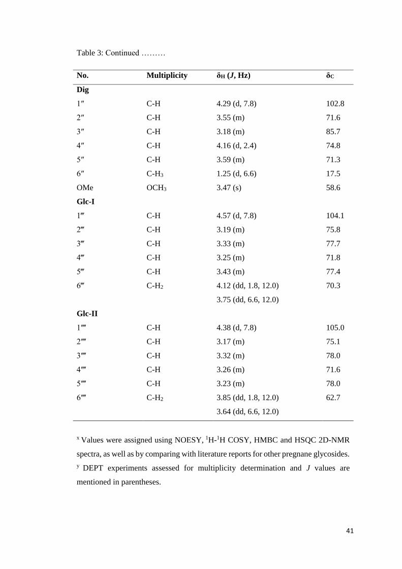
Table 3: Continued ………
No. Multiplicity δH (J, Hz) δC
Dig
1″ C-H 4.29 (d, 7.8) 102.8
2″ C-H 3.55 (m) 71.6
3″ C-H 3.18 (m) 85.7
4″ C-H 4.16 (d, 2.4) 74.8
5″ C-H 3.59 (m) 71.3
6″ C-H3 1.25 (d, 6.6) 17.5
OMe OCH3 3.47 (s) 58.6
Glc-I
1‴ C-H 4.57 (d, 7.8) 104.1
2‴ C-H 3.19 (m) 75.8
3‴ C-H 3.33 (m) 77.7
4‴ C-H 3.25 (m) 71.8
5‴ C-H 3.43 (m) 77.4
6‴ C-H2 4.12 (dd, 1.8, 12.0) 70.3
3.75 (dd, 6.6, 12.0)
Glc-II
1ʹ‴ C-H 4.38 (d, 7.8) 105.0
2ʹ‴ C-H 3.17 (m) 75.1
3ʹ‴ C-H 3.32 (m) 78.0
4ʹ‴ C-H 3.26 (m) 71.6
5ʹ‴ C-H 3.23 (m) 78.0
6ʹ‴ C-H2 3.85 (dd, 1.8, 12.0) 62.7
3.64 (dd, 6.6, 12.0)
x Values were assigned using NOESY, 1H-1H COSY, HMBC and HSQC 2D-NMR
spectra, as well as by comparing with literature reports for other pregnane glycosides.
y DEPT experiments assessed for multiplicity determination and J values are
mentioned in parentheses.

42
3.2.3 Characterization of Desmiflavaside C (3)
It was obtained as white solid which was readily soluble in MeOH. Its molecular
formula was established as C49H68O12.
Percentage purity: 85%
[α]D25: –4.5 (CH3OH, c 0.05)
UV (CH2Cl2) λmax (log ε): 217 (3.01), 227 (2.91) nm
IR (KBr): 3410, 1710, 1615, 1450, 1060 cm-1
ESI-MS (m/z): 887.1 [M+Na]+ (85) (C49H68NaO12).
HR-ESIMS: 871.4606 (calculated for C49H68NaO12, 871.4608).
1H and 13C NMR (600 and 150 MHz respectively, CD3OD): Table 4

43
Table 4: 1H and 13C NMR data of Desmiflavaside C (3)x,y
No. Multiplicity δH (J, Hz) δC
1. C-H2 1.66 (m), 1.04 (m) 38.0
2. C-H2 1.82 (m), 1.28 (m) 30.3
3. C-H 3.63 (m) 78.5
4. C-H2 1.63 (m), 1.20 (m) 35.5
5. C-H 1.13 (m) 45.6
6. C-H2 1.37 (m), 1.27 (m) 29.8
7. C-H2 2.03 (m), 1.13 (m) 28.6
8. C-H 1.69 (m) 41.2
9. C-H 1.08 (m) 47.4
10. C - 36.9
11. C-H2 1.72 (m), 1.46 (m) 27.5
12. C-H 4.95 (dd, 4.5, 12.0) 79.5
13. C - 53.7
14. C - 87.2
15. C-H2 1.89 (m), 1.67 (m) 32.3
16. C-H2 2.03 (m), 1.66 (m) 26.0
17. C-H 2.25 (m) 50.9
18. C-H3 1.09 (s) 10.0
19. C-H3 0.82 (s) 12.4
20. C-H 5.24 (dd, 6.0, 10.0) 75.6
21. C-H3 1.20 (d, 6.0) 19.7
Bz(12)
C=O C - 168.0
1ʹ C - 132.0
2ʹ, 6ʹ C-H, C-H 8.03 (d, 7.2) 130.7
3ʹ, 5ʹ C-H, C-H 7.50 (t, 7.2) 129.8
4 ʹ C-H 7.68 (t, 7.2) 134.3

44
Table 4: Continued ………
No. Multiplicity δH (J, Hz) δC
Bz(20)
C=O C - 167.4
1ʹ C - 132.0
2ʹ, 6ʹ C-H, C-H 7.73 (d, 7.2) 130.3
3ʹ, 5ʹ C-H, C-H 7.22 (t, 7.2) 129.4
4ʹ C-H 7.51 (t, 7.2) 133.8
CymI
1″ C-H 4.85 (dd, 1.8, 9.6) 97.1
2″ C-H2 2.04 (m), 1.51 (m) 36.6
3″ C-H 3.83 (m) 78.5
4″ C-H 3.25 (m) 83.8
5″ C-H 3.79 (dq, 6.0, 10.0) 69.9
6″ C-H3 1.19 (d, 6.0) 18.5
OMe O-CH3 3.41 (s) 58.4
CymII
1‴ C-H 4.57 (dd, 1.8, 9.6) 102.7
2‴ C-H2 2.31 (m), 1.34 (m) 37.3
3‴ C-H 2.96 (t, 9.0) 76.9
4‴ C-H 3.18 (m) 81.6
5‴ C-H 3.25 (dd, 6.0, 10.0) 73.2
6‴ C-H3 1.26 (d, 6.0) 18.3
OMe O-CH3 3.41 (s) 57.3
x Values were assigned using NOESY, 1H-1H COSY, HMBC and HSQC 2D NMR
spectra, as well as by comparing with literature reports for other pregnane glycosides.
y DEPT experiments assessed for multiplicity determination and J values are
mentioned in parentheses.

45
3.2.4 Characterization of Desmiflavaside D (4)
It was obtained as white solid which was readily soluble in MeOH. Its molecular
formula was established as C55H78O17.
Percentage purity: 90%
[α]D25: –4.1 (CH3OH, c 0.05)
UV (CH2Cl2) λmax (log ε): 217 (3.01), 227 (2.91) nm
IR (KBr): 3410, 1715, 1610, 1455, 1070 cm-1
ESI-MS (m/z): 1033.1 [M+Na]+ (89) (C55H78NaO17).
HR-ESIMS: 1033.5125 (calculated for C55H78NaO17, 1033.5137).
1H and 13C NMR (600 and 150 MHz respectively, CD3OD): Table 5
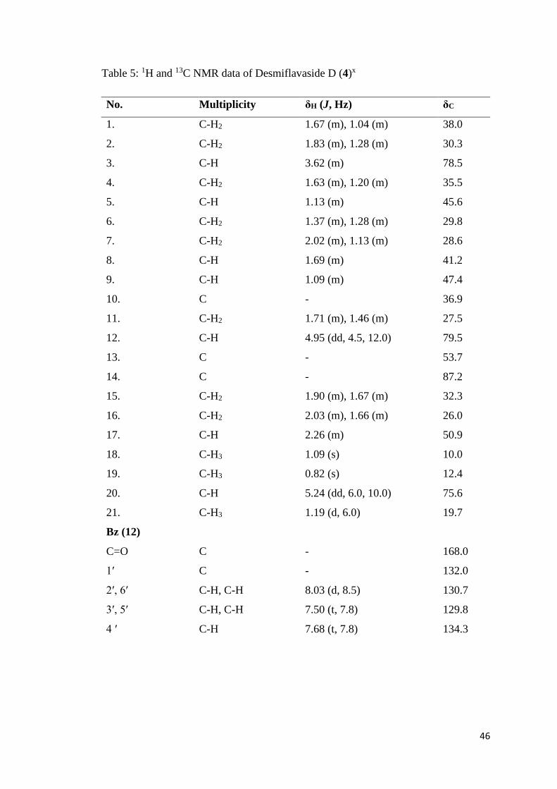
46
Table 5: 1H and 13C NMR data of Desmiflavaside D (4)x
No. Multiplicity δH (J, Hz) δC
1. C-H2 1.67 (m), 1.04 (m) 38.0
2. C-H2 1.83 (m), 1.28 (m) 30.3
3. C-H 3.62 (m) 78.5
4. C-H2 1.63 (m), 1.20 (m) 35.5
5. C-H 1.13 (m) 45.6
6. C-H2 1.37 (m), 1.28 (m) 29.8
7. C-H2 2.02 (m), 1.13 (m) 28.6
8. C-H 1.69 (m) 41.2
9. C-H 1.09 (m) 47.4
10. C - 36.9
11. C-H2 1.71 (m), 1.46 (m) 27.5
12. C-H 4.95 (dd, 4.5, 12.0) 79.5
13. C - 53.7
14. C - 87.2
15. C-H2 1.90 (m), 1.67 (m) 32.3
16. C-H2 2.03 (m), 1.66 (m) 26.0
17. C-H 2.26 (m) 50.9
18. C-H3 1.09 (s) 10.0
19. C-H3 0.82 (s) 12.4
20. C-H 5.24 (dd, 6.0, 10.0) 75.6
21. C-H3 1.19 (d, 6.0) 19.7
Bz (12)
C=O C - 168.0
1ʹ C - 132.0
2ʹ, 6ʹ C-H, C-H 8.03 (d, 8.5) 130.7
3ʹ, 5ʹ C-H, C-H 7.50 (t, 7.8) 129.8
4 ʹ C-H 7.68 (t, 7.8) 134.3

47
Table 5: Continued ………
No. Multiplicity δH (J, Hz) δC
Bz (20)
C=O C - 167.4
1ʹ C - 132.0
2ʹ, 6ʹ C-H, C-H 7.73 (d,7.8) 130.3
3ʹ, 5ʹ C-H, C-H 7.22 (t, 7.8) 129.4
4ʹ C-H 7.51 (t, 7.8) 133.8
CymI
1″ C-H 4.86 (dd, 2.0, 12.0) 97.1
2″ C-H2 2.04 (m), 1.51(m) 36.6
3″ C-H 3.82 (m) 78.5
4″ C-H 3.25 (m) 83.8
5″ C-H 3.80 (m) 69.8
6″ C-H3 1.19 (d, 6.0) 18.5
OMe O-CH3 3.41 (s) 58.4
CymII
1‴ C-H 4.59 (dd, 2.0, 12.0) 102.5
2‴ C-H2 2.32 (m), 1.43 (m) 37.9
3‴ C-H 3.40 (m) 80.1
4‴ C-H 3.26 (m) 83.5
5‴ C-H 3.39 (m) 72.7
6‴ C-H3 1.36 (d, 6.0) 18.7
OMe O-CH3 3.46 (s) 58.2
Glc
1ʹ‴ C-H 4.43 (d, 7.8) 104.1
2ʹ‴ C-H 3.15 (dd, 7.8, 9.0) 75.5
3ʹ‴ C-H 3.32 (t, 8.7) 78.0
4ʹ‴ C-H 3.22 (m) 71.8
5ʹ‴ C-H 3.22 (m) 78.3
6ʹ‴ C-H2 3.85 (dd, 2.0, 12.0) 63.0
3.62 (dd, 6.0, 12.0)
x Values were assigned using 1D and 2D NMR spectra.

48
3.2.5 Characterization of Nizwaside (5)
It was obtained as white solid which was readily soluble in MeOH. Its molecular
formula was established as C56H80O16.
Percentage purity: 80%
[α]D25: –4.0 (CH3OH, c 0.04)
UV (CH2Cl2) λmax (log ε): 229 (2.90), 219 (3.04) nm
IR (KBr): 1710, 3400, 1620, 1060, 1450 cm-1
ESI-MS (m/z): 1031.1 [M+Na]+ (88) (C56H80NaO16).
HR-ESIMS: 1031.5332 (calculated for C56H80NaO16, 1031.5338).
1H and 13C NMR (600 and 150 MHz respectively, CD3OD): Table 6

49
Table 6: 1H and 13C NMR data of Nizwaside (5)x
No. Multiplicity δH (J, Hz) δC
1. C-H2 0.94 (m), 1.65 (m) 38.0
2. C-H2 2.05 (m), 2.13 (m) 28.7
3. C-H 3.65 (m) 78.5
4. C-H2 1.22 (m), 1.68 (m) 35.5
5. C-H 1.27 (m) 45.6
6. C-H2 1.44 (m), 1.71 (m) 27.5
7. C-H2 1.53 (m), 2.01 (m) 36.6
8. C-H 1.70 (m) 41.2
9. C-H 1.10 (m) 47.4
10. C - 36.9
11. C-H2 1.66 (m), 2.02 (m) 26.0
12. C-H 4.95 (dd, 4.5, 12.0) 79.4
13. C - 53.7
14. C - 87.2
15. C-H2 1.38 (m), 1.83 (m) 29.8
16. C-H2 1.66 (m), 1.90 (m) 32.3
17. C-H 2.25 (m) 50.9
18. C-H3 1.09 (s) 10.0
19. C-H3 0.82 (s) 12.4
20. C-H 5.24 (dd, 6.0, 10.0) 75.7
21. C-H3 1.35 (d, 6.0) 18.9
Bz (12)
C=O C 168.0
1ʹ C 132.0
2ʹ/6ʹ C-H, C-H 8.03 (dd, 1.2, 8.4) 130.7
3ʹ/5ʹ C-H, C-H 7.50 (t, 8.4) 129.8
4ʹ C-H 7.67 (t, 8.4) 134.3

50
Table 6: Continued ………
No. Multiplicity δH (J, Hz) δC
Bz (20)
C=O C 167.4
1ʹ C 132.0
2ʹ/6ʹ C-H, C-H 7.73 (dd, 1.2, 8.4) 130.3
3ʹ/5ʹ C-H, CH 7.22 (t, 8.4) 129.5
4ʹ C-H 7.52 (t, 8.4) 133.9
Allom
1″ C-H 4.71 (d, 8.4) 102.6
2″ C-H 3.36 (m) 72.5
3″ C-H 3.61 (m) 83.7
4″ C-H 3.30 (m) 73.6
5″ C-H 3.33 (m) 72.5
6″ C-H3 1.19 (d, 6.0) 18.9
OMe O-CH3 3.59 (s) 62.5
Cym-I
1‴ C-H 4.57 (dd, 2.0, 9.0) 102.2
2‴ C-H2 1.22 (m), 1.67 (m) 35.5
3‴ C-H 3.83 (m) 78.5
4‴ C-H 3.67 (m) 83.8
5‴ C-H 3.64 (m) 71.2
6‴ C-H3 1.22 (d, 6.0) 18.2
OMe O-CH3 3.40 (s) 57.4
Cym-II
1ʹ‴ C-H 4.84 (dd, 2.0, 9.0) 97.1
2ʹ‴ C-H2 1.55 (m), 2.02 (m) 36.6
3ʹ‴ C-H 3.83 (m) 78.7
4ʹ‴ C-H 3.24 (m) 83.9
5ʹ‴ C-H 3.80 (m) 69.9
6ʹ‴ C-H3 1.20 (d, 6.0) 18.5
OMe O-CH3 3.41 (s) 58.4
x Values were assigned using 1D and 2D NMR spectra.

51
3.2.6 Characterization of Desflavaside A (6)
It was obtained as white solid which was readily soluble in MeOH. Its molecular
formula was established as C62H90O21.
Percentage purity: 90%
[α]D25: –5.4 (CH3OH, c 0.06)
UV (CH2Cl2) λmax (log ε): 219 (3.00), 229 (2.93) nm
IR (KBr): 3410, 1710, 1610, 1455, 1060 cm-1
ESI-MS (m/z): 1193.1 [M+Na]+ (90) (C62H90NaO21).
HR-ESIMS: 1193.5885 (calculated for C62H90NaO21, 1193.5872).
1H and 13C NMR (600 and 150 MHz respectively, CD3OD): Table 7

52
Table 7: 1H and 13C NMR data of Desflavaside A (6)x,y
No. Multiplicity δH (J, Hz) δC
1. C-H2 1.66 (m), 1.06 (m) 38.0
2. C-H2 1.82 (m), 1.28 (m) 30.3
3. C-H 3.62 (m) 78.5
4. C-H2 1.63 (m), 1.20 (m) 35.5
5. C-H 1.14 (m) 45.6
6. C-H2 1.38 (m), 1.28 (m) 29.8
7. C-H2 2.03 (m), 1.13 (m) 28.7
8. C-H 1.70 (m) 41.2
9. C-H 1.09 (m) 47.4
10. C 36.9
11. C-H2 1.72 (m), 1.47 (m) 27.5
12. C-H 4.94 (dd, 4.2, 12.0) 79.5
13. C 53.7
14. C 87.2
15. C-H2 1.89 (m), 1.67 (m) 32.3
16. C-H2 2.03 (m), 1.66 (m) 26.0
17. C-H 2.25 (m) 50.8
18. C-H3 1.09 (s) 10.0
19. C-H3 0.82 (s) 12.4
20. C-H 5.24 (m) 75.6
21. C-H3 1.20 (d, 6.6) 19.7
Bz (12)
C=O C 168.0
1ʹ C 132.0
2ʹ, 6ʹ C-H, C-H 8.03 (dd, 1.0, 6.0) 130.7
3ʹ, 5ʹ C-H, C-H 7.50 (t, 6.0) 129.8
4 ʹ C-H 7.67 (t, 6.0) 134.3

53
Table 7: Continued ………
No. Multiplicity δH (J, Hz) δC
Bz (20)
C=O C 167.4
1ʹ C 132.0
2ʹ, 6ʹ C-H, C-H 7.73 (dd, 1.0, 6.0) 130.3
3ʹ, 5ʹ C-H, C-H 7.22 (t, 6.0) 129.4
4ʹ C-H 7.51 (t, 6.0) 133.8
Cym-I C-H 4.85 (dd, 2.0, 6.0) 97.1
1″ C-H2 2.03 (m), 1.51 (m) 36.6
2″ C-H 3.36 (m) 80.4
3″ C-H 3.24 (m) 83.9
4″ C-H 3.81 (m) 69.9
5″ C-H3 1.18 (d,6.0) 18.5
6″ O-CH3 3.40 (s) 57.5
OMe
Glc
1‴ C-H 4.33 ( d, 7.2) 106.2
2‴ C-H 3.17 (m) 75.5
3‴ C-H 3.33 (m) 77.9
4‴ C-H 3.24 (m) 78.5
5‴ C-H 3.28 (m) 78.0
6‴ C-H2 3.90 (dd, 2.0, 11.7) 63.0
3.64 (dd, 6.0, 11.7)
Cym-II
1ʹ‴ C-H 4.56 (dd, 2.0, 10.0) 102.5
2ʹ‴ C-H2 2.30 (m), 1.40 (m) 37.5
3ʹ‴ C-H 3.82 (m) 78.5
4ʹ‴ C-H 3.17(m) 83.8
5ʹ‴ C-H 3.35 (m) 72.5
6ʹ‴ C-H3 1.29 (d, 6.0) 18.1
OMe O-CH3 3.41 (s) 58.4

54
Table 7: Continued ………
No. Multiplicity δH (J, Hz) δC
Allom
1″‴ C-H 4.70 (d, 8.0) 102.1
2″‴ C-H 3.31 (m) 72.9
3″‴ C-H 3.94 (t, 3.5) 83.2
4″‴ C-H 3.32 (m) 83.9
5″‴ C-H 3.81 (m) 70.1
6″‴ C-H3 1.35 (d, 6.0) 18.9
OMe O-CH3 3.58 (s) 61.9
x Values were assigned using NOESY, 1H-1H COSY, HMBC and HSQC 2D-NMR
spectra, as well as by comparing with literature reports for other pregnane glycosides.
y DEPT experiments assessed for multiplicity determination and J values are
mentioned in parentheses.
55
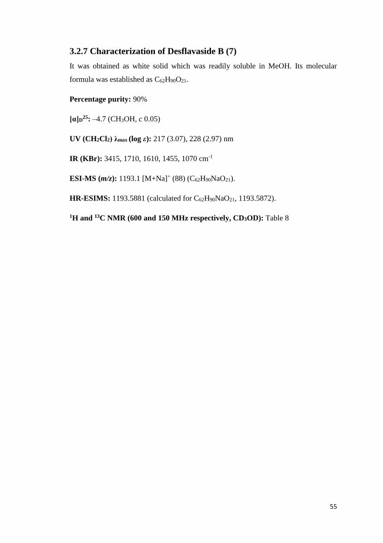
3.2.7 Characterization of Desflavaside B (7)
It was obtained as white solid which was readily soluble in MeOH. Its molecular
formula was established as C62H90O21.
Percentage purity: 90%
[α]D25: –4.7 (CH3OH, c 0.05)
UV (CH2Cl2) λmax (log ε): 217 (3.07), 228 (2.97) nm
IR (KBr): 3415, 1710, 1610, 1455, 1070 cm-1
ESI-MS (m/z): 1193.1 [M+Na]+ (88) (C62H90NaO21).
HR-ESIMS: 1193.5881 (calculated for C62H90NaO21, 1193.5872).
1H and 13C NMR (600 and 150 MHz respectively, CD3OD): Table 8
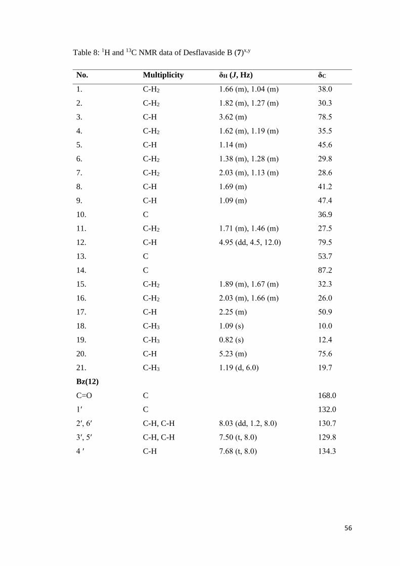
56
Table 8: 1H and 13C NMR data of Desflavaside B (7)x,y
No. Multiplicity δH (J, Hz) δC
1. C-H2 1.66 (m), 1.04 (m) 38.0
2. C-H2 1.82 (m), 1.27 (m) 30.3
3. C-H 3.62 (m) 78.5
4. C-H2 1.62 (m), 1.19 (m) 35.5
5. C-H 1.14 (m) 45.6
6. C-H2 1.38 (m), 1.28 (m) 29.8
7. C-H2 2.03 (m), 1.13 (m) 28.6
8. C-H 1.69 (m) 41.2
9. C-H 1.09 (m) 47.4
10. C 36.9
11. C-H2 1.71 (m), 1.46 (m) 27.5
12. C-H 4.95 (dd, 4.5, 12.0) 79.5
13. C 53.7
14. C 87.2
15. C-H2 1.89 (m), 1.67 (m) 32.3
16. C-H2 2.03 (m), 1.66 (m) 26.0
17. C-H 2.25 (m) 50.9
18. C-H3 1.09 (s) 10.0
19. C-H3 0.82 (s) 12.4
20. C-H 5.23 (m) 75.6
21. C-H3 1.19 (d, 6.0) 19.7
Bz(12)
C=O C 168.0
1ʹ C 132.0
2ʹ, 6ʹ C-H, C-H 8.03 (dd, 1.2, 8.0) 130.7
3ʹ, 5ʹ C-H, C-H 7.50 (t, 8.0) 129.8
4 ʹ C-H 7.68 (t, 8.0) 134.3

57
Table 8: Continued ………
No. Multiplicity δH (J, Hz) δC
Bz(20)
C=O C 167.4
1ʹ C 132.0
2ʹ, 6ʹ C-H, C-H 7.73 (dd, 1.0, 7.8) 130.3
3ʹ, 5ʹ C-H, C-H 7.22 (t, 7.8) 129.4
4ʹ C-H 7.51 (t, 7.8) 133.8
Cym-I
1″ C-H 4.85 (dd, 2.0, 9.6) 97.1
2″ C-H2 2.03 (m), 1.51 (m) 36.6
3″ C-H 3.82 (m) 78.5
4″ C-H 3.24 (m) 83.8
5″ C-H 3.80 (m) 69.9
6″ C-H3 1.19 (d,6.0) 18.8
OMe O-CH3 3.41 (s) 58.4
Glc
1‴ C-H 4.41( d, 7.8) 104.3
2‴ C-H 3.15 (m) 75.6
3‴ C-H 3.33 (m) 78.0
4‴ C-H 3.24 (m) 78.5
5‴ C-H 3.22 (m) 78.3
6‴ C-H2 3.85 (m), 3.62 (m) 63.1
Cym-II
1ʹ‴ C-H 4.57 (dd, 2.0, 9.6) 102.5
2ʹ‴ C-H2 2.30 (m), 1.40 (m) 37.6
3ʹ‴ C-H 3.36 (m) 80.2
4ʹ‴ C-H 3.19 (m) 84.3
5ʹ‴ C-H 3.37 (m) 72.5
6ʹ‴ C-H3 1.36 (d, 6.0) 18.4
OMe O-CH3 3.40 (s) 57.6

58
Table 8: Continued ………
No. Multiplicity δH (J, Hz) δC
Thev
1″‴ C-H 4.43 (d, 7.8) 104.2
2″‴ C-H 3.22 (m) 75.2
3″‴ C-H 3.16 (m) 86.3
4″‴ C-H 3.34 (m) 82.9
5″‴ C-H 3.42 (m) 72.6
6″‴ C-H3 1.36 (d,6.0) 18.5
OMe O-CH3 3.61 (s) 61.2
x Values were assigned using NOESY, 1H-1H COSY, HMBC and HSQC 2D NMR
spectra, as well as by comparing with literature reports for other pregnane glycosides.
y DEPT experiments assessed for multiplicity determination and J values are
mentioned in parentheses.

59
3.2.8 Characterization of Desflavaside C (8)
It was obtained as white solid which was readily soluble in MeOH. Its molecular
formula was established as C61H88O22.
Percentage purity: 90%
[α]D25: –6.1 (CH3OH, c 0.06)
UV (CH2Cl2) λmax (log ε): 220 (3.11), 228 (2.92) nm
IR (KBr): 3415, 1715, 1615, 1455, 1070 cm-1
ESI-MS (m/z): 1195.1 [M+Na]+ (81) (C61H88NaO22).
HR-ESIMS: 1195.5673 (calculated for C61H88NaO22, 1195.5665).
1H and 13C NMR (600 and 150 MHz respectively, CD3OD): Table 9

60
Table 9: 1H and 13C NMR data of Desflavaside C (8)x,y
No. Multiplicity δH (J, Hz) δC
1. C-H2 1.66 (m), 1.04 (m) 38.0
2. C-H2 1.82 (m), 1.27 (m) 30.3
3. C-H 3.64 (m) 78.5
4. C-H2 1.63 (m), 1.19 (m) 35.5
5. C-H 1.14 (m) 45.6
6. C-H2 1.38 (m), 1.28 (m) 29.8
7. C-H2 2.03 (m), 1.12 (m) 28.7
8. C-H 1.69 (m) 41.2
9. C-H 1.09 (m) 47.4
10. C 36.9
11. C-H2 1.71 (m), 1.47 (m) 27.5
12. C-H 4.95 (dd, 4.8, 12.0) 79.5
13. C 53.7
14. C 87.2
15. C-H2 1.89 (m), 1.67 (m) 32.3
16. C-H2 2.03 (m), 1.66 (m) 26.0
17. C-H 2.25 (m) 50.9
18. C-H3 1.09 (s) 10.0
19. C-H3 0.82 (s) 12.4
20. C-H 5.24 (m) 75.7
21. C-H3 1.19 (d, 6.6) 19.7
Bz (12)
C=O C 168.0
1ʹ C 131.9
2ʹ, 6ʹ C-H, C-H 8.03 (dd, 1.2, 7.8) 130.7
3ʹ, 5ʹ C-H, C-H 7.49 (t, 7.8) 129.8
4 ʹ C-H 7.68 (t, 7.8) 134.3

61
Table 9: Continued ………
No. Multiplicity δH (J, Hz) δC
Bz (20)
C=O C 167.4
1ʹ C 131.9
2ʹ, 6ʹ C-H, C-H 7.73 (dd, 1.2, 7.8) 130.3
3ʹ, 5ʹ C-H, C-H 7.22 (t, 7.8) 129.5
4ʹ C-H 7.51 (t, 7.8) 133.9
Cym-I
1″ C-H 4.86 (dd, 2.0, 6.0) 97.1
2″ C-H2 2.03 (m), 1.51 (m) 36.6
3″ C-H 3.82 (m) 78.5
4″ C-H 3.25 (m) 83.7
5″ C-H 3.80 (m) 69.9
6″ C-H3 1.19 (d, 6.0) 18.5
OMe O-CH3 3.41 (s) 58.4
Glc-I
1‴ C-H 4.47 ( d, 7.8) 104.0
2‴ C-H 3.22 (m) 74.9
3‴ C-H 3.38 (m) 76.7
4‴ C-H 3.50 (m) 80.9
5‴ C-H 3.50 (m) 76.4
6‴ C-H2 3.91 (dd, 12.0, 2.0) 62.1
3.80 (dd, 12.0, 6.0)
Glc-II
1ʹ‴ C-H 4.38 (d, 7.8) 104.6
2ʹ‴ C-H 3.22 (m) 75.3
3ʹ‴ C-H 3.33 (m) 78.1
4ʹ‴ C-H 3.28 (m) 79.5
5ʹ‴ C-H 3.33 (m) 77.8
6ʹ‴ C-H2 3.64 (dd, 6.0, 12.0) 62.4
3.86 (dd, 11.8, 2.1)

62
Table 9: Continued ………
No. Multiplicity δH (J, Hz) δC
Cym-II
1‴″ C-H 4.59 (dd, 1.7, 9.6) 102.5
2‴″ C-H2 3.32 (m), 1.43 (m) 37.7
3‴″ C-H 3.40 (m) 80.1
4‴″ C-H 3.25 (m) 83.8
5‴″ C-H 3.39 (m) 72.6
6‴″ C-H3 1.37 (d, 6.0) 18.7
OMe O-CH3 3.44 (s) 57.9
x Values were assigned using NOESY, 1H-1H COSY, HMBC and HSQC 2D-NMR
spectra, as well as by comparing with literature reports for other pregnane glycosides.
y DEPT experiments assessed for multiplicity determination and J values are
mentioned in parentheses.

63
3.2.9 Characterization of Desflavaside D (9)
It was obtained as white solid which was readily soluble in MeOH. Its molecular
formula was established as C46H78O22.
Percentage purity: 80%
[α]D25: –3.9 (CH3OH, c 0.04)
IR (KBr): 3410, 1715, 1610, 1450, 1075 cm-1
ESI-MS (m/z): 1005.1 [M+Na]+ (87) (C46H78NaO22).
HR-ESIMS: 1005.4876 (calculated for C46H78NaO22, 1005.4882).
1H and 13C NMR (600 and 150 MHz respectively, CD3OD): Table 10

64
Table 10: 1H and 13C NMR data of Desflavaside D (9)x,y
No. Multiplicity δH (J, Hz) δC
1. C-H2 1.74 (m), 0.99 (m) 38.3
2. C-H2 1.85 (m), 1.51 (m) 30.4
3. C-H 3.64 (m) 79.5
4. C-H2 1.69 (m), 1.31 (m) 35.3
5. C-H 1.07 (m) 45.7
6. C-H2 1.45 (m), 1.27 (m) 22.2
7. C-H2 1.32 (m), 1.26 (m) 30.0
8. C-H 1.60 (m) 41.4
9. C-H 0.91 (m) 50.9
10. C 36.9
11. C-H2 2.02 (m), 1.09 (m) 28.8
12. C-H2 1.37 (m), 1.26 (m) 42.0
13. C 54.7
14. C 85.7
15. C-H2 1.95 (m), 1.53 (m) 33.2
16. C-H2 1.90 (m), 1.81 (m) 20.4
17. C-H 1.63 (m) 57.7
18. C-H3 1.07 (s) 15.5
19. C-H3 0.83 (s) 12.6
20. C-H 3.98 (m) 79.0
21. C-H3 1.27 (d, 6.0) 21.3
Glc-I
1″ C-H 4.58 (d, 7.7) 104.1
2″ C-H 2.19 (m) 75.8
3″ C-H 3.34 (m) 77.9
4″ C-H 3.26 (m) 71.9
5″ C-H 3.44 (m) 77.4
6″ C-H2 4.13 (dd, 2.0, 11.6) 70.3
3.75 (dd, 2.0, 11.6)

65
Table 10: Continued ………
No. Multiplicity δH (J, Hz) δC
Glc-II
1‴ C-H 4.38 ( d, 7.8) 104.3
2‴ C-H 3.17 (m) 75.6
3‴ C-H 3.32 (m) 78.0
4‴ C-H 3.26 (m) 78.5
5‴ C-H 3.34 (m) 78.3
6‴ C-H2 3.86 (dd, 6.0, 11.6) 63.1
3.75 (dd, 12.0, 6.0)
Glc-III
1ʹ‴ C-H 4.36 (d, 7.9) 104.4
2ʹ‴ C-H 3.12 (m) 75.2
3ʹ‴ C-H 3.25 (m) 78.0
4ʹ‴ C-H 3.27 (m) 79.0
5ʹ‴ C-H 3.34 (m) 77.8
6ʹ‴ C-H2 3.84 (dd, 1.6, 11.6) 62.7
3.64 (dd, 6.0, 11.6)
Dig
1‴″ C-H 4.34 (d, 7.8) 102.7
2‴″ C-H 3.63 (m) 71.3
3‴″ C-H 3.22 (m) 85.8
4‴″ C-H 4.18 (d, 2.6) 74.8
5‴″ C-H 3.58 (m) 71.3
6‴″ C-H3 1.27 (d, 6.0) 17.6
OMe O-CH3 3.61 (s) 58.6
x Values were assigned using NOESY, 1H-1H COSY, HMBC and HSQC 2D-NMR
spectra, as well as by comparing with literature reports for other pregnane glycosides.
y DEPT experiments assessed for multiplicity determination and J values are
mentioned in parentheses.

66
3.3 Biological Activities
3.3.1 Anticancer Activity
Breast (MDA-MB-231) and ovarian (SKOV-3) cancer cells were maintained in
dulbecco's modified eagle medium (DMEM) and the media was supplemented with 1%
antimycotic antibiotic and 10% fetal bovine serum (FBS). Cancer cells were cultured
in 5% CO2 humidified atmosphere at 37 oC. Normal breast epithelial (MCF-10-2A) cell
line was propagated in DMEM/F-12 supplemented with 5% horse serum, 500 ng/ml
hydrocortisone, 20 ng/ml EGF, 10 mg/ml insulin, 0.1 mg/ml cholera toxin, 100 units/ml
penicillin, and 100 mg/ml streptomycin in a 5% CO2 atmosphere at 37 oC. A 5 mg/ml
stock solution of 3-(4,5-dimethyl-thiazol-2-yl)-2,5-diphenyltetrazolium bromide
(MTT) was prepared in phosphate buffer saline (PBS).
Cells were seeded in 96-well (1 x 10⁴/well) culture plates. After 24 hours of incubation,
normal growth medium was replaced with either fresh medium or various
concentrations of test samples in medium, diluted from a 2 mg/ml (for Doxorubicin,
CR, ME and its fractions) or 2 mM stock (for compounds 3-5). After 24 hours of
incubation, MTT solution was added to each well (5 mg/ml in PBS for Doxorubicin,
CR, ME and its fractions whereas 0.1 mg/ml in DMEM for compounds (3-5) and
incubated further for 4 hours at 37 oC. Upon termination, the supernatant was aspirated
and the MTT formazan, formed by metabolically viable cells, was dissolved in a
solubilization solution containing DMSO (100 μl) by mixing for 5 minutes on a
gyratory shaker. The absorbance was measured at 540 nm on an Ultra Multifunctional
Microplate Reader. Absorbance of control was considered as 100% cell survival.
Doxorubicin was used as positive control. Values are presented as Mean ± SD of four
duplicates.

67
Figure 6: Anticancer activity of crude crystals, methanolic extract and its fractions against breast cancer cells (MDA-MB-231).
0
20
40
60
80
100
120
CR ME HX DM EA BU AQ
Cel
l S
urv
ival
(%)
Control 25 ug/ml 50 ug/ml 75 ug/ml 100 ug/ml DOX 100 µg/ml
68
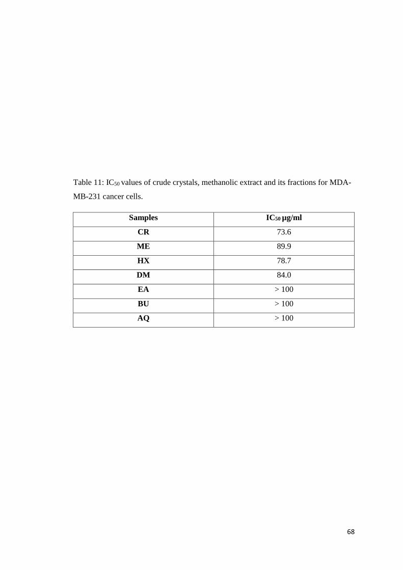
Table 11: IC50 values of crude crystals, methanolic extract and its fractions for MDA-
MB-231 cancer cells.
Samples IC50 µg/ml
CR 73.6
ME 89.9
HX 78.7
DM 84.0
EA > 100
BU > 100
AQ > 100
69
Compound 3 Compound 4 Compound 5
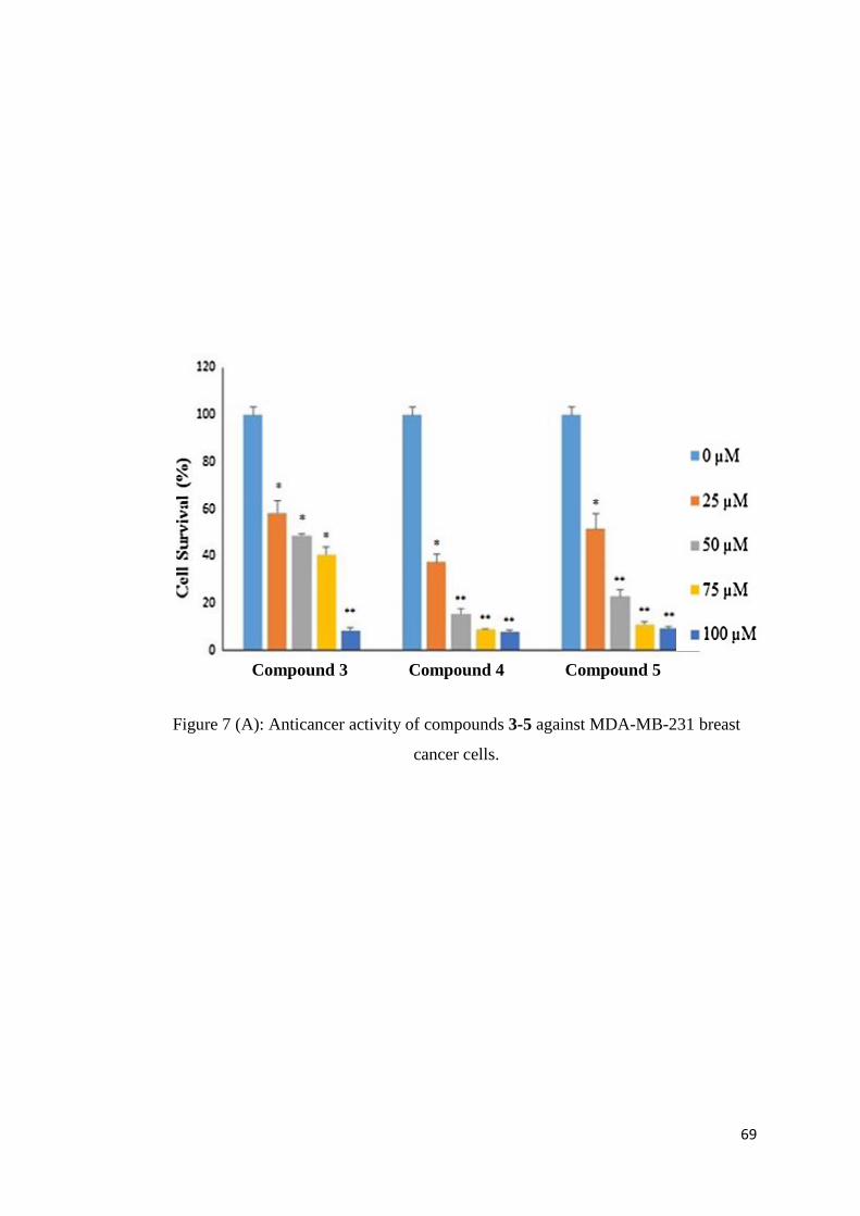
Figure 7 (A): Anticancer activity of compounds 3-5 against MDA-MB-231 breast
cancer cells.

70
Compound 3 Compound 4 Compound 5
Figure 7 (B): Anticancer activity of compounds 3-5 against SKOV-3 ovarian cancer
cells.
71
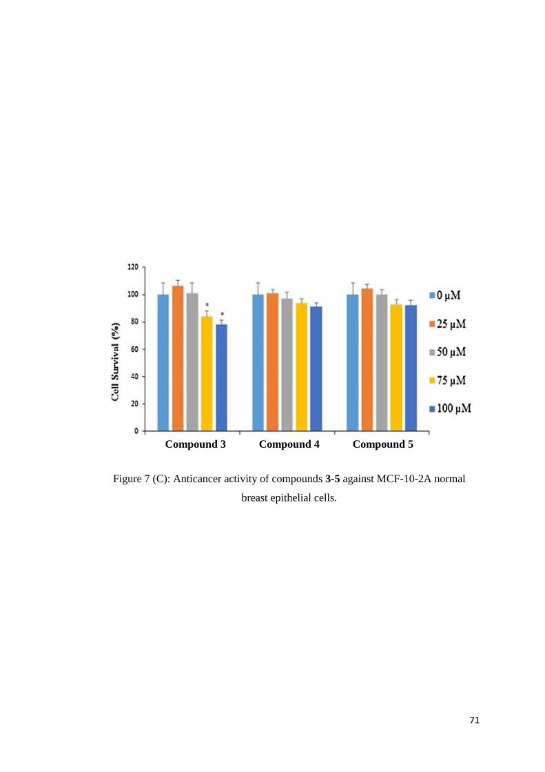
Compound 3 Compound 4 Compound 5
Figure 7 (C): Anticancer activity of compounds 3-5 against MCF-10-2A normal
breast epithelial cells.
72
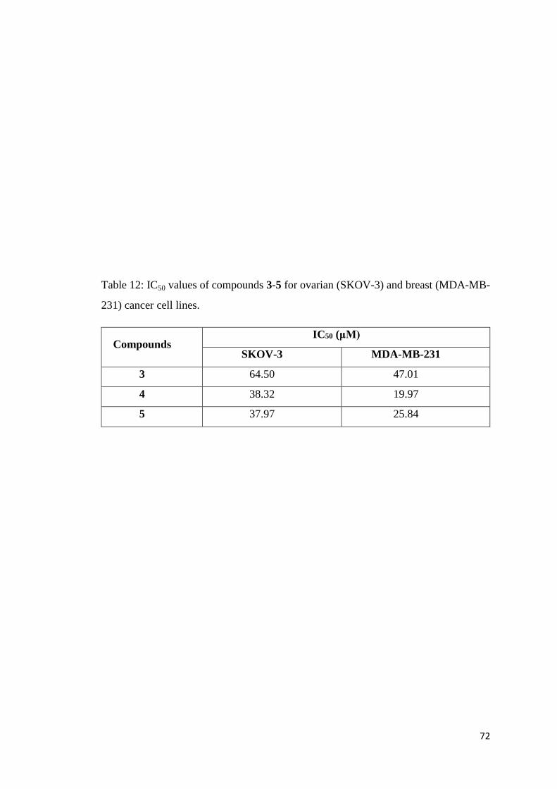
Table 12: IC50 values of compounds 3-5 for ovarian (SKOV-3) and breast (MDA-MB-
231) cancer cell lines.
Compounds IC50 (μM)
SKOV-3 MDA-MB-231
3 64.50 47.01
4 38.32 19.97
5 37.97 25.84
73
3.3.2 Enzyme Inhibition Activity
Urease Enzyme Inhibition: A solution comprising 25 μL of Jack bean Urease, 55 μL
of phosphate buffer and 100 mM urea was incubated with 5 μL (0.5 mg/mL) of the test
samples (CR, HX, DM, EA, BU, AQ, compounds 1-5) at 30 °C for 15 minutes in 96-
well plate. The production of NH3 was measured by indophenol method and used to
find the urease inhibitory activity. The phenol reagent (45 μL, 1% w/v C6H5OH and
0.005% w/v C5FeN6Na2O) and alkali reagent (70 μL, 0.5% w/v NaOH and 0.1%
NaOCl) were added to each well and the absorbance was measured at 630 nm after fifty
minutes, using a microplate reader (Molecular Device, USA). The assays were
performed at pH 8.2 (0.01 M K2HPO4.3H2O, 1.0 mM EDTA and 0.01 M LiCl2). The
experiment was replicated three times.
α-Glucosidase enzyme Inhibition: In this assay, 0.1 mg of α-glucosidase (type-1, from
Saccharomyces cerevisiae) was dissolved in 10 mL of phosphate buffer (pH 6.8). In
96-well plate, 20 μL of sample (CR, HX, DM, EA, BU, AQ, compounds 1-5) of
concentration 0.5 mg/mL was premixed with 120 μL of 50 mM phosphate buffer (pH
6.8) and 20 μL of 5 mM p-nitrophenyl α-D-glucopyranoside. The reaction mixture was
pre-incubated at 37 ◦C for 5 minutes. After that 20 μL α-glucosidase was added in
reaction wells and incubated at 37 ◦C for 15 minutes. The reaction was terminated by
the addition of 100 μL Na2CO3 (200 mM). Inhibition activity was determined
spectrophotometrically at 400 nm on spectrophotometer (SpectroMax Molecular
Devices, USA).
Acetylcholinesterase Enzyme Inhibition: In this assay, 96-well plate was used and
each reaction well contained 25 μL of 15 mM acetylcholinesterase (type VI-S from
electric eel) dissolved in water, 125 μL of 3 mM DTNB in Buffer (50 mM Tris–HCl,
pH 8.0), and 25 μL of test sample (CR, HX, DM, EA, BU, AQ, compounds 1-5) of
concentration 0.5 mg/mL dissolved in DMSO. In blank sample 25 μL inhibitor sample
was replaced by 25 μL of Tris-HCl buffer. In control sample also 25 μL of Tris-HCl
buffer was used in place of 25 μL of sample. 96-well plate was then incubated at 25 oC
for 15 minutes. Thereafter, 25 μL acetylcholinesterase (0.2 U/mL) was added to the
wells and the absorbance was measured five times consecutively after every 45 seconds.
The reaction was monitored for 5 minutes at 405 nm.
The percentage inhibition for above enzymes assays were calculated using the equation:
Inhibition % = 100 – (ODtest well
/ODcontrol
) × 100.

74
Table 13: Enzyme inhibition activities of crude crystals and fractions of methanolic
extract.
Samples
(0.5 mg/ml)
Urease enzyme
inhibition (%)
α-Glucosidase
enzyme Inhibition
(%)
Acetylcholinesterase
Inhibition (%)
CR 15 NA NA
HX 10 NA NA
DM NA 20 10
EA 12 15 10
BU NA NA 15
AQ NA 10 12
Thiourea 90 - -
Acrabose - 72 -
Galantamine - - 90
NA = Not Active
The values are mean of three replicates.
75
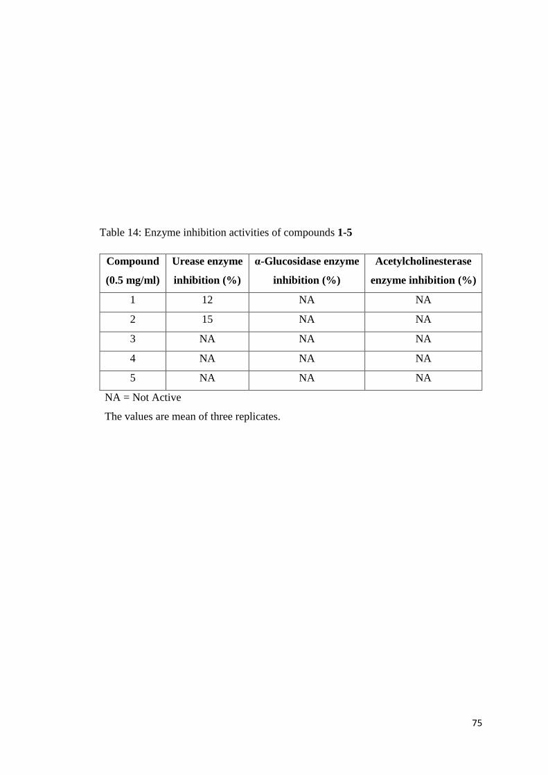
Table 14: Enzyme inhibition activities of compounds 1-5
Compound
(0.5 mg/ml)
Urease enzyme
inhibition (%)
α-Glucosidase enzyme
inhibition (%)
Acetylcholinesterase
enzyme inhibition (%)
1 12 NA NA
2 15 NA NA
3 NA NA NA
4 NA NA NA
5 NA NA NA
NA = Not Active
The values are mean of three replicates.
76
3.3.3 DPPH radical scavenging activity
Free radical scavenging capacity of CR, fractions (HX, DM, EA, BU, AQ) and
compounds 1-5 was determined by measuring the change in absorbance of DPPH (l,l-
Diphenyl-2-picrylhydrazyl radical) at 515 nm using spectrophotometer. In this assay,
reaction mixture was comprised of 95 µL (0 .3mM) of methanolic solution of DPPHֹ
and 5 µL of the test samples (0.5 mg/mL) dissolved in DMSO. Ascorbic acid was used
as standard. Following equation was used for measuring the scavenging activity.

77
Table 15: DPPH radical scavenging activity of crude crystals and fractions of
methanolic extract.
Fraction (0.5 mg/mL) DPPH radical scavenging (%)
CR 12
HX 28
DM 25
EA 15
BU 30
AQ NA
Ascorbic Acid 95
NA = Not Active
The values are mean of three replicates.

78
Table 16: DPPH radical scavenging activity of compounds 1-5
Compounds (0.5 mg/mL) DPPH radical scavenging (%)
1 10
2 15
3 NA
4 NA
5 15
NA = Not Active
The values are mean of three replicates.

79
3.4 Molecular Docking Studies
Compounds 3-5 were used as ligand molecules and their structures were obtained by
using ChemBioDraw software. The three dimensional structure of the protein tyrosine
phosphatases (PTPs) was obtained from the protein Data Bank (PDBID: 4RH5). The
MOL format was first converted to SMILE format using an OpenBabel tool and then
transferred onto the Molecular Operating Enviroment (MOE) software. The coordinates
of the ligand compounds and target protein were optimized by MOE software which
represented the most stable conformation and minimum energy. The drugable
properties (toxicity, molecular weight and partition coefficient) of these pregnane
glycosides were evaluated using Molinspiration server
[http://www.molinspiration.com].
Docking studies were carried out to investigate any binding interactions between the
PTPs target sites with compounds 3-5 by employing MOE software. Default parameters
were used and the energy of binding interaction at every step of the simulation was
measured using atomic affinity potentials. Docking studies were performed by Dr. Syed
Aun Muhammad, Bahauddin Zakariya University, Multan, Pakistan.

80
Figure 8: (A) Protein tyrosine phosphatase binding sites.
81
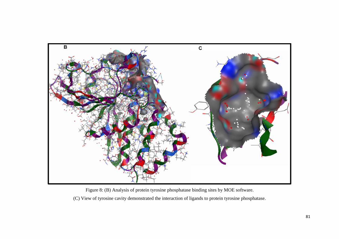
Figure 8: (B) Analysis of protein tyrosine phosphatase binding sites by MOE software.
(C) View of tyrosine cavity demonstrated the interaction of ligands to protein tyrosine phosphatase.

82
Figure 9 (A): Molecular docking views of ligand (3).

83
Figure 9 (B): Molecular docking views of ligand (4).

84
Figure 9 (C): Molecular docking views of ligand (5).

85
Figure 10: Heat map showing the toxicity analysis and pharmacokinetics of compounds 3-5.
86
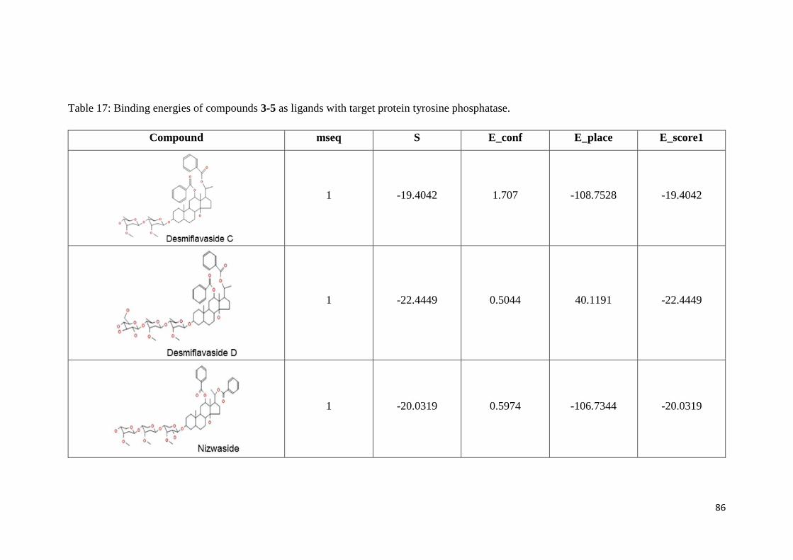
Table 17: Binding energies of compounds 3-5 as ligands with target protein tyrosine phosphatase.
Compound mseq S E_conf E_place E_score1
1 -19.4042 1.707 -108.7528 -19.4042
1 -22.4449 0.5044 40.1191 -22.4449
1 -20.0319 0.5974 -106.7344 -20.0319

87
4. RESULTS
AND
DISCUSSION

88
4.1 New Compounds from Caralluma flava
4.1.1 Desmiflavaside A (1)
The IR spectrum of compound 1 showed bands at 1610, 1710 and 3400 cm-1 which
indicated the presence of aryl ring, ester and hydroxyl functionalities in the molecule.
The ESIMS data analysis showed pseudo-molecular ion peak at m/z 963.1 [M+Na] in
positive ion mode and HRESIMS demonstrated quasi-molecular ion peak at m/z
963.4553 [M+Na]+ (calculated for C47H72NaO19, 963.4560). Based on HRESIMS and
13C NMR data, the molecular formula of the compound was suggested to be C47H72O19.
The 13C NMR (Broad band decoupled and DEPT) displayed signals of 47 carbons
which include 28 carbons of aglycone moiety and 19 carbons of glycoside part.
The structure of the aglycone portion was interpreted from NMR data (Table 2) which
demonstrated one doublet of secondary methyl at δH 1.07 (Me-21); δC 21.9 and two
singlets of tertiary methyls at δH 0.84; δC 12.8 and δH 1.11; δC 15.6 assigned to Me-19
and Me-18, respectively. It further showed signals of seven methines including two
oxymethines at δH 3.58 (m, 1H, H-3); δC 79.4 and δH 5.60 (t, J = 8.4 Hz, 1H, H-15); δC
77.3 and two quaternary carbons (C-10 and C-13) appeared at δC 37.1 and 48.0,
respectively (Abdallah et al., 2013; Abdel-Sattar et al., 2001, 2002, 2007; Hayashi et
al., 1988). Moreover, the signal of oxygenated quaternary carbon at δ 82.4 is
characteristic for pregnanes having an OH group at C-14 (Elsebai et al., 2015; Tanaka
et al., 1990). The significant HMBC interactions (Fig. 11) of Me-19 with C-1, C-5, C-
9 and C-10; Me-18 with C-12, C-13, C-14, and C-17; H-20 with C-13, C-16, C-17 and
C-21; H-15 with C-13, C-14, C-16 and C-17 justified the 3,14,20-trioxygenated
pregnane skeleton (Abdallah et al., 2013; Abdel-Sattar et al., 2001, 2002, 2007; Tanaka
et al., 1990).
The presence of one benzoyl group was indicated by its 1H NMR signals at δH 8.09 (dd,
J = 2.0, 7.0 Hz, H-2′/H-6′), δH 7.47 (t, J = 7.0 Hz, H-3′/H-5′), δH 7.60 (t, J = 7.0 Hz, H-
4′) and from an ester carbonyl resonance at δC 167.8 in the 13C NMR spectrum
(Abdallah et al., 2013; Abdel-Sattar et al., 2002; Tanaka et al., 1990; Hayashi et al.,
1988). The attachment of this benzoyl group to C-15 was confirmed by the HMBC
interaction of H-15 (δH 5.60) with benzoyl ester C=O (δC 167.8). The structure of
aglycone part was confirmed by its important connectivities in 1H-1H COSY and 1D
TOCSY spectra (Fig. 12).

89
The presence of three sugar anomeric proton signals at δH 4.57 (d, H-1ʹʹ); δH 4.38 (d,
H-1ʹʹʹ) and δH 4.30 (d, H-1ʹʹʹʹ); one Me group at δH 1.26 (d, H-6''); and one sugar OMe
group at δH 3.49 (s) in the NMR spectra suggesting it to be a triglycoside (Abdallah et
al., 2013; Abdel-Sattar et al., 2007; De Leo et al., 2005). The 13C NMR signals at δC
102.8 (C-1ʹʹ), 71.3 (C-2ʹʹ), 85.7 (C-3ʹʹ), 74.8 (C-4ʹʹ), 71.6 (C-5ʹʹ), 17.5 (C-6ʹʹ), 58.5
(OMe); δC 104.1 (C-1ʹʹʹ), 75.8 (C-2ʹʹʹ), 77.8 (C-3ʹʹʹ), 71.8 (C-4ʹʹʹ), 77.4 (C-5ʹʹʹ), 70.3 (C-
6ʹʹʹ) and δC 105.0 (C-1ʹʹʹʹ), 75.1 (C-2ʹʹʹʹ), 78.0 (C-3ʹʹʹʹ), 71.6 (C-4ʹʹʹʹ), 78.0 (C-5ʹʹʹʹ), 62.0
(C-6ʹʹʹʹ) resulted in establishing the presence of one D-digitalose and two glucoside
sugar units (Abdel-Sattar et al., 2001; Lee-Juian et al., 1994; Al-Massarani et al., 2012).
The β-linkages of the three sugar units were evident from the large coupling constants
(J = 7.8) of the anomeric protons (H-1ʹʹ, H-1ʹʹʹ, H-1ʹʹʹʹ) (Abdel-Sattar et al., 2007; De
Leo et al., 2005).
The Selective 1D TOCSY NMR experiment was employed on the anomeric proton
signals of the three hexose units at δH 4.30 (d, J =7.8), 4.38 (d, J =7.8) and 4.57 (d, J
=7.8) and their respective resonances H-1 to H-6 were assigned. Further, the NOESY,
COSY, HSQC and HMBC correlations resulted in confirming the presence of one D-
digitalose and two glucoside sugar units (Abdel-Sattar et al., 2001; Lee-Juian et al.,
1994). This was aided by comparison with previous reports for similar systems. The
identification of digitalose unit in desmiflavaside A (1) was assessed from its upfield
shifted OMe group at δH 3.49, δC 58.5 in comparison to the downfield OMe groups of
allomerose and thevetose (δH > 3.55, δC > 61.0). In addition, the characteristic doublet
at δH 4.15, assigned to H-4 of the digitalose moiety, had a distinctly small coupling
constant (d, J =2.4 Hz) that was consistent with the equatorial orientation of this proton
and its weak coupling to the axially oriented H-3 and undetected coupling to H-5.
Furthermore, the presence of a NOESY correlation between H-3/H-4 (δH 3.18/ δH 4.15),
and the absence of a similar correlation between H-2/H-4 (δH 3.55) required an
equatorial orientation of H-4 (Abdel-Sattar et al., 2001; Al-Massarani et al., 2012).
The connectivities of the sugar units to each other and of the digitalose sugar unit to
pregnane skeleton was confirmed via key HMBC correlations (Fig. 11): D-digitalose
anomeric proton H-1ʹʹ and C-3; H-3 and C-1ʹʹ; D-glucose-1 anomeric proton H-1ʹʹʹ and
C-4ʹʹ; H-4ʹʹ and C-1ʹʹʹ; D-glucose-2 anomeric proton H-1ʹʹʹʹ with C-4ʹʹʹ; and H-4ʹʹʹ with
C-1ʹʹʹʹ.
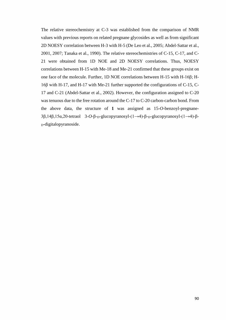
90
The relative stereochemistry at C-3 was established from the comparison of NMR
values with previous reports on related pregnane glycosides as well as from significant
2D NOESY correlation between H-3 with H-5 (De Leo et al., 2005; Abdel-Sattar et al.,
2001, 2007; Tanaka et al., 1990). The relative stereochemistries of C-15, C-17, and C-
21 were obtained from 1D NOE and 2D NOESY correlations. Thus, NOESY
correlations between H-15 with Me-18 and Me-21 confirmed that these groups exist on
one face of the molecule. Further, 1D NOE correlations between H-15 with H-16β; H-
16β with H-17, and H-17 with Me-21 further supported the configurations of C-15, C-
17 and C-21 (Abdel-Sattar et al., 2002). However, the configuration assigned to C-20
was tenuous due to the free rotation around the C-17 to C-20 carbon-carbon bond. From
the above data, the structure of 1 was assigned as 15-O-benzoyl-pregnane-
3β,14β,15α,20-tetraol 3-O-β-D-glucopyranosyl-(1→4)-β-D-glucopyranosyl-(1→4)-β-
D-digitalopyranoside.

91
OH
OH
OO
O
MeO
OO
HO
OHO
HO
OH1''
3''
1'''3'''
6''
6'''6''''
1''''3''''
1
4
6
9 14
131115
21
19
OH
OHOH
OHO
O
H
Figure 11: Significant HMBC interactions of 1
OH
OH
OO
O
MeO
OO
HO
OHO
HO
OH1''
3''
1'''3'''
6''
6'''6''''
1''''3''''
1
4
6
9 14
131115
21
19
OH
OHOH
OHO
O
H
Figure 12: — Significant 1H-1H COSY and 1D TOCSY interactions of 1

92
4.1.2 Desmiflavaside B (2)
Compound 2 showed pseudo-molecular ion peak at m/z 961.1 [M+Na] in positive ion
mode ESIMS. Based on the molecular ion peak in the HRESIMS spectrum (observed
961.4403 (calculated for C47H70NaO19, 961.4404) its molecular formula was found to
be C47H70O19.
The spectral data of 2 illustrated its close resemblance with desmiflavaside A (1). The
1H and 13C NMR resonances (Table 3) for the aglycone part and three sugar moieties
were almost similar to those of compound 1 (Table 2) except with few differences in
the pregnane skeleton. The most prominent changes appeared in the chemical shifts of
the methyl doublet at δH 1.07 (d, 6.6 Hz, H-21); δC 21.9 and oxymethine multiplet at δH
4.03 (m, H-20); δC 66.0 present in 1 that were absent and replaced by a singlet methyl
signal at δH 2.26 (s, H-21); δC 32.6 and a ketone signal (δC 218.2) in the NMR spectra
of 2 (Abdel-Sattar et al., 2002). This was further confirmed from HRESIMS of 2 which
illustrated two units of mass less than 1 (961.4403 vs 963.4553). Moreover, the
dissimilarities were also evident by comparison of HMBC interactions (Fig. 13); Me-
21 to C-17 and C-20; H-17 to C-20 and C-21. Therefore, the structure of compound 2
was elucidated as 15-O-benzoyl-pregnane-3β,14β,15α-triol 3-O-β-D-glucopyranosyl-
(1→4)-β-D-glucopyranosyl-(1→4)-β-D-digitalopyranoside.
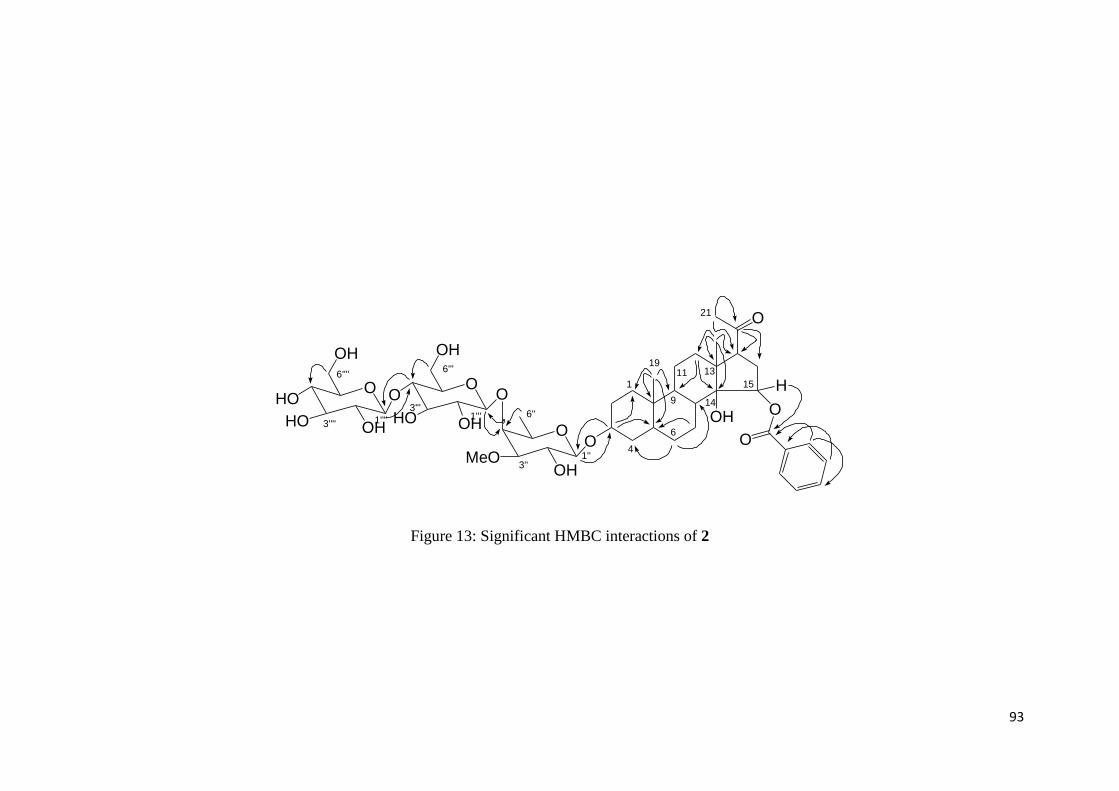
93
O
OH
OO
O
MeO
OO
HO
OHO
HO
OH1''
3''
1'''3'''
6''
6'''6''''
1''''3''''
1
4
6
9 14
131115
21
19
OH
OHOH
OHO
O
H
Figure 13: Significant HMBC interactions of 2

94
4.1.3 Desmiflavaside C (3)
The IR spectrum of compound 3 exhibited bands at 3410, 1710 and 1615 cm-1
indicating the presence of OH, ester C=O and aryl group in the molecule.
Desmiflavaside C showed a pseudo-molecular ion peak in ESIMS at m/z 871.4 [M+Na]
corresponding to the molecular formula C49H68O12 based on HRESIMS spectrum
(observed 871.4606 (calculated for C49H68NaO12, 871.4608). The 1H NMR spectrum
(Table 4) indicated two singlet resonances for two tertiary methyl protons at δ 1.09 (3H,
s, Me-18) and 0.82 (3H, s, Me-19) and one three protons doublet resonance for a
secondary Me group at δ 1.20 (3H, d, J= 6.0 Hz, Me-21). Its 13C NMR spectrum also
supported the presence of these three Me groups by displaying signals at δ 10.0 (Me-
18), 12.4 (Me-19), and 19.7 (Me-21), respectively. The presence of three oxymethine
groups in molecule were supported by its NMR spectra which displayed signals at δH
3.63 (1H, m, H-3); δC 78.5, δH 4.95 (1H, J= 4.5, 12.0 Hz, dd, H-12); δC 79.5 and δH
5.24 (1H, J= 6.0, 10.0 Hz, dd, H-20); δC 75.6 along with signals of two quaternary
carbons at δ 36.9 (C-10), 53.7 (C-13) and one oxygenated quaternary carbon at δ 87.2
(C-14) (Abdallah et al., 2013; Abdel-Sattar et al., 2007, 2002, 2001; Tanaka et al.,
1990). All the above data confirmed the pregnane skeleton of compound 3. Further
complete structure confirmation was achieved by COSY, 1D 1H-1H TOCSY (Fig. 15)
and HMBC correlations (Fig. 14). The 3,14 dioxygenated pregnane moiety was
confirmed by its key HMBC interactions between Me-21 (δ 1.19) with C-13, C-17, and
C-20; H-20 (δ 5.24) with C-13, C-16, C-17 and C-21; Me-19 (δ 0.82) with C-1, C-5,
C-9 and C-10; Me-18 (δ 1.09) and C-12, C-13, C-14, and C-17 (Abdallah et al., 2013;
Abdel-Sattar et al., 2007, 2002, 2001; Tanaka et al., 1990).
The 1H NMR spectra of desmiflavaside C showed signals for two benzoyl groups Bz-
12: δH 8.03 (J= 7.2 Hz, br d, H-2'/H-6'), 7.68 (J= 7.2 Hz, t, H-4'), 7.50 (J= 7.2 Hz, d, H-
3'/H-5') and Bz-20: δH 7.73 (J= 7.2 Hz, br d, H-2'/H-6'), 7.50 (J= 7.2 Hz, t, H-4'), 7.22
(J= 7.2 Hz, d, H-3'/H-5') (Abdallah et al., 2013; Tanaka et al., 1990; Hayashi et al.,
1988). These two benzoyl moieties were attached to C-12 and C-20 based on significant
HMBC interactions of H-12 and H-20 to the C=O resonances at δc 168.0 and 167.4,
respectively.
The NMR spectra displayed two sugar anomeric signals at δH 4.85 (J = 2.0, 12.0 Hz,
dd, H-1ʹʹ); δC 97.1 (C-1ʹʹ) and 4.57 (J = 1.8, 9.6 Hz, dd, H-1ʹʹ′); δC 102.7 (C-1ʹʹ′). NMR

95
spectra further exhibited two sugar methyl group signals at δH 1.19 (J = 6.0 Hz, d, H-
6′′); δC 18.5 (C-6ʹʹ) and δH 1.26 (J = 6.0 Hz, d, H-6‴); δc 18.3 (C-6‴) and two sugar OMe
group signals at δH 3.41 (s); δC 58.4 and δH 3.41 (s); δC 57.3 which indicated compound
3 to be a diglycoside with a β-linkage (J = 8.4–9.6 Hz; large coupling constant) (De
Leo et al., 2005; Abdel-Sattar et al., 2007).
The selective 1D TOCSY interactions showed the presence of two cymarose sugar units
which were confirmed by significant HSQC and HMBC interactions and were aided by
comparing NMR data with previous reports on cymarose (Abdel-Sattar et al., 2002,
2007). Attachment of the disaccharide chain to C-3 of the pregnane moiety was
confirmed from HMBC correlations between first cymarose anomeric proton (H-1ʹʹ)
and C-3. It was interesting to note that one cymarose unit was glycosylated at C-4 and
was evident from the downfield shift observed for C-4Cym-I [δ 83.8 (C-4Cym-I) vs 81.6
(C-4Cym-II)]. Moreover 3J HMBC cross peaks between H-1ʹʹʹ (H-1Cym-II) and C-4ʹʹ (C-
4Cym-I) indicated the sequence of the disaccharide chain.
The relative stereochemistry at C-3 was established by the significant NOESY
interaction of H-3 with H-5 and was confirmed by comparison of NMR data of C-3 [δH
3.65 (m, H-3); δC 78.5 (C-3)] with previous reports on related pregnane glycosides [Lit:
δH 3.63-3.68 (m, H-3); δC 78.1-78.4 (C-3)] (De Leo et al., 2005). Furthermore, the α-
configuration of H-12 and H-17 were positively identified based on NOE interactions
between H-12 with H-11α and H-17α and was further supported from the lack of
interaction of H-12 with Me-18. On the other hand, the stereochemistry of C-20 was
identified to be S from the NOE interaction of H-20 with H-21 and H-18, and H-l6α
with H-21 and the lack of NOE interactions between H-21 and H-18 (Tanaka et al.,
1990; Qiu et al., 1997; Abdel-Sattar et al., 2001 and 2007; Itokawa et al., 1988).
However, the configuration assigned to C-20 was tenuous due to the free rotation
around the C-17 to C-20 carbon-carbon bond. Based on above mentioned data, the
structure of 3 was elucidated as 12,20-di-O-benzoyl-20S-pregnane-3β,12β,14β,20-
tetraol 3-O-β-D-cymaropyranosyl-(1→4)-β-D-cymaropyranoside.

96
O
OH
OOO
OMe
OHO
OMe
1''3''1'''3'''
6''6'''
1
4
6
9 14
131115
21
19O
OO
H
H
Figure 14: Significant HMBC interactions of 3
O
OH
OOO
OMe
OHO
OMe
1''3''1'''3'''
6''6'''
1
4
6
9 14
131115
21
19O
OO
H
H
Figure 15: — Significant 1H-1H COSY and 1D TOCSY interactions of 3

97
4.1.4 Desmiflavaside D (4)
Compound 4 showed molecular ion peak at m/z 1033.5 [M+Na]+ in ESIMS having
molecular formula C55H78O17 obtained through HR-ESIMS (observed 1033.5125
(calculated for C55H78NaO17, 1033.5137).
The IR spectrum of 4 was identical to desmiflavaside C (3) having same functional
groups such as hydroxyl, carbonyl and aromatic ring (3410, 1715 and 1610 cm-1).
The NMR spectral data of 4 (Table 5) was comparable to 3 (Table 4) as it possesses
same pregnane aglycone skeleton and two benzoyl units present at C-12 and C-20.
Further, the three sugar units in 4 were positively suggested from the anomeric signals
at δH 4.86; δC 97.1; δH 4.59; δC 102.5 and δH 4.43; δC 104.1. The two cymarose sugar
units along with their attachments were completely identical to those in 3 but an
additional glucose unit was found in 4 which was present at C-4 of Cymarose-II. This
was positively identified from the significant HMBC interaction (Fig. 16) of H-4''' to
C-1'''' and from the downfield shift in the 13C NMR value of C-4''' (δ 83.5) in 4 compared
to C-4''' (δ 81.6) in 3. Thus, the structure of desmiflavaside D was characterized as
12,20-di-O-benzoyl-20S-pregnane-3β,12β,14β,20-tetraol 3-O-β-D-glucopyranosyl-
(1→4)-β-D-cymaropyranosyl-(1→4)-β-D-cymaropyranoside.

98
O
OH
OOO
OMe
OO
OMe
1''3''1'''3'''
6''6'''
1
4
6
9 14
131115
21
19O
OO
H
H
OHO
HO
6''''
1''''3''''
OH
OH
Figure 16: Significant HMBC interactions of 4

99
4.1.5 Nizwaside (5)
Compound 5 had a close resemblance with 4 i.e having pregnane skeleton, two benzoyl
groups and three sugar moieties. The dissimilarity was the presence of 6-deoxy-3-O-
methyl-D-allopyranose in 5 instead of D-glucopyranose.
The IR spectrum of compound 5 showed the presence of hydroxyl (3400cm-1), ester
(1710cm-1) and aromatic ring (1610cm-1) in the molecule. Its molecular formula was
found to be C56C80H16 based on a quasi-molecular ion peak at m/z 1031.5332
(calculated for C56H80NaO16, 1031.5338).
The 1H and 13C NMR spectra of 5 (Table 6) showed resonances for three methyls at δH
1.09 (s, 3H, Me-18); δC 10.0 (C-18); δH 0.82 (s, 3H, Me-19); δC 12.4 (C-19); δH 1.35
(d, J = 6.6 Hz, 3H, Me-21); δC 18.9 (C-21) and three oxygenated methine signals at δH
3.62 (m, 1H, H-3); δC 78.5 (C-3); δH 4.95 (dd, J = 4.8, 12.0 Hz, 1H, H-12); δC 79.4 (C-
12) and δH 5.24 (q, J = 6.6 Hz, 1H, H-20); δC 75.7 (C-20) which were assigned to the
pregnane part (Abdallah et al., 2013; Abdel-Sattar et al., 2001, 2002, 2007; Tanaka et
al., 1990). This was further confirmed by its COSY, selective 1D TOCSY and HMBC
interactions (Fig. 17 and 18). The significant HMBC interactions of Me-19 with C-1,
C-5, C-9, and C-10, Me-18 with C-12, C-13, C-14, and C-17; CH2-11 with C-8, C-9,
C-12, and C-13; H-20 with C-13, C-16, C-17, and C-21; H-12 with C-9, C-11, C-13,
C-14, C-17 and Me-18 confirmed the 3,12,20-trioxygenated pregnane skeleton
(Abdallah et al., 2013; Abdel-Sattar et al., 2001, 2002, 2007; Hayashi et al., 1988). The
pregnane skeleton bearing OH group at C-14 was justified by its characteristic peak
appeared at δH 87.2 in 13C NMR spectrum (Elsebai et al., 2015; Tanaka et al., 1990).
Further, the NMR signals indicated the presence of two benzoyl groups in compound
5. These benzoyl groups were located at C-12 and C-20 which were confirmed by the
prominent HMBC interactions of H-12 (δ 4.95) and H-20 (δ 5.24) with carbonyl
carbons of benzoyl groups (δ 168.0 and 167.4 respectively) (Abdallah et al., 2013;
Tanaka et al., 1990; Hayashi et al., 1988).
NMR spectra (Table 6) showed signals of three sugar units which were positively
identified as one 6-deoxy-3-O-methyl-D-allopyranose and two cymarose (= 2,6-
dideoxy-3-O-methyl-ribohexose) (Abdel-Sattar et al., 2002). The interactions of sugar
units with each other and with aglycone moiety were identified by their significant
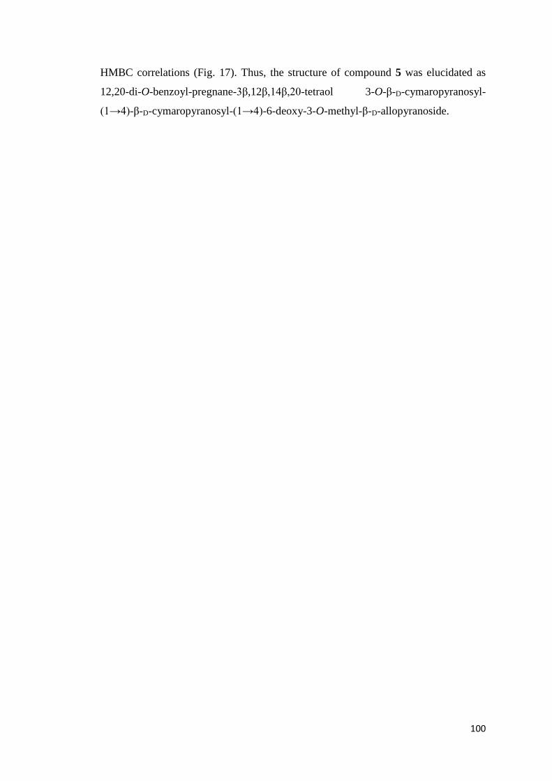
100
HMBC correlations (Fig. 17). Thus, the structure of compound 5 was elucidated as
12,20-di-O-benzoyl-pregnane-3β,12β,14β,20-tetraol 3-O-β-D-cymaropyranosyl-
(1→4)-β-D-cymaropyranosyl-(1→4)-6-deoxy-3-O-methyl-β-D-allopyranoside.
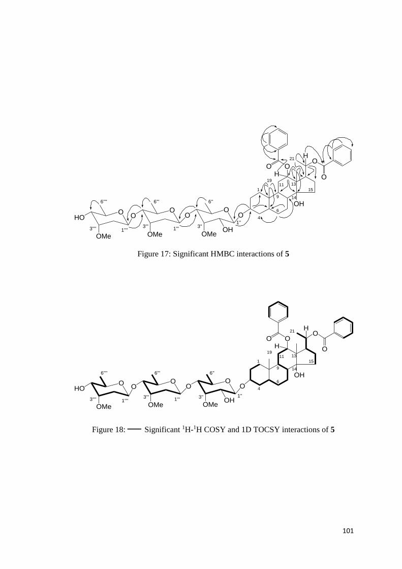
101
O
OH
1
4
6
9 14
131115
21
19O
OO
H
H
OO
O
OMe
OO
OMe
OHO
OMeOH
1''3''
1'''3'''
6''6'''6''''
1''''3''''
Figure 17: Significant HMBC interactions of 5
O
OH
1
4
6
9 14
131115
21
19O
OO
H
H
OO
O
OMe
OO
OMe
OHO
OMeOH
1''3''1'''
3'''
6''6'''6''''
1''''3''''
Figure 18: — Significant 1H-1H COSY and 1D TOCSY interactions of 5

102
4.1.6 Desflavaside A (6)
The molecular formula of compound 6 was established as C62C90H21 based on quasi-
molecular ion peak in the HR-ESIMS at m/z 1193.5885 (calculated for C62H90NaO21,
1193.5872).
The IR spectrum of 6 showed the presence of OH, ester C=O and aryl moieties in the
molecule. Further, the NMR spectrum (Table 7) indicated the resonances for two
tertiary and one secondary methyls at δH 1.09 (δC 10.0), δH 0.82 (δC 12.4) and δH 1.20
(δC 19.7). The pregnane skeleton of compound 6 was suggested by the appearance of
three oxymethine group signals at δH 3.63 (δC 78.5), δH 4.94 (δC 79.5) and δH 5.24 (δC
75.6) along with two quaternary carbons at δC 36.9 (C-10), 53.7 (C-13) and a
characteristic oxygenated C-14 at δC 87.2 (Abdallah et al., 2013; Abdel-Sattar et al.,
2001, 2002, 2007; Tanaka et al., 1990). The significant COSY, selective 1D TOCSY
and HMBC interactions (Fig. 19 and 20) also confirmed the pregnane moiety of 6. The
presence of two benzoyl groups illustrated by their resonances in 1H NMR spectrum at
δ 8.03 (H-2'/H-6'), 7.67 (H-4') and 7.50 (H-3'/H-5') were assigned to benzoyl group at
C-12 and signals at δ 7.73 (H-2'/H-6'), 7.51 (H-4') and 7.22 (H-3'/H-5') defined to
benzoyl group at C-20 (Abdallah et al., 2013; Tanaka et al., 1990; Hayashi et al., 1988).
Furthermore, the HMBC interactions of H-12 and H-20 to the C=O carbons at δC 168.0
and 167.0 confirmed their respective locations, respectively.
The tetraglycosidic nature of desflavaside A was positively identified by the resonances
for four anomeric sugar centers at δH 4.85 (J = 2.0, 6.0 Hz, dd, H-1ʹʹ); δC 97.1 (C-1ʹʹ);
δH 4.33 (J = 7.2 Hz, d, H-1ʹʹ′); δC 106.2 (C-1ʹʹ′); δH 4.56 (J = 2.0, 10.0 Hz, dd, H-1ʹʹʹʹ);
δC 102.5 (C-1ʹʹʹʹ); δH 4.70 (J = 6.0 Hz, d, H-1ʹʹʹʹʹ); δC 102.1 (C-1ʹʹʹʹʹ); three deoxy sugar
methyl resonances at δH 1.18 (J = 6.0 Hz, d, H-6′′); δC 18.5 (C-6ʹʹ); δH 1.29 (J = 6.0 Hz,
d, H-6ʹʹʹʹ); δc 18.1 (C-6ʹʹʹʹ); δH 1.35 (J = 6.0 Hz, d, H-6ʹʹʹʹʹ); δc 18.9 (C-6ʹʹʹʹʹ) and three
sugar OMe resonances at δH 3.40 (s); δC 57.5; δH 3.41 (s); δC 58.4; δH 3.58 (s); δC 61.9.
The above mentioned resonances and large coupling constants (8.4–9.6 Hz) indicated
that the group attached at C-3 of the aglycone portion is a tetraglycoside with a β-
linkage (Abdel-Sattar et al., 2002, 2007; De Leo et al., 2005). The 13C NMR, selective
1D TOCSY and 1H-1H COSY spectra (Fig. 20) positively suggested that the
tetrasaccharide is comprised of one glucose, one 6-deoxy-3-O-D-methylallose and two
cymarose (=2,6-dideoxy-3-O-methyl-ribohexose) units which were strongly supported

103
by a comparison of the spectra with previous compounds 4 and 5. Connectivity of the
saccharide chain to C-3 of the pregnane part was confirmed from significant HMBC
interactions between the first cymarose anomeric proton (H-1ʹʹ) and C-3. Moreover, the
sequence of the tetrasaccharide in the chain was confirmed from the H-1ʹʹʹʹʹ to C-4ʹʹʹʹ;
H-4ʹʹʹʹ to C-1ʹʹʹʹʹ; H-1ʹʹʹʹ to C-4ʹʹʹ; H-4ʹʹʹ to C-1ʹʹʹʹ; H-1ʹʹʹ to C-4ʹʹ and H-4ʹʹ to C-1ʹʹʹ 3J
correlations.
The relative stereochemistry at C-3 was established by the prominent NOESY
interaction of H-3 with H-5 and was further confirmed by comparison of NMR values
related to this position [δH 3.62 (m, H-3); δC 78.5 (C-3)] with literature reports of related
pregnane glycosides Lit: δH 3.63-3.68 (m, H-3); δC 78.1-78.4 (C-3) (De Leo et al.,
2005). Further, the α-configurations of H-12 and H-17 were confirmed from NOE
interactions of H-12 with H-11α together with H-17α and positively supported by lack
of NOE correlations with Me-18. On the other hand the stereochemistry of C-20 was
confirmed as S by a similar analysis to that reported for other pregnane glycosides viz.,
an NOE correlation between H-20 with H-21 and H-18 on the one hand and H-l6α with
H-21 together with lack of an NOE correlation between H-21 and H-l8 (Itokawa et al.,
1988; Abdel-Sattar et al., 2001, 2007; Qiu et al., 1997; Tanaka et al., 1990). However,
the configuration assigned to C-20 was tenuous due to the free rotation around the C-
17 to C-20 carbon-carbon bond. Therefore, the structure of 6 was deduced as 12,20-di-
O-benzoyl-20S-pregnane-3β,12β,14β,20-tetraol 3-O-6-deoxy-3-O-methyl-β-D-
allopyranosyl-(1→4)-β-D-cymaropyranosyl-(1→4)-β-D-glucopyranosyl-(1→4)-β-D-
cymaropyranoside.

104
O
OH
OOO
OMe
OO
1''3''1'''3'''
6''6'''
1
4
6
9 14
131115
21
19O
OO
H
H
OO
6''''
1''''3''''
OH
OHOMe
O
OMe
3'''''
6'''''
OH
HO
1''''' HO
Figure 19: Significant HMBC interactions of 6

105
O
OH
OOO
OMe
OO
1''3''1'''3'''
6''6'''
1
4
6
9 14
131115
21
19O
OO
H
H
OO
6''''
1''''3''''
OH
OHOMe
O
OMe
3'''''
6'''''
OH
HO
1''''' HO
Figure 20: — Significant 1H-1H COSY and 1D TOCSY interactions of 6

106
4.1.7 Desflavaside B (7)
The molecular formula of compound 7 was identical to 6 that is C62C90H21 (HRESIMS
observed 1193.5881, calculated for C62H90NaO21, 1193.5872). IR spectrum of 7
showed bands at 3415, 1710, 1610 cm-1 indicated the presence of OH, ester C=O and
aromatic ring in the molecule.
The NMR data of aglycone part of 7 (Table 8) was similar to 6 (Table 7) whereas the
difference was observed in the glycoside chain indicating the existence of β-D-
thevetopyranosyl terminal sugar unit in 7 instead of β-D-allopyranosyl sugar. The
presence of terminal β-D-thevetopyranosyl unit was positively supported by 13C NMR
data of this unit [δC 104.2 (C-1′′′′′), 75.2 (C-2′′′′′), 86.3 (C-3′′′′′), 82.9 (C-4′′′′′), 72.6 (C-
5′′′′′), 18.5 (C-6′′′′′), 61.2 (OMe)] with the literature [Lit: δC 104.2 (C-1), 75.0 (C-2),
86.1 (C-3), 82.6 (C-4), 72.3 (C-5), 18.5 (C-6), 61.0 (OMe)] (De Leo et al., 2005). On
behalf of above differences, the structure of 7 was assigned as 12,20-di-O-benzoyl-20S-
pregnane-3β,12β,14β,20-tetraol 3-O-β-D-thevetopyranosyl-(1→4)-β-D-
cymaropyranosyl-(1→4)-β-D-glucopyranosyl-(1→4)-β-D-cymaropyranoside.
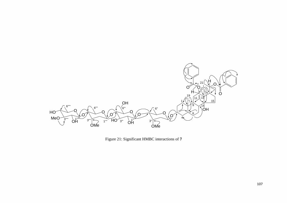
107
O
OH
OOO
OMe
OO
1''3''1'''3'''
6''6'''
1
4
6
9 14
131115
21
19O
OO
H
H
OO
6''''
1''''3''''
OH
OHOMe
O
MeO 3'''''
6'''''
OH
HO
1''''' HO
Figure 21: Significant HMBC interactions of 7
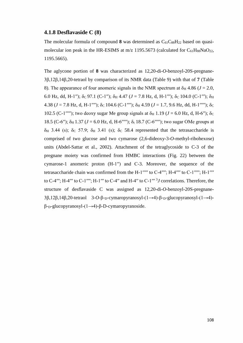
108
4.1.8 Desflavaside C (8)
The molecular formula of compound 8 was determined as C61C88H22 based on quasi-
molecular ion peak in the HR-ESIMS at m/z 1195.5673 (calculated for C61H88NaO22,
1195.5665).
The aglycone portion of 8 was characterized as 12,20-di-O-benzoyl-20S-pregnane-
3β,12β,14β,20-tetraol by comparison of its NMR data (Table 9) with that of 7 (Table
8). The appearance of four anomeric signals in the NMR spectrum at δH 4.86 (J = 2.0,
6.0 Hz, dd, H-1ʹʹ); δC 97.1 (C-1ʹʹ); δH 4.47 (J = 7.8 Hz, d, H-1ʹʹ′); δC 104.0 (C-1ʹʹ′); δH
4.38 (J = 7.8 Hz, d, H-1ʹʹʹʹ); δC 104.6 (C-1ʹʹʹʹ); δH 4.59 (J = 1.7, 9.6 Hz, dd, H-1ʹʹʹʹʹ); δC
102.5 (C-1ʹʹʹʹʹ); two deoxy sugar Me group signals at δH 1.19 (J = 6.0 Hz, d, H-6′′); δC
18.5 (C-6ʹʹ); δH 1.37 (J = 6.0 Hz, d, H-6ʹʹʹʹʹ); δc 18.7 (C-6ʹʹʹʹʹ); two sugar OMe groups at
δH 3.44 (s); δC 57.9; δH 3.41 (s); δC 58.4 represented that the tetrasaccharide is
comprised of two glucose and two cymarose (2,6-dideoxy-3-O-methyl-ribohexose)
units (Abdel-Sattar et al., 2002). Attachment of the tetraglycoside to C-3 of the
pregnane moiety was confirmed from HMBC interactions (Fig. 22) between the
cymarose-1 anomeric proton (H-1ʹʹ) and C-3. Moreover, the sequence of the
tetrasaccharide chain was confirmed from the H-1ʹʹʹʹʹ to C-4ʹʹʹʹ; H-4ʹʹʹʹ to C-1ʹʹʹʹʹ; H-1ʹʹʹʹ
to C-4ʹʹʹ; H-4ʹʹʹ to C-1ʹʹʹʹ; H-1ʹʹʹ to C-4ʹʹ and H-4ʹʹ to C-1ʹʹʹ 3J correlations. Therefore, the
structure of desflavaside C was assigned as 12,20-di-O-benzoyl-20S-pregnane-
3β,12β,14β,20-tetraol 3-O-β-D-cymaropyranosyl-(1→4)-β-D-glucopyranosyl-(1→4)-
β-D-glucopyranosyl-(1→4)-β-D-cymaropyranoside.

109
O
OH
OOO
OMe
OO
1''3''1'''3'''
6''6'''
1
4
6
9 14
131115
21
19O
OO
H
H
OO
6''''
1''''3''''
OH
OH
O
3'''''
6'''''
OH
HO
1''''' HO
OH
HOOMe OH
Figure 22: Significant HMBC interactions of 8

110
O
OH
OOO
OMe
OO
1''3''1'''3'''
6''6'''
1
4
6
9 14
131115
21
19O
OO
H
H
OO
6''''
1''''3''''
OH
OH
O
3'''''
6'''''
HO
1''''' HO
OH
HOOMe OH
18
Figure 23: — Significant 1H-1H COSY and 1D TOCSY interactions of 8

111
4.1.9 Desflavaside D (9)
The HRESIMS analysis of compound 9 showed the quasi-molecular ion peak at m/z
1005.4876 (calculated for C46H78NaO22, 1005.4882) assigned for molecular formula
C46H78O22.
Two major differences in the NMR signals of the aglycone of 9 (Table 10) were
observed as compared to compound 8 (Table 9). Firstly, the NMR peaks for two
benzoyls were completely absent in compound 9. Secondly, the methylene signals for
ring C appeared at δH 1.26 (m) and 1.37 (m) in the 1H NMR spectrum of 9 instead of
an oxymethine signal at δH 4.95 (1H, m, H-12) which was also supported by the
presence of a methylene signal at δC 42.0 in the 13C NMR spectrum instead of a methine
signal at δC 79.5 (C-12). Therefore, the aglycone portion of 9 was characterized as 20S-
pregnane-3β,14β,20-triol.
Further analysis of the NMR spectra of desflavaside D indicated the presence of four
anomeric signals at δH 4.58 (J = 7.7 Hz, d, H-1ʹʹ); δC 104.1 (C-1ʹʹ); δH 4.38 (J = 7.8 Hz,
d, H-1ʹʹ′); δC 104.3 (C-1ʹʹ′); δH 4.36 (J = 7.9 Hz, d, H-1ʹʹʹʹ); δC 104.4 (C-1ʹʹʹʹ) and δH 4.34
(J = 7.8 Hz, d, H-1ʹʹʹʹʹ); δC 102.7 (C-1ʹʹʹʹʹ) suggested compound 9 to be tetrasaccharide
glycoside. Its 1D and 2D NMR spectra displayed the presence of one digitalose (3-O-
methyl-6-deoxy-galactopyranose) and three glucose units.
Attachment of the saccharide chain to C-3 and the sequence of the saccharides in the
chain was confirmed from H-1ʹʹʹʹʹ to C-4ʹʹʹʹ; H-4ʹʹʹʹ to C-1ʹʹʹʹʹ; H-1ʹʹʹʹ to C-4ʹʹʹ; H-4ʹʹʹ to
C-1ʹʹʹʹ; H-1ʹʹʹ to C-4ʹʹ and H-4ʹʹ to C-1ʹʹʹ; H-1ʹʹ to C-3; H-3 to C-1ʹʹ 3J HMBC correlations
(Fig. 24). From the foregoing evidences, compound 9 was characterized as 20S-
pregnane-3β,14β,20-triol 3-O-β-D-digitalopyranosyl-(1→4)-β-D-glucopyranosyl-
(1→4)-β-D-glucopyranosyl-(1→6)-β-D-glucopyranoside.
The NMRs of all the isolated compounds (1-9) indicated that none of the compound
was isolated in a completely pure state and minor impurities were observed in the
NMRs of all the isolates. It was also to be noted that no known compound was isolated
from C. flava in our research.
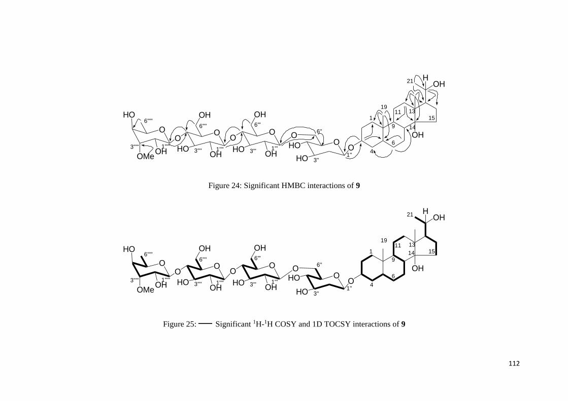
112
OH
OH
OO
1''3''
6''
1
4
6
9 14
131115
21
19
H
HO
OOO
1'''3'''
6'''
OO
6''''
1''''3''''
OH
OH
O
3'''''
6'''''
OH
HO
1''''' HO
OH
HOOMe OH
HO
Figure 24: Significant HMBC interactions of 9
OH
OH
OO
1''3''
6''
1
4
6
914
131115
21
19
H
HO
OOO
1'''3'''
6'''
OO
6''''
1''''3''''
OH
OH
O
3'''''
6'''''
OH
HO
1''''' HO
OH
HOOMe OH
HO
Figure 25: — Significant 1H-1H COSY and 1D TOCSY interactions of 9
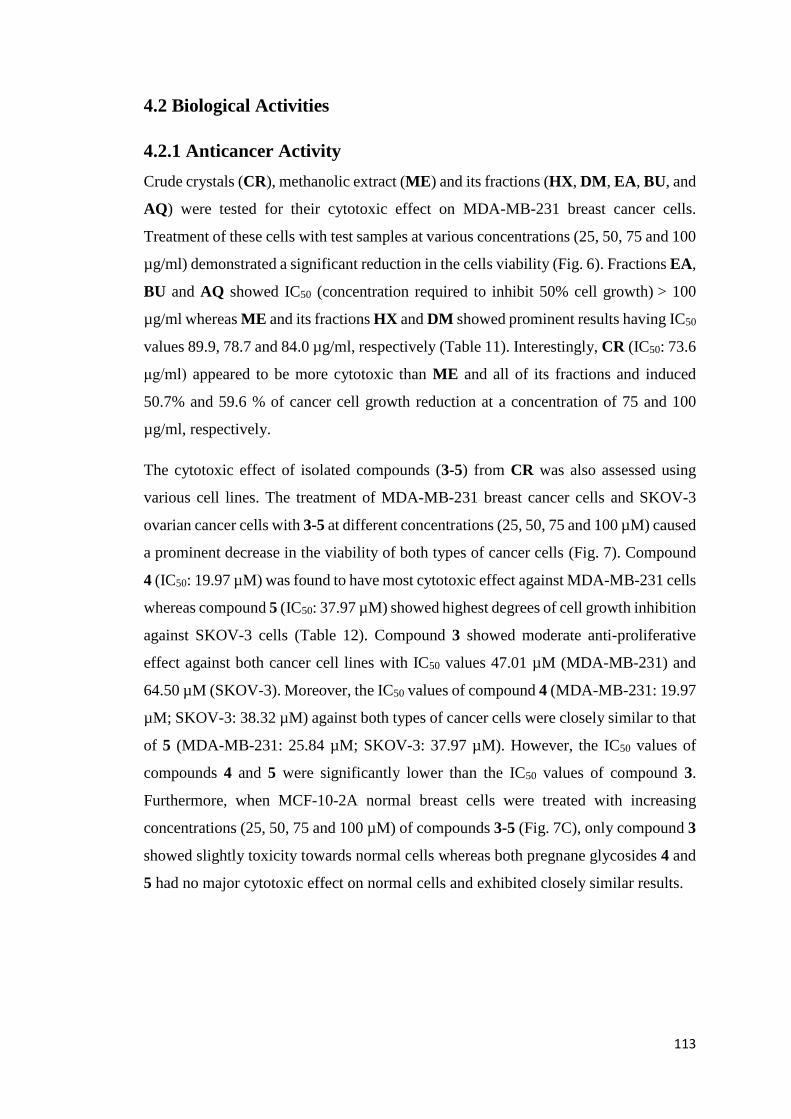
113
4.2 Biological Activities
4.2.1 Anticancer Activity
Crude crystals (CR), methanolic extract (ME) and its fractions (HX, DM, EA, BU, and
AQ) were tested for their cytotoxic effect on MDA-MB-231 breast cancer cells.
Treatment of these cells with test samples at various concentrations (25, 50, 75 and 100
µg/ml) demonstrated a significant reduction in the cells viability (Fig. 6). Fractions EA,
BU and AQ showed IC50 (concentration required to inhibit 50% cell growth) > 100
µg/ml whereas ME and its fractions HX and DM showed prominent results having IC50
values 89.9, 78.7 and 84.0 µg/ml, respectively (Table 11). Interestingly, CR (IC50: 73.6
μg/ml) appeared to be more cytotoxic than ME and all of its fractions and induced
50.7% and 59.6 % of cancer cell growth reduction at a concentration of 75 and 100
µg/ml, respectively.
The cytotoxic effect of isolated compounds (3-5) from CR was also assessed using
various cell lines. The treatment of MDA-MB-231 breast cancer cells and SKOV-3
ovarian cancer cells with 3-5 at different concentrations (25, 50, 75 and 100 µM) caused
a prominent decrease in the viability of both types of cancer cells (Fig. 7). Compound
4 (IC50: 19.97 µM) was found to have most cytotoxic effect against MDA-MB-231 cells
whereas compound 5 (IC50: 37.97 µM) showed highest degrees of cell growth inhibition
against SKOV-3 cells (Table 12). Compound 3 showed moderate anti-proliferative
effect against both cancer cell lines with IC50 values 47.01 µM (MDA-MB-231) and
64.50 µM (SKOV-3). Moreover, the IC50 values of compound 4 (MDA-MB-231: 19.97
µM; SKOV-3: 38.32 µM) against both types of cancer cells were closely similar to that
of 5 (MDA-MB-231: 25.84 µM; SKOV-3: 37.97 µM). However, the IC50 values of
compounds 4 and 5 were significantly lower than the IC50 values of compound 3.
Furthermore, when MCF-10-2A normal breast cells were treated with increasing
concentrations (25, 50, 75 and 100 µM) of compounds 3-5 (Fig. 7C), only compound 3
showed slightly toxicity towards normal cells whereas both pregnane glycosides 4 and
5 had no major cytotoxic effect on normal cells and exhibited closely similar results.
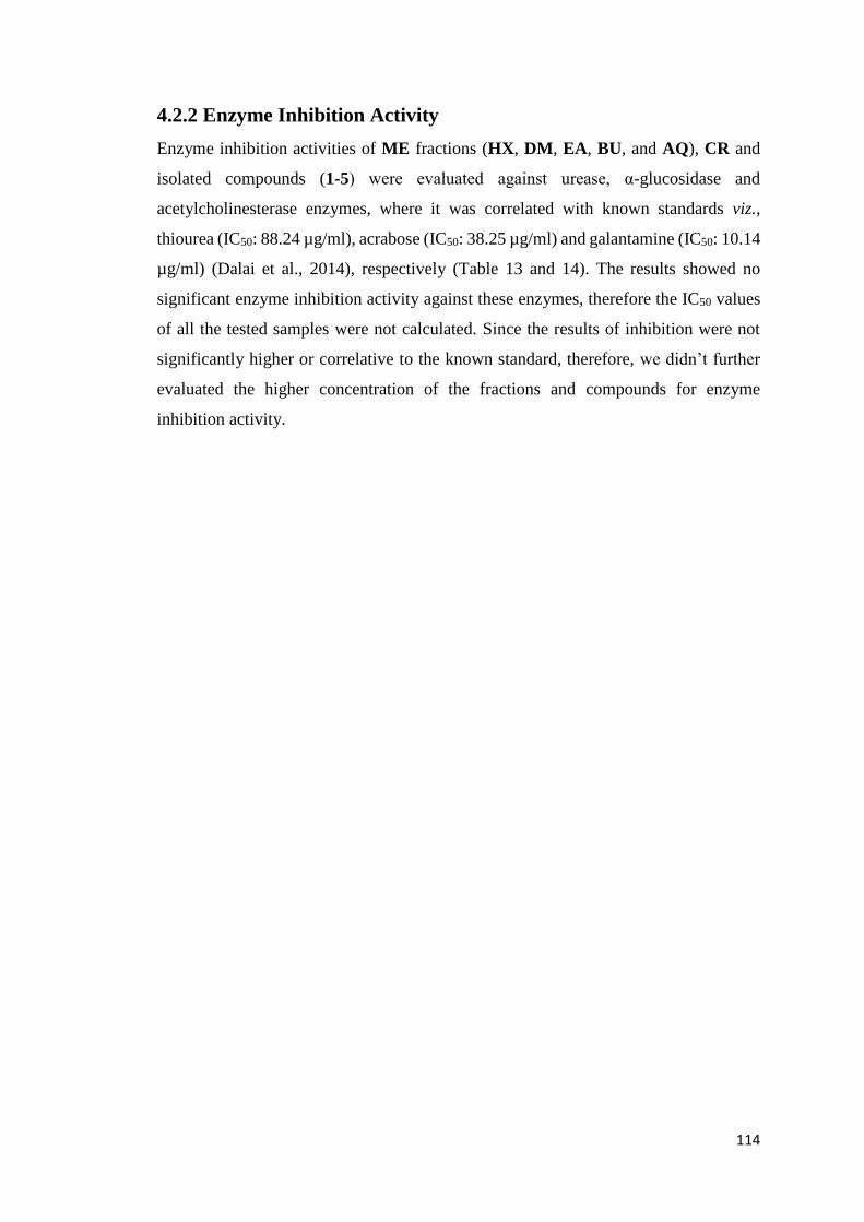
114
4.2.2 Enzyme Inhibition Activity
Enzyme inhibition activities of ME fractions (HX, DM, EA, BU, and AQ), CR and
isolated compounds (1-5) were evaluated against urease, α-glucosidase and
acetylcholinesterase enzymes, where it was correlated with known standards viz.,
thiourea (IC50: 88.24 µg/ml), acrabose (IC50: 38.25 µg/ml) and galantamine (IC50: 10.14
µg/ml) (Dalai et al., 2014), respectively (Table 13 and 14). The results showed no
significant enzyme inhibition activity against these enzymes, therefore the IC50 values
of all the tested samples were not calculated. Since the results of inhibition were not
significantly higher or correlative to the known standard, therefore, we didn’t further
evaluated the higher concentration of the fractions and compounds for enzyme
inhibition activity.
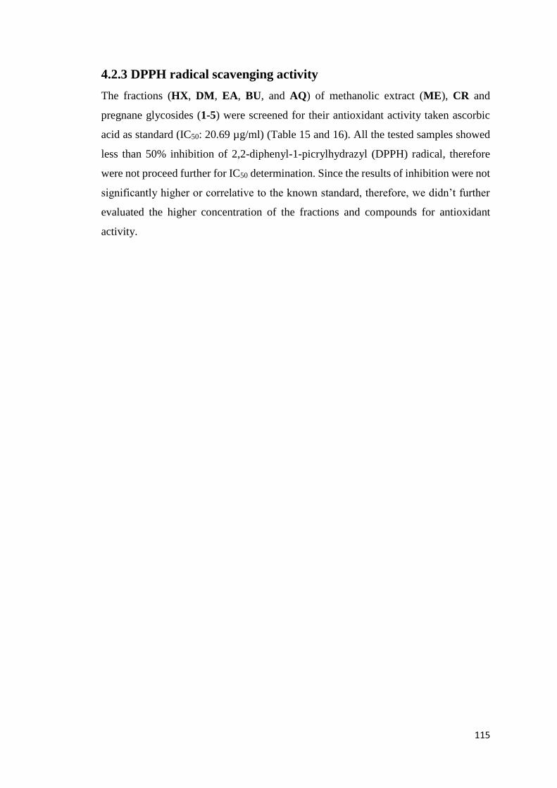
115
4.2.3 DPPH radical scavenging activity
The fractions (HX, DM, EA, BU, and AQ) of methanolic extract (ME), CR and
pregnane glycosides (1-5) were screened for their antioxidant activity taken ascorbic
acid as standard (IC50: 20.69 µg/ml) (Table 15 and 16). All the tested samples showed
less than 50% inhibition of 2,2-diphenyl-1-picrylhydrazyl (DPPH) radical, therefore
were not proceed further for IC50 determination. Since the results of inhibition were not
significantly higher or correlative to the known standard, therefore, we didn’t further
evaluated the higher concentration of the fractions and compounds for antioxidant
activity.

116
4.3 Molecular Docking Studies
Molecular docking studies were carried out to ascertain if it was possible to predict any
important binding orientations required of small molecule drug candidates with
suggested protein target molecules for the purposes of being able to predict the affinity
and activity to an acceptable degree by such compounds. The normal role of protein
tyrosine phosphatases (PTPs) enzyme is to regulate cell proliferation and tumor
suppression while its overexpression leads to breast cancer. Therefore, newly
synthesized PTPs inhibitors have a therapeutic role.
Pregnane glycosides 3-5 were used as ligand molecules to inhibit the PTPs
overexpression activity in order to understand the drug receptor interaction. The
isolated binding pocket of PTPs contained amino acid residues: ALA-707, ARG-875,
ASP-872, ASP-879, GLU-704, ILE-705, ILE-868, ILE-873, LEU-861, LEU-866,
LYS-876, MET-703, PRO-706, PRO-867, THR-857 and TYR-869 (Fig. 8). Results of
the present studies illustrated that the targeted enzyme was docked by these ligands and
sixteen tyrosine residues demonstrated interactions with these inhibitor molecules. The
ASP-872, PRO-706, ASP-879 and ARG-875 amino acid residues of protein target were
mainly interacting with the solvent-residues (H-OH), arene-H of aromatic rings and
electro-negative O-hetero atoms of compound (3) (Fig. 9A). The solvent residues (H-
OH) of compound (4) exhibited binding affinity with ALA-707 and PRO-857, while
the ASP-879 amino acid interacted with the hydroxyl group of the C41-42 group of the
ligand molecule (Fig. 9B). Similarly, compound (5) demonstrated that the arene-H of
the aromatic ring possess promising binding affinity with LYS-876; ALA-707, PRO-
706 and PRO-867 with the C1-O7 group and ASP-879 with carbonyl group (Fig. 9C).
Compound (3) underwent the most important interactions with GLU-704 (acidic
contact), PRO-706 (sidechain donor), ASP-872 (acidic receptor contact), LYS-876
(basic contact), ARG-875 (basic receptor contact), ASP-879 (acidic contact), ILE-705
(greasy contact) and LEU-851 (greasy contact). Similarly, compound (4) interacted
with ARG-875 (basic receptor contact), PRO-857 (greasy receptor contact), ASP-879
(acidic receptor contact), ASP-872 (acidic contact), LEU-856 (greasy contact), ALA-
707 (greasy receptor contact) and ARG-875 (basic receptor contact) while compound
(5) showed good interactions with ALA-707, PRO-857, PRO-705, LEU-851 (greasy

117
receptor contact), GLU-704 (acidic receptor contact), ASP-879, ASP-872 (acidic
receptor contact), LYS-876 (basic receptor contact).
In terms of binding interaction with different amino acid residues, compound 4
exhibited better affinity as compared to 3 and 5. In the best docked position, 4 formed
three hydrogen bonds with the receptor, involving residues ASP-879, PRO-857 and
ALA-707. Residue ASP-879 was strongly involved in hydrogen bond formation with
pregnane glycosides 3-5 showing a similar orientation of these molecules, which was
also obvious from a closer analysis of their relative docking positions (Fig. 9).
All ligand molecules demonstrated the best interaction with target protein and their
calculated docked energies were found to be: desmiflavaside C (3), -19.4042 kcal/mol,
desmiflavaside D (4), -22.4449 kcal/mol and nizwaside (5), -20.0314 kcal/mol (Table
17). Compound 4 showed a comparatively promising binding energy as compared to 3
and 5.

118
5. CONCLUSIONS

119
In conclusion, this study found that the plant of C. flava growing in the high mountains
of the Sultanate of Oman possesses promising anti-cancer properties and can be used
to cope with breast cancer in the Sultanate which has been one of the most common
cancer in Omani females and a leading cause of their mortality. Our findings clearly
showed that both, the plant extract and its exudate sap possess significant anticancer
activity towards breast cancer.
Among the five different fractions of the methanolic extract of C. flava, hexane fraction
showed the most promising anticancer effects in-vitro towards breast cancer. The
therapeutical activities of this plant were carried out scientifically and reported for the
first time in this research. Further work on isolation and characterization of the possible
bioactive compounds contained therein is required for further investigation. Moreover,
in-vivo verification of the anti-cancer therapeutic effectiveness of the active
components are also needed.
In comparison with C. flava methanolic extract and all of its fractions, the viscous sap
of the plant was found to be the most antiproliferative against breast cancer and turned
out to be a rich source of pregnane glycosides. On chemical investigation, it first yielded
crude shiny yellow crystals and later their chromatographic separation provided nine
new pregnane glycosides namely desmiflavaside A (1), desmiflavaside B (2),
desmiflavaside C (3), desmiflavaside D (4), nizwaside (5), desflavaside A (6),
desflavaside B (7), desflavaside C (8) and desflavaside D (9).
The anticancer properties of the sap of C. flava attributed to the pregnane glycosides
present therein. The result of anticancer activities illustrated that all the pregnane
glycosides tested viz., desmiflavasides C (3) and D (4) and nizwaside (5) exhibited
significant potential to effectively suppress the growth of ovarian and breast cancer
cells without disturbing the growth of normal breast epithelial cells. These pregnane
glycosides can have anticancer effects on other cancer cell lines as well which need to
be further explored.
The molecular docking study results of three pregnane glycosides namely
desmiflavasides C (3) and D (4) and nizwaside (5) illustrated that all these quite
extended ligand molecules have a good potential to accurately interact with the target
protein tyrosine phosphatase sites reasonably specifically to inhibit its overexpression
activity which leads to breast cancer. This is why, these isolated pregnane glycosides

120
from C. flava can serve as strong candidates for future anti-cancer drug development
and therefore need to be further explored for their anti-cancer action in the cure of breast
cancer, in-vivo models of tumorigenesis and an understanding of the mechanisms
thereof.
The plant of C. flava showed no significant potential to inhibit urease,
acetylcholinesterase and α-glucosidase enzymes and therefore may not be considered
to be very much effective in the treatment of Alzheimer’s disease, diabetes, peptic ulcer
and infection-induced urinary stones. Further, the scientific examination for the
traditional claims for antidiabetic use of this plant may also not be justified. Further
studies are needed to explore the possible claimed actions of this plants. The antioxidant
ability of the plant was also found to be weak. It is important to note that the failure of
a plant extract or isolated compounds to demonstrate in-vitro activity during the general
screening process does not necessarily imply a total absence of inherent medicinal
value. The possible presence of synergistic interactions between the different
constituents in crude preparations may result in activities that are not exhibited by
isolated compounds, and should not be excluded. Furthermore, the plant or extract may
react differently in-vivo.
As modern cultures and scientific advances spread around the world, the breadth of the
knowledge store of traditional healers still remains crucial. The full significance of the
indigenous knowledge forfeited may not be realized. It is thus important that the
knowledge be documented and the traditional use given some credence through modern
scientific studies.
121
6. REFRENCES

122
Abdallah HM, Osman AM, Almehdar H, Abdel-Sattar E. Acylated pregnane glycosides
from Caralluma quadrangula. Phytochemistry. 2013 Apr 30;88:54-60.
Abdel-Sattar E, Ahmed AA, Hegazy ME, Farag MA, Al-Yahya MA. Acylated
pregnane glycosides from Caralluma russeliana. Phytochemistry. 2007 May
31;68(10):1459-63.
Abdel-Sattar E, Al-Yahya MA, Nakamura N, Hattori M. Penicillosides A-C, C-15
oxypregnane glycosides from Caralluma penicillata. Phytochemistry. 2001 Aug
31;57(8):1213-7.
Abdel-Sattar E, Harraz FM, Al-Ansari SM, El-Mekkawy S, Ichino C, Kiyohara H,
Ishiyama A, Otoguro K, Omura S, Yamada H. Acylated pregnane glycosides from
Caralluma tuberculata and their antiparasitic activity. Phytochemistry. 2008 Aug
31;69(11):2180-6.
Abdel-Sattar E, Meselhy MR, Al-Yahya MA. New oxypregnane glycosides from
Caralluma penicillata. Planta medica. 2002 May;68(05):430-4.
Adnan M, Jan S, Mussarat S, Tariq A, Begum S, Afroz A, Shinwari ZK. A review on
ethnobotany, phytochemistry and pharmacology of plant genus Caralluma R. Br.
Journal of Pharmacy and Pharmacology. 2014 Oct 1;66(10):1351-68.
Ahmad VU, Basha A. Spectroscopic data of steroid glycosides. Volume 5. Springer
Science & Business Media; 2007.
Albalawi MA, Bashir NA, Tawfik A. Anticancer and Antifolate Activities of Extracts
of Six Saudi Arabian Wild Plants Used in Folk Medicine. Journal of Life Sciences.
2015;9:334-40.
Albers F, Meve U. Illustrated Handbook of Succulent Plants: Asclepiadaceae. Springer
Science & Business Media; 2002.
Alhakmani F, Khan SA, Ahmad A. Determination of total phenol, invitro antioxidant
and anti-inflammatory activity of seeds and fruits of Zizyphus spina-christi grown
in Oman. Asian Pacific Journal of Tropical Biomedicine. 2014 Jul 31;4:S656-60.
Al-Lawati JA, Kumar CRS, Mohammed AJ, Jaffer MA. Cancer incidence in Oman, 1
993-1 997. Eastern Mediterranean health journal. 1999;5(5).

123
Al-Madouj A, Eldali A, Al-Zahrani A. Ten Year Cancer Incidence Among Nationals
of the GCC States 1998–2007. Cancer Incidence Report of the Cooperation
Council States. P. 2011:16-9.
Al-Massarani SM, Bertrand S, Nievergelt A, El-Shafae AM, Al-Howiriny TA, Al-
Musayeib NM, Cuendet M, Wolfender JL. Acylated pregnane glycosides from
Caralluma sinaica. Phytochemistry. 2012 Jul 31;79:129-40.
Al-Massarani SM, El-Shafae AM. Pharmacognostical and Biological Study of
Caralluma sinaica Growing in Saudi Arabia. 2011.
Al-Yahya MAZ, Abdel-Sattar E, Guittet E. Pregnane glycosides from Caralluma
russeliana. Journal of natural products. 2000 Oct 27;63(10):1451-3.
Bruyns PV, al Farsi A, Hedderson T. Phylogenetic relationships of Caralluma R. Br.
(Apocynaceae). Taxon. 2010 Aug 1;59(4):1031-43.
Burney IA, Furrukh M, Al-Moundhri MS. What are our Options in the Fight against
Breast Cancer?. Sultan Qaboos University Medical Journal. 2014 May;14(2):e149.
Dalai MK, Bhadra S, Chaudhary SK, Bandyopadhyay A, Mukherjee PK. Anti-
cholinesterase activity of the standardized extract of Syzygium aromaticum L.
Pharmacognosy magazine. 2014 May 1;10(38):276.
De Leo M, De Tommasi N, Sanogo R, Autore G, Marzocco S, Pizza C, Morelli I, Braca
A. New pregnane glycosides from Caralluma dalzielii. Steroids. 2005 Aug
31;70(9):573-85.
Deepak D, Khare A, Khare MP. Plant pregnanes. Phytochemistry. 1989 Jan
1;28(12):3255-63.
Deepak D, Srivastav S, Khare A. Pregnane glycosides. InFortschritte der Chemie
organischer Naturstoffe/Progress in the Chemistry of Organic Natural Products
1997 (pp. 169-325). Springer Vienna.
Elsebai MF, Mohamed IE. New pregnane glycoside derivative from Caralluma
retrospiciens (Ehrenb). Natural product research. 2015 Aug 3;29(15):1426-31.

124
Formisano C, Senatore F, Della Porta G, Scognamiglio M, Bruno M, Maggio A,
Rosselli S, Zito P, Sajeva M. Headspace volatile composition of the flowers of
Caralluma europaea NE. Br. (Apocynaceae). Molecules. 2009 Nov
11;14(11):4597-613.
Ghazanfar SA, Al-Sabahi AMA. Medicinal plants of northern and central Oman
(Arabia). Economic Botany. 1993 Jan 1;47(1):89-98.
Ghazanfar SA. Handbook of Arabian medicinal plants. CRC press; 1994 Aug 24.
Ghazanfar SA. Quantitative and biogeographic analysis of the flora of the Sultanate of
Oman. Global ecology and biogeography letters. 1992 Nov 1:189-95.
Grulich V, Caralluma flava N. E. Br., Botany. Cz, 2 Dec. 2015. Web 29 Jun. 2016.
http://botany.cz/cs/caralluma-flava/
Hayashi K, Iida I, Nakao Y, Nakao Y, Kaneko K. Four pregnane glycosides,
boucerosides A-I, A-II, B-I and B-II, from Boucerosia aucheriana.
Phytochemistry. 1988 Dec 31;27(12):3919-24.
Hussain J, Rehman NU, Khan AL, Ali L, Kim JS, Zakarova A, Al-Harrasi A, Shinwari
ZK. Phytochemical and biological assessment of medicinally important plant
Ochradenus arabicus. Pak. J. Bot. 2014 Dec 1;46(6):2027-34.
Itokawa H, Xu J, Takeya K. Pregnane glycosides from an antitumour fraction of
Periploca sepium. Phytochemistry. 1988 Dec 31;27(4):1173-9.
Jürgens A, Dötterl S, Meve U. The chemical nature of fetid floral odors in stapeliads
(Apocynaceae‐Asclepiadoideae‐Ceropegieae). New Phytologist. 2006 Nov
1;172(3):452-68.
Kunert O, Rao BV, Babu GS, Padmavathi M, Kumar BR, Alex RM, Schühly W, Simic
N, Kühnelt D, Rao AV. Novel steroidal glycosides from two Indian Caralluma
species, C. stalagmifera and C. indica. Helvetica chimica acta. 2006 Feb
1;89(2):201-9.
Kunert O, Rao VG, Babu GS, Sujatha P, Sivagamy M, Anuradha S, Rao BV, Kumar
BR, Alex RM, Schühly W, Kühnelt D. Pregnane glycosides from Caralluma
adscendens var. fimbriata. Chemistry & biodiversity. 2008 Feb 1;5(2):239-50.

125
Lee-Juian L, Long-Ze L, Gil RR, Cordelly GA, Ramesh M, Srilatha B, Reddy B, Rao
AV. Pregnane glycosides from Caralluma umbellata. Phytochemistry. 1994 Apr
19;35(6):1549-53.
Lupton D, Al-Jabri T, Al-Hinai A, Al-Hatmi S. People and plants in the Sultanate of
Oman: Ethnobotany at Oman Botanic Garden. Volume 9 Number 2 - July 2012
Marwah RG, Fatope MO, Al Mahrooqi R, Varma GB, Al Abadi H, Al-Burtamani SK.
Antioxidant capacity of some edible and wound healing plants in Oman. Food
chemistry. 2007 Dec 31;101(2):465-70.
Mosti S, Raffaelli M. Desmidorchis tardellii (Asclepiadaceae), a new species from
Dhofar, southern Oman. Webbia. 2004 Jan 1;59(2):285-91.
Oyama M, Iliya I, Tanaka T, Iinuma M. Five new steroidal glycosides from Caralluma
dalzielii. Helvetica chimica acta. 2007 Jan 1;90(1):63-71.
Patzelt A, Oman Plant Red Data Book. (Oman Botanic Garden Publication, No. 1)
2015; many col. photogr. 312 p. 4 to. Hard-cover.
Qiu SX, Cordell GA, Kumar BR, Rao YN, Ramesh M, Kokate C, Rao AV.
Bisdesmosidic pregnane glycosides from Caralluma lasiantha. Phytochemistry.
1999 Feb 10;50(3):485-91.
Qiu SX, Lin LZ, Cordell GA, Ramesh M, Kumar BR, Radhakrishna M, Mohan GK,
Reddy BM, Rao YN, Srinivas B, Thomas NS. Acylated C-21 steroidal
bisdesmosidic glycosides from Caralluma umbellata. Phytochemistry. 1997 Sep
30;46(2):333-40.
Raees MA, Hussain H, Al-Rawahi A, Csuk R, Muhammad SA, Khan HY, Rehman NU,
Abbas G, Al-Broumi MA, Green IR, Elyassi A. Anti-proliferative and
Computational Studies of Two New Pregnane Glycosides from Desmidorchis
flava. Bioorganic Chemistry. 2016 May 31.
Reddy KD, Rao BV, Babu GS, Kumar BR, Braca A, Vassallo A, De Tommasi N, Rao
GV, Rao AV. Minor pregnanes from Caralluma adscendens var. gracilis and
Caralluma pauciflora. Fitoterapia. 2011 Oct 31;82(7):1039-43.

126
Renganathan L, Ramasubramaniam S, Al-Touby S, Seshan V, Al-Balushi A, Al-Amri
W, Al-Nasseri Y, Al-Rawahi Y. What do Omani women know about breast cancer
symptoms?. Oman medical journal. 2014 Nov;29(6):408.
Tanaka T, Tsukamoto S, Hayashi K. Pregnane glycosides from Boucerosia aucheriana.
Phytochemistry. 1990 Jan 1;29(1):229-37.
Waheed A, Barker J, Barton SJ, Khan GM, Najm-us-Saqib Q, Hussain M, Ahmed S,
Owen C, Carew MA. Novel acylated steroidal glycosides from Caralluma
tuberculata induce caspase dependent apoptosis in cancer cells. Journal of
ethnopharmacology. 2011 Oct 11;137(3):1189-96.
Walter KS, Gillett HJ. 1997 I.U.C.N. Red List of threatened plants. I.U.C.N.; 1998.
WHO: World Health Organization - Cancer Country Profiles, 2014
WHO: World Health Organization. Country cooperation strategy for W.H.O. and Oman
2010–2015.
)سلطنۃ عمان دیوان البالط السطالنی الم یر یۃ الوامۃ للخ مات الطبمۃ عمادۃ الطب الشوبی ،
تالمف: سهم مبارک عب ہللا الو مبی(

127
7. GLOSSARY

128
Plant sap
Sugary water solution in a plant is known as plant sap.
COSY
Homonuclear correlation spectroscopy is the first and very popular
2D NMR experiment and used for the identification of spins which are coupled to each
other.
NOESY
A two dimensional NMR technique used to determine NOE correlation between
protons within a molecule and useful for analyzing the spatial proximity of protons to
find the stereochemistry and conformation of molecules.
HMBC
Heteronuclear Multiple-Bond Correlation is a two dimensional NMR experiment used
for the identification of long-range (refers to 2- or 3-bonds) couplings between 1H and
13C.
HSQC
The Heteronuclear Single Quantum Coherence experiment is a highly sensitive and an
inverse 2D-NMR experiment used to describe the direct shift correlations in a 1H-13C
system.
HOHAHA or TOCSY
A two dimensional homonuclear correlation experiment used to determine scalar (J)
coupling system of protons with in a spin network. The spectrum resembles with 2D
COSY spectrum. This technique is useful in analyzing the overlapped crowded spectra
and popular for analyzing the oligosaccharides and peptides where molecules are
usually composed of discrete subunits (spin systems) ie. Saccharide units or amino-
acids.
DEPT
Distortionless Enhancement by Polarisation Transfer (DEPT) is a one dimensional
NMR experiment. Which is used for increasing the sensitivity of 13C observation and
for editing of carbon spectra. The technique allows the distinction between C, CH, CH2
and CH3 carbons. Intensity of carbon signals are enhanced via polarization transfer
from more sensitive to less sensitive nuclei such as proton.

129
Broadband
It is a completely decoupled 13C-NMR technique and shows all four types of carbons
(C, CH, CH2, CH3) in a single spectrum.
IC50
It is the inhibitor concentration at which response is reduced by half.
IR Spectrum
Infra-red (IR) spectroscopy is one of the most important analytical technique and IR
spectrum is obtained by the absorption of infra-red radiations which triggers the
molecular vibrations and provide information about the absence and presence of certain
functional groups in the molecule.
UV spectrum
Transition of valence electrons to higher energy state occurs when UV radiation strikes
on an organic compound and molecule absorb these radiations. Ultraviolet
spectroscopy (UV) provides information about the presence of double and triple bonds
and conjugated π systems in the molecule.
Coupling Constant
In proton NMR spectra, signals are split into multiplet, doublet of doublet or singlet etc.
This splitting occurs due to chemically nonequivalent protons on the same carbon atom
or on adjacent carbon. Coupling constant (J) is independent of the applied magnetic
field strength and is expressed in Hertz.
Stereochemistry
The branch of chemistry refers to the three dimensional spatial deposition of atoms
present in molecule.
Glycosides
Glycosides are naturally occurring molecules found in various plants. Basically, these
are molecules in which a sugar is attached to a non-sugar molecule, usually small
natural molecule. Glycosides play various functions in all living organisms.

130
8. APPENDIX

131
List of Tables
1: Pregnane glycosides of various Caralluma species……………………. 16
2: 1H and 13C NMR data of Desmiflavaside A (1)…………….……….. 37
3: 1H and 13C NMR data of Desmiflavaside B (2)……………………… 40
4: 1H and 13C NMR data of Desmiflavaside C (3)……………………… 43
5: 1H and 13C NMR data of Desmiflavaside D (4)……………………… 46
6: 1H and 13C NMR data of Nizwaside (5)……………………………… 49
7: 1H and 13C NMR data of Desflavaside A (6)………………………… 52
8: 1H and 13C NMR data of Desflavaside B (7)………………………… 56
9: 1H and 13C NMR data of Desflavaside C (8)………………………… 60
10: 1H and 13C NMR data of Desflavaside D (9)………………………… 64
11: IC50 vlaues of crude crystals, methanolic extract and its fractions for
breast cancer cells (MDA-MB-231)…………………………………… 68
12: IC50 values of compounds 3-5 for ovarian (SKOV-3) and breast (MDA-
MB-231) cancer cell lines……………………………………………… 72
13: Enzyme inhibition activities of crude crystals and fractions of
methanolic extract……….…………………………………………….. 74
14: Enzyme inhibition activities of compounds 1-5……………………… 75
15: DPPH radical scavenging activity of crude crystals and fractions of
methanolic extract……………...……………………………………… 77
16: DPPH radical scavenging activity of compounds 1-5………………….. 78
17: Binding energies of compounds 3-5 as ligands with target protein
tyrosine phosphatase…………………………………………………… 86

132
List of Figures
1: Scientific studies of Caralluma species in different countries…………. 4
2: Pharmacological activities of Caralluma species……………………… 4
3: Cancer incidences of Omani females…………………………………... 5
4: Cancer mortality profile of Omani females…………………………….. 5
5: (A) Sap exudate of C. flava (B) Sap precipitates (C) Crude crystals…… 29
6: Anticancer activity of crude crystals, methanolic extract and its
fractions against MDA-MB-231 cancer cells………..………………… 67
7: (A) Anticancer activity of compounds 3-5 against MDA-MB-231 breast
cancer cells…………………………………………………………….. 69
7: (B) Anticancer activity of compounds 3-5 against SKOV-3 ovarian
cancer cell line…………………………………………………………. 70
7: (C) Anticancer activity of compounds 3-5 against MCF-10-2A normal
breast epithelial cell line……………………………………………….. 71
8: (A) Protein tyrosine phosphatase binding sites………………………… 80
8: (B) Analysis of protein tyrosine phosphatase binding sites by MOE
software………………………………………………………………… 81
8: (C) View of tyrosine cavity demonstrated the interaction of ligands to
protein tyrosine phosphatase…………………………………………… 81
9: (A) Molecular docking views of ligand (3)…………………………….. 82
9: (B) Molecular docking views of ligand (4)…………………………….. 83
9: (C) Molecular docking views of ligand (5)……………………………... 84
10: Heat map showing the toxicity analysis and pharmacokinetics of
compounds 3-5……………………………………………………….. 85

133
11: Significant HMBC interactions of 1……………………………………. 91
12: Significant 1H-1H COSY and 1D TOCSY interactions of 1…………… 91
13: Significant HMBC interactions of 2……………………………………. 93
14: Significant HMBC interactions of 3……………………………………. 96
15: Significant 1H-1H COSY and 1D TOCSY interactions of 3…………… 96
16: Significant HMBC interactions of 4……………………………………. 98
17: Significant HMBC interactions of 5……………………………………. 101
18: Significant 1H-1H COSY and 1D TOCSY interactions of 5…………… 101
19: Significant HMBC interactions of 6……………………………………. 104
20: Significant 1H-1H COSY and 1D TOCSY interactions of 6…………… 105
21: Significant HMBC interactions of 7……………………………………. 107
22: Significant HMBC interactions of 8……………………………………. 109
23: Significant 1H-1H COSY and 1D TOCSY interactions of 8…………… 110
24: Significant HMBC interactions of 9……………………………………. 112
25: Significant 1H-1H COSY and 1D TOCSY interactions of 9…………… 112

134
List of Abbreviations
Bz Benzoyl
COSY correlated spectroscopy
CHCl3 Chloroform
Hz hertz
NOESY nuclear over-hauser enhancement spectroscopy
MeOH methanol
EtOH ethanol
s singlet
m multiplet
d doublet
ppm parts per million
EtOAc ethyl acetate
BuOH butanol
DCM dichloromethane
Glc glucose
Thev thevetose
Dig digitalose
Allom allomethylose
Cym cymarose
MOE molecular operating environment
PTPs protein tyrosine phosphatases
CC column chromatography
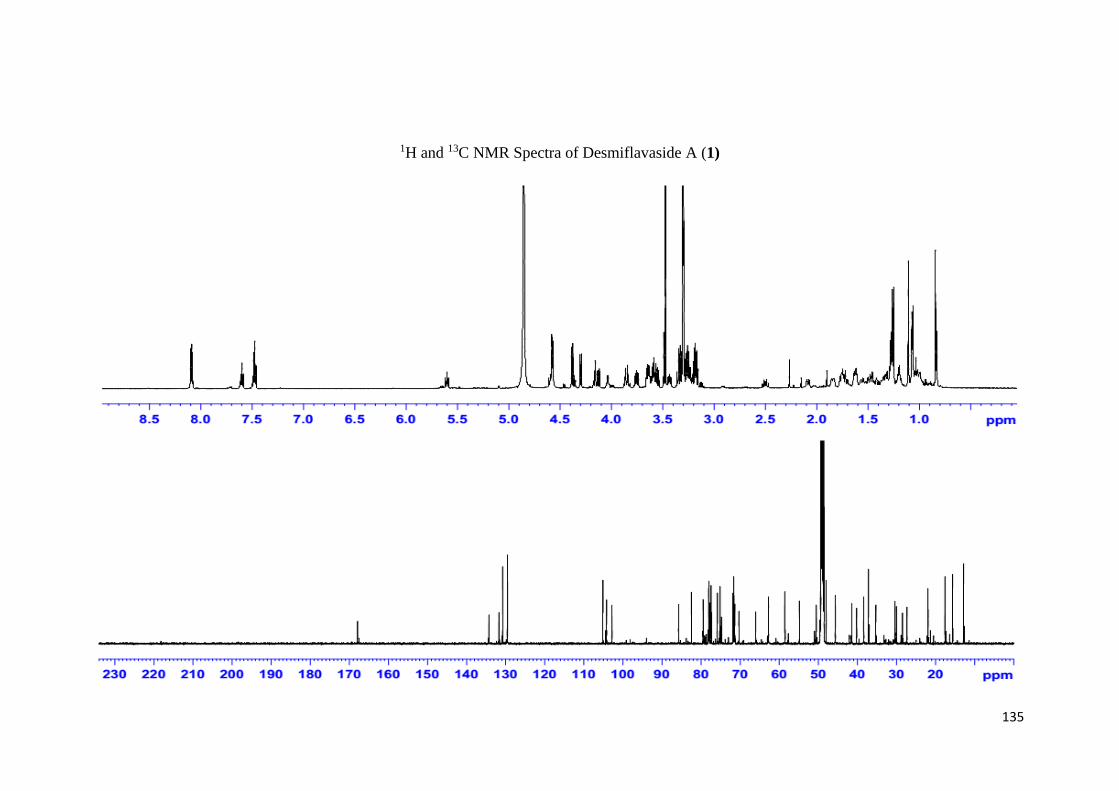
135
1H and 13C NMR Spectra of Desmiflavaside A (1)
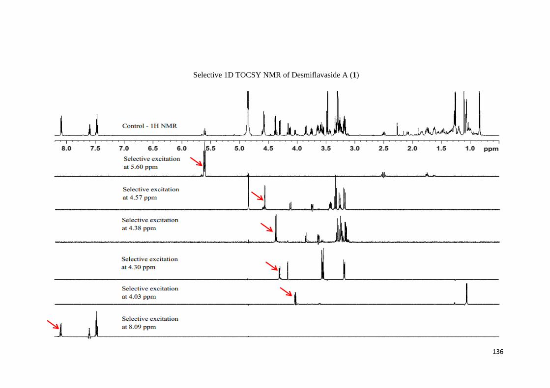
136
Selective 1D TOCSY NMR of Desmiflavaside A (1)

137
1H and 13C NMR Spectra of Desmiflavaside B (2)

138
Selective 1D TOCSY NMR of Desmiflavaside B (2)

139
1H and 13C NMR Spectra of Desmiflavaside C (3)
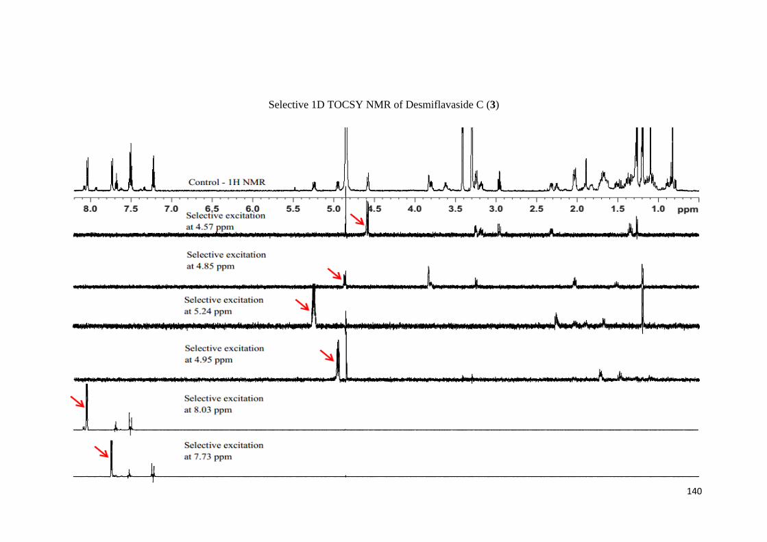
140
Selective 1D TOCSY NMR of Desmiflavaside C (3)

141
1H and 13C NMR Spectra of Desmiflavaside D (4)

142
Selective 1D TOCSY NMR of Desmiflavaside D (4)
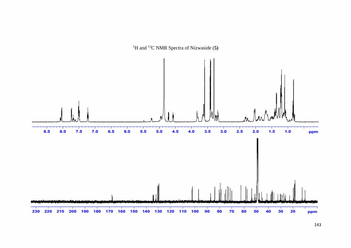
143
1H and 13C NMR Spectra of Nizwaside (5)

144
Selective 1D TOCSY NMR of Nizwaside (5)

145
1H and 13C NMR Spectra of Desflavaside A (6)
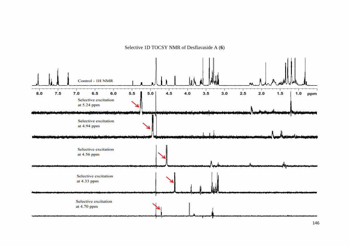
146
Selective 1D TOCSY NMR of Desflavaside A (6)
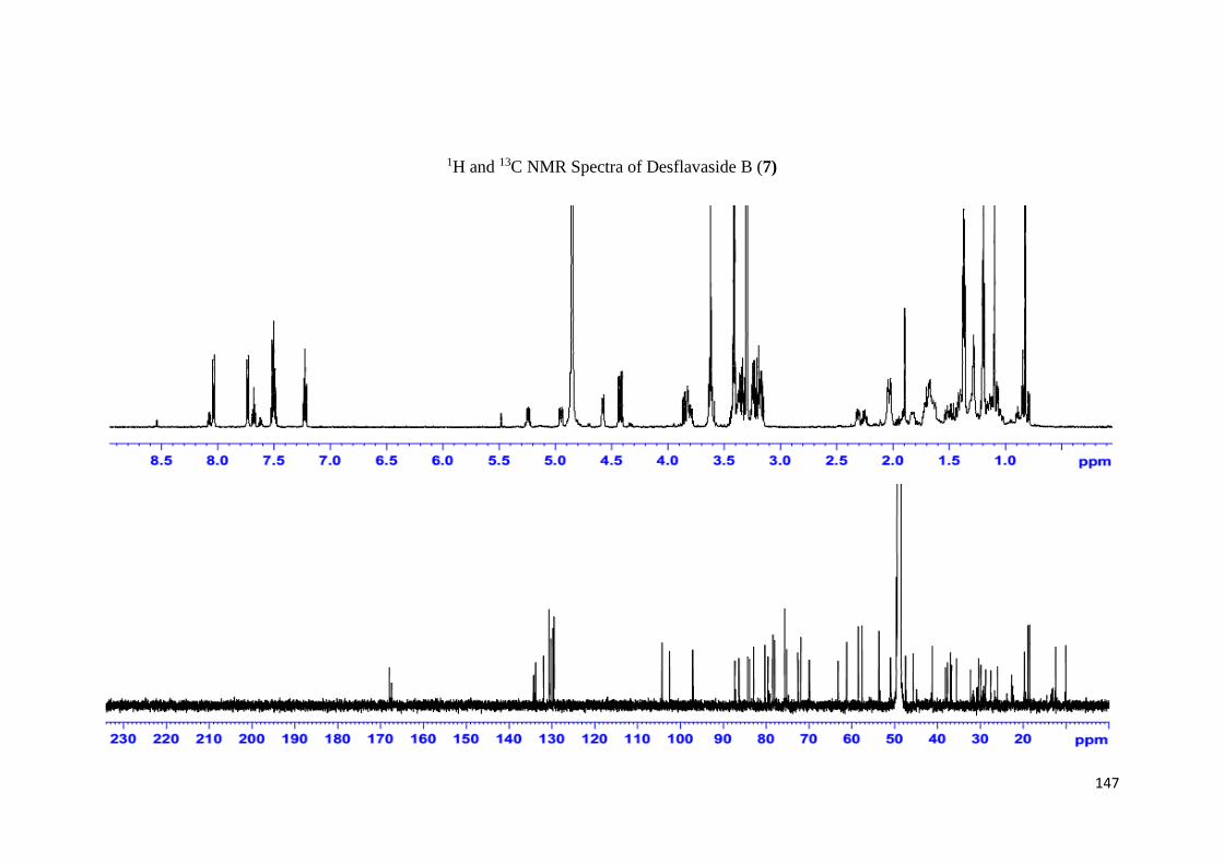
147
1H and 13C NMR Spectra of Desflavaside B (7)

148
Selective 1D TOCSY NMR of Desflavaside B (7)

149
1H and 13C NMR Spectra of Desflavaside C (8)

150
Selective 1D TOCSY NMR of Desflavaside C (8)
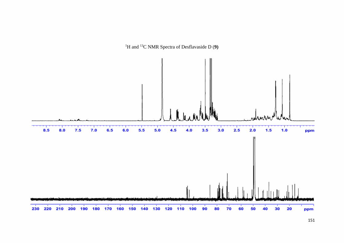
151
1H and 13C NMR Spectra of Desflavaside D (9)

152
Selective 1D TOCSY NMR of Desflavaside D (9)

153
9. PUBLICATIONS
154

155
156

157
158
CURRICULUM VITAE
I, Mr. Muhammad Adil s/o Raees Ahmed was born on June 7th, 1987 in Karachi,
Pakistan. I had my secondary school education with 1st division from Dhaka Boys
Secondary School, Karachi. I did higher secondary education with 1st division from
Saint Patrick’s Govt. College, Karachi during 2005-2006. I earned the degree of
Bachelors in Science (Analytical Chemistry) from Department of Chemistry, Federal
Urdu University, Karachi during 2007-2010. I joined University of Karachi as Trainee
NMR Spectroscopy in 2011. Along with my job, I continued my education and took
admission in M.S leading to Ph.D program in Department of Chemistry, Federal Urdu
University in 2012. I had an opportunity to join University of Nizwa, Sultanate of Oman
in the position of NMR Spectroscopist in 2013 and worked there with 600 MHz NMR
spectrometer having CryoProbe. I had my training in Advanced NMR Methods offered
by Bruker in Fällanden/Zürich, Switzerland in 2015. I performed most of my
experimental work of M.S and Ph.D in University of Nizwa, Sultanate of Oman. It is
the most attractive opportunity for me to complete my Ph. D. under the kind supervision
of Dr. Talat Mahmood.