proteins: workshop ii - louisiana tech universityupali/chem121/protein pogil.pdf · 6 d. tertiary...
TRANSCRIPT
1
Name _________________
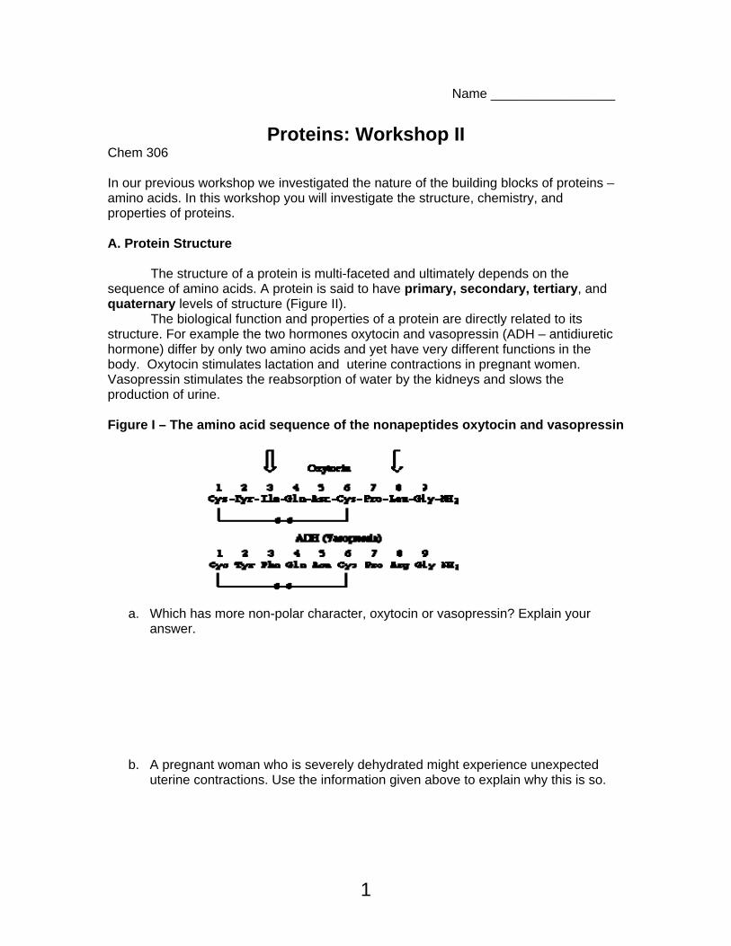
Proteins: Workshop II Chem 306 In our previous workshop we investigated the nature of the building blocks of proteins – amino acids. In this workshop you will investigate the structure, chemistry, and properties of proteins. A. Protein Structure The structure of a protein is multi-faceted and ultimately depends on the sequence of amino acids. A protein is said to have primary, secondary, tertiary, and quaternary levels of structure (Figure II). The biological function and properties of a protein are directly related to its structure. For example the two hormones oxytocin and vasopressin (ADH – antidiuretic hormone) differ by only two amino acids and yet have very different functions in the body. Oxytocin stimulates lactation and uterine contractions in pregnant women. Vasopressin stimulates the reabsorption of water by the kidneys and slows the production of urine. Figure I – The amino acid sequence of the nonapeptides oxytocin and vasopressin
a. Which has more non-polar character, oxytocin or vasopressin? Explain your answer.
b. A pregnant woman who is severely dehydrated might experience unexpected uterine contractions. Use the information given above to explain why this is so.

2
Figure II – The four levels of protein structure.
3
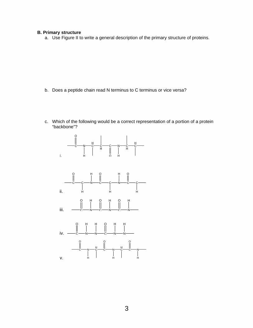
B. Primary structure a. Use Figure II to write a general description of the primary structure of proteins.
b. Does a peptide chain read N terminus to C terminus or vice versa?
c. Which of the following would be a correct representation of a portion of a protein
“backbone”?
i.
C
O
N
H
HC C
HC N
H
CH
HC
O
ii.
C
O
C
H
N
H
C
O
C N
H
C
O
C
HH
iii. C
O
N
H
C
O
N
H
C
O
N
H
iv. C
O
N
H
N
H
C
O
N
H
N
H
v.
C
O
N
H
HC C
O
NHC C
O
N
HH

4
C. Secondary structure – The secondary structure of a polypeptide is the spatial arrangement of the polypeptide and is held in place by H-bonds between backbone carbonyls & amino groups.
a. Circle the H-bonds between the backbone carbonyls in the structure below.
There are three major conformations found in the secondary structure of a polypeptide - helices, sheets, and random coils. All of these structures are found in both fibrous and globular proteins
• α-helix: The polypeptide backbone twists so that H-bonds form between C=O and an N-H group 4 residues (amino acids) further along in the sequence. Each amino acid R group face the outside of the helix. The α-helix is a right handed helix which means that it spirals in a clockwise direction. Hair, skin, and nails are composed of bundles of α-helices.
a. The amino acid proline is not commonly found in alpha helices unless it is one of
the first three amino acids. Why do you think this is so?
5
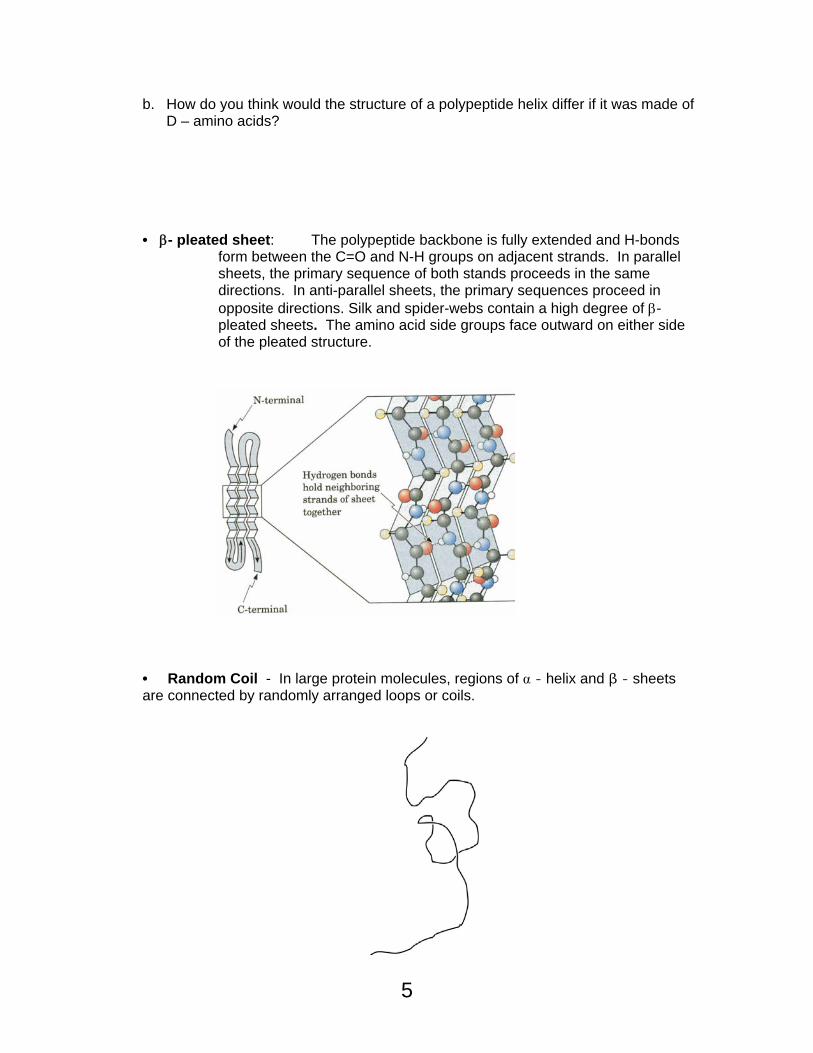
b. How do you think would the structure of a polypeptide helix differ if it was made of D – amino acids?
• β- pleated sheet: The polypeptide backbone is fully extended and H-bonds
form between the C=O and N-H groups on adjacent strands. In parallel sheets, the primary sequence of both stands proceeds in the same directions. In anti-parallel sheets, the primary sequences proceed in opposite directions. Silk and spider-webs contain a high degree of β- pleated sheets. The amino acid side groups face outward on either side of the pleated structure.
• Random Coil - In large protein molecules, regions of - helix and β - sheets are connected by randomly arranged loops or coils.
6
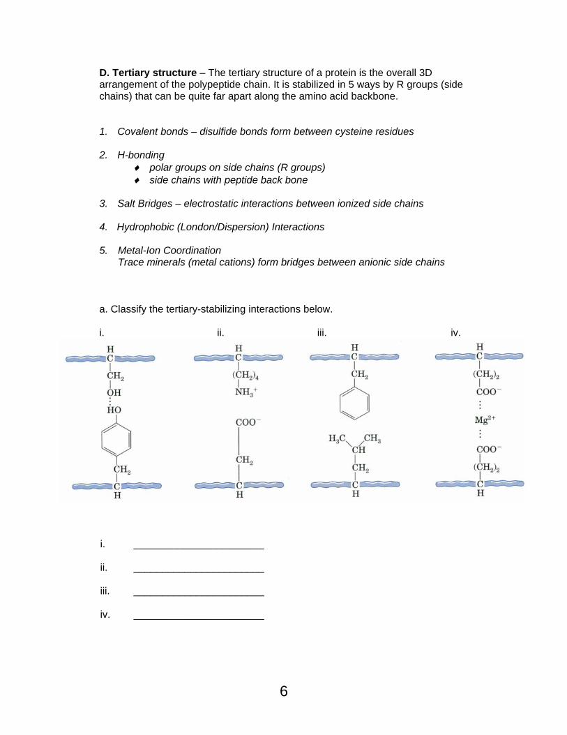
D. Tertiary structure – The tertiary structure of a protein is the overall 3D arrangement of the polypeptide chain. It is stabilized in 5 ways by R groups (side chains) that can be quite far apart along the amino acid backbone.
1. Covalent bonds – disulfide bonds form between cysteine residues 2. H-bonding
♦ polar groups on side chains (R groups) ♦ side chains with peptide back bone
3. Salt Bridges – electrostatic interactions between ionized side chains 4. Hydrophobic (London/Dispersion) Interactions 5. Metal-Ion Coordination Trace minerals (metal cations) form bridges between anionic side chains a. Classify the tertiary-stabilizing interactions below. i. ii. iii. iv.
i. _______________________ ii. _______________________ iii. _______________________ iv. _______________________
7
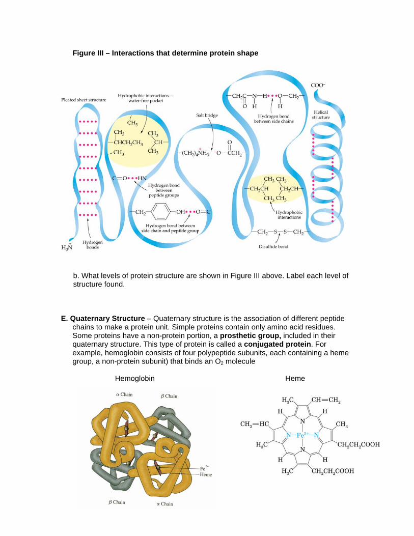
Figure III – Interactions that determine protein shape b. What levels of protein structure are shown in Figure III above. Label each level of structure found.
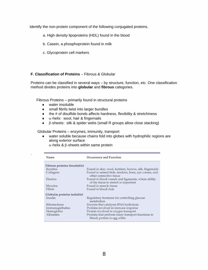
E. Quaternary Structure – Quaternary structure is the association of different peptide
chains to make a protein unit. Simple proteins contain only amino acid residues. Some proteins have a non-protein portion, a prosthetic group, included in their quaternary structure. This type of protein is called a conjugated protein. For example, hemoglobin consists of four polypeptide subunits, each containing a heme group, a non-protein subunit) that binds an O2 molecule
Hemoglobin Heme
8
Identify the non-protein component of the following conjugated proteins. a. High density lipoproteins (HDL) found in the blood b. Casein, a phosphoprotein found in milk c. Glycoprotein cell markers F. Classification of Proteins – Fibrous & Globular Proteins can be classified in several ways – by structure, function, etc. One classification method divides proteins into globular and fibrous categories.
Fibrous Proteins – primarily found in structural proteins ♦ water insoluble ♦ small fibrils twist into larger bundles ♦ the # of disulfide bonds affects hardness, flexibility & stretchiness ♦ α-helix: wool, hair & fingernails ♦ β-sheets: silk & spider webs (small R groups allow close stacking)
Globular Proteins – enzymes, immunity, transport
♦ water soluble because chains fold into globes with hydrophilic regions are along exterior surface
α-helix & β-sheets within same protein .
9
a. Which of the following amino acids would most likely be found on the external surface of a globular protein? Which would be found in the internal regions of a globular protein? Explain your answer. Asp Phe Ser Leu Arg b. Which example of fibrous proteins most likely has the most disulfide bonds between protein chains – finger nails or hair?
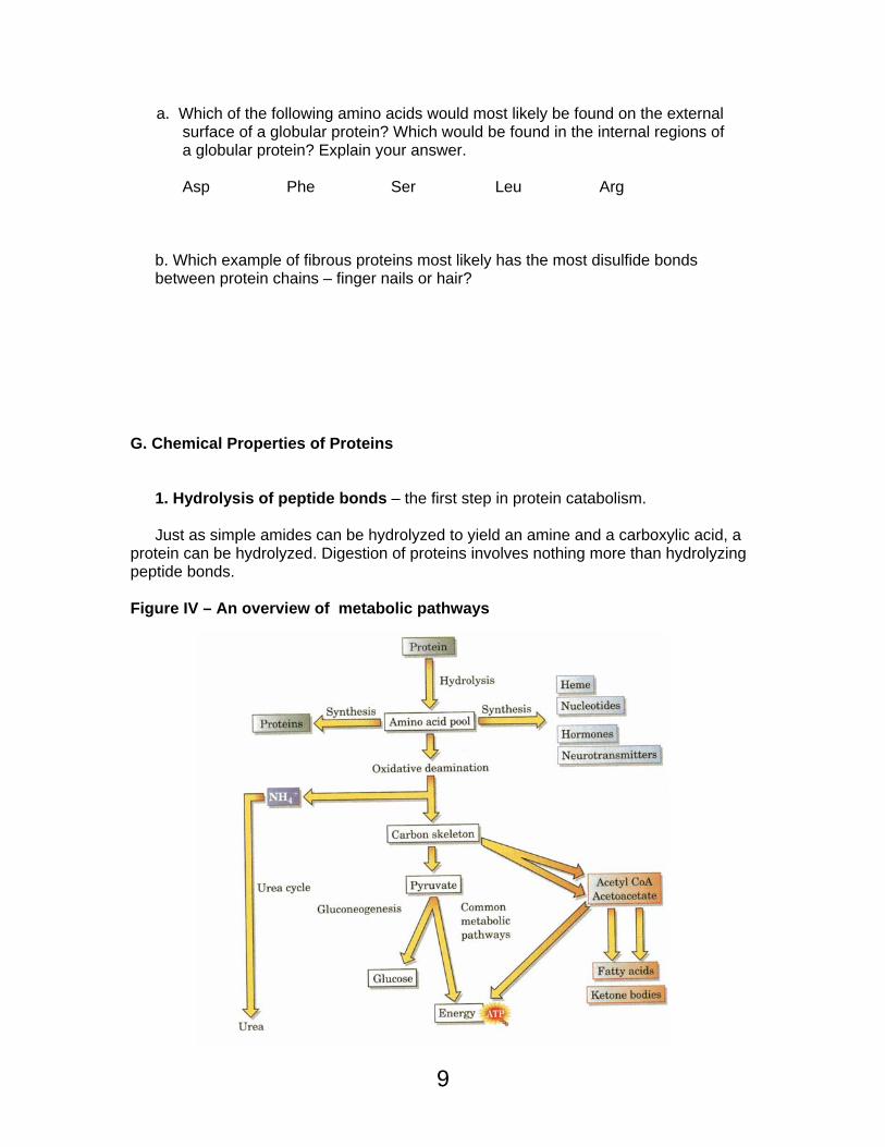
G. Chemical Properties of Proteins 1. Hydrolysis of peptide bonds – the first step in protein catabolism. Just as simple amides can be hydrolyzed to yield an amine and a carboxylic acid, a protein can be hydrolyzed. Digestion of proteins involves nothing more than hydrolyzing peptide bonds. Figure IV – An overview of metabolic pathways

10
Predict the acid hydrolysis products of the following tripeptide. Label each amino acid product.
+ H3N CH C
CH2
HN
O
HN
CH C
CH
HN
O
OH
CH3
CH C
CH2
O -
O
CH2
CH2
CH2
NH3 + 2. Denaturation
An active protein must be in the shape in which it functions in living systems (native conformation). The primary structure is composed of amino acids connected with covalent peptide bonds that are not easily broken. However, the secondary, tertiary, & quaternary structures are held together with weaker, noncovalent interactions. These structures can be disturbed by heat, agitation, drastic pH changes, detergents, salts, organic solvents, etc. in a process called denaturing. Denatured proteins are often said to be unfolded and are no longer able to perform their intended function. Some proteins can spontaneously refold, but most denaturations are essentially irreversible, inactivating the protein.

11
Table IV – Modes of Protein Denaturation (Destruction of Secondary and Higher Structures
a. Which amino acid is most frequently involved in denaturation by reducing agents?
b. What type of interactions are destroyed by organic solvents such as hexane?
c. Why do phlebotomists use 70% alcohol to wipe the skin clean before giving injections?
d. Ceviche is a fish dish that uses lemon juice to “cook” the fish. Use what you know about denaturation to explain this process and describe how it is related to cooking fish with heat.
12
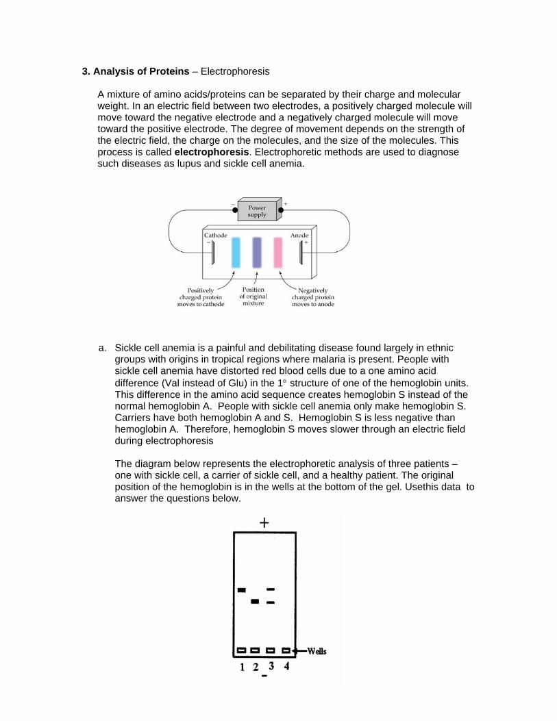
3. Analysis of Proteins – Electrophoresis
A mixture of amino acids/proteins can be separated by their charge and molecular weight. In an electric field between two electrodes, a positively charged molecule will move toward the negative electrode and a negatively charged molecule will move toward the positive electrode. The degree of movement depends on the strength of the electric field, the charge on the molecules, and the size of the molecules. This process is called electrophoresis. Electrophoretic methods are used to diagnose such diseases as lupus and sickle cell anemia.
a. Sickle cell anemia is a painful and debilitating disease found largely in ethnic groups with origins in tropical regions where malaria is present. People with sickle cell anemia have distorted red blood cells due to a one amino acid difference (Val instead of Glu) in the 1° structure of one of the hemoglobin units. This difference in the amino acid sequence creates hemoglobin S instead of the normal hemoglobin A. People with sickle cell anemia only make hemoglobin S. Carriers have both hemoglobin A and S. Hemoglobin S is less negative than hemoglobin A. Therefore, hemoglobin S moves slower through an electric field during electrophoresis The diagram below represents the electrophoretic analysis of three patients – one with sickle cell, a carrier of sickle cell, and a healthy patient. The original position of the hemoglobin is in the wells at the bottom of the gel. Usethis data to answer the questions below.

13
Why does hemoglobin S move slower than hemoglobin A in an electric field? Which lane is from a person with sickle cell anemia? Which lane is from a person with normal hemoglobin? Which lane is from a carrier for sickle cell anemia?
b. Three dipeptides were separated by electrophoresis at pH 6.4, and the results are shown below. The position of the original mixture is at point B. If the three dipeptides are Lys-Trp, Glu-Tyr, and Ala-Cys, identify each spot with the appropriate dipeptide.
+ Origin - A B C ● ● ●