protein synthesis in neurons and glial cells of the
TRANSCRIPT

Journal of Neurochemistry, 1971, Vol. IS, pp. 1445 to 1460. Pergamon Press. Printed in Great Britain.
PROTEIN SYNTHESIS IN NEURONS AND GLIAL CELLS OF THE DEVELOPING RAT BRAIN: AN IN V W O STUDY'
D. E. JOHN SON^ and 0. Z. SELLINCER Mental Health Research Institute, The University of Michigan Medical Center,
AM Arbor, Michigan
(Received 20 October 1970. Accepted 8 January 1971)
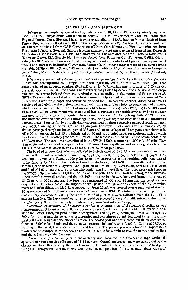
Abstract-A technique for the isolation of pure neuronal perikarya and intact glial cells from cerebral cortex has been developed for routine use. The yield of neuronal perikarya and glial cells was greater from highly immature (5-10 days) rat cerebral cortex than from the cortex of older rats (18-43 days). The perikarya/glia yield ratio decreased with age indicating that, as the glial population matured, the procedure succeeded in isolating a gradually smaller proportion of the existing neurons. The perikarya/glia ratio was highest for the 5-day- old cortex in which no mature glial cells could be identified.
After a 10-min pulse in vivo of intrathecally injected [14C]phenylalanine, the specific radioactivity of the neuronal proteins was higher than that of the glial proteins in the 5-, 10- and 18-day-old rat but was lower in the 43-day-old rat. The values for absolute specific radioactivity of the ''C-labelled proteins in both cell types were greater, the younger the brain. The 14C-labelling of neuronal and glial proteins in the 18-day-old rat was assessed in vivo as a function of time by determining the incorporation of [14C]phenylalanine into such proteins at 5, 10, 20 and 45 min after administration of the amino acid. The rate of incorporation of ['4C]phenylalanine into the glial cells was faster than into the neurons since higher specific radioactivities of the glial proteins could be achieved at earlier times. Also, a biphasic pattern of W-labelling of the glial proteins was noted, suggesting, perhaps, a sequen- tial involvement of the oligodendrocytes and astrocytes.
Homogenates of prelabelled neuronal perikarya were fractionated into the nuclear, mitochondrial, microsomal and soluble cell sap fractions. In the 18-day-old cerebral cortex, the proteins of the microsomal fraction exhibited the highest specific radioactivity at the end of 10 min, whereas by 20 min proteins of the mitochondrial fraction were most highly labelled. The specific radioactivity of the nuclear proteins increased over the entire 45-min experimental period. On the contrary, the proteins of the soluble cell sap, in which the specific radioactivity was at all times by far the lowest, were maximally labelled by 5 min. Examination of the labelling of the neuronal subcellular fractions as a function of age revealed that at 10 min after administration of [14C]phenyIalanine, the specSc radioactivities of all l4C-labe1led proteins were highest in the youngest (5-day-old) neurons. The proteins of the microsomal fraction were most rapidly labelled at all ages. During this interval the proteins of the soluble cell sap were only moderately labelled in the 5-day-old neurons and were totally unlabelled in the 43-day-old neurons, indicating age-dependent differences in the rate of utilization of the amino acid precursor by the neurons.
THE BRAIN is characterized by a high rate of protein synthesis (for reviews see LAJTHA, 1964 a, b). The rate of synthesis of cerebral proteins and of RNA is higher in the im- mature animal than in the adult (MILLER, 1969; ROBERTS, ZOMZELY and BONDY, 1970). BARONDES (1964) and YAMAGAMI, FRITZ and RAPPOPORT (1966) have shown that RNA polymerase and RNA are more active in brains of immature animals than in those of adults. SCHAIN, CARVER, COPENHAVER and UNDERDAHL (1967) examined the rate in vivo of incorporation of amino acids into pig brain and observed that the maximum
This work was supported in part by training grant MH-07417 from the National Institute of
Post-doctoral trainee. Mental Health and grant no. NB 06294 from the United States Public Health Service.
Abbreviations used: BSA, bovine serum albumin; G, glial cells; NP, neuronal perikarya; PPO, 2,5 diphenyloxazole; dimethyl POPOP, 1,4 bis [2-(4-methyl-5-phenyloxazolyl)]-benzene; PVP, polyvinylpyrrolidone.
1445

1446 D. E. JOHNSON and 0.2. SELLINGER
decrease in protein synthesis occurred soon after birth and was later followed by a more gradual decline. ABDEL-LATIF and ABOOD (1966) studied the incorporation in vivo of [14C]serine into the subcellular fractions of the developing rat brain and found that the proteins of all fractions were more highly labelled in the immature brain than in the older brain. In general, the in vivo experiments have been confirmed by in vitro studies which show that the rates of protein and nucleic acid synthesis decrease in mammalian brain during development (LAHIRI and LAJTHA, 1964; MURTHY and RAPPOPORT, 1965; MURTHY, 1966; YAMAGAMI ef al., 1966; ADAMS and LIM, 1966; JOHNSON and LUTTGES, 1966; JOHNSON, 1967, 1968; BONDY and ROBERTS, 1968; JOHNSON and BELYTSCHKO, 1969; YAMAGAMI and MORI, 1970).
In addition to the general pattern of decreasing protein synthesis with age, a marked change occurs in protein turnover which becomes progressively slower with age. Changes in the qualitative nature of the proteins also appear to relate to the development of brain tissue (GAITONDE and RICHTER, 1956; LAJTHA, FURST, GERSTEIN and WAELSCH, 1957; LAJTHA, 1964~ ; ROBERTS el al., 1970) and may thus reflect the physiological activity of particular brain cells which synthesize specific 'developmental' proteins such as the acidic S-100 protein (MOORE, PEREZ and GEHRING, 1968; FRIED- MAN and WENGER, 1970), the basic protein associated with myelin (HERRIMAN and HUNTER, 1965) and microtubular protein (DUTTON and BARONDES, 1969; TUOMEY and WYTTENBACH, 1970). Brain tissue consists principally of neurons and glial cells, the relative proportions of which change markedly with the age of the brain. In a develop- mental study on rats ranging from 5 to 730 days of age, BRIZZEE, VOGT and KHAK- ETCHKO (1964) found the glial population to be relatively small in the cerebral cortex of immature rats but to exceed by far that of the neurons in adult animals. Since virtually all previous studies on cerebral protein synthesis have utilized intact brain and thus perforce disregarded the problems of the changing proportions of, and of the quantita- tive differences between, neurons and glial cells known to characterize cerebral maturation, we thought it desirable to assess the synthetic contribution of the two cell types after their separation from one another.
Four distinct but only partially satisfactory procedures have been described for the separation of neurons from glial cells: (a) brain is treated with a mixture of acetone- glycerol and water prior to mincing and centrifugation (SATAKE and ABE, 1966; SATAKE, HASEGAWA, ABE and TANAKA, 1968 ; FREYSZ, BIETH, JUDES, SENSENBRENNER, JACOB and MANDEL, 1968); (b) brain mince is sieved and subjected to gradient centri- fugation (ROSE, 1967; ROSE and SINHA, 1969; BLOMSTRAND and HAMBERGEK, 1969); (c) whole brain is disrupted in a tissue press and zonal centrifugation used to separate the cellular elements (FLANGAS and BOWMAN, 1968) and (d) chopped brain is incubated under oxygen at 37°C in the presence of trypsin (NORTON and PODUSLO, 1970). An additional procedure has recently been developed in our laboratory which avoids some of the pitfalls of previous procedures and which makes it possible to isolate pure neuronal perikarya (NP) and intact glial cells (G) from as little as 1 g of cerebral cortex and in yields which permit the subsequent subcellular fractionation of the isolated cells (SELLINGER and AZCURRA, 1970; SELLINGER, AZCURRA, JOHNSON, OHLSSON and LODIN, 1971). Using this procedure, we have carried out an investigation of the time course in uiuo of protein synthesis in the neurons and the glial cells of rat cerebral cortex during development. A preliminary account of some of the results has appeared (JOHNSON and SELLINGER, 1970).
Protein synthesis in neurons and glia 1447
MATERIALS A N D METHODS Animals and materials. Sprague-Dawley, male rats of 5, 10, 18 and 43 days of postnatal age were
used. ~-[U-'~C]Phenylalanine with a specific activity of > 350 mCi/mmol was obtained from New England Nuclear Corp. (Boston, Mass.). Bovine serum albumin (BSA; fraction V) was obtained from Pentex Biochemicals (Kankakee, Ill.). Polyvinylpyrrolidone (PVP; Plasdone C, average mol. wt. 40,000) was purchased from GAF Corporation (Calvert City, Kentucky). Ficoll was obtained from Pharmacia (Uppsala, Sweden). Sucrose (special enzyme grade) was purchased from Mann Research Laboratories (New York, N.Y.). PPO and dimethyl POPOP were obtained from Packard Instruments (Downers Grove, Ill.). Biosolv No. 3 was purchased from Beckman Co. (Fullerton, Calif.). Glutar- aldehyde (70%, v/v, solution sealed under nitrogen in 2 ml ampoules) and Epon 812 were purchased from Ladd Research Industries (Burlington, Vermont). All other reagents were of the purest grade available. Millipore filters (Ga-6,0.45 pm pore size) were obtained from Gelman Instrument Company (Ann Arbor, Mich.). Nylon bolting cloth was purchased from Tobler, Ernst and Traber (Elmsford, N.Y.).
Injection procedure and isolation of neuronal perikarya and glial cells. Labelling of brain proteins in vivo was accomplished by a single intrathecal injection, while the rats were under light ether anaesthesia, of an aqueous solution (0.05 ml) of ~-[U-'~C]phenyIalanine in a dose of 6-25 pCi per brain. At specified intervals the animals were subsequently killed by decapitation. Neuronal perikarya and glial cells were isolated from the cerebral cortex according to the method of SELLINCER et al. (1971). Ten animals were killed and the brains were rapidly excised and placed on an inverted Petri dish covered with 6lter paper and resting on crushed ice. The cerebral cortices, dissected as free as possible of underlying white matter, were chopped with a razor blade into the consistency of a mince, which was transferred into 10-15 ml of an ice-cold solution of 7.5% (w/v) PVP, 1 % (w/v) BSA and 10 m-CaC12. The tissue mince was then eased into a truncated disposable syringe and the plunger was used to push the mince suspension through one thickness of nylon bolting cloth of 333 pm pore size stretched over the open end of the syringe. This sieving was repeated twice and the last filtrate was allowed to stand on ice for 10 min. Sieving was continued by three successive passes through an inner layer of 333 pm and an outer layer of 110 pm pore size mylon mesh and, after 10 min on ice, by similar passage through an inner layer of 333 pm and an outer layer of 73 pm pore size nylon mesh. After 20 min on ice, the last '73-pm filtrate' (about 65 ml) was divided into three portions, each of which was layered over a two-step gradient of 6 ml of 1.0 M-sucrose and 5 ml of 1.75 M-sucrose containing 1 % (w/v) BSA. The tubes were centrifuged in the SW-25.1 Spinco rotor at 41,000 g for 30 min. They then contained a top band of myelin, a band of nerve fibres, capillaries and impure glial cells at the 1.0 M-1.75 M-sucrose interface and a pellet of pure neuronal perikarya.
The band of impure glial cells was collected to include most of the 1.75 M-sucrose under it and was mixed with 1.5 vol. of a solution containing 5 % (w/v) Ficoll, 7.5 % (w/v) PVP and 1 % (w/v) BSA, whereupon it was centrifuged at 500 g for 10 min. A suspension of the resulting pellet was passed thrice through the 73 pm nylon mesh and was brought to a vol. of 45-60 ml. It was divided into three samples, each of which was layered over a gradient of 3 ml of 30% (w/v) Ficoll, 6 ml of 1.2 M-sucrose and 5 ml of 1.65 M-sucrose, all solutions also containing 1 % (w/v) BSA. The tubes were centrifuged in the SW-25.1 Spinco rotor at 41,000 g for 30 min. The pellets and the bands collecting at the sucrose- Fjcoll interface were discarded and the 1.2-1.65 M-Sucrose bands were kept and brought to a vol. of 18-22 ml with 0.32 M-sucrose. The tube was centrifuged at 500 g for 12 rnin and the pellet was re- suspended in 0.32 M-sucrose. The suspension was passed through one thickness of the 73 p m nylon mesh and, after dilution with 0.32 M-sucrose to about 20 ml, was layered over a gradient of 6 ml of 1-3 M-sucrose and 5 ml of 1.65 M-sucrose which were free of BSA. The tubes were centrifuged in the SW-25.1 Spinco rotor at 2500 g for 20 min. Purified glial cells were collected from the 1.3-1.65 M sucrose interface. The last centrifugation step might be repeated in case of significant contamination of the glia by capillaries, as routinely monitored by phase-contrast microscopy.
Subcellular fractionation of the neuronal perikarya. A suspension of the neuronal perikarya was homogenized in 0.25 M-sucrose with six up-and-down strokes rotating at about 1300 rev./min of a standard Potter-Elvehjem glass-Teflon homogenizer. The 5 % (w/v) homogenate was centrifuged at 800 g for 10 min and the pellet was resuspended and centrifuged as just described twice more. The final pellet was designated the nuclear fraction. The pooled post-nuclear supernatant fluids were centri- fuged at 18,000 g for 15 min and the pellet was resuspended and recentrifuged for 12 rnin at 17,300 8, yielding as the pellet, the crude mitochondria1 fraction. The pooled post-mitochondria1 supernatant fluids were centrifuged in the Spinco 65 rotor at 100,OOO g for 60 rnin to give the microsomal (pellet) and the cell sap (soluble) fractions.
Measurement of radioactivity. The radioactivity was measured in a Nuclear Chicago Unilux II spectrometer at a counting efficiency of 75-85 per cent. Quenching corrections were carried out by the channels-ratio method and by the use of an internal standard. The c.p.m. were converted to d a m . using a suitable program on the PDP-8 computer. The composition of the scintillation fluid was 4 g of

1445 D. E. JOHNSON and 0. Z. SELLINGER
PPO and 0.1 g of dimethyl POPOP per 1. of toluene. The total radioactivity was determined by mixing 1 ml of the sample with 2 ml of Biosolv-3 and 10 ml of the toluene scintillation fluid. The I4C-labelled protein was precipitated by 1 ml of ice-cold 10% (w/v) TCA and was adsorbed on Millipore filters (pore size: 0.45 pm). The filters were treated successively with 3 and 2 ml of 5 % (w/v) ice-cold TCA, 3 and 2 ml of 95 % (v/v) ethanol and 5 ml of ether. Heating in 5 % (w/v) TCA in the presence of ['2C]phenylalanine carrier, as recommended by ZOMZELY, ROBERTS, GRUBER and BROWN (1968), was found to be unnecessary and was therefore omitted. The filters were transferred to counting vials and dried under an infra-red lamp; 10 ml of the toluene scintillation fluid were added for counting. Protein was determined by the method of LOWRY, ROSEBROUGH, FARR and RANDALL (1951) and RNA was measured by the procedure of FLECK and BEGG (1965). For RNA, a factor of 41.6 was used to convert the Az6,, readings (taken in a final vol. of 1.3 ml) to pg of RNA/ml.
Morphological examination of the cells and subcehlar fractions. The neuronal perikarya and the dial cells were examined by phase-contrast microscopy (Fig. 1, A-C) and, together with the sub- cellular fractions of the neuronal perikarya, also by electron microscopy (Fig. 2, A to D). The neuronal perikarya were fixed in pellet form for 2.5 h at 4°C in solution GPS composed of 3 % (v/v) glutaralde- hyde buffered at pH 7.4 with 0.1 M-phosphate buffer and containing 5 % (w/v) sucrose. After fixation, the pellet was minced in the phosphate buffer and was stored at 4°C. The glial cells, collected as a band (see above), were fixed for 1 h at 4°C in solution GPS in which the concentration of glutaraldehyde was 1.5 % (v/v). The cells were centrifuged at 3020 g for 10 min and the pellet was minced in the phosphate buffer and stored at 4°C. The subcellular fractions were collected in pellet form and were fixed in solution GPS for 6 h at 4°C. After fixation, the pellets were transferred to the phosphate buffer and were stored at 4°C. Post-fixation was the same for all samples and was for 4 h in 2% (w/v) ice-cold osmium tetroxide in 0.1 M-phosphate buffer (pH 7.4) containing 5 % (w/v) sucrose. After a brief buffer wash, the samples were brought to room temperature, dehydrated in a graded series of alcohols and treated thrice with propylene oxide. They were kept overnight in a 1 : 1 (v/v) mixture of Epon 812 and propylene oxide and were then treated with pure Epon 812 for 6 h before being flat-embedded. The ultra-thin sections mounted on bare W m e s h copper grids were stained with 5 % (w/v) uranyl acetate and counterstained with lead citrate. The sections were examined in a JEOLCO, JEM 6-A electron microscope.
R E S U L T S Comparison of the neuronal perikarya and the glial cells. The yield (in terms of
protein) of neuronal perikarya and glial cells depended markedly on the age of the animal (Table 1); hence, a considerably higher proportion of neuronal perikarya was isolated from the younger rats. Taking the cortical suspension as the parent fraction for reference, the yield of neuronal perikarya dropped from 42.4 per cent in the 5-day- old rat to 0.68 per cent in the 43-day-old rat. When expressed this way, the yield of glial cells changed very little with age; no cells morphologically identifiable as mature glial cells were present in the preparation obtained from a 5-day-old rat, an observa- tion in confirmation of the morphological findings of CALEY and MAXWELL (1968b). There was a common but unequal decrease in the yield of both cell types with matura- tion. Hence, the NP/G ratio (Table 1) decreased in absolute value, confirming that the cell separation procedure led to the isolation of higher proportions of neuronal perikarya than of glial cells in the younger animal.
For studies relating the yield of both cell types to their selective labelling as a function of postnatal age (Table 2), a ratio of cell yield to cell I4C-labelled protein > 1 indicated the selective isolation of a larger proportion of unlabelled cells, whereas, conversely, a ratio < I indicated that some of the isolated cells were preferentially labelled. In the neuronal perikarya this ratio was > 1 only at 5 days and was < 1 later in life, whereas for the glial cells it decreased from values > 1 at 10 days to a low value at 43 days of 0.42. The incorporation of ~-['~C]phenylalanine into neuronal and glial proteins was studied in 5-, lo-, 18- and 43-day-old rats after a 10 min pulse in vim of ~-['~C]phenylalanine (Table 3). As indicated by the specific radioactivities of the isolated 14C-labelled proteins, the ability of neurons to synthesize protein decreased

FIG. 1.-Phase contrast photomicrographs. (A) Neuronal perikarya ( x 200); IS-day-old cerebral cortex of the rat; (B) Neuronal perikarya ( x 800); 18-day-old cerebral cortex; (C) Glial cells (x 1000) the two cells were from the cerebral cortex of a 38-day-old rat.
Note that not all structural features could be photographed in focus. 1448


FIG 2C" and C'
FIG. 2.--Electron micrographs. (A) Neuronal perikaryon from the cerebral cortex of an IS-day-old rat, detail ( ;. l0,OOO). (B) Same as A ( . 10,000); (C) Field showing the appearance of the microsomal fraction; 18-day-old cerebral cortex was used ( x 10,200), with a section through the top (C)' and middle (C") portions of the microsomal pellet (see Methods); the inset ( C ' ) shows a detail of a microsomal preparation demonstrating the presence of 'bound' ribosomes (studding the membrane of the endoplasmic reti- culum); (D) GIial cell from the cerebral cortex of an 18-day-old rat ( Y 10,000). Scale marker represents one pm. Abbret:iu/ions used: N, nucleus; NI, nucleolus: M, mito- chondrion; FR. free ribosomes; RER, rough endoplasmic reticulum; PM, plasma
membrane; NM, nuclear membrane.
TAB
= 1 .-
THE
YIELD
OF
NE
UR
ON
AL
PE
RIK
AR
YA
AND
GL
IAC
CELLS A
S A
FU
NC
~O
N O
F PO
STN
AT
AL
AG
E*
Age
C
orte
x su
spen
sion
t N
euro
nal p
erik
arya
(N
p)
Glia
l cel
ls (G
) R
atio
(d
ays)
T
otal
(m
g)
(mg/
d T
otal
Ow
) (m
g/g)
Y
ield
(%)
Tot
al (m
g)
(mg/
g)
Yie
ld (%
) N
PIG
Z 2
0, &. 3 Fi
17.4
5'
(n
= 3
) g 5
-
-
-
5 10
1 30
42.8
12
.2
42-4
4
(10.
3, 1
4.1)
0-
067
164.
2 10
37
7 63
f 1
-73
41.3
6.
9 f 1
.37
11.0
0.253
0.04
2 f 0-
005
18
757
113
rt: 1
.2
10.9
1-63 f 0.
15
1-44
04
22
0063
0.
014
0.05
6 25
-7
43
814
90 +
4.93
5-5
0.66
f 0.
00
0.68
0-
343
0.038 f 0
.002
0.
042
In =
2)
(37, 23
)
(n =
3)
&'
(i~ =
16)
El g G.
Neu
rona
l pe
rikar
ya a
nd g
lial c
ells
wer
e is
olat
ed a
s gi
ven
in M
etho
ds.
lu
* All
valu
es re
pres
ent p
rote
in c
onte
nt d
eter
min
ed by
the
met
hod
of L
OWRY
et a
l. (1
951)
on
pool
ed sa
mpl
es fr
om 1
0 rat
cer
ebra
l cor
tices
for e
ach
expe
rimen
t. t T
he c
ereb
ral c
ortic
es w
ere
chop
ped
with
a r
azor
bla
de a
nd p
lace
d in
10
mM
-CaC
I,.
To
obta
in u
ncon
tam
inat
ed p
rote
in v
alue
s, B
SA a
nd P
VP
wer
e pu
r-
pose
ly le
ft ou
t of t
he so
lutio
n in
whi
ch th
e cor
tices
wer
e ba
thed
for m
inci
ng. T
he m
ince
was
pas
sed
thro
ugh
333,
110
and
73 p
m n
ylon
mes
hes a
nd h
omog
eniz
ed
(six
up-
and-
dow
n st
roke
s) in
10
mM
-CaC
1,.
The
prot
ein
valu
es o
f th
e ne
uron
al p
erik
arya
and
glia
l fra
ctio
ns w
ere
obta
ined
in
sepa
rate
exp
erim
ents
. Mea
ns f
S.E.
M.
are
give
n.
Rat
ios
of m
g of
NP
prot
ein
to mg
of G
pro
tein
, eac
h pe
r g o
f sa
mpl
e.
Ther
e w
as n
o m
orph
olog
ical
evi
denc
e of
dia
l cel
ls in
the
5-da
y-ol
d sa
mpl
es.
E W

1450 D. E. JOHNSON and 0. Z. SELLINGER
TABLE 2.-&LL YIELD VS. CELL "c-LABELLED PROTEIN :* THE INFLUENCE OF AGE
% Neuronal perikarya % Glial cells Age
(days) % 14C-labelled protein? % 14C-labelled protein:
5 (n = 2) 1.89 (M 10 (n = 3) 0.90 f 0025 1.60 f 069 18 (n = 4) 0.64 f 0.021 1.00 f 0031 43 (n = 3) 0.84 & 0028 0.42 i 0.020
(1.87, 1.91)
The in uiuo pulse of ~-['~C]phenylalanine was 10 min. Neuronal perikarya and glial cells were isolated as described in Methods.
Except where individual values are listed (5 days), mean values =t S.E.M. are given. The percentage of glial cells was obtained by dividing the protein of the glial cells by the protein of the cortex suspension (Table 1) x 100.
* 14C-labelled protein refers to TCA-precipitable, Millipore-treated radioactivity, as described under Methods.
x 100 t Percentage '*C-labelled protein =i d.p.m. in cortex suspension d.p.m. in NP
x 100 d.p.m. in G-
d.p.m. in cortex suspension Percentage '*C-labelled protein =
4 There was no morphological evidence of glial cells in the 5-day-old samples.
progressively, whereas that of the glial cells increased during the period 10 to 18 days and then decreased during the period 18 to 43 days of age. The specific radioactivity of both cell types was lowest in the 43-day-old rat. Also, the specific radioactivity of the neurons was higher than that of the glial cells at all ages except at 43 days, when that of the glial cells was higher. Therefore, at 43 days the NP/G ratio for these data was c 1. The time course of neuronal and glial protein synthesis was assessed in viuo by deter- mining the incorporation of ~-['~C]phenylalanine into 14C-labelled protein at 5, 10,
TABLE 3.-THE SPECmC RADIOACTIVITY OF NEURONAL AND GLIAL l4c-LABELLED PROTEIN :* THE EFFECT OF AGE
5 days 10 days 18 days 43 days type (d.p.m./mg, n = 2) (d.p.m./mg, n = 3) (d.p.m./mg, n = 4) (d.p.m./mg, n= 3)
Neuro nal 3204 3127 f 320 2869 f 326 463 f 99
Glia (G) ? t 1793 4 415 2205 f 368 853 f 85 NP/G ? 1.74 1.30 0.54
perikarya (NP) (3892; 2515)
Except where individual values are listed (5 days), mean values f S.E.M. are given. Rats were injected intrathecally with 50 pl of aqueous ~-[U-'*C]phenylanaline in a dose of 6.25 pCi
per brain and killed after an in uiuo pulse of 10 min. Neuronal perikarya and glial cells were isolated as described in Methods.
* 14C-labelled protein refers to TCA-precipitable, Millipore-treated radioactivity, as described under Methods.
t There was no morphological evidence of glial cells in the 5-day-old samples.

Protein synthesis in neurons and glia 1451
I P
0 Cortex Suspenrron
A Neuronal Psriharfl
a Glial Ce/k
u 5 I0 I5 20 25 30 35 40 45
CI4 Pulse(h4inutes)
FIG. 3.-Incorporation of ~-['~C]phenylalanine into proteins of the cerebral cortex, neuronal perikarya and glial cells of 18-day-old rats as a function of pulse time. Rats were injected intrathecally with 50 pl of an aqueous solution of ~-[U-'~C]phenylalanine in a dose of 625 pCi per brain and killed at the desired times. The cerebral cortex, neuronal perikarya and glial cells were isolated as described in Methods and the specific radioactivities (d.p.m./mg of protein) were determined. Values are the averages of four experiments for the cerebral cortex and neuronal perikarya and three experiments for the glial cells. The S.E.M. for the 5, 10, 20 and 45-min pulses were respectively: for cortex suspension: &97, 1334, &451 and 1448; for neuronal perikarya: ~539, 1326, 5764
and &316; for glial cells: 1756, &520, &429 and *1116.
20 and 45 min after administration (Fig. 3). Labelling of the protein was rapid but unequal, reaching near maximum values in the neurons within 10 min and in the glial cells within 20 min from the time of administration of ~-['~C]phenylalanine. The relative rates of incorporation of ~-['~C]phenylalanine were initially faster for the glial cells than for the neurons, as indicated by higher values for specific radioactivity in glial cells at 5 min. At 10 min, however, the specific radioactivity of the neuronal 14C-labelled protein exceeded that of the glial 14C-labelled protein.
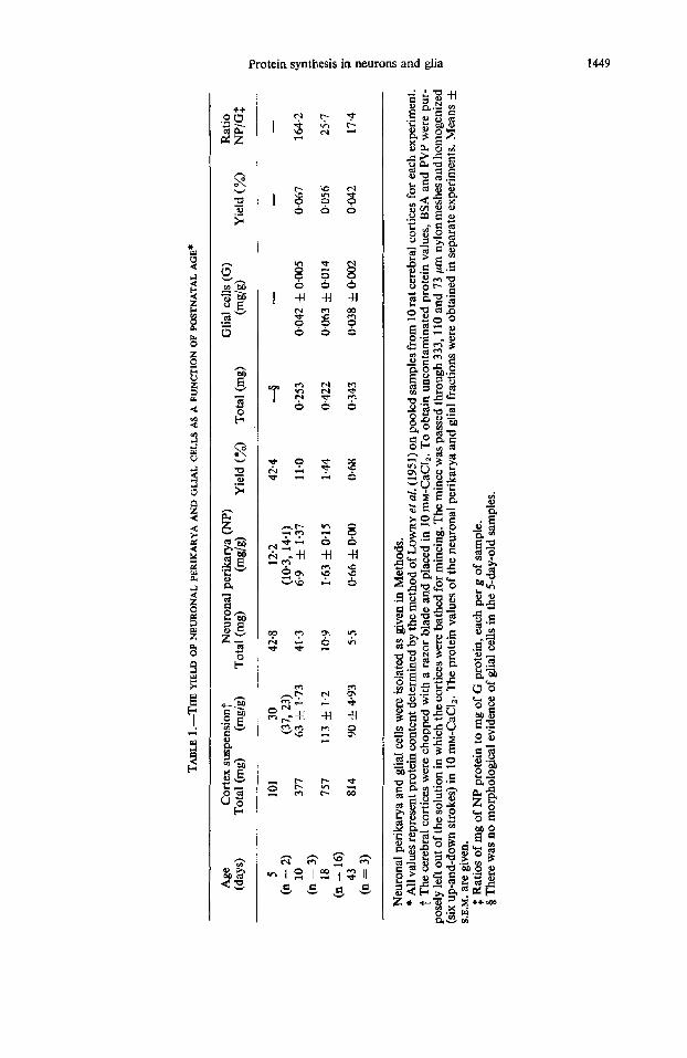
Protein and RNA content in the subcellular fractions of the neuronalperikarya. The protein content of the subcellular fractions of the neuronal perikarya varied with age (Table 4). Thus, the amount of protein in the nuclear and microsomal fractions decreased and that in the mitochondrial and cell sap fractions increased with increasing postnatal age (MURTHY and RAPPOPORT, 1965). The subcellular distribution of RNA in the neuronal perikarya was determined in the 18-day-old rat (Table 5). A high pro- portion of RNA (54.4 per cent) characterized the microsomal fraction (Fig. 2C), whereas the cell sap fraction contained only 10.3 per cent of the total RNA. Conse- quently, the ratios of percentage RNA/percentage protein ranged from 0.19 for the cell sap to 4.3 for the microsomal fraction, with the remaining fractions averaging about 1.0.
Efect of pulse duration and of age on the incorporation of [14C]phenylalanine into proteins in the subcellular fractions of neuronal perikarya. Subfractionation of a homo- genate of the neuronal perikarya derived from 18-day-old rats injected with L-['~C]- phenylalanine revealed that the distribution of the 14C-labelled protein was affected by the pulse time in uivo (Table 6). The mitochondria1 fraction comprised about
NEURO 1818-F
TA
BL
E 4.-NE
URON
AL
PRO
TEIN
CO
NTE
NT AND
ITS
SUB
CEL
LULA
R D
ISTR
IBU
TIO
N: E
FFEC
T OF A
GE
5 da
ys (n
= 2
) 10
day
s (n
= 3
) 18
day
s (n
= 1
6)
43 d
ays (n =
3)
Frac
tion
Tot
al (m
g)
( %)
Tota
l (m
g)
( %)
Tota
l (m
g)
( %)
Tota
l (m
g)
( %)
U
Hom
oaen
ate
42.8
10
0 41
.3 f 1
0.1
100
10.9
f 0.
71
100
5.5 f 0.
21
100
bd
-
Nuc
lear
Mito
chon
dria
1
Mic
roso
inal
Cel
l sap
* Rec
over
y, %
1'1
(37.
6, 4
8.0)
4
10.2
32
.3
11.8
f 3.
54
30.2
1.
45 f 0.
18
15.5
0.
55 f 0.
15
12.6
8
(10.
1, 1
0.4)
z
4.65
14
.7
5.4 f 0.
60
13.8
1.
65 f 0.
13
17.6
E (3
.9, 5
.4)
18.5
1.
20 f 0.
15
12.7
0.
40
0.02
9.
2 P
5.
9 18
.6
7.2
& 1
.61
P 11
.0
34.4
14
.65 f 3-
73
37.5
5.
1 f 0.
43
54.2
2.
48 f 0.
05
0.93
f 0.
18
21.3
H
569
.N
74.2
94
4 85
.9
80.0
I F
(5.7
, 6.1
)
(13.
9, 9
.0)
k! Th
e ne
uron
al p
erik
arya
and
thei
r su
bcel
lula
r fra
ctio
ns w
ere
isol
ated
as
desc
ribed
in M
etho
ds. T
he m
orph
olog
ical
app
eara
nce
of th
e m
icro
som
al fr
actio
n is
* Sum
of
prot
ein
in f
ract
ions
Ten
rat c
ereb
ral c
ortic
es w
ere
pool
ed fo
r eac
h ex
perim
ent.
The
perc
enta
ge v
alue
s are
corr
ecte
d fo
r diff
eren
ces i
n re
cove
ry. E
xcep
t whe
re in
divi
dual
val
ues
are
8 ill
ustra
ted
in F
ig. 2
C.
-x lo
o. pr
otei
n in
hom
ogen
ate
liste
d (5
day
s), m
ean
valu
es &
S.E
.M.
are
give
n.

Protein synthesis in neurons and glia 1453
TABLE 5.-THE SUBCELLULAR DISTRIBUTION OF RNA IN MURONAL PERIKARYA
RNA RNA Fraction (mg) (%) % RNA/% protein
Homogenate 1.516 100 1 .o Nuclear 0.199 f 0.007 16.0 1.03 Mitochondrial 0.241 f 0,004 19.3 1.10 Microsomal 0.678 f 0.024 54,4 4.28 Cell sap 0.129 f 0.003 10.3 0.19 * Recovery, % 82-3
The cerebral cortices of ten 18-day-old rats were used for each experiment. Initial weight of the pooled cortices: 6 7 g.
Values are normalized to 100 per cent recovery and are the means f S.E.M. of six experiments. See Methods for details of fractionation and analytical Drocedures.
* Sum of RNA in fractions RNA in homogenate x loo.
50 per cent of the 14C-labelled proteins after a 20-min pulse, whereas at that time the microsomal fraction comprised less than 30 per cent of the total. At all pulse times the cell sap comprised less than 10 per cent of the l4C-labe1led proteins. SELLINGER, AZCURRA, IDOYAGA-VARGAS and SANrIAGo (unpublished) have shown peaks of specific activity of succinate and glycerol-phosphate dehydrogenases (EC 1.3.99.1 and 1.1.1.8) in the mitochondria1 fraction, of N-acetyl-j3-D-glucosaminidase in the lysosomal frac- tion and of lactate dehydrogenase (EC 1.1.1.27) in the soluble cell sap fraction, thus confirming the relative intactness of the isolated cell bodies. On the other hand, more recently (JOHNSON and SELLINGER, in preparation) the soluble perikaryal proteins could be appreciably labelled, provided the exposure of the cells to ~-['~C]phenylalanine
TABLE 6.-THE SUBCELLULAR DISTRIBUTION OF "C-LABELLED PROTEIN* I N NEURONAL PERIKARYA : THE EFPECT OF PUJSE DURATION
Fraction 5 min 10 min 20 min 45 min ( %) ( %) ( %) %)
Nuclear 19.0 1.54 19.4 & 1.98 19.3 f 3-91 23.4 & 2.98 Mitochondria1 34.5 f 4.36 35.9 & 5.50 50.6 f 6.16 38.4 3.78 Microsomal 37.9 f 4-09 36.5 f 3.77 25.2 f 1.02 29.2 f 3.90 Cell sap 8.6 f 1.37 8.2 f 2.93 4.9 f 1.39 9.0 & 3.17 t Recovery, % 92.8 106.5 89.6 92,4
Rats of 18 days were injected as described in Table 3 and killed at the desired times. The neuronal
Values are expressed in percentages and are corrected for differences in recovery. Each value is the
* 14C-labelled protein refers to TCA-precipitable, Millipore-treated radioactivity, as described in
t Sum of d.p.m. in fractions
perikarya and their subcellular fractions were isolated as described in Methods.
mean of four experiments f S.E.M.
Methods.
x loo. d.p.m. in homogenate

1454 D. E. JOHNSON and 0. Z. SELLINGER
Or;/ 5 10 IS 20 25 30 35 b 25 I I I I I I I
Ct4 Pulse (Minutes)
FIG. 4.-Incorporation of ~-['~C]phenylalanine into proteins of the subcellular fractions of neuronal perikarya from 18-day-old rat brains as a function of pulse time. Rats were injected intrathecally with 50 pl of an aqueous solution of ~-[U-'~C]phenyIalanine in a dose of 6.25 pCi per brain and killed at the desired times. The subcellular fractions were isolated as described in Methods and the specific radioactivities (d.p.m./mg of protein) were determined. Values are the averages of four experiments. The S.E.M. for the 5, 10, 20 and 45-min pulses were respectively: for cell sap: *173, 1165, 1 3 6 1 and f217; for nuclei: f185, k418, 11158 and f1652; for mitochondria: 792,1673,2314,1437;
for microsornes; f990, *1964, 12208 and f 1020.
in uiuo was of longer duration (1 day to 2 weeks). The microsomal fraction exhibited the highest specific radioactivity at 10 min (Fig. 4) but by 20 min, the mitochondrial fraction became the most highly labelled fraction. The specific radioactivity of the nuclear fraction increased steadily over the 45-min experimental period, whereas that of the cell sap changed very little and remained the lowest of all fractions.
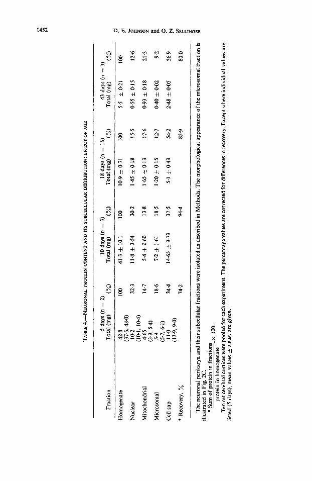
The distribution of the ''C-labelled proteins among the subcellular fractions of the neuronal perikarya derived, after a 10-min pulse C i uiuo, from 5-, lo-, 18- and 43-day- old rats revealed age-dependent differences (Table 7). Thus, while the nuclear fraction comprised about 20 per cent of the '4C-labelled protein at all ages, the share of the mitochondrial fraction rose progressively from about 13 per cent in the 5-day-old rat to 46 per cent in the 43-day-old rat and conversely, the amount of 14C-labelled protein in the microsomal and cell sap fractions decreased progressively over the same age period. A comparison of the specific radioactivities also revealed age-dependent differences (Fig. 5). The labelling of the nuclear fraction was fairly constant up to 18 days but declined thereafter, whereas that of the mitochondrial fraction increased from 5 to 18 days and also declined thereafter. The microsomal fraction was labelled most rapidly at all ages, but microsomes from the 43-day-old rat exhibited only 15 per cent of the activity found in the microsomes from 5-day-old rats. Under the conditions of our experiments, the soluble neuronal proteins were only moderately labelled in the 5-day-old rat and, as already mentioned, not at all in the 43-day-old animal. In absolute terms, the cell sap fraction was the least labelled of all the sub- cellular fractions studied at any age by the pulse-labelling technique.
TCA-soluble rudiuuctioity. To determine the effect of the duration of the radioactive pulse and of age on the efficiency of utilization of ~-['~C]phenylalanine, we attempted
Protein synthesis in neurons and glia
,,,HO#OGENATE lUCLEAR MITOCHOWDRIAL MICROSONAL CELL SAP
lob
1455
FIG. 5.-The incorporation in uiuo of [14C]phenylalanine into proteins of the neuronal perikarya and their subcellular fractions from rats of various ages. Rats were injected intrathecally with 50 p1 of an aqueous solution of ~-[U-'~C]phenyIalanine in a dose of 6.25 pCi per brain and killed after an in uiuo pulse of 10 min. The neuronal perikarya and the subcellular fractions were isolated as described in Methods and the specific radioactivities (d.p.m./mg of protein) were determined. Each value is the average of four experiments and the vertical lines on the bars represent the range of values. Note
the value of zero d.p.m./mg for the cell sap of 43-day-old rat.
TABLE 7.-THE SUBCELLULAR DISTRIBUTION OF 14C-LABELLED PROTEIN* IN NEURON& PERIKARY.4: EFFECT OF AGE
5 days (n = 2) 10 days (n = 3) 18 days (n = 4) 43 days (n = 3) Fraction ( %) (%) ( %) ( %I
Nuclear 23-0 24.0 f 1.94 19.4 f 1.98 23.2 & 4.47
Mi tochondrial 12.9 21.5 f 3.49 35.9 & 5.50 45.8 & 3.17 (23-5, 22.3)
Microsomal (106, 15.1)
(46.9, 45.0) 460 38.7 f 2.17 36.5 f 3.77 31.0 & 4.57
Cell sap . 18.1 15.8 & 1-76 8-2 f 2.93 0.0 f 0.00 (18.9, 17.6)
t Recovery, % 945 103.0 106.5 99.6
Conditions of injection and killing are given in Table 3. The neuronal perikarya and their sub-
Except where individual percentages are listed ( 5 days) mean values f S.E.M. are given. All values
* *4C-labelled protein refers to TCA-precipitable, Millipore-treated radioactivity, as described in
t Sum of d.p.m. in fractions d.p.m. in homogenate
cellular fractions were isolated as described in Methods.
were corrected for differences in recovery.
Methods.
x loo.
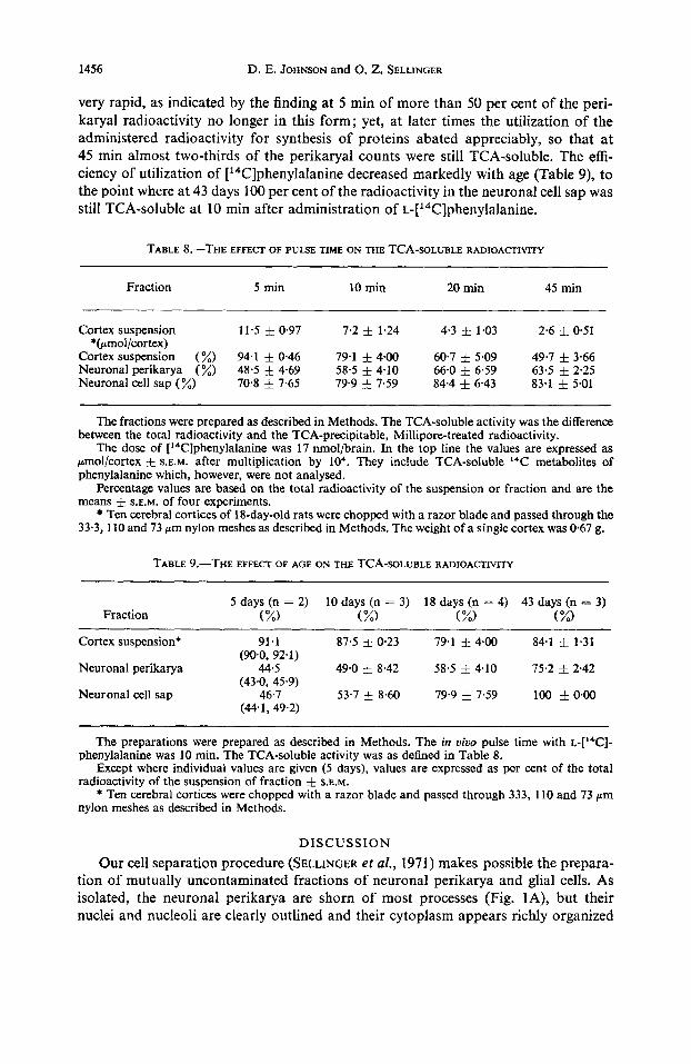
to estimate, after a 10 min pulse, the TCA-soluble radioactivity in three fractions of the 18-day-old brain cortex (Table 8) and at four ages (Table 9). TCA-soluble radioactivity decreased rapidly in the cortical suspension during the period between 5 and 45 min (Table 8). In the neuronal perikarya, the loss of TCA-soluble counts was initially also
1456 D. E. JOHNSON and 0. Z. SELLINGER
very rapid, as indicated by the finding at 5 min of more than 50 per cent of the peri- karyal radioactivity no longer in this form; yet, at later times the utilization of the administered radioactivity for synthesis of proteins abated appreciably, so that at 45 min almost two-thirds of the perikaryal counts were still TCA-soluble. The effi- ciency of utilization of [14C]phenylalanine decreased markedly with age (Table 9), to the point where at 43 days 100 per cent of the radioactivity in the neuronal cell sap was still TCA-soluble at 10 min after administration of ~-['~C]phenylalanine.
TABLE 8.-THE EFFECT OF PULSE TIME ON THE TCA-SOLUBLE RADIOACnVlTY
Fraction 5 min 10 min 20 min 45 min
Cortex suspension 11.5 0.97 7.2 f 1.24 4.3 f 1.03 2.6 f 0 5 1
Cortex suspension (%) 94.1 f 0.46 79.1 f 4.00 60.7 f 5.09 49.7 f 3.66 Neuronal perikarya (%) 48.5 f 4.69 58.5 f 4.10 66.0 i 6.59 63-5 f 2.25 Neuronal cell sap (%) 70.8 f 7.65 79.9 f 7.59 84.4 f 6.43 83.1 f 5.01
*(prnol/cortex)
The fractions were prepared as described in Methods. The TCA-soluble activity was the difference between the total radioactivity and the TCA-precipitable, Millipore-treated radioactivity.
The dose of ['4C]phenylalanine was 17 nmol/brain. In the top line the values are expressed as pmol/cortex f S.E.M. after multiplication by lo4. They include TCA-soluble I4C metabolites of phenylalanine which, however, were not analysed.
Percentage values are based on the total radioactivity of the suspension or fraction and are the means f S.E.M. of four experiments.
Ten cerebral cortices of 18-day-old rats were chopped with a razor blade and passed through the 33.3, 110 and 73 pm nylon meshes as described in Methods. The weight of a single cortex was 0.67 g.
TABLE 9.-THE EFFECT OF AGE ON THE TCA-SOLUBLE RADIOACTIVITY
5 days (n = 2) 10 days (n = 3) 18 days (n = 4) 43 days (n = 3) Fraction ( %) ( %) ( %) (%I
Cortex suspension* 91.1 87.5 & 0.23 79.1 f 4.00 84.1 1-31 (90.0, 92.1)
Neuronal perikarya 44.5 49.0 f 8.42 58.5 i 4.10 75.2 & 2.42
Neuronal cell sap 46.7 53.7 & 8.60 79.9 7.59 100 f 0.00 (43.0, 45.9)
(44.1,49.2)
The preparations were prepared as described in Methods. The in vioo pulse time with L-['~C]-
Except where individual values are given (5 days), values are expressed as per cent of the total
* Ten cerebral cortices were chopped with a razor blade and passed through 333, 110 and 73 pm
phenylalanine was 10 min. The TCA-soluble activity was as defined in Table 8.
radioactivity of the suspension of fraction f S.E.M.
nylon meshes as described in Methods.
DISCUSSION
Our cell separation procedure (SELLINGER et al., 1971) makes possible the prepara- tion of mutually uncontaminated fractions of neuronal perikarya and glial cells. As isolated, the neuronal perikarya are shorn of most processes (Fig. lA), but their nuclei and nucleoli are clearly outlined and their cytoplasm appears richly organized

Protein synthesis in neurons and glia 1457
(Fig. 1B). Electron microscopy of the neuronal perikarya (Fig. 2, A and B) and of the glial cells (Fig. 2D) revealed largely, if not entirely, intact plasma membranes, rather well-preserved intracellular organelles and a normal double-layered nuclear envelope. Our fraction of glial cells (Fig. 1C) most closely compares with the preparation described by NORTON and PODUSLO (1970), since the numerous processes appeared preserved and the nucleus comprised the major portion of the cellular space. With our technique, the yield (on the basis of protein) of both cell types, but particularly that of the neuronal perikarya, decreased with age, a feature previously not noted by most of the other authors who have applied similar cell-separation techniques at only one age (ROSE, 1967; FLANGAS and BOWMAN, 1968; BLOMSTRAND and HAMBERGER, 1969). NORTON and PODUSLO (1970), who worked with 10-30-day-old rats, reported an approximately five-fold higher yield of neuronal perikarya than of glial cells (NP/G: 5). Our NP/G ratios (Table 1) for the same age span ranged from a high value of 164 at 10 days to a low value of 17.4 at 43 days. We cannot assess whether this age- dependent decrease in the relative yield of cells is a consequence of the technical arbitrariness of imposing a constant pore-size limit to the passage of cells derived from cortices of varying age or whether, instead, it is the result of developmental changes in the physical state and/or the cellular and hence chemical composition of the brain cortex (CALEY and MAXWELL, 1968a, b). The high initial NP/G ratio (Table 1 ; 5 days) and the pronounced drop of this ratio within 1 week suggests that, as the number of glial cells increased in relation to the number of existing neurons to reach a constant density in presumably less than 23 days (DALTON, HOMMES and LEBLOND, 1968), the isolation of neuronal perikarya became relatively more difficult than at very early post-natal times when neurons relatively process-free (CALEY and MAXWELL, 1968~) are known to predominate numerically (BRIZZEE et al., 1964).
The incorporation rates in uiuo of labelled amino acids into cerebral proteins are known to be faster in young animals than in adults, with the major decrease in rate occurring soon after birth and followed subsequently by a lesser and more gradual decline (ABDEL-LATIF and ABOOD, 1966; SCHAIN et al., 1967; OJA, 1967). The results of studies in vitro generally support this conclusion (MURTHY and RAPPOPORT, 1965; MURTHY, 1966, 1970; JOHNSON and BELYTSCHKO, 1969; LERNER and JOHNSON, 1970; ROBERTS et al., 1970). Although our results on neuronal and glial protein synthesis in vivo confirm previous findings obtained for intact brain, they also bring out qualita- tive and quantitative differences between the two cell types. Thus, in the neuronal perikarya the specific radioactivity of the 14C-labelled proteins in the 43-day-old rat was 1/7 of that of the 14C-labelled proteins in the 5- or 10-day-old animal (Table 3), whereas the 43-day values were merely one-half of the 10-day values in the glial cells (Table 3). Furthermore, although the specific radioactivity of the glial 14C-labelled proteins increased perceptibly between days 10 and 18, namely, at a time of maximal synthesis of myelin by the oligodendrocytes, the specific radioactivity of the neuronal 14C-labelled proteins maintained a steady decline from its highest 5-day value to its lowest 43-day value. BLOMSTRAND and HAMBERGER (1969) noted that 1.5 h after the intravenous administration of [3H]leucine to adult rabbits, the neuronal proteins were 1.5- to 3-fold more highly labelled than the glial proteins and that this difference persisted up to 72 h post-administration. Quite to the contrary, our experiments (Fig. 3) indicate that in the cerebral cortex of the 18-day-old rat the glial proteins were more highly labelled at 5 min and again from 20 min onward, after a single injection of
1458 D. E. JOHNSON and 0. Z. SELLINGER
[14C]phenylalanine. Only at 10 min were the neuronal proteins more highly labelled than the glial proteins. This biphasic labelling pattern of the glial proteins (Fig. 3) may indicate a sequential and specific involvement of the astrocytes and the oligodendro- cytes, there being no microglia in the 18-day-old cortex of the rat (CALEY and MAX- WELL, 19686).
The labelling of the suspension of cerebral cortex (Fig. 3) which still contained the majority of the fibres with their nerve endings, proceeded apparently more slowly than that of either cell type, presumably because fibres and particularly nerve endings were labelled only secondarily, i.e. as a result of the transport of labelled perikaryal proteins into and down the axon and eventually into the endings. Direct evidence in support of this interpretation was obtained when we noted that the proteins of the fraction of nerve fibres and endings which is collected early in the isolation procedure (from the 1-0 M-1.75 M-sucrose interface; see Methods) were most highly labelled 20 min after the administration of ~-[‘~C]phenyIalanine in contrast to the fraction of neuronal perikarya which was maximally labelled as early as 10 min after injection of label (Fig. 3). The study of the time-course of incorporation of ~-[‘~C]phenylalanine into proteins of the subcellular fractions of the neuronal perikarya of the 18-day-old cerebral cortex revealed a relatively sustained 19-21 per cent and 8-9 per cent, respec- tively, of the total radioactivity in the nuclear and cell sap fractions. The partition of the radioactivity between the mitochondrial and the microsomal fractions shifted from an equal distribution of 35-37 per cent in each at 5 and 10 rnin to a 2: 1 proportion of 50.6 per cent vs. 25-2 per cent in favour of the mitochondrial fraction at 20 min (Table 6). The progressively higher radioactivity of the mitochondrial fraction becomes even more evident by comparing the specific radioactivities of the 14C-labelled proteins in the two fractions. Thus, the higher values in the microsomal fraction at 5 and 10 min (Fig. 4) shifted to the mitochondrial fraction by 20 min. Interestingly, the specific radioactivity of all fractions except the soluble cell sap was virtually the same at 45 min. Such results emphasize the remarkably rapid synthetic response of the 18-day-old neuron and illustrate the high capacity of neuronal ribosomes to carry out the process.
A comparison of the incorporation of ~-[“T]phenylalanine into the proteins of the subcellular fractions of the neuronal perikarya as a function of age also revealed significant differences (Fig. 5, Table 7). These differences were particularly noticeable in the mitochondrial fraction which contained 12-9 per cent of the radioactivity at 5 days, compared to 45.8 per cent at 43 days (Table 7). Interestingly, the protein in this fraction (Table 4) increased by only 50 per cent (from 14-7 per cent to 21.3 per cent), a finding suggesting that the more than 300 per cent increase in the mitochondrial radio- activity (Table 7) reflects a true, age-dependent enhancement of the rate of synthesis of presumably essential, structural mitochondrial proteins. The opposite situation prevailed in the microsomal fraction which lost one-half of its protein between days 5 and 43 (from 18.6 per cent to 9.2 per cent; Table 4) while losing only about one-third of its share of the total neuronal radioactivity (from 46.0 per cent to 31-0 per cent; Table 7). MURTHY and RAPPOWRT (1965) also observed a decrease in microsomal protein of rat brain that amounted to about 75 per cent between neonatal and adult animals. The results suggest a peak in the rate of synthesis of microsomal structures such as the membranes of the endoplasmic reticulum and the ribosomes before the animals are 5 days old and subsequently a very rapid decrease to much lower levels. Some evidence in support of this suggestion is provided by the finding that the greatest

Protein synthesis in neurons and glia 1459
drop in specific radioactivity between days 5 and 43 was for the proteins in the micro- soma1 fraction (Fig. 5) . The reduction of protein synthesis in the microsomal fraction may reflect a decrease in the number of active polysomes which combine mRNA. MURTHY (1966) and, more recently, YAMAGAMI and MORI (1970) have shown that polysomal preparations from adult rat brains contained significantly fewer heavy components than the corresponding polysomes from immature brain. Although the soluble neuronal proteins were moderately labelled within 10 min in the 5-day-old rat, they remained totally unlabelled in the 43-day-old animal presumably because rapid synthesis of soluble perikaryal proteins ceased by about 20 days PUTTON and BARONLWS, 1969 ; JOHNSON, SANTIAGO and SELLINGER, unpublished). The finding that the specific radioactivity of the soluble 14C-labelled proteins decreased to zero by 43 days (Table 7) would seem to indicate the formation of proteins with increasingly slower rates of turnover with age. This finding provides direct confirmation of the results of LAJTHA et a/. (1957) and LAJTHA (1964~1, b) who found that the average half- life of cerebral proteins in the rat increased with age as a result of the formation in the older brains of proteins with proportionately slower rates of turnover than those found in the immature brain.
Although the present experiments reveal differences in protein synthesis between neuronal and glial cells that we have tried to relate to parameters possibly governing the independent maturation of each cell type, the results were obtained admittedly from only one brain region, namely the cerebral cortex. As FISH and WINICK (1969) have recently stated, however, there exist in brain ‘distinct regional patterns of cellular growth during postnatal development’, and these must be considered before generaliza- tions are warranted. Moreover, in agreement with BASS, NETSKY and YOUNG (1969), we fully realize that the biochemical ‘maturation of the rat cerebrum is a complex process involving many stages of cellular migration and differentiation that are both critical and interdependent’, and thus that our approach only opens the door to future investigations of this highly complex phenomenon.
Acknowledgements-We thank Dr. V. IDOYAGA-VARGAS and Mrs. PATRICIA D. PETIET for assistance in electron microscopy. Appreciation is expressed to Mr. WAYNE G. OHLSSON for his invaluable technical assistance.
REFERENCES ABDEL-LATIF A. A. and ABOOD L. G. (1966) J. Neurochem. 13, 1189. ADAMS D. H. and LIM L. (1966) Biochem. J. 99,261. BARONDES S. H. (1964) J. Neurochem. 11, 663. BASS N. H., NETSKY M. G. and YOUNG E. (1969) Neurofogy, Minneap. 19, 258. BLOMSTRAND C. and HAMBERGER A. (1969) J. Neurochem. 16, 1401. BONDY S. C. and ROBERTS S. (1968) Biochem. J. 109, 533. BRIZZEE K. R., VOOT J. and KHARETCHKO X. (1964) In Growth and Maturation of the Brain (Edited by
PURPURA D. P. and SCHADO J. P.) Progress in Brain Research, vol. 4, p. 136. Elsevier, Amsterdam. CALEY D. W. and MAXWELL D. S. (1968~) J. comp. Neurol. 133, 17. CALEY D. W. and MAXWELL D. S. (19686) J. comp. Neurol. 133,45. DALTON M. M., HOMMFS 0. R. and LEBLOND C. P. (1968) J. comp. Neurol. 134,397. D ~ O N G. R. and BARONDW S. (1969) Science, N . Y. 166, 1637. FISH I. and WINICK M. (1969) Pediat. Res. 3,407. FLANGAS A. L. and BOWMAN R. E. (1968) Science, N. Y. 161,1025. FLECK A. and BEGG P. (1965) Biochim. biophys. Acta 108,333. FREYSZ L., BIETH R., JUDES C., SENSENBRENNER M., JACOB M. and MANDEL P. (1968) J. Neurochem
FRIEDMAN H. P. and WENGER B. S. (1970) J. Embryol. exp. Morph. 23,289. GAITONDE M. K. and RICHTER D. (1956) Proc. R . SOC. 145,83.
15, 307.

1460 D. E. JOHNSON and 0. Z. SELLINGER
HERRIMAN I. D. and HUNTER G. D. (1965) J. Neurochem. 12, 937. JOHNSON T. C. (1967) J. Neurochem. 14, 1075. JOHNSON T. C. (1968) J . Neurochem. 15, 1189. JOHNSON T. C. and BELYTSCHKO G. (1969) Proc. natn. A c d . Sci. U.S.A. 62,844. JOHNSON T. C. and LUITGFS M. W. (1966) J. Neurochem. 13, 545. JOHNSON D. E. and SELUNGER 0. Z. (1970) J. Cell Biol. 47, 100a. LAHIRI S. and LAJTHA A. (1964) J. Neurochem. 11,77. LAJTHA A. (1964a) Int. Rev. Neurobiol. 6, 1. LAJTHA A. (1964b) Int. Rev. Neurobiol. 7 , 1. LAJTHA A., FURST A., GERSTEIN A. and WAELSCH H. (1957) J. Neurochem. 1,289. LERNER M. P. and JOHNSON T. C. (1970) J. biol. Chem. 245, 1388. LOWRY 0. H., ROSEBROUGH W. J., FARR A. L. and RANDALL R. J. (1951) J. biol. Chem. 193,265. MILLER S . A. (1969) In Mammalian Protein Metabolism (Edited by MUNRO H. N.) Vol. 3, p. 200.
MOORE B. W., PEREZ V. J. and GEH~UNG M. (1968) J. Neurochem. 15,265. MURTHY M. R. V. (1966) Biochim. biophys. Acta 119, 599. MURTHY M. R. V. (1970) In Protein Metabolism of the Nervous System (Edited by LAJTHA A.) p. 109.
M m m ~ M. R. V. and RAPPOPORT D. A. (1965) Biochim. biophys. Acta 95, 121. NORTON W. T. and PODUSLO S. E. (1970) Science, N. Y. 167, 1144. OJA S . S . (1967) Ann. Acad. Sci. Fenn. 131, 1. ROBERTS S., ZOMZELY C. E. and BONDY S. C. (1970) In Protein Metabolism of the Nervous System
ROSE S. P. R. (1967) Biochem. J. 102, 33. ROSE S. P. R. and SINHA A. K. (1969) J. Neurochern. 16, 1319. SATAKE M. and ABE S. (1966) J. Biochem., Tokyo 59,12. SATAKE M., HASEGAWA S., ABE S. and TANAKA R. (1968) Brain Res. 11,246. SCHAM R. J., CARVER M. J., COPENHAVER J. H. and UNDERDAHL W. R. (1967) Science, N.Y. 156,984. SELLINGER 0. Z. and AZCURRA J. M. (1970) Trans. Am, SOC. Neurochem. 1,22. SELLINGER 0. Z., AZCURRA J. M., JOHNSON D. E., OHLSON W. G. and LODIN Z. (1971) Nature.Lond.
TUOMEY S. L. and WY~TENBACH C. R. (1970) Trans. Am. SOC. Neurochem. 1, 73. YAMAGAMI S., FRITZ R. R. and RAPPOPORT D. A. (1966) Biochem. biophys. Acta 129,532. YAMAGAMI S . and MORI K. (1970) J. Neurochem. 17,721. ZOMZELY C . E., ROBERTS S., GRUBER C. P. and BROWN D. M. (1968) J. biol. Chem. 243,5396.
Academic Press, New York.
Plenum Press, New York.
(Edited by LAJTHA A.) p. 3. Plenum Press, New York.
230,253.