protein structure prediction (part i)
TRANSCRIPT

Protein structure prediction (Part I)
CS/CME/BioE/Biophys/BMI 279 Oct. 7, 2021
Ron Dror
1

Outline
• Why predict protein structure? • Can we use (pure) physics-based methods? • Knowledge-based methods • Two major approaches to protein structure
prediction – Template-based (“homology”) modeling (e.g., Phyre2) – Ab initio modeling (e.g., Rosetta)
• An additional approach: analysis of multiple sequences (coevolution)
• Structure prediction games2I will include additional material about recent progress in protein
structure prediction in a subsequent lecture
Why predict protein structure?
3

Problem definition
• Given the amino acid sequence of a protein, predict its three-dimensional structure
• Proteins sample many structures. We want the average structure, which is roughly what’s measured experimentally.
4
SVYDAAAQLTADVKKDLRDSW KVIGSDKKGNGVALMTTLFAD NQETIGYFKRLGNVSQGMAND KLRGHSITLMYALQNFIDQLD NPDSLDLVCS…….
Average structure = the one that the proteins spends most of it time in

How are predicted structures used?• Drug development
– Computational screening of candidate drug compounds – Figuring out how to optimize a promising candidate
compound – Figuring out which binding site to target
• Identifying the mechanism by which a protein functions – How do genetic mutations alter that function (e.g., cause
disease)? – How one might alter that protein’s function (e.g., with a
drug)? • Interpreting experimental data
– For example, a computationally predicted approximate structure can help in determining an accurate structure experimentally, as we’ll see later in this course 5

Why not just solve the structures experimentally?
• Some structures are very difficult to solve experimentally – Sometimes many labs work for decades to solve the structure of one protein
• Sequence determination far outpaces experimental structure determination – We already have far more sequences than experimental structures, and this
gap will likely grow
6http://www.dnastar.com/blog/wp-content/ uploads/2015/08/ProteinDBGrowthBar3.png
Experimentally derived structures are more accurate but difficult to solve than computationally derived ones.

Can we use (pure) physics-based methods?
7

Example: Simulation vs. experiment for 12 fast-folding proteins, up to 80 residues each
Lindorff-Larsen et al., Science, 2011
Why not just simulate the folding process by molecular dynamics?
8
Chignolin BBA
NTL9WW domain
VillinTrp-cage
Protein GHomeodomain
Protein BBBL
λ-repressorα3D
This is possible for some proteins.

For most proteins, this doesn’t (yet) work
1. Folding timescales are usually much longer than simulation timescales.
2. Current molecular mechanics force fields aren’t always sufficiently accurate.
3. Disulfide bonds form during the real folding process. This is hard to mimic in simulation.
Simulating folding is important for understand how the folding process works (that is, how a protein gets from its unfolded state to its folded state—the original “protein folding problem”), but is not necessary to predict structure. Although many people refer to structure prediction as “the protein folding problem,” structure prediction is an easier problem (easier, but still tough!).

Can we find simpler physics-based rules that predict protein structure?
• For example, look at patterns of hydrophobic, hydrophilic, or charged amino acids?
• People have tried for a long time without much success
10

Knowledge-based methods
11
Most methods used in practice utilize knowledge based methods.

Basic idea behind knowledge-based (data-driven) methods
• We have about 150,000 experimentally determined protein structures
• Can we use that information to help us predict new structures?
• Yes!
12http://www.duncanmalcolm.com/blog/startup-data-analytics-metric-
We can also use the >50 million protein sequences in the UniProt database
Can find in PDB (protein data bank)
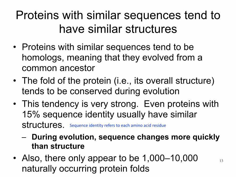
Proteins with similar sequences tend to have similar structures
• Proteins with similar sequences tend to be homologs, meaning that they evolved from a common ancestor
• The fold of the protein (i.e., its overall structure) tends to be conserved during evolution
• This tendency is very strong. Even proteins with 15% sequence identity usually have similar structures. – During evolution, sequence changes more quickly
than structure • Also, there only appear to be 1,000–10,000
naturally occurring protein folds13
Sequence identity refers to each amino acid residue

For most human protein sequences, we can find a homolog with known structure
14
Schwede, Structure 2013
The plot shows the fraction of amino acids in human proteins that can be mapped to similar sequences in PDB structures. Different colors indicate % sequence identity.
Unstructured (disordered) amino acids
As this graph stops at 2012, percentages are likely even higher in 2021
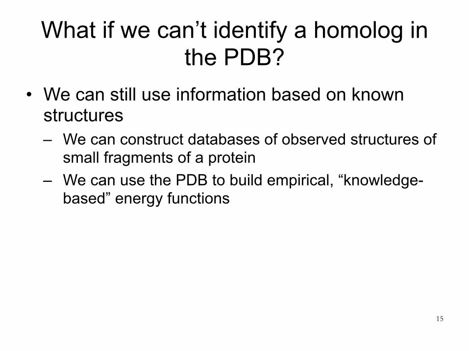
What if we can’t identify a homolog in the PDB?
• We can still use information based on known structures – We can construct databases of observed structures of
small fragments of a protein – We can use the PDB to build empirical, “knowledge-
based” energy functions
15

Two major approaches to protein structure prediction
16
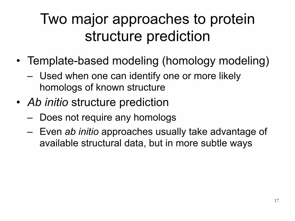
Two major approaches to protein structure prediction
• Template-based modeling (homology modeling) – Used when one can identify one or more likely
homologs of known structure • Ab initio structure prediction
– Does not require any homologs – Even ab initio approaches usually take advantage of
available structural data, but in more subtle ways
17

18
Template-based (“homology”) modeling (e.g., Phyre2)
Two major approaches to protein structure prediction

Template-based structure prediction: basic workflow
• User provides a query sequence with unknown structure
• Search the PDB for proteins with similar sequence and known structure. Pick the best match (the template).
• Build a model based on that template – One can also build a model based on multiple
templates, where different templates are used for different parts of the protein.
19
Query sequence = seq of protein whose structure you are interested in

What does it mean for two sequences to be similar?
• Basic measure: count minimum number of amino acid residues one needs to change, add, or delete to get from one sequence to another – Sequence identity: amino acids that match exactly
between the two sequences – Not trivial to compute for long sequences, but there
are efficient dynamic programming algorithms to do so
20

What does it mean for two sequences to be similar?
• We can do better – Some amino acids are chemically similar to one
another (example: glutamic acid and aspartic acid) • Sequence similarity is like sequence identity, but does
not count changes between similar amino acids
21Glutamic acid Aspartic acid
Similar in terms of chemical properties (ie. Acidic, basic, nonpolar, aromaticity)

What does it mean for two sequences to be similar?
• We can do even better – Once we’ve identified some homologs to a query
sequence (i.e., similar sequences in the sequence database), we can create a profile describing the probability of mutation to each amino acid at each position
– We can then use this profile to search for more homologs – Iterate between identification of homologs and profile
construction – Measure similarity of two sequences by comparing their
profiles – Often implemented using Hidden Markov Models (HMMs)
• For example, the HHBlits software tool • You are not responsible for knowing about HMMs 22

We’ll use the Phyre2 template-based modeling server as an example
• Try it out: http://www.sbg.bio.ic.ac.uk/phyre2/ • Why use Phyre2 as an example of template-
based modeling? – Among the better automated structure
prediction servers – Among the most widely used, and arguably
the easiest to use – Approach is similar to that of other template-
based modeling methods – Great name!
23

Phyre2 algorithmic pipeline
24LA Kelley et al., Nature Protocols 10:845 (2015)

Phyre2 algorithmic pipeline
25
Identify similar sequences in protein sequence database

Phyre2 algorithmic pipeline
26
Choose a template structure by: (1) comparing sequence profiles and (2) predicting secondary structure for each residue in the query sequence and comparing to candidate template structures. Secondary structure (alpha helix, beta sheet, or neither) is predicted for segments of query sequence using a neural network trained on known structures.

Phyre2 algorithmic pipeline
27
Compute optimal alignment of query sequence to template structure

Build a crude backbone model (no side chains) by simply superimposing corresponding amino acids. Some of the query residues will not be modeled, because they don’t have corresponding residues in the template (insertions). There will be some physical gaps in the modeled backbone, because some template residues don’t have corresponding query residues (deletions).
Phyre2 algorithmic pipeline

Phyre2 algorithmic pipeline
29
Use loop modeling to patch up defects in the crude model due to insertions and deletions. For each insertion or deletion, search a large library of fragments (2-15 residues) of PDB structures for ones that match local sequence and fit the geometry best. Tweak backbone dihedrals within these fragments to make them fit better.

Phyre2 algorithmic pipeline
30
Add side chains. Use a database of commonly observed structures for each side chain (these structures are called rotamers). Search for combinations of rotamers that will avoid steric clashes (i.e., atoms ending up on top of one another).

Modeling based on multiple templates• In “intensive mode,” Phyre
2 will use multiple templates that cover (i.e., match well to) different parts of the query sequence. – Build a crude backbone
model for each template – Extract distances between
residues for “reliable” parts of each model
– Perform a simplified protein folding simulation in which these distances are used as constraints. Additional constraints enforce predicted secondary structure
– Fill in the side chains, as for single-template models
31
LA Kelley et al., Nature Protocols 10:845 (2015) You’re not responsible for this

32
Ab initio modeling (e.g., Rosetta)
Two major approaches to protein structure prediction

Two major approaches to protein structure prediction
• Template-based modeling (homology modeling) – Used when one can identify one or more likely
homologs of known structure • Ab initio structure prediction
– Does not require any homologs – Even ab initio approaches usually take advantage of
available structural data, but in more subtle ways
33

Ab initio structure prediction
• Also known as “de novo structure prediction” • Many approaches proposed over time • Probably the most successful is fragment
assembly, as exemplified by the Rosetta software package
34

We’ll use Rosetta as an example of ab initio structure prediction
• Software developed over the last 20–25 years by David Baker (U. Washington) and collaborators
• Software at: https://www.rosettacommons.org/software • Structure prediction server: http://robetta.bakerlab.org/ • Why use Rosetta as an example?
– Among the better ab initio modeling packages (for some years it was the best)
– Approach is similar to that of many ab initio modeling packages
– Rosetta provides a common framework that has become very popular for a wide range of molecular prediction and design tasks, such as protein design and RNA structure prediction 35

Key ideas behind Rosetta
• Knowledge-based energy function – In fact, two of them:
• The “Rosetta energy function,” which is coarse-grained (i.e., does not represent all atoms in the protein), is used in early stages of protein structure prediction
• The “Rosetta all-atom energy function,” which depends on the position of every atom, is used in late stages
• A knowledge-based strategy for searching conformational space (i.e., the space of possible structures for a protein) – Fragment assembly forms the core of this method
36
Gives an approximation of side chains
Called “knowledge-based” because these strategies are informed by structures in PDB and are not necessarily homologs of the protein you are predicting

Rosetta energy function• At first this was the only energy function used by
Rosetta (hence the name) • Based on a simplified representation of protein
structure: – Do not explicitly represent solvent (e.g., water) – Assume all bond lengths and bond angles are fixed – Represent the protein backbone using torsion angles
(three per amino acid: Φ, Ψ, ω) – Represent side chain position using a single “centroid,”
located at the side chain’s center of mass • Centroid position determined by averaging over all
structures of that side chain in the PDB37

Rosetta energy function
38From Rohl et al., Methods in Enzymology 2004You’re not responsible for the details!

Rosetta energy function
39From Rohl et al., Methods in Enzymology 2004
You’re not responsible for the details!Updated version with more terms: Alford et al., Journal of Chemical Theory and Computation, 2017

Rosetta energy function: take-aways
• The (coarse-grained) Rosetta energy function is essentially entirely knowledge-based – Based on statistics computed from the PDB
• Many of the terms are of the form –loge[P(A)], where P(A) is the probability of some set A – This is essentially the free energy of set A. Recall
definition of free energy:
40
P(A) = exp −GAkBT( )GA = −kBT loge P(A)( )
k_b = Boltzmann’s constant T = temperature

Rosetta all-atom energy function
• Still makes simplifying assumptions: – Do not explicitly represent solvent (e.g., water) – Assume all bond lengths and bond angles are fixed
• Functional forms are a hybrid between molecular mechanics force fields and the (coarse-grained) Rosetta energy function – Partly physics-based, partly knowledge-based
41

Are these potential energy functions or free energy functions?
• The energy functions of previous lectures were potential energy functions
• One can also attempt to construct a free energy function, where the energy associated with a conformation is the free energy of the set of “similar” conformations (for some definition of “similar”)
• The Rosetta energy functions are approximate free energy functions (despite sometimes being referred to as potential energy functions) – This means that searching for the “minimum” energy is more valid
(as a way to determine structure) – Nevertheless, typical protocol is to repeat the search process
many times, cluster the results, and report the largest cluster as the solution. This rewards wider and deeper wells. 42

How does Rosetta search the conformational space?
• Two steps: – Coarse search: fragment assembly – Refinement
• Perform coarse search many times, and then perform refinement on each result
43

Coarse search: fragment assembly
• Uses a large database of 3-residue and 9-residue fragments, taken from structures in the PDB
• Monte Carlo sampling algorithm proceeds as follows: 1. Start with the protein in an extended conformation 2. Randomly select a 3-residue or 9-residue section 3. Find a fragment in the library whose sequence resembles it 4. Consider a move in which the backbone dihedrals of the
selected section are replaced by those of the fragment. Calculate the effect on the entire protein structure.
5. Evaluate the Rosetta energy function before and after the move.
6. Use the Metropolis criterion to accept or reject the move. 7. Return to step 2
• The real search algorithm adds some bells and whistles44

Refinement
• Refinement is performed using the Rosetta all-atom energy function, after building in side chains
• Refinement involves a combination of Monte Carlo moves and energy minimization
• The Monte Carlo moves are designed to perturb the structure much more gently than those used in the coarse search – Many still involve the use of fragments
45

An additional approach: analysis of multiple sequences (coevolution)
46

We’ve discussed two approaches to protein structure prediction
• Template-based modeling (homology modeling) – Used when one can identify one or more likely
homologs of known structure • Ab initio structure prediction
– Does not require any homologs – Even ab initio approaches usually take advantage of
available structural data, but in more subtle ways
47
What if we know sequences of many homologs, but don’t have structures for any of them?
Tricky to decide if you want to use template based modeling or ab initio structure prediction…

Amino acids in direct physical contact tend to covary or “coevolve” across related proteins
...GANPMHGRDQSGAVASLTSVA...
...GANPMHGRDQEGAVASLTSVA...
...GANPMHGRDEKGAVASLTSVG...
...GANPMHGRDSHGWLASCLSVA...
...GANPMNGRDVKGFVAAGASVA...
...GANPMHGRDRDGAVASLTSVA...
...GANPMHGRDQVGAVASLTSVA...
...GANPMHGRDQEGAVASLTSVA...
...VEDLMKEVVTYRHFMNASGG...
...VEALMARVLSYRHFMNASGG...
...VATVMKQVMTYRHYLRATGG...
...VARAMREIGKYAQVLKISRG...
...VPELMQDLTSYRHFMNASGG...
...ADHVLRRLSDFVPALLPLGG...
...FERARTALEAYAAPLRAMGG...
...VPEVMKKVMSYRHYLKATGG...
For example, a mutation that causes one amino acid to get bigger is more likely to preserve protein structure and function (and thus survive) if another amino acid gets smaller to make space

Can we use this observation to predict structure?
• Given many sequences of related proteins (whose structure is assumed to be similar), look for amino acids that coevolve. They are probably in contact
• This idea has been around for some time, but it has become practically useful recently, thanks to: – A dramatic increase in amount of sequence data
available – Better computational methods
49
Allows you to find more sequences of homologs’

Some key papers on this approach
Protein 3D Structure Computed from EvolutionarySequence VariationDebora S. Marks1*., Lucy J. Colwell2., Robert Sheridan3, Thomas A. Hopf1, Andrea Pagnani4, Riccardo
Zecchina4,5, Chris Sander3
1 Department of Systems Biology, Harvard Medical School, Boston, Massachusetts, United States of America, 2 MRC Laboratory of Molecular Biology, Hills Road,
Cambridge, United Kingdom, 3 Computational Biology Center, Memorial Sloan-Kettering Cancer Center, New York, New York, United States of America, 4 Human Genetics
Foundation, Torino, Italy, 5 Politecnico di Torino, Torino, Italy
Abstract
The evolutionary trajectory of a protein through sequence space is constrained by its function. Collections of sequencehomologs record the outcomes of millions of evolutionary experiments in which the protein evolves according to theseconstraints. Deciphering the evolutionary record held in these sequences and exploiting it for predictive and engineeringpurposes presents a formidable challenge. The potential benefit of solving this challenge is amplified by the advent ofinexpensive high-throughput genomic sequencing. In this paper we ask whether we can infer evolutionary constraints froma set of sequence homologs of a protein. The challenge is to distinguish true co-evolution couplings from the noisy set ofobserved correlations. We address this challenge using a maximum entropy model of the protein sequence, constrained bythe statistics of the multiple sequence alignment, to infer residue pair couplings. Surprisingly, we find that the strength ofthese inferred couplings is an excellent predictor of residue-residue proximity in folded structures. Indeed, the top-scoringresidue couplings are sufficiently accurate and well-distributed to define the 3D protein fold with remarkable accuracy. Wequantify this observation by computing, from sequence alone, all-atom 3D structures of fifteen test proteins from differentfold classes, ranging in size from 50 to 260 residues., including a G-protein coupled receptor. These blinded inferences are denovo, i.e., they do not use homology modeling or sequence-similar fragments from known structures. The co-evolutionsignals provide sufficient information to determine accurate 3D protein structure to 2.7–4.8 A Ca-RMSD error relative to theobserved structure, over at least two-thirds of the protein (method called EVfold, details at http://EVfold.org). This discoveryprovides insight into essential interactions constraining protein evolution and will facilitate a comprehensive survey of theuniverse of protein structures, new strategies in protein and drug design, and the identification of functional geneticvariants in normal and disease genomes.
Citation: Marks DS, Colwell LJ, Sheridan R, Hopf TA, Pagnani A, et al. (2011) Protein 3D Structure Computed from Evolutionary Sequence Variation. PLoSONE 6(12): e28766. doi:10.1371/journal.pone.0028766
Editor: Andrej Sali, University of California San Francisco, United States of America
Received November 10, 2011; Accepted November , 2011; Published December 7, 2011
Copyright: ! 2011 Marks et al. This is an open-access article distributed under the terms of the Creative Commons Attribution License, which permitsunrestricted use, distribution, and reproduction in any medium, provided the original author and source are credited.
Funding: CS and RS have support from the Dana Farber Cancer Institute-Memorial Sloan-Kettering Cancer Center Physical Sciences Oncology Center (NIH U54-CA143798). LC is supported by an Engineering and Physical Sciences Research Council fellowship (EP/H028064/1). TH has support from the German NationalAcademic Foundation. RZ has support from European Community grant 267915. No other financial support was received for the research. The funders had no rolein study design, data collection and analysis, decision to publish, or preparation of the manuscript.
Competing Interests: The authors have declared that no competing interests exist.
* E-mail: [email protected]
. These authors contributed equally to this work.
Introduction
Exploiting the evolutionary record in protein familiesThe evolutionary process constantly samples the space of
possible sequences and, by implication, structures consistent with afunctional protein in the context of a replicating organism.Homologous proteins from diverse organisms can be recognizedby sequence comparison because strong selective constraintsprevent amino acid substitutions in particular positions frombeing accepted. The beauty of this evolutionary record, reportedin protein family databases such as PFAM [1], is the balancebetween sequence exploration and constraints: conservation offunction within a protein family imposes strong boundaries onsequence variation and generally ensures similarity of 3D structureamong all family members [2] (Figure 1).
In particular, to maintain energetically favorable interactions,residues in spatial proximity may co-evolve across a protein family
[2,3]. This suggests that residue correlations could provideinformation about amino acid residues that are close in structure[4,5,6,7,8,9,10,11]. However, correlated residue pairs within aprotein are not necessarily close in 3D space. Confounding residuecorrelations may reflect constraints that are not due to residueproximity but are nevertheless true biological evolutionaryconstraints or, they could simply reflect correlations arising fromthe limitations of our insight and technical noise. Evolutionaryconstraints on residues involved in oligomerization, protein-protein, or protein-substrate interactions or other spatially indirector spatially distributed interactions can result in co-variationbetween residues not in close spatial proximity within a proteinmonomer. In addition, the principal technical causes of con-founding residue correlations are transitivity of correlations,statistical noise due to small numbers and phylogenetic samplingbias in the set of sequences assembled in the protein family[12,13,14,15]. One does not know a priori the relative contributions
PLoS ONE | www.plosone.org 1 December 2011 | Volume 6 | Issue 12 | e28766
14
Distance-based protein folding powered bydeep learningJinbo Xua,1
aToyota Technological Institute at Chicago, Chicago, IL 60637
Edited by David Baker, University of Washington, Seattle, WA, and approved July 15, 2019 (received for review December 14, 2018)
Direct coupling analysis (DCA) for protein folding has made verygood progress, but it is not effective for proteins that lack manysequence homologs, even coupled with time-consuming confor-mation sampling with fragments. We show that we can accuratelypredict interresidue distance distribution of a protein by deep learning,even for proteins with ∼60 sequence homologs. Using only the geo-metric constraints given by the resulting distance matrix we may con-struct 3D models without involving extensive conformation sampling.Our method successfully folded 21 of the 37 CASP12 hard targetswith a median family size of 58 effective sequence homologswithin 4 h on a Linux computer of 20 central processing units. Incontrast, DCA-predicted contacts cannot be used to fold any ofthese hard targets in the absence of extensive conformation sam-pling, and the best CASP12 group folded only 11 of them by inte-grating DCA-predicted contacts into fragment-based conformationsampling. Rigorous experimental validation in CASP13 shows thatour distance-based folding server successfully folded 17 of 32 hardtargets (with a median family size of 36 sequence homologs) andobtained 70% precision on the top L/5 long-range predicted con-tacts. The latest experimental validation in CAMEO shows that ourserver predicted correct folds for 2 membrane proteins while all ofthe other servers failed. These results demonstrate that it is nowfeasible to predict correct fold for many more proteins lack ofsimilar structures in the Protein Data Bank even on a personalcomputer.
protein folding | deep learning | protein contact prediction |protein distance prediction | direct coupling analysis
Computational structure prediction of proteins without de-tectable homology to experimentally solved structures is a
very challenging problem. Even after decades of research, prog-ress on this problem has been slow, and many methods requireconsiderable computational resources, even for relatively smallproteins. Nevertheless, in recent years good progress has beenachieved thanks to accurate contact prediction enabled by directcoupling analysis (DCA) (1–9) and deep convolutional neural net-works (DCNN) (10–16). As such, contact-assisted protein foldinghas gained a lot of attention and contact prediction has garneredconsiderable research effort.We have developed the CASP12- and CASP13-winning method
RaptorX-Contact (10) that uses deep and fully convolutional re-sidual neural network (ResNet) to predict contacts. ResNet is onetype of DCNN (17) but is much more powerful than traditionalDCNN. RaptorX-Contact has good accuracy even for some proteinswith only dozens of sequence homologs. The precision of RaptorX-Contact decreases more slowly than DCA when more predictedcontacts are evaluated, especially when the protein under study hasfew sequence homologs (10). As reported in refs. 10 and 12, withoutextensive fragment-based conformation sampling, the 3D modelsconstructed from contacts predicted by RaptorX-Contact have muchbetter quality than those built from contacts predicted by DCAmethods such as CCMpred (6) and the CASP11 winner Meta-PSICOV (18). RaptorX-Contact also works well for membraneproteins (MPs) even trained by soluble proteins (12) and for com-plex contact prediction even trained by single-chain proteins (19).
Both our ResNet and DCA are global prediction methodsbecause they predict the contact/distance score or probability ofone residue pair by considering its correlation with other residuepairs at distant sequence positions, which is the key to the sig-nificant improvement in contact prediction. In principle, whenmany convolutional layers are used, it is possible to capturecorrelation between any two residue pairs across the wholecontact/distance matrix. However, ResNet differs from DCA inthat 1) ResNet can capture higher-order residue correlation(e.g., structure motifs) while DCA mainly focuses on pairwiserelationships, 2) ResNet tries to learn the global context of acontact matrix, and 3) existing DCA methods are roughly linearmodels with tens of millions of parameters estimated from asingle protein family, while ResNet is a nonlinear model withparameters estimated from thousands of protein families. Deeplearning (DL) models such as CMAPpro (20) and Deep BeliefNetworks (DBN) (21) were used for contact prediction before,but ResNet is a DL method that greatly outperforms shallowmethods such as MetaPSICOV (18). Different from ResNet andDCA, DBN and MetaPSICOV are local prediction methods, asthey predict the label (i.e., contact or distance) of 1 residue pairwithout considering the labels of others. This is one of the majorreasons why DBN and MetaPSICOV underperformed RaptorX-Contact. Inspired by the success of RaptorX-Contact, manyCASP13 predictors have employed fully ResNet or DCNN (13, 15,22), as shown in the CASP13 abstract book. Notably, the Chenggroup, who developed DBN, has switched to DCNN for contact
Significance
Accurate description of protein structure and function is afundamental step toward understanding biological life andhighly relevant in the development of therapeutics. Althoughgreatly improved, experimental protein structure determina-tion is still low-throughput and costly, especially for membraneproteins. As such, computational structure prediction is oftenresorted. Predicting the structure of a protein without similarstructures in the Protein Data Bank is very challenging andusually needs a large amount of computing power. This papershows that by using a powerful deep learning technique, evenwith only a personal computer we can predict new folds muchmore accurately than ever before. This method also works wellon membrane protein folding.
Author contributions: J.X. designed research, performed research, contributed new re-agents/analytic tools, analyzed data, and wrote the paper.
The author declares no conflict of interest.
This article is a PNAS Direct Submission.
Published under the PNAS license.
Data deposition: Stand-alone code related to this paper is available at https://github.com/j3xugit/RaptorX-Contact. Our web server is available at http://raptorx.uchicago.edu/AbInitio-Folding/.1Email: [email protected].
This article contains supporting information online at www.pnas.org/lookup/suppl/doi:10.1073/pnas.1821309116/-/DCSupplemental.
Published online August 9, 2019.
16856–16865 | PNAS | August 20, 2019 | vol. 116 | no. 34 www.pnas.org/cgi/doi/10.1073/pnas.1821309116
Down
load
ed a
t STA
NFO
RD U
NIV
MED
CEN
TER
on O
ctob
er 1
, 202
0
706 | Nature | Vol 577 | 30 January 2020
Article
Improved protein structure prediction using potentials from deep learning
Andrew W. Senior1,4*, Richard Evans1,4, John Jumper1,4, James Kirkpatrick1,4, Laurent Sifre1,4, Tim Green1, Chongli Qin1, Augustin Žídek1, Alexander W. R. Nelson1, Alex Bridgland1, Hugo Penedones1, Stig Petersen1, Karen Simonyan1, Steve Crossan1, Pushmeet Kohli1, David T. Jones2,3, David Silver1, Koray Kavukcuoglu1 & Demis Hassabis1
Protein structure prediction can be used to determine the three-dimensional shape of a protein from its amino acid sequence1. This problem is of fundamental importance as the structure of a protein largely determines its function2; however, protein structures can be di!cult to determine experimentally. Considerable progress has recently been made by leveraging genetic information. It is possible to infer which amino acid residues are in contact by analysing covariation in homologous sequences, which aids in the prediction of protein structures3. Here we show that we can train a neural network to make accurate predictions of the distances between pairs of residues, which convey more information about the structure than contact predictions. Using this information, we construct a potential of mean force4 that can accurately describe the shape of a protein. We "nd that the resulting potential can be optimized by a simple gradient descent algorithm to generate structures without complex sampling procedures. The resulting system, named AlphaFold, achieves high accuracy, even for sequences with fewer homologous sequences. In the recent Critical Assessment of Protein Structure Prediction5 (CASP13)—a blind assessment of the state of the "eld—AlphaFold created high-accuracy structures (with template modelling (TM) scores6 of 0.7 or higher) for 24 out of 43 free modelling domains, whereas the next best method, which used sampling and contact information, achieved such accuracy for only 14 out of 43 domains. AlphaFold represents a considerable advance in protein-structure prediction. We expect this increased accuracy to enable insights into the function and malfunction of proteins, especially in cases for which no structures for homologous proteins have been experimentally determined7.
Proteins are at the core of most biological processes. As the function of a protein is dependent on its structure, understanding protein struc-tures has been a grand challenge in biology for decades. Although several experimental structure determination techniques have been developed and improved in accuracy, they remain difficult and time-consuming2. As a result, decades of theoretical work has attempted to predict protein structures from amino acid sequences.
CASP5 is a biennial blind protein structure prediction assessment run by the structure prediction community to benchmark progress in accuracy. In 2018, AlphaFold joined 97 groups from around the world in entering CASP138. Each group submitted up to 5 structure predictions for each of 84 protein sequences for which experimentally determined structures were sequestered. Assessors divided the proteins into 104 domains for scoring and classified each as being amenable to template-based modelling (TBM, in which a protein with a similar sequence has a known structure, and that homologous structure is modified in accordance with the sequence differences) or requiring free model-ling (FM, in cases in which no homologous structure is available), with
an intermediate (FM/TBM) category. Figure 1a shows that AlphaFold predicts more FM domains with high accuracy than any other system, particularly in the 0.6–0.7 TM-score range. The TM score—ranging between 0 and 1—measures the degree of match of the overall (back-bone) shape of a proposed structure to a native structure. The assessors ranked the 98 participating groups by the summed, capped z-scores of the structures, separated according to category. AlphaFold achieved a summed z-score of 52.8 in the FM category (best-of-five) compared with 36.6 for the next closest group (322). Combining FM and TBM/FM categories, AlphaFold scored 68.3 compared with 48.2. AlphaFold is able to predict previously unknown folds to high accuracy (Fig. 1b). Despite using only FM techniques and not using templates, AlphaFold also scored well in the TBM category according to the assessors’ for-mula 0-capped z-score, ranking fourth for the top-one model or first for the best-of-five models. Much of the accuracy of AlphaFold is due to the accuracy of the distance predictions, which is evident from the high precision of the corresponding contact predictions (Fig. 1c and Extended Data Fig. 2a).
https://doi.org/10.1038/s41586-019-1923-7
Received: 2 April 2019
Accepted: 10 December 2019
Published online: 15 January 2020
1DeepMind, London, UK. 2The Francis Crick Institute, London, UK. 3University College London, London, UK. 4These authors contributed equally: Andrew W. Senior, Richard Evans, John Jumper, James Kirkpatrick, Laurent Sifre. *e-mail: [email protected]
PLoS ONE, 2011
PNAS, 2019
Nature, 2020
This paper from DeepMind describes the original AlphaFold method, but the current AlphaFold method is substantially different, as we’ll see later
Structure prediction games
51

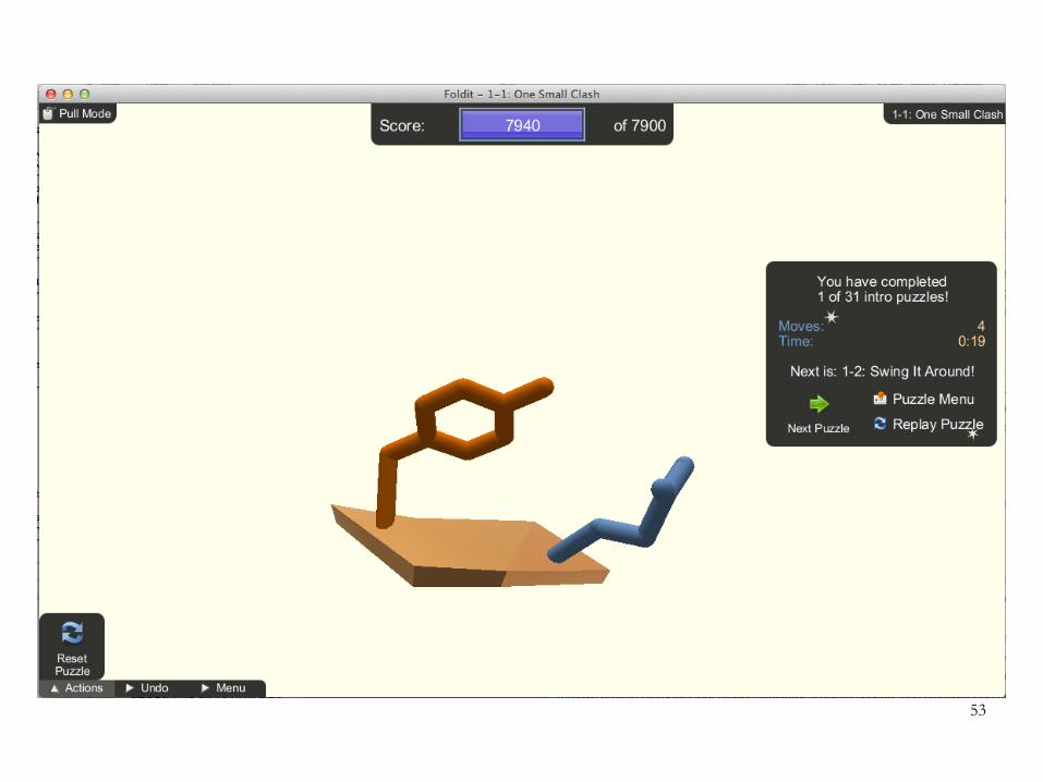
FoldIt: Protein-folding game• https://fold.it/ • Basic idea: allow players to optimize the Rosetta
all-atom energy function – Game score is negative of the energy (plus a constant)
52
53
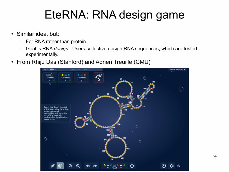
EteRNA: RNA design game • Similar idea, but:
– For RNA rather than protein. – Goal is RNA design. Users collective design RNA sequences, which are tested
experimentally. • From Rhiju Das (Stanford) and Adrien Treuille (CMU)
54