protein structure databases and classification · protein structure databases and classification...
TRANSCRIPT

Protein Structure Databases andClassification
•SCOP, CATH classification schemes, what they mean.
•Motifs: classic turn types. Extended turn types.
•TOPS: drawing a protein molecule

The SCOP database
• Contains information about classification ofprotein structures and within thatclassification, their sequences
• Go to http://scop.berkeley.edu

SCOP classification heirarchy
(1) class
(2) fold
(3) superfamily
(4) family
(5) protein
(6) species
global characteristics (noevolutionary relation)
Similar “topology” . Distantevolutionary cousins?
Clear structural homology
Clear sequence homology
functionally identical
unique sequences
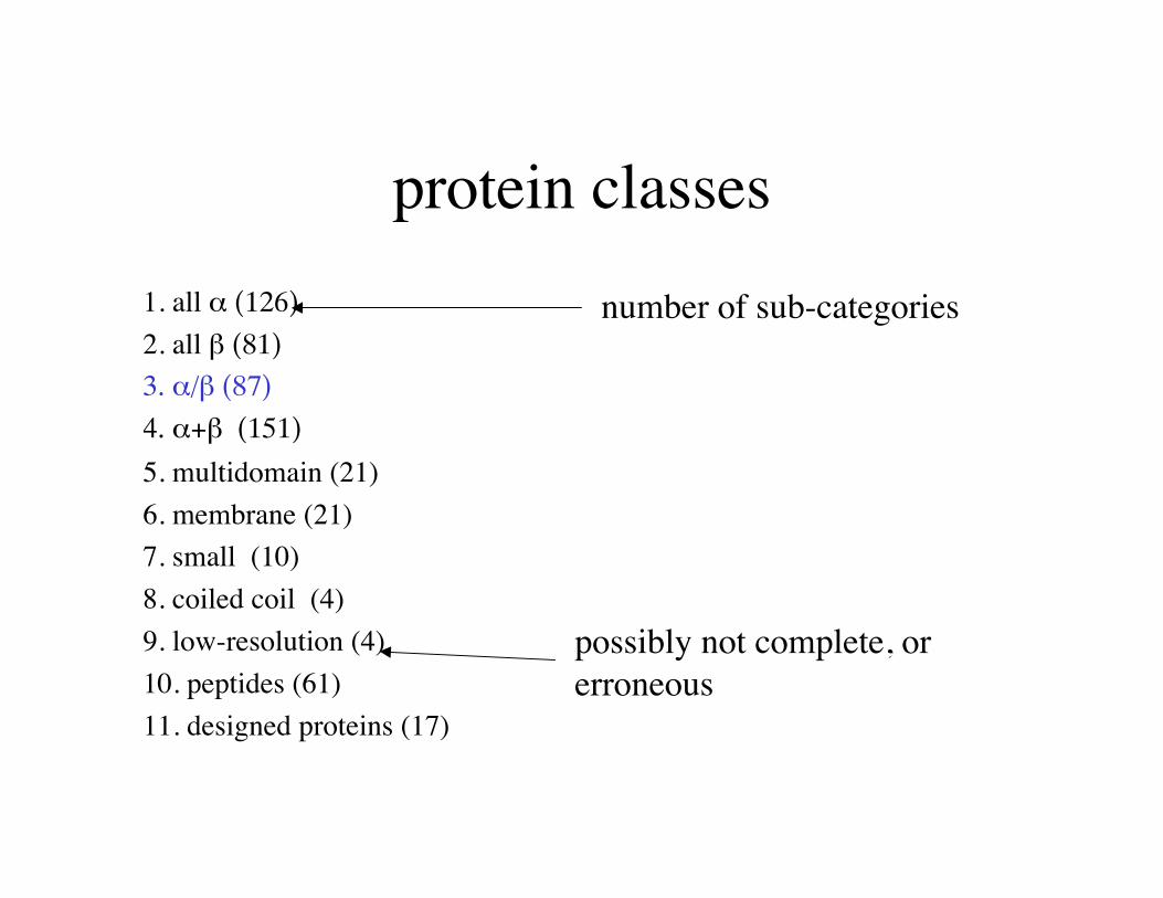
protein classes1. all α (126)
2. all β (81)
3. α/β (87)
4. α+β (151)
5. multidomain (21)6. membrane (21)7. small (10)8. coiled coil (4)9. low-resolution (4)10. peptides (61)11. designed proteins (17)
number of sub-categories
possibly not complete, orerroneous

class: α/β proteinsFolds:TIM-barrel (22)
swivelling beta/beta/alpha domain (5)
spoIIaa-like (2)
flavodoxin-like (10)
restriction endonuclease-like (2)
ribokinase-like (2)
chelatase-like (2)
Mainly parallel beta sheets (beta-alpha-beta units)
Many folds have historical names.“TIM” barrel was first seen in TIM.These classifications are done byeye, mostly.

fold: flavodoxin-likeSuperfamilies:
1.Catalase, C-terminal domain (1)
2.CheY-like (1)
3.Succinyl-CoA synthetase domains (1)
4.Flavoproteins (3)
5.Cobalamin (vitamin B12)-binding domain (1)
6.Ornithine decarboxylase N-terminal "wing" domain (1)
7.Cutinase-like (1)
8.Esterase/acetylhydrolase (2)
9.Formate/glycerate dehydrogenase catalytic domain-like (3)
10.Type II 3-dehydroquinate dehydratase (1)
3 layers, α/β/α; parallel beta-sheet of 5 strand, order 21345
Note the term: “layers”
These are not domains.No implication ofstructural independence.
Note how beta sheets aredescribed: number ofstrands, order (N->C)

fold-level similaritycommon topological features
catalase flavodoxin
At the fold level, a common core of secondary structure isconserved. Outer secondary structure units may not beconserved.

Superfamily:FlavoproteinsFlavodoxin-related (7) NADPH-cytochrome p450
reductase, N-terminal domain Quinone reductase
These molecules do not superimpose well, but side-by-sideyou can easily see the similar topology. Sec struct’s align 1-to-1, mostly.

Family: quinone reductasesbinds FAD
NADPH quinonereductase
quinonereductase type 2
Proteins:
Different members of the same family superimpose well. At thislevel, a structure may be used as a molecular replacement model.
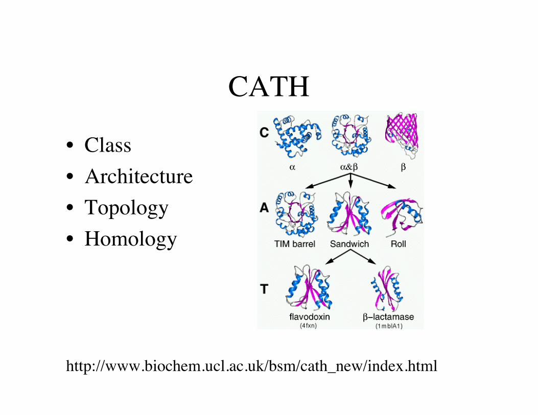
CATH
• Class• Architecture• Topology• Homology
http://www.biochem.ucl.ac.uk/bsm/cath_new/index.html

Structural heirarchy of proteins
• Primary structure• Secondary structure• Local structure• super-secondary structure• domains, folds• Global, multi-domain (tertiary structure)• Quaternary structure

Secondary structure
Alpha helix Right-handed Overall dipole N+->C-3.6 residues/turni->i+4 H-bonds

3 types of Alpha helix
ae
b
f c
g
d Two ways to display position of sidechain on a helix.
non-polar
amphipathic
polar
For amphipathic and non-polar, sidechainsline up in a favorable way.

beta-strandAntiparallel:
Parallel:

Two types of beta strandAmphipathic
Found at the edges of a sheet,or when one side of the sheet isexposed to solvent (i.e. 2-layerproteins).
Found in the buried middlestrands of sheets in 3-layerproteins.
Hydrophobic
Not
e pr
efer
ence
for b
eta-
bran
ched
aa’
s: I,V
,T

Local structure: beta hairpins
Two adjacent antiparallel beta strands = a beta hairpin
Shown are “tight turns”, 2 residues in the loop region (shaded).Hairpins can have as many as 20 residues in the loop region.

hairpin sequence motifs
“Serine beta-hairpin” (alsocalled an “alpha turn”). Aspecific pattern (DPESG)forms an alpha-helical turn 4-residues long.
“Extended Type-1 hairpin”. A type-1“tight turn” has only 2 residues in theturn. This motif, more common thanthe tight turn, has an additional Pro orpolar sidechain. Pattern: PDG.

diverging turn motif
“Diverging turns”have a Type-2 betaturn and two strandsthat do not pair. Theconsensus sequencepattern is PDG. Theresidue before G can beanything polar, but not aD or an N.

Proline Proline helix C-cap motifhelix C-cap motif
Sequence pattern=...nppnnpp[HNYF]P[DE]n
structural variability
Locations of non-polar(magenta) and polar (green)sidechains
note:hydrophobiccluster
Pro blocks helix D or E stabilizestight turn
“n”=non-polar“p”=polar[...]=alternative aa’s

Supersecondary: Two Helix motifs
α−α corner(helix-turn-helix)
EF-hand
(binds Ca2+)

The EF-hand

red=1CLL
green=1cfd
Without Ca2+ bound, the helices have more contact. Backboneangles do not change very much when Ca2+ binds.
Supersecondary motifs are plastic.

α−α corner motif
motif pattern (summary):nnpp[nH]Gn[PDS][Px]pn
“n”=non-polar“p”=polar[...]=alternative aa’s
red=favorableblue = unfavorablegreen=neutral
backbone angles:green=psired=phi

Can you see the α−α motif inthe sequence?
1 GLSDGEWQQV LNVWGKVEAD IAGHGQEVLI RLFTGHPETL EKFDKFKHLK ___HHHHHHH HHHHHHHHHT HHHHHHHHHH HHHHHTHHHH HTTTTTTT__
51 TEAEMKASED LKKHGTVVLT ALGGILKKKG HHEAELKPLA QSHATKHKIP SHHHHHTTHH HHHHHHHHHH HHHHHHHTTT __HHHHHHHH HHHHHTS___
101 IKYLEFISDA IIHVLHSKHP GDFGADAQGA MTKALELFRN DIAAKYKELG HHHHHHHHHH HHHHHHHHST TSS_HHHHHH HHHHHHHHHH HHHHHHHHHT
151 FQG
motif pattern (summary):nnpp[nH]Gn[PDS][Px]pn

βαβ motifWhen a helix occurs betweentwo strands, they are oftenpaired in a parallel sheet.
The cross-over from onestrand to the next isalmost always right-handed, possibly forenergetic reasons,possibly for kineticreasons.

Greek key motif
2 3 4 1
beta meander
One of the most commonarrangements of four strands.
Two permutations. 2341 and3214
Exercise: Download 2PLT.Find the Greek key motifs.

TOPS topology cartoonsA simple way to draw a protein
beta strandpointing up
beta strandpointingdown
alpha helix
A parallel beta sheet
An anti- parallel beta sheet
connections

TOPS topology cartoons
A right-handed βαβ unit
A left-handed βαβ unit(rarely seen)
connection in middlemeans on top.connection on sidemeans on bottom.

How to draw TOPSSelect one molecule and Hide the others.Render-->Backbone-->Cartoon Render-->Backbone-->Color-->terminus (to help see the chaindirection)

How to draw TOPSLine up the molecule along the beta sheet, if present.Otherwise choose a direction so that secondary structures aremostly perpendicular to the screen.

TOPS diagram
• Draw secondary structures first.

TOPS diagram
• number them and connect them
4 3 1 2
4 2
3 1
Be careful to draw connections to the center orside, when it is in front or in back, respectively.
N
C

SCOP-style naming
• 3 layers, 2-4-2 αβα, mixed sheet, 4312
4 3 1 2
4 2
3 1 N
C

SCOP-style naming
• all anti-parallel beta-barrel, closed. n=6
6 5
N
C 3
1 2
4

βαβαβ motif1 2 3
12 3
31 2
Only 3 arepossible.
(with R-handedcrossovers)

Efimov’s 7 trees
A. Efimov showed thatalmost all proteinstructures can beclassified as being one of7 trees, each startingwith a motif and“growing” by onesecondary structure unitat time.
Does structuralphylogeny recapitulatefolding?

Multidomain proteinsDomain boundaries can be seen as "weak" connections inthe structure.
"Weak" means few contacts and few chain cross-overs.
Domain boundaries can be seen in multiple sequence alignments if thealignments are of whole genes.