protein kinase ck2-dependent phosphorylation of the human regulators of calcineurin reveals a novel...
TRANSCRIPT

Biochimica et Biophysica Acta 1833 (2013) 2311–2321
Contents lists available at SciVerse ScienceDirect
Biochimica et Biophysica Acta
j ourna l homepage: www.e lsev ie r .com/ locate /bbamcr
Protein kinase CK2-dependent phosphorylation of the humanRegulators of Calcineurin reveals a novel mechanism regulatingthe calcineurin–NFATc signaling pathway
Sergio Martínez-Høyer a, Álvaro Aranguren-Ibáñez a, Javier García-García b, Eva Serrano-Candelas a,Jordi Vilardell c, Virginia Nunes e, Fernando Aguado d, Baldo Oliva b, Emilio Itarte c, Mercè Pérez-Riba a,⁎a Cellular Signaling group, Cancer and Molecular Genetics Program, Bellvitge Biomedical Research Institute — IDIBELL, L'Hospitalet de Llobregat, 08908 Barcelona, Catalonia, Spainb Structural Bioinformatics Goup (GRIB-IMIM), Departament de Ciències Experimentals i de la Salut, Universitat Pompeu Fabra, Barcelona Research Park of Biomedicine (PRBB),08003 Barcelona, Catalonia, Spainc Unitat de Bioquímica de Biociències, Departament de Bioquímica i Biologia Molecular, Universitat Autònoma de Barcelona, 08193 Bellaterra, Catalonia, Spaind Department of Cell Biology, University of Barcelona, Barcelona, Spaine Laboratorio de Genética Molecular, IDIBELL, 2U-730 (CIBERER), and Departament de Ciències Fisiològiques II, Facultad de Medicina, Universitat de Barcelona, Barcelona, Spain
Abbreviations: Cn, calcineurin; CnA, calcineurin cataRCAN (formerly calcipressin) inhibitor of calcineurin sequNFATc, cytosolic Nuclear Factor of Activated T-cells; RCCsA, cyclosporin A; Io, Ionomycin; PMA, Phorbol 12-miris⁎ Corresponding author at: HumanMolecular Genetics L
Genetics Program, Bellvitge Biomedical Research InstituLlobregat, Barcelona 08908, Spain. Tel.: +34 932607427; fa
E-mail address: [email protected] (M. Pérez-Riba).
0167-4889/$ – see front matter © 2013 Published by Elhttp://dx.doi.org/10.1016/j.bbamcr.2013.05.021
a b s t r a c t
a r t i c l e i n f oArticle history:Received 14 December 2012Received in revised form 21 May 2013Accepted 22 May 2013Available online 1 June 2013
Keywords:Calcineurin–NFATc signalingRCANPXIXITCK2ImmunosuppressionPhosphorylation
Cyclosporine A and FK506 produce immunosuppression by blocking calcineurin phosphatase activity andconsequently activation of cytosolic Nuclear Factor of Activated T-cell (NFATc) transcription factor. Due tothe chronic toxicity associated with their administration, the development of more specific immunosuppres-sants is currently an important unmet medical need. In this context, an immunosuppressant peptide derivedfrom the CIC motif of the human Regulators of Calcineurin (RCAN) proteins has been shown to inhibit NFATcsignaling without affecting general phosphatase activity of calcineurin. Here we show that protein kinase CK2phosphorylates a conserved serine residue within the CIC motif of vertebrate RCANs, which increases its af-finity for calcineurin and consequently its inhibition of NFATc-dependent gene expression in activatedT-cells. Molecular modeling studies have led us to identify a positively charged interaction site on the surfaceof calcineurin where the phosphorylated serine residue of the CIC motif would normally locate. Finally, wehave also identified RCAN3 as a new phosphoprotein with multiple phosphorylation sites. Therefore, ourfindings reveal for the first time a novel molecular mechanism underlying the regulation of calcineurin–NFATc signaling by means of phosphorylation of the CIC motif of RCAN proteins. The knowledge of howRCAN proteins modulate the calcineurin–NFATc pathway paves the way for the development of potentnovel selective immunosuppressant drugs.
© 2013 Published by Elsevier B.V.
1. Introduction
The highly conserved serine/threonine phosphatase calcineurin(Cn, also known as PPP3, formerly PP2B) is a cellular sensor enzymethat plays a pivotal role in transducing Ca2+ signals into cellular re-sponses [1]. The activation of Cn, which is ubiquitously expressed,results in the dephosphorylation of its substrates, including the cyto-solic members of the Nuclear Factor of Activated T-cell (NFATc) fam-ily of transcription factors [2]. Once dephosphorylated, NFATcproteins translocate to the nucleus where, in cooperation with other
lytic subunit A; CIC sequence,ence; CK2, protein kinase CK2;AN, Regulators of Calcineurin;tate 13-acetateaboratory, Cancer and Molecularte — IDIBELL. L'Hospitalet dex: +34 932607414.
sevier B.V.
transcription factors, they trigger the expression of genes involvedin diverse biological processes including T-cell activation, cardiac hy-pertrophy, bone remodeling or angiogenesis among others [3]. Theimportance of this pathway came to light with the discovery thatthe most widely used current immunosuppressant drugs, cyclospor-ine A (CsA) and FK506, elicited their clinical response by inhibitingCn enzymatic activity and therefore also activation of NFATc [4].However, further studies revealed that the inhibition of Cn by thesedrugs inhibits dephosphorylation of all Cn substrates and is associat-ed to severe undesirable side effects [5].
The Regulators of Calcineurin (RCAN; previously called DSCR,MCIP or calcipressin (CALP)) family of proteins are specific endoge-nous modulators of the Cn–NFATc signaling axis [6,7]. This proteinfamily, defined by several highly conserved motifs in Eukarya suchas the FLISPP motif (FLISPPASPP sequence), is conserved from yeastto human and in vertebrates constitutes a highly conserved subfamilycomprised of three proteins: RCAN1, RCAN2 and RCAN3 [8,9], each ofwhich is expressed as different isoforms [9]. The specificity towards

2312 S. Martínez-Høyer et al. / Biochimica et Biophysica Acta 1833 (2013) 2311–2321
NFATc signaling modulation by these proteins is explained by twoconserved Cn-interactingmotifs shared by both NFATc and RCAN pro-teins: the LXXP (LXVP in NFATc proteins) and PXIXIT motifs [10–14].The PXIXIT motif has been described as the major Cn-anchoring site ofNFATc proteins [15]. The crystal structure of the synthetic VIVIT peptidederived from the PXIXIT sequence of NFATc [16] in complex with Cn,helped to clarify how this sequence interacts in a hydrophobic cleftformed by strands β14 and β11 of Cn [17]. Variations in the consensusPXIXIT sequence modulate the binding affinity for Cn of the differentPXIXIT-containing proteins and therefore the biological consequenceof such interaction [18–20]. RCAN proteins possess a PSVVVH sequencewhich has been shown to behave as a PXIXITmotif [12–14]. This PXIXITsequence, together with a C-terminal ELHA sequence, comprise theRCAN inhibitor of Cn (CIC) motif, conserved in all vertebrate RCANs[8,21]. We and others have shown that in RCANs, the CIC motif needsboth the ELHA and PXIXIT sequences to bind with high affinity to Cnand inhibit downstream NFATc signaling [12,14].
Both inhibitory and facilitative roles on the Cn–NFATc signalingpathway have been associated with the RCANs, depending on the spe-cific tissue or cell type and expression level [22–24]. In addition, RCANproteins are also known to be regulated by post-translational modifica-tion including phosphorylation. For instance, phosphorylation at theconserved FLISPPmotif of RCAN1 byGSK3β, primed by phosphorylationby either BMK, MAPK or DYRK1A, facilitates Cn–NFATc signaling[25–28]. Also, phosphorylation of RCAN1 by TAK1 has been related toa facilitative role towards the Cn–NFATc signaling pathway [29]. Onthe other hand, phosphorylated RCAN1 at both serine residues of theFLISPP motif has been shown to be a better inhibitor of NFATc activity[26]. In addition, phosphorylation of RCAN1 has also been associatedto an increased protein half life and therefore and enhanced inhibitionof Cn–NFATc signaling [28,30].
Protein kinase CK2 is a ubiquitous enzyme with pleiotropic activ-ity, as it has been shown to phosphorylate many substrates, includingNFATc [31,32]. CK2 consists of two catalytic (α or α′) and two β reg-ulatory subunits arranged as a tetramer [33]. Although it is generallyregarded as a constitutively active enzyme, some natural regulators ofits activity have been described [34]. In this work, we identify a phos-phorylated serine within a protein kinase CK2 consensus sequence atthe conserved CIC motif in all vertebrate RCANs. Additionally, weshow that this phosphorylation increases the affinity of the CICmotif of RCAN for Cn and consequently potentiates the inhibition to-wards Cn–NFATc pathway activation and T-cell activation.
2. Materials and methods
2.1. Peptides and reagents
All peptides were purchased from the Peptide 2.0 Company(Chantilly, VA) and synthesized as acetylated N-terminal andC-terminal amides for the unlabeled peptides and N-carboxyfluorescein(CF) and C-terminal amide for the labeled peptides. The sequencesof the peptides used are the following: KYELHAGTESTPSVVVHVCESfor the RCAN183–203 peptides; KYELHAGTESTPSVVVHVCESETEEE forthe RCAN3183–208 peptides; KYELHAGTESTPSVVVHVCE(pS)ETEEE forthe phosphoRCAN3183–208 peptides; ASGLSPRIEITPSHEL for theNFATc2-SPRIEIT (SPRIEIT) peptides; MAGPHPVIVITGPHEE for theVIVIT peptide; and MAGPHPVIVITGPHAA for the mutated VIVIT(VIVIT–AA) peptide. The N-termini CF-labeled peptides were syn-thesized with a two-glycine linker between the fluorescent probeand the original N-terminus residue. All peptides were resuspended in10% DMSO at 7.5 mM. Concentration was validated by spectrophotom-etry when possible. Peptides not containing aromatic residues in theirsequence were assessed by bicinchoninic acid assay (BCA). CyclosporinA (CsA)was obtained from Sandoz. Ionomycin (Io) sodium salt, Phorbol12-myristate 13-acetate sodium salt (PMA) and cycloheximide wereobtained from Sigma. RNAiMAX transfection reagent was purchased
from Invitrogen. The anti-calcineurin A antibody was purchased fromBD Biosciences, the anti-CK2α (H-286 clone) from Santa Cruz, and theanti-HA (Clone 12CA5) and monoclonal anti-α-tubulin (P-6074) werefrom Sigma. The protease and phosphatase inhibitor cocktails werefrom Calbiochem.
2.2. Plasmid constructs and site-directed mutagenesis
The different pGEX5X–RCAN3, pGEX6P–CnA (human CnAαamino acids 2–347), HA–RCAN3 (NP_038469.1) and HA–RCAN1(NP_004405.3) plasmid constructs used for this work have been de-scribed previously [8]. RCAN2-3 cDNA (NM_001251973.1) was a giftfrom Dr. X. Cao and was subcloned as an N-terminal HA-tagged fu-sion in pcDNA3.1-HA vector using BamHI restriction sites. TheFlag–hCnAα and GST–NFATc2 expression plasmids were kindly pro-vided by Dr. J. M. Redondo (CNIC, Madrid, Spain), the 3xNFAT-lucplasmid construct by Dr. J. Aramburu (Universitat Pompeu Fabra,Barcelona, Spain), and the pRL-null plasmid, which lacks a promoterdriving Renilla expression, from Dr. C. Marín (Universidad deLeón, León, Spain). The EGFPc–RCAN3178–203 construct has beenpreviously described [12]. The RCAN3178–210 protein fragment wasPCR-amplified from the HA–RCAN3 plasmid using specific primersand subcloned as an EGFPc-fusion protein using BamHI restrictionsites. Oligonucleotide sequences are listed in Supplementary Table 1.
2.3. Recombinant protein expression and purification
GST fusionproteinswere transformed inBL21-CodonPlus®strain. Sol-uble extracts were produced and GST fusion protein was purified usingGlutathione Sepharose 4B beads (GE Healthcare) as described previously[21]. Free CnAwas obtainedbyPrescission protease (GEHealthcare) incu-bationof the purifiedGST–CnA.Quantification, integrity andpurity of CnAwere assessed by spectrophotometry and 10% SDS-PAGE.
2.4. Cell lines and transfection
Human Jurkat T-cells were maintained in RPMI with Glutamax(Invitrogen) supplemented with 10% fetal bovine serum (FBS). HEK293T cells were maintained in DMEM with Glutamax (Invitrogen)supplemented with 10% FBS, 100 U/ml penicillin and 100 μg/mlstreptomycin. For immunoprecipitation and reporter gene assays,HEK 293T cells were transfected with the indicated plasmids usingpolyethyleneimine (PEI)-mediated transfection. Briefly, plasmid DNAand PEI (linear 25,000 MW polyethyleneimine, Polysciences) wereadded in separate tubes to 0.9% NaCl. Then, DNA–PEI complexes (DNA:PEI ratio of 1:2.5 (w:w)) were prepared by adding PEI solution to theDNA and immediately vortexing for a short time. After 20 min incubationat room temperature, complexes were added to the cells. The followingday, the medium was replaced and the cells were left incubating at37 °C for an additional 24 h. For luciferase assays, cells were analyzed24 h post-transfection. One day before HA–RCAN3 plasmid transfection,pooled siRNA against CK2α (Dharmacon) was transfected in HEK 293Tat 25 nM with RNAiMAX reagent (Invitrogen) following themanufacturer's instructions to deplete CK2α in the cells.
2.5. NFAT-luciferase reporter gene assay
HEK 293T cells were seeded at 30% confluence in 24-well plates.24 h later, each well was transfected with 100 ng of 3xNFAT-luc re-porter plasmid, 0.9 ng of pRLNull as an internal transfection controland decreasing concentrations of the indicated constructs (200 ng,20 ng, 2 ng and 0.2 ng for HA–RCAN3 and 500 ng, 250 ng, 125 ngand 62.5 ng for the EGFPc–RCAN3178–210). The total amount of plas-mid DNA was kept constant in all conditions using empty vector.24 h after transfection, cells were stimulated with 1 μM ionomycin,10 ng/ml PMA and 10 mM CaCl2. Cyclosporin A (CsA, 1 μM) was

2313S. Martínez-Høyer et al. / Biochimica et Biophysica Acta 1833 (2013) 2311–2321
used as a positive control for calcineurin inhibition. After 6 h of stim-ulation, cells were analyzed for luciferase gene expression using theDual-Luciferase Reporter Assay (Promega) following the manufac-turer’s protocol on a multiplate luminometer (FLUOstar Optima,BMG). Luciferase units were normalized to Renilla luciferase values.
2.6. In vivo phosphorylation
HA and EGFP-tagged protein constructs were transfected in HEK293T cells in 6-well plates for RCAN1 and RCAN3 and in p100 dishesfor RCAN2 and EGFP–CIC in vivo phosphorylation. After 24 h, cellswere incubated in phosphate-free DMEM (Invitrogen) supplementedwith 10% dialyzed FBS for 3 h. Then, cells were labeled with 1 ml forp6-well plates and 5 ml for p100 dishes of phosphate-free DMEMsupplementedwith [32P]Pi at a concentration of 50 μCi/ml for additional3 h. Where indicated, CX-4945 was added at 10 μM to the culture me-dium 16 h prior to immunoprecipitation. Cells were then washed incold phosphate buffered saline (PBS) and lysed in ice-cold RIPA buffer(50 mM Tris–HCl, pH 7.5, 150 mM NaCl, 1% TX-100, 5 mM EGTA, 0.5%sodium deoxycholate, 0.1% SDS, 1 mM PMSF and protease and phos-phatase inhibitor cocktails). Protein extracts were centrifuged at16,000 ×g at 4 °C and the soluble HA or EGFP-tagged proteins from su-pernatants were immunoprecipitated by incubation with anti-HA oranti-GFP coupled to protein G Sepharose beads (GE Heatlhcare) in arocking platform for 1 h in ice. Beads were washed extensively inRIPA buffer, resuspended in 2× Laemmli sample buffer and boiled for10 min. Immunoprecipitated proteins were resolved on 10% SDS-PAGE,transferred onto nitrocellulose membranes and subjected to autoradiog-raphy (CL-XPosure Film, Thermo Scientific). Later, membranes were in-cubated with anti-HA antibody (1:1000, 1 h at room temperature) oranti-GFP (1:1000, 1 h at room temperature) and the signals were quan-tified to normalize the radioactive signal of the immunoprecipitatedproteins.
2.7. GST-pull-down competition assays
In GSTRCAN3 pull-down competition assays, HEK 293T cells werelysed in co-immunoprecipitation buffer (50 mM Tris–HCl, pH 7.5,100 mM NaCl, 2 mM CaCl2, 5 mM MgCl2, 1% IGEPAL, 1 mM DTT,2 mM PMSF and protease and phosphatase inhibitor cocktails) andthe CIC-derived peptides were added to the extracts at the indicatedconcentrations for 20 min. Then, the soluble extracts were incubatedwith GST–RCAN3 bound to Glutathione Sepharose beads for 1 hat 4 °C. After extensive washing, co-precipitated proteins were elutedby resuspension in 2× Laemmli buffer and boiling for 10 min. ForGST–NFATc2 pull-down competition assays, HEK 293T cells transfectedwith FLAG–CnA were lysed in a buffer containing 50 mM Tris–HCl,pH 8, 100 mM NaCl, 1.5 mM CaCl2, 6 mM MgCl2, 0.2% TX-100, 1 mMPMSF and protease and phosphatase inhibitor cocktails.
2.8. In vitro CK2 kinase assay
Phosphorylation of wild-type and mutant GST–RCAN3 constructs(7.5 μg each) was carried out for 60 min at 30 °C in a mediumcontaining human His6–CK2 holoenzyme (reconstituted by mixing1.25 pmol His6–CK2α and 1.25 pmol His6–CK2β) or of His6–CK2αalone (1.25 pmol) and 100 μM of [γ-32P] ATP. The reactions werestopped by adding 2× Laemmli buffer and boiling, resolved onto a12% SDS/PAGE and analyzed by autoradiography. Coomassie bluestaining of the gel was performed to ensure equal amounts of pro-tein in the assay. The activity of the reconstituted enzymes was test-ed previously using the CK2 substrate peptide RRRADDSDDDDD(Jena Bioscience).
2.9. Fluorescence polarization
To calculate Cn-binding affinities, 10 nM of CF-labeled peptidein 50 μl of PBS (pH 7.4) containing 1% (w/v) fraction V bovine serumalbumin (BSA) (Roche Diagnostics, IN) was titrated with increasingconcentrations of purified CnA. For the competitive binding assays,the peptide–CnA complex was performed using 10 nM CF-labeled pep-tides and 4 μMCnA for the CF–RCAN3183–203, CF–RCAN3183–208 and CF–phosphoRCAN3183–208 peptides and 10 μMCnA for the CF–SPRIEIT pep-tide. Unlabeled competitor peptides were pre-incubated with CnA atincreasing concentrations for 15 min before adding the fluorescence-labeled peptides. Experiments were performed in a OptiPlate black384-well flat-bottom plates (PerkinElmer Life Sciences, Waltham, MA)and measured using a Wallac VICTOR™ X5 2030 Multilabel Reader(PerkinElmer Life Sciences) with excitation and emission wavelengthsof 485 nm and 535 nm, respectively. All binding and competition as-says were performed for 15 min at room temperature. All data wereobtained from at least three independent experiments performed intriplicates.
2.10. Circular dichroism
CD spectra were recorded at 20 °C and 5 °C on a Jasco J-810spectropolarimeter (JASCO Corporation, Tokyo, Japan) in quartz cellsof 0.1 cm path length. Peptides were dissolved at 20 μM in PBS andtheir ability to adopt a secondary conformation was analyzed byMeOH and 2,2,2,-trifluoroethanol (TFE) titrations. Each CD spectrumwas the average of 20 scans performed at 1 nm intervals. The resultsare expressed as mean molar residue ellipticities (deg cm2 dmol−1).
2.11. Lentiviral production, purification and infection
The lentivirus encoding EGFP-fusion peptides (pWPT, Addgene:12255) were obtained by transient calcium phosphate transfection ofHEK 293T cells. At 48 h post-transfection the supernatants of thetransfected cells were collected, filtered through a 45 μM membraneand ultracentrifuged at 20,000 rpm for 1.5 h in a SW-28 rotor(Beckmann Coulter). Concentrated viruses were collected in cold PBSand stored frozen at −80 °C. Exponentially growing Jurkat cells weretransduced overnight at MOI = 10 at a cell density of 500,000 cells/mlin RPMI supplemented with 10% FBS and 8 μg/ml polybrene. At 16 hpost-infection, the medium was replaced with fresh RPMI 10% FBS.
2.12. Cell sorting
Transduced Jurkat cells were resuspended (107 cells/ml) in PBSsupplemented with 0.5% BSA and 2 mM EDTA to prevent cell aggre-gation. The EGFP positive population was sorted in three differentgroups depending on the protein levels of EGFP using MoFlo Astriosequipment (Beckman Coulter, Scientific Services at the University ofBarcelona). The same sorting windows were applied for the twoEGFP–peptide expressing cells. Cells expressing EGFP alone at thewindow corresponding to the highest level of EGFP protein wereused as controls. Sorted cells were recovered in fresh RPMI 10% FBSand incubated at 37 °C, 5% CO2 and 95% relative humidity until theywere assayed.
2.13. RNA extraction and real-time PCR
Sorted Jurkat cells were stimulated for 4 h with 1 μM ionomycinand 10 ng/ml PMA. Then, the cells were pelleted and RNA wasextracted using RNAeasy kit (Quiagen). At least 300 ng of RNA wasused to synthesize cDNA using Superscript III (Invitrogen) followingthemanufacturer's protocols. Quantitative PCR (qPCR) experimentswereperformed using Universal probe Libraries (UPL, Roche) followingmanu-facturer’s instructions. PCR reactions were carried out in triplicates in a

2314 S. Martínez-Høyer et al. / Biochimica et Biophysica Acta 1833 (2013) 2311–2321
Lightcycler 480 System (Roche). Human HPRT1 gene amplification wasused as a housekeeping control. The human UPL probes used forreal-time experiments were UPL probe 65 for IL2, UPL probe 81 forRCAN1-4, UPL probe 21 for IFNγ, UPL probe 60 for IL3, UPL probe 3 forCSF2, UPL probe 29 for TNF-α and UPL probe 73 for HPRT1. The primersused are shown in Supplementary Table 1.
2.14. In silico modeling
Template complexes of the interaction between CnA and VIVIT pep-tides were extracted from the Protein Data Bank database (PDB) [35]and used to model the interaction of several peptides similar to VIVIT.As a template we used the complex structure of PDB ID: 2P6B, a 2.3 ÅX-ray structure of Cn bound with the optimized NFAT-derived VIVITpeptide. The structures of four different peptide sequences weremodeled on its interaction with CnA using the MODELLER program[36]. A total of 500 models were generated with the MODELLER foreach peptide–CnA complex. For the sake of comparison, the templatewas also modeled upon itself to obtain a collection of 500 conforma-tions. Models were evaluated using the distribution of the ZRANKdocking-score [37]. Distributions of ZRANK scores were comparedusing a t-test. A threshold of 0.001 of significance in the t-test wasused to assess if the distributions were similar or different. To evaluatethe normality of the distributions we used the Shapiro–Wilk test with0.001 significance thresholds. UCSF Chimera package and Pymol [38]were used for visual inspection of the interactionmodels, for the graph-ical representations and for the calculation of accessible surfaces. Elec-trostatic potentials over the accessible surface of Cn were calculatedwith Adaptative Poisson–Boltzmann Solver (APBS) software [39].
3. Results
3.1. RCAN proteins are phosphorylated at the conserved CIC motif in vivo
In order to explore the regulatory mechanisms governing the CICmotif function in the human RCAN proteins more deeply, we focusedon the post-translational modifications predicted for the RCANs. To be
Fig. 1. RCAN proteins are phosphorylated at the CIC motif in eukaryotic cells. (A) Scheme shoprotein family (upper) and at the N- and C-termini of RCAN3 (lower scheme). An arrow dCK2-target S18 in RCAN3. (B–C) Radioactive labeling with [32P]-orthophosphate of the HAdetected by membrane autoradiography. Western blot using anti-HA is shown as a contrchain and two asterisks (**) to the light IgG chain. Radioactive signal was quantified by denware (Bio-Rad). Densitometry values are presented below each lane as a fraction of the radietry values below the panel are expressed as mean with standard deviation (SD) from the
specific, by using Net Phos 2.0 server [40] we identified a CK2 phos-phorylation site at a serine residue within the CIC motif that was con-served in all vertebrate RCAN proteins (Fig. 1A). Furthermore, therewere two additional sites present exclusively in human RCAN3-2 iso-form: T205 next to the conserved S203 at the CIC motif of RCAN3 andT19 at the non-conserved N-terminus. Finally, although not fulfillingthe consensus CK2 target sequence, we also considered S18 as a po-tential phosphorylation site based on other CK2 phosphorylated pro-teins [31].
Bymeans of in vivo radioactive labeling of transfected HEK 293Twith[32P]-orthophosphate we observed that human RCAN1-1, RCAN2-3 andRCAN3-2 isoforms [7] (hereafter referred to as RCAN1, RCAN2 andRCAN3, respectively) were phosphorylated in basal conditions (Fig. 1Band C, wild-type (wt) proteins). Additionally, when mutating the twoRCAN2 serine residues at the FLISPP motif (hereafter termed RCAN2AA, which corresponds to serine residues S158 and S162 mutated to al-anine) and of the RCAN3 FLISPP motif (hereafter termed R3 AA, whichcorresponds to RCAN3 S148 and S152 residues mutated to alanine) theincorporation of radioactive phosphate by the immunoprecipitated pro-teins was reduced compared with the wild-type protein (Fig. 1B and C),indicating that at least one of the serine residues at the FLISPP motif ofRCAN2 and RCAN3 is phosphorylated in vivo. This observation suggeststhat the phosphorylation at the CIC motif is independent of that occur-ring at the FLISPP motif of RCAN proteins.
Also, when the CK2-target serine residue within the CIC motif in thethree RCANs (S218 for RCAN1; S213 for RCAN2 and S203 in RCAN3)wasmutated to alanine, we observed a reduction in the radioactive signalconfirming that this serine of the CIC motif of all human RCAN is phos-phorylated in vivo (Fig. 1B and C).
Apart from the CICmotif, mutation of S18 to alanine in RCAN3 also re-duced its radioactive phosphorylation signal, suggesting that S18 is alsophosphorylated in vivo at basal conditions (Fig. 1C, compare R3AA S18Avs R3AA). Furthermore, mutating both CK2 predicted phosphorylationsites in RCAN3 (R3AA S18A/S203A: R3AA with the additional mutationsto alanine at S18 and S203) resulted in a further reduction in the incorpo-ration of radioactivity by themutant protein (Fig. 1C, compare R3AAwithR3AA S18A/S203A). Mutating T19 and T205 to alanine in the R3AA S18A/
wing the predicted CK2 target sequence (S/TxxE/D) at the CIC motif of the human RCANesignates the conserved CK2-target serine residue at the CIC motif and an asterisk the-RCAN wt or mutant proteins transfected in HEK 293T cells. [32P] incorporation wasol of the immunoprecipitated protein. One asterisk (*) corresponds to the heavy IgGsitometry and normalized to the immunoprecipitated protein using Quantity One soft-oactive phosphorylation signal referred to the signal in the wt protein. In 1C, densitom-indicated number of experiments performed (n).

2315S. Martínez-Høyer et al. / Biochimica et Biophysica Acta 1833 (2013) 2311–2321
S203A backbone (R3AA S18A/T19A/S203A/T205A) further reduced theincorporation of a radioactive signal, suggesting that these residuesmight also be phosphorylated in vivo. Finally, since R3AA S18A/T19A/S203A/T205A is still phosphorylated, it is suggested that there is at leastone additional phosphorylated site in the protein in basal conditions.
3.2. Protein kinase CK2 phosphorylates the CIC motif of the RCANs invitro
It has been reported that both the tetrameric CK2 enzyme and theCK2α and CK2α′ free subunits are catalytically active in vitro [31]. Inorder to differentiate whether the CK2 holoenzyme (CK2αβ) or thefree CK2α catalytic subunit were able to phosphorylate RCAN3 atthe predicted phosphoacceptor sites, we performed a kinase assayby incubating RCAN3 wild-type (wt), RCAN3 N-term (amino acids 2to 144) and RCAN3 C-term (amino acids 176 to 241) with eitherCK2 holoenzyme or free CK2α catalytic subunit. First, we observedthat both CK2 forms phosphorylated RCAN3 wt protein (Fig. 2A). Im-portantly, RCAN3 wt protein showed a stronger radioactive signalwhen phosphorylated with the holoenzyme as compared with thephosphorylation with CK2α alone (Fig. 2A, compare lanes 3 and 4).Secondly, we observed that the N-terminus of RCAN3 was preferen-tially phosphorylated by CK2α whereas the C-terminus of RCAN3was preferentially phosphorylated by CK2 holoenzyme, highlightinga role for CK2β regulatory subunit in the phosphorylation of the con-served region of RCAN3.
In accordance with what was observed in eukaryotic cells, radioactivelabeling due to CK2 holoenzyme phosphorylation was absent when mu-tating the RCAN3 CK2 predicted phosphorylation sites, i.e. S18A/T19A atthe N-terminus and S203A/T205A at the C-terminus of RCAN3 protein(Fig. 2B). Moreover, in the same kinase assay the C-terminus of RCAN1(amino acids 170 to 252) was also phosphorylated by CK2 in vitro(Fig. 2B, lane 8), suggesting a conserved feature of the RCAN protein fam-ily. Point mutations at the C-terminus of RCAN3 identified position S203of RCAN3 located within the conserved CIC motif, as the preferential tar-get for CK2 phosphorylation (Fig. 2C). Additionally, we detected a weakphosphorylation signal in the S203A mutant, suggesting that T205 couldalso be phosphorylated, but to a much lesser extent than S203. The ab-sence of radioactive signal in the S203A/T205A mutant confirmed thatthere are no other CK2 target sites in the conserved C-terminus of RCAN3.
3.3. Protein kinase CK2 phosphorylates the CIC motif of RCAN proteins invivo
In order to determine if CK2 could be involved in RCAN phosphor-ylation at the CK2 predicted sites in vivo, HEK 293T cells transfected
Fig. 2. Protein Kinase CK2 phosphorylates human RCAN proteins including the CIC motif infusion proteins and deletion mutants in the presence of radioactive [γ-32P]-ATP. Coomass(A) GST-RCAN3 fusion protein (wt) and deletion mutants were incubated with either puGST–RCAN3175–241. (B and C) GST–RCAN3 (R3) and the indicated mutants and GST–RCANRCAN1170–252.
with RCAN1 and RCAN3, either wt or mutated at the conserved serineresidue at the CIC motif were radiolabeled in the presence of a specificCK2 inhibitor CX-4945 [41,42]. As shown in Fig. 3A, incubating thecells with CX-4945 dramatically reduced the radioactive phosphateincorporation to RCAN1 and RCAN3 proteins. When mutating theCK2 target residue of RCAN1 protein we observed reduced phosphor-ylation of the protein and most importantly, its radiolabeling levelswere not further reduced in the presence of CX-4945. A similar phos-phorylation profile is observed in RCAN1 when transfecting the cellswith a specific siRNA against CK2α (Fig. 3B), where there is no furtherreduction of RCAN1 S218A phosphorylation in the presence of siRNACK2α, which confirms that this residue is the only target site for CK2for RCAN1. These results suggest that S218 in RCAN1 is the only res-idue phosphorylated by CK2 in eukaryotic cells and therefore subjectto the effects of the CK2 inhibitor. As for RCAN3, incubation withCX-4945 did reduce the phosphorylation status not only of the wtprotein but also of the mutated protein at the conserved S203,suggesting that there are other CK2 dependent phosphorylationsites in RCAN3. In accordance with this, RCAN3 presents at least oneextra CK2 target site at the N-terminus of the protein, S18, asshown in Figs. 1A and 2B. CK2α depletion has a much smaller effecton RCAN3 phosphorylation probably due to the presence of addition-al phosphorylation sites targeted by CK2 and/or other kinases (datanot shown). Taken together our results strongly suggest that the pro-tein kinase CK2 phosphorylates a conserved serine residue within theCIC motif of human RCAN proteins. However, we cannot rule out thepossibility that other kinases could also be involved in the regulationof the conserved serine residue at the CIC motif.
3.4. The phosphorylation at the CIC motif of RCANs increases the disruptivepotential of the motif towards the Cn–NFATc interaction
It has been previously described that, in addition to the CIC motif,the central region of RCAN proteins contributes to the binding and in-hibition of Cn [13]. In order to exclude a masking effect by these otherregions in the context of the full-length protein, we studied the ef-fects of CK2 phosphorylation on Cn binding using the CIC motif ofRCAN3 alone. Thus, an experiment was set up to define the bindingaffinities for CnA of the CIC-derived peptides by fluorescence anisot-ropy. This extended what had been previously described as the min-imal NFATc-inhibitory sequence in RCANs, the R3183–203 peptide [12],at its C-terminus to include the CK2 target sequence when phosphor-ylated or not (that is RCAN3183–203, RCAN3183–208 and RCAN3183–208
phosphorylated at S203).We found that the Kd value of the CF-phosphoR3183–208 peptidewhen
binding to CnA was lower compared with the non-phosphorylated
vitro. In vitro CK2 phosphorylation assay using recombinant GST-RCAN3 or GST-RCAN1ie gel staining is shown as loading control. GST alone was used as negative control.rified human CK2α or CK2 holoenzyme (CK2αβ). N-term: GST–RCAN32–144; C-term:1 C-terminus (R1 C-term) were incubated with CK2 holoenzyme. R1 C-term: GST–

Fig. 3. Protein kinase CK2 phosphorylates the CIC motif of human RCAN proteins in vivo. HA-RCAN1 or HA-RCAN3 or the indicated mutants were transfected into HEK 293T cells andthen radioactive incorporation was analyzed in the presence of the CK2 inhibitor CX-4945 (A) or siRNA against CK2α (B). After incubation with radioactive phosphate for 3 h, im-munoprecipitation was carried out using anti-HA antibody. Radioactive labeling of the proteins was detected by membrane autoradiography. CK2 was detected by using theanti-CK2α antibody. Western blot using anti-HA is shown as a control of the immunoprecipitated protein and anti-α-tubulin as a loading control. Radioactive signal was quantifiedby densitometry and normalized to the immunoprecipitated protein using Quantity One software (Bio-Rad Densitometry values is presented below each lane as a fraction of theradioactive phosphorylation signal referred to the signal in the wt protein. An asterisk (*) corresponds to the heavy IgG chain. IP: immunoprecipitation.
Fig. 4. Phosphorylation of the CIC motif at the conserved CK2 target site increases its potential to disrupt Cn–NFATc interaction. (A) Binding of CF–R3183–203, CF–R3183–208
and CF–phosphoR3183–208 peptides to increasing amounts of CnA assessed by fluorescence anisotropy. Anisotropic fluorescence emission values (mP) are represented asmean ± SEM of three independent experiments performed in triplicates. (B) SPRIEIT–CnA interaction was competed with increasing amounts of unlabeled R3183–203,R3183–208 and phosphoR3183–208 peptides. An unrelated peptide was included as negative control. Data is represented as mean percentage of CF–SPRIEIT bound to CnA(±SEM) of three independent experiments performed in triplicates. Signal in the absence of competing peptide was considered as 100% of CF–SPRIEIT bound to Cn. Appar-ent KD (A) and IC50 (B) values for all the interactions are displayed below each graph respectively. (C–D) Endogenous CnA (C) or Flag–CnA (D) coprecipitation assays usingGST–RCAN3 (C) or GST–NFATc2 (D) as bait and competed with increasing concentrations of CIC-derived peptides. GST alone was used as negative control. Ponceau stainingof the membrane shows equal GST fusion protein loading of each lane.
2316 S. Martínez-Høyer et al. / Biochimica et Biophysica Acta 1833 (2013) 2311–2321

2317S. Martínez-Høyer et al. / Biochimica et Biophysica Acta 1833 (2013) 2311–2321
CIC-derived peptides (Fig. 4A; Kd for CF-R3183–203 = 3.04 ± 0.02 μM,CF-R3183–208 = 3.3 ± 0.04 μM and CF-phosphoR3183–208 = 2.36 ±0.03 μM), suggesting a higher affinity of CF-phosphoR3183–208 peptidefor CnA. The specificity of the interaction with CnA of each peptide isshown in Supplementary Fig. 1. Then, we analyzed how the differentCIC-derived peptides, which include a PXIXIT-like motif, were able todisrupt the Cn–NFATc complex. We performed competition assayswhere the complex formed by Cn and a peptide spanning the PXIXIT se-quence of NFATc2 (termed SPRIEIT), was competed with unlabeledR3183–203, R3183–208 or phosphoR3183–208. The results showed thatthe extended peptide R3183–208 was similar to R3183–203 (Fig. 4B; IC50for R3183–203 = 2.66 ± 0.12 μM vs R3183–208 = 3.46 ± 0.12 μM).However, when the R3183–208 peptide was phosphorylated at the con-served serine (i.e., phosphoR3183–208) the IC50 value was lower thanthe non-phosphorylated R3183–208 and R3183–203 peptides (IC50 phos-phoR3183–208 = 1.78 ± 0.18 μM) indicating that it is the best compet-itor of the peptides examined. These results were further analyzed ina cellular context by performing a competitive pull-down analysis, inwhich increasing amounts of CIC-derived peptides where incubatedwith the complex formed by CnA and either GST–RCAN3 or GST–NFATc2, both containing a PXIXIT motif. In line with the IC50 valuesobtained above, the phosphoR3183–208 proved to be a better competi-tor, compared with either R3183–203 or R3183–208, of any of these pro-teins in complex with CnA (Fig. 4C and D). It is worth noting that theenhanced disruptive potential of the phosphoR3183–208 was best ob-served over the CnA–NFATc2 complex (Fig. 4D). Taken together, ourdata strongly suggest that the phosphorylation of the CIC motif ofRCAN proteins enhances the disruptive potential of a CIC-derived pep-tide towards the Cn–NFATc interaction.
3.5. Phosphorylation of the CIC motif of the RCANs enhances its inhibitorypotential towards Cn–NFATc signaling
In order to test the extent to which this increased inhibitory activ-ity of the phosphoR3183–208 translated into a downstream signalingoutcome, we compared the CIC- and phosphoCIC-derived peptidesin their ability to inhibit NFATc-dependent promoter activation. HEK293T cells transfected with the EGFP fusion peptides encoding forR3178–203, R3178–210 and R3178–210 S203A and radiolabeled using[32P]-orthophosphate showed that the EGFP-R3178–210 peptide isphosphorylated in vivo. In contrast, neither the EGFP nor the EGFP–R3178–203 (lacking the CK2 phosphorylation target sequence) wasphosphorylated (Fig. 5A). In line with our previous observations, mu-tation at S203 of the EGFP–R3178–210 peptide dramatically diminishedthe radioactive labeling. Interestingly, we also observed a faint phos-phorylation signal in the EGFP–R3178–210 S203A peptide, suggestingthat other residues in the extended peptide, most likely T205, mightalso be phosphorylated. Next, we transfected the EGFP fusion peptidesencoding for R3178–203, R3178–210, R3178–210 S203A and R3178–210
S203E sequences together with an NFATc-dependent luciferasegene reporter plasmid in HEK 293T cells (Fig. 5B). We observedthat R3178–203, which does not include the C-terminal CK2 phosphoryla-tion target sequence, had a decreased ability to inhibit NFATc promoteractivation as compared to the extended R3178–210 peptide, where S203is phosphorylated. Furthermore, the R3178–210 S203A peptide produceda similar inhibitory profile as the R3183–203 peptide, whereas mutatingthe S203 to glutamic acid, mimicking a negative charge at that position,increased the NFATc-inhibitory effect of the peptide. Altogether, theseresults point to an important role for the phosphorylated residue inthe inhibition of NFATc activation.
Additionally, we analyzed how the phosphorylated CIC motifcould influence the expression of NFATc-dependent genes byreal-time PCR in Jurkat cells after stimulation with ionomycin andPMA for 4 h. To do so, we previously transduced the cells with a len-tivirus encoding either the EGFP–R3178–210 or EGFP–R3178–210 S203Apeptides and sorted them in three different groups depending on
their level of EGFP–peptide expression. At lower levels of EGFP–RCAN peptide the inhibition of NFATc dependent transcription wasnot saturated (data not shown) and therefore we only used this pop-ulation for the analysis. As shown in Fig. 5C, the phosphorylatedR3178–210 peptide significantly inhibited IL2, IL3, CSF2 and RCAN1-4and to a lower extent IFNy gene expression. In sharp contrast, the mu-tated R3178–210 S203A peptide, even though it conserves an intactPXIXIT sequence, was less efficient in the inhibition of NFATc depen-dent gene expression. It is worth noting that not all NFATc genes wereequally altered by the phosphorylation status of the CIC peptide(compare IL2 vs TNF-α), suggesting a gene-specific regulatory rolefor the phosphorylation of the CIC motif of RCAN proteins on the out-put signal of an activated Cn–NFATc signaling pathway.
3.6. A negatively charged tail C-terminal to the RCAN3 PXIXIT-likesequence enhances the disruption of Cn–NFATc interaction
Since no structural information is available for the RCAN–CnA in-teraction, we modeled the R3192–204–CnA interaction based on thepreviously described VIVIT–CnA crystal structure (PDB: 2P6B) [17].Two different sequences were used to model each CIC-derived pep-tide: i) only the PXIXIT core sequence (Supplementary Fig. 2A,underlined sequence) and ii) the PXIXIT core sequence plus addition-al C-terminal amino acid residues. We calculated the correlation be-tween experimental binding affinities of different PXIXIT sequencesand ZRANK mean values (Supplementary Fig. 2A). It has to be notedthat in accordance with the observed significant correlation betweenexperimentally determined affinity and average of ZRANK scores, theauthors of ZRANK have used a similar approach to generalize the ap-plication to predict the affinity of protein–protein interactions [37].The Spearman's rank correlation was 0.70 (with significant p-valueb0.01) when using the PXIXIT core motif alone. This correlation in-creased to 0.83 (with significant p-value = 0.003) when using longerpeptides (extended 4 or 5 residues in C-terminus and 2 residues inN-terminus). In summary, these results suggest a significant correla-tion between the experimentally determined affinity and average ofZRANK scores of the models of the interaction between peptidesand Cn.
The structure of the peptide sequence R3192–204 with a mutation atS203 to aspartic acid (R3192–204 S203D, STPSVVVHVCEDE) wasmodeled to mimic the effect of a negative charge, provided by thephosphorylation of the conserved serine at the C-terminus of the pep-tide when it interacts with CnA. We also modeled a mutated VIVITpeptide with the two C-terminal glutamic acids substituted by ala-nine (VIVIT–AA) and compared it with the original VIVIT peptidewith a double negative charge at position +4 and +5 from thePXIXIT motif (MAGPHPVIVITGPHEE), which is at the same distanceas the phosphorylated serine from the PXIXIT sequence of RCANs.The distribution of scores of the interaction of R3192–204 peptidewith Cn was significantly higher than the distribution of R3192–204
S203D, which suggests an increased binding affinity for R3192–204
S203D as compared to R3192–204 (Supplementary Fig. 2b; p-valueb0.01). These results suggest that the presence of negatively chargedresidues in the C-terminal region of the peptide increase its bindingaffinity for Cn.
The graphical representation of the model of the R3192–204 S203Dpeptide interacting with CnA shows the location of the D203 residuefacing a positively charged area of CnA (Fig. 6A). To be precise, K100and N335 in CnA lie at 4 Å from the D203 in the RCAN3-derived pep-tide, whereas R332 and Q333 are 5 Å away. Also in the model, E202 ofthe RCAN3-derived peptide could help to position the side-chain ofH199 to pack against the guanidium group of R332 in CnA. Asshown in Fig. 6B, the C-terminus of the VIVIT peptide in the 2P6Bstructure is also facing the aforementioned positively charged surfaceof Cn. Therefore, it is suggested that the electrostatic complementar-ity between this positively charged region of CnA and a C-terminal
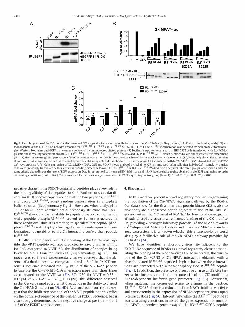
Fig. 5. Phosphorylation of the CIC motif at the conserved CK2 target site increases the inhibition towards the Cn–NFATc signaling pathway. (A) Radioactive labeling with [32P]-or-thophosphate of the EGFP fusion peptides encoding for R3178–203, R3178–210 and R3178–210 S203A in HEK 293 T cells. [32P] incorporation was detected by membrane autoradiogra-phy. Western blot using anti-EGFP is shown as a control of the immunoprecipitated protein. (B) Luciferase reporter gene assays in HEK 293T cells transfected with 3xNFAT-lucplasmid and increasing concentrations of EGFP–R3178–203, EGFP–R3178–210, EGFP–R3178–210 S203A and EGFP–R3178–210 S203E fusion peptides. Data is one representative experiment(N = 3) given as mean (±SEM) percentage of NFAT activation where the 100% is the activation achieved by the mock vector with ionomycin (Io)/PMA/CaCl2 alone. The expressionof each construct in each condition was assessed by western blot using anti-EGFP antibody. (−) no stimulation; (+) stimulated with Io/PMA/Ca2+; (CsA) stimulated with Io/PMA/Ca2+/cyclosporine A. (C) Gene expression of IL2, IL3, IFNγ, TNFα, CSF2 and RCAN1-4 was analyzed by real-time PCR in transduced Jurkat cells after Io/PMA/Ca2+ stimulation. Jurkatcells were previously transduced with a lentivirus encoding either EGFP alone, EGFP–R3178–210 or EGFP–R3178–210 S203A fusion peptides. The three groups were sorted under thesame criteria depending on the level of EGFP expression. Data is represented as mean (±SEM) fold change of mRNA levels relative to that obtained in the EGFP expressing group instimulating conditions (dashed line). T-test was used for statistical analyses compared to EGFP expressing control group (N = 3). *p b 0.05; **p b 0.01; ***p b 0.001.
2318 S. Martínez-Høyer et al. / Biochimica et Biophysica Acta 1833 (2013) 2311–2321
negative charge in the PXIXIT-containing peptides plays a key role inthe binding affinity of the peptides for CnA. Furthermore, circular di-chroism (CD) spectroscopy revealed that the two peptides, R3183–208
and phosphoR3183–208, adopt random conformation in phosphatebuffer solution (Supplementary Fig. 3). However, when analyzed inTFE or MeOH, both of which act as secondary structure stabilizers,R3183–208 showed a partial ability to populate β-sheet conformationwhile peptide phosphoR3183–208 proved to be less structured inthese conditions. Thus, it is tempting to speculate that peptide phos-phoR3183–208 could display a less rigid environment-dependent con-formational adaptability to the Cn interacting surface than peptideR3183–208.
Finally, in accordance with the modeling of the CIC derived pep-tide, the VIVIT peptide was also predicted to have a higher affinityfor CnA compared to VIVIT–AA, the distribution of energies beinglower for VIVIT than for VIVIT–AA (Supplementary Fig. 2B). Thismodel was confirmed experimentally, as we observed that the ab-sence of a double negative charge at +4 and +5 of the PXIXIT con-sensus sequence increased the IC50 value of the VIVIT–AA peptideto displace the CF–SPRIEIT–CnA interaction more than three timesas compared to the VIVIT wt (Fig. 6C; IC50 for VIVIT = 0.57 ±0.15 μM vs VIVIT–AA = 1.78 ± 0.13 μM). This difference observedin the IC50 value implied a dramatic reduction in the ability to disruptthe Cn–NFATc2 interaction (Fig. 6D). As a conclusion, our results sug-gest that the inhibitory potential of the VIVIT peptide not only relieson the optimized sequence of the consensus PXIXIT sequence, but isalso strongly determined by the negative charge at position +4 and+5 of the PXIXIT core sequence.
4. Discussion
In this work we present a novel regulatory mechanism governingthe modulation of the Cn–NFATc signaling pathway by the RCANs.Our data show for the first time that protein kinase CK2 is able tophosphorylate a conserved serine adjacent to the PXIXIT-like se-quence within the CIC motif of RCANs. The functional consequenceof such phosphorylation is an enhanced binding of the CIC motif toCn, providing a stronger inhibitory potential of the RCANs towardsCa2+-dependent NFATc activation and therefore NFATc-dependentgene expression. It is unknown whether this phosphorylation couldalso play a facilitative role of the Cn–NFATc pathway provided bythe RCANs [24].
We have identified a phosphorylation site adjacent to thePXIXIT-like sequence of RCANs as a novel regulatory element modu-lating the binding of the motif towards Cn. To be precise, the disrup-tion of the Cn–RCAN3 or Cn–NFATc interaction obtained with aphosphorylated R3183–208 peptide is higher than when these interac-tions are competed with a non-phosphorylated R3183–208 peptide(Fig. 4). In addition, the presence of a negative charge at the CK2 tar-get serine increases the inhibitory potential of the CIC motif on aNFATc-dependent luciferase gene promoter (Fig. 5B). Conversely,when mutating the conserved serine to alanine in the peptide,R3178–210 S203A, there is a reduction of the NFATc-inhibitory activityand consequently in the expression of NFATc-dependent genes uponT-cell activation (Fig. 5C). Interestingly, while the R3178–210 peptide atnon-saturating conditions inhibited the gene expression of most ofthe NFATc dependent genes assayed, the R3178–210 S203A peptide

Fig. 6. An acidic tail C-terminal to the PXIXIT sequence enhances the Cn–NFATc disruptive potential of PXIXIT-derived peptides. (A) Structural representation of the best ZRANKranked model of Cn–R3194–204 S203D interaction. Cn surface is colored by electrostatic potential: Blue (positive) and red (negative). Peptide is shown using stick representation.The C-terminal region of the peptide is faced in a positively charged area in the Cn surface. (B) The acidic tail of the VIVIT peptide in the 2P6B structure faces the same positivesurface of Cn as R3194–204 S203D. (C) SPRIEIT–CnA interaction competed with either VIVIT or VIVIT–AA peptides. Data is presented as in Fig. 4b. Apparent IC50 values for each pep-tide are displayed on the right. (D) Flag-CnA coprecipitation using GST–NFATc2 as bait competed with either VIVIT or VIVIT–AA peptides performed as in Fig. 4d. (E) Proposed newmodel for the CK2 mediated regulation of the Cn–NFATc signaling pathway via phosphorylation of the CIC motif of RCAN proteins. A regulator of RCAN protein dephosphorylation isunknown (X in the scheme). P indicates a phosphate group; CK2: protein kinase CK2; CK1: protein kinase CK1; DYRK1: Dual specificity tyrosine phosphorylation-regulated kinase1; GSK3β: Glycogen synthase kinase 3β; Cn: calcineurin; NFATc: Nuclear Factor of Activated T-cells cytosolic.
2319S. Martínez-Høyer et al. / Biochimica et Biophysica Acta 1833 (2013) 2311–2321
could still inhibit the expression of IL2, pointing to a dependence on ahigh NFATc activity for the expression of this gene. Altogether, theseobservations suggest that the non-phosphorylated CIC derived pep-tides allows for an increased NFATc activity and therefore, the extentto which the CIC motif phosphorylation influences NFATc dependentgene expression may rely on the specific NFATc activity thresholdthat each gene requires for its transcription. Therefore, we postulatethat this phosphorylation is an important determinant in the modula-tion of the Cn–NFATc pathway by the RCANs in vivo. Moreover, a bio-informatic search regarding all known vertebrate PXIXIT sequencesyielded no other proteins apart from RCANs bearing a phosphorylableamino acid at position +4 from the PXIXIT sequence. This observa-tion suggests that the regulation by phosphorylation of the PXIXITaffinity to Cn is exclusive to the RCANs. Previous work from our labo-ratory has set up the experimental basis for a drug screen based onthe Cn–RCAN interaction, which as a first hit identified dipyridamoleas a drug with immunosuppressant properties [12]. The observationthat the phosphorylation adjacent to the PXIXIT-like motif increasesthe displacement of NFAT from Cn provides a detailed mechanismof such an interaction and may therefore help in the search formore selective and potent immunosuppressant drugs.
We took advantage of the crystallographic data from the VIVIT in-teraction with Cn [17] to model the interaction of the CIC motif withCn. The model predicted that the PSVVVH sequence would interact in
a similar fashion to the VIVIT peptide (Fig. 6A and B). Moreover, thephosphorylated serine at +4 from the PXIXIT sequence would lie inclose contact with a positively charged region in CnA, that is, within4 Å distance from a positive charge provided by the K100 residue ofCnA. The interaction between both residues would explain the phos-phorylation at this position, which adds a double negative charge atthe conserved serine, enhances the interaction of the CIC motif viaan ionic interaction with Cn and therefore increases its Cn–NFATc dis-ruptive potential. This model of interaction could also explain theVIVIT–Cn interaction as the mutation of two glutamic acid residuesto alanine at the C-terminus of the VIVIT peptide, even though thecore PXIXIT sequence is intact, dramatically increases the IC50 of themutated peptide (Fig. 6C) and its ability to disrupt the Cn–NFATccomplex in cells (Fig. 6D). The data presented here suggest that notonly the sequence definition of the PXIXIT core, but also the context inwhich it is inserted, determines the overall affinity of the motif for Cn.
We have previously described that deleting the first three aminoacids (K183, Y184 and E185) of the R3183–203 peptide abrogates itsbinding to CnA [12], suggesting that the PSVVVH sequence, probablydue to its high variation from the consensus PXIXIT, needs some otherelements to firmly anchor to the Cn binding pocket [14]. This de-creased affinity for Cn could be rescued in RCANs by the negativecharge at the C-terminus of the PSVVVH. It is unknown how theKYELHA sequence at the N-terminus of the RCAN CIC motif affects

2320 S. Martínez-Høyer et al. / Biochimica et Biophysica Acta 1833 (2013) 2311–2321
theoverall affinity for Cn. In any case, it is clear that theNFATc-inhibitoryactivity of the CIC motif could be detached in three conserved modules:i) the KYELHA sequence; ii) the PXIXIT-like sequence, and iii) thephosphorylable acidic region at the C-terminus. Future studies focusedon solving the crystal structure of the complete CIC motif of RCANswith Cn would shed light onto the exact role played by each of thethree elements comprising the functional motif in the binding to Cn.
We have shown for the first time and by direct means that in addi-tion to the CIC motif, the FLISPP motif and N-terminus of RCAN3 arephosphorylated in eukaryotic cells (Fig. 1C). Ongoing studies are ad-dressing the functional consequences of RCAN3 S18 phosphorylation.Moreover, our data suggest that protein kinase CK2 phosphorylatesthe CIC motif of vertebrate RCANs in vitro and in vivo, although we can-not rule out the possibility that other kinasesmay be implicated aswell.Accordingly, the phosphorylated serine identified at the CIC motif ofhuman RCAN proteins has also been regarded as phosphorylated invivo in Rcan1 and Rcan2 mouse proteins (Phosphomouse database).We therefore propose a mechanism of regulation of the Cn–NFATcaxis in which the high activity of CK2 counteracts Cn-mediated activa-tion of NFATc and provides the pathway with a reliable switch to shutdown the signal when activated at two levels: increasing the inhibitionof the Cn-dependent activation of NFATs provided by the RCANs andpriming the nuclear export of NFATc by other kinases [32] (Fig. 6E). Itis unknown whether the phosphorylation at the CIC motif is subject toregulation by protein phosphatases.
As a conclusion, we present a novel regulatorymechanism bywhichRCANs modulate the signal output of the Cn–NFATc pathway. We pro-pose that protein kinase CK2 is responsible for the phosphorylation ofa conserved serine residue adjacent to the PXIXIT-like sequence of ver-tebrate RCAN proteins, which results in an enhanced binding of the CICmotif of RCANs to Cn and consequently an increased inhibition of NFATcsignaling in T-cells. Moreover, we have unraveled a novel general regu-latory mechanism driving the dynamics of the PXIXIT sequence ofRCANs binding to Cn that provides important insights at the molecularlevel. We propose that the detailed knowledge of the molecular mech-anisms governing the PXIXIT–Cn interactionmay also prove very usefulin the rational design of future immunosuppressant drugs.
Supplementary data to this article can be found online at http://dx.doi.org/10.1016/j.bbamcr.2013.05.021.
Acknowledgements
This work has been supported by grants SAF2009-08216, BFU2009-10189 and BFU2010-22132 from MICINN and 2009SGR1490 from theGeneralitat de Catalunya. S.M-H is a recipient of an IDIBELL PhD fellow-ship, A.A-I. of a FI fellowship from the Generalitat de Catalunya andE.S.-C. was supported by SAF2009-08216.
We specially thank E. Pérez-Payá and Mar Orzáez for technicalsupport and insightful comments on this work.
References
[1] J. Aramburu, A. Rao, C.B. Klee, Calcineurin: from structure to function, Curr. Top.Cell. Regul. 36 (2000) 237–295.
[2] A. Rao, Signaling to gene expression: calcium, calcineurin and NFAT, Nat. Immunol.10 (2009) 3–5.
[3] H. Wu, A. Peisley, I.A. Graef, G.R. Crabtree, NFAT signaling and the invention ofvertebrates, Trends Cell Biol. 17 (2007) 251–260.
[4] J. Liu, J.D. Farmer Jr.,W.S. Lane, J. Friedman, I.Weissman, S.L. Schreiber, Calcineurin isa common target of cyclophilin-cyclosporin A and FKBP-FK506 complexes, Cell 66(1991) 807–815.
[5] S. Martinez-Martinez, J.M. Redondo, Inhibitors of the calcineurin/NFAT pathway,Curr. Med. Chem. 11 (2004) 997–1007.
[6] J.J. Fuentes, L. Genesca, T.J. Kingsbury, K.W. Cunningham, M. Perez-Riba, X.Estivill, S. de la Luna, DSCR1, overexpressed in Down syndrome, is an inhibitorof calcineurin-mediated signaling pathways, Hum. Mol. Genet. 9 (2000)1681–1690.
[7] T.J. Kingsbury, K.W. Cunningham, A conserved family of calcineurin regulators,Genes Dev. 14 (2000) 1595–1604.
[8] M.C. Mulero, A. Aubareda, A. Schluter, M. Perez-Riba, RCAN3, a novel calcineurininhibitor that down-regulates NFAT-dependent cytokine gene expression,Biochim. Biophys. Acta 1773 (2007) 330–341.
[9] K.J. Davies, G. Ermak, B.A. Rothermel, M. Pritchard, J. Heitman, J. Ahnn, F.Henrique-Silva, D. Crawford, S. Canaider, P. Strippoli, P. Carinci, K.T. Min, D.S. Fox,K.W. Cunningham, R. Bassel-Duby, E.N. Olson, Z. Zhang, R.S. Williams, H.P. Gerber,M. Perez-Riba, H. Seo, X. Cao, C.B. Klee, J.M. Redondo, L.J. Maltais, E.A. Bruford, S.Povey, J.D. Molkentin, F.D. McKeon, E.J. Duh, G.R. Crabtree, M.S. Cyert, S. de la Luna,X. Estivill, Renaming the DSCR1/Adapt78 gene family as RCAN: regulators ofcalcineurin, FASEB J. 21 (2007) 3023–3028.
[10] F.J. Garcia-Cozar, H. Okamura, J.F. Aramburu, K.T. Shaw, L. Pelletier, R. Showalter,E. Villafranca, A. Rao, Two-site interaction of nuclear factor of activated T cellswith activated calcineurin, J. Biol. Chem. 273 (1998) 23877–23883.
[11] J. Aramburu, F. Garcia-Cozar, A. Raghavan, H. Okamura, A. Rao, P.G. Hogan, Selec-tive inhibition of NFAT activation by a peptide spanning the calcineurin targetingsite of NFAT, Mol. Cell 1 (1998) 627–637.
[12] M.C. Mulero, A. Aubareda, M. Orzaez, J. Messeguer, E. Serrano-Candelas, S.Martinez-Hoyer, A. Messeguer, E. Perez-Paya, M. Perez-Riba, M. Orzáez, S.Martínez-Hoyer, E. Pérez-Payá, M. Pérez-Riba, Inhibiting the calcineurin–NFAT(nuclear factor of activated T cells) signaling pathway with a regulator ofcalcineurin-derived peptide without affecting general calcineurin phosphataseactivity, J. Biol. Chem. 284 (2009) 9394–9401.
[13] S. Mehta, H. Li, P.G. Hogan, K.W. Cunningham, Domain architecture of the regula-tors of calcineurin (RCANs) and identification of a divergent RCAN in yeast, Mol.Cell. Biol. 29 (2009) 2777–2793.
[14] S. Martinez-Martinez, L. Genesca, A. Rodriguez, A. Raya, E. Salichs, F. Were, M.D.Lopez-Maderuelo, J.M. Redondo, S. de la Luna, The RCAN carboxyl end mediatescalcineurin docking-dependent inhibition via a site that dictates binding to sub-strates and regulators, Proc. Natl. Acad. Sci. U. S. A. 106 (2009) 6117–6122.
[15] J. Roy, M.S. Cyert, Cracking the phosphatase code: docking interactions determinesubstrate specificity, Sci. Signal. 2 (2009) re9.
[16] J. Aramburu, M.B. Yaffe, C. López-Rodríguez, L.C. Cantley, P.G. Hogan, A. Rao,Affinity-driven peptide selection of an NFAT inhibitor more selective than cyclo-sporin A, Science 285 (1999) 2129–2133.
[17] H. Li, L. Zhang, A. Rao, S.C. Harrison, P.G. Hogan, Structure of calcineurin in com-plex with PVIVIT peptide: portrait of a low-affinity signalling interaction, J. Mol.Biol. 369 (2007) 1296–1306.
[18] J. Roy, H. Li, P.G. Hogan, M.S. Cyert, A conserved docking site modulates substrateaffinity for calcineurin, signaling output, and in vivo function, Mol. Cell 25 (2007)889–901.
[19] M.R. Muller, Y. Sasaki, I. Stevanovic, E.D. Lamperti, S. Ghosh, S. Sharma, C. Gelinas,D.J. Rossi, M.E. Pipkin, K. Rajewsky, P.G. Hogan, A. Rao, M.R. Müller, Requirementfor balanced Ca/NFAT signaling in hematopoietic and embryonic development,Proc. Natl. Acad. Sci. U. S. A. 106 (2009) 7034–7039.
[20] H. Li, M. Pink, J. Murphy, A. Stein, M. Dell’acqua, P.G. Hogan, Balanced interactionsof calcineurin with AKAP79 regulate Ca(2+)-calcineurin-NFAT signaling, Nat.Struct. Mol. Biol 19 (2012) 337–345.
[21] A. Aubareda, M.C. Mulero, M. Perez-Riba, Functional characterization of thecalcipressin 1 motif that suppresses calcineurin-mediated NFAT-dependent cyto-kine gene expression in human T cells, Cell. Signal. 18 (2006) 1430–1438.
[22] S. Ryeom, R.J. Greenwald, A.H. Sharpe, F. McKeon, The threshold pattern ofcalcineurin-dependent gene expression is altered by loss of the endogenous in-hibitor calcipressin, Nat. Immunol. 4 (2003) 874–881.
[23] Z. Hilioti, D.A. Gallagher, S.T. Low-Nam, P. Ramaswamy, P. Gajer, T.J.Kingsbury, C.J. Birchwood, A. Levchenko, K.W. Cunningham, GSK-3 kinases en-hance calcineurin signaling by phosphorylation of RCNs, Genes Dev. 18 (2004)35–47.
[24] B. Sanna, E.B. Brandt, R.A. Kaiser, P. Pfluger, S.A.Witt, T.R. Kimball, E. van Rooij, L.J. DeWindt, M.E. Rothenberg, M.H. Tschop, S.C. Benoit, J.D. Molkentin, Modulatorycalcineurin-interacting proteins 1 and 2 function as calcineurin facilitators in vivo,Proc. Natl. Acad. Sci. U. S. A. 103 (2006) 7327–7332.
[25] R.B. Vega, J. Yang, B.A. Rothermel, R. Bassel-Duby, R.S. Williams, Multiple domainsof MCIP1 contribute to inhibition of calcineurin activity, J. Biol. Chem. 277 (2002)30401–30407.
[26] L. Genesca, A. Aubareda, J.J. Fuentes, X. Estivill, S. De La Luna, M. Perez-Riba, Phos-phorylation of calcipressin 1 increases its ability to inhibit calcineurin and de-creases calcipressin half-life, Biochem. J. 374 (2003) 567–575.
[27] S. Abbasi, J.D. Lee, B. Su, X. Chen, J.L. Alcon, J. Yang, R.E. Kellems, Y. Xia, Proteinkinase-mediated regulation of calcineurin through the phosphorylation of modu-latory calcineurin-interacting protein 1, J. Biol. Chem. 281 (2006) 7717–7726.
[28] M.S. Jung, J.H. Park, Y.S. Ryu, S.H. Choi, S.H. Yoon, M.Y. Kwen, J.Y. Oh, W.J. Song,S.H. Chung, Regulation of RCAN1 protein activity by Dyrk1A protein-mediatedphosphorylation, J. Biol. Chem. 286 (2011) 40401–40412.
[29] Q. Liu, J.C. Busby, J.D. Molkentin, Interaction between TAK1-TAB1-TAB2 andRCAN1-calcineurin defines a signalling nodal control point, Nat. Cell Biol. 11 (2009)154–161.
[30] S.S. Kim, Y. Oh, K.C. Chung, S.R. Seo, Protein kinase A phosphorylates Down syn-drome critical region 1 (RCAN1), Biochem. Biophys. Res. Commun. 418 (2012)657–661.
[31] F. Meggio, L.A. Pinna, One-thousand-and-one substrates of protein kinase CK2?FASEB J. 17 (2003) 349–368.
[32] C.M. Porter, M.A. Havens, N.A. Clipstone, Identification of amino acid residues andprotein kinases involved in the regulation of NFATc subcellular localization, J. Biol.Chem. 275 (2000) 3543–3551.
[33] L.A. Pinna, Protein kinase CK2: a challenge to canons, J. Cell Sci. 115 (2002)3873–3878.

2321S. Martínez-Høyer et al. / Biochimica et Biophysica Acta 1833 (2013) 2311–2321
[34] M.Montenarh, Cellular regulators of protein kinase CK2, Cell Tissue Res. 342 (2010)139–146.
[35] H.M. Berman, J. Westbrook, Z. Feng, G. Gilliland, T.N. Bhat, H. Weissig, I.N.Shindyalov, P.E. Bourne, The Protein Data Bank, Nucleic Acids Res. 28 (2000)235–242.
[36] N. Eswar, B. Webb, M.A. Marti-Renom, M.S. Madhusudhan, D. Eramian, M.-Y.Shen, U. Pieper, A. Sali, Comparative protein structure modeling using MODELLER,Curr. Protoc. Protein Sci. 50 (2007) 2.9.1–2.9.31.
[37] B. Pierce, Z.Weng, ZRANK: reranking protein docking predictions with an optimizedenergy function, Proteins 67 (2007) 1078–1086.
[38] L.L.C. Schrodinger, The PyMOL Molecular Graphics System, Version 13r1,2010.
[39] N.A. Baker, D. Sept, S. Joseph, M.J. Holst, J.A. McCammon, Electrostatics ofnanosystems: application to microtubules and the ribosome, Proc. Natl. Acad.Sci. U. S. A. 98 (2001) 10037–10041.
[40] N. Blom, S. Gammeltoft, S. Brunak, Sequence and structure-based prediction ofeukaryotic protein phosphorylation sites, J. Mol. Biol. 294 (1999) 1351–1362.
[41] M. Salvi, E. Trashi, G. Cozza, A. Negro, P.I. Hanson, L.A. Pinna, Tools to discriminate be-tween targets of CK2 vs PLK2/PLK3 acidophilic kinases, Biotechniques 2012 (2012).
[42] R. Battistutta, G. Cozza, F. Pierre, E. Papinutto, G. Lolli, S. Sarno, S.E. O'Brien, A.Siddiqui-Jain, M. Haddach, K. Anderes, D.M. Ryckman, F. Meggio, L.A. Pinna, Un-precedented selectivity and structural determinants of a new class of protein ki-nase CK2 inhibitors in clinical trials for the treatment of cancer, Biochemistry 50(2011) 8478–8488.