protein-based salivary profiles as novel …downloads.hindawi.com/journals/dm/2018/6141845.pdf1oral...
TRANSCRIPT

Review ArticleProtein-Based Salivary Profiles as Novel Biomarkers forOral Diseases
Alejandro I. Lorenzo-Pouso ,1Mario Pérez-Sayáns,1 Susana B. Bravo,2 Pía López-Jornet ,3
María García-Vence,2 Manuela Alonso-Sampedro,4 Javier Carballo,5
and Abel García-García1
1Oral Medicine, Oral Surgery and Implantology Unit, Faculty of Medicine and Dentistry, Universidade de Santiago de Compostela,Health Research Institute of Santiago de Compostela (IDIS), Santiago de Compostela, A Coruña, Spain2Proteomic Unit, Health Research Institute of Santiago de Compostela (IDIS), Santiago de Compostela, A Coruña, Spain3Department of Oral Medicine, Faculty of Medicine, Regional Campus of International Excellence “Campus Mare Nostrum”,University of Murcia, Espinardo, Murcia, Spain4Department of Internal Medicine and Clinical Epidemiology, Santiago de Compostela University Hospital Complex (CHUS),Health Research Institute of Santiago de Compostela (IDIS), Santiago de Compostela, Galicia, Spain5Department of Food Technology, Faculty of Sciences, University of Vigo-Ourense Campus, Ourense, Spain
Correspondence should be addressed to Alejandro I. Lorenzo-Pouso; [email protected]
Received 7 August 2018; Revised 26 September 2018; Accepted 4 October 2018; Published 7 November 2018
Guest Editor: Anna Zalewska
Copyright © 2018 Alejandro I. Lorenzo-Pouso et al. This is an open access article distributed under the Creative CommonsAttribution License, which permits unrestricted use, distribution, and reproduction in any medium, provided the original workis properly cited.
The Global Burden of Oral Diseases affects 3.5 billion people worldwide, representing the number of people affected by the burdenof untreated dental caries, severe periodontal disease, and edentulism. Thus, much more efforts in terms of diagnostics andtreatments must be provided in the fight of these outcomes. In this sense, recently, the study of saliva as biological matrix hasbeen identified as a new landmark initiative in the search of novel and useful biomarkers to prevent and diagnose theseconditions. Specifically, saliva is a rich reservoir of different proteins and peptides and accessible due to recent advances inmolecular biology and specially in targeted and unbiased proteomics technologies. Nonetheless, emerging barriers are anobstacle to the study of the salivary proteome in an effective way. This review aims at giving an overall perspective of salivarybiomarkers identified in several oral diseases by means of molecular biology approaches.
1. Introduction
Saliva is a complex biological matrix generated by the sali-vary glands. Each salivary gland emits considerably differentsecretions with a highly variable composition depending onsympathetic and parasympathetic stimulation, circadianrhythm, eating habits, health-illness spectrum, drug intake,and other conditions [1]. The basic secretory units of sali-vary glands are clusters of cells called acini. The main threepairs of salivary glands in humans (parotid, submaxillary,and sublingual) together with the minor salivary glandsgenerate 0.75–1.5 liters of this exocrine secretion per day.
This physiological secretion remains high during the day,reducing significantly during the night [2].
Besides water, saliva contains a large number of electro-lytes (i.e., Ca2+, Cl−, H2PO4
−, HCO3−, I−, K+, Mg2+, Na+,
and SCN−), proteins (i.e., mucins, enzymes, and immuno-globulins), lipids, and other molecules [3]. Saliva plays a piv-otal role in the early stages of digestion, allowing a correctphysiological homeostasis in human through nutrition [4].Salivary antioxidant capacity is mainly related to someenzymes (i.e., salivary peroxidase, superoxide dismutase,catalase, glutathione peroxidase, and myeloperoxidase), uricacid, and, to a less extent, ascorbic acid and albumin [5]. In
HindawiDisease MarkersVolume 2018, Article ID 6141845, 22 pageshttps://doi.org/10.1155/2018/6141845

this sense, saliva is the first line of defence against oxidativestress (OE), reactive oxygen species (ROS), and free radicals[6]. Imbalance between the systemic manifestation of ROShas been implicated in the pathogenesis of over 100 patho-logical conditions and also in the prevailing free-radicaltheory of aging [7].
Recently, the term liquid biopsy (LP) was coined inanalytic chemistry as the sampling and analysis of nonsolidbiological tissues, primarily blood and also saliva and otherbiofluids. LP methodologies allow the biomonitoring of sev-eral biomarkers such as proteins, nucleic acids, circulatingtumor cells, or disease drivers related to infections whichproved usefulness in the diagnosis, prognosis, and stagingof a large number of pathologies [8]. In principle, when salivais compared with other biofluids (e.g., blood serum, amnioticfluid, cerebrospinal fluid, and bronchoalveolar lavage fluid),this matrix seems attractive over the others due to its nonin-vasive nature, its lower economic cost, and its greater clinicalsafety. Although certain pathologies and adverse drug reac-tions may limit the bioavailability of this fluid [9], salivaremains as a window of opportunity for modern medicine[10]. In this sense, this matrix has been used by medicinefor the biomonitoring of physiological functions for morethan a century. A good example would be salivary cortisoldeterminations which have been widely used in medicineand behavioural research in the last 150 years for their easyconservation and handling. Therefore, salivary cortisol isstable at room temperature for 1–2 days and at 4°C for oneweek [11].
Currently, several sensitive analytical techniques allowthe detection and quantification of a large number of bio-markers in saliva such as mass spectrometry (MS), reversetranscription-polymerase chain reaction (RT-PCR), micro-arrays, nanoscale sensors, magnetic resonance spectros-copy (MRS), Western blot, immunoassay techniques, or
enzymatic assays. A continuous and exponential growthin the saliva-related research lines has occurred throughoutthe last decades and new relevant concepts as point-of-care(POC) diagnostics have emerged [12]. In the past, cost-effectiveness analysis applied to these techniques showedthem as not appropriate for clinical purposes; however,nowadays, these barriers are being effectively addressed,and this approaches are being progressively translated toclinical practice [13]. Currently, five alphabets (also knownas “OMICS”) of biomarkers present in saliva are known:proteome, transcriptome,microRNA (miRNA),metabolome,and microbiome [14].
In the field of salivanomics, the greatest advances inrecent decades have focused on the analysis on nucleic acids;despite this, some interest has also been placed on protein-based techniques. Human saliva is a rich reservoir of proteinsand peptides; in fact, it gathers more than 3652 proteins and12,562 peptides and shares almost 51% of the proteins and79% of the peptides contained in the plasma [15, 16](Figure 1). Recent advances in proteomics techniques havebrought the discovery of a large number of biomarkers andtherapeutic targets in a large number of oral diseases and sys-temic pathologies with repercussions in the oral cavity [17].A new landmark in salivanomics has been the discovery ofthe presence of exosomes and its outstanding stability insaliva. Exosomes are extracellular vesicles involved in inter-cellular traffic [18]. These vesicles comprise genetic material(i.e., miRNAs) and proteins. Exosomes play a pivotal role inimmune system modulation, inflammation, and oncogenesis[19]. On the other hand, the discovery of the function of cer-tain salivary peptides has helped in the development of newantibiotics [20].
In the present review, the most relevant scientific infor-mation published to date related to the salivary proteomewithin the spectrum of oral diseases is collected and critically
Anti-bacterialAmylases, cystatins, histatins,mucins, peroxidases,lactoferrin, lysozyme,agglutinin
BufferingCarbonic anhydrases, histatins
DigestionAmylases, mucins, lipases
MineralizationCystatins, histatins, proline-richproteins, statherins
Viscoelasticity and lubricationMucins, statherins
Anti-viralMucins, lactoferrin,cystatins, immunoglobulins,peroxidases
Anti-fungalLactoferrin, peroxidases,immunoglobulins, mucins, histatins
Tissue coatingAmylases, cystatins, mucins,proline-rich proteins, statherins
Salivaryproteome
Figure 1: Biological function of the salivary proteome (adapted from Van Nieuw Amerongen et al. [159]).
2 Disease Markers

discussed. This paper is mainly focused on proteins of humanorigin present in saliva and not on the oral disease driver-related proteins or the ones related to the pathogen-host-environment interplay.
1.1. Methods for Collecting Saliva. Protein kinetics and itsconcentrations in saliva are influenced by several factors. Inthis line, quantity and composition of extracted saliva areaffected by the time of day, degree of hydration, body position,psychological stimuli, drug intake, health-related behaviours,systemic/oral health, and other factors [21]. In addition, defi-cits in sample collection, sample handling, and sample trans-port to the laboratory can trigger preprocessing problems.Thus, proteomic literature has extensively expressed thenecessity of highly standardized protocols and tailored to fitthe experimental design [22].
At this point, it is important to highlight that saliva canbe collected under resting or stimulated conditions. Salivarygland stimulation can be achieved by means of different stim-uli such as chewing (gums or swabs), taste stimuli (citricacid), or pharmacologic and electric stimulants [22]. Salivaryflow is controlled by the autonomic nervous system. Para-sympathetic stimulation produces a higher flow rate, whilesympathetic stimulation produces a small flow but richer inproteins and peptides. This stimulation provides clear differ-ences in the snapshot of the salivary proteome and also in therelative amount of specific proteins detected [23].
On the other side, saliva can be collected as whole saliva(WS) or individual gland saliva. Different approaches havebeen described in order to obtain single gland fluids. Regard-ing to parotid gland saliva, different methods can be usedsuch as the Lashley’s cup [24] or the modified Carlson-Crittenden device [25]. Submandibular and sublingual glandsaliva can be collected by means of Truelove’s V-shaped col-lector [26] or Fox’s micropipette [27]. Minor gland secretionscan be collected by pipettes, absorbent papers, or capillarytubes [28]. A relevant drawback in relation to the majorityof these methodologies is the requirement of duct cauteriza-tion, which in practice is technically demanding and uncom-fortable for patients [22].
In the case of WS, regardless of the approach used,patients should refrain from eating, drinking, and oralhygiene procedures for at least one hour before collection,and just before this process, use deionized water as a mouthrinse. Specifically, to collect unstimulated whole saliva(USWS), the patient must be kept comfortably seated avoid-ing orofacial movements during 5 minutes [29]. Navazeshdescribed four approaches to collect WS: draining, spitting,suction, and the swab method. Due to the preference ofcollecting USWS, the gold standard method is draining [22].Different devices have been developed in order to collect pas-sive drool such as Salivette® (Sarstedt, Nümbrecht, Germany),Quantisal® (Immunalysis, Pomona, CA, USA), Orapette®(Trinity Biotech, Dublin, Ireland), and SCS® (Greiner-Bio-One, Kremsmünster, Austria) [30]. Several reports haveshown that the protein coverage does not suffer relevantchanges in relation to different collection devices. The onlywell-known WS drawback versus single gland saliva is that ithas a higher proportion of certain nonsalivary materials such
as desquamated epithelial cells, food debris, bacteria, or leuko-cyte in WS when compared to single gland saliva [1].
The published scientific literature on the effect of preana-lytical variables on saliva profiling is scarce. Controversies arespecially accentuated when the focus is put on centrifugationspeed, addition of a protease inhibitor cocktail (PIC), andstorage temperature range [31]. Schipper et al. demonstratedthat in the case of MS-based techniques, centrifugation speeddoes not have an effect on the number of proteins but a smalleffect on the intensity of the peaks [31]. Mohamed et al.reported that centrifugation can compromise the identifi-cation and quantification of larger proteins [32]. PICs(e.g., aprotinin, leupeptin, antipain, pepstatin A, phenyl-methylsulfonyl fluoride, EDTA, and thimerosal) can avoidproteolysis through the inhibition of serine-, cysteine-, aspar-tic-, and metallo-proteases. Nevertheless, PICs cannot fullyinhibit proteolysis, and this phenomenon can occur duringcentrifugation especially on low-molecular-weight proteins[33]. It is worth mentioning that the addition of somereagents such as sodium azide can cause interference inimmunoassays with horseradish peroxidase [33]. Despitethese limitations, the majority of the described protocolsuse PICs to stabilize this matrix [29]. Collected samplesmust be collected in an ice container and proceeded inthe laboratory within one hour; this methodology avoidsbacterial action and minimizes posttranslational modifica-tions (PTMs) [21]. More than 700 different species of micro-organism cohabit in saliva [34]. A significant part of thesemicroorganisms produce a variety of proteolytic and otherenzymes that can trigger PTMs [29]. Moreover, temperatureis known to play a pivotal role in proteostasis; for example,some proteases can function as chaperones (i.e., “helper”proteins) at low temperatures, but they act as proteasesat elevated temperatures [35]. After processing, storageat−80°Chave shown to provide the same spectra as fresh sam-ples, while at −20°C temperature results can be distorted [31].
Finally, many salivary proteins of low abundance, suf-fer a strong interference with other more abundant proteins(i.e., lysozyme and α-amylase) resulting in a low ionizationefficiency in MS-based analysis. There are mainly threemethods for the removal of high-abundance salivary pro-teins: enzyme substrate absorption method used for alpha-amylase affinity removal, immunodepletion method, andthe combinatorial peptide ligand library [14].
1.2. Analysis. Quantitative molecular biology techniquesremain as the gold standard in the study of the salivary pro-teome [36]. These techniques are classified into absolutequantification techniques in which the exact concentrationof proteins in a matrix is detected and the relative techniquesin which the difference in protein concentration betweensamples is measured. Relative quantification techniques fit avery broad field of experimental designs; in this sense,semiquantitative ELISA, MS, and two-dimensional gel elec-trophoresis (2-DE) have been widely used. Nonetheless, abso-lute quantification approaches such as quantitative ELISAassays or multiplexed immunobead-based assay have alsobeen used [37]. Recently, in the search for salivary biomarkers,nontargeted techniques have been successfully introduced. In
3Disease Markers

this sense, the current state-of-art techniques are 2-DEtechniques coupled to matrix-assisted laser desorption/ionization-time of flight mass spectrometry (MALDI-TOFMS) or liquid chromatography tandem-mass spectrometry(LC-MS/MS) [38]. Moreover, other non-gel-based approachessuch as isobaric tags for relative and absolute quantitation(iTRAQ) or label-free quantification have been used forthe quantitative analysis of the salivary proteome [39].Minority, surface-enhanced laser desorption/ionization-time of flight (SELDI-TOF) MS was also used [40].
2. The Salivary Proteome in theHealth-Illness Spectrum
2.1. Salivary Proteomic Profile in Health. A recent collabora-tive study among three reference centres in the salivaresearch revealed the presence of 1939 different proteinsobtained from 19,474 unique peptides in whole saliva [41].Despite this, there may be variations in this number depend-ing on the equipment and techniques used [42]. Zhao et al.recently studied the number of matching proteins in fivebody fluids (i.e., plasma, urine, cerebrospinal fluid, amnioticfluid, and saliva) finding a total of 564 common proteins[43]. It has been hypothesized that the common proteinspresent in both plasma and saliva may be due to the intimatecontact of saliva with crevicular fluid present at the periodon-tal pocket of sulcus level (such as albumin, transferrin, andimmunoglobulins G andM) [34]. Nevertheless, several trans-port mechanisms capable to allow this communication havebeen identified such as passive diffusion, pinocytosis, andfusion pores at acinar cells [44]. Most of the salivary proteinshave a low molecular weight. Specifically, 70% of the salivaryproteome is made up of proline-rich proteins (PRPs) synthe-sized from the genome contained in chromosome 12 [45];the rest of the proteins are synthesized from genome belong-ing to chromosomes 4 and 20 [46]. The salivary proteome ishighly dynamic. Its proteins are affected by a large number ofPTMs such glycosylation, phosphorylation, acetylation,ubiquitination, methylation, deamidation, sulfation, or pro-teolysis. The homeostatic mechanisms that regulate thesemodifications are not well known, but they constitute a par-ticular “biological signature” not included in the genome[47]. ROS can also affect salivary proteins; in this sense, theycan damage proteoglycans and can cause the oxidation ofsome relevant proteases. Some of these PTMs may increasethe molecular weight of these proteins [48]. In addition, thesalivary “interactome” of these proteins has been recentlyinvestigated. In this sense, most proteins interact with otherscreating protein complexes (e.g., amylase with MUC 5B,MUC 7, histatin 1, and histatin 5) [49].
Due to the limitations that the use of single OMICtechnique entails, recently, they tend to be combined inorder to obtain a better vision of the disease and its pro-gression [50–53]. In this regard, current theories point toa bidirectional relationship between salivary microbiomeand proteome. The salivary proteome thereby conferslong-term stability to the composition and activity of theoral microbiota [50].
2.2. Dental and Periodontal Diseases. Table 1 summarizes theuse of protein-based techniques for salivary biomarkeridentification in dental and periodontal diseases.
2.2.1. Periodontal and Peri-Implant Diseases. The mostcommon forms of periodontal disease are gingivitis andperiodontitis. Gingivitis is defined as a plaque-inducedinflammation of the marginal gingiva, whereas periodontitis(PD) implies a chronic inflammation that causes the destruc-tionof the connective tissueof the tooth and surrounding alve-olar bone [54]. PD is one of the most frequent inflammatoryevents in humans; in fact, one of every two Americans aged30 or older are affected by PD (i.e., 64.7 million people) [55].
Schenck et al. demonstrated that high levels of salivaryIgA were related with higher susceptibility to gingivitis whenthe host response to several bacteria was investigated [56].Another nontargeted salivary proteomics research designedwith Löe’s concept of experimental gingivitis analysed using2-DE found that, in patients suffering from gingivitis, therewas a greater presence of serum-related proteins such asimmunoglobulins and keratins in relation to the controlgroup [57]. Nonetheless, the majority of the investigationsanalyse the inflammatory condition proteome in the gingivalcrevicular fluid and not in the saliva [39, 58]. A problemreflected in the literature regarding MS (specifically LC-iontrap MS, LC-Orbitrap MS, or LC-FTMS) is its lower sensitiv-ity to detect certain proinflammatory and anti-inflammatorycytokines versus ELISA techniques [59]. These cytokines arevery relevant in the genesis of the periodontium pathology[60, 61]. If we take a closer look at the studies that use ELISAtechniques to detect different levels of proteins in salivafrom patients with gingivitis compared to controls, we willfind a large number of overexpressed proteins in affectedsubjects: TNF-α, IL-1, Annexin-1, HBD-1, HBD-2, HBD-3,25-hydroxy-vitamin D3, PGE2, Cystatin C, etc. [62–66].Due to the reversible character of this outcome, the two mostused patients’ subgroups in this type of research have beenchildren and pregnant women.
Chemical studies applied in the study of PD have beenconstant in the medical literature for the last 70 years [67].However, due to the lack of stable criteria and classificationto diagnose this family of pathologies [68], all these investi-gations went through great biases until the last 30 years.PD-related salivary proteins have been classified in foursubgroups [69].
The most specific salivary group biomarkers are theimmunoglobulin (Ig) family proteins. Igs are glycoproteinsof the γ-globulin type that acts at the saliva level in the iden-tification and neutralization of bacterial agents. Immunoflu-orescence studies have shown that these Igs are synthesizedby plasma B cells located at the level of salivary glands [12].In this regard, countless studies have studied the differentiallevels of IgA, IgG, and IgM expression in control patients ver-sus patients with different forms of PD [70, 71]. The mainanalytical techniques used to determine these Igs in salivaare radial immunodiffusion (RID), nephelometry, and ELISA[72]. Several studies have shown that the levels of these Igs inboth chronic and aggressive periodontitis are higher than inhealthy patients [69]. At the same time, it has also been
4 Disease Markers
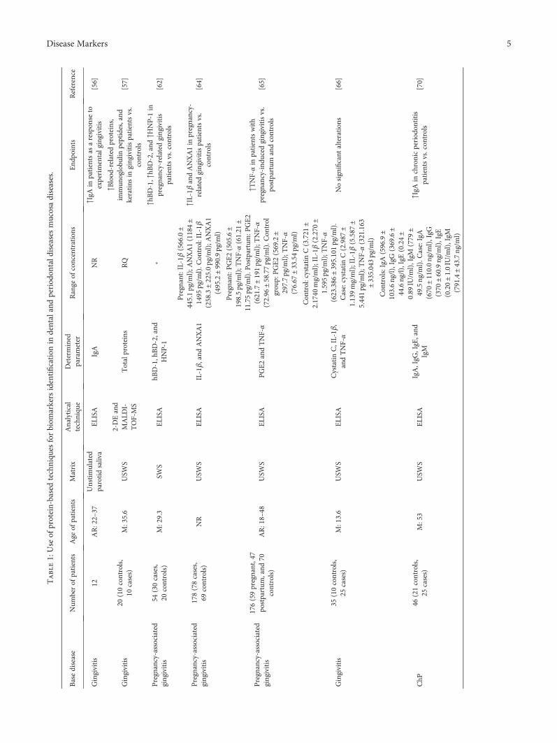
Table1:Use
ofprotein-basedtechniqu
esforbiom
arkers
identification
indentalandperiod
ontald
iseasesmucosadiseases.
Basedisease
Num
berof
patients
Age
ofpatients
Matrix
Analytical
techniqu
eDetermined
parameter
Range
ofconcentrations
End
points
Reference
Gingivitis
12AR:22–37
Unstimulated
parotidsaliva
ELISA
IgA
NR
↑IgA
inpatientsas
arespon
seto
experimentalgingivitis
[56]
Gingivitis
20(10controls,
10cases)
M:35.6
USW
S2-DEand
MALD
I-TOF-MS
Totalproteins
RQ
↑Blood
-related
proteins,
immun
oglobu
linpeptides,and
keratins
ingingivitispatientsvs.
controls
[57]
Pregnancy-associated
gingivitis
54(30cases,
20controls)
M:29.3
SWS
ELISA
hBD-1,h
BD-2,and
HNP-1
∗↑h
BD-1,↑hB
D-2,and
↑HNP-1
inpregnancy-relatedgingivitis
patientsvs.con
trols
[62]
Pregnancy-associated
gingivitis
178(78cases,
69controls)
NR
USW
SELISA
IL-1β,and
ANXA1
Pregnant:IL-1β(566.0±
445.1pg/m
l);A
NXA1(1184±
1495
pg/m
l).C
ontrol:IL-1β
(258.3±225.0pg/m
l);A
NXA1
(495.2±990.9pg/m
l)
↑IL-1β
andANXA1in
pregnancy-
relatedgingivitispatientsvs.
controls
[64]
Pregnancy-associated
gingivitis
176(59pregnant,47
postpartum
,and
70controls)
AR:18–48
USW
SELISA
PGE2andTNF-α
Pregnant:PGE2(505.6±
198.5pg/m
l);T
NF-α(61.21
±11.75pg/m
l).P
ostpartum:P
GE2
(621.7±191pg/m
l);T
NF-α
(72.96
±58.77pg/m
l).C
ontrol
grou
p:PGE2(569.2±
297.7pg/m
l);T
NF-α
(76.67
±33.54pg/m
l)
↑TNF-αin
patientswith
pregnancy-indu
cedgingivitisvs.
postpartum
andcontrols
[65]
Gingivitis
35(10controls,
25cases)
M:13.6
USW
SELISA
CystatinC,IL-1β
,andTNF-α
Con
trol:cystatinC(3.721
±2.1740
mg/ml);IL-1β
(2.270
±1.595pg/m
l);T
NF-α
(623.386
±395.101pg/m
l).
Case:cystatin
C(2.987
±1.139mg/ml);IL-1β
(5.587
±5.441pg/m
l);T
NF-α(321.163
±335.0 43pg/m
l)
Nosignificant
alteration
s[66]
ChP
46(21controls,
25cases)
M:53
USW
SELISA
IgA,IgG
,IgE
,and
IgM
Con
trols:IgA(596.9±
103.6ng/l),IgG(369.6±
44.6ng/l),IgE(0.24±
0.89
IU/m
l),IgM
(779
±49.5ng/m
l).C
ase:IgA
(670
±110.0ng/m
l),IgG
(370
±60.9ng/m
l),IgE
(0.20±1.0IU
/ml),IgM
(791.4±43.7ng/m
l)
↑IgA
inchronicperiod
ontitis
patientsvs.con
trols
[70]
5Disease Markers

Table1:Con
tinu
ed.
Basedisease
Num
berof
patients
Age
ofpatients
Matrix
Analytical
techniqu
eDetermined
parameter
Range
ofconcentrations
End
points
Reference
ChP
70(20controls,
50cases)
AR:18–45
USW
SELISA
IgA,IL-1β
,and
MMP-8
Con
trol:IgA
(81.23
±24.61μg/ml);IL-1β
(89.93
±25.48pg/m
l);M
MP-8
(57.95
±31.64ng/m
l).C
ase:IgA
(196.48±54.61μg/ml);IL-1β
(530.76±343.85
pg/m
l);
MMP-8
(672.18±
411.0ng/m
l)
↑IgA
,↑IL-1β,and
↑MMP-8
inchronicperiod
ontitispatientsvs.
controls
[71]
Aggressive
period
ontitis
10(5
controlsand
5cases)
M:24
USW
S
2-DEand
LC-M
S/MS
valid
ationvia
ELISA
Totalproteins
RQ
↓Lactoferrin,↑IgA2,and↑album
inin
aggressive
period
ontitispatients
vs.con
trols
[74]
ChP
20(10controlsand10
cases)
AR:26–50
USW
S2-DEand
MALD
I-TOF-MS
Totalproteins
RQ
↑α-A
mylasevariantsin
chronic
period
ontitispatientsvs.con
trols
[75]
ChP
57(38obesepatients
(13period
ontitisand
33no
nperiodo
ntitis)
and19
healthy
patients)
AR:35–65
SWS
SELD
I-TOF-MS
Totalproteins
RQ
↑Album
in,↑aandbhemoglobin,↑
α-defensins
1,2,and3in
period
ontitisobesepatientsvs.
controls
[76]
ChP
25(15period
ont itis
patientswithtype
two
diabetes
and
10controls)
AR:40–60
USW
S2-DEand
LC-M
S/MS
Totalproteins
RQ
↑Immun
oglobu
linJchain,
↓polym
ericim
mun
oglobu
linreceptor,↑plastin-2,↓actin-related
protein,
↓interleuk
in-1
receptor
antagonist,and
↑leukocyteelastase
chronicperiod
ontitispatientsvs.
controls
[77]
ChP
andgingivitis
67(17chronic
period
ontitis,17
gingivitis,and
33healthy)
AR:20–64
USW
SLC
-MS/MS
valid
ationvia
MRM
Totalproteins
RQ
↑Matrixmetalloproteinase-9,
↑Ras-related
protein-1,↑actin-
relatedprotein2/3complex
subu
nit5,↓clusterin,and
↓deleted
inmalignant
braintumors1in
chronicperiod
ontitisand
gingivitispatientsvs.con
trols
[78]
ChP
54(20controls,
43cases)
AR:20–73
USW
SELISA
IL-1β,IL-1ra,
platelet-derived
grow
thfactor-BB,
VEGF,
MMP-8,
MMP-9,C
RP,and
lactoferrin
Con
trol:M
MP-8
(36.8
(16.9–295.5ng/m
l));lactoferrin
(10,877
(5808–20,937
ng/m
l)).
Case:MMP-8
(203.7
(86.8–609.2ng/m
l));lactoferrin
(10,87715,801
(12707–18,687ng/m
l))
↑IL-1β
and↑M
MP-8
inchronic
period
ontitisandgingivitis
patientsvs.con
trols
[80]
6 Disease Markers

Table1:Con
tinu
ed.
Basedisease
Num
berof
patients
Age
ofpatients
Matrix
Analytical
techniqu
eDetermined
parameter
Range
ofconcentrations
End
points
Reference
ChP
46(20controls,
26cases)
M:49
USW
SELISA
HGF
Con
trol:H
GF(0.06to
5.38
ng/m
l).C
ase:HGF
(0.68ng/m
l(range:0–7.33))
↑HGFin
chronicperiod
ontitis
patientsvs.con
trols
[83]
ChP
52(24controls,
28cases)
AR:20–50
USW
SELISA
IL-1β
Con
trol:IL-1β
(161.51pg/m
l).
Case:IL-1β(1312.75
pg/m
l)↑IL-1β
inchronicperiod
ontitis
patientsvs.con
trols
[84]
ChP
42(15controls,
27cases)
AR:35–55
SWS
ELISA
MMP-8,M
MP-9,
TIM
P-1,T
IMP-2,
andMPO
NR
↑MMP-8,↑TIM
P-1,and
↑MPOin
chronicperiod
ontitispatientsvs.
controls
[88]
Ortho
dontically
indu
ced
inflam
matoryroot
resorption
72(24controls,
48cases)
AR:10–30
USW
S
2-DEand
LC-M
S/MS
valid
ationvia
Western
blot
Totalproteins
RQ
↑P21-A
RCand↑C
DC42
inorthod
ontically
indu
ced
inflam
matoryroot
resorption
vs.
controls
[91]
Caries
32(16controls,
16cases)
AR:18–29
USW
S2-DEand
LC-M
S/MS
Totalproteins
RQ
↑Amylase,↑IgA
,and
↑lactoferrin,
↓cystatins,↓acidicPRPs,and
↓li pocalin-1
indentalcarie s
patientsvs.con
trols
[99]
Caries
30(10controls,
20cases)
AR:10–12
USW
SiTRAQ-LCMS/
MSvalid
ationvia
MRM
Totalproteins
RQ
↑Mucin
7,↑m
ucin
5B,↑histatin
1,↑cystatinS,↑cystatinSN
,and
↑basicsalivary↑p
rolin
e-rich
protein2in
dentalcaries
patients
vs.con
trols
[100]
Caries
100(50controls,
50cases)
AR:4–6
USW
S2-DE
Totalproteins
RQ
↑Prolin
e-rich
proteinband
sin
dentalcaries
patientsvs.con
trols
[101]
Caries
26(13controls,
13cases)
AR:3–4
SWS
MALD
I-TOFMS
combinedwith
magneticbeads
Totalproteins
RQ
↑2specificpeptides
withm/z
values
3162.0Daand3290.4Dain
caries
patientsvs.con
trols
[104]
Caries
30(10controls,
20cases)
AR:4.7
SWS
MALD
I-TOFMS
combinedwith
magneticbeads
Totalproteins
RQ
↑Histatin-1in
caries
patientsvs.
controls
[105]
Abbreviations:2-D
E:two-dimension
algelelectroph
oresis;A
NXA:ann
exin;A
R:age
range;ARC:activity-regulatedcytoskeleton
-associatedprotein;
CDC:cell-division
cycleprotein;
ChP
:chron
icperiod
ontitis;
CRP:c-reactiveprotein;
ELISA
:enzym
e-lin
kedim
mun
osorbent
assay;HDB:h
emoglobinsubu
nitdelta;H
GF:
hepatocyte
grow
thfactor;H
NP:h
uman
neutroph
ilpeptide;Ig:immun
oglobu
lin;IL:
interleukin;
ITRAQ:isobaric
tags
forrelative
andabsolute
quantitation
;LC
-MS/MS:
liquidchromatograph
ytand
em-m
assspectrom
etry;M:mean;
NR:no
trepo
rted;MALD
I-TOF:
matrix-assisted
laserdesorption
/ionization
;MMP:matrixmetalloproteinase;MPO:myeloperoxidase;MRM:multiplereaction
mon
itoring;
PG:prostaglandin;
PRP:proline-rich
protein;
RQ:relative
quantification
;SW
S:stim
ulated
who
lesaliva;TIM
P:tissueinhibitorof
metalloproteinases;TNF:
tumor
necrosisfactor;U
WSW
:unstimulated
who
lesaliva;VEGF:
vascular
endo
thelialgrowth
factor.∗foraddition
aldata
seeoriginalsource.
7Disease Markers
shown that the level of these proteins decreases significantlywith periodontal treatment. In addition, oral dysbiosis maytrigger the production of specific proteases against Igs [73].
The second group comprises nonspecific markers. In thisregard, there is an innumerable amount of nonspecific pro-teins that have been found altered in patients with periodon-tal disease versus healthy patients. Among them, we find, forexample, albumins, amylases, mucins, lactoferrins, lyso-zymes, histatins, or proteins related to oxidative stress (OS).Nontargeted proteomic techniques are the most used to iden-tify these nonspecific biomarkers [74–77]. Bostanci et al.demonstrated through label-free quantitative proteomicsthat patients with PD had lower levels of lactoferrin, lacritin,sCD14, Mucin 5B, and Mucin 7 vs. control [78]. This findingpoints to a reduction in the salivary antimicrobial anddefence properties among PD-affected patients.
The third group comprises proteins related to systemicand local inflammation at the soft gingival tissues level. Inthis sense, C-reactive protein (CRP) and cytokines standout. At the same time, within the group of cytokines, thereare several remarkable subfamilies such as those of IL-1(11 proteins), TNF-α (19 proteins), chemokines, growthfactors, or bone metabolism-related cytokines (i.e., RANK/RANKL/OPG) [79].
CRP is an acute phase protein, whose levels rise inresponse of inflammation. This analyte can be detectable insaliva by means of ELISA [80] and integrated microfluidicplatforms [81]. According to a recent systematic review, highsalivary levels of CRP have been correlated with local inflam-mation (PD) and systemic inflammation [82]. At present,the most widely studied PD-related cytokines have beeninterleukin-1 beta and hepatocyte growth factor. Severalcase-control studies confirmed that both proteins are over-expressed in PD-affected patients vs. control [83, 84].
The RANKL/RANK/OPG pathway is responsible forcontrolling osteoclastogenesis [85]. Apparently, at the sali-vary level, high and low levels of RANKL of OPG, respec-tively, have being found during PD [86].
The last groups of proteins are metalloproteinases(MMPs). MMPs are a subfamily of zinc-dependent proteasesresponsible of extracellular matrix (ECM) remodelling. Asidefrom their initial role as ECM modifiers, MMPs also interactwith several cell-surface molecules (i.e., chemokines, cyto-kines, growth factors, intercellular junction proteins, otherproteases, and cell receptors). Imbalance in the ECM equilib-rium has been linked to alterations at tissue remodelling,inflammatory response, cell growth, andmigration [87]. Manyscientific reports have given insight intoMMPs and their rela-tionship with periodontal inflammation and destruction dueto the pivotal role of these proteases in collagen degradation.
The MMPs 8 and 9 are the main detectable ones in saliva.One of the actual gold standard biomarkers of PD is salivaryMMP8, as several ELISA and POC platforms have ascer-tained [87]. Meschiari et al. demonstrated that salivaryMMP9 (also known as gelatinase B) is overexpressed inPD-affected patients by zymography approaches [88].
Recent reports have used proteomic techniques in thesearch of salivary biomarkers in peri-implant diseases(i.e., peri-implantitis and peri-implant mucositis). These
reports have shown a series of markedly overexpressedproteins in these pathological conditions, especially cyto-kines (i.e., IL-1b and RANK/RANKL/OPG) and MMPs(MMP8) [89]. These biomarkers are very close to thosedescribed in the PD; this finding supports the epidemio-logical relationship between PD and peri-implant diseases[90]. A particular proteomic signature has been alsodetected in the processes of root resorption induced byorthodontic movements by means of 2-DE coupled toMALDI-TOF-MS [91, 92].
2.2.2. Caries. Caries is a biofilm-mediated carbohydrate-driven pathological condition. This outcome produces themineral breakdown of the dental tissues [93]. Dental cariesat permanent dentition is the most common human diseases,affecting 2.4 billion people (40% of the global population)[94]. Classically, the diagnosis of this condition has beenmade through conventional clinical diagnosis and radiologi-cal techniques [95]; however, recent studies at the salivarylevel have also served to find new useful biomarkers in thediagnosis and response to treatment of this outcome [14].Different salivary parameters outside the proteome have beenstudied and correlated with the predisposition to dentalcaries such as dysbiosis of microbiota, evaluation of pH,buffering capacity, viscosity, and flow rate levels [96]. Thebiomarkers currently detected at the salivary proteome levelwere recently classified by Gao et al. into three subgroups:Igs, innate (nonimmune) host defence proteins and pep-tides, and proteins and peptides implicated on calciumphosphate chemistry.
In relation to Igs, the evidence is limited in relation to IgAand salivary IgG [97]. Nonetheless, Fidalgo et al. recentlydeveloped a meta-analysis of case-control studies to exploresalivary IgA levels in dental caries concluding that highlevels of IgA were higher in patients with caries (0.27 OR[0.17–0.38]) [98].
Regarding nonspecific proteins, different case-controlstudies with nontargeted proteomic techniques have founddifferential expression of different proteins [99, 100]. Numer-ous investigations have pointed out that a low number ofPRPs is associated with an increased risk of dental caries[101, 102]. On the other hand, different studies have shownthat the presence of mucins in patients with caries was signif-icantly higher than in patients without this pathology [103].Regarding other proteins (i.e., agglutinins, amylase, lactofer-rin, and lysozyme), the results have been disparate andcontradictory. Finally, in relation to salivary antibacterialpeptides, there are contradictory results regarding their diag-nostic value (i.e., alpha-defensins, cathelicidins, histatins, andstaterins) [104, 105].
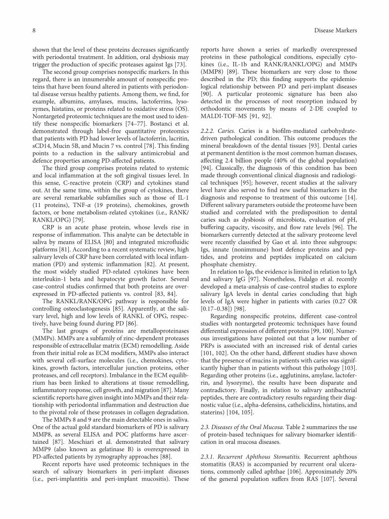
2.3. Diseases of the Oral Mucosa. Table 2 summarizes the useof protein-based techniques for salivary biomarker identifi-cation in oral mucosa diseases.
2.3.1. Recurrent Aphthous Stomatitis. Recurrent aphthousstomatitis (RAS) is accompanied by recurrent oral ulcera-tions, commonly called aphthae [106]. Approximately 20%of the general population suffers from RAS [107]. Several
8 Disease Markers
Table2:Use
ofprotein-basedtechniqu
esforbiom
arkersidentification
inoralmucosadiseases.
Basedisease
Num
berof
patients
Age
ofpatients
Matrix
Analytical
techniqu
eDetermined
parameter
Range
ofconcentrations
End
points
Reference
RAS
30(10controls,
20cases)
M:35.9
USW
SELISA
Cortisol
Con
trol:cortisol
(0.64±0.36
mg/dl);case:
cortisol
(0.57±0.25
mg/dl)
↑Cortisolinrecurrent
aphtho
usstom
atitis
patientsvs.con
trols
[108]
RAS
68(34controls,
34cases)
M:23.29
USW
SELISA
Cortisoland
amylase
Con
trol:cortisol
(3.65±2.5ng/dl);amylase
(128.74±86.3U/m
l);case:
cortisol
(3.35±1.8ng/dl);
amylase(155.09±
116.1U/m
l)
Nosignificant
differences
[109]
RAS
75(25controls,
50cases)
M:27.5
USW
SELISA
MPO
Con
trol:M
PO
(21.36
±14.73Ug−
1 );case:MPO
(19.22
±18.97Ug−
1 ).
Nosignificant
differences
[111]
RAS
62(30controls,
32cases)
AR:14–46
Unstimulated
parotidsaliva
ELISA
Superoxide
dism
utase,
glutathion
eperoxidase,
andcatalase
Con
trol:sup
eroxide
dism
utase(0.56±
0.11
U/m
gprotein);catalase
(0.78±0.03
U/m
g);
glutathion
eperoxidase
(2.88±0.18
U/m
g).C
ase:
superoxide
dism
utase
0.90
±0.04
(U/m
g);catalase
(0.90±0.04
U/m
g);
glutathion
eperoxidase
(1.70±0.1U/m
g).
↑Sup
eroxidedism
utase,
↑glutathione
peroxidase,
and↓catalasein
recurrent
aphtho
usstom
atitis
patientsvs.con
trols
[112]
RAS
52(26controls,
26cases)
AR:22–64
USW
SELISA
IL-6
and
TNF-α
Con
trol:IL-6(9.38±
9.23
pg/m
l);T
NF-α
(7.88±8.45
pg/m
l);case:
IL-6
(12.5±17.51pg/m
l);
TNF-α(28±26.19pg/m
l).
↑IL-6in
recurrentaph
thou
sstom
atitisvs.con
trols
[114]
Behçet’s
disease
andRAS
119(60controls
and59
cases
(33Behçet’s
disease
and16
recurrent
aphtho
usstom
atitis))
AR:16–45
USW
SELISA
Salivary
epidermal
grow
thfactor
Con
trol:salivaryepidermal
grow
thfactor
(2758.7±
81,657.9pg/m
l).B
ehçet’s
disease:salivaryepidermal
grow
thfactor
(1939.7±
81,561.5pg/m
l).R
ecurrent
aphtho
usstom
atitis:
salivaryepidermalgrow
thfactor
(1650.5±
8704.7pg/m
l)
↓Salivaryepidermalgrow
thfactor
inrecurrentaph
thou
sstom
atitis,and
Behçet’s
diseasevs.con
trols
[115]
9Disease Markers

Table2:Con
tinu
ed.
Basedisease
Num
berof
patients
Age
ofpatients
Matrix
Analytical
techniqu
eDetermined
parameter
Range
ofconcentrations
End
points
Reference
Pem
phigus
vulgaris
127(77controls,
50cases)
M:46.84
USW
SELISA
Desmoglein
1and3
Con
trol:allcontrolswere
belowcut-off
value;case:
desm
oglein
1(58.25
±47.52
indexvalue);d
esmoglein
3(144.47±53.42indexvalue)
↑Desmoglein
1and↑3
inpemph
igus
vulgarispatients
vs.con
trols
[118]
BP
100(50controls,
50cases)
AR:38–91
USW
SELISA
BP180
NC16aand
BP230-C3
NR
BP180NC16auseful
asdiagno
sticmarkerfor
pemph
igoid
[119]
Mucou
smem
brane
pemph
igoid
114(50controls,
50cases)
AR:26–87
USW
Sand
stim
ulated
parotidsaliva
ELISA
IgGandIgA
NR
IgAuseful
asdiagno
stic
markerforpemph
igoid
[120]
BMS
60(30controls,
30cases)
M:63.8
USW
SELISA
Cortisoland
α-amylase
Con
trol:cortisol(3.69
±3.07
ng/m
l);amylase
(146.22±130.4IU
/l);case:
cortisol
(4.50±3.68
ng/m
l);
amylase(351.68±
142.5IU
/l)
↑Cortisoland
↑α-amylase
inBMSpatientsvs
controls
[122]
BMS
29(14controls,
15cases)
M:65.7
USW
SandSW
SELISA
Cortisol,17b-
estradiol,
progesterone,
dehydroepiandrosterone,
andα-amylase
∗
↑Cortisolin
USW
S,andof
↑17b-
estradiolin
SWSin
BMS
vs.con
trols
[123]
BMS
270(90controls,
180cases)
AR:15–88
USW
SELISA
Album
in,
lysozyme,
amylase,IgM,
IgG,
andIgA
Con
trol:album
in(9.36±3.44
mg/dl);
lysozyme(24.03
±3.38
mg/ml);amylase
(1638.0±372.0IU
/l);IgM
(1.02±0.06
mg/dl);IgG
(0.79±0.15
mg/dl);IgA
(24.34
±1.26
mg/dl).Case:
albu
min
(18.04
±2.56
mg/dl);lysozyme
(28.10
±3.48
mg/dl);
amylase(3030.0±
470.0IU
/l);IgM
(2.19±0.68
mg/dl);IgG
(4.47±0.76
mg/dl);
IgA(33.15
±3.53
mg/dl)
↑Album
in,↑IgA,↑IgG,
↑IgM
,and
↑lysozym
ein
BMSpatientsvs.con
trols
[124]
10 Disease Markers

Table2:Con
tinu
ed.
Basedisease
Num
berof
patients
Age
ofpatients
Matrix
Analytical
techniqu
eDetermined
parameter
Range
ofconcentrations
End
points
Reference
BMS
45(30controls,
15cases)
M:55.2
USW
SELISA
IgA
Con
trol:IgA
(164.71±
158.80
μg/ml).C
ase:IgA
(176.14±97.23μg/ml).
Nosignificant
differences
[125]
BMS
97(50controls,
47cases(BMS,oral
lichenplanus,and
RAS))
NR
USW
SELISA
IgE
Con
trol:IgE
(20.6±
66.6mg/dl).Case:IgE
(8.07±30.4mg/dl)
Nosignificant
differences
[126]
BMS
38(19controls,
19cases)
NR
USW
SLC
-MS/MS
valid
ationvia
ELISA
Total
proteins
RQ
↑Alpha-eno
lase,↑IL-18,
and↑K
LK13
inBMSvs.
controls
[127]
Abbreviations:AR:agerange;BMS:
burningmou
thsynd
rome;BP:bu
llous
pemph
igoid;
ELISA
:enzyme-lin
kedim
mun
osorbent
assay;
Ig:im
mun
oglobu
lin;IL:interleukin;
KLK
:kallikrein-relatedpeptidase;
LC-M
S/MS:
liquidchromatograph
ytand
em-m
assspectrom
etry;M:meanNR:no
trepo
rted;MPO:myeloperoxidase;NC:no
ncollageno
us;RAS:
recurrentaphtho
usstom
atitis;RQ:relative
quantification
;SW
S:stim
ulated
who
lesaliva;
TNF:
tumor
necrosis
factor;UWSW
:un
stim
ulated
who
lesaliva.
∗foraddition
aldata
seeoriginal
source.
11Disease Markers

reports have investigated the salivary proteome of patientssuffering from this pathology. In particular, the most studiedmolecules have been cortisol, the OE-related peptides, Igs,and certain cytokines.
Different ELISA-based reports have found higher cortisollevels in patients with RAS than healthy controls [108, 109].It has been hypothesized that these altered levels may belinked to the stress and anxiety present in these patients,establishing a neurobiological basis for this pathology. Totalantioxidant capacity (TAC) is not related to the aetiology ofthis pathology; however, patients with RAS do tend to havealtered levels of molecules related to OS [110–112]. Numer-ous studies have shown that levels of IgA and IgG increaseconsiderably in RAS disease outbreaks [113]. Differentinflammatory mediators, especially cytokines, can stimulatethe production of MHC class I and II antigens in epithelialcells [106]. These cells trigger a cytotoxic response in T lym-phocytes causing ulceration. In relation to this etiopatho-genic model, numerous cytokines are found in greateramounts in patients with RAS (i.e., TNF-α, PGE2, VEGF,and IL-6) [114–116].
2.3.2. Pemphigus and Pemphigoid. Vesiculobullous disordersare autoimmune-based pathologies characterized by thepresence of antibodies against epithelial tissue-specific adhe-sion molecules. Its prevalence is 0.2 to 3 people out of every100,000 [117].
Hallaji et al. demonstrated that by ELISA techniques, inthe case of pemphigus, salivary desmoglein 1 and desmoglein3 had sensitivities of 70% and 94%, respectively, in the diag-nosis of this dermatological condition [118]. In the case ofthe pemphigoid, Esmaili et al. proved that the salivary con-centration of BP180-NC16a is useful in the diagnosis of thisdisease [119]. It has also been shown that IgA and IgG sali-vary are markedly increased during pemphigoid and can begood alternatives in its diagnosis [120].
2.3.3. Glossodynia or Burning Mouth Syndrome. The Interna-tional Headache Society (IHS) defines burning mouth syn-drome (BMS) as an intraoral burning or dysestheticsensation, which is repeated daily for more than 2 hours/dayfor more than 3 months, without clinically evident causinglesions. BMS prevalence is barely 4% in the general popula-tion but reaches 18%–33% of postmenopausal women [121].
Due to the psychosomatic profile of this aetiology of thisdisease, stress-related proteins (such as cortisol and α-amy-lase) have been related to its presentation [122, 123]. Thereare few studies investigating the role of salivary Igs in thispathology, and the existing ones have contradictory results.Regarding cytokine-based investigations, the results arealso contradictory for a large number of proteins (i.e.,IL-1β, IL-6, IL-8, and TNF-α) [124–126].
Recently, nontargeted proteomic techniques have discov-ered other novel biomarkers for this pathology. A recentcase-control study based on the LC-MS/MS and iTRAQfound 50 altered proteins (39 overexpressed and 11 subex-pressed); three of them were validated through ELISA:alpha-enolase, IL-18, and KLK13 [127].
2.4. Oral Cancer and Potentially Malignant Oral Lesions.Table 3 summarizes the use of protein-based techniques forsalivary biomarker identification in oral cancer and poten-tially malignant disorders.
2.4.1. Oral Lichen Planus. Oral lichen planus (OLP) is a rela-tively common mucocutaneous disorder. OLP is originatedthrough a chronic inflammation triggered by the epithelialcells apoptosis mediated by autocytotoxic T lymphocytes.According to the World Health Organization (WHO), OLPis considered an oral potentially malignant oral disorder(OPMD). There are several prospective long-term studiesthat show a malignant transformation rate of 1% over a5-year average period [128]. Despite the progress of molecu-lar biology in recent decades, there is no useful biomarker inassessing the risk of malignant transition of this entity; how-ever, recent research based on salivary proteome analysismay be a step forward. The protein-based biomarkers mostwidely investigated in relation to the diagnosis of OLP havebeen cortisol, OS-related molecules, Igs, and cytokines.
In relation to cortisol, numerous investigations haveinvestigated the relationship between psychological statusand levels of this hormone in patients with OLP. Somecase-control studies suggest that the elevated levels of thisglucocorticoid are common among affected individuals[129, 130]. However, some reports do not find significantdifferences [131] or even find lower cortisol levels inOLP-affected patients [132]. Theoretically, cortisol generatesa reduction in the number of lymphocytes and other immunecells and also dysfunctions in the hypothalamus-pituitary-adrenal (HPA) axis which trigger reduction in its production[133]. Lopez-Jornet et al. demonstrated that the levels ofadiponectin were higher in OLP patients. In relation to Igsanalysed via ELISA, IgA and IgG are considerably increasedin patients with OLP compared to controls [129].
OLP aetiology is based on an imbalance betweenTh1/Th2 lymphocytes. The proinflammatory mediatorsthat justify this imbalance are significantly increased inOLP-affected patients: IL-4, IL-10, IL-18, TNF-α, NF-κB-related cytokines, CD44, and CD14 [113]. Interestingly, treat-ment with immunosuppressants such as corticosteroids ornonantibiotic macrolides and alternative therapies such asplant extracts and polyphenols have shown a relevant reduc-tion in these inflammation-based biomarkers [133–135]. Itshould be noted that no research has yet provided a validsalivary biomarker to predict OLPmalignant transformation.
Recently, nontargeted proteomic studies based onMS-based studies have provided new perspectives regardingthe aetiology and diagnosis of the OLP [136, 137].
2.4.2. Oral Leukoplakia. Oral leukoplakia (OL) is defined as“a white plaque of questionable risk having excluded (other)known diseases or disorders that carry no increased risk forcancer” [138]. The pooled estimated prevalence rate of OLvaries between 1.7 and 2.7% in general population. OL is con-sidered by the WHO as OPMD. Malignant transformation oforal leukoplakia in annual average is 1%. Despite the molec-ular biology progress to date, there is no certain marker topredict OL malignant transformation.
12 Disease Markers

Table3:Use
ofprotein-basedtechniqu
esforbiom
arkersidentification
inoralcancer
andpo
tentially
malignant
disorders.
Basedisease
Num
berof
patients
Age
ofpatients
Matrix
Analytical
techniqu
eDetermined
parameter
Range
ofconcentrations
End
points
Reference
OLP
65(32controls,
33cases)
M:57
USW
SELISA
Cortisol,IgA,and
adipon
ectin
Con
trol:IgA
(48.9±32.8mg/l);
cortisol
(0.4±0.2μg/dl);
adipon
ectin(20.1±24.9mg/ml).
Case:IgA(80.3±51.3mg/l);
cortisol
(0.5±0.3μg/dl);
adipon
ectin(38.2±63.5mg/ml)
↑Cortisoland
↑IgA
inOLP
patientsvs.con
trols
[129]
OLP
61(31controls,
30cases)
M:54
USW
SELISA
Cortisol
Con
trol:cortisol(5.21
±2.54
mg/ml).C
ase:cortisol
(4.67±0.33
mg/ml)
Nosignificant
differences
[130]
OLP
62(31controls,
31cases)
M:30
USW
SELISA
Cortisoland
dehydroepiandrosterone
Con
trol:cortisol(14.10
[8.60–18–30]
nmol/l);
dehydroepiandrosterone
(0.66(0.51–1.22)nm
ol/l);
case:cortisol(13.50
(10.50–21–30),nm
ol/l);
dehydroepiandrosterone
(0.75(0.46–0.99)nm
ol/l)
Nosignificant
differences
[131]
OLP
20(10controls,
10cases)
M:58.1
Unstimulated
parotidsaliva
ELISA
Cortisol
∗Nosignificant
differences
[132]
OLP
20(10controls,
10cases)
M:57
USW
SLC
-MS/MS
valid
ationvia
MRM
Totalproteins
RQ
↑S100A
8,↑S100A
9,and
↑haptoglobin
inOLP
vs.
controls
[136]
OLP
12(6
controls,
6cases)
AR:23–60
USW
S2-DEand
MALD
I-TOF-MS
Totalproteins
RQ
↑Urinary
prokallikrein
and
↓PLU
NCin
OLP
vs.con
trols
[137]
OLandOSC
C88
(31controls,
29OL,28
OSC
C)
M:60.89
USW
SELISA
IL-1β,IL-6,andTNF-α
Con
trol:IL-1β
(354
±61.51pg/m
l);IL-6(16±
3.91
pg/m
l);T
NF-α
(38±3.23
pg/m
l).O
L:IL-1β
(143
±54.74pg/m
l);IL-6
(18±5.19
pg/m
l);T
NF-α
(30±3.01
pg/m
l).O
SCC:IL-1β
(906
±62.21pg/m
l);IL-6
(129
±66.59pg/m
l);T
NF-α
(34±21.58pg/m
l)
↑IL-1β
and↑IL-6in
OLand
OSC
Cpatientsvs.con
trols
[139]
OLandOSC
C
75(25controls,
25OL/oral
subm
ucosa
fibrosis
(OPMDs),
25oralcancer)
M:53.2
USW
SELISA
IL-8
Con
trol:IL-8(210.096
±142.302pg/m
l).O
L/oral
subm
ucosafibrosis:IL-8
(299.513
±158.165pg/m
l).
OSC
C:IL-8(1718.610±
668.294pg/m
l)
↑IL-8in
OSC
Cpatientsvs.
controlsandOL
[140]
13Disease Markers

Table3:Con
tinu
ed.
Basedisease
Num
berof
patients
Age
ofpatients
Matrix
Analytical
techniqu
eDetermined
parameter
Range
ofconcentrations
End
points
Reference
OLandOSC
C110(48OL,
62OSC
C)
NR
USW
SELISA
C4d
OL:
Cd4
IL-8
(0.04±
0.03
μg/ml−1 ).O
SCC:C
d4(0.07±0.07
μg/ml−1 )
↑C4d
inOSC
Cpatientsvs.
OLpatients
[141]
OLandOSC
C
69(20OSC
C,
15OSC
Ccured,
15OL,
and
20controls)
AR:32–89
USW
SELISA
End
othelin
-1NR
Nosignificant
differences
[142]
OL
25(10controls,
15cases)
M:73.8
USW
S2-DEand
MALD
I-TOF-MS
Totalproteins
RQ
↓Apo
lipop
rotein
A-1
and↑cystatin
SNprecursorin
OLpatientsvs.
controls
[143]
Headand
neck
cancer
24(8
controls,16
cases)
M:51.88
USW
S2-DEand
LC-M
S/MS
Totalproteins
RQ
↑Betafibrin,↑S100
calcium-binding
protein,
↑transferrin,↑
immun
oglobu
linheavychain
constant
region
gamma,↑cofi
lin-1
and,
↓transthyretin
inHeadand
neck
cancer
patientsvs.con
trols
[150]
OSC
C38
(19controls,
19cases)
M:66
USW
SELISA
Maspin,
phosph
orSrc,
CycD1,Ki67,MMP-9,
andLD
H
∗↓M
aspin,
↓pho
spho
Src,↑C
ycD1,
↑Ki67,↑M
MP-9,and
↑LDH
inOSC
Cpatientsvs.con
trols
[151]
OSC
C53
(26controls,
27cases)
M:53
USW
SELISA
ErbB2andCEA
Con
trol:E
rbB2(4.9±
2.1ng/m
l);C
EA(22.6±
22.1ng/m
l);C
ase:ErbB2
(5.2±1.8ng/m
l);C
EA
(42.6±21.1ng/m
l)
↑CEAin
OSC
Cpatientsvs.
controls
[153]
OSC
C54
(25controls,
29cases)
M:61.9
USW
SLC
-MS/MS
valid
ationvia
ELISA
IL-1α,IL-1β
,IL-6,IL-8,
TNF-α,V
EGF,
catalase,
profi
lin-1,S100A
9,CD59,
galectin-3-bindigprotein,
CD44,thioredoxin,and
keratin-19
∗↑S100A
9and↑IL-6in
OSC
Cpatientsvs.con
trols
[154]
OSC
C128(64controls,
64cases)
M:54
USW
S
2-DEand
LC-M
S/MS
valid
ationvia
ELISA
Totalproteins
RQ
↑M2B
P,↑MRP14,↑CD59,
↑catalase,and↑p
rofilin
inOSC
Cpatientsvs.con
trols
[156]
OSC
C77
(30controls,
47cases)
M:53.3
USW
S
MALD
I-TOFMS
combined
with
magnetic
beads
valid
ationvia
ELISA
Totalproteins
RQ
↑24-mer
ZNF510
peptidein
OSC
Cvs.con
trols
[156]
OSC
CM:50.7
USW
STotalproteins
RQ
[157]
14 Disease Markers
Table3:Con
tinu
ed.
Basedisease
Num
berof
patients
Age
ofpatients
Matrix
Analytical
techniqu
eDetermined
parameter
Range
ofconcentrations
End
points
Reference
460(96controls,
103low-risk
OPMDs,
130high-risk
OPMDs,and131
OSC
C)
LC–M
S/MS
valid
ationvia
MRM
↑MMP1,↑K
NG1,↑A
NXA2,and
↑HSP
A5in
OSC
Cpatientsvs.
controls
OSC
C37
(17controls,
20cases)
M:57
USW
SLC
–MS/MS
valid
ationvia
ELISA
Totalproteins
RQ
Nosignificant
differences
[158]
Abbreviations:2-D
E:two-dimension
algelelectroph
oresis;A
NXA:ann
exin;A
R:age
range;C4d:com
plem
entcompo
nent
4;CD:cell-surfaceprotein;
CEA:carcino
embryonicantigen;
CycD1:cyclin
D1;ELISA
:enzyme-lin
kedim
mun
osorbent
assay;
erbB
-2:h
uman
epidermal
grow
thfactor
receptor
2;HSP
:heatshockprotein;
Ig:immun
oglobu
lin;IL:
interleukin;
KNG:k
ininogen;L
C-M
S/MS:
liquidchromatograph
ytand
em-m
assspectrom
etry;LD
H:lactatedehydrogenase;
M2B
P:hu
man
Mac-2-binding
protein;
M:mean;
MALD
I-TOF:
matrix-assisted
laserdesorption
/ion
ization;
Maspin:
mam
maryserine
protease
inhibitor;MMP:matrixmetalloproteinases;MRP:migration
inhibitory
factor-related
protein;
NR:no
trepo
rted;OL:
oral
leucop
lakia;
OLP
:oral
lichenplanus;OPMD:oral
potentially
malignant
disorder;
OSC
C:oral
squamou
scellcarcinom
a;ph
osph
or-SRC:ph
osph
orylated
SRC;PLU
NC:palate,lung,andnasalepitheliu
mclon
eprotein;
RQ:relative
quantification
;SW
S:stim
ulated
who
lesaliva;TNF:
tumor
necrosisalph
a;UWSW
:unstimulated
who
lesaliva;VEGF:
vascular
endo
thelialgrowth
factor;Z
NF:
zinc
finger
protein.
∗foraddition
aldata
seeoriginalsource.
15Disease Markers

Proteomic studies focused on saliva to anticipate thismalignancy are scarce, and the study of cytokines is basedon ELISA techniques (i.e., IL-6, IL-8, and TNF-α) [139,140]. Other reported proteins that were also useful to discernbetweenOLandoral squamous cell carcinomahave beenC4d,MDA, endothelin-1, and lactate dehydrogenase [141, 142].Camisasca et al. recently reported that in a 2-DE gel-basedproteomic study, 22 spots are much more abundant inpatients with OL than in controls. One spot corresponded toCK10. Later, the authors validated this marker by means ofimmunohistochemistry [143].
2.4.3. Oral Squamous Cell Carcinoma.Oral squamous cell car-cinoma (OSCC) is the eighth most common cancer world-wide. Oral carcinogenesis is modulated by environmentaland genetic factors [144]. The most extensively modifiablerisk factors for this entity are tobacco and alcohol consump-tion [145, 146]. In the last 20 years, the study of HPV as a car-cinogenic factor has also taken on strength [147]. The mostextensively described OSCC-related modifiable risk factorsare tobacco and alcohol consumption. In the last 20 years,the study of HPV as a carcinogenic factor has also raised.Despite all efforts on the side of public health and transna-tional research, a significant improvement in the 5-yearsurvival rate of this neoplasm has not been achieved [144].
In relation to oral diseases, OSCC is by far the one inwhich proteomics research has employed its greatest efforts.A recent meta-analysis suggested that the use of simple orcombined salivary biomarkers for the OSCC may be usefulfor diagnostic purposes [148]. One of the first family of pro-teins that aroused interest as OSCC biomarkers was interleu-kins family; in this sense, there are a large number of studiesthat ascertained their concentrations in saliva. Specifically,the most studied interleukins have been IL-6, IL-8, IL-1,and TNF-α. High levels of these proteins in saliva have beenassociated with OSCC. The biological plausibility of thesehigh levels is found in the proangiogenic and proinflamma-tory functions of these analytes [149]. Elevated levels of IgGhave also been detected in OSCC-affected patients versuscontrols, which ascertains the pivotal role of angiogenesis inoral carcinogenesis [150]. On the other hand, by means ofELISA techniques, Shpitzer et al. found that the salivarylevels of Ki-67 and Cyclin D1 were also altered in thesepatients [151]. These findings are compatible with numerousimmunohistochemistry reports in OSCC [152]. On the otherhand, different investigations mainly based on Western blot,or MRS-based targeted proteomics techniques have foundcell-surface glycoproteins overexpressed in patients withOSCC such as CD44, CD59, or CEA. Other biomarkersrelated to the zinc finger protein family (ZNF) such asZNF510, Cyfra 21-1, and CK19 have also been reported[153–155]. In this sense, Jou et al. reported a sensitivity andspecificity greater than 95% for salivary ZNF510 in the dis-crimination of tumors in early stages (T1+T2) vs. advancedstages (T3+T4) [156].
Nontargeted proteomic techniques have provided otherunique proteins or panels useful as oncological markers. Huet al. reported in a ROC curve analysis that a panel consistingof 5 proteins (M2BP, MRP14, CD59, catalase, and profilin)
had a sensitivity of 90% and a specificity of 83% in thediagnosis of the OSCC via LC-MS/MS [155]. A Taiwanesegroup composed another panel with 4 proteins (MMP1,KNG1, ANXA2, and HSPA5) that able to diagnose OSCCand also to predict OPMDs malignant transformation[157]. Csosz et al. failed to validate some of the biomarkersdescribed by other authors; this Hungarian group justifiedthis fact by the ethnic and geographical variability of thetarget populations [158].
3. Conclusion and Future Perspectives
The advance in the field of salivanomics is a teragnostic rev-olution in oral pathology. The salivary proteome has a Janusrole in oral pathology; oral proteins can provide cytoprotec-tive functions in many of the oral diseases, and, at the sametime, they can contribute to inflammation, infection, andeven tumorigenesis in this cavity. In this sense, salivary pro-teome plays a pivotal role in oral homeostasis; imbalances atimmunological and nonimmunological salivary defence sys-tems can cause a myriad of possible mechanisms leading tooral pathologies.
Moreover, the salivary proteome is an immense sourceof useful biomarkers in the diagnosis and prognosis of thisburden of diseases. However, the precise mechanismsunderlying the role of oral proteins in the initiation andprogression of these conditions are still largely unknown.Further research and a standardization of the analyticalprocesses involved in its study are necessary to give a stepforward. The study of the salivary proteome will mean aninexorable change in current dentistry.
Disclosure
The authors have no relevant affiliations or financial involve-ment with any organization or entity with a financial interestin or financial conflict with the subject matter or materialsdiscussed in the manuscript. This includes employment,consultancies, honoraria, stock ownership or options, experttestimony, grants or patents received or pending, or royalties.
Conflicts of Interest
The authors declare that they have no conflicts of interest.
Authors’ Contributions
Alejandro I. Lorenzo-Pouso and María García-Vence wrotethe main body of the paper. Pía López-Jornet, Susana B.Bravo, Javier Carballo, Mario Pérez-Sayáns, Abel García-García, and Manuela Alonso-Sampedro helped in the litera-ture search and correction. All authors have approved thefinal version of the article.
Acknowledgments
Alejandro I. Lorenzo-Pouso is supported by a fellowshipfrom Health Research Institute of Santiago de Compostela(IDIS). Our sincere apologies to all the authors whose workcould not be cited due to space constraints.
16 Disease Markers
References
[1] E. Kaufman and I. B. Lamster, “The diagnostic applicationsof saliva—a review,” Critical Reviews in Oral Biology andMedicine, vol. 13, no. 2, pp. 197–212, 2002.
[2] K. V. Holmberg and M. P. Hoffman, “Anatomy, biogenesisand regeneration of salivary glands,” Monographs in OralScience, vol. 24, pp. 1–13, 2014.
[3] S. P. Humphrey and R. T. Williamson, “A review of saliva:normal composition, flow, and function,” The Journal ofProsthetic Dentistry, vol. 85, no. 2, pp. 162–169, 2001.
[4] I. Lombaert, M. M. Movahednia, C. Adine, and J. N. Ferreira,“Concise review: salivary gland regeneration: therapeuticapproaches from stem cells to tissue organoids,” Stem Cells,vol. 35, no. 1, pp. 97–105, 2017.
[5] P. Zukowski, M. Maciejczyk, and D. Waszkiel, “Sources offree radicals and oxidative stress in the oral cavity,” Archivesof Oral Biology, vol. 92, pp. 8–17, 2018.
[6] M. Battino, M. S. Ferreiro, I. Gallardo, H. N. Newman, andP. Bullon, “The antioxidant capacity of saliva,” Journal ofClinical Periodontology, vol. 29, no. 3, pp. 189–194, 2002.
[7] I. L. C. Chapple, “Reactive oxygen species and antioxidants ininflammatory diseases,” Journal of Clinical Periodontology,vol. 24, no. 5, pp. 287–296, 1997.
[8] G. Siravegna, S. Marsoni, S. Siena, and A. Bardelli, “Integrat-ing liquid biopsies into the management of cancer,” NatureReviews Clinical Oncology, vol. 14, no. 9, pp. 531–548, 2017.
[9] E. Csosz, G. Kallo, B. Markus, E. Deak, A. Csutak, andJ. Tozser, “Quantitative body fluid proteomics in medici-ne—a focus on minimal invasiveness,” Journal of Proteomics,vol. 153, pp. 30–43, 2017.
[10] M. Greabu, M. Battino, M.Mohora et al., “Saliva–a diagnosticwindow to the body, both in health and in disease,” Journal ofMedicine and Life, vol. 2, no. 2, pp. 124–132, 2009.
[11] U. Turpeinen and E. Hamalainen, “Determination of cortisolin serum, saliva and urine,” Best Practice & Research. ClinicalEndocrinology & Metabolism, vol. 27, no. 6, pp. 795–801,2013.
[12] M. Groschl, “Saliva: a reliable sample matrix in bioanalytics,”Bioanalysis, vol. 9, no. 8, pp. 655–668, 2017.
[13] K. Aro, F. Wei, D. T. Wong, and M. Tu, “Saliva liquid biopsyfor point-of-care applications,” Frontiers in Public Health,vol. 5, 2017.
[14] K. E. Kaczor-Urbanowicz, C. Martin Carreras-Presas, K. Aro,M. Tu, F. Garcia-Godoy, and D. T. W. Wong, “Saliva diag-nostics - current views and directions,” Experimental Biologyand Medicine, vol. 242, no. 5, pp. 459–472, 2016.
[15] F. Amado, M. J. C. Lobo, P. Domingues, J. A. Duarte, andR. Vitorino, “Salivary peptidomics,” Expert Review of Proteo-mics, vol. 7, no. 5, pp. 709–721, 2010.
[16] F. M. L. Amado, R. M. P. Vitorino, P. M. D. N. Domingues,M. J. C. Lobo, and J. A. R. Duarte, “Analysis of the humansaliva proteome,” Expert Review of Proteomics, vol. 2, no. 4,pp. 521–539, 2005.
[17] N. Spielmann and D. T. Wong, “Saliva: diagnostics andtherapeutic perspectives,” Oral Diseases, vol. 17, no. 4,pp. 345–354, 2011.
[18] Y. Ogawa, Y. Miura, A. Harazono et al., “Proteomic analysisof two types of exosomes in human whole saliva,” Biological& Pharmaceutical Bulletin, vol. 34, no. 1, pp. 13–23, 2011.
[19] M. Yáñez-Mó, P. R.-M. Siljander, Z. Andreu et al., “Biologicalproperties of extracellular vesicles and their physiologicalfunctions,” Journal of Extracellular Vesicles, vol. 4, no. 1,article 27066, 2015.
[20] F. Trindade, F. Amado, J. Pinto da Costa et al., “Salivarypeptidomic as a tool to disclose new potential antimicro-bial peptides,” Journal of Proteomics, vol. 115, pp. 49–57,2015.
[21] W. L. Siqueira and C. Dawes, “The salivary proteome: chal-lenges and perspectives,” Proteomics. Clinical Applications,vol. 5, no. 11-12, pp. 575–579, 2011.
[22] M. Navazesh, “Methods for collecting saliva,” Annals of theNew York Academy of Sciences, vol. 694, pp. 72–77, 1993.
[23] G. B. Proctor and G. H. Carpenter, “Salivary secretion: mech-anism and neural regulation,” Monographs in Oral Science,vol. 24, pp. 14–29, 2014.
[24] K. Inenaga, T. Inangaki, R. Hosokawa, and K. Ono, “Parotidsalivary secretion induced by stimulation of periodontalregions with toothbrush in humans,” The Journal of MedicalInvestigation, vol. 56, p. 277, 2009.
[25] A. Sproles and L. D. Schaeffer, “An advanced design of theCarlson-Crittenden cup for collection of parotid fluid,” Bio-materials, Medical Devices, and Artificial Organs, vol. 2,no. 2, pp. 95–101, 1974.
[26] E. L. Truelove, D. Bixler, and A. D. Merritt, “Simplifiedmethod for collection of pure submandibular saliva in largevolumes,” Journal of Dental Research, vol. 46, no. 6,pp. 1400–1403, 1967.
[27] P. C. Fox, P. F. van der Ven, B. C. Sonies, J. M. Weiffenbach,and B. J. Baum, “Xerostomia: evaluation of a symptom withincreasing significance,” Journal of the American DentalAssociation, vol. 110, no. 4, pp. 519–525, 1985.
[28] A. H. Kutscher, I. D. Mandel, E. V. Zegarelli et al., “A tech-nique for collecting the secretion of minor salivary glands: I.Use of capillary tubes,” Journal of Oral Therapeutics andPharmacology, vol. 3, no. 5, pp. 391-392, 1967.
[29] B. S. Henson and D. T. Wong, “Collection, storage, andprocessing of saliva samples for downstreammolecular appli-cations,” Methods in Molecular Biology, vol. 666, pp. 21–30,2010.
[30] Z. Khurshid, S. Zohaib, S. Najeeb, M. Zafar, P. Slowey, andK. Almas, “Human saliva collection devices for proteomics:an update,” International Journal of Molecular Sciences,vol. 17, no. 6, 2016.
[31] R. Schipper, A. Loof, J. de Groot, L. Harthoorn, E. Dransfield,andW. van Heerde, “SELDI-TOF-MS of saliva: methodologyand pre-treatment effects,” Journal of Chromatography B,vol. 847, no. 1, pp. 45–53, 2007.
[32] R. Mohamed, J. L. Campbell, J. Cooper-White, G. Dimeski,and C. Punyadeera, “The impact of saliva collection andprocessing methods on CRP, IgE, and myoglobin immunoas-says,” Clinical and Translational Medicine, vol. 1, no. 1, p. 19,2012.
[33] S. Chiappin, G. Antonelli, R. Gatti, and E. F. De Palo, “Salivaspecimen: a new laboratory tool for diagnostic and basicinvestigation,” Clinica Chimica Acta, vol. 383, no. 1-2,pp. 30–40, 2007.
[34] A. Simon-Soro, I. Tomas, R. Cabrera-Rubio, M. D. Catalan,B. Nyvad, and A. Mira, “Microbial geography of the oralcavity,” Journal of Dental Research, vol. 92, no. 7, pp. 616–621, 2013.
17Disease Markers

[35] C. Leidhold and W. Voos, “Chaperones and proteases—-guardians of protein integrity in eukaryotic organelles,”Annals of the New York Academy of Sciences, vol. 1113,no. 1, pp. 72–86, 2007.
[36] Y. C. Hsiao, L. J. Chu, Y. T. Chen et al., “Variability assess-ment of 90 salivary proteins in intraday and interday samplesfrom healthy donors by multiple reaction monitoring-massspectrometry,” Proteomics. Clinical Applications, vol. 12,no. 2, 2018.
[37] R. Aebersold andM.Mann, “Mass spectrometry-based prote-omics,” Nature, vol. 422, no. 6928, pp. 198–207, 2003.
[38] S. Hu, Y. Xie, P. Ramachandran et al., “Large-scale identi-fication of proteins in human salivary proteome by liquidchromatography/mass spectrometry and two-dimensionalgel electrophoresis-mass spectrometry,” Proteomics, vol. 5,no. 6, pp. 1714–1728, 2005.
[39] N. Bostanci, P. Ramberg, A. Wahlander et al., “Label-freequantitative proteomics reveals differentially regulated pro-teins in experimental gingivitis,” Journal of ProteomeResearch, vol. 12, no. 2, pp. 657–678, 2013.
[40] S. Shintani, H. Hamakawa, Y. Ueyama, M. Hatori, andT. Toyoshima, “Identification of a truncated cystatin SA-Ias a saliva biomarker for oral squamous cell carcinoma usingthe SELDI ProteinChip platform,” International Journal ofOral and Maxillofacial Surgery, vol. 39, no. 1, pp. 68–74,2010.
[41] W. Yan, R. Apweiler, B. M. Balgley et al., “Systematiccomparison of the human saliva and plasma proteomes,”Proteomics. Clinical Applications, vol. 3, no. 1, pp. 116–134,2009.
[42] S. Bandhakavi, M. D. Stone, G. Onsongo, S. K. Van Riper, andT. J. Griffin, “A dynamic range compression and three-dimensional peptide fractionation analysis platform expandsproteome coverage and the diagnostic potential of wholesaliva,” Journal of Proteome Research, vol. 8, no. 12,pp. 5590–5600, 2009.
[43] M. Zhao, Y. Yang, Z. Guo et al., “A comparative proteomicsanalysis of five body fluids: plasma, urine, cerebrospinal fluid,amniotic fluid and saliva,” Proteomics-Clinical applications,no. article 1800008, 2018.
[44] X. Cong, Y. Zhang, Q. H. He et al., “Endothelial tight junc-tions are opened in cholinergic-evoked salivation in vivo,”Journal of Dental Research, vol. 96, no. 5, pp. 562–570,2017.
[45] E. A. Azen, P. A. Goodman, and P. A. Lalley, “Humansalivary proline-rich protein genes on chromosome 12,”American Journal of Human Genetics, vol. 37, no. 2,pp. 418–424, 1985.
[46] E. A. Azen, “Genetic polymorphism of basic proteins fromparotid saliva,” Science, vol. 176, no. 4035, pp. 673-674, 1972.
[47] M. Castagnola, T. Cabras, A. Vitali, M. T. Sanna, andI. Messana, “Biotechnological implications of the salivaryproteome,” Trends in Biotechnology, vol. 29, no. 8, pp. 409–418, 2011.
[48] S. Tsuchida, M. Satoh, H. Umemura et al., “Proteomicanalysis of gingival crevicular fluid for discovery of novelperiodontal disease markers,” Proteomics, vol. 12, no. 13,pp. 2190–2202, 2012.
[49] K. T. B. Crosara, D. Zuanazzi, E. B. Moffa, Y. Xiao, M. A. d. A.M. Machado, and W. L. Siqueira, “Revealing the amylaseinteractome in whole saliva using proteomic approaches,”
BioMed Research International, vol. 2018, Article ID6346954, 15 pages, 2018.
[50] N. Grassl, N. A. Kulak, G. Pichler et al., “Ultra-deep andquantitative saliva proteome reveals dynamics of the oralmicrobiome,” Genome Medicine, vol. 8, no. 1, p. 44, 2016.
[51] S. Hu, Y. Li, J. Wang et al., “Human saliva proteome andtranscriptome,” Journal of Dental Research, vol. 85, no. 12,pp. 1129–1133, 2006.
[52] K. Bao, N. Bostanci, N. Selevsek, T. Thurnheer, and G. N.Belibasakis, “Quantitative proteomics reveal distinct proteinregulations caused by Aggregatibacter actinomycetemcomi-tans within subgingival biofilms,” PLoS One, vol. 10, no. 3,article e0119222, 2015.
[53] J. Groenink, E. Walgreen-Weterings, K. Nazmi et al.,“Salivary lactoferrin and low-Mr mucin MG2 in Actino-bacillus actinomycetemcomitans-associated periodontitis,”Journal of Clinical Periodontology, vol. 26, no. 5, pp. 269–275, 1999.
[54] B. L. Pihlstrom, B. S. Michalowicz, and N. W. Johnson, “Peri-odontal diseases,” Lancet, vol. 366, no. 9499, pp. 1809–1820,2005.
[55] P. I. Eke, B. A. Dye, L. Wei, G. O. Thornton-Evans, R. J.Genco, and CDC Periodontal Disease Surveillance work-group: James Beck (University of North Carolina, ChapelHill, USA), Gordon Douglass (Past President, AmericanAcademy of Periodontology), Roy Page (University ofWashin, “Prevalence of periodontitis in adults in the UnitedStates: 2009 and 2010,” Journal of Dental Research, vol. 91,no. 10, pp. 914–920, 2012.
[56] K. Schenck, D. Poppelsdorf, C. Denis, and T. Tollefsen,“Levels of salivary IgA antibodies reactive with bacteria fromdental plaque are associated with susceptibility to experimen-tal gingivitis,” Journal of Clinical Periodontology, vol. 20,no. 6, pp. 411–417, 1993.
[57] R. Goncalves Lda, M. R. Soares, F. C. Nogueira et al.,“Analysis of the salivary proteome in gingivitis patients,”Journal of Periodontal Research, vol. 46, no. 5, pp. 599–606,2011.
[58] M. M. Grant, A. J. Creese, G. Barr et al., “Proteomic analysisof a noninvasive human model of acute inflammation and itsresolution: the twenty-one day gingivitis model,” Journal ofProteome Research, vol. 9, no. 9, pp. 4732–4744, 2010.
[59] J. A. Stenken and A. J. Poschenrieder, “Bioanalytical chemis-try of cytokines–a review,” Analytica Chimica Acta, vol. 853,pp. 95–115, 2015.
[60] N. Bostanci, G. Emingil, B. Afacan et al., “Tumor necrosisfactor-α-converting enzyme (TACE) levels in periodontaldiseases,” Journal of Dental Research, vol. 87, no. 3,pp. 273–277, 2008.
[61] F. D'Aiuto, L. Nibali, M. Parkar, K. Patel, J. Suvan, andN. Donos, “Oxidative stress, systemic inflammation, andsevere periodontitis,” Journal of Dental Research, vol. 89,no. 11, pp. 1241–1246, 2010.
[62] M. Gursoy, U. K. Gursoy, A. Liukkonen, T. Kauko,S. Penkkala, and E. Kononen, “Salivary antimicrobial defen-sins in pregnancy,” Journal of Clinical Periodontology,vol. 43, no. 10, pp. 807–815, 2016.
[63] G. Dogusal, B. Afacan, E. Bozkurt, and I. Sonmez, “Gingivalcrevicular fluid and salivary resistin and tumor necrosisfactor-alpha levels in obese children with gingivitis,” Journalof Periodontology, vol. 89, no. 8, pp. 973–982, 2018.
18 Disease Markers

[64] M. N. Hassan, G. N. Belibasakis, P. Gumus, V. O. Ozturk,G. Emingil, and N. Bostanci, “Annexin-1 as a salivarybiomarker for gingivitis during pregnancy,” Journal ofPeriodontology, vol. 89, no. 7, pp. 875–882, 2018.
[65] P. Gumus, V. O. Ozturk, E. Bozkurt, and G. Emingil,“Evaluation of the gingival inflammation in pregnancyand postpartum via 25-hydroxy-vitamin D3, prostaglandinE2 and TNF-α levels in saliva,” Archives of Oral Biology,vol. 63, pp. 1–6, 2016.
[66] A. E. Ulker, O. Tulunoglu, N. Ozmeric, M. Can, andS. Demirtas, “The evaluation of cystatin C, IL-1β, and TNF-αlevels in total saliva and gingival crevicular fluid from 11- to16-year-old children,” Journal of Periodontology, vol. 79,no. 5, pp. 854–860, 2008.
[67] M. Berg, D. Y. Burrill, and L. S. Fosdick, “Chemical studiesin periodontal disease III: putrefaction of salivary proteins,”Journal of Dental Research, vol. 25, no. 4, pp. 231–246,1946.
[68] J. G. Caton, G. Armitage, T. Berglundh et al., “A new classifi-cation scheme for periodontal and peri-implant diseases andconditions - introduction and key changes from the 1999classification,” Journal of Clinical Periodontology, vol. 45,Supplement 20, pp. S1–S8, 2018.
[69] W. V. Giannobile, T. Beikler, J. S. Kinney, C. A. Ramseier,T. Morelli, and D. T. Wong, “Saliva as a diagnostic tool forperiodontal disease: current state and future directions,” Peri-odontology 2000, vol. 50, no. 1, pp. 52–64, 2009.
[70] O. A. Olayanju, S. K. Rahamon, I. O. Joseph, and O. G.Arinola, “Salivary immunoglobulin classes in Nigerianswith periodontitis,” The Journal of Contemporary DentalPractice, vol. 13, no. 2, pp. 163–166, 2012.
[71] V. Rangbulla, A. Nirola, M. Gupta, P. Batra, and M. Gupta,“Salivary IgA, interleukin-1β and MMP-8 as salivarybiomarkers in chronic periodontitis patients,” The ChineseJournal of Dental Research, vol. 20, no. 1, pp. 43–51, 2017.
[72] C. Schneider, M. Illi, M. Lotscher, M. Wehrli, and S. vonGunten, “Isolation of antibodies from human plasma, saliva,breast milk, and gastrointestinal fluid,”Methods in MolecularBiology, vol. 1643, pp. 23–31, 2017.
[73] M. Nakayama and N. Ohara, “Molecular mechanisms ofPorphyromonas gingivalis-host cell interaction on periodon-tal diseases,” Japanese Dental Science Review, vol. 53, no. 4,pp. 134–140, 2017.
[74] Y. Wu, R. Shu, L. J. Luo, L. H. Ge, and Y. F. Xie, “Initial com-parison of proteomic profiles of whole unstimulated salivaobtained from generalized aggressive periodontitis patientsand healthy control subjects,” Journal of PeriodontalResearch, vol. 44, no. 5, pp. 636–644, 2009.
[75] L. Da Rós Gonçalves, M. R. Soares, F. C. S. Nogueira et al.,“Comparative proteomic analysis ofwhole saliva fromchronicperiodontitis patients,” Journal of Proteomics, vol. 73, no. 7,pp. 1334–1341, 2010.
[76] H. Range, T. Leger, C. Huchon et al., “Salivary proteomemodifications associated with periodontitis in obesepatients,” Journal of Clinical Periodontology, vol. 39, no. 9,pp. 799–806, 2012.
[77] H. H. Chan, Z. H. A. Rahim, K. Jessie, O. H. Hashim, andT. B. Taiyeb-Ali, “Salivary proteins associated with periodon-titis in patients with type 2 diabetes mellitus,” InternationalJournal of Molecular Sciences, vol. 13, no. 4, pp. 4642–4654,2012.
[78] N. Bostanci, N. Selevsek, W. Wolski et al., “Targeted proteo-mics guided by label-free quantitative proteome analysis insaliva reveal transition signatures from health to periodontaldisease,” Molecular & Cellular Proteomics, vol. 17, no. 7,pp. 1392–1409, 2018.
[79] K. M. Jaedicke, P. M. Preshaw, and J. J. Taylor, “Salivary cyto-kines as biomarkers of periodontal diseases,” Periodontology2000, vol. 70, no. 1, pp. 164–183, 2016.
[80] C. H. Lee, Y. W. Chen, Y. K. Tu, Y. C. Wu, and P. C. Chang,“The potential of salivary biomarkers for predicting thesensitivity and monitoring the response to nonsurgicalperiodontal therapy: a preliminary assessment,” Journal ofPeriodontal Research, vol. 53, no. 4, pp. 545–554, 2018.
[81] N. Christodoulides, S. Mohanty, C. S. Miller et al., “Appli-cation of microchip assay system for the measurement ofC-reactive protein in human saliva,” Lab on a Chip,vol. 5, no. 3, pp. 261–269, 2005.
[82] V. Gohel, J. A. Jones, and C. J. Wehler, “Salivary biomarkersand cardiovascular disease: a systematic review,” ClinicalChemistry and Laboratory Medicine, vol. 56, no. 9,pp. 1432–1442, 2018.
[83] M. Wilczynska-Borawska, J. Borawski, O. Kovalchuk,L. Chyczewski, and W. Stokowska, “Hepatocyte growth fac-tor in saliva is a potential marker of symptomatic periodontaldisease,” Journal of Oral Science, vol. 48, no. 2, pp. 47–50,2006.
[84] R. Kaushik, R. K. Yeltiwar, and K. Pushpanshu, “Salivaryinterleukin-1β levels in patients with chronic periodontitisbefore and after periodontal phase I therapy and healthycontrols: a case-control study,” Journal of Periodontology,vol. 82, no. 9, pp. 1353–1359, 2011.
[85] M. Perez-Sayans, J. M. Somoza-Martin, F. Barros-Angueira,J. M. G. Rey, and A. Garcia-Garcia, “RANK/RANKL/OPGrole in distraction osteogenesis,”Oral Surgery, Oral Medicine,Oral Pathology, Oral Radiology, and Endodontics, vol. 109,no. 5, pp. 679–686, 2010.
[86] N. Buduneli and D. F. Kinane, “Host-derived diagnosticmarkers related to soft tissue destruction and bone degrada-tion in periodontitis,” Journal of Clinical Periodontology,vol. 38, Supplement 11, pp. 85–105, 2011.
[87] T. Sorsa, U. K. Gursoy, S. Nwhator et al., “Analysis of matrixmetalloproteinases, especially MMP-8, in gingival creviclularfluid, mouthrinse and saliva for monitoring periodontal dis-eases,” Periodontology 2000, vol. 70, no. 1, pp. 142–163,2016.
[88] C. A. Meschiari, A. M. Marcaccini, B. C. Santos Moura, L. R.Zuardi, J. E. Tanus-Santos, and R. F. Gerlach, “SalivaryMMPs, TIMPs, and MPO levels in periodontal diseasepatients and controls,” Clinica Chimica Acta, vol. 421,pp. 140–146, 2013.
[89] P. Emecen-Huja, I. Hasan, and C. S. Miller, “Biologic markersof failing implants,” Dental Clinics of North America, vol. 59,no. 1, pp. 179–194, 2015.
[90] L. J. A. Heitz-Mayfield, “Peri-implant diseases: diagnosis andrisk indicators,” Journal of Clinical Periodontology, vol. 35,Supplement 8, pp. 292–304, 2008.
[91] K. E. Kaczor-Urbanowicz, O. Deutsch, B. Zaks, G. Krief,S. Chaushu, and A. Palmon, “Identification of salivary pro-tein biomarkers for orthodontically induced inflammatoryroot resorption,” Proteomics Clinical Applications, vol. 11,no. 9-10, 2017.
19Disease Markers

[92] M. F. Ellias, S. H. Zainal Ariffin, S. A. Karsani, M. AbdulRahman, S. Senafi, and R. Megat Abdul Wahab, “Proteomicanalysis of saliva identifies potential biomarkers for ortho-dontic tooth movement,” The Scientific World Journal,vol. 2012, Article ID 647240, 6 pages, 2012.
[93] N. B. Pitts, D. T. Zero, P. D. Marsh et al., “Dental caries,”Nature Reviews Disease Primers, vol. 3, article 17030, 2017.
[94] N. J. Kassebaum, E. Bernabe, M. Dahiya, B. Bhandari, C. J. L.Murray, and W. Marcenes, “Global burden of untreatedcaries: a systematic review and metaregression,” Journal ofDental Research, vol. 94, no. 5, pp. 650–658, 2015.
[95] R. H. Selwitz, A. I. Ismail, and N. B. Pitts, “Dental caries,”Lancet, vol. 369, no. 9555, pp. 51–59, 2007.
[96] C. Martins, A. K. Buczynski, L. C. Maia, W. L. Siqueira, andG. F. B. A. Castro, “Salivary proteins as a biomarker for dentalcaries—a systematic review,” Journal of Dentistry, vol. 41,no. 1, pp. 2–8, 2013.
[97] X. Gao, S. Jiang, D. Koh, and C. Y. S. Hsu, “Salivary bio-markers for dental caries,” Periodontology 2000, vol. 70,no. 1, pp. 128–141, 2016.
[98] T. K. da Silva Fidalgo, L. B. Freitas-Fernandes, M. Ammari,C. T. Mattos, I. P. R. de Souza, and L. C. Maia, “The relation-ship between unspecific s-IgA and dental caries: a systematicreview and meta-analysis,” Journal of Dentistry, vol. 42,no. 11, pp. 1372–1381, 2014.
[99] R. Vitorino, S. de Morais Guedes, R. Ferreira et al., “Two-dimensional electrophoresis study of in vitro pellicle forma-tion and dental caries susceptibility,” European Journal ofOral Sciences, vol. 114, no. 2, pp. 147–153, 2006.
[100] K. Wang, Y. Wang, X. Wang et al., “Comparative salivaryproteomics analysis of children with and without dentalcaries using the iTRAQ/MRM approach,” Journal of Transla-tional Medicine, vol. 16, no. 1, p. 11, 2018.
[101] S. Bhalla, S. Tandon, and K. Satyamoorthy, “Salivary proteinsand early childhood caries: a gel electrophoretic analysis,”Contemporary Clinical Dentistry, vol. 1, no. 1, pp. 17–22,2010.
[102] A. S. Hemadi, R. Huang, Y. Zhou, and J. Zou, “Salivary pro-teins and microbiota as biomarkers for early childhood cariesrisk assessment,” International Journal of Oral Science, vol. 9,no. 11, article e1, 2017.
[103] B. L. Slomiany, J. Piotrowski, A. Czajkowski, andA. Slomiany, “Control of mucin molecular forms expressionby salivary protease: differences with caries,” The Interna-tional Journal of Biochemistry, vol. 25, no. 5, pp. 681–687,1993.
[104] C. Tian, X. Sun, X. Liu, X. Huang, F. Chen, and S. Zheng,“Salivary peptidome profiling analysis for occurrence ofnew carious lesions in patients with severe early childhoodcaries,” PLoS One, vol. 12, no. 8, article e0182712, 2017.
[105] X. Sun, X. Huang, X. Tan et al., “Salivary peptidome profilingfor diagnosis of severe early childhood caries,” Journal ofTranslational Medicine, vol. 14, no. 1, p. 240, 2016.
[106] S. R. Porter, C. Scully, and A. Pedersen, “Recurrent aphthousstomatitis,” Critical Reviews in Oral Biology and Medicine,vol. 9, no. 3, pp. 306–321, 1998.
[107] I. I. Ship, “Epidemiologic aspects of recurrent aphthous ulcer-ations,” Oral Surgery, Oral Medicine, and Oral Pathology,vol. 33, no. 3, pp. 400–406, 1972.
[108] A. Eguia-del Valle, R. Martinez-Conde-Llamosas, J. Lopez-Vicente, A. Uribarri-Etxebarria, and J. M. Aguirre-Urizar,
“Salivary cortisol determination in patients from the BasqueCountry with recurrent aphthous stomatitis. A pilot study,”Medicina Oral Patología Oral y Cirugia Bucal, vol. 18,pp. e207–e211, 2013.
[109] U. K. Kunikullaya, M. A. Kumar, V. Ananthakrishnan, andG. Jaisri, “Stress as a cause of recurrent aphthous stomatitisand its correlation with salivary stress markers,” The ChineseJournal of Physiology, vol. 60, no. 4, pp. 226–230, 2017.
[110] S. Tugrul, A. Kocyigit, R. Dogan et al., “Total antioxidant sta-tus and oxidative stress in recurrent aphthous stomatitis,”International Journal of Dermatology, vol. 55, no. 3,pp. e130–e135, 2016.
[111] F. Caglayan, Miloglu, O. Altun, Erel, and A. B. Yilmaz, “Oxi-dative stress and myeloperoxidase levels in saliva of patientswith recurrent aphthous stomatitis,” Oral Diseases, vol. 14,no. 8, pp. 700–704, 2008.
[112] Y. Karincaoglu, K. Batcioglu, T. Erdem, M. Esrefoglu, andM. Genc, “The levels of plasma and salivary antioxidants inthe patient with recurrent aphthous stomatitis,” Journal ofOral Pathology & Medicine, vol. 34, no. 1, pp. 7–12, 2005.
[113] Y. Hassona and C. Scully, “Salivary changes in oral mucosaldiseases,” Periodontology 2000, vol. 70, no. 1, pp. 111–127,2016.
[114] V. V. Boras, J. Lukac, V. Brailo, P. Picek, D. Kordic, and I. A.Zilic, “Salivary interleukin-6 and tumor necrosis factor-alphain patients with recurrent aphthous ulceration,” Journal ofOral Pathology & Medicine, vol. 35, no. 4, pp. 241–243, 2006.
[115] E. Adisen, A. Aral, C. Aybay, and M. A. Gurer, “Salivaryepidermal growth factor levels in Behçet’s disease andrecurrent aphthous stomatitis,” Dermatology, vol. 217, no. 3,pp. 235–240, 2008.
[116] K. Chaudhuri, K. K. Nair, and L. Ashok, “Salivary levels ofTNF-α in patients with recurrent aphthous stomatitis: across-sectional study,” Journal of Dental Research, DentalClinics, Dental Prospects, vol. 12, no. 1, pp. 45–48, 2018.
[117] H. C. Nousari and G. J. Anhalt, “Pemphigus and bullouspemphigoid,” The Lancet, vol. 354, no. 9179, pp. 667–672,1999.
[118] Z. Hallaji, H. Mortazavi, V. Lajevardi et al., “Serum and sali-vary desmoglein 1 and 3 enzyme-linked immunosorbentassay in pemphigus vulgaris: correlation with phenotypeand severity,” Journal of the European Academy of Dermatol-ogy and Venereology, vol. 24, no. 3, pp. 275–280, 2010.
[119] N. Esmaili, H. Mortazavi, K. Kamyab-Hesari et al., “Diagnos-tic accuracy of BP180 NC16a and BP230-C3 ELISA in serumand saliva of patients with bullous pemphigoid,” Clinicaland Experimental Dermatology, vol. 40, no. 3, pp. 324–330,2015.
[120] S. Ali, C. Kelly, S. J. Challacombe et al., “Salivary IgA and IgGantibodies to bullous pemphigoid 180 noncollagenousdomain 16a as diagnostic biomarkers in mucous membranepemphigoid,” The British Journal of Dermatology, vol. 174,no. 5, pp. 1022–1029, 2016.
[121] “Headache Classification Committee of the InternationalHeadache Society (IHS). The International Classification ofHeadache Disorders, 3rd edition,” Cephalalgia, vol. 38,no. 1, pp. 1–211, 2018.
[122] T. Nosratzehi, S. Salimi, and A. Parvaee, “Comparison of sal-ivary cortisol and α-amylase levels and psychological profilesin patients with burning mouth syndrome,” Special Care inDentistry, vol. 37, no. 3, pp. 120–125, 2017.
20 Disease Markers

[123] H. I. Kim, Y. Y. Kim, J. Y. Chang, J. Y. Ko, and H. S. Kho, “Sal-ivary cortisol, 17β-estradiol, progesterone, dehydroepian-drosterone, and α-amylase in patients with burning mouthsyndrome,” Oral Diseases, vol. 18, no. 6, pp. 613–620, 2012.
[124] O. Hershkovich and R. M. Nagler, “Biochemical analysis ofsaliva and taste acuity evaluation in patients with burningmouth syndrome, xerostomia and/or gustatory disturbances,”Archives of Oral Biology, vol. 49, no. 7, pp. 515–522, 2004.
[125] H. Imura, M. Shimada, Y. Yamazaki, and K. Sugimoto,“Characteristic changes of saliva and taste in burning mouthsyndrome patients,” Journal of Oral Pathology & Medicine,vol. 45, no. 3, pp. 231–236, 2016.
[126] G. Campisi and C. Di Liberto, “Role of total IgE in unspeci-fied burning oral symptoms. Serum and salivary comparativelevels in a case-control study,” Minerva Stomatologica,vol. 52, no. 7-8, pp. 381–391, 2003.
[127] E. H. Ji, C. Diep, T. Liu et al., “Potential protein biomarkersfor burning mouth syndrome discovered by quantitative pro-teomics,” Molecular Pain, vol. 13, 2016.
[128] G. Lodi, M. Carrozzo, S. Furness, and K. Thongprasom,“Interventions for treating oral lichen planus: a systematicreview,” The British Journal of Dermatology, vol. 166, no. 5,pp. 938–947, 2012.
[129] P. Lopez-Jornet, C. A. Cayuela, A. Tvarijonaviciute, F. Parra-Perez, D. Escribano, and J. Ceron, “Oral lichen planus: salivalbiomarkers cortisol, immunoglobulin A, adiponectin,” Jour-nal of Oral Pathology & Medicine, vol. 45, no. 3, pp. 211–217,2016.
[130] A. Mansourian, S. Najafi, N. Nojoumi, P. Parhami, andM. S. Moosavi, “Salivary cortisol and salivary flow rate inclinical types of oral lichen planus,” Skinmed, vol. 16, no. 1,pp. 19–22, 2018.
[131] C. Girardi, C. Luz, K. Cherubini, M. A. Z. . Figueiredo,M. L. T. Nunes, and F. G. Salum, “Salivary cortisol anddehydroepiandrosterone (DHEA) levels, psychological factorsin patients with oral lichen planus,” Archives of Oral Biology,vol. 56, no. 9, pp. 864–868, 2011.
[132] P. O. Rodstrom, M. Jontell, M. Hakeberg, U. Berggren, andG. Lindstedt, “Erosive oral lichen planus and salivary corti-sol,” Journal of Oral Pathology & Medicine, vol. 30, no. 5,pp. 257–263, 2001.
[133] M. A. Gonzalez-Moles and C. Scully, “HPA-suppressiveeffects of aqueous clobetasol propionate in the treatment ofpatients with oral lichen planus,” Journal of the EuropeanAcademy of Dermatology and Venereology, vol. 24, no. 9,pp. 1055–1059, 2010.
[134] E. M. Varoni, G. Lodi, A. Sardella, A. Carrassi, and M. Iriti,“Plant polyphenols and oral health: old phytochemicals fornew fields,” Current Medicinal Chemistry, vol. 19, no. 11,pp. 1706–1720, 2012.
[135] P. Lopez Jornet and C. Aznar-Cayuela, “Efficacy of topicalchamomile management vs. placebo in patients with orallichen planus: a randomized double-blind study,” Journal ofthe European Academy of Dermatology and Venereology,vol. 30, no. 10, pp. 1783–1786, 2016.
[136] M. M. Souza, G. P. Florezi, M. Nico, F. de Paula, F. M. Paula,and S. V. Lourenco, “Salivary proteomics in lichen planus: arelationship with pathogenesis?,” Oral Diseases, vol. 24,no. 5, pp. 784–792, 2018.
[137] L.-L. Yang, X.-Q. Liu, W. Liu, B. Cheng, and M.-T. Li, “Com-parative analysis of whole saliva proteomes for the screening
of biomarkers for oral lichen planus,” InflammationResearch, vol. 55, no. 10, pp. 405–407, 2006.
[138] S. Warnakulasuriya, N. W. Johnson, and I. van der Waal,“Nomenclature and classification of potentially malignantdisorders of the oral mucosa,” Journal of Oral Pathology &Medicine, vol. 36, no. 10, pp. 575–580, 2007.
[139] V. Brailo, V. Vucicevic-Boras, J. Lukac et al., “Salivary andserum interleukin 1 beta, interleukin 6 and tumor necrosisfactor alpha in patients with leukoplakia and oral cancer,”Medicina Oral Patología Oral y Cirugia Bucal, vol. 17,pp. e10–e15, 2012.
[140] S. R. Punyani and R. S. Sathawane, “Salivary level ofinterleukin-8 in oral precancer and oral squamous cell car-cinoma,” Clinical Oral Investigations, vol. 17, no. 2,pp. 517–524, 2013.
[141] D. Ajona, M. J. Pajares, M. D. Chiara et al., “Complementactivation product C4d in oral and oropharyngeal squamouscell carcinoma,” Oral Diseases, vol. 21, no. 7, pp. 899–904,2015.
[142] R. R. Hoffmann, L. S. Yurgel, andM.M. Campos, “Evaluationof salivary endothelin-1 levels in oral squamous cell carci-noma and oral leukoplakia,” Regulatory Peptides, vol. 166,no. 1-3, pp. 55–58, 2011.
[143] D. R. Camisasca, L. da Rós Gonçalves, M. R. Soares et al., “Aproteomic approach to compare saliva from individuals withand without oral leukoplakia,” Journal of Proteomics, vol. 151,pp. 43–52, 2017.
[144] S. Warnakulasuriya, “Global epidemiology of oral andoropharyngeal cancer,” Oral Oncology, vol. 45, no. 4-5,pp. 309–316, 2009.
[145] Y. C. Ko, Y. L. Huang, C. H. Lee, M. J. Chen, L. M. Lin, andC. C. Tsai, “Betel quid chewing, cigarette smoking and alco-hol consumption related to oral cancer in Taiwan,” Journalof Oral Pathology & Medicine, vol. 24, no. 10, pp. 450–453,1995.
[146] N. E. Allen, V. Beral, D. Casabonne et al., “Moderate alcoholintake and cancer incidence in women,” Journal of theNational Cancer Institute, vol. 101, no. 5, pp. 296–305,2009.
[147] R. Herrero, X. Castellsagué, M. Pawlita et al., “Human papil-lomavirus and oral cancer: the International Agency forResearch on Cancer multicenter study,” Journal of theNational Cancer Institute, vol. 95, no. 23, pp. 1772–1783,2003.
[148] E. N. S. Guerra, A. C. Acevedo, A. F. Leite, D. Gozal,H. Chardin, and G. de Luca Canto, “Diagnostic capability ofsalivary biomarkers in the assessment of head and neck can-cer: a systematic review and meta-analysis,” Oral Oncology,vol. 51, no. 9, pp. 805–818, 2015.
[149] F. Balkwill and A. Mantovani, “Inflammation and cancer:back to Virchow?,” The Lancet, vol. 357, no. 9255, pp. 539–545, 2001.
[150] P. Dowling, R. Wormald, P. Meleady, M. Henry, A. Curran,and M. Clynes, “Analysis of the saliva proteome frompatients with head and neck squamous cell carcinoma revealsdifferences in abundance levels of proteins associated withtumour progression and metastasis,” Journal of Proteomics,vol. 71, no. 2, pp. 168–175, 2008.
[151] T. Shpitzer, Y. Hamzany, G. Bahar et al., “Salivary analysis oforal cancer biomarkers,” British Journal of Cancer, vol. 101,no. 7, pp. 1194–1198, 2009.
21Disease Markers

[152] C. Rivera, A. K. Oliveira, R. A. P. Costa, T. De Rossi, and A. F.Paes Leme, “Prognostic biomarkers in oral squamous cellcarcinoma: a systematic review,” Oral Oncology, vol. 72,pp. 38–47, 2017.
[153] M. Honarmand, L. Farhad-Mollashahi, A. Nakhaee, andM. Nehi, “Salivary levels of ErbB2 and CEA in oral squamouscell carcinoma patients,” Asian Pacific Journal of CancerPrevention, vol. 17, Supplement 3, pp. 77–80, 2016.
[154] E. Csosz, P. Labiscsak, G. Kallo et al., “Proteomics investiga-tion of OSCC-specific salivary biomarkers in a Hungarianpopulation highlights the importance of identification ofpopulation-tailored biomarkers,” PLoS One, vol. 12, no. 5,article e0177282, 2017.
[155] S. Hu, M. Arellano, P. Boontheung et al., “Salivary proteo-mics for oral cancer biomarker discovery,” Clinical CancerResearch, vol. 14, no. 19, pp. 6246–6252, 2008.
[156] Y. J. Jou, C. D. Lin, C. H. Lai et al., “Salivary zinc finger pro-tein 510 peptide as a novel biomarker for detection of oralsquamous cell carcinoma in early stages,” Clinica ChimicaActa, vol. 412, no. 15-16, pp. 1357–1365, 2011.
[157] J. S. Yu, Y. T. Chen, W. F. Chiang et al., “Saliva proteinbiomarkers to detect oral squamous cell carcinoma in ahigh-risk population in Taiwan,” Proceedings of the NationalAcademy of Sciences of the United States of America, vol. 113,no. 41, pp. 11549–11554, 2016.
[158] E. Csosz, B. Markus, Z. Darula et al., “Salivary proteomeprofiling of oral squamous cell carcinoma in a Hungarianpopulation,” FEBS Open Bio, vol. 8, no. 4, pp. 556–569, 2018.
[159] A. Van Nieuw Amerongen, J. G. M. Bolscher, and E. C. I.Veerman, “Salivary proteins: protective and diagnostic valuein cariology?,” Caries Research, vol. 38, no. 3, pp. 247–253,2004.
22 Disease Markers

Stem Cells International
Hindawiwww.hindawi.com Volume 2018
Hindawiwww.hindawi.com Volume 2018
MEDIATORSINFLAMMATION
of
EndocrinologyInternational Journal of
Hindawiwww.hindawi.com Volume 2018
Hindawiwww.hindawi.com Volume 2018
Disease Markers
Hindawiwww.hindawi.com Volume 2018
BioMed Research International
OncologyJournal of
Hindawiwww.hindawi.com Volume 2013
Hindawiwww.hindawi.com Volume 2018
Oxidative Medicine and Cellular Longevity
Hindawiwww.hindawi.com Volume 2018
PPAR Research
Hindawi Publishing Corporation http://www.hindawi.com Volume 2013Hindawiwww.hindawi.com
The Scientific World Journal
Volume 2018
Immunology ResearchHindawiwww.hindawi.com Volume 2018
Journal of
ObesityJournal of
Hindawiwww.hindawi.com Volume 2018
Hindawiwww.hindawi.com Volume 2018
Computational and Mathematical Methods in Medicine
Hindawiwww.hindawi.com Volume 2018
Behavioural Neurology
OphthalmologyJournal of
Hindawiwww.hindawi.com Volume 2018
Diabetes ResearchJournal of
Hindawiwww.hindawi.com Volume 2018
Hindawiwww.hindawi.com Volume 2018
Research and TreatmentAIDS
Hindawiwww.hindawi.com Volume 2018
Gastroenterology Research and Practice
Hindawiwww.hindawi.com Volume 2018
Parkinson’s Disease
Evidence-Based Complementary andAlternative Medicine
Volume 2018Hindawiwww.hindawi.com
Submit your manuscripts atwww.hindawi.com