protection from pneumonic infection with burkholderia ...iai.asm.org/content/77/4/1579.full.pdf ·...
TRANSCRIPT

INFECTION AND IMMUNITY, Apr. 2009, p. 1579–1588 Vol. 77, No. 40019-9567/09/$08.00�0 doi:10.1128/IAI.01384-08Copyright © 2009, American Society for Microbiology. All Rights Reserved.
Protection from Pneumonic Infection with Burkholderia Species byInhalational Immunotherapy�
Andrew Goodyear,1 Lisa Kellihan,1 Helle Bielefeldt-Ohmann,1† Ryan Troyer,1Katie Propst,1 and Steven Dow1,2*
Department of Microbiology, Immunology, and Pathology1 and the Department of Clinical Sciences,2 Colorado State University,Ft. Collins, Colorado 80523
Received 12 November 2008/Returned for modification 4 December 2008/Accepted 19 January 2009
Burkholderia mallei and B. pseudomallei are important human pathogens and cause the diseases glanders andmelioidosis, respectively. Both organisms are highly infectious when inhaled and are inherently resistant tomany antimicrobials, thus making it difficult to treat pneumonic Burkholderia infections. We investigatedwhether it was possible to achieve rapid protection against inhaled Burkholderia infection by using inhaledimmunotherapy. For this purpose, cationic liposome DNA complexes (CLDC), which are potent activators ofinnate immunity, were used to elicit the activation of pulmonary innate immune responses. We found thatmucosal CLDC administration before or shortly after bacterial challenge could generate complete or nearlycomplete protection from inhalational challenge with 100% lethal doses of B. mallei and B. pseudomallei.Protection was found to be dependent on the CLDC-mediated induction of gamma interferon responses in lungtissues and was partially dependent on the activation of NK cells. However, CLDC-mediated protection was notdependent on the induction of inducible nitric oxide synthase, as assessed by depletion studies. We concludedthat the potent local activation of innate immune responses in the lung could be used to elicit rapid andnonspecific protection from aerosol exposure to both B. mallei and B. pseudomallei.
Pathogenic Burkholderia species, including B. mallei and B.pseudomallei, are gram-negative facultative intracellular bacte-ria. Burkholderia pseudomallei is a soil bacterium that causes adisease known as melioidosis, while B. mallei is an obligatemammalian pathogen that causes glanders (26, 37). The pri-mary host for B. mallei is equines, though the organism alsocan infect other mammals, including humans (21, 22). Withoutantimicrobial therapy, infection with B. mallei is nearly 100%fatal (37). Both B. mallei and B. pseudomallei are classified ascategory B select agents by the Centers for Disease Controland Prevention due to their high potential for use as bioweap-ons and their high resistance to antibiotics (1, 28, 33, 39).Currently, prolonged (up to 6 months) antimicrobial therapy ofBurkholderia infection is required (31, 40). Currently there isno vaccine available for preventing infection with Burkholderiaorganisms.
Proinflammatory cytokines are critical for generating pro-tective immunity to acute Burkholderia infection. For example,mice unable to produce interleukin-12 (IL-12) are highly sus-ceptible to infection with B. mallei, and treatment with recom-binant IL-12 provides partial protection against infection. (2,29). Tumor necrosis factor � (TNF-�) also plays an importantprotective role in B. pseudomallei infection, as the antibodyneutralization of TNF-� increases susceptibility to infection,and TNF-��/� and TNF-�-receptor�/� (TNF-�-R�/�) miceare highly susceptible to lethal infection (4, 30). Gamma inter-
feron (IFN-�) also is critical in generating protective immunityto Burkholderia infection (29, 30). Indeed, even very low con-centrations of IFN-� are sufficient to generate protectionagainst B. pseudomallei infection (14).
Given the inherent antimicrobial resistance of Burkholderiaand the lack of effective vaccines, there is a need for alternativeapproaches to rapidly protect from aerosol infection. The non-specific activation of innate immunity by the administration ofimmunotherapeutics represents one such approach. For exam-ple, it was shown previously that the systemic (intraperitoneal[i.p.]) administration of CpG oligonucleotides prior to in-fection provided protection against low-dose aerosol B. mal-lei challenge (38). A similar CpG oligonucleotide treatmentapproach also was shown to be effective against systemicchallenge with B. pseudomallei (41). However, an inhaledimmunotherapeutic could be more rapidly administered andgenerate protective immunity more rapidly than a parenter-ally administered agent. Therefore, we evaluated the poten-tial effectiveness of a mucosally administered immunother-apeutic for protection from inhaled Burkholderia infection.The studies described here used cationic liposome-DNAcomplexes (CLDC) as immunotherapeutics, as our previousinvestigations have shown CLDC to be potent inducers ofinnate immunity (11).
We investigated the effects of the timing of mucosal CLDCadministration on the induction of protective immunity andwhether CLDC immunotherapy could protect against both B.mallei and B. pseudomallei infection. These studies also used arigorous high-dose inhalational Burkholderia challenge modeland investigated immunological mechanisms of protection. Wereport here that the intranasal (i.n.) administration of aliposome-based immunotherapeutic was capable of elicitingsignificant protection from pneumonic Burkholderia infec-
* Corresponding author. Mailing address: Department of Microbi-ology, Immunology and Pathology, Colorado State University, Ft. Col-lins, CO 80523. Phone: (970) 491-6144. Fax: (970) 491-0603. E-mail:[email protected].
† Present address: School of Veterinary Science, University of Queens-land, St. Lucia, Qld 4072, Australia.
� Published ahead of print on 29 January 2009.
1579
on Novem
ber 10, 2018 by guesthttp://iai.asm
.org/D
ownloaded from

tion. These results suggest that this approach is an effectivestrategy for generating rapid and nonspecific protectionfrom the inhalation of acutely pathogenic bacteria.
MATERIALS AND METHODS
Mice. Female BALB/c mice and IFN-��/� mice on the BALB/c backgroundwere used in this study (Jackson Laboratories, Bar Harbor, ME). All mice were6 to 8 weeks of age at the time of infection and were housed under pathogen-freeconditions. We determined that BALB/c mice were more susceptible to B. malleiinfection by the i.n. route than C57BL/6 mice, which is consistent with resultsobtained by other investigators in prior studies (12, 37).
Preparation and administration of CLDC. Sterile complexes of cationic lipo-somes were prepared using equimolar amounts of DOTIM [octadecanoyloxy(ethyl-2-heptadecenyl-3-hydroxyethyl) imidazolinium chloride] and cholesterol,which were prepared as described previously, except that the liposomes wereextruded through a final filter diameter of 200 nm rather than 100 nm (32).Liposome-DNA complexes were formed just prior to injection by gently mixingcationic liposomes with plasmid DNA in 5% dextrose in water at room temper-ature. The final plasmid DNA concentration in the complexes was 200 �g DNAper ml. Plasmid DNA was isolated from Escherichia coli DH5� using the QiagenEndo-free Giga kit according to the manufacturer’s instructions (Qiagen, Va-lencia, CA). The plasmid used for these experiments was a eukaryotic expressionplasmid that contained no transgene insert, as described previously (11). CLDCwere administered to mice i.n. in a 20-�l volume (10 �l per nostril).
Preparation of Burkholderia mallei and B. pseudomallei stocks and animalinfections. Burkholderia mallei (strain ATCC 23344) and Burkholderia pseudoma-llei (strain 1026b) both were kindly provided by Herbert Schweizer, ColoradoState University. All animal procedures were approved by the Animal Care andUse Committee at Colorado State University. All procedures involving Burk-holderia were performed in a biosafety level 3 facility, in accordance with selectagent regulations and with Institutional Biosafety Committee oversight at Col-orado State University.
For B. mallei studies, the ATCC 23344 B. mallei strain first was seriallypassaged three times by animal infection in mice. The use of animal passage hasbeen reported previously to increase the virulence of B. mallei, and we observedthe same phenomenon in our studies (27, 37). In addition, prior to each in vivoor in vitro infection with B. mallei, fresh broth cultures were grown in brucellabroth supplemented with 4% glycerol (BB4G) (Remel, Lenexa, KS). Bacteriawere harvested during the log phase of growth, titers were determined based onoptical density values, and bacterial dilutions were prepared in sterile phosphate-buffered saline (PBS). Bacterial titers (in CFU) of each inocula were determinedby plating serial dilutions of the inocula on BB4G agar plates (Remel).
Prior to infection, mice were anesthetized by the i.p. injection of ketamine(Fort Dodge Animal Health, Overland Park, KS) and xylazine (Ben VenueLaboratories, Bedford, OH) solution prepared in sterile water. Mice were in-fected with B. mallei i.n. using a total volume of 20 �l of inoculum (10 �l pernostril). Animal challenge studies were conducted to determine the 50% lethaldose (LD50) and LD100 of B. mallei in BALB/c mice. The LD50 of B. mallei inBALB/c mice by the i.n. route was determined by the Reed-Meunch method tobe 820 CFU, and the experimental LD100 was 4.2 � 103 CFU. For in vitroexperiments with B. mallei, frozen stocks of known titers of the animal-passagedB. mallei were diluted in cell culture media, added to cells, and incubated at 37°Cfor the indicated amount of time and at the indicated MOI.
Frozen stocks of B. pseudomallei of known titers were prepared from culturesgrown in Luria-Bertani (LB) broth (BD Biosciences, San Jose, CA) by freezingthe cultures in LB medium containing 20% glycerol. Inocula for in vivo infectionwith B. pseudomallei were prepared by thawing and diluting frozen stocks insterile PBS. The LD50 of B. pseudomallei in BALB/c mice by the i.n. route wasdetermined by the Reed-Meunch method to be 900 CFU, and the experimentalLD100 was 4.5 � 103 CFU.
Determination of bacterial burden in tissues. To determine bacterial burdensin lung, liver, and spleen tissues of infected mice, the mice were humanelyeuthanized, and organs from each mouse were harvested and placed in 5 mlsterile PBS. Organs were homogenized using a Stomacher 80 Biomaster (Seward,Bohemia, NY). Serial 10-fold dilutions of supernatants then were prepared insterile saline and plated on BB4G agar plates (Remel). Agar plates were incu-bated at 37°C for 48 h, and colonies were counted. The limit of detection for thedetermination of the bacterial burden was either 50 or 100 CFU/organ, depend-ing on the experiment.
Histological analyses. Lung, liver, and spleen tissues were harvested andplaced in 10% formalin for 24 h. In the case of lung tissues, the lungs were
inflated with formalin via tracheal injection prior to being removed and thenwere placed in formalin solution for 24 h. After 24 h, organs were transferredinto 70% ethanol for another 7 days. Tissues then were sectioned and stainedwith hematoxylin and eosin. Organ pathology was assessed by an experiencedveterinary pathologist (H. Bielefeldt-Ohmann).
Cytokine quantitation. To assess the effects of immunotherapy on cytokineproduction in the lungs, CLDC (or diluent) was administered i.n. to mice (n �4 per group), which were euthanized 6 or 24 h later. Lungs were harvested andhomogenized using a Stomacher 80 Biomaster (Seward) in 4 ml sterile saline.The lung homogenate was clarified by centrifugation, and the supernatants werefrozen at �80°C prior to cytokine analysis. Concentrations of IL-12p40 andIFN-� were measured by commercial enzyme-linked immunosorbent assays(ELISAs) according to the manufacturer’s instructions (R&D Systems, Minne-apolis, MN).
In vitro infection of macrophages with B. mallei. The mouse alveolar macro-phage cell line (AMJ2; American Type Tissue Collection, Manassas, VA) wasused to investigate the ability of CLDC supernatants to enhance bactericidalactivity in vitro. Cells were cultured in 24-well plates in complete mediumwithout antibiotics. Cells were cultured in complete medium that consisted ofminimal essential medium (Invitrogen, Carlsbad, CA) containing 10% fetal bo-vine serum (Gemini Bio-Products, West Sacramento, CA), 2 mM L-glutamine(Invitrogen), 1� nonessential amino acids (Invitrogen), and 0.075% sodiumbicarbonate (EMD Science, Gibbstown, NJ). Adherent cells were infected withB. mallei at a multiplicity of infection (MOI) of 2 for 1 h in 250 �l medium at37°C and 5% CO2. Extracellular bacteria were removed by washing the cellsthree times with PBS, followed by treatment with medium plus 350 �g/ml kana-mycin (Sigma-Aldrich) for 1 h. After incubation with kanamycin for 1 h, the cellswere washed and then cultured in complete medium with 10 �g/ml kanamycinfor an additional 24 h. To quantitate intracellular bacterial numbers, the cellswere washed three times with PBS and then lysed with sterile double-distilledwater containing 0.01% Triton X-100. Serial dilutions of lysate were plated onBB4G agar plates, and plates were incubated at 37°C for 48 h, at which timecolonies were counted.
Cytokine inhibition of macrophage infection with B. mallei. The ability ofcytokines elicited by CLDC immunotherapy to inhibit the intracellular replica-tion of B. mallei in infected macrophages was assessed using AMJ2 alveolarmacrophages. AMJ2 cells (1 � 105 per well in triplicate wells of 24-well plates)were pretreated for 24 h before infection with diluted (1:10 or 1:100) superna-tants from spleen cells of mice treated in vivo with CLDC or with supernatantsfrom control spleen cells. Briefly, supernatants were generated as describedpreviously by using overnight cultures of spleen cells prepared from mice injectedintravenously (i.v.) with 200 �l CLDC 3 h prior to sacrifice (11). It was deter-mined by ELISA that the supernatants from spleen cells of CLDC-injected micecontained 3 ng/ml IFN-� and 140 pg/ml TNF-� (data not shown).
Neutralizing antibodies were used to identify key cytokines that may have beenresponsible for generating antibacterial activity in supernatants from spleen cells.Thus, supernatants were treated with 10 �g/ml anti-IFN-� antibody (cloneR4.6A2) (eBioscience, San Diego, CA), 10 �g/ml of anti-TNF-� antibody (cloneTN3-19.12) (eBioscience), or a mixture of equal concentrations of both antibod-ies. Controls included incubating the supernatants with equivalent amounts ofirrelevant isotype-matched antibodies (clones eBRG1 and eBio299Arm; eBio-sciences) at 37°C for 30 min prior to the addition of supernatants to cells. Cellswere incubated with supernatants for 24 h and then infected with B. mallei, andintracellular bacterial concentrations were determined as noted above. The ef-fects of individual cytokines on inhibiting the B. mallei infection of AMJ2 cellsalso were evaluated. AMJ2 cells were incubated with recombinant murine IFN-�(10 ng/ml; Preprotec, Rocky Hill, NJ) or murine TNF-� (10 ng/ml; Preprotec) for24 h prior to B. mallei infection. Twenty-four hours after infection, intracellularconcentrations of bacteria were determined as indicated above.
NK cell depletion. NK cells were depleted systemically in mice (n � 5 pergroup) by the injection of anti-asialo GM1 antibody (Wako Chemicals, Rich-mond, VA), as described previously (9, 11, 13). Briefly, mice were injected i.p.with 50 �g of anti-asialo GM1 antibody 24 h prior to CLDC treatment. Thistreatment was found to decrease the number of splenic and lung NK cells bymore than 75% (data not shown). The mice then were infected i.n. with B. mallei24 h after that (48 h after the injection of asialo-GM1 antibody). Controlsincluded mice injected i.p. with an equivalent amount of rabbit immunoglobulinG antibody (Jackson Immunoresearch, West Grove, PA).
Inhibition of nitric oxide production in vivo. Inducible nitric oxide synthase(iNOS) activity was inhibited by the treatment of mice with aminoguanidine(AG) (Sigma-Aldrich). Mice were injected i.p twice daily with 0.2 ml of a50-mg/ml solution of AG prepared in PBS, starting 5 days prior to infection and
1580 GOODYEAR ET AL. INFECT. IMMUN.
on Novem
ber 10, 2018 by guesthttp://iai.asm
.org/D
ownloaded from
continuing until 7 days post infection, using a previously described protocol (5,7). Control mice were injected with an equal volume of sterile PBS.
Statistical analysis. Statistical analysis was performed using Prism 5.0 software(GraphPad, La Jolla, CA). For the comparison of two groups, two-tailed t testswere performed. For the comparison of more than two groups, one-way analysisof variance (ANOVA) was done, followed by Tukey’s multiple means compar-ison test. Survival times were compared using Kaplan-Meier curves and thelog-rank test. Data were considered statistically significant for P � 0.05.
RESULTS
Mucosal administration of CLCD immunotherapy protectsmice from acute B. mallei pneumonic infection. Previous stud-iesdemonstratedthatsystemic immunotherapyusingCpGoligo-nucleotides could provide protection from chronic infectionfollowing low-dose B. mallei pneumonia (38). However, wewished to determine whether mucosal immunotherapy with aliposome-based immunotherapeutic was effective in an acutelylethal B. mallei pneumonia model. Therefore, we conductedprotection studies in mice challenged with a high dose (10�LD50) of B. mallei by the i.n. route.
In the first studies, we assessed the effects of the timing ofCLDC administration on the induction of protective immunity.BALB/c mice (n � 5 per group) were administered 20 �l ofCLDC by the i.n. route, either 24 h prior to infection, concur-rently with infection, or 24 h after infection. Mice then wereinfected i.n. with 10� LD50 (approximately 1 � 104 CFU) of B.mallei and monitored for effects on survival. We observed thatthe administration of CLDC 24 h prior to infection generatedcomplete protection from lethal B. mallei infection (Fig. 1).Concurrent i.n. administration of CLDC immunotherapy pro-vided partial protection from lethal infection, whereas treat-ment 24 h after infection did not generate significant protec-tion (Fig. 1). In addition, the administration of CLDC 6 h afterinfection also failed to elicit significant protection (data notshown). We also noted that protection elicited by mucosalCLDC immunotherapy prevented lethal acute infection butfailed to completely protect from chronic infection, as approx-imately 50% of treated mice developed chronic infection byday 60 after challenge (data not shown). Thus, pretreatment bythe i.n. administration of liposome-based immunotherapy wassufficient to generate significant protection from high-dose in-halational challenge with B. mallei.
Experiments then were conducted to assess the dose respon-siveness of CLDC protection from B. mallei challenge by usingthe 24 h of pretreatment model. Mice (n � 5 per group) werepretreated with the standard concentration of CLDC or withCLDC diluted 1:2 or 1:20 in diluent (Fig. 1). We found that theadministration of CLDC diluted 1:2 still provided significant(P � 0.01) protection, whereas CLDC diluted 1:20 failed toelicit protection.
We next assessed the ability of CLDC immunotherapy toprotect from a higher B. mallei challenge dose. Mice weresubjected to challenge with 50� LD50 (5 � 104 CFU), and theeffects on survival time were assessed. We found that CLDCimmunotherapy still provided significant protection in thishigher-dose challenge model. Though 80% of the mice suc-cumbed to this high-dose challenge, the median survival timefor CLDC-treated mice was 4 days, whereas it was 2 days foruntreated mice (P � 0.003) (data not shown). We also assessedwhether increasing the CLDC dose increased protection. In-
creasing the dose of CLDC administered by doubling the con-centration of liposomes and plasmid DNA (and still adminis-tering the same i.n. volume of CLDC) did not, however,increase survival significantly over that elicited by the admin-istration of the standard amount of CLDC (data not shown).
Burkholderia mallei and B. pseudomallei are related patho-gens, but they also possess some key differences with respect togenomic complexity and the numbers of potential virulencefactors (17, 25). Therefore, we conducted challenge studieswith B. pseudomallei to determine whether CLDC immuno-therapy also was capable of eliciting protective immunityagainst pneumonic infection with this organism. BALB/c mice(n � 5 per group) were treated with CLDC i.n. 24 h prior toinfection and then were subject to i.n. infection with 10� LD50
(1 � 104 CFU) of B. pseudomallei, and the effects on survivaltimes were assessed (Fig. 1). We found that CLDC immuno-therapy was effective in completely protecting mice from lethalpneumonic infection with B. pseudomallei. Thus, CLDC immu-notherapy was capable of eliciting broadly protective immunityagainst two species of Burkholderia.
Reduced bacterial burden and organ pathology in micetreated with CLDC immunotherapy. The preceding studiesdemonstrated that CLDC immunotherapy could protectagainst lethal respiratory Burkholderia infection in a dose-de-pendent fashion. Therefore, studies were conducted next toelucidate the mechanisms of protection. First, the effects ofCLDC treatment on bacterial burdens in the lungs, spleen, andliver were assessed. BALB/c mice (n � 5 per group) were shamtreated or were treated with CLDC i.n. 24 h prior to infectionand then were subjected to infection with approximately 1 �104 CFU B. mallei i.n. The mice were euthanized 3 days afterinfection and bacterial were burdens determined, as describedin Materials and Methods. We found that there were signifi-cant (P � 0.0001) reductions in the bacterial burdens in lung,liver, and spleen in CLDC-treated mice compared to that inuntreated mice (Fig. 2).
CLDC immunotherapy did not, however, generate completesterilizing immunity. For example, nearly all CLDC-treatedmice, though they survived acute infection, eventually devel-oped chronic disseminated B. mallei infection in the liver andspleen. Bacterial burdens in the lung remained below the limitof detection (50 CFU) on day 21, 30, 45, or 60 after infection.However, increasing bacterial burdens were noted in the liverand especially the spleen at later time points following CLDCtreatment and B. mallei challenge (data not shown).
Pretreatment with CLDC immunotherapy also substantiallyreduced acute organ pathology in B. mallei-infected mice. Inuntreated mice (n � 4 per group) euthanized 3 days after B.mallei challenge, there was marked neutrophil infiltration inthe lungs, along with hemorrhage from pulmonary vessels, thenecrosis of respiratory epithelium, and the apoptosis of leuko-cytes and lung parenchyma (Fig. 3). In contrast, in CLDC-treated mice the lung neutrophil infiltrate was reduced andhemorrhage was not observed in pulmonary vessels.
In untreated B. mallei-infected mice, hepatic lesions con-sisted of infiltrates of neutrophils and monocytes. In the liversof untreated mice there were large areas of necrosis, and insome mice hepatic lesions coalesced, whereas hepatic lesionsin CLDC treated mice showed no evidence of necrosis andgenerally were much smaller (Fig. 3). Important changes in
VOL. 77, 2009 IMMUNOTHERAPY FOR PNEUMONIC BURKHOLDERIA INFECTION 1581
on Novem
ber 10, 2018 by guesthttp://iai.asm
.org/D
ownloaded from

spleen pathology were not observed between CLDC-treatedand untreated mice. Thus, these studies demonstrated thatCLDC immunotherapy significantly reduced bacterial replica-tion and organ pathology in mice infected with B. mallei com-pared to those for infected mice not receiving immunotherapy.
CLDC-elicited cytokines block B. mallei infection of alveolarmacrophages in vitro. The fact that the i.n. administration of
CLDC elicited a significant decrease in bacterial replication inthe lungs suggested that treatment triggered the release ofcytokines that were capable of eliciting antibacterial activity inmacrophages. For example, it is known that the delivery ofliposome-DNA complexes to the lungs can elicit the local pro-duction of IFN-� and TNF-� (6). To investigate this mecha-nism further and identify possible protective cytokines, we
FIG. 1. Protective effects of CLDC against pneumonic Burkholderia infection are dependent on the timing and dose of CLDC administered.(A) BALB/c mice (n � 5 animals per group) were challenged i.n. with 1 � 104 CFU B. mallei, and the effects of the timing of the administrationof CLDC immunotherapy on survival were assessed. Mice were untreated or were treated with CLDC 24 h prior to infection, at the time ofinfection, or 24 h after infection, and survival times were determined as described in Materials and Methods. (B) The effects of the CLDC doseon the induction of protective immunity were assessed. Mice (n � 5 animals per group) were treated by the i.n. administration of standard CLDCor CLDC diluted 1:2 or 1:20 24 h before i.n. challenge with 1 � 104 CFU B. mallei, and survival times were determined. (C) BALB/c mice (n �5 per group) were untreated or were treated with CLDC 24 h prior to i.n. infection with 10� LD50 (1 � 104 CFU) of B. pseudomallei. Statisticaldifferences for the data shown were determined by Kaplan-Meier analysis followed by a log-rank test (**, P � 0.01). Data shown in all three panelsare representative of two independent experiments each.
1582 GOODYEAR ET AL. INFECT. IMMUN.
on Novem
ber 10, 2018 by guesthttp://iai.asm
.org/D
ownloaded from

used an in vitro infection system consisting of AMJ2 cells (analveolar macrophage line) and supernatants from spleens ofmice that had been injected i.v. with CLDC. In a previousstudy, we found that spleen cells were a major source of cyto-kine production in CLDC-treated mice (11). AMJ2 cells weretreated with CLDC supernatants (or supernatants from un-
treated control mice) for 24 h. The cells then were infectedwith B. mallei at an MOI of 2, and 24 h later the numbers ofintracellular bacteria were determined.
The preincubation of macrophages with supernatants fromCLDC-treated mice, but not supernatants from untreated con-trol mice, resulted in significant reduction in the numbers ofviable intracellular B. mallei (Fig. 4). We also observed theinhibition of B. mallei infection in macrophage cultures withsupernatants from CLDC-stimulated lungs, but the effect wasmuch less potent than that with spleen supernatants (data notshown). Significant antibacterial activity was observed whensupernatants were diluted 1:1 (not shown) or 1:10, but it waslost when supernatants were diluted 1:100. In previous studies,CLDC were shown to elicit large amounts of IFN-� andsmaller amounts of TNF-�, both of which have antibacterialactivity against Burkholderia (4, 29, 30). Therefore, we usedneutralization experiments to assess the relative contributionof each cytokine to antibacterial activity. The neutralization ofIFN-� activity resulted in the significant abrogation of themacrophage antibacterial activity of CLDC supernatants (Fig.4). In contrast, the neutralization of TNF-� activity had a muchsmaller effect. However, the neutralization of both cytokinesresulted in the greater inhibition of antibacterial activity, sug-gesting that TNF-� production contributed to the effectivenessof IFN-� in suppressing B. mallei replication in infected mac-rophages. Finally, the pretreatment of AMJ2 cells with 10, 1, or
FIG. 2. Effects of CLDC immunotherapy on bacterial burden inthe lung, liver, and spleen of mice following inhalational challenge withB. mallei. BALB/c mice (n � 5 per group) were treated with CLDC byi.n. administration 24 h prior to challenge and then challenge by theinhalation of 1 � 104 CFU B. mallei. Bacterial burdens (in CFU) weredetermined in each organ 72 h after B. mallei challenge, as describedin Materials and Methods. Significant reductions in bacterial burdenswere seen in mice treated with CLDC in all three organs analyzed, asassessed by a two-tailed Student’s t test (***, P � 0.001). Data arerepresentative of two independent experiments.
FIG. 3. Comparison of lung and liver pathology in untreated and CLDC-treated mice following lethal B. mallei challenge. BALB/c mice (n �5 per group) were untreated or were treated with CLDC 24 h prior to i.n. infection with 104 CFU B. mallei. On day 3 after infection, mice wereeuthanized and lung and liver tissues were collected and processed for histopathological evaluation, as described in Materials and Methods.Representative sections from lung (�2 magnification) and liver (�20 magnification) from untreated and CLDC-treated mice were photographed.Images are representative of those obtained in two independent experiments.
VOL. 77, 2009 IMMUNOTHERAPY FOR PNEUMONIC BURKHOLDERIA INFECTION 1583
on Novem
ber 10, 2018 by guesthttp://iai.asm
.org/D
ownloaded from

0.1 ng/ml recombinant IFN-� resulted in 100, 98, and 98%inhibition of bacterial growth, respectively (data not shown).These results indicated that the CLDC-induced production ofIFN-� mediated many of the protective effects of CLDC im-munotherapy noted in vivo, particularly since we have ob-served that alveolar macrophages are one of the primary early
target cells for Burkholderia infection in the lungs (data notshown).
i.n. administration of CLDC elicits production of IL-12 andIFN-� in the lungs. Both IL-12 and IFN-� are known to playcritical roles in protective immunity to Burkholderia infection(2, 18–20, 29, 30). In addition, the preceding in vitro studiesindicated that IFN-� could control Burkholderia replication inalveolar macrophages, a likely target cell for the inhalationalroute of infection. Therefore, we assessed the ability of theinhalational delivery of CLDC to elicit IL-12 and IFN-� pro-duction in the lungs. Six and 24 h after the i.n. administrationof CLDC, IL-12 and IFN-� concentrations in lung tissues weredetermined by ELISA (Fig. 5). Significant increases in theconcentrations of both IL-12 and IFN-� were observed in lungtissues 24 h following the i.n. delivery of CLDC, with smallerincreases noted at 6 h after administration. These results indi-cated that the inhalational delivery of relatively low doses ofCLDC to the lung could trigger the significant production of akey cytokine (IFN-�) responsible for suppressing the intracel-lular replication of B. mallei. The kinetics of cytokine inductionin the lung in response to CLDC immunotherapy also mayexplain why pretreatment with CLDC 24 h before challengedid elicit protective immunity, whereas treatment 6 h afterchallenge did not.
FIG. 4. IFN-� elicited by CLDC treatment inhibits the B. malleiinfection of alveolar macrophages in vitro. (A) AMJ2 (an alveolarmacrophage cell line) cells in triplicate wells of 24-well plates weretreated 24 h prior to infection with supernatants generated fromCLDC-treated or untreated mouse spleen cells, as described in Mate-rials and Methods. The cells then were infected with B. mallei, and 24 hlater the numbers of intracellular B. mallei organisms were deter-mined. The treatment of AMJ2 cells with CLDC supernatant resultedin a significant reduction (P � 0.01) in the numbers of intracellular B.mallei organisms. (B) Neutralizing antibodies to IFN-� and TNF-�were utilized to determine the key cytokines present in CLDC-stimu-lated spleen supernatants that were responsible for the inhibition of B.mallei infection in macrophages, as described in Materials and Meth-ods. The neutralization of IFN-� resulted in the significant (P � 0.05)inhibition of the antibacterial activity of CLDC supernatants againstthe B. mallei infection of AMJ2 cells. In contrast, the inhibition ofTNF-� alone did not significantly inhibit CLDC activity. Statisticalsignificance was assessed using ANOVA, followed by Tukey’s multiplemeans comparison test. These results are representative of those ob-tained in three independent experiments.
FIG. 5. Induction of IL-12p40 and IFN-� production in the lungsfollowing the i.n. administration of CLDC. BALB/c mice (n � 5 pergroup) were treated with CLDC i.n. and then were euthanized 6 or24 h after treatment. Concentrations of IL-12p40 and IFN-� weredetermined in homogenized lung tissues by ELISA, as described inMaterials and Methods. A one-way ANOVA followed by Tukey’smultiple means comparison test was used to determine significantdifferences between the three treatment groups (**, P � 0.01; ***,P � 0.001; compared to results for untreated samples). Significantincreases in both IL-12 and IFN-� were observed 24 h after the ad-ministration of CLDC. Data are representative of two independentexperiments.
1584 GOODYEAR ET AL. INFECT. IMMUN.
on Novem
ber 10, 2018 by guesthttp://iai.asm
.org/D
ownloaded from

IFN-� is necessary for CLDC-mediated protection in vivo.Experiments next were conducted to directly elucidate the invivo role of IFN-� in CLDC-mediated protection from lethalchallenge with B. mallei. For these experiments, we used IFN-��/� mice on the BALB/c background. In addition, a muchlower challenge dose of B. mallei was administered (0.5 LD50
or 500 CFU), since IFN-��/� mice are known to be exquisitelysensitive to Burkholderia infection (29, 30). Untreated IFN-��/� mice, IFN-��/� mice treated with CLDC 24 h prior toinfection (IFN-��/� plus CLDC), and wild-type BALB/c micewere infected with 0.5 LD50 (500 CFU) of B. mallei i.n. (Fig. 6).We observed that the CLDC pretreatment of IFN-��/� micefailed to elicit protective immunity, indicating that IFN-� wasa key component of the protective immunity elicited by CLDCimmunotherapy.
The role of IFN-� in suppressing B. mallei replication in vivoalso was assessed. Bacterial burdens in the lung, liver, and
spleen of IFN-��/� mice treated with CLDC were comparedto those of untreated IFN-��/� mice and wild-type mice onday 2 after low-dose i.n. challenge with B. mallei. As expected,the numbers of bacteria in all three organs from IFN-��/�
mice were significantly higher than those in organs from wild-type mice (Fig. 6). However, significant differences were notobserved when the numbers of bacteria in untreated IFN-��/�
mice and CLDC-treated IFN-��/� mice were compared (Fig.6). Thus, these data are consistent with the conclusion thatIFN-� was necessary for CLDC immunotherapy to effectivelysuppress B. mallei replication and to increase survival followingpulmonary challenge.
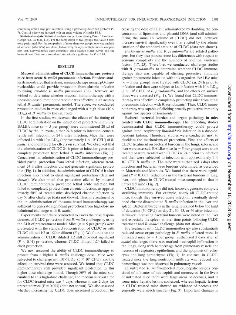
Role of NK cells in CLDC-induced protection from B. malleichallenge. Previous studies have indicated that NK cells werethe major source of IFN-� production following systemic (i.v.)treatment with CLDC (10, 34). However, the role of NK cellsin mediating CLDC-induced pulmonary immunity to bacterialchallenge has not been explored previously. Prior studies haverevealed that multiple cell types, including macrophages,CD8� T cells, and NK cells, produce IFN-� in response toBurkholderia infection (14, 29). To elucidate the role of NKcells in CLDC protection, NK cells were depleted systemicallyusing the asialo GM1 antibody (9, 11, 13, 16). The antibodywas administered 48 h prior to infection, and then the micewere treated 24 h prior to infection by the i.n. administration ofCLDC. Mice then were infected with 10� LD50 of B. malleiand monitored for survival.
There was a significant reduction in survival (P � 0.01) ofCLDC-treated mice depleted of NK cells compared to that ofCLDC-treated mice not depleted of NK cells (Fig. 7). How-ever, CLDC-treated and NK-depleted mice still had a signifi-cant improvement in survival compared to that of untreatedmice (Fig. 7). Thus, these results indicated that NK cells werenecessary for full CLDC-mediated protection. However, par-tial protection still was observed in NK cell-depleted mice,suggesting either that other cell types besides NK cells alsocontributed to the protective effects of CLDC therapy or thatthe residual NK cells not depleted by the administration ofanti-asialo GM1 antibody were sufficient to elicit partial pro-tection.
The role of NK cells in regulating the bacterial burden alsowas assessed. Bacterial burdens in the lung, liver, and spleen ofmice depleted of NK cells and treated with CLDC were de-termined 3 days after infection. Bacterial burdens in NK cell-depleted and CLDC-treated mice were significantly increasedin all three organs compared to those of mice treated withCLDC immunotherapy alone (Fig. 7). In addition, significantreductions in bacterial burdens also were observed in NK cell-depleted and CLDC-treated mice compared to those of un-treated mice. These results are consistent with the idea thatNK cells activated by CLDC immunotherapy contribute toantibacterial activity, most likely by the secretion of IFN-�.
Nitric oxide production is not necessary for CLDC-mediatedprotection. The preceding in vitro and in vivo experimentsdemonstrated that IFN-� was critical to the therapeutic effec-tiveness of CLDC immunotherapy. IFN-� also is known to bea potent inducer of NOS2 (iNOS) activity, with the subsequentproduction of nitric oxide (24). Several in vitro studies utilizingboth B. pseudomallei and B. mallei have demonstrated that thepreactivation of cells with IFN-� results in nitric oxide produc-
FIG. 6. IFN-� is necessary for the in vivo protective of CLDCimmunotherapy. (A) BALB/c wild-type mice (n � 5 per group) orIFN-��/� mice (n � 5) were treated with CLDC 24 h prior to infectionor were left untreated. Mice were infected by low-dose challenge usingthe i.n. administration of 5 � 102 CFU B. mallei, and survival wasassessed. Survival times in the IFN-��/� mice treated with i.n.-admin-istered CLDC were not significantly different in terms of infection thanuntreated IFN-��/� mice. Statistical differences were determined byKaplan-Meier analysis using a log-rank test (**, P � 0.01 compared toresults for the wild type). Similar results were obtained in a secondexperiment done in IFN-��/� mice on a C57BL/6 background.(B) Wild-type BALB/c mice (n � 5) or IFN-��/� mice (n � 5) weretreated with i.n.-administered CLDC 24 h prior to infection or wereleft untreated. Mice were infected with 5 � 102 CFU B. mallei, and onday 2 after infection mice were euthanized and bacterial burdens in thelungs, liver, and spleen were determined, as described in Materials andMethods. The bacterial burdens in all three organs of IFN-��/� micetreated with CLDC were not significantly different from those of un-treated IFN-��/� mice. Similar results were obtained in a secondexperiment done in IFN-��/� mice on a C57BL/6 background. Statis-tical analysis was performed using a one-way ANOVA followed byTukey’s multiple means comparison test (***, P � 0.001 compared toresults for the wild type).
VOL. 77, 2009 IMMUNOTHERAPY FOR PNEUMONIC BURKHOLDERIA INFECTION 1585
on Novem
ber 10, 2018 by guesthttp://iai.asm
.org/D
ownloaded from
tion, which in turn mediates bacterial killing. Other in vitrostudies have shown that Burkholderia species may downmodu-late nitric oxide production in order to enhance its survival (3,15, 23, 35, 36). In contrast to the results obtained in these invitro studies, in vivo B. pseudomallei challenge studies usingNOS2�/� mice have failed to demonstrate a role for nitricoxide in controlling infection (8).
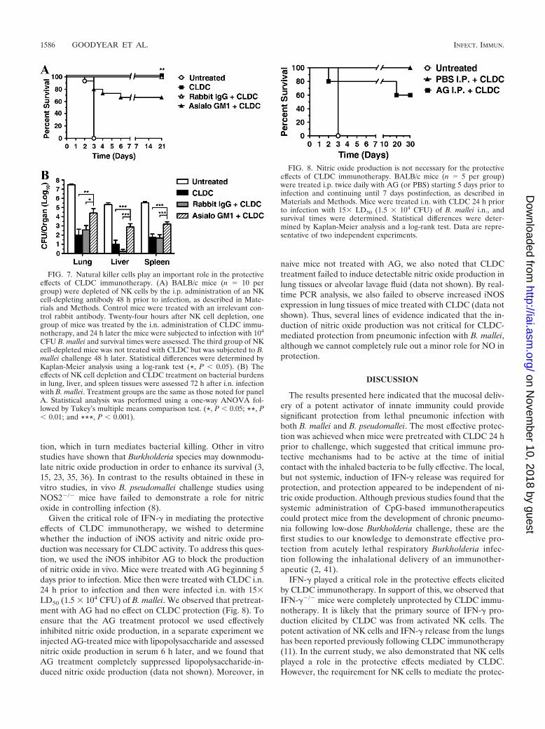
Given the critical role of IFN-� in mediating the protectiveeffects of CLDC immunotherapy, we wished to determinewhether the induction of iNOS activity and nitric oxide pro-duction was necessary for CLDC activity. To address this ques-tion, we used the iNOS inhibitor AG to block the productionof nitric oxide in vivo. Mice were treated with AG beginning 5days prior to infection. Mice then were treated with CLDC i.n.24 h prior to infection and then were infected i.n. with 15�LD50 (1.5 � 104 CFU) of B. mallei. We observed that pretreat-ment with AG had no effect on CLDC protection (Fig. 8). Toensure that the AG treatment protocol we used effectivelyinhibited nitric oxide production, in a separate experiment weinjected AG-treated mice with lipopolysaccharide and assessednitric oxide production in serum 6 h later, and we found thatAG treatment completely suppressed lipopolysaccharide-in-duced nitric oxide production (data not shown). Moreover, in
naive mice not treated with AG, we also noted that CLDCtreatment failed to induce detectable nitric oxide production inlung tissues or alveolar lavage fluid (data not shown). By real-time PCR analysis, we also failed to observe increased iNOSexpression in lung tissues of mice treated with CLDC (data notshown). Thus, several lines of evidence indicated that the in-duction of nitric oxide production was not critical for CLDC-mediated protection from pneumonic infection with B. mallei,although we cannot completely rule out a minor role for NO inprotection.
DISCUSSION
The results presented here indicated that the mucosal deliv-ery of a potent activator of innate immunity could providesignificant protection from lethal pneumonic infection withboth B. mallei and B. pseudomallei. The most effective protec-tion was achieved when mice were pretreated with CLDC 24 hprior to challenge, which suggested that critical immune pro-tective mechanisms had to be active at the time of initialcontact with the inhaled bacteria to be fully effective. The local,but not systemic, induction of IFN-� release was required forprotection, and protection appeared to be independent of ni-tric oxide production. Although previous studies found that thesystemic administration of CpG-based immunotherapeuticscould protect mice from the development of chronic pneumo-nia following low-dose Burkholderia challenge, these are thefirst studies to our knowledge to demonstrate effective pro-tection from acutely lethal respiratory Burkholderia infec-tion following the inhalational delivery of an immunother-apeutic (2, 41).
IFN-� played a critical role in the protective effects elicitedby CLDC immunotherapy. In support of this, we observed thatIFN-��/� mice were completely unprotected by CLDC immu-notherapy. It is likely that the primary source of IFN-� pro-duction elicited by CLDC was from activated NK cells. Thepotent activation of NK cells and IFN-� release from the lungshas been reported previously following CLDC immunotherapy(11). In the current study, we also demonstrated that NK cellsplayed a role in the protective effects mediated by CLDC.However, the requirement for NK cells to mediate the protec-
FIG. 7. Natural killer cells play an important role in the protectiveeffects of CLDC immunotherapy. (A) BALB/c mice (n � 10 pergroup) were depleted of NK cells by the i.p. administration of an NKcell-depleting antibody 48 h prior to infection, as described in Mate-rials and Methods. Control mice were treated with an irrelevant con-trol rabbit antibody. Twenty-four hours after NK cell depletion, onegroup of mice was treated by the i.n. administration of CLDC immu-notherapy, and 24 h later the mice were subjected to infection with 104
CFU B. mallei and survival times were assessed. The third group of NKcell-depleted mice was not treated with CLDC but was subjected to B.mallei challenge 48 h later. Statistical differences were determined byKaplan-Meier analysis using a log-rank test (*, P � 0.05). (B) Theeffects of NK cell depletion and CLDC treatment on bacterial burdensin lung, liver, and spleen tissues were assessed 72 h after i.n. infectionwith B. mallei. Treatment groups are the same as those noted for panelA. Statistical analysis was performed using a one-way ANOVA fol-lowed by Tukey’s multiple means comparison test. (*, P � 0.05; **, P� 0.01; and ***, P � 0.001).
FIG. 8. Nitric oxide production is not necessary for the protectiveeffects of CLDC immunotherapy. BALB/c mice (n � 5 per group)were treated i.p. twice daily with AG (or PBS) starting 5 days prior toinfection and continuing until 7 days postinfection, as described inMaterials and Methods. Mice were treated i.n. with CLDC 24 h priorto infection with 15� LD50 (1.5 � 104 CFU) of B. mallei i.n., andsurvival times were determined. Statistical differences were deter-mined by Kaplan-Meier analysis and a log-rank test. Data are repre-sentative of two independent experiments.
1586 GOODYEAR ET AL. INFECT. IMMUN.
on Novem
ber 10, 2018 by guesthttp://iai.asm
.org/D
ownloaded from

tive effects of CLDC was more apparent at lower challengedoses. For example, NK-depleted, CLDC-treated mice chal-lenged with 8� LD50 (8 � 103 CFU) had a 60 to 80% survivalrate, whereas mice infected with 12� LD50 (1.2 � 104 CFU)has only a 20% survival rate. Nevertheless, both of these chal-lenge doses produced 100% mortality in untreated mice.
Despite a series of in vitro studies that have shown that nitricoxide is important for the bactericidal effects of IFN-� againstboth B. pseudomallei and B. mallei, in our studies we failed tofind a role for nitric oxide in CLDC-mediated protective ac-tivity (3, 15, 23, 35, 36). Our findings thus are in agreementwith a previous in vivo study wherein mice lacking the iNOSgene (NOS2�/�) did not exhibit increased susceptibility to B.pseudomallei infection (8). Thus, we concluded that CLDC-mediated protection from lethal B. mallei pneumonia waslargely independent of nitric oxide production.
Although the mucosal administration of CLDC elicitedcomplete protection against acute B. mallei infection, the in-fection was not completely eliminated. For example, whenmice surviving the acute challenge were monitored out to 60days, we observed the recrudescence of infection in the spleensand livers of 80 to 90% of mice. Thus, it would be important tofollow up CLDC immunotherapy with conventional antimicro-bial therapy to assure the complete eradication of Burkhold-eria. In summary, these studies indicate that appropriatelytimed mucosally administered immunotherapy may induce ef-fective nonspecific protection against pneumonic intracellularbacterial pathogens such as B. mallei and B. pseudomallei.
ACKNOWLEDGMENTS
We acknowledge Abby Jones and Amber Troy for their assistancewith these studies. The assistance of Herbert Schweitzer in providingthe B. mallei and B. pseudomallei strains used in these studies also isacknowledged.
This work was supported by a grant (U54AI1065357) from the NIHand by the Rocky Mountain RCE.
REFERENCES
1. Alibek, K., and S. Handelman. 1999. Biohazzard: the chilling true story ofthe largest covert biological weapons program in the world told from insideby the man who ran it. Random House, New York, NY.
2. Amemiya, K., J. L. Meyers, S. R. Trevino, T. C. Chanh, S. L. Norris, andD. M. Waag. 2006. Interleukin-12 induces a Th1-like response to Burkhold-eria mallei and limited protection in BALB/c mice. Vaccine 24:1413–1420.
3. Arjcharoen, S., C. Wikraiphat, M. Pudla, K. Limposuwan, D. E. Woods, S.Sirisinha, and P. Utaisincharoen. 2007. Fate of a Burkholderia pseudomalleilipopolysaccharide mutant in the mouse macrophage cell line RAW 264.7:possible role for the O-antigenic polysaccharide moiety of lipopolysaccharidein internalization and intracellular survival. Infect. Immun. 75:4298–4304.
4. Barnes, J. L., N. L. Williams, and N. Ketheesan. 2008. Susceptibility toBurkholderia pseudomallei is associated with host immune responses involv-ing tumor necrosis factor receptor-1 (TNFR1) and TNF receptor-2(TNFR2). FEMS Immunol. Med. Microbiol. 52:379–388.
5. Beckerman, K. P., H. W. Rogers, J. A. Corbett, R. D. Schreiber, M. L.McDaniel, and E. R. Unanue. 1993. Release of nitric oxide during the Tcell-independent pathway of macrophage activation. Its role in resistance toListeria monocytogenes. J. Immunol. 150:888–895.
6. Blezinger, P., B. D. Freimark, M. Matar, E. Wilson, A. Singhal, W. Min, J. L.Nordstrom, and F. Pericle. 1999. Intratracheal administration of interleukin12 plasmid-cationic lipid complexes inhibits murine lung metastases. Hum.Gene Ther. 10:723–731.
7. Bowman, M. A., O. G. Simell, A. B. Peck, J. Cornelius, R. Luchetta, Z. Look,N. K. Maclaren, and M. A. Atkinson. 1996. Pharmacokinetics of aminogua-nidine administration and effects on the diabetes frequency in nonobesediabetic mice. J. Pharmacol. Exp. Ther. 279:790–794.
8. Breitbach, K., S. Klocke, T. Tschernig, N. Van Rooijen, U. Baumann, and I.Steinmetz. 2006. Role of inducible nitric oxide synthase and NADPH oxi-dase in early control of Burkholderia pseudomallei infection in mice. Infect.Immun. 74:6300–6309.
9. Byrne, P., P. McGuirk, S. Todryk, and K. H. G. Mills. 2004. Depletion of NKcells results in disseminating lethal infection with Bordetella pertussis associ-ated with a reduction of antigen-specific Th1 and enhancement of Th2, butnot Tr1 cells. Eur. J. Immunol. 34:2579–2588.
10. Dow, S. W., R. E. Elmslie, L. G. Fradkin, D. H. Liggitt, T. D. Heath, A. P.Willson, and T. A. Potter. 1999. Intravenous cytokine gene delivery by lipid-DNA complexes controls the growth of established lung metastases. Hum.Gene Ther. 10:2961–2972.
11. Dow, S. W., L. G. Fradkin, D. H. Liggitt, A. P. Willson, T. D. Heath, and T. A.Potter. 1999. Lipid-DNA complexes induce potent activation of innate im-mune responses and antitumor activity when administered intravenously.J. Immunol. 163:1552–1561.
12. Fritz, D. L., P. Vogel, D. R. Brown, D. DeShazer, and D. M. Waag. 2000.Mouse model of sublethal and lethal intraperitoneal glanders (Burkholderiamallei). Vet. Pathol. 37:626–636.
13. Habu, S., H. Fukui, K. Shimamura, M. Kasai, Y. Nagai, K. Okumura, andN. Tamaoki. 1981. In vivo effects of anti-asialo GM1. I. Reduction of NKactivity and enhancement of transplanted tumor growth in nude mice. J. Im-munol. 127:34–38.
14. Haque, A., A. Easton, D. Smith, A. O’Garra, N. Van Rooijen, G. Lertmemon-gkolchai, R. W. Titball, and G. J. Bancroft. 2006. Role of T cells in innateand adaptive immunity against murine Burkholderia pseudomallei infection.J. Infect. Dis. 193:370–379.
15. Jones-Carson, J., J. Laughlin, M. A. Hamad, A. L. Stewart, M. I. Voskuil,and A. Vazquez-Torres. 2008. Inactivation of [Fe-S] metalloproteins medi-ates nitric oxide-dependent killing of Burkholderia mallei. PLoS ONE3:e1976.
16. Kasai, M., M. Iwamori, Y. Nagai, K. Okumura, and T. Tada. 1980. Aglycolipid on the surface of mouse natural killer cells. Eur. J. Immunol.10:175–180.
17. Kim, H. S., M. A. Schell, Y. Yu, R. L. Ulrich, S. H. Sarria, W. C. Nierman,and D. DeShazer. 2005. Bacterial genome adaptation to niches: divergenceof the potential virulence genes in three Burkholderia species of differentsurvival strategies. BMC Genomics 6:174–187.
18. Koo, G. C., and Y. H. Gan. 2006. The innate interferon gamma response ofBALB/c and C57BL/6 mice to in vitro Burkholderia pseudomallei infection.BMC Immunol. 7:19–31.
19. Lauw, F. N., A. J. Simpson, J. M. Prins, M. D. Smith, M. Kurimoto, S. J. H.van Deventer, P. Speelman, W. Chaowagul, N. J. White, and T. van der Poll.1999. Elevated plasma concentrations of interferon (IFN)-� and the IFN-�-inducing cytokines interleukin (IL)-18, IL-12, and IL-15 in severe melioid-osis. J. Infect. Dis. 180:1878–1885.
20. Lertmemongkolchai, G., G. Cai, C. A. Hunter, and G. J. Bancroft. 2001.Bystander activation of CD8� T cells contributes to the rapid production ofIFN-� in response to bacterial pathogens. J. Immunol. 166:1097–1105.
21. McGilvray, C. D. 1944. The transmission of glanders from horse to man.Can. J. Public Health 35:286–275.
22. Miller, W. R., L. Pannell, L. Cravitz, W. A. Tanner, and T. Rosebury. 1948.Studies on certain biological characteristics of Malleomyces mallei and Mal-leomyces pseudomallei. II. Virulence and infectivity for animals. J. Bacteriol.55:127–135.
23. Miyagi, K., K. Kawakami, and A. Saito. 1997. Role of reactive nitrogen andoxygen intermediates in gamma interferon-stimulated murine macrophagebactericidal activity against Burkholderia pseudomallei. Infect. Immun. 65:4108–4113.
24. Nathan, C. F., T. J. Prendergast, M. E. Wiebe, E. R. Stanley, E. Platzer, H. G.Remold, K. Welte, B. Y. Rubin, and H. W. Murray. 1984. Activation ofhuman macrophages. Comparison of other cytokines with interferon-�. J.Exp. Med. 160:600–605.
25. Ong, C., C. H. Ooi, D. Wang, H. Chong, K. C. Ng, F. Rodrigues, M. A. Lee,and P. Tan. 2004. Patterns of large-scale genomic variation in virulent andavirulent Burkholderia species. Genome Res. 14:2295–2307.
26. Peacock, S. J. 2006. Melioidosis. Curr. Opin. Infect. Dis. 19:421–428.27. Romero, C. M., D. DeShazer, T. Feldblyum, J. Ravel, D. Woods, H. S. Kim,
H. S. Kim, Y. Yu, C. M. Ronning, and W. C. Nierman. 2006. Genomesequence alterations detected upon passage of Burkholderia mallei ATCC23344 in culture and in mammalian hosts. BMC Genomics 7:228.
28. Rotz, L. D., A. S. Khan, S. R. Lillibridge, S. M. Ostroff, and J. M. Hughes.2002. Public health assessment of potential biological terrorism agents.Emerg. Infect. Dis. 8:225–230.
29. Rowland, C. A., G. Lertmemongkolchai, A. Bancroft, A. Haque, M. S. Lever,K. F. Griffin, M. C. Jackson, M. Nelson, A. O’Garra, R. Grencis, G. J.Bancroft, and R. A. Lukaszewski. 2006. Critical role of type 1 cytokines incontrolling initial infection with Burkholderia mallei. Infect. Immun. 74:5333–5340.
30. Santanirand, P., V. S. Harley, D. A. Dance, B. S. Drasar, and G. J. Bancroft.1999. Obligatory role of gamma interferon for host survival in a murinemodel of infection with Burkholderia pseudomallei. Infect. Immun. 67:3593–3600.
31. Srinivasan, A., C. N. Kraus, D. DeShazer, P. M. Becker, J. D. Dick, L.Spacek, J. G. Bartlett, W. R. Byrne, and D. L. Thomas. 2001. Glanders in amilitary research microbiologist. N. Engl. J. Med. 345:256–258.
VOL. 77, 2009 IMMUNOTHERAPY FOR PNEUMONIC BURKHOLDERIA INFECTION 1587
on Novem
ber 10, 2018 by guesthttp://iai.asm
.org/D
ownloaded from

32. Templeton, N. S., D. D. Lasic, P. M. Frederik, H. H. Strey, D. D. Roberts,and G. N. Pavlakis. 1997. Improved DNA: liposome complexes for increasedsystemic delivery and gene expression. Nat. Biotechnol. 15:647–652.
33. Thibault, F. M., E. Hernandez, D. R. Vidal, M. Girardet, and J. D. Cavallo.2004. Antibiotic susceptibility of 65 isolates of Burkholderia pseudomallei andBurkholderia mallei to 35 antimicrobial agents. J. Antimicrob. Chemother.54:1134–1138.
34. U’Ren, L., R. Kedl, and S. Dow. 2006. Vaccination With liposome-DNAcomplexes elicits enhanced antitumor immunity. Cancer Gene Ther. 13:1033–1044.
35. Utaisincharoen, P., S. Arjcharoen, K. Limposuwan, S. Tungpradabkul, andS. Sirisinha. 2006. Burkholderia pseudomallei RpoS regulates multinucleatedgiant cell formation and inducible nitric oxide synthase expression in mousemacrophage cell line (RAW 264.7). Microb. Pathog. 40:184–189.
36. Utaisincharoen, P., N. Tangthawornchaikul, W. Kespichayawattana, P.Chaisuriya, and S. Sirisinha. 2001. Burkholderia pseudomallei interferes with
inducible nitric oxide synthase (iNOS) production: a possible mechanism ofevading macrophage killing. Microbiol. Immunol. 45:307–313.
37. Waag, D. M., D. DeShazer, L. E. Lindler, F. J. Lebeda, G. W. Korch, and M.Meselson. 2005. Glanders, new insights into an old disease, p. 209–238.Biological weapons defense; infectious diseases and counterbioterrorism,vol. 2. Humana Press, Totowa, NJ.
38. Waag, D. M., M. J. McCluskie, N. Zhang, and A. M. Krieg. 2006. A CpGoligonucleotide can protect mice from a low aerosol challenge dose ofBurkholderia mallei. Infect. Immun. 74:1944–1948.
39. Wheelis, M. 1998. First shots fired in biological warfare. Nature 395:213.40. White, N. J. 2003. Melioidosis. Lancet 361:1715–1722.41. Wongratanacheewin, S., W. Kespichayawattana, P. Intachote, S. Pichy-
angkul, R. W. Sermswan, A. M. Krieg, and S. Sirisinha. 2004. Immunos-timulatory CpG oligodeoxynucleotide confers protection in a murine modelof infection with Burkholderia pseudomallei. Infect. Immun. 72:4494–4502.
Editor: R. P. Morrison
1588 GOODYEAR ET AL. INFECT. IMMUN.
on Novem
ber 10, 2018 by guesthttp://iai.asm
.org/D
ownloaded from