protease inhibitors from marine venomous animals and their
TRANSCRIPT

Mar. Drugs 2013, 11, 2069-2112; doi:10.3390/md11062069
Marine Drugs ISSN 1660-3397
www.mdpi.com/journal/marinedrugs
Review
Protease Inhibitors from Marine Venomous Animals and Their
Counterparts in Terrestrial Venomous Animals
Caroline B. F. Mourão and Elisabeth F. Schwartz *
Laboratory of Toxinology, Department of Physiological Sciences, University of Brasília, Brasília,
DF, 70910-900, Brazil; E-Mail: [email protected]
* Author to whom correspondence should be addressed; E-Mail: [email protected];
Tel.: +55-61-31073106; Fax: +55-61-31073107.
Received: 19 April 2013; in revised form: 28 May 2013 / Accepted: 30 May 2013 /
Published: 14 June 2013
Abstract: The Kunitz-type protease inhibitors are the best-characterized family of serine
protease inhibitors, probably due to their abundance in several organisms. These inhibitors
consist of a chain of ~60 amino acid residues stabilized by three disulfide bridges, and was
first observed in the bovine pancreatic trypsin inhibitor (BPTI)-like protease inhibitors,
which strongly inhibit trypsin and chymotrypsin. In this review we present the protease
inhibitors (PIs) described to date from marine venomous animals, such as from sea
anemone extracts and Conus venom, as well as their counterparts in terrestrial venomous
animals, such as snakes, scorpions, spiders, Anurans, and Hymenopterans. More emphasis
was given to the Kunitz-type inhibitors, once they are found in all these organisms. Their
biological sources, specificity against different proteases, and other molecular blanks
(being also K+ channel blockers) are presented, followed by their molecular diversity.
Whereas sea anemone, snakes and other venomous animals present mainly Kunitz-type
inhibitors, PIs from Anurans present the major variety in structure length and number of
Cys residues, with at least six distinguishable classes. A representative alignment of PIs
from these venomous animals shows that, despite eventual differences in Cys assignment,
the key-residues for the protease inhibitory activity in all of them occupy similar positions
in primary sequence. The key-residues for the K+ channel blocking activity was also compared.
Keywords: protease inhibitor; Kunitz-type; serine protease; toxin; trypsin;
chymotrypsin; venom
OPEN ACCESS

Mar. Drugs 2013, 11 2070
1. Introduction
Protease inhibitors (PIs) are proteins or peptides capable of inhibiting the catalytic activity of
proteolytic enzymes. They are widely distributed in nature and can be found in all kingdoms of cellular
life and also in viral genomes [1,2]. A typical mammalian genome contains 2%–4% of genes encoding
for proteases or protease inhibitors, reflecting the importance of proteolysis in their biological
processes [3]. PIs have been known since the end of 20th century, and their identification can become
more effective nowadays because of the current appliance of protease degradomics [4], which in
association with proteomic tools and enzymatic assays can lead to the characterization of innumerous
novel protease inhibitors.
Interest in characterizing new PIs and understanding their physiological significance has increased
due to their biological relevance for all living processes, such as blood coagulation system,
complement cascade, apoptosis, cell cycle and hormone processing pathways [5–8]. Furthermore,
deficiencies or alterations in the regulation of these enzymes underlie several pathological conditions,
such as cancer, arthritis, neurodegenerative and cardiovascular diseases [9,10].
According to the proteases they inhibit, PIs can be grouped primarily as serine, cysteine, aspartic
and metallo protease inhibitors [11]. Among these, serine protease inhibitors are the largest and most
widely distributed superfamily of PIs [1,12,13], and based upon their possession of conserved
functional motifs they can be subdivided in many classes, being the Kunitz-type inhibitors the best
characterized of them, probably due to their abundance in several organisms [2,14–17]. The
Kunitz-type motif consists of a polypeptide chain of ~60 amino acid residues stabilized by three
disulfide bridges (CI–CVI, CII–CIV, CIII–CV). The Kunitz-type inhibitors interact with proteases by the
classical substrate-like mechanism [13] mainly through the P1–S1 interaction, based on the
nomenclature of Schechter and Berger [18]. The standard mechanism implies that substrates/inhibitors
contain the reactive site residues P3, P2, P1, P1′, P2′, P3′, located in the most exposed region of the
protease-binding loop, that bind to the substrate amino acid side chains S3, S2, S1, S1′, S2′, S3′, which
form the groove or cleft where amide bond hydrolysis occurs. Non-prime and prime designations
specify amino- and carboxy-terminal sides of cleavage site, respectively [11,18]. The Kunitz-type
motif was first observed in the bovine pancreatic trypsin inhibitor (BPTI)-like protease inhibitors,
which strongly inhibit serine proteases such as trypsin and chymotrypsin [19–21].
In this review we present the protease inhibitors described to date from marine venomous animals,
most of which have been obtained from sea anemone extracts, but also from Conus species, as well as
their counterparts in terrestrial venomous animals, such as snakes, scorpions, spiders, frogs and toads,
and bees and wasps. Due to the big amount of data, we have given more emphasis on the Kunitz-type
serine protease inhibitors, once they are the most studied compounds among PIs and are found in all
these organisms. Initially, the PIs are presented according to their biological sources, together with
their main characteristics and activities against different proteases. Then, their dual function including
potassium channel blocking activity is discussed, followed by the molecular diversity of protease
inhibitor compounds.

Mar. Drugs 2013, 11 2071
2. Protease Inhibitors from Sea Anemones
The first reports on the existence of protease inhibitors in sea anemones species date from the
70s [22,23]. Currently, protease inhibitor peptides and neurotoxins are isolated from sea anemone
whole bodies, tentacles, secreted mucus and aggressive organs such as acrorhagi, which is present in
some species from Actiniidae family [24].
Several PIs have already been isolated or partially purified and characterized from the sea
anemone species Actinia equina [24–26], Anemonia sulcata [27–30], Radianthus koseirensis [31],
Heteractis crispa (also named Radianthus macrodactylus) [32–34], Rhodactis rhodostoma [31],
Stoichactis sp. [35], Stoichactis helianthus [36–39], Stichodactyla haddoni [40], Anthopleura aff.
xanthogrammica [24,41], Anthopleura elegantissima [42] and Anthopleura fuscoviridis [24] (Table 1).
Most of these characterized PIs are homologous to Kunitz-type inhibitors. However, some of them
belong to different superfamilies.
Table 1. Protease inhibitors from venomous animals. Some protease inhibitors with less
information about sequence or biological activity, as well as some putative protease
inhibitors only found by means of transcriptomic approach but not tested against proteases,
were not included in this table (some of them are found within the text, with the respective
UniProtKB code). Organisms from which the PIs were obtained are indicated by the
symbols at left: # sea anemones; + snakes; § scorpions; ¥ spiders; ¤ Anurans;
ø Hymenopterans. Capital letters denote the proteases inhibited: T, trypsin; C,
chymotrypsin; CL, cathepsin L; CB, cathepsin B; P, papain; K, kallikrein; PK, plasma
kallikrein; TK, tissue kallikrein; Pl, plasmin; E, elastase; nE, neutrophil elastase; pE,
pancreatic elastase; X, factor Xa; XII, α-factor XIIa; SA, subtilisin A; ptK, proteinase K;
Th, thrombin. Structural classes are indicated by symbols: ♦ Kunitz-type motif protease
inhibitors; ∎ Kazal-type protease inhibitors; ● thyroglobulin type-1domain; ◊ Ascaris-type
motif; Bowman-Birk-type motif.
Specie Toxin UniProt KB AA a Protease inhibited Inhibitory activity b–g References
# Actinia equina
♦ AEPI-I - 59 III - - [26]
♦ AEPI-II - 59 III - - [26]
● Equistatin P81439 199 III
Cathepsin L *,
papain,
cathepsin B
0.051 (CL),
0.57 (P),
1.4 (CB) b
[25,43]
♦ AEAPI - 57 III Trypsin *, plasmin 700 IU/mg (T) c [24]
# Anemonia sulcata
■ Elastase inhibitor P16895 48 III Porcine elastase 1.0 d [28,44]
♦ Kalicludin-1 or AsKC1 Q9TWG0 58 III Trypsin <30 d [27]
♦ Kalicludin-2 or AsKC2 Q9TWF9 58 III Trypsin <30 d [27]
♦ Kalicludin-3 or AsKC3 Q9TWF8 59 III Trypsin <30 d [27]
♦ SA5 II P10280 62 III Kallikrein, Trypsin 0.3 (T) d [29,32]
# Anthopleura aff.
xanthogrammica
♦ AXPI-I or AXAPI P81547 58 III Trypsin *, chymotrypsin,
elastase, thermolysin 1900 IU/mg (T) c [24,41]
♦ AXPI-II P81548 58 III Trypsin *, chymotrypsin 490 IU/mg (T) c [41]

Mar. Drugs 2013, 11 2072
Table 1. Cont.
# Anthopleura elegantissima ♦ APEKTx1 P86862 65 III Trypsin 124 d [42]
# Anthopleura fuscoviridis ♦ AFAPI-I - 56 III Trypsin *, plasmin 950 IU/mg (T) d [24]
♦ AFAPI-III - 56 III Trypsin *, plasmin 900 IU/mg (T) d [24]
# Heteractis crispa
♦ Jn-IV P16344 56 III Trypsin 9.6 d [32,33]
♦ InhVJ - 56 III Trypsin *,
chymotrypsin
2.49 (T),
21.7 (C) d [45–47]
♦ APHC1 B2G331 56 III Trypsin,
chymotrypsin
1000 (T),
~5000 (C) d [34]
# Rhodactis rhodostoma ♦ Inhibitor 4 - 48 II
Trypsin,
kallikrein *,
chymotrypsin
0.95 (T),
0.49 (K),
33 (C) d
[31]
# Stichodactyla haddoni ♦ SHTX-3 B1B5I8 62 III Trypsin 203 IU/mg c [40]
# Stoichactis helianthus
♦ ShPI-1 P31713 55 III
Serine *,
cysteine,
aspartic proteases
0.11 (T),
2.3 (C),
2.7 (Pl) d
[36,37]
♦ ShPI-2 P81129 55 III Serine, cysteine,
aspartic proteases n.f. [38]
+ Dendroaspis polylepis
polylepis ♦ Dendrotoxin E P00984 59 III
Trypsin *,
chymotrypsin
1.0 (T),
100 (C) d [48]
+ Daboia siamensis
♦ BBPTI-1 - 60 III Chymotrypsin 4.77 b [49]
♦ CBPTI-1 A8Y7N4 66 III Trypsin 407 b [50]
♦ CBPTI-2 A8Y7N5 60 III Trypsin 666 b [50]
♦ CBPTI-3 A8Y7N6 60 III Chymotrypsin 2.55 b [50]
+ Hemachatus haemachatus ♦ HHV inhibitor II P00985 57 III
Trypsin,
chymotrypsin,
kallikrein, plasmin
- [51]
+ Naja nivea ♦ NNV inhibitor II P00986 57 III Trypsin - [51]
+ Vipera ammodytes
♦ Trypsin inhibitor I P00991 61 III
Trypsin *,
chymotrypsin,
kallikrein, plasmin
0.34 (T),
270 (C) b [52,53]
♦ Trypsin inhibitor II - 62 III
Trypsin *,
chymotrypsin,
kallikrein, plasmin
0.56 (T),
300 (C) b [52]
♦ Chymotrypsin inhibitor P00992 65 III
Chymotrypsin *,
trypsin,
plasma kallikrein
4.3 (C),
5100 (T) b [52]
+ Bungarus fasciatus
♦ BF9 P25660 65 III Chymotrypsin 58 d [54]
♦ Bungaruskunin B2KTG1 59 III Chymotrypsin *,
trypsin, elastase 6100 (C) b [55]
+ Naja naja ♦ Trypsin inhibitor P20229 57 III Trypsin 0.0035 b [56]
+ Naja atra ♦ NACI Q5ZPJ7 57 III Chymotrypsin 25 b [57,58]
+ Ophiophagus hannah
♦ Oh11-1 P82966 58 III Chymotrypsin 3520 b [59]
♦ OH-TCI B6RLX2 58 III Chymotrypsin *,
trypsin
84.6 (C),
391 (T) b [60]
+ Bungarus multicinctus ♦ PILP-1 B4ESA2 58 III Trypsin 55.62 b [61]

Mar. Drugs 2013, 11 2073
Table 1. Cont.
+ Oxyuranus scutellatus
scutellatus ♦ TSPI B7S4N9 62 III
Kallikrein *,
trypsin, plasmin,
elastase, factor Xa,
α-factor XIIa
0.057 (PK),
0.23 (TK),
0.31 (T), 6.1 (Pl),
201 (E), 871 (X),
2380 (XII) b
[62,63]
+ Pseudonaja textilis
textilis
♦ Textilinin-1 Q90WA1 59 III Plasmin *,
trypsin
0.49 ± 0.02 (Pl),
0.76 ± 0.02 (T) b [7,63]
♦ Textilinin-2 Q90WA0 59 III Plasmin 2.2 b [7]
+ Macrovipera lebetina
transmediterranea ♦ PIVL I2G9B4 67 III Trypsin - [64]
+ Pseudechis australis
♦ Pr-mulgin 1 E7FL11 59 III Metalloprotease 2 60 b [65]
♦ Pr-mulgin 2 E7FL12 59 III
Trypsin *,
chymotrypsin,
plasmin
5 (T),
40 (C),
40 (Pl) b
[65]
♦ Pr-mulgin 3 E7FL13 59 III Trypsin,
plasmin
5 (T),
100 (Pl) b [65]
§ Mesobuthus tamulus ♦ Fraction IX-1-a - - Trypsin *,
kallikrein
19.2 IU/mg (T),
87 IU/mg (K) c [66]
§ Hadrurus gertschi ♦ rHg1 P0C8W3 67 III Trypsin 107 d [67,68]
§ Lychas mucronatus
♦ rLmKTT-1a P0DJ46 59 III Trypsin 140 d [16,68]
♦ rLmKTT-1b or SdPI P0DJ45 59 III Trypsin 160 d [16,68]
♦ rLmKTT-1c P0DJ48 59 III Trypsin 124 d [68]
§ Mesobuthus martensii
♦ rBmKTT-1 P0DJ49 59 III Trypsin 136 d [68]
♦ rBmKTT-2 P0DJ50 58 IV Trypsin 420 d [68]
♦ rBmKTT-3 P0DJ47 70 III Trypsin 760 d [68]
§ Scorpiops jendeki ◊ rSjAPI - 64 V Chymotrypsin *,
elastase
97.1 (C),
3700 (E) b [69]
¥ Ornithoctonus huwena ♦ HWTX-XI P68425 55 III Trypsin 0.23 d [14]
¥ Araneus ventricosus
♦ AvKTI K7YYJ2 57 III
Trypsin,
chymotrypsin,
plasmin *,
neutrophil elastase
7.34 (T),
37.75 (C),
4.89 (Pl),
169.07 (E) b
[70]
♦ AvCI L7X735 70 IV
Chymotrypsin,
subtilisin A,
proteinase K,
neutrophil elastase,
pancreatic elastase
49.85 (C),
20.51 (SA),
65.42 (ptK),
8.74 (nE),
11.32 (pE) b
[71]
¤ Bombina bombina ◊ BSTI Q90248 60 V Trypsin,
thrombin
80–100 (T),
1300 (Th) b [72]
¤ Bombina maxima ◊ BMTI Q8QFP3 60 V Trypsin 60 b [73]
¤ Rana areolata ◊ Trypsin inhibitor - 61 V Trypsin ~20,000 e [74]
¤ Bombina orientalis ◊ BOTI Q800F0 60 V - - [75]
¤ Bombina variegata ◊ BVTI Q800E9 60 V - - [75]
¤ Bombina
microdeladigitora
◊ BMSI 1 B1P2F8 60 V Trypsin, thrombin 20 (T), 150 (Th) b [76]
◊ BMSI 2 B1P2F9 60 V - - [76]
Mar. Drugs 2013, 11 2074
Table 1. Cont.
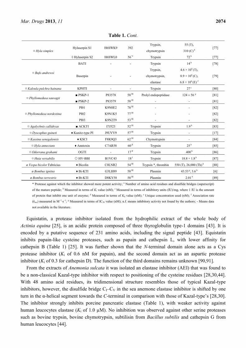
¤ Hyla simplex Hylaserpin S1 H6SWK9 392
Trypsin,
chymotrypsin
55 (T),
310 (C) b [77]
◊ Hylaserpin S2 H6SWL0 56 V Trypsin 72 b [77]
¤ Bufo andrewsi
BATI - - Trypsin 14 b [78]
Baserpin - -
Trypsin,
chymotrypsin,
elastase
4.6 × 10⁶ (T),
8.9 × 10⁶ (C),
6.8 × 10⁶ (E) f
[79]
¤ Kaloula pulchra hainana KPHTI - - Trypsin 27 c [80]
¤ Phyllomedusa sauvagii ■ PSKP-1 P83578 58 III Prolyl endopeptidase 124 ± 56 g [81]
■ PSKP-2 P83579 58 III - - [81]
¤ Phyllomedusa nordestina
PI01 K9N0E2 78 III - - [82]
PI02 K9N1K5 77 III - - [82]
PI03 K9N2T9 53 III - - [82]
¤ Agalychnis callidryas ■ ACKTI I7J523 52 III Trypsin 1.9 b [83]
¤ Dyscophus guineti ♦ Kunitz-type PI J9UVV9 57 III Trypsin - [17]
¤ Kassina senegalensis ♦ KSCI F8K9Q3 62 III Chymotrypsin - [84]
¤ Hyla annectans ♦ Anntoxin C7AR58 60 II Trypsin 25 b [85]
¤ Odorrana grahami OGTI - 17 II Trypsin 400 b [86]
¤ Huia versabilis HV-BBI B1VC43 18 I Trypsin 18.8 + 1.8 b [87]
ø Vespa bicolor Fabricius ♦ Bicolin C0LNR2 54 III Trypsin *, thrombin 550 (T), 26,000 (Th) b [88]
ø Bombus ignitus ♦ Bi-KTI G3LH89 58 III Plasmin 43.53 g, 3.6 b [6]
ø Bombus terrestris ♦ Bt-KTI D8KY58 58 III Plasmin 2.01 b [89]
* Protease against which the inhibitor showed more potent activity; a Number of amino acid residues and disulfide bridges (superscript)
of the mature peptide; b Measured in terms of Ki value (nM); c Measured in terms of inhibitory units (IU)/mg, where 1 IU is the amount
of protein that inhibit one unit of enzyme; d Measured in terms of Kd value (nM); e Unique concentration used (nM); f Association rate
(kass) measured in M−1·s−1; g Measured in terms of IC50 value (nM); n.f. means inhibitory activity not found by the authors; - Means data
not available in the literature.
Equistatin, a protease inhibitor isolated from the hydrophilic extract of the whole body of
Actinia equina [25], is an acidic protein composed of three thyroglobulin type-1 domains [43]. It is
encoded by a putative sequence of 231 amino acids, including the signal peptide [43]. Equistatin
inhibits papain-like cysteine proteases, such as papain and cathepsin L, with lower affinity for
cathepsin B (Table 1) [25]. It was further shown that the N-terminal domain alone acts as a Cys
protease inhibitor (Ki of 0.6 nM for papain), and the second domain act as an aspartic protease
inhibitor (Ki of 0.3 for cathepsin D). The function of the third domains remains unknown [90,91].
From the extracts of Anemonia sulcata it was isolated an elastase inhibitor (AEI) that was found to
be a non-classical Kazal-type inhibitor with respect to positioning of the cysteine residues [28,30,44].
With 48 amino acid residues, its tridimensional structure resembles those of typical Kazal-type
inhibitors, however, the disulfide bridge CI–CV in the sea anemone elastase inhibitor is shifted by one
turn in the α-helical segment towards the C-terminal in comparison with those of Kazal-type’s [28,30].
The inhibitor strongly inhibits porcine pancreatic elastase (Table 1), with weaker activity against
human leucocytes elastase (Ki of 1.0 µM). No inhibition was observed against other serine proteases
such as bovine trypsin, bovine chymotrypsin, subtilisin from Bacillus subtilis and cathepsin G from
human leucocytes [44].

Mar. Drugs 2013, 11 2075
Type II toxins from sea anemone are a peptide group that block Kv1 channel currents—although
with much less potency than the sea anemone type I toxins, which are potent Kv1 channel
blockers [92]—and are characterized by a polypeptide chain of 58–63 amino acid residues and three
disulfide bridges [42,93]. They are homologous to Kunitz-type inhibitors of serine proteases and their
biological role is still unclear. It is supposed that these protease inhibitors could (1) defend sea
anemones from the protease of their victims; (2) protect the toxins injected into preys or predators
from fast degradation; (3) act on the regulation of digestive mechanisms, including self-digestion by
their own enzymes or by those of symbiotic microorganism; (4) and also, due to their dual activity,
they could also be used to paralyze preys [31,35,92].
Among these Kunitz-type inhibitor toxins (KTTs), the sea anemone kalicludines (AsKC1 to AsKC3,
Table 1), from Anemonia sulcata, are examples of toxins with both protease inhibitor and potassium
channel blocking activities (Table 2) [27]. With 58–59 amino acid residues cross-linked by three
disulfide bridges, all the three kalicludines inhibit trypsin with very similar inhibition profiles, in a
1:1 molar ratio, such as BPTI. From titration curves Kd values below 30 nM could be deduced [27].
Another KTT from A. sulcata is the Kunitz-type protease inhibitor 5 II (SA5 II, Table 1), which
inhibits both tissue and plasma kallikreins [29]. Previous to these studies, at least ten KTTs with
inhibitory effects against trypsin, chymotrypsin, plasmin and kallikrein have been reported from
A. sulcata extracts [22,23].
Table 2. Potassium channel blocking activity of Kunitz-type motif protease inhibitor
peptides from animal venoms. Scorpion and spider mutated peptides were not considered.
Source Peptide IC50 (nM) Target Ref.
Sea anemone
APEKTx1 * 0.9 ± 0.1 Kv1.1 [42]
AsKC1 * 2800 Kv1.2 [27]
AsKC2 * 1100 Kv1.2 [27]
AsKC3 * 1300 Kv1.2 [27]
SHTX-3 * 650 Synap a [40]
Cone snail Conk-S1 1.33 ± 0.5 Shaker [94]
Snake
α-DTX 0.4–150 Kv1.1, Kv1.2, Kv1.6 [95]
DTX-I 0.13–50 Kv1.1, Kv1.2, Kv1.6 [95]
DTX-K 0.03 Kv1.1 [96]
δ-DTX 0.029–1.8 Kv1.1 [96,97]
DaE 300 Kv1.1 [98]
Scorpion
Hg1 * 6.2 ± 1.2 Kv1.3 [68]
LmKTT-1a * 1580 Kv1.3 [68,99]
LmKTT-1b * >1000 Kv1.3 [68]
LmKTT-1c * >1000 Kv1.3 [68]
BmKTT-1 * 129.7 ± 31.3 Kv1.3 [68]
BmKTT-2 * 371.3 ± 82.1 Kv1.3 [68]
BmKTT-3 * >1000 Kv1.3 [68]
Spider HWTX-XI * 2570 Kv1.1 [14]
* Peptides that also present protease inhibitory activity, as shown in Table 1; a Activity obtained through
synaptosomal binding assay.

Mar. Drugs 2013, 11 2076
In the 80s, three protease inhibitors composed of 52–56 amino acid residues were isolated from
Stoichactis sp. [35]. Inhibitors 2 and 3 had estimated molecular masses of ~6000 Da. Inhibitor 2,
which was the main component, had its approximate molecular mass (5800 Da) determined by gel
filtration. Inhibitors 2 and 3 lack tryptophan in their structures and contain two and three disulfide
bridges, respectively. They both inhibited trypsin almost completely (98%) and, to a lesser extent, also
chymotrypsin (86% and 92%, respectively). Inhibitor 1, which was still impure, presented a lower
inhibitory activity (80% against trypsin and 50% against chymotrypsin).
In the same decade, protease inhibitors were isolated from the body extracts of Radianthus
koseirensis and Rhodactis rhodostoma [31]. From R. rhodostoma, the main protease inhibitor has
5459 Da and only two disulfide bridges. Named inhibitor 4 (Table 1), it had a high affinity for serine
proteases (trypsin, kallikrein and chymotrypsin), and was less active in terms of the molar ratio
(enzyme:inhibitor). Whereas typical trypsin inhibitors act stoichiometrically 1:1, inhibitor 4 from
R. rhodostoma had a ratio of 1:2 for trypsin and 1:2.4 for chymotrypsin and kallikrein. From
R. koseirensis, an inhibitor fraction was obtained, but it had very low affinity for serine proteases and
did not inhibit chymotrypsin. The authors suggest that the ineffectiveness towards serine proteases of
R. koseirensis fraction may have its origin in the neutral to slightly acidic properties of its inhibitor
molecule. Differently, PIs from R. rhodostoma are basic in nature, such as BPTI and many KTTs [31].
Until now, many PIs were isolated from the body extract of Heteractis crispa (=Radianthus
macrodactylus), but only few of them were fully characterized. Protease inhibitors of about 6000 Da
were obtained from a water-ethanol extract of H. crispa, and the primary structure was elucidated for
one of them, named Kunitz-type trypsin inhibitor IV or Jn-IV (Table 1) [33]. After that, four trypsin
inhibitors were isolated (InI–InIV), and one of them (InI) was partially characterized. Composed by
59 amino acid residues, its molecular mass determined by SDS-PAGE was 7100 Da, whereas its
molecular mass calculated by the amino acid composition was 6304 Da [100]. Posteriorly, two serine
protease inhibitors were isolated [32]. RmIn I and RmIn II have 6322.8 and 6096.0 Da, respectively,
according to their amino acid composition, and both have three disulfide bridges and no methionine or
tryptophan residues [32], which is common in Kunitz-type protease inhibitors [35,51,101], with few
exceptions (Figure 1). The N-terminal amino acid sequences of RmIn I and RmIn II were determined
and they were 66%–86% identical with the analogous fragment of Kunitz-type trypsin inhibitor IV,
isolated from the same source, suggesting that they are isoforms. Both RmIn I and RmIn II were active
against trypsin and α-chymotrypsin, with Ki values of 2.4 and 2.5 nM for trypsin and 23 and 30 nM for
α-chymotrypsin, respectively. Moreover, both polypeptides were not toxic to mice (10–100 µg of
peptide per kg of animal) and exhibited antihistamine activity in a dose-dependent manner [32].
Also from Heteractis crispa extract, a Kunitz-type toxin designated as InhVJ was isolated
(Table 1) [45,46]. It has 6106 Da, three disulfide bridges and also no Met or Trp residues [45,47].
Its putative sequence encodes a 78-amino acid residues polypeptide, including a signal peptide with
22 amino acid residues [47]. InhVJ is highly specific toward trypsin and α-chymotrypsin and did not
inhibit other serine proteases (such as thrombin, kallikrein and plasmin), neither cysteine (papain) nor
aspartic (pepsin) proteases [45,46]. Recently characterized, the analgesic polypeptide HC1
(or APHC1), with 6187.0 Da, presents very weak inhibition against trypsin and chymotrypsin
(Table 1), although it completely inhibits both at a 1:1 molar ratio. Its main activity is related to an
analgesic effect due to modulation of mammalian TRPV1-receptors [34].

Mar. Drugs 2013, 11 2077
The peptides ShPI-1 and ShPI-2 were obtained from the crude extract of Stoichactis helianthus.
With 6110.6 Da and six cysteine residues, ShPI-1 was active against many proteases of different
mechanistic classes [37]. It inhibited serine, cysteine and aspartic proteases, and did not affect the one
metalloenzyme tested (bacterial collagenase). The strongest inhibition capacity was against trypsin
(Ki = 1.1 × 10⁻¹⁰
M). Other serine proteases such as plasmin and chymotrypsin, and cysteine proteases
(bromelain and papain) were inhibited with smaller intensity. The smallest degrees of inhibition were
obtained against the serine protease kallikrein and elastase and against aspartic proteases [37].
Four serine protease inhibitors (AEPI-I to -IV) were isolated from the sea anemone
Actinia equina [26]. Whereas only 35 and 37 N-terminal amino acid residues were determined for
AEPI-III and -IV, the complete primary sequence was determined to AEPI-I and -II, which contain
59 amino acid residues and three disulfide bonds. Both peptides (Table 1) share structural similarity
with Kunitz-type inhibitors, however, their protease inhibitory activity has not been measured yet.
From the whole body extract of Stichodactyla haddoni, four peptide toxins were isolated and named
SHTX-1 to SHTX-4 [40]. SHTX-1–3 induced paralysis in crabs with ED50 values of 430, 430 and
183 µg/kg, respectively, exhibiting no lethality even at 1000 µg/kg. SHTX-4 was lethal to crab with
an estimated LD50 of 93 µg/kg. Among them, SHTX-3 was the only one with an antitryptic activity
(Table 1), as well as potassium channel blocking activity (Table 2). SHTX-3 toxin has 7035.0 Da and
is cross-linked by three disulfide bridges. Its cloned gene encodes a putative sequence of 19 amino
acid residues for the signal peptide and 62 for the mature peptide, without a propart between them.
Because it was previously proposed that the propart with two basic residues at its end functions as a
signal directing toxins into nematocyst [102], it is assumed that SHTX III is not contained in these
stinging cnidarian organelles [40].
AXPI-I and -II (Table 1) were isolated from the whole body extract of Anthopleura aff.
xanthogrammica [41]. Both are basic peptides, with three disulfide bonds, 58 amino acid residues and
belong to the Kunitz-type family. Both inhibited trypsin at a molar ratio of 2:1 (inhibitor: enzyme).
AXPI-I was moderately or weakly inhibitory against the serine proteases α-chymotrypsin and elastase,
and against the metallo-proteases thermolysin and Streptomyces griseus protease. AXPI-II also
presented weak inhibition against α-chymotrypsin. Neither of them inhibited the cysteine proteases
papain nor bromelain [41].
AXAPI, which was another name given to AXPI-I (Table 1), was further isolated from the
acrorhagial extract of this same species [24]. Since much higher protease inhibitory activity was
displayed by the acrorhagi of A. aff. xanthogrammica (59 IU/mL) than the remaining tissues
(2.9 IU/mL), the authors suggested that AXPI-I is derived from the acrorhagi. Competitive inhibition
experiments with 125
I-α-dendrotoxin and rat synaptosomal membranes suggest that this peptide has
no potassium channel toxicity even at 10 µM [24]. In the same study, two Kunitz-type PIs that were
purified from the acrorhagial extracts of Anthopleura fuscoviridis (AFAPI-I and AFAPI-III, Table 1)
and one from Actinia equina (AEAPI, Table 1) were also described [24]. Similar to AXAPI,
they all inhibited both trypsin and plasmin and did not inhibit potassium channels at the same
concentration [24].
The toxin APEKTx1 was isolated from Anthopleura elegantissima and is slightly larger than most
of the KTTs (Table 1) [42]. With 63 amino acid residues, 7468.5 Da and three disulfide bridges, this
toxin inhibits serine proteases and also blocks potassium channels (Table 2), similar to the kalicludines

Mar. Drugs 2013, 11 2078
and SHTX-3 [27,40]. APEKTx1 is presumably a competitive trypsin inhibitor, with a dissociation
constant (Kd) of 124 nM. However, a 3-fold molecular excess of toxin over trypsin was needed to
completely inhibit the protease, which is about 3 times the amount of BPTI or the kalicludines
necessary to reach total trypsin inhibition [42].
3. PIs from Conus Venoms
Conotoxins are small disulfide-rich peptide toxins from cone snails usually with 10–30 amino acid
residues that have evolved from at least 16 genetically distinct superfamilies, many of which are
subject to diverse types of posttranslational modifications, such as C-terminal amidation,
hydroxyprolines, or glycosylation of serine or threonine [103,104]. They can be divided into several
families according to the cysteine bridge pattern and biological activities [104]. In relation to most of
Conus venom toxins, conkunitzin-S1 (Conk-S1, UniProtKB P0C1X2), from the venom of
Conus striatus, is unusually long (60 amino acid residues) and has a C-terminal amidation as the only
posttranslational modification [105]. This potassium channel pore-blocking polypeptide (Table 2) also
belongs to the Kunitz-type inhibitor domain, although it is stabilized by only two of the three disulfide
bridges normally found in KTTs [105].
The cDNA library constructed with the mRNA from Conus californicus venom ducts revealed the
presence of four new putative Kunitz-type inhibitors (Cal9.1a to Cal9.1d, UniProtKB D2Y488,
D2Y489, D2Y490 and D2Y491, respectively) [106]. The mature peptides of these four compounds
have 56 amino acid residues and share high sequence identity. Despite having about the same length of
the Conk-S1, these toxins have six cysteine residues. Their activity as protease inhibitors is still
uncertain [106]. In addition, another putative Kunitz-type inhibitor (conkunitzin-B1, UniProtKB
P0CY85) was obtained from the transcriptome of Conus bullatus venom ducts by means of
next-generation sequencing techniques [107].
4. PIs from Terrestrial Venomous Animals
4.1. PIs from Snake Venoms
Similar to those from marine venomous animals, most of protease inhibitors characterized so far
from snake venoms present the Kunitz-type motif. These snake venom polypeptides have been
demonstrated to specifically inhibit the proteolytic activity of trypsin or chymotrypsin. However, some
snake neurotoxins, such as dendrotoxins, calcicludine and B chain of β-bungarotoxin, possess
a Kunitz/BPTI-like domain although they exhibit little or no protease inhibitory activity.
In the beginning of 70’s, from the venom of the black mamba (Dendroaspis polylepis polylepis), the
first trypsin inhibitor homologs (toxins I and K) were reported [108]. Related toxins containing
57–60 amino acid residues and cross-linked by three disulfide bridges were subsequently characterized
from different mamba venoms and eventually named dendrotoxins (DTXs), such as the α-dendrotoxin
from the green mamba snake (Dendroaspis angusticeps) [95,109]. Dendrotoxins are potent blockers of
certain subtypes of voltage-gated potassium channels (Table 2) [95,110]. Differently from toxins I
and K, dendrotoxin E, from black mamba, was shown to inhibit both trypsin and chymotrypsin
(Table 1) [48].

Mar. Drugs 2013, 11 2079
Also from Dendroaspis angusticeps, calcicludine (CaC, UniProtKB P81658) is a 60-amino acid
residue polypeptide that does not display protease inhibitory activity either K+ or Na
+ channel
modulator activities. CaC specifically blocks most of the high-threshold Ca2+
channels in the
nanomolar range, specially the L-type component of the Ca2+
current [111,112]. β-Bungarotoxin was
previously isolated from Bungarus multicinctus venom and comprises two dissimilar polypeptide
chains, the A chain (~14 kDa) and B chain (~7 kDa), which are linked by an interchain disulfide
bridge [113]. The B chain of β-bungarotoxin is homologous to venom basic protease inhibitors but,
similar to most of dendrotoxins, displays no protease inhibitory activity and also blocks voltage-gated
K+ channels [114].
Despite the dendrotoxin related toxins, several Kunitz-type protease inhibitors have been isolated
from snake venoms, particularly from Elapidae and Viperidae snakes. The Russell’s viper venom
(RVV) inhibitor, from Daboia russelii (=Vipera russelli), seems to be the first of them [101]. It is
a basic polypeptide with deduced 60 amino acid residues and inhibitory activity against plasma and
pancreatic kallikreins, plasmin and trypsin, being more potent against trypsin. A Kunitz-type inhibitor
homolog (RVV inhibitor II, UniProtKB P00990) was further sequenced from the venom of the Eastern
Russel’s viper (=Daboia siamensis) [115]. Recently, similar polypeptides were identified in the venom
of Daboia siamensis [49,50]. From the Burmese specie, BBPTI-1 (Table 1) was shown to strongly
inhibit chymotrypsin activity, with no detectable inhibitory activity against trypsin [49]. From the
Chinese one, two trypsin inhibitors (CBPTI-1 and CBPTI-2, Table 1) and one chymotrypsin inhibitor
(CBPTI-3, Table 1) were purified and cloned [50].
Five PIs were isolated from the venoms of the African Elapids Hemachatus haemachatus and
Naja nivea [51]. The isolated trypsin inhibitors have 52 to 57 amino acid residues, and were all devoid
of tryptophan. Except for one peptide from Naja nivea venom, designated NNV inhibitor la, which had
two disulfide bonds, all the others contained three. Two high homologous peptides (HHV inhibitor II
and NNV inhibitor II, Table 1) were completely sequenced. Similar to HHV inhibitor I, HHV inhibitor
II was active against trypsin, α-chymotrypsin, plasma kallikreins and plasmin from bovine sources.
NNV inhibitor II was only tested against trypsin.
From the venom of the long-nosed viper Vipera ammodytes, three basic PIs were isolated [52].
Two of them strongly inhibit trypsin, whereas the third one primarily inhibits chymotrypsin (Table 1).
Other enzymes were also inhibited, although with lower potency. The precursors of the chymotrypsin
inhibitor and trypsin inhibitor I were further obtained [116]. An homologous putative trypsin inhibitor
was also sequenced from the venom of the leaf-nosed viper, Eristicophis macmahoni (UniProtKB
P24541) [117].
Until now, two PIs have been isolated from Bungarus fasciatus venom [54,55]. The first of them
was denoted protease inhibitor IX (or BF9, Table 1) and consists of a polypeptide chain of
65 amino acid residues that, when cleaved, generates the protease inhibitor VIIIb [54]. BF9
specifically inhibits α-chymotrypsin in a 1:1 molar ratio and is structurally similar to the Kunitz-type
toxins [54,118]. The second is a Kunitz-type serine protease inhibitor named bungaruskunin (Table 1),
which was characterized from venom and also cloned from venom glands [55]. The predicted
precursor contains 83 amino acid residues, including a 24-residues long signal peptide. The mature
bungaruskunin inhibits trypsin, chymotrypsin, and elastase, but not thrombin.

Mar. Drugs 2013, 11 2080
From the venom of the cobra Naja naja, a strong trypsin inhibitor (Table 1) [56] and a putative
57-amino acid residue chymotrypsin inhibitor (UniProtKB P19859) were characterized [119]. With
almost 90% identity to the latter, a chymotrypsin inhibitor was also characterized from the venom of
Naja atra (NACI, Table 1) [57]. No detectable inhibitory effects were observed on trypsin or plasmin.
Its precursor was further characterized and it was shown that the expressed protein without the first
three residues at its N-terminal led to a moderate increase in the inhibitory constant against
chymotrypsin [58].
A weak chymotrypsin inhibitor with no inhibitory action against trypsin was characterized from the
venom of the king cobra Ophiophagus hannah (Oh11-1, Table 1) [59]. After that, a peptide named
OH-TCI was shown to inhibit both chymotrypsin and trypsin with similar potencies [60]. The
inhibition constants of the native peptide for chymotrypsin and trypsin were both around 170 nM,
while the recombinant peptide presented no more than 5-fold difference between both values (Table 1).
No detectable effects were observed against thrombin and subtilisin [60].
The structural organization of the genes encoding three protease inhibitor-like proteins (PILP-1 to
PILP-3) from Bungarus multicinctus was reported [61]. Recombinant PILP-1 inhibited trypsin
(Table 1), whereas PILP-2 (UniProtKB B4ESA3) and PILP-3 (UniProtKB B4ESA4) did not inhibit
chymotrypsin or trypsin. PILP genes share identical organization with B chain genes, with three exons
interrupted by two introns in similar positions. Comparative analysis of PILP and B chain genes,
together with the high degree of sequence similarity of the polypeptides, suggested that these genes
have originated from a common ancestor and that they should be evolved by gene duplication followed
by divergence [61].
Two distinct types of KTTs have already been characterized from the venom of Australian elapid
snakes. Taicatoxin serine protease inhibitor (TSPI, Table 1) was purified from the venom of
Oxyuranus scutellatus scutellatus as part of a multimeric complex that blocks high threshold calcium
channels and small conductance Ca2+
-activated K+ channels [62,120]. In its isolated form, TSPI
exhibits antifibrinolytic activity and is a broad spectrum inhibitor, being active against plasma and
tissue kallikrein, trypsin, elastase, factor Xa, and α-factor XIIa [62,63]. Subsequently, a second family
of Kunitz-type inhibitors was purified, cloned and characterized from the venom of the common
brown snake Pseudonaja textilis [7,121]. Two of them, textilinin-1 and -2 (Table 1), were shown to be
potent plasmin inhibitors [7,63]. Textilinin-1 is also a potent trypsin inhibitor, with little effect on both
plasma and tissue kallikrein, and no effect on many other proteases tested [63,122]. Because textilin-1
has been shown to inhibit plasmin and reduce bleeding in a small animal model [7], further studies
have suggested its use as a therapeutic alternative for aprotinin [122,123], which is widely used in
surgery as an anti-bleeding agent but is associated with numerous side effects [124].
From the New Guinean Pseudechis australis (=Pseudechis rossignolii), three cDNAs encoding
Kunitz-type protease inhibitors were isolated and named Pr-mulgins 1, 2 and 3 [65]. The putative
polypeptides are 92.4%–99.3% identical with their orthologs in the Australian Pseudechis australis [125].
Recombinant Pr-mulgin 1 significantly affected matrix metalloprotease (MMP) 2; Pr-mulgin 2 and 3
strongly inhibited trypsin and plasma plasmin; and Pr-mulgin 2 also inhibited α-chymotrypsin (Table 1).
No inhibitory effects were observed against other proteases such as serine proteases (elastase,
kallikrein and pepsin), cysteine protease (cathepsin G), and MMPs (1,3,7,8,9,10,12,13 and 14). These
peptides did not block Drosophila K+ channels (Shaker) and rat K
+ channels (Kv1.1).
Mar. Drugs 2013, 11 2081
Recently, a trypsin inhibitor named PIVL (Table 1) was characterized from the venom of the
Tunisian snake Macrovipera lebetina transmediterranea [64]. Besides trypsin inhibitory activity,
PIVL also exhibits anti-tumor effect and displays integrin inhibitory activity without being cytotoxic.
No inhibitory effect was detected on chymotrypsin.
Transcriptomic approaches are also showing numerous serine PIs in snake venoms. Transcriptome
of the venom glands of the Australian elapid snake Drysdalia coronoides revealed that serine protease
inhibitors are one of the major components from this venom [126]. The presence of putative
Kunitz-type inhibitors was also shown in the venom gland transcriptomes of the elapid snakes
Bungarus multicinctus and Naja atra [127], of the viper snake Vipera nikolskii [128], of the Brazilian
coral snake Micrurus altirostris [129], and from several Australian elapid snakes [125].
Other classes of PIs in snake venoms comprise the cystatins and the bradykinin-potentiating
peptides (BPPs). The cystatins are a family of cysteine protease inhibitors with homology to chicken
cystatin/ovocystatin and human cystatin C [130]. To date, few snake venom cystatins have been fully
characterized, and some of them were shown to inhibit papain-like cysteine proteases [131–134]. BPPs
consist of about 5–14 amino acid residues that specifically block the endothelial metalloprotease
angiotensin I-converting enzyme (ACE). The first report of a “bradykinin-potentiating factor (BPF)”
in the venom of Bothrops jararaca was in 1965 [135]. These factors were subsequently purified and
characterized as peptides responsible for potentiating the activity of bradykinin and inhibition of
ACE [136,137]. Among a series of analogs of BPPs studied, one of them was modified to produce
captopril, an orally available peptidomimetic that, together with further modified molecules, become
widely used for the treatment of hypertension [138]. Although not presented in detail in this review,
other BPPs have been identified in snake venoms or venoms glands [139–143].
4.2. PIs from Scorpion Venoms
The first report on a protease inhibitor from scorpion venom was made in 1981 by Chhatwal and
Habermann [66]. The peptide was purified from the venom of the Indian red scorpion
Mesobuthus tamulus and consisted of 77 amino acid residues and 8500 Da (Table 1). It inhibited
trypsin and also kallikrein, but had weak or no activity against chymotrypsin, plasmin and
thrombin [66]. Its sequence, however, was not revealed.
Posteriorly, Schwartz and collaborators [67] reported for the first time, by means of transcriptomic
analysis, the presence of a Kunitz type precursor in scorpions. The mature peptide, named Hg1, from
the Mexican scorpion Hadrurus gertschi, was not found in the venom and its activity remained
unknown, until it was recently expressed by Escherichia coli BL21(DE3) cells, purified and tested [68].
Together with Hg1, other six recombinant scorpion peptides (LmKTT-1a, LmKTT-1b, LmKTT-1c,
BmKTT-1, BmKTT-2 and BmKTT-3) were also characterized as trypsin inhibitors (Table 1), with no
activity against chymotrypsin or elastase even at high doses [68]. Among them, recombinant Hg1
presented the lowest dissociation constant against trypsin, while rBmKTT-3, from Mesobuthus
martensii, had the highest [68]. LmKTT-1b, also named SdPI, from Lychas mucronatus, was the first
functionally characterized Kunitz-type peptide from scorpion [16]. All these seven recombinant
peptides seem to have similar secondary structure to BPTI, as analyzed by circular dichroism
spectroscopy [68]. Moreover, some of them are also blockers of potassium channels (Table 2) [68].

Mar. Drugs 2013, 11 2082
From the cDNA libraries constructed from the venom glands of three scorpion species,
four putative serine protease inhibitors belonging to the Ascaris-type peptides were recently identified:
SjAPI (Scorpiops jendeki Ascaris-type protease inhibitor), SjAPI-2 (Scorpiops jendeki Ascaris-type
protease inhibitor 2), CtAPI (Chaerilus tricostatus Ascaris-type protease inhibitor), and BmAPI
(Buthus martensii Ascaris-type protease inhibitor) [69]. Members of the Ascaris-type peptides’ family
are characterized by the presence of five disulfide bonds [144]. The recombinant SjAPI (Table 1)
inhibited both α-chymotrypsin and elastase, with no detectable effect on trypsin. SjAPI is produced as
a precursor polypeptide of 94 amino acid residues, which includes a signal peptide of 24 residues,
a propeptide of 6 residues, and a 64-residue mature peptide [69].
Besides these serine protease inhibitors, a plasmin inhibitor named discreplasminin was isolated
from Tityus discrepans scorpion venom. It has an antifibrinolytic mechanism similar to aprotinin and
probably interacts with the active sites of plasmin and tPA, a tissue-type plasminogen activator [145].
Moreover, a putative serpin peptidase inhibitor-like protein (UniProtKB C5J8C1) was identified in the
cDNA venom gland library of Opisthacanthus cayaporum. However, it is composed by 176 encoded
amino acid residues and lacks the signal peptide region [146].
4.3. PIs from Spider Venoms
Yuan and collaborators reported the first superfamily of Kunitz-type toxins from spiders [14]. It is
composed by HWTX-XI, the prototype member of this superfamily, and 45 additional putative KTTs,
which were found by means of two venom gland cDNA libraries constructions of the two Chinese bird
spiders Ornithoctonus huwena and O. haiana (34 and 11 putative KTTs, respectively). HWTX-XI
(Table 1) has 6166.23 Da and six cysteine residues. It was isolated from both venom and venom gland
extract of O. huwena. HWTX-XI inhibits trypsin stoichiometrically in a 1:1 ratio and strongly binds to
trypsin, while presenting lower affinity to α-chymotrypsin. It had about 30-fold higher inhibition
potential to trypsin compared with BPTI, with a Kd value of 2.3 × 10⁻¹⁰
M, while BPTI had a Kd of
6.57 × 10⁻⁹
M under the same experimental conditions [14]. Similar to many KTTs, HWTX-XI also
inhibits potassium channel currents (Table 2).
After that, a putative serine protease inhibitor was revealed from the cDNA library of
Loxosceles intermedia venom gland [147]. This single EST (cluster LIS209) presented high similarity
with serine protease inhibitors from mammals, such as Mus musculus (UniProtKB O35684) and
Pan troglodytes, and also from Ambliomma americanum tick.
Recently, the first spider Kunitz-type inhibitor with plasmin and elastase inhibitory activity
(AvKTI, Table 1) was identified in Araneus ventricosus [70]. However, its coding clone was selected
from the expressed sequence tags (ESTs) that were generated from a cDNA library constructed using
A. ventricosus whole bodies. Because AvKTI is expressed only in the epidermis, it is suggested that it
is a Kunitz-type inhibitor derived from the spider body, but not from venom [70]. Recombinant AvKTI
inhibited both trypsin and chymotrypsin, with no detectable inhibitory effects on factor Xa, thrombin
or tissue plasminogen activator (tPA). However, it inhibited plasmin and neutrophil elastase [70].
Thus, among the proteases tested, the most prominent inhibitory effect of AvKTI was observed on
plasmin, resembling other PIs with antifibrinolytic activity, such as scorpion discreplasminin [145] and
snake textilinins [7,122]. In addition, another Kunitz-type inhibitor (AvCI, Table 1) was obtained from

Mar. Drugs 2013, 11 2083
A. ventricosus in a similar manner to AvKTI [71]. AvCI inhibits chymotrypsin, the microbial serine
proteases subtilisin A and proteinase K, and also the human neutrophil elastase and porcine pancreatic
elastase. It was ineffective against trypsin, factor Xa, thrombin, tPA and plasmin [71].
4.4. PIs from the Skin Secretion of Anurans
The skin granular glands of Anurans (frogs and toads) produce a remarkably diverse range of
bioactive polypeptides [17]. Among them, almost all the serine protease inhibitors from amphibians
contain 3–5 conserved disulfide bridges, with few exceptions [85–87], and their biological role is still
not clear [87]. Unless otherwise stated, all amphibian PIs described in this section were obtained from
their skin secretions.
From the toad Bombina bombina, a 60-residue polypeptide cross-linked by five disulfide bridges
was isolated and cloned [72]. Named BSTI, it inhibits both porcine trypsin and human thrombin
(Table 1), with no activity against chymotrypsin. Its precursor contains 84 amino acid residues,
including a 24-residue signal peptide [72]. Structurally homologous to BSTI, a trypsin inhibitor named
BMTI (Table 1) was characterized from the toad Bombina maxima [73]. Its precursor also contains
84 amino acid residues, with a putative signal peptide of 24 residues. Unlike BSTI, BMTI had
no inhibitory effect on thrombin. Chymotrypsin, elastase and bacterial subtilisin were also not
inhibited at up to 10 µM concentration. Also with five disulfide bridges, a weak trypsin inhibitor was
isolated from the frog Rana areolata (Table 1) [74].
In addition, two trypsin inhibitor analogs (BOTI and BVTI) were isolated from the toads
Bombina orientalis and Bombina variegata (Table 1) [75]. By means of a new non-invasive technique,
two cDNA libraries were constructed from the dermal venom of these two species and both precursors
were obtained, which contain 84 amino acid residues, including a 24-residue signal peptide. The
trypsin inhibitory activity of BOTI and BVTI was not measured [75].
From the toad Bombina microdeladigitora, two serine protease inhibitors structurally homologous
to BSTI were identified (BMSI 1 and BMSI 2, Table 1) [76]. BMSI 1 inhibits both trypsin and
thrombin. No inhibition of the hydrolysis of substrates by elastase, chymotrypsin and subtilisin were
observed with an inhibitor concentration up to 10 µM.
From Hyla simplex, two serine protease inhibitors were isolated (Table 1)—an alpha-1-antitrypsin-like
serpin (hylaserpin-S1) and a wasp venom-like toxin (hylaserpin-S2) [77]. Hylaserpin-S2 precursor is
composed by 83 amino acid residues, including a predicted signal peptide of 27 residues. It is
structurally related to those amphibian serine protease inhibitors containing five disulfide bridges from
Bombina and Rana genera [72–75]. Hylaserpin-S1 is a much longer peptide, with 44 kDa, and is
encoded by a 415-amino acid residues precursor, with a signal peptide of 23 amino acid residues.
Hylaserpin-S1 inhibited both trypsin and chymotrypsin, whereas hylaserpin-S2 only inhibited trypsin,
similarly to BSTI [72,77]. Moreover, hylaserpin-S2 had bacteriostatic effect against Gram-positive
bacteria Bacillus subtilis, and hylaserpin-S1 displayed direct microorganism-killing abilities against
B. subtilis, E. coli, and Candida albicans [77].
Another long protease inhibitor, consisting of a basic single chain glycoprotein with about 22 kDa,
was isolated from the toad Bufo andrewsi (BATI, Table 1). BATI inhibits trypsin, but displays
no inhibitory activity against chymotrypsin, thrombin and elastase [78]. Also from Bufo andrewsi,

Mar. Drugs 2013, 11 2084
an irreversible serine protease inhibitor (baserpin, Table 1) also consisting of a single chain
glycoprotein with ~60 kDa was purified. Baserpin, which possibly belongs to the serpin superfamily,
inhibits the catalytic activity of trypsin, chymotrypsin and elastase, with no detectable inhibitory
effects on thrombin [79]. Similar to BATI, the peptide KPHTI (Table 1), from the frog
Kaloula pulchra hainana, is a single chain glycoprotein with about 23 kDa, and also inhibits trypsin,
but not chymotrypsin, thrombin, elastase or subtilisin [80]. Only few N-terminal residues from BATI,
baserpin and KPHTI were sequenced [78–80].
An albumin-like protein with approximately 60 kDa (albumin-1, UniProtKB Q3T479) was isolated
and cloned from Bombina maxima skin [148]. Purified from skin homogenate, this protein inhibits
trypsin (Kd of 1.92 nM), with no inhibition against thrombin, chymotrypsin, elastase or subtilisin.
However, this protein, which shares similar biochemical and immunochemical properties as those of
the B. maxima serum albumin, was not detected in the skin secretions [148].
Two peptides analogous to the Kazal family of serine protease inhibitors were obtained from the
skin extract of Phyllomedusa sauvagii (PSKP-1 and PSKP-2, Table 1) [81]. Recombinant PSKP-1
inhibited a serum prolyl endopeptidase from blood serum, but was not active against trypsin, chymotrypsin,
V8 protease or proteinase K. In addition, PSKP-1 displays bactericidal activity and induces agglutination
of red cells and bacteria [81]. Similar to these peptides, two putative protease inhibitors (PI01 and
PI02) were obtained from the cDNA library of Phyllomedusa nordestina (Table 1). The third of them
(PI03) is shorter and structurally different [82]. A different Kazal-type protein with potent trypsin
inhibitory activity (ACKTI, Table 1) was characterized from the frog Agalychnis callidryas [83]. Its
precursor contains 78 amino acid residues, including a signal peptide with 26 residues.
A Kunitz-type protease inhibitor (Table 1) was isolated from the skin secretion of the tomato frog
Dyscophus guineti [17]. It contains 57 amino acid residues, including six cysteine residues, and
inhibits trypsin. In addition, a chymotrypsin inhibitor of Kunitz-type (KSCI, Table 1) was characterized
from Kassina senegalensis [84]. Its precursor sequence contains 84 amino acid residues, including a
22-residue putative signal peptide.
From the frog Hyla annectans, a different Kunitz-type inhibitor peptide (anntoxin, Table 1)
with additional (although weak) sodium channel activity was characterized [85]. It inhibits
tetrodotoxin-sensitive (TTX-S) sodium channel currents elicited from adult rat dorsal root ganglion
neurons with an IC50 value of 3.4 µM, and is also lethal to different potential predators, like insects,
snake, birds, and mice. Calcium and potassium currents were few or not inhibited even at
concentrations up to 10 µM. Anntoxin inhibits trypsin and is homologous to KTTs, but contains only
two of the three highly conserved disulfide bridges [85].
The first small serine protease inhibitor known to contain only a single disulfide bond characterized
from animals was isolated and cloned from the skin secretion of the frog Odorrana grahami [86]. Five
different precursors, although highly conserved, encode OGTI. All of them contain 70 amino acid
residues including a signal peptide of 22 residues, an acidic spacer peptide with 31 residues and equal
mature peptides with 17 residues. OGTI toxin inhibited trypsin (Table 1), but had no inhibitory effects
on thrombin, chymotrypsin, elastase, subtilisin, plasmin or furin at concentrations up to 10 µM [86].
Similar precursors encoding mature peptides equal to OGTI seem to have been found in the skin
secretions of Odorrana andersonii (UniProtKB E3SZL1), Odorrana margaretae (UniProtKB
E1AWD0) and Rana schmackeri (UniProtKB D5LXG2).

Mar. Drugs 2013, 11 2085
Another small peptide with only one disulfide bond (HV-BBI, Table 1) was characterized from the
frog Huia versabilis (=Odorrana versabilis) [87]. This Bowman-Birk type protease inhibitor is
encoded by a precursor of 62 amino acid residues, including a 22-residue signal peptide, a 22-residue
propeptide, and a mature peptide consisting of 18 amino acid residues and a C-terminal amidation.
The synthetic peptide was found to behave as a competitive reversible inhibitor of trypsin.
No chymotrypsin inhibitory activity was observed [87].
4.5. PIs from Hymenopterans’ Venoms
There are few reports on serine protease inhibitors from the venom of Hymenopterans, such as
wasps and bees. Moreover, some of them are putative PIs, obtained by means of transcriptomic tools,
and have not been tested yet.
From the venom of the solitary spider wasp Anoplius samariensis, a peptide named As-fr-19
(UniProtKB Q589G4) was purified and cloned [149]. Its precursor encodes a peptide with 75 amino
acid residues, containing a signal peptide of 17 residues and a mature toxin of 58 residues. As-fr-19
contains six cysteine residues and presents sequence similarity to some sea anemone and snake toxins,
such as the kalicludines from Anemonia sulcata [27] and snake dendrotoxin I and K and
calciludine [108,111]. Thus, As-fr-19 may exert inhibitory effects on serine proteases and on
potassium and/or calcium channels [149].
Using random sequencing analysis, novel putative toxins from the venom of the parasitoid wasp
Pimpla hypochondriaca have been sequenced [150]. Among the cysteine-rich venom proteins, four of
them (cvp1, cvp2, cvp4 and cvp6) presented sequence similarity with protease inhibitors. Both cvp1
and cvp6 contain 10 cysteine residues and are similar to the chymotrypsin inhibitor AMCI 1 from the
larval hemolymph of Apis mellifera (UniProtKB P56682). Mature cvp2 shares high sequence
similarity to Kunitz-type serine protease inhibitors, and cvp4 consists of a three times repeated
six cysteine motif and is similar to pacifastin, a protease inhibitor from locust (UniProtKB Q8WQ22).
A serine protease inhibitor polypeptide named bicolin (Table 1) was characterized from the venom
of the wasp Vespa bicolor Fabricius [88]. Its precursor is composed of 77 amino acid residues
comprising a 23-residue predicted signal peptide and the 54-residue mature toxin cross-linked by three
disulfide bonds. Bicolin is homologous to As-fr-19 and cvp2 and showed inhibitory activity against
trypsin and thrombin, but had no effect on elastase and chymotrypsin. Bicolin was found to own
anticoagulation function, possibly due to its thrombin-inhibitory activity [88].
From Bombus ignitus bumblebee venom, a Kunitz-type serine protease inhibitor (Bi-KTI, Table 1)
with plasmin inhibitory activity was characterized [6]. Its precursor consists of 82 amino acid residues,
with a predicted 24-residue signal peptide and a 58-residue mature peptide, including six cysteine
residues. It strongly inhibits plasmin, although its inhibitory activity was approximately two-fold
weaker than that of aprotinin. Bi-KTI did not inhibit other enzymes from the hemostatic system, such
as factor Xa, thrombin, or tPA. These results are similar to those of snake textilinins [7,122]. Bi-KTI,
which acts as an antifibrinolytic agent, and Bi-VSP, a Bombus ignitus venom serine protease
previously characterized that acts as a fibrin(ogen)olytic agent [151], may act in a cooperative fashion
to promote the spread of bee venom under anti-bleeding conditions [6].

Mar. Drugs 2013, 11 2086
Similar results were obtained with the Kunitz-type inhibitor Bt-KTI (Table 1), from
Bombus terrestris bumblebee venom [89]. Bt-KTI shares high sequence similarity to Bi-KTI and also
consists of a 58-amino acid residue mature peptide, including six conserved cysteine residues.
Moreover, it displays strong inhibitory activity against plasmin, exhibiting an antifibrinolytic activity,
and does not inhibit factor Xa, thrombin or tPA [89].
From a cDNA library constructed using Asiatic honeybee (Apis cerana) whole bodies, a putative
chymotrypsin inhibitor (AcCI) with ten conserved disulfide bridges was identified [152]. Based on the
RT-PCR done with samples of epidermis, fat body, midgut, and venom gland, the AcCI gene was
found to be constitutively expressed in all of these tissues. The recombinant AcCI (rAcCI), obtained
by a baculovirus/insect cell expression system, inhibited the activity of chymotrypsin (IC50: 24.71 nM),
with an inhibitory constant (Ki) of 11.27 nM, and of human neutrophil (IC50: 38.50 nM) and porcine
pancreatic (IC50: 70.21 nM) elastases, with Ki values of 61.05 nM and 101.89 nM, respectively. rAcCI
had no inhibitory activity against trypsin, factor Xa, thrombin, tPA, or plasmin. It is worthy to mention
that although AcCI precursor codes to a 65-amino acid residue mature peptide, with a predicted
molecular mass of 7.2 kDa, rAcCI was identified as a 16-kDa protein, which was interpreted by
authors as due to the presence of carbohydrate moieties [152].
5. PIs and Potassium Channel Activity
It is suggested that the evolution process of Kunitz-type toxins has probably three stages: old
functional molecules, bi-functional toxins and new function toxins [14]. Thus, some Kunitz-type
protease inhibitors could acquire the neurotoxin function, whereas others would even lose their
protease inhibitory role and act on voltage-gated ion channels [14,116]. Potent and specific neurotoxic
K+ channel blockers with Kunitz-type motif are particularly developed in snakes, while sea anemone,
scorpion and spider toxins have developed only the dual-functional KTTs, frequently with weak K+
channel blocking activity. One possible explanation is that the selective pressure would have act in
order to keep the dual-functional KTTs with weak K+ channel blocking activity in these latter animals,
which already have other potent neurotoxins in their venoms/body extracts. By contrast, only K+
channel blockers presenting the Kunitz-type motif were characterized from snake venoms so far [14].
Typical examples of this are the snake dendrotoxins. Although being similar to Kunitz-type
protease inhibitors in amino acid sequences and three-dimensional conformation [153], dendrotoxins
display few or no serine protease inhibitory activity. By contrast, they are strong blockers of potassium
channels (Table 2) [95,110]. Both dendrotoxin (or α-dendrotoxin, UniProtKB P00980) from green
mamba Dendroaspis angusticeps and toxin I (UniProtKB P00979), from the black mamba
Dendroaspis polylepis, block cloned Kv1.1, Kv1.2 and Kv1.6 channels in the low nanomolar range
(Kd < 20 nM) [95,154]. Toxin K (UniProtKB P00981), also from the black mamba D. polylepis,
preferentially blocks Kv1.1 channels and is active at picomolar concentrations [96,155]. Other
subtypes of potassium channels are also blocked by some dendrotoxins, although with lower
affinity [95]. The protein E homologues from green mamba venom are also able to block Kv1.1
channels expressed in oocytes, although higher concentrations are needed (Table 2) [98]. Named
“DaE1” and “DaE2”, these polypeptides share 98% and 95% identity, respectively, to trypsin inhibitor
E from black mamba venom [156].

Mar. Drugs 2013, 11 2087
By means of chemical modifications of native dendrotoxins and genetic engineering to produce
mutated toxins, the amino acid residues that are essential for their interaction with potassium channels
have been identified [95,110]. Lys5 seems to be the major determinant of the binding affinity of
α-DTX and DTX-I for K+ channels (Figure 1) [157,158]. Along with it, the neighbor hydrophobic
residue Leu9 is also important for binding to K+ channels [159]. Lys19, which is around the
“anti-protease” site, caused only a little loss of activity when acetylated in DTX-I [158] or replaced by
Ala in α-DTX [159].
Besides Lys5, or the equivalent Lys3 in DTX-K, the amino acids at the β-turn region
(residues 24–28 in toxin K, in particular Trp25 and Lys26) are responsible for the potassium channel
activity [160,161]. In DTX-I, acetylation of Lys29 inactivated the toxin, possibly by producing large
structural perturbations [158]. Moreover, although acetylation of Lys28 alone had little effect, the
toxin became almost inactive when both Lys28 and Tyr24 were modified [158]. Differently, in α-DTX,
modification of the Lys triplet 28–30 to Ala-Ala-Gly exhibited only small decreases in biological
activity [162]. Seven amino acid residues (Lys3, Tyr4, Lys6, Leu7, Pro8, Arg10 and Lys26) from
δ-dendrotoxin were shown to be important for the toxin’s interaction with a Shaker channel
variant [163].
The heterodimeric snake neurotoxin β-bungarotoxin inhibits the release of acetylcholine from motor
nerve endings [164]. It consists of a PLA2 subunit and a K+ channel binding subunit, which is
a member of the Kunitz-type protease-inhibitor superfamily, both linked by a disulfide bridge [165].
β-Bungarotoxin acts on a presynaptic potassium channel and then, with the phospholipase A2 unit
activity, blocks neurotransmission, altering the acetylcholine release [166]. β-Bungarotoxin (2 µM/L)
partially blocked fast K+ outward current (IK.F) through the fast K
+ channels and also Ca
2+-dependent
K+ current (IK(Ca)) through Ca
2+-activated K
+ channel in motor nerve terminals of snake [167]. The
mutated B(C55S)-bungarotoxin chain, where Cys55 was replaced by Ser55, at 200 nM, blocked the
outward K+ current through synaptosomal membranes. However, the B chain is not the only essential
subunit for the binding of β-bungarotoxin to its target given that it has been shown that Ca2+
is required
for the binding of β-bungarotoxin with its receptors [168] and that the A chain is the Ca2+
-binding
subunit of the toxin [169].
Some dendrotoxin homologues have been characterized in other venomous animals, such as sea
anemones, cone snails, scorpions and spiders (Figure 1 and Table 2), raising the possibility that the
protease inhibitor structural framework has been used to create potassium channel blocking activity,
being the new active site different from and independent of the old one [14].
Figure 1. Representative alignment of toxins with the Kunitz-type motif from venomous
animals. Organisms from which the polypeptides were obtained are indicated by the
colored lines at left: blue, sea anemones; grey, snakes; red, scorpions; yellow, spider;
green, Anurans; purple, Hymenopterans; black, cone snail. The toxins that present
potassium channel blocking activity are indicated by symbols after their names:
●, K+ channel blockers with no protease inhibitory activity or still not tested against
proteases; ♦ and ◊, dual-function toxins, where ♦ denotes toxins with stronger potency in
K+ channels than those indicated by ◊. The other polypeptides, without symbols, are all
serine protease inhibitors. The alignment was generated by ClustalW [170] and the

Mar. Drugs 2013, 11 2088
consensus sequence was colored using Chroma and manual edition [171]. Key residues for
K+ channel blocking activity are highlighted in red. Key residues for protease inhibition
with more specificity to trypsin or chymotrypsin are highlighted in magenta and cyan,
respectively. Some of them were suggested to be key residues by sequence similarity with
other toxins. The P1 site residues are pointed by an arrow. Capital letters denote amino
acids. Lower-case letters denote: b, big; p, polar; h, hydrophobic; a, aromatic. The known
conserved disulfide bridges are labeled in black lines. The black dotted line suggests a
possible new disulfide bridge in scorpion venom PIs [99]. The numbers within parenthesis
mean amino acid residues from the C-terminus of the peptides that were not completely
shown in this alignment.
The kalicludines AsKC1, AsKC2 and AsKC3, from the sea anemone Anemonia sulcata, were
shown to inhibit the binding of 125
I-α-dendrotoxin to rat brain membranes in a competitive way, with

Mar. Drugs 2013, 11 2089
IC50 values of 375 nM for AsKC1 and 500 nM for AsKC3. AsKC2 had an inhibition constant Ki of
20 nM. All these toxins were able to inhibit Kv1.2 channels expressed in Xenopus oocytes
(Table 2) [27]. Similarly, SHTX-3, from the sea anemone Stichodactyla haddoni, inhibited the binding
of 125
I-α-dendrotoxin to rat synaptosomal membranes [40]. Its IC50 value (Table 2) indicated that this
toxin is about 110 times less potent than α-dendrotoxin, which under the same conditions presented
an estimated IC50 value of 5.7 nM.
The sea anemone peptide APEKTx1, from A. elegantissima, was subject to a wide screening on
23 ion channels expressed in Xenopus laevis oocytes: 13 cloned voltage-gated K+ channels
(Kv1.1–Kv1.6, Kv1.1 triple mutant, Kv2.1, Kv3.1, Kv4.2, Kv4.3, hERG, the insect channel Shaker IR);
2 cloned hyperpolarization-activated cyclic nucleotide-sensitive Ca2+
non-selective channels (HCN1
and HCN2) and 8 cloned voltage-gated Na+ channels (Nav1.2–Nav1.8 and the insect channel
DmNav1) [42]. With exception of Kv1.1 channel, no significant effects could be observed on the other
ion channel isoforms at concentrations up to 1 µM. The IC50 value for Kv1.1 channel (Table 2)
indicated that APETx1 inhibits Kv1.1 channels with the same potency as DTX-I and α-DTX. The
inhibition of Kv1.1 channels by this toxin is reversible and not voltage-dependent. APETx1 did not
alter channel gating and it presumably acts by blocking the pore in the open state of the Kv channel. It
is suggested that APEKTx1 acts on the channel’s extracellular site [42].
It was investigated whether the sensitivity of APEKTx1 for Kv1.1 channels could be affected by
mutating 3 amino acid residues (A352P, E353S and Y379H) in the dendrotoxin binding site, which is
located in the H5 loop between the transmembrane domains S5 and S6 of Kv1.1 α-subunit. By doing
these mutations, the pore region of this triple mutant closely resembles the one of Kv1.3 channels.
Even at 100 µM concentration, APEKTx1 only induced 53% current inhibition at the mutated channel,
yielding an IC50 value of 10.8 ± 0.6 µM, highlighting the crucial interaction of these channel residues
with the toxin. The same experiment was performed with DTX-K and it was obtained an IC50 value of
0.51 ± 0.064 µM for wild-type Kv1.1 channels and 5.28 ± 0.23 µM for the triple mutant channel,
which represents a 10-fold decrease in sensitivity [42].
Despite the high sequence similarity between InhVJ toxin, from the sea anemone A. sulcata, and
these other sea anemone polypeptides (up to 50%), InhVJ did not modified potassium channel currents
(Kv1.1–Kv1.6, Shaker, Kv2.1, Kv3.1, Kv4.2, Kv4.3 and cardiac hERG channel) expressed in
Xenopus oocytes in a concentration range of up to 50 µM [47]. This inactivity was attributed mainly to
the absence of a functional dyad consisting of basic and hydrophobic amino acid residues in the
structure of InhVJ. In addition, it was suggested that the small positive charge (+0.02) of InhVJ was
apparently not enough to inhibit Kv channels, since heavily charged polypeptides would interact better
with the negative electrostatic potential of the extracellular part of potassium channels [47].
Recombinant conkunitzin-S1, from the cone snail Conus striatus, was shown to inhibit the
Shaker potassium channel expressed in Xenopus oocytes (Table 2) [94,105]. Conkunitzin-S1presented
high affinity for mutated K427D Shaker-Δ6-46 channels, with an IC50 value that was in good
agreement to that of Conk-S1 obtained from natural source [94].
All seven scorpion protease inhibitors described by Chen and collaborators [68] were tested on
Kv1.3 channels (Table 2). The toxins rLmKTT-1a, rLmKTT-1b and rLmKTT-1c inhibited ~50% of
Kv1.3 channel currents at 1 µM concentration, whereas rHg1, rBmKTT-1 and rBmKTT-2 promoted
~60%–80% current inhibition at the same concentration. rHg1 was also active at 100 nM concentration,
Mar. Drugs 2013, 11 2090
and rBmKTT-3 had only a weak effect on Kv1.3 channel currents. rHg1 was also shown to inhibit
<50% of the Kv1.1 and Kv1.2 channel currents, with little effect on SKCa3 and BKCa channel currents,
all at 1 µM concentration. These results indicate that Hg1 is specific for Kv1.3 channels [68].
Different Hg1 mutants (generated by the alanine-scanning strategy) were tested on Kv1.3
channels [68], showing no apparent effects of His2, His3, Asn4, Arg5, Leu9 and Lys13 residues on the
pharmacological activity of the toxin. Since basic amino acid residues from animal toxins are usually
essential for the potassium channel blocking activity [172,173], mutations were performed on the
second cluster of basic residues of Hg1, located at the C-terminus of the toxin. Replacement of Lys56,
Arg57, Phe61 and Lys63 residues (Figure 1) by alanine significantly reduced the Kv1.3 blocking
activity of mutants by about 94-, 49-, 58- and 74-fold, respectively. Since no molecular conformational
changes were observed by circular dichroism analysis between mutants and native peptide, these data
indicate that Hg1 mainly uses its C-terminal residues, and not those from its N-terminal, to inhibit the
Kv1.3 channel. This is different from the known mechanism of the KTTs such as HWTX-XI and
δ-dendrotoxin, which use their N-terminal residues to block Kv1.1 channels [14,174]. A structural
model of the Hg1-Kv1.3 complex was computationally obtained, supporting the importance of these
four C-terminal residues of Hg1 as the channel-interacting surface. Lys56 is the pore-blocking residue;
Phe61 contacts residues of the channel A and D chains; and Arg57 and Lys63 mainly contacts residues
of the channel D chain [68].
The spider venom toxin HWTX-XI inhibited potassium channels expressed in rat dorsal
root ganglion neurons, reducing the amplitude of potassium currents by 41.7% ± 1.8% at 1 µM
concentration [14]. Inhibition was voltage- and concentration-dependent, with an IC50 value of
11.6 nM. Experiments with K+ channel subtypes expressed in X. laevis oocytes showed that
HWTX-XI was more active on Kv1.1 channels (78% ± 7% inhibition) than on Kv1.2 and Kv1.3
(10% ± 2% and 28% ± 3% inhibition, respectively), although a high dose (5 µM) was needed
(Table 2) [14].
Studies with 18 mutants of HWTX-XI [14], which were constructed through site-directed
mutagenesis using HWTX-XI gene as template, showed that the residue Leu6 seems to be essential for
the K+ channel blocking activity (Figure 1). Its mutation to Ala or Tyr produced about 200-fold
reduction in the inhibitory potency. The residues Arg5 and Arg25 seem to have a secondary role in the
blocking function, once the mutations R5I and R25A produced about 14 and 4-fold reduction in the
potassium channel activity, respectively.
Comparisons between the primary (Figure 1) and tertiary structures of HWTX-XI and DTX-K
provided some clues to explain why HWTX-XI is weaker than DTX K for the Kv channel blocking
activity [14]. It was shown that the key residue for channel binding Lys3 in DTX-K is replaced by
Asp2 in HWTX-XI, which is negatively charged and has a shorter side chain. Although this amino
acid residue may contribute to the smaller blocking activity of HWTX-XI, the mutation D2K in this
molecule increased the activity by only about 4-fold. It is possible that the side chain of Lys at the
mutant was not at the molecular surface. Another difference between the two compounds is that
HWTX-XI does not possess the equivalent residues to Trp25 and Lys26 of DTX-K, which may offer a
hydrophobic surface for binding to the turret of Kv1.1 subunits [14].
Mar. Drugs 2013, 11 2091
6. Molecular Diversity
As shown in this review, there are several protease inhibitors from venomous animals isolated and
characterized from their venoms, body extracts or skin secretion so far. In addition, many of them were
identified from transcriptomic approaches and their activity as protease inhibitors is still uncertain.
In sea anemones, most of these polypeptides present the Kunitz-type serine protease inhibitor motif,
with three conserved disulfide bridges (with exception of Inhibitor 4, from Rhodactis rhodostoma,
which has only two disulfide bridges [31]), and thus are structurally homologous to the bovine
pancreatic trypsin inhibitor (BPTI). Only two sea anemone polypeptides were shown to present distinct
structural motifs to date—the cysteine inhibitor equistatin, with three thyroglobulin type-1 domains [43],
and the non-classical Kazal-type elastase inhibitor (AEI) [30].
The tridimensional structure of BPTI determined by both crystallography and NMR reveals
an α/β/α structural motif [19–21]. BPTI contains a hydrophobic core and three disulfide bridges
(CI–CVI, CII–CIV, CIII–CV), and its structure is characterized by a 310-helix at its N-terminal (residues 3
to 7), a β-hairpin of residues 18 to 35, an antiparallel β-strand involving residue 45 in contact to
residue 21, and an α-helix formed by residues 47–56 at the C-terminal [19]. Structural-function
relationship analysis of BPTI-enzyme complex has shown that a solvent exposed loop formed by
residues 8 to 19 is highly complementary to the enzyme active site (S1 pocket), wherein the P1 residue
(Lys15 in BPTI) deeply penetrates to interact with the S1 binding pocket of the protease. Trypsin,
which has Asp189 as its S1 site, is particularly suited to interact with the basic side chains of Lys15
from BPTI [175,176] (Figure 2E).
Regarding the serine superfamily of PIs, three major classes are designated as trypsin-like
(positively charged residues Lys/Arg preferred at P1), elastase-like (small hydrophobic residues Ala/Val
at P1) or chymotrypsin-like (large hydrophobic residues Phe/Trp/Tyr/Leu/Val at P1) [12,84,177]. The
P1 site residues Met, Asn and His for chymotrypsin binding were also reported [50,59,118,177,178]. A
catalytic triad of residues (usually Ser, His and Asp) on the enzymes’ pocket is responsible for amide
bond hydrolysis. Analysis of various mutants of BPTI and other trypsin inhibitors revealed that the
interaction inhibitor:trypsin is almost independent of the nature of the basic residue at P1 position, with
no significant changes in the association energies with trypsin after the mutation K15R in BPTI [179].
By contrary, inhibition for kallikrein favors Arg over Lys [180,181], while plasmin inhibition is
enhanced by Lys at P1 site [182]. The P1′ site of venom trypsin/chymotrypsin inhibitors is occupied by
a hydrophobic amino acid residue (Ala, Gly and Phe), being the Ala residue the most commonly used
(Figure 1).
The solution structure of ShPI-1, a Kunitz-type protease inhibitor purified from the sea anemone
Stoichactis helianthus, was determined by NMR spectroscopy [36]. Despite low sequence similarity
between the two polypeptides (~35%), ShPI-1 (Figure 2A) has an almost identical molecular
architecture to BPTI, with a 310-helix involving residues 1 to 5, a twisted β-harpin of residues 16 to 33,
a single-residue antiparallel β strand of residue 43 and an α-helix at the C-terminal (residues 45 to 54) [36].
Moreover, the crystallographic structure of ShPI-1 in complex with bovine pancreatic trypsin was
revealed (PDB ID: 3M7Q) [39]. The overall structure of this complex is highly similar to the homologous
complexes with BTPI (PDB ID: 2FTL and 3OTJ) [183], which is characterized by the lowest
dissociation constant known so far for an inhibitor-protease interaction (Ki of 6 × 10–14
M) [184].

Mar. Drugs 2013, 11 2092
Around 40% of the total interactions at the interface rShPI-1A:trypsin are formed by Lys13 at P1
position (Figures 1 and 2A), whose side chain interacts with Asp189 at the trypsin catalytic pocket,
similarly to BPTI [175]. Additional contributions to the stability of the complex ShPI-1:trypsin are
indicated by residues Arg11 (P3 site) and Ile32. The side chain of Arg11 points directly into a pocket
on the enzyme surface (S3 site), establishing additional H-bonds at the complex in comparison to
BPTI, whose proline residue at this position does not deeply enter the S3 pocket due to its cyclic
nature [178,183]. The in silico Arg11Ala mutation in ShPI-1 led to a 10-fold increase in the theoretical
Ki value against trypsin.
Figure 2. Tridimensional structures of polypeptides from venomous animals with the
Kunitz-type inhibitor motif (A–D) and bovine pancreatic trypsin inhibitor (BPTI): trypsin
complex (E). (A) ShPI-1 (PDB code 1SHP) from the sea anemone Stoichactis helianthus;
(B) conkunitzin-S1 (PDB code 2CA7) from the cone snail Conus striatus; (C) HWTX-XI
(PDB code 2JOT) from the spider Ornithoctonus huwena; (D) anntoxin (PDB code 2KCR)
from the frog Hyla annectans; (E) BPTI:trypsin complex (PDB code 3OTJ). The structures
are shown as ribbon diagrams, where α-helixes are indicated by red color, β-strands by
yellow, 310-helix by dark blue; and disordered structure by silver. In detail, Lys residue at
P1 position (cyan at A, C and D). In BPTI:trypsin complex, interacting amino acid residues
are shown in dark grey for BPTI (Lys15 residue at P1 position) and in orange for trypsin
(Asp189 at S1 position). A, C, D and E were obtained with the software VMD (visual
molecular dynamics) [185], whereas B was obtained with the software Discovery Studio 3.5
(Accelrys, Inc., San Diego, CA, USA).

Mar. Drugs 2013, 11 2093
The sea anemone protease inhibitors kalicludines (AsKC1 to AsKC3) and ShPI-2, from
Anemonia sulcata and Stoichactis helianthus, respectively, along with some Kunitz-type PIs from
snakes, scorpions and spiders (Figure 1) also present Arg residue at the P3 position. Sea anemone
kalicludines contain the residues Arg15 and Ala16 at P1 and P1′ positions, respectively, which are
important for good trypsin binding. However, the presence of Pro19—equivalent to Pro21 in
dendrotoxin, which is suggested to cause unfavorable feature for trypsin binding [153]—may explain
the weaker inhibitory activities of kalicludines in comparison to BPTI, which has an Ile residue at the
equivalent position [27].
The molecular diversity of Kunitz-type inhibitor homologs in Heteractis crispa was examined by
using PCR-based cloning techniques, revealing that the H. crispa polypeptides are encoded by a
multigene superfamily, which was divided into four distinct groups according to the N-terminal amino
acid residues (GS-, RG-, GG-, and GN-) [15]. According to the nature of P1 residue, 33 deduced
mature GS-polypeptides were categorized into three groups: group I (with Lys at P1 site), group II
(Thr at P1), and group III (Arg at P1). Most of transcripts were placed in group I (66.1%),
24% generated group II, and 9.9% was in group III. Phylogenetic analysis suggested that the
GS-polypeptide genes, as well as those from other sea anemone Kunitz-type inhibitors, have evolved
from a common ancestor. The ancestral polypeptide more likely contains Arg as the P1 residue and
Gly in position 38, as these important residues are shared by most of sea anemone Kunitz-type
inhibitors (Figure 1) and members of group III [15].
The P1 site is occupied by a Thr residue, instead of Lys/Arg, in the sea anemone polypeptides
InhVJ, Jn-IV and APHC1-APHC3 (Figure 1). Because the serine protease inhibitor InhVJ, from the
sea anemone Heteractis crispa, is homologous to ShPI-1 (87% identity), its spatial structure could be
model using the structure of ShPI-1 as a template, and the model was used to predict the structure of
the complex InhVJ:trypsin [47]. Docking analysis showed a smaller number (three) of H-bond formed
by the P1 Thr residue of InhVJ with the trypsin active site in comparison to those of ShPI-1 (five) and
of BPTI (seven), which correlate well with the decreasing Ki values of their complexes. Electrostatic
interactions and H-bonds of Glu45 at the weak contact site with protease Lys60 and Tyr39 were also
important to stabilize the InhVJ:trypsin complex [47]. The conformational stability of InhVJ was
analyzed by circular dichroism (CD) spectroscopy [186]. It was shown to be highly stable to changes
in temperature and pH. Even after 70 °C, which is the point of the thermal conformational transition of
the polypeptide, InhVJ still retained 80% of the inhibitory activity. Significant changes were observed
on the level of tertiary structure of the molecule in the pH range 11.0–13.0, which may be explained by
the ionization of tyrosine residues. InhVJ is conformationally stable at a low pH value (2.0).
Quenching experiments showed that two tyrosine residues are completely accessible for the quencher,
whereas the third residue is partially accessible [186].
From cone snail, only few PIs have been studied. Although sharing similar size and tridimensional
structure to the Kunitz-type inhibitors, conkunitzin-S1 (Figure 2B) has only two disulfide
bridges [105]. The solution structure of conkunitzin-S1 consists of a 310-helix of residues 6–8,
a contorted β-hairpin of residues 20–36, and an α-helix in the C-terminus (residues 50–56). The CI–CIV
pair connects the two helices, and the C-terminal helix is connected to the β-sheet by the disulfide
bridge between CII and CIII [187]. To evaluate the importance of the third disulfide bond to the
K+-channel blocker activity, a double cysteine mutant (Conk-S1
CC) with the additional cysteine bridge

Mar. Drugs 2013, 11 2094
between the positions 16 and 40 was constructed. Interestingly, wild type and mutant Conk-S1
displayed the same inhibitory activity on Shaker channels [105]. Most of the K+-channel blockers from
venomous animals have conserved residues, which are responsible for block of the ion conduction
pore and promote a high affinity binding within the K+ channel pore vestibule, such as a lysine residue
and an aromatic residue at 6.6 ± 1.0 Å from the α carbon of that lysine, respectively, a motif termed
the “functional dyad” [188]. However, different interaction mechanisms have been described, among
them, the “basic ring” (four or five non-identical basic residues that might stabilize the interaction with
the channels) [189]. In the crystal structure of conkunitzin-S1, positive electrostatic potential is
concentrated on one side of the molecule and there is apparently no dyad-like lysine-hydrophobic pair,
suggesting that conkunitzin-S1 could interact with K+-channels through its arrangement of positively
charged residues.
The scorpion mature peptide rLmKTT-1b (SdPI), from Lychas mucronatus, presents sequence
similarity with Kunitz-type inhibitors. However, different from the typical Kunitz-type motif,
rLmKTT-1b—along with the scorpion toxins rLmKTT-1a, rLmKTT-1c, and rBmKTT-1 [68]—possess
a unique cysteine framework, where the normal CII–CIV disulfide bridge is absent, such as
conkunitizin-S1, but two additional cysteine residues at the C-terminus of the mature peptide might
generate a new disulfide bridge. As a result, a distinct disulfide assignment may be generated
(Figure 1) [16]. Also from scorpion venom PIs, recombinant BmKTT-2 was found to form four
disulfide bridges (Table 1 and Figure 1), which is different from all known Kunitz-type animal
toxins [68]. AvCI, from the spider Araneus ventricosus (Table 1) also presents the Kunitz-type motif
and four disulfide bridges, and although a protease inhibitory activity was observed, this toxin was
suggested to derive from the spider whole body, but not from venom [71].
These propositions were recently revealed with the solution structure of rLmKTT-1a [99]. NMR
experiments of rLmKTT-1a showed a typical Kunitz-type fold, characterized by a N-terminal helix
from Lys2 to Cys4, a double-stranded anti-parallel β-sheets from Phe17 to Asn23 and Lys28 to Tyr34,
and a C-terminal helix from Asp49 to Ala55, where the disulfide link between Cys51 and Cys59 is
presented. Although the sequence similarity is low between LmKTT-1a and classical Kunitz-type
toxins, its structure resembles the fold of other inhibitors, such as conkunitzin-S1, dendrotoxin,
HWTX-XI, and APEKTx1, suggesting that, despite of molecular diversity, these Kunitz-type peptides
hold a structural conservation [99]. The mutant LmKTT-1a-C51A/C59A, with the same disulfide
bridge pattern as those of cone snail conkunitzin-S1, and preserving a secondary structure similar to
LmKTT-1a, presented a 5-fold lower trypsin inhibitory activity than the native LmKTT-1a, but the
same Kv1.3 channel blocking activity.
The LmKTT-1a gene structure contains two introns in the mature peptide and differs from the
genomic organization of the four families of scorpion KTxs, α-, β-, γ-, and κ-KTxs. Considering the
structural, and functional features, and the genomic organization of scorpion Kunitz-type toxins, it has
been proposed a new KTx subfamily to classify these scorpion peptides, the δ-KTx, where the seven
members would be divided in three groups based on the disulfide bridge patterns [99].
Molecular dynamics simulation with a proposed rLmKTT-1b:trypsin complex model [16], based on
the BPTI interaction with trypsin, showed the active site formed by Lys12, Gly13, Lys14 and Ala15
(Figure 1) inside the S1 pocket of trypsin. In the proximity of the active site there were also strong
polar and nonpolar interactions among residues. Peptide Phe17 may also contribute to enhancing the

Mar. Drugs 2013, 11 2095
rLmKTT-1b:trypsin interaction. The side chain of Lys14, the P1 residue of rLmKTT-1b, protrudes
inside the S1 pocket and forms hydrogen bonds with trypsin residues Asp176 and Ser192. Therefore, it
is suggested that besides its new disulfide bridge connection, rLmKTT-1b likely interacts with trypsin
in a similar way to other Kunitz-type toxins [16].
Recently, four putative serine PIs belonging to the Ascaris-type peptides were identified in scorpion
cDNA libraries [69]. Members of the Ascaris-type peptides’ family are characterized by four short
β-strands arranged in two approximately perpendicular β-sheets and stabilized by five disulfide
bridges [144].
The NMR solution structure of spider Kunitz-type toxin HWTX-XI (Figure 2C) [14] resembles the
structures of BPTI and DTX-K [19,190]. In the N-terminal of HWTX-XI, from Thr3 to Arg5, there is a
310-helix, and in the C-terminal, there is an α-helix from Gln45 to Cys52 and a triple-stranded
anti-parallel β-sheet formed by Glu18–Asn23, Thr26–Ile31 and Lys41–Phe42. The two helixes are
connected by the disulfide bond Cys4–Cys52, and the α-helix is connected to the β-sheet by the
disulfide bond Cys27–Cys48.
The studies with 18 mutants of HWTX-XI [14] showed that Lys14 is the main amino acid residue
responsible for the trypsin inhibition function, since the mutation K14N reduced in approximately
105-fold the inhibitory potency and K14A presented no trypsin binding activity at all. Thus, HWTX-XI
counts with the presence of two separate and functionally independent sites—one for serine protease
inhibition and another for potassium channel blockage (Figure 1), as discussed in the previous section.
By comparing the primary and tertiary structures of HWTX-XI and BPTI, the authors suggest some
possible reasons for making HWTX-XI a stronger trypsin inhibitor than BPTI [14]. First, the
replacement of Arg21 in BPTI by the smaller and non-charged residue Ser16 in HWTX-XI (Figure 1)
increases the interaction of Lys14 with the negatively charged residue Asp189 at the bottom of the S1
pocket of trypsin. Second, the Phe17 of HWTX-XI has a larger side chain (phenyl group) than Ile22 of
BPTI, and this may lead to a broader hydrophobic surface to interact with the hydrophobic pocket
formed by Phe41 and Lys60 of trypsin. Third, the amino groups of the long hydrophobic side chain of
Arg12 of HWTX-XI fit perfectly the positive pocket formed by the hydroxyls of Lys145, Ser146 and
Gly148 of trypsin. These interactions may be strengthened by the potential hydrogen bonds between
them [14].
As shown, most of snake venom serine protease inhibitors characterized to date also belong to the
Kunitz-type motif, although some of them do not present inhibitory activity due to the absence of the
key residues at P3–P3′ (Figure 1).
Among the known protease inhibitors from venomous animals, PIs from Anurans display the major
variety in structure length and number of cysteine residues. At least six different classes can be
distinguished: long PIs, consisting of a basic single chain glycoprotein varying from 22 to 60 kDa;
inhibitors with 56–61 amino acid residues and five disulfide bridges that, similar to scorpion SjAPI,
are also members of the Ascaris-type peptides’ family; Kazal-type serine protease inhibitors varying
from 52 to 78 amino acid residues and containing three disulfide bridges; Kunitz-type inhibitors also
with three disulfide bridges and 57–62 residues long; long-chain inhibitors with 60 residues and only
two disulfide bridges; and the two smallest protease inhibitors from venomous animals, with
17–18 amino acid residues and a single disulfide bridge.
Mar. Drugs 2013, 11 2096
The mature OGTI, from the frog Odorrana grahami, is one of the smallest protease inhibitors ever
found (17 amino acid residues) and contains a six-residue loop cross-linked by one disulfide
bridge [86]. Circular dichroism spectroscopy analysis indicated that its conformational distribution is
essentially random (94.8%). The importance of Lys13 to the inhibitory activity was confirmed by the
mutation K13A, being the mutated peptide not active against trypsin [86].
The Bowman-Birk type trypsin inhibitor HV-BBI, from the frog Huia versabilis, is also one of the
smallest PIs characterized from animals to date [87]. This 18-amino acid residue trypsin inhibitor
contains a disulfide loop between Cys5 and Cys15. Substitution of Lys8 at the presumed P1 position in
the conserved canonical BBI motif, -TKSIPP-, by Arg resulted in 3-fold decrease in trypsin inhibitory
activity (Ki of ~57 nM), whereas replacement by the aromatic residue Phe resulted in complete
ineffectiveness. Both the Lys-P1 and Arg-P1 variants had no inhibitory activity against chymotrypsin
even at a concentration of 100 µM. In contrast, the Phe-P1 variant presented a modest chymotrypsin
inhibitory activity (Ki of ~389 nM). Thus, the Lys residue that occupies the P1 position seems to be
optimal for potency of action against trypsin [87].
The amphibian peptide anntoxin, from Hyla annectans, is similar to conkunitzin-S1, having the
same four cysteine framework and spacing [85]. The NMR structure of the recombinant peptide
(Figure 2D), which presented protease inhibitory activity equal to the native peptide, was also similar
to those of conkunitzin-S1 [105]. Its structure consists of a twisted β-hairpin (Thr20–Phe34),
an α-helix (Leu49–Ala59), and a short 310-helix (Tyr4–Cys6) in the N-terminus. Their two disulfide
bridges are formed by Cys6–Cys56 and Cys31–Cys52. In anntoxin, the conserved trypsin inhibitor
domain and conserved interactive sites were found to be the residues 14
KGSGST20
[85].
Two other Kunitz-type inhibitors characterized from Anurans, KTPI (Kunitz-type protease inhibitor)
and KSCI, were isolated from Dyscophus guineti and Kassina senegalensis, respectively [17,84], and
their P1 residues (Figure 1) are in complete agreement with their trypsin (Arg16 in KTPI) and
chymotrypsin (Phe17 in KSCI) inhibitory activities.
Among the few protease inhibitors functionally characterized from wasp and bees until now, three
of them (bicolin, Bi-KTI and Bt-KTI) share the three conserved disulfide bridges of Kunitz/BPTI-type
inhibitors (Figure 1). Besides having an unusual P1 residue (Gln15), bicolin, from the venom of the
wasp Vespa bicolor, still shows trypsin and thrombin inhibitory activity [88].
7. Therapeutic Potential
Besides being one of the most studied small globular proteins used as tool for protein 3D structure,
protein/protein interaction, and molecular recognition studies, the clinical use of BPTI has been
advised in cardiac surgery and orthotopic liver transplantation. BPTI has a very high specificity for
plasmin, and exerts a significant reduction in hemorrhagic complications, blood-transfusion requirements,
and the inflammatory status associated to extra-corporeal circulation [191,192]. Indeed, patients treated
with BPTI submitted to repeated cardiac surgery, valve replacement and coronary-artery bypass grafting,
presented up to 80 percent less blood loss and up to 60 percent lower transfusion requirements than
control groups [193,194]. The potential use of textilin-1, a serine protease inhibitor isolated from snake
venom, is proposed to reduce blood loss during cardiopulmonary surgery [7,122,195].

Mar. Drugs 2013, 11 2097
As broadly known and emphasized in the present review, proteases are essential for most
physiological processes and, because of that, protease inhibitors have a wide therapeutic potential on
cancer and on CNS, cardiovascular, inflammatory and neurodegenerative diseases, including
Alzheimer's disease (reviewed in [11]), as well as on viral and parasitic infections associated to
overexpressed or unregulated enzyme activity. More than 50 human diseases have been associated to
specific mutations in protease genes [196].
Cysteine protease inhibitors have been proposed as antimalarial drugs, because of the critical role
for these proteases in hemoglobin hydrolysis at the trophozoite stage of malaria parasite [197]. These
proteases also take part in many human physiological pathways such as antigen processing and
prohormone activation, as in some diseases including arthritis, Alzheimer’s disease, and cancer-cell
invasion, reinforcing the importance of finding new cysteine protease inhibitors.
According to practice guidelines published by the American Association for the Study of Liver
Diseases (AASLD) in 2011, the best treatment for genotype 1 chronic hepatitis C virus (HCV)
infection is a triple therapy that consists of ribavirin and pegylated interferon-α together with a serine
protease inhibitor (telaprevir or boceprevir) [198]. In the Acquired Immunodeficiency Syndrome
(AIDS), the HIV protease has become an important therapeutic target leading to the development of
several protease inhibitors [199,200], and their use as therapeutic drugs has been associated with a
drastic reduction in AIDS morbidity and mortality, although the benefits have been compromised by
viral mutation and drug-resistance [201].
8. Conclusions
Several protease inhibitors have been isolated from venomous animals, mainly sea anemones,
snakes and Anurans. Nevertheless, the characterization of PIs from scorpions, spiders and
Hymenopterans has attracted increasing interest from the scientific community, whereas those from
cone snails remain less studied. Among the serine protease inhibitors, these polypeptides usually have
distinct specificity for different proteases, such as trypsin, chymotrypsin, elastase, plasmin, and others.
Because most of toxins from venomous animals are characterized as Kunitz-type serine protease
inhibitors and present similar tridimensional structural motifs, their specificity towards serine proteases
is mainly associated with P1 amino acid. However, besides the amino acid residues surrounding the
reactive site, the residues present in the weak contact loop are also important for the different
interactions with various serine proteases.
In addition to the protease inhibitory activity, some of these polypeptides are also blockers of
potassium channels, being the site of interaction with these channels different from and independent of
that for the protease binding. Evolutionary features suggest that new potassium channel blockers or
dual-function neurotoxins have been originated from ancient molecules.
Because of the increasing diversity of known Kunitz-type polypeptides, primary sequence
comparisons, along with studies comprising site-directed mutagenesis and conformational analysis,
could help better understand which amino acid residues are essential for protease inhibition and also
for the dual-function. By analyzing the sequences of BmKTT-1, BmKTT-2 and BmKTT-3 (Figure 1),
for example, it is possible to propose that the higher potency of BmKTT-1 on Kv1.3 channels in
relation to these other toxins is because only BmKTT-1 possesses the dyad residues Lys2 and Leu5

Mar. Drugs 2013, 11 2098
equivalent to those in dendrotoxins. Conversely, LmKTT-1a also possesses these dyad residues, but
displays little blocking activity. One possible explanation could be the lack of the second cluster of key
residues in the equivalent β-turn region of dendrotoxins. The sea anemone toxin APEKTx1, in turn, is
a potent blocker of Kv1.1 channels, and although not containing the corresponding Lys5 and Leu9
residues of α-DTX and DTX-I, it contains the residues Leu7 and Lys10, and also the second cluster of
key residues. These two regions could be responsible for the higher potassium channel activity in
relation to SHTX-3, which is a weak channel blocker. However, the sea anemone kalicludines, which
present most of the key residues for K+ channel blocking activity, are weak inhibitors of Kv1.2
channels. Thus, it would be interesting to test them on different subtypes of potassium channels, such
as Kv1.1 and Kv1.3.
This sort of analysis could lead to the design of more potent protease inhibitors and dual-function
polypeptides, and also to their strategically minimization in size, fashioning enhanced and low-priced
drugs for diverse therapeutic and biotechnological applications. Moreover, comparative analysis could
lead to a global evolutionary model that comprises all these PIs, which are undoubtedly provided with
important physiological functions in the source organisms.
Acknowledgments
The authors are grateful to Solange Rego, from Laboratório de Toxinologia, Universidade de
Brasília, for helping with the figure with tridimensional structures. Financial support: CNPq
(306524/2012-0 and 564223/2010-7), FAPDF (Rede Pró-Centro-Oeste INOVATOXIN). C.B.F.M.
received scholarship from CNPq.
Conflict of Interest
The authors declare no conflict of interest.
References
1. Di Cera, E. Serine proteases. IUBMB Life 2009, 61, 510–515.
2. Lingaraju, M.H.; Gowda, L.R. A Kunitz trypsin inhibitor of Entada scandens seeds: Another
member with single disulfide bridge. Biochim. Biophys. Acta 2008, 1784, 850–855.
3. Puente, X.S.; Sanchez, L.M.; Gutierrez-Fernandez, A.; Velasco, G.; Lopez-Otin, C. A genomic
view of the complexity of mammalian proteolytic systems. Biochem. Soc. Trans. 2005, 33,
331–334.
4. López-Otín, C.; Overall, C.M. Protease degradomics: A new challenge for proteomics. Nat. Rev.
Mol. Cell Biol. 2002, 3, 509–519.
5. Oliva, M.L.; Souza-Pinto, J.C.; Batista, I.F.; Araujo, M.S.; Silveira, V.F.; Auerswald, E.A.;
Mentele, R.; Eckerskorn, C.; Sampaio, M.U.; Sampaio, C.A. Leucaena leucocephala serine
proteinase inhibitor: Primary structure and action on blood coagulation, kinin release and rat paw
edema. Biochim. Biophys. Acta 2000, 1477, 64–74.

Mar. Drugs 2013, 11 2099
6. Choo, Y.M.; Lee, K.S.; Yoon, H.J.; Qiu, Y.; Wan, H.; Sohn, M.R.; Sohn, H.D.; Jin, B.R.
Antifibrinolytic role of a bee venom serine protease inhibitor that acts as a plasmin inhibitor.
PLoS One 2012, 7, e32269.
7. Masci, P.P.; Whitaker, A.N.; Sparrow, L.G.; de Jersey, J.; Winzor, D.J.; Watters, D.J.;
Lavin, M.F.; Gaffney, P.J. Textilinins from Pseudonaja textilis textilis. Characterization of two
plasmin inhibitors that reduce bleeding in an animal model. Blood Coagul. Fibrinolysis 2000, 11,
385–393.
8. Salvador, L.A.; Taori, K.; Biggs, J.S.; Jakoncic, J.; Ostrov, D.A.; Paul, V.J.; Luesch, H. Potent
elastase inhibitors from cyanobacteria: Structural basis and mechanisms mediating cytoprotective
and anti-inflammatory effects in bronchial epithelial cells. J. Med. Chem. 2013, 56, 1276–1290.
9. Kennedy, A.R. Chemopreventive agents: Protease inhibitors. Pharmcol. Ther. 1998, 78,
167–209.
10. Turk, B. Targeting proteases: Successes, failures and future prospects. Nat. Rev. Drug Discov.
2006, 5, 785–799.
11. Abbenante, G.; Fairlie, D.P. Protease inhibitors in the clinic. Med. Chem. 2005, 1, 71–104.
12. Hedstrom, L. Serine protease mechanism and specificity. Chem. Rev. 2002, 102, 4501–4524.
13. Krowarsch, D.; Cierpicki, T.; Jelen, F.; Otlewski, J. Canonical protein inhibitors of serine
proteases. Cell Mol. Life Sci. 2003, 60, 2427–2444.
14. Yuan, C.H.; He, Q.Y.; Peng, K.; Diao, J.B.; Jiang, L.P.; Tang, X.; Liang, S.P. Discovery of a
distinct superfamily of Kunitz-type toxin (KTT) from tarantulas. PLoS One 2008, 3, e3414.
15. Isaeva, M.P.; Chausova, V.E.; Zelepuga, E.A.; Guzev, K.V.; Tabakmakher, V.M.;
Monastyrnaya, M.M.; Kozlovskaya, E.P. A new multigene superfamily of Kunitz-type protease
inhibitors from sea anemone Heteractis crispa. Peptides 2012, 34, 88–97.
16. Zhao, R.; Dai, H.; Qiu, S.; Li, T.; He, Y.; Ma, Y.; Chen, Z.; Wu, Y.; Li, W.; Cao, Z. SdPI, the
first functionally characterized Kunitz-type trypsin inhibitor from scorpion venom. PLoS One
2011, 6, e27548.
17. Conlon, J.M.; Kim, J.B. A protease inhibitor of the Kunitz family from skin secretions of the
tomato frog, Dyscophus guineti (Microhylidae). Biochem. Biophys. Res. Commun. 2000, 279,
961–964.
18. Schechter, I.; Berger, A. On the size of the active site in proteases. I. Papain. Biochem. Biophys.
Res. Commun. 1967, 27, 157–162.
19. Berndt, K.D.; Guntert, P.; Orbons, L.P.; Wuthrich, K. Determination of a high-quality nuclear
magnetic resonance solution structure of the bovine pancreatic trypsin inhibitor and comparison
with three crystal structures. J. Mol. Biol. 1992, 227, 757–775.
20. Wagner, G.; Braun, W.; Havel, T.F.; Schaumann, T.; Go, N.; Wuthrich, K. Protein structures in
solution by nuclear magnetic resonance and distance geometry. The polypeptide fold of the basic
pancreatic trypsin inhibitor determined using two different algorithms, DISGEO and DISMAN.
J. Mol. Biol. 1987, 196, 611–639.
21. Deisenhofer, J.; Steigemann, W. Crystallographic refinement of the structure of bovine
pancreatic trypsin inhibitor at l.5 A resolution. Acta Cryst. 1975, 31, 238–250.

Mar. Drugs 2013, 11 2100
22. Fritz, H.; Brey, B.; Beress, L. Polyvalent isoinhibitors for trypsin, chymotrypsin, plasmin and
kallikreins of sea anemones (Anemonia sulcata), isolation, inhibitory behavior and amino acid
composition. Hoppe Seyler Z. Physiol. Chem. 1972, 353, 19–30.
23. Wunderer, G.; Beress, L.; Machleidt, W.; Fritz, H. Broad-specificity inhibitors from sea
anemones. Methods Enzymol..1976, 45, 881–888.
24. Minagawa, S.; Sugiyama, M.; Ishida, M.; Nagashima, Y.; Shiomi, K. Kunitz-type protease
inhibitors from acrorhagi of three species of sea anemones. Comp. Biochem. Physiol. B Biochem.
Mol. Biol. 2008, 150, 240–245.
25. Lenarcic, B.; Ritonja, A.; Strukelj, B.; Turk, B.; Turk, V. Equistatin, a new inhibitor of cysteine
proteinases from Actinia equina, is structurally related to thyroglobulin type-1 domain. J. Biol.
Chem. 1997, 272, 13899–13903.
26. Ishida, M.; Minagawa, S.; Miyauchi, K.; Shimakura, K.; Nagashima, Y.; Shiomi, K. Amino acid
sequences of Kunitz-type protease inhibitors from the sea anemone Actinia equina. Fish. Sci.
1997, 63, 794–798.
27. Schweitz, H.; Bruhn, T.; Guillemare, E.; Moinier, D.; Lancelin, J.M.; Beress, L.; Lazdunski, M.
Kalicludines and kaliseptine. Two different classes of sea anemone toxins for voltage sensitive
K+ channels. J. Biol. Chem. 1995, 270, 25121–25126.
28. Tschesche, H.; Kolkenbrock, H.; Bode, W. The covalent structure of the elastase inhibitor from
Anemonia sulcata—A “non-classical” Kazal-type protein. Biol. Chem. Hoppe Seyler 1987, 368,
1297–1304.
29. Wunderer, G.; Machleidt, W.; Fritz, H. The broad-specificity proteinase inhibitor 5 II from the
sea anemone Anemonia sulcata. Methods Enzymol. 1981, 88, 816–820.
30. Hemmi, H.; Kumazaki, T.; Yoshizawa-Kumagaye, K.; Nishiuchi, Y.; Yoshida, T.; Ohkubo, T.;
Kobayashi, Y. Structural and functional study of an Anemonia elastase inhibitor, a “nonclassical”
Kazal-type inhibitor from Anemonia sulcata. Biochemistry 2005, 44, 9626–9636.
31. Mebs, D.; Liebrich, M.; Reul, A.; Samejima, Y. Hemolysins and proteinase inhibitors from sea
anemones of the Gulf of Aqaba. Toxicon 1983, 21, 257–264.
32. Sokotun, I.N.; Il’ina, A.P.; Monastyrnaya, M.M.; Leychenko, E.V.; Es’kov, A.A.;
Anastuk, S.D.; Kozlovskaya, E.P. Proteinase inhibitors from the tropical sea anemone
Radianthus macrodactylus: Isolation and characteristic. Biochemistry. Biokhimiia 2007, 72,
301–306.
33. Zykova, T.A.; Vinokurov, L.M.; Markova, L.F.; Kozlovskaya, E.P.; Elyakov, G.B. Amino acid
sequence of trypsin inhibitor IV from Radianthus macrodactylus. Bioorg. Khim. 1985, 11,
293–301.
34. Andreev, Y.A.; Kozlov, S.A.; Koshelev, S.G.; Ivanova, E.A.; Monastyrnaya, M.M.;
Kozlovskaya, E.P.; Grishin, E.V. Analgesic compound from sea anemone Heteractis crispa
is the first polypeptide inhibitor of vanilloid receptor 1 (TRPV1). J. Biol. Chem. 2008, 283,
23914–23921.
35. Mebs, D.; Gebauer, E. Isolation of proteinase inhibitory, toxic and hemolytic polypeptides from
a sea anemone, Stoichactis sp. Toxicon 1980, 18, 97–106.
Mar. Drugs 2013, 11 2101
36. Antuch, W.; Berndt, K.D.; Chavez, M.A.; Delfin, J.; Wuthrich, K. The NMR solution structure
of a Kunitz-type proteinase inhibitor from the sea anemone Stichodactyla helianthus. Eur. J.
Biochem. 1993, 212, 675–684.
37. Delfín, J.; Martínez, I.; Antuch, W.; Morera, V.; González, Y.; Rodríguez, R.; Márquez, M.;
Saroyán, A.; Larionova, N.; Díaz, J.; Padrón, G.; Chávez, M. Purification, characterization and
immobilization of proteinase inhibitors from Stichodactyla helianthus. Toxicon 1996, 34,
1367–1376.
38. Díaz, J.; Morera, V.; Delfin, J.; Huerta, V.; Lima, G.; Rodríguez de la Vega, M.; García, B.;
Padrón, G.; Assfalg-Machleidt, I.; Machleidt, W.; Chávez, M. Purification and partial
characterization of a novel proteinase inhibitor from the sea anemone Stichodactyla helianthus.
Toxicon 1998, 36, 1275–1276.
39. García-Fernández, R.; Pons, T.; Perbandt, M.; Valiente, P.A.; Talavera, A.;
González-González, Y.; Rehders, D.; Chávez, M.A.; Betzel, C.; Redecke, L. Structural insights
into serine protease inhibition by a marine invertebrate BPTI Kunitz-type inhibitor. J. Struct.
Biol. 2012, 180, 271–279.
40. Honma, T.; Kawahata, S.; Ishida, M.; Nagai, H.; Nagashima, Y.; Shiomi, K. Novel peptide
toxins from the sea anemone Stichodactyla haddoni. Peptides 2008, 29, 536–544.
41. Minagawa, S.; Ishida, M.; Shimakura, K.; Nagashima, Y.; Shiomi, K. Isolation and amino acid
sequences of two Kunitz-type protease inhibitors from the sea anemone Anthopleura aff.
xanthogrammica. Comp. Biochem. Physiol. B Biochem. Mol. Biol. 1997, 118, 381–386.
42. Peigneur, S.; Billen, B.; Derua, R.; Waelkens, E.; Debaveye, S.; Beress, L.; Tytgat, J. A
bifunctional sea anemone peptide with Kunitz type protease and potassium channel inhibiting
properties. Biochem. Pharmacol. 2011, 82, 81–90.
43. Galesa, K.; Strukelj, B.; Bavec, S.; Turk, V.; Lenarcic, B. Cloning and expression of functional
equistatin. Biol. Chem. 2000, 381, 85–88.
44. Kolkenbrock, H.; Tschesche, H. A new inhibitor of elastase from the sea anemone
(Anemonia sulcata). Biol. Chem. Hoppe Seyler 1987, 368, 93–99.
45. Sokotun, I.N.; Leichenko, E.V.; Vakorina, T.I.; Es’kov, A.A.; Il’ina, A.P.; Monastyrnaia, M.M.;
Kozlovskaia, E.P. A serine protease inhibitor from the anemone Radianthus macrodactylus:
Isolation and physicochemical characteristics (in Russian). Bioorg. Khim. 2007, 33, 448–455.
46. Sokotun, I.N.; Gnedenko, O.V.; Leichenko, E.V.; Monastyrnaia, M.M.; Kozlovskaia, E.P.;
Mol'nar, A.A.; Ivanov, A.C. Interaction investigation of trypsin inhibitor from sea anemone
Radianthus macrodactylus with proteases (in Russian). Biomed. Khim. 2006, 52, 595–600.
47. Gladkikh, I.; Monastyrnaya, M.; Leychenko, E.; Zelepuga, E.; Chausova, V.; Isaeva, M.;
Anastyuk, S.; Andreev, Y.; Peigneur, S.; Tytgat, J.; Kozlovkaya, E. Atypical reactive center
Kunitz-type inhibitor from the sea anemone Heteractis crispa. Mar. Drugs 2012, 10, 1545–1565.
48. Strydom, D.J. Snake venom toxins. Purification and properties of low-molecular-weight
polypeptides of Dendroaspis polylepis polylepis (black mamba) venom. Eur. J. Biochem. 1976,
69, 169–176.
49. Guo, C.T.; McClean, S.; Shaw, C.; Rao, P.F.; Ye, M.Y.; Bjourson, A.J. Purification,
characterization and molecular cloning of chymotrypsin inhibitor peptides from the venom of
Burmese Daboia russelii siamensis. Peptides 2013, 43C, 126–132.

Mar. Drugs 2013, 11 2102
50. Guo, C.T.; McClean, S.; Shaw, C.; Rao, P.F.; Ye, M.Y.; Bjourson, A.J. Trypsin and
chymotrypsin inhibitor peptides from the venom of Chinese Daboia russellii siamensis. Toxicon
2013, 63, 154–164.
51. Hokama, Y.; Iwanaga, S.; Tatsuki, T.; Suzuki, T. Snake venom proteinase inhibitors. III.
Isolation of five polypeptide inhibitors from the venoms of Hemachatus haemachatus (Ringhal’s
corbra) and Naja nivea (Cape cobra) and the complete amino acid sequences of two of them.
J. Biochem. 1976, 79, 559–578.
52. Ritonja, A.; Turk, V.; Gubensek, F. Serine proteinase inhibitors from Vipera ammodytes venom.
Isolation and kinetic studies. Eur. J. Biochem. 1983, 133, 427–432.
53. Ritonja, A.; Meloun, B.; Gubensek, F. The primary structure of Vipera ammodytes venom
trypsin inhibitor I. Biochim. Biophys. Acta 1983, 748, 429–435.
54. Liu, C.S.; Wu, T.C.; Lo, T.B. Complete amino acid sequences of two protease inhibitors in the
venom of Bungarus fasciatus. Int. J. Pept. Protein Res. 1983, 21, 209–215.
55. Lu, J.; Yang, H.; Yu, H.; Gao, W.; Lai, R.; Liu, J.; Liang, X. A novel serine protease inhibitor
from Bungarus fasciatus venom. Peptides 2008, 29, 369–374.
56. Shafqat, J.; Beg, O.U.; Yin, S.J.; Zaidi, Z.H.; Jornvall, H. Primary structure and functional
properties of cobra (Naja naja naja) venom Kunitz-type trypsin inhibitor. Eur. J. Biochem. 1990,
194, 337–341.
57. Zhou, X.D.; Jin, Y.; Lu, Q.M.; Li, D.S.; Zhu, S.W.; Wang, W.Y.; Xiong, Y.L. Purification,
characterization and primary structure of a chymotrypsin inhibitor from Naja atra venom. Comp.
Biochem. Physiol. B Biochem. Mol. Biol. 2004, 137, 219–224.
58. Cheng, Y.C.; Yan, F.J.; Chang, L.S. Taiwan cobra chymotrypsin inhibitor: Cloning, functional
expression and gene organization. Biochim. Biophys. Acta 2005, 1747, 213–220.
59. Chang, L.; Chung, C.; Huang, H.B.; Lin, S. Purification and characterization of a chymotrypsin
inhibitor from the venom of Ophiophagus hannah (King Cobra). Biochem. Biophys. Res.
Commun. 2001, 283, 862–867.
60. He, Y.Y.; Liu, S.B.; Lee, W.H.; Qian, J.Q.; Zhang, Y. Isolation, expression and characterization
of a novel dual serine protease inhibitor, OH-TCI, from king cobra venom. Peptides 2008, 29,
1692–1699.
61. Chang, L.S.; Wang, J.J.; Cheng, Y.C.; Chou, W.M. Genetic organization of Bungarus
multicinctus protease inhibitor-like proteins. Toxicon 2008, 51, 1490–1495.
62. Possani, L.D.; Martin, B.M.; Yatani, A.; Mochca-Morales, J.; Zamudio, F.Z.; Gurrola, G.B.;
Brown, A.M. Isolation and physiological characterization of taicatoxin, a complex toxin with
specific effects on calcium channels. Toxicon 1992, 30, 1343–1364.
63. Earl, S.T.; Richards, R.; Johnson, L.A.; Flight, S.; Anderson, S.; Liao, A.; de Jersey, J.;
Masci, P.P.; Lavin, M.F. Identification and characterisation of Kunitz-type plasma kallikrein
inhibitors unique to Oxyuranus sp. snake venoms. Biochimie 2012, 94, 365–373.
64. Morjen, M.; Kallech-Ziri, O.; Bazaa, A.; Othman, H.; Mabrouk, K.; Zouari-Kessentini, R.;
Sanz, L.; Calvete, J.J.; Srairi-Abid, N.; El Ayeb, M.; et al. PIVL, a new serine protease inhibitor
from Macrovipera lebetina transmediterranea venom, impairs motility of human glioblastoma
cells. Matrix Biol. 2013, 32, 52–62.

Mar. Drugs 2013, 11 2103
65. Inagaki, H.; Kimoto, H.; Yamauchi, Y.; Toriba, M.; Kubo, T. Functional characterization of
Kunitz-type protease inhibitor Pr-mulgins identified from New Guinean Pseudechis australis.
Toxicon 2012, 59, 74–80.
66. Chhatwal, G.S.; Habermann, E. Neurotoxins, protease inhibitors and histamine releasers in the
venom of the Indian red scorpion (Buthus tamulus): Isolation and partial characterization.
Toxicon 1981, 19, 807–823.
67. Schwartz, E.F.; Diego-Garcia, E.; Rodríguez de la Vega, R.C.; Possani, L.D. Transcriptome
analysis of the venom gland of the Mexican scorpion Hadrurus gertschi (Arachnida:
Scorpiones). BMC Genomics 2007, 8, 119.
68. Chen, Z.Y.; Hu, Y.T.; Yang, W.S.; He, Y.W.; Feng, J.; Wang, B.; Zhao, R.M.; Ding, J.P.;
Cao, Z.J.; Li, W.X.; Wu, Y.L. Hg1, novel peptide inhibitor specific for Kv1.3 channels from first
scorpion Kunitz-type potassium channel toxin family. J. Biol. Chem. 2012, 287, 13813–13821.
69. Chen, Z.; Wang, B.; Hu, J.; Yang, W.; Cao, Z.; Zhuo, R.; Li, W.; Wu, Y. SjAPI, the first
functionally characterized Ascaris-type protease inhibitor from animal venoms. PLoS One 2013,
8, e57529.
70. Wan, H.; Lee, K.S.; Kim, B.Y.; Zou, F.M.; Yoon, H.J.; Je, Y.H.; Li, J.; Jin, B.R.
A spider-derived Kunitz-type serine protease inhibitor that acts as a plasmin inhibitor and
an elastase inhibitor. PLoS One 2013, 8, e53343.
71. Wan, H.; Lee, K.S.; Kim, B.Y.; Yuan, M.; Zhan, S.; You, H.; Li, J.; Jin, B.R. A spider
(Araneus ventricosus) chymotrypsin inhibitor that acts as an elastase inhibitor and a microbial
serine protease inhibitor. Comp. Biochem. Physiol. B Biochem. Mol. Biol. 2013, 165, 36–41.
72. Mignogna, G.; Pascarella, S.; Wechselberger, C.; Hinterleitner, C.; Mollay, C.; Amiconi, G.;
Barra, D.; Kreil, G. BSTI, a trypsin inhibitor from skin secretions of Bombina bombina related to
protease inhibitors of nematodes. Protein Sci. 1996, 5, 357–362.
73. Lai, R.; Liu, H.; Lee, W.H.; Zhang, Y. Identification and cloning of a trypsin inhibitor from skin
secretions of Chinese red-belly toad Bombina maxima. Comp. Biochem. Physiol. B Biochem.
Mol. Biol. 2002, 131, 47–53.
74. Ali, M.F.; Lips, K.R.; Knoop, F.C.; Fritzsch, B.; Miller, C.; Conlon, J.M. Antimicrobial peptides
and protease inhibitors in the skin secretions of the crawfish frog, Rana areolata. Biochim.
Biophys. Acta 2002, 1601, 55–63.
75. Chen, T.; Shaw, C. Identification and molecular cloning of novel trypsin inhibitor analogs from
the dermal venom of the Oriental fire-bellied toad (Bombina orientalis) and the European
yellow-bellied toad (Bombina variegata). Peptides 2003, 24, 873–880.
76. Lu, X.; Ma, Y.; Wu, J.; Lai, R. Two serine protease inhibitors from the skin secretions of the
toad, Bombina microdeladigitora. Comp. Biochem. Physiol. B Biochem. Mol. Biol. 2008, 149,
608–612.
77. Wu, J.; Liu, H.; Yang, H.; Yu, H.; You, D.; Ma, Y.; Ye, H.; Lai, R. Proteomic analysis of skin
defensive factors of tree frog Hyla simplex. J. Proteome Res. 2011, 10, 4230–4240.
78. Zhao, Y.; Jin, Y.; Lee, W.H.; Zhang, Y. Isolation and preliminary characterization of
a 22-kDa protein with trypsin inhibitory activity from toad Bufo andrewsi skin. Toxicon 2005,
46, 277–281.

Mar. Drugs 2013, 11 2104
79. Zhao, Y.; Jin, Y.; Wei, S.S.; Lee, W.H.; Zhang, Y. Purification and characterization of
an irreversible serine protease inhibitor from skin secretions of Bufo andrewsi. Toxicon 2005, 46,
635–640.
80. Zhang, Y.; Wang, M.; Wei, S. Isolation and characterization of a trypsin inhibitor from the skin
secretions of Kaloula pulchra hainana. Toxicon 2010, 56, 502–507.
81. Gebhard, L.G.; Carrizo, F.U.; Stern, A.L.; Burgardt, N.I.; Faivovich, J.; Lavilla, E.;
Ermacora, M.R. A Kazal prolyl endopeptidase inhibitor isolated from the skin of
Phyllomedusa sauvagii. Eur. J. Biochem. 2004, 271, 2117–2126.
82. Neiva, M.; Vargas, D.C.; Conceição, K.; Rádis-Baptista, G.; Assakura, M.T.; Jared, C.;
Hayashi, M.A. Gene expression analysis by ESTs sequencing of the Brazilian frog
Phyllomedusa nordestina skin glands. Toxicon 2013, 61, 139–150.
83. Li, R.; Wang, H.; Jiang, Y.; Yu, Y.; Wang, L.; Zhou, M.; Zhang, Y.; Chen, T.; Shaw, C. A novel
Kazal-type trypsin inhibitor from the skin secretion of the Central American red-eyed leaf frog,
Agalychnis callidryas. Biochimie 2012, 94, 1376–1381.
84. Wang, H.; Wang, L.; Zhou, M.; Yang, M.; Ma, C.; Chen, T.; Zhang, Y.; Zeller, M.;
Hornshaw, M.; Shaw, C. Functional peptidomics of amphibian skin secretion: A novel
Kunitz-type chymotrypsin inhibitor from the African hyperoliid frog, Kassina senegalensis.
Biochimie 2012, 94, 891–899.
85. You, D.; Hong, J.; Rong, M.; Yu, H.; Liang, S.; Ma, Y.; Yang, H.; Wu, J.; Lin, D.; Lai, R.
The first gene-encoded amphibian neurotoxin. J. Biol. Chem. 2009, 284, 22079–22086.
86. Li, J.; Wu, J.; Wang, Y.; Xu, X.; Liu, T.; Lai, R.; Zhu, H. A small trypsin inhibitor from the frog
of Odorrana grahami. Biochimie 2008, 90, 1356–1361.
87. Song, G.; Zhou, M.; Chen, W.; Chen, T.; Walker, B.; Shaw, C. HV-BBI—A novel amphibian
skin Bowman-Birk-like trypsin inhibitor. Biochem. Biophys. Res. Commun. 2008, 372, 191–196.
88. Yang, X.; Wang, Y.; Lu, Z.; Zhai, L.; Jiang, J.; Liu, J.; Yu, H. A novel serine protease inhibitor
from the venom of Vespa bicolor Fabricius. Comp. Biochem. Physiol. B Biochem. Mol. Biol.
2009, 153, 116–120.
89. Qiu, Y.; Lee, K.S.; Choo, Y.M.; Kong, D.; Yoon, H.J.; Jin, B.R. Molecular cloning and
antifibrinolytic activity of a serine protease inhibitor from bumblebee (Bombus terrestris)
venom. Toxicon 2013, 63, 1–6.
90. Lenarcic, B.; Turk, V. Thyroglobulin type-1 domains in equistatin inhibit both papain-like
cysteine proteinases and cathepsin D. J. Biol. Chem. 1999, 274, 563–566.
91. Strukelj, B.; Lenarcic, B.; Gruden, K.; Pungercar, J.; Rogelj, B.; Turk, V.; Bosch, D.;
Jongsma, M.A. Equistatin, a protease inhibitor from the sea anemone Actinia equina is composed
of three structural and functional domains. Biochem. Biophys. Res. Commun. 2000, 269, 732–736.
92. Honma, T.; Shiomi, K. Peptide toxins in sea anemones: Structural and functional aspects.
Mar. Biotechnol. (NY) 2006, 8, 1–10.
93. Frazão, B.; Vasconcelos, V.; Antunes, A. Sea anemone (Cnidaria, Anthozoa, Actiniaria) toxins:
An overview. Mar. Drugs 2012, 10, 1812–1851.
94. Bayrhuber, M.; Graf, R.; Ferber, M.; Zweckstetter, M.; Imperial, J.; Garrett, J.E.; Olivera, B.M.;
Terlau, H.; Becker, S. Production of recombinant Conkunitzin-S1 in Escherichia coli. Protein
Expr. Purif. 2006, 47, 640–644.

Mar. Drugs 2013, 11 2105
95. Harvey, A.L. Twenty years of dendrotoxins. Toxicon 2001, 39, 15–26.
96. Owen, D.G.; Hall, A.; Stephens, G.; Stow, J.; Robertson, B. The relative potencies of
dendrotoxins as blockers of the cloned voltage-gated K+ channel, mKv1.1 (MK-1), when stably
expressed in Chinese hamster ovary cells. Br. J. Pharmacol. 1997, 120, 1029–1034.
97. Hopkins, W.F. Toxin and subunit specificity of blocking affinity of three peptide toxins for
heteromultimeric, voltage-gated potassium channels expressed in Xenopus oocytes. J. Pharmacol.
Exp. Ther. 1998, 285, 1051–1060.
98. Tytgat, J.; Vandenberghe, I.; Ulens, C.; van Beeumen, J. New polypeptide components purified
from mamba venom. FEBS Lett. 2001, 491, 217–221.
99. Chen, Z.; Luo, F.; Feng, J.; Yang, W.; Zeng, D.; Zhao, R.; Cao, Z.; Liu, M.; Li, W.; Jiang, L.;
Wu, Y. Genomic and structural characterization of Kunitz-type peptide LmKTT-1a highlights
diversity and evolution of scorpion potassium channel toxins. PLoS One 2013, 8, e60201.
100. Monastyrnaya, M.M.; Zykova, T.A.; Apalikova, O.V.; Shwets, T.V.; Kozlovskaya, E.P.
Biologically active polypeptides from the tropical sea anemone Radianthus macrodactylus.
Toxicon 2002, 40, 1197–1217.
101. Takahashi, H.; Iwanaga, S.; Suzuki, T. Isolation of a novel inhibitor of kallikrein, plasmin and
trypsin from the venom of Russell’s viper (Vipera russelli). FEBS Lett. 1972, 27, 207–210.
102. Anderluh, G.; Podlesek, Z.; Macek, P. A common motif in proparts of Cnidarian toxins and
nematocyst collagens and its putative role. Biochim. Biophys. Acta 2000, 1476, 372–376.
103. Craig, A.G.; Bandyopadhyay, P.; Olivera, B.M. Post-translationally modified neuropeptides from
Conus venoms. Eur. J. Biochem. 1999, 264, 271–275.
104. Lewis, R.J.; Dutertre, S.; Vetter, I.; Christie, M.J. Conus venom peptide pharmacology.
Pharmacol. Rev. 2012, 64, 259–298.
105. Bayrhuber, M.; Vijayan, V.; Ferber, M.; Graf, R.; Korukottu, J.; Imperial, J.; Garrett, J.E.;
Olivera, B.M.; Terlau, H.; Zweckstetter, M.; Becker, S. Conkunitzin-S1 is the first member of
a new Kunitz-type neurotoxin family. Structural and functional characterization. J. Biol. Chem.
2005, 280, 23766–23770.
106. Elliger, C.A.; Richmond, T.A.; Lebaric, Z.N.; Pierce, N.T.; Sweedler, J.V.; Gilly, W.F. Diversity
of conotoxin types from Conus californicus reflects a diversity of prey types and a novel
evolutionary history. Toxicon 2011, 57, 311–322.
107. Hu, H.; Bandyopadhyay, P.K.; Olivera, B.M.; Yandell, M. Characterization of the Conus bullatus
genome and its venom-duct transcriptome. BMC Genomics 2011, 12, 60.
108. Strydom, D.J. Protease inhibitors as snake venom toxins. Nat. New Biol. 1973, 243, 88–89.
109. Harvey, A.L.; Karlsson, E. Dendrotoxin from the venom of the green mamba, Dendroaspis
angusticeps. Naunyn Schmiedebergs Arch. Pharmacol. 1980, 312, 1–6.
110. Harvey, A.L.; Robertson, B. Dendrotoxins: Structure-activity relationships and effects on
potassium ion channels. Curr. Med. Chem. 2004, 11, 3065–3072.
111. Schweitz, H.; Heurteaux, C.; Bois, P.; Moinier, D.; Romey, G.; Lazdunski, M. Calcicludine, a
venom peptide of the Kunitz-type protease inhibitor family, is a potent blocker of high-threshold
Ca2+
channels with a high affinity for L-type channels in cerebellar granule neurons. Proc. Natl.
Acad. Sci. USA 1994, 91, 878–882.
Mar. Drugs 2013, 11 2106
112. Gilquin, B.; Lecoq, A.; Desne, F.; Guenneugues, M.; Zinn-Justin, S.; Menez, A. Conformational
and functional variability supported by the BPTI fold: Solution structure of the Ca2+
channel
blocker calcicludine. Proteins 1999, 34, 520–532.
113. Kondo, K.; Toda, H.; Narita, K.; Lee, C.Y. Amino acid sequence of β2-bungarotoxin from
Bungarus multicinctus venom. The amino acid substitutions in the B chains. J. Biochem. 1982,
91, 1519–1530.
114. Wu, P.F.; Wu, S.N.; Chang, C.C.; Chang, L.S. Cloning and functional expression of B chains of
β-bungarotoxins from Bungarus multicinctus (Taiwan banded krait). Biochem. J. 1998, 334,
87–92.
115. Takahashi, H.; Iwanaga, S.; Kitagawa, T.; Hokama, Y.; Suzuki, T. Snake venom proteinase
inhibitors. II. Chemical structure of inhibitor II isolated from the venom of Russell’s viper
(Vipera russelli). J. Biochem. 1974, 76, 721–733.
116. Zupunski, V.; Kordis, D.; Gubensek, F. Adaptive evolution in the snake venom Kunitz/BPTI
protein family. FEBS Lett. 2003, 547, 131–136.
117. Siddiqi, A.R.; Zaidi, Z.H.; Jornvall, H. Purification and characterization of a Kunitz-type trypsin
inhibitor from Leaf-nosed viper venom. FEBS Lett. 1991, 294, 141–143.
118. Chen, C.; Hsu, C.H.; Su, N.Y.; Lin, Y.C.; Chiou, S.H.; Wu, S.H. Solution structure of a
Kunitz-type chymotrypsin inhibitor isolated from the elapid snake Bungarus fasciatus. J. Biol.
Chem. 2001, 276, 45079–45087.
119. Shafqat, J.; Zaidi, Z.H.; Jornvall, H. Purification and characterization of a chymotrypsin Kunitz
inhibitor type of polypeptide from the venom of cobra (Naja naja naja). FEBS Lett. 1990, 275,
6–8.
120. Doorty, K.B.; Bevan, S.; Wadsworth, J.D.; Strong, P.N. A novel small conductance
Ca2+
-activated K+ channel blocker from Oxyuranus scutellatus taipan venom. Re-evaluation of
taicatoxin as a selective Ca2+
channel probe. J. Biol. Chem. 1997, 272, 19925–19930.
121. Filippovich, I.; Sorokina, N.; Masci, P.P.; de Jersey, J.; Whitaker, A.N.; Winzor, D.J.;
Gaffney, P.J.; Lavin, M.F. A family of textilinin genes, two of which encode proteins with
antihaemorrhagic properties. Br. J. Haematol. 2002, 119, 376–384.
122. Flight, S.; Johnson, L.; Trabi, M.; Gaffney, P.; Lavin, M.; de Jersey, J.; Masci, P. Comparison of
textilinin-1 with aprotinin as serine protease inhibitors and as antifibrinolytic agents.
Pathophysiol. Haemost. Thromb. 2005, 34, 188–193.
123. Flight, S.M.; Johnson, L.A.; Du, Q.S.; Warner, R.L.; Trabi, M.; Gaffney, P.J.; Lavin, M.F.;
de Jersey, J.; Masci, P.P. Textilinin-1, an alternative anti-bleeding agent to aprotinin: Importance
of plasmin inhibition in controlling blood loss. Br. J. Haematol. 2009, 145, 207–211.
124. Mangano, D.T.; Tudor, I.C.; Dietzel, C. The risk associated with aprotinin in cardiac surgery.
N. Engl. J. Med. 2006, 354, 353–365.
125. St Pierre, L.; Earl, S.T.; Filippovich, I.; Sorokina, N.; Masci, P.P.; De Jersey, J.; Lavin, M.F.
Common evolution of waprin and kunitz-like toxin families in Australian venomous snakes. Cell
Mol. Life Sci. 2008, 65, 4039–4054.

Mar. Drugs 2013, 11 2107
126. Chatrath, S.T.; Chapeaurouge, A.; Lin, Q.; Lim, T.K.; Dunstan, N.; Mirtschin, P.; Kumar, P.P.;
Kini, R.M. Identification of novel proteins from the venom of a cryptic snake
Drysdalia coronoides by a combined transcriptomics and proteomics approach. J. Proteome Res.
2011, 10, 739–750.
127. Jiang, Y.; Li, Y.; Lee, W.; Xu, X.; Zhang, Y.; Zhao, R.; Wang, W. Venom gland transcriptomes
of two elapid snakes (Bungarus multicinctus and Naja atra) and evolution of toxin genes. BMC
Genomics 2011, 12, 1.
128. Ramazanova, A.S.; Fil'kin, S.; Starkov, V.G.; Utkin Iu, N. Molecular cloning and analysis of
cDNA sequences encoding serine proteinase and Kunitz type inhibitor in venom gland of
Vipera nikolskii viper (in Russian). Bioorg. Khim. 2011, 37, 374–385.
129. Corrêa-Netto, C.; Junqueira-de-Azevedo Ide, L.; Silva, D.A.; Ho, P.L.; Leitão-de-Araújo, M.;
Alves, M.L.; Sanz, L.; Foguel, D.; Zingali, R.B.; Calvete, J.J. Snake venomics and venom gland
transcriptomic analysis of Brazilian coral snakes, Micrurus altirostris and M. corallinus.
J. Proteomics 2011, 74, 1795–1809.
130. Turk, V.; Bode, W. The cystatins: protein inhibitors of cysteine proteinases. FEBS Lett. 1991,
285, 213–219.
131. Richards, R.; St Pierre, L.; Trabi, M.; Johnson, L.A.; de Jersey, J.; Masci, P.P.; Lavin, M.F.
Cloning and characterisation of novel cystatins from elapid snake venom glands. Biochimie
2011, 93, 659–668.
132. Evans, H.J.; Barrett, A.J. A cystatin-like cysteine proteinase inhibitor from venom of the African
puff adder (Bitis arietans). Biochem. J. 1987, 246, 795–797.
133. Brillard-Bourdet, M.; Nguyen, V.; Ferrer-di Martino, M.; Gauthier, F.; Moreau, T. Purification
and characterization of a new cystatin inhibitor from Taiwan cobra (Naja naja atra) venom.
Biochem. J. 1998, 331, 239–244.
134. Mashiko, H.; Takahashi, H. Cysteine proteinase inhibitors in elapid and hydrophiid snake
venoms. Toxicon 2002, 40, 1275–1281.
135. Ferreira, S.H. A bradykinin-potentiating factor (BFP) present in the venom of Bothrops jararca.
Br. J. Pharmacol. Chemother. 1965, 24, 163–169.
136. Ferreira, S.H.; Bartelt, D.C.; Greene, L.J. Isolation of bradykinin-potentiating peptides from
Bothrops jararaca venom. Biochemistry 1970, 9, 2583–2593.
137. Stewart, J.M.; Ferreira, S.H.; Greene, L.J. Bradykinin potentiating peptide PCA-Lys-Trp-Ala-Pro:
An inhibitor of the pulmonary inactivation of Bradykinin and conversion of angiotensin I to II.
Biochem. Pharmacol. 1971, 20, 1557–1567.
138. Opie, L.H.; Kowolik, H. The discovery of captopril: From large animals to small molecules.
Cardiovasc. Res. 1995, 30, 18–25.
139. Higuchi, S.; Murayama, N.; Saguchi, K.; Ohi, H.; Fujita, Y.; da Silva, N.J., Jr.;
de Siqueira, R.J.; Lahlou, S.; Aird, S.D. A novel peptide from the ACEI/BPP-CNP precursor in
the venom of Crotalus durissus collilineatus. Comp. Biochem. Physiol. C Toxicol. Pharmacol.
2006, 144, 107–121.
140. Cidade, D.A.; Simão, T.A.; Davila, A.M.; Wagner, G.; Junqueira-de-Azevedo, I.L.; Ho, P.L.;
Bon, C.; Zingali, R.B.; Albano, R.M. Bothrops jararaca venom gland transcriptome: Analysis of
the gene expression pattern. Toxicon 2006, 48, 437–461.

Mar. Drugs 2013, 11 2108
141. Munawar, A.; Trusch, M.; Georgieva, D.; Spencer, P.; Frochaux, V.; Harder, S.;
Arni, R.K.; Duhalov, D.; Genov, N.; Schluter, H.; Betzel, C. Venom peptide analysis of
Vipera ammodytes meridionalis (Viperinae) and Bothrops jararacussu (Crotalinae) demonstrates
subfamily-specificity of the peptidome in the family Viperidae. Mol. Biosyst. 2011, 7,
3298–3307.
142. Chi, C.W.; Wang, S.Z.; Xu, L.G.; Wang, M.Y.; Lo, S.S.; Huang, W.D. Structure-function studies
on the bradykinin potentiating peptide from Chinese snake venom (Agkistrodon halys Pallas).
Peptides 1985, 6, 339–342.
143. Higuchi, S.; Murayama, N.; Saguchi, K.; Ohi, H.; Fujita, Y.; Camargo, A.C.; Ogawa, T.;
Deshimaru, M.; Ohno, M. Bradykinin-potentiating peptides and C-type natriuretic peptides from
snake venom. Immunopharmacology 1999, 44, 129–135.
144. Grasberger, B.L.; Clore, G.M.; Gronenborn, A.M. High-resolution structure of Ascaris trypsin
inhibitor in solution: Direct evidence for a pH-induced conformational transition in the reactive
site. Structure 1994, 2, 669–678.
145. Brazón, J.; D'Suze, G.; D'Errico, M.L.; Arocha-Piñango, C.L.; Guerrero, B. Discreplasminin, a
plasmin inhibitor isolated from Tityus discrepans scorpion venom. Arch. Toxicol. 2009, 83,
669–678.
146. Silva, E.C.; Camargos, T.S.; Maranhao, A.Q.; Silva-Pereira, I.; Silva, L.P.; Possani, L.D.;
Schwartz, E.F. Cloning and characterization of cDNA sequences encoding for new venom
peptides of the Brazilian scorpion Opisthacanthus cayaporum. Toxicon 2009, 54, 252–261.
147. Gremski, L.H.; da Silveira, R.B.; Chaim, O.M.; Probst, C.M.; Ferrer, V.P.; Nowatzki, J.;
Weinschutz, H.C.; Madeira, H.M.; Gremski, W.; Nader, H.B.; et al. A novel expression profile
of the Loxosceles intermedia spider venomous gland revealed by transcriptome analysis. Mol.
Biosyst. 2010, 6, 2403–2416.
148. Zhang, Y.X.; Lai, R.; Lee, W.H.; Zhang, Y. Frog albumin is expressed in skin and characterized
as a novel potent trypsin inhibitor. Protein Sci. 2005, 14, 2469–2477.
149. Hisada, M.; Satake, H.; Masuda, K.; Aoyama, M.; Murata, K.; Shinada, T.; Iwashita, T.;
Ohfune, Y.; Nakajima, T. Molecular components and toxicity of the venom of the solitary wasp,
Anoplius samariensis. Biochem. Biophys. Res. Commun. 2005, 330, 1048–1054.
150. Parkinson, N.M.; Conyers, C.; Keen, J.; MacNicoll, A.; Smith, I.; Audsley, N.; Weaver, R.
Towards a comprehensive view of the primary structure of venom proteins from the parasitoid
wasp Pimpla hypochondriaca. Insect Biochem. Mol. Biol. 2004, 34, 565–571.
151. Choo, Y.M.; Lee, K.S.; Yoon, H.J.; Kim, B.Y.; Sohn, M.R.; Roh, J.Y.; Je, Y.H.; Kim, N.J.; Kim,
I.; Woo, S.D.; et al. Dual function of a bee venom serine protease: Prophenoloxidase-activating
factor in arthropods and fibrin(ogen)olytic enzyme in mammals. PLoS One 2010, 5, e10393.
152. Kim, B.Y.; Lee, K.S.; Wan, H.; Zou, F.M.; Choi, Y.S.; Yoon, H.J.; Kwon, H.W.; Je, Y.H.;
Jin, B.R. Anti-elastolytic activity of a honeybee (Apis cerana) chymotrypsin inhibitor. Biochem.
Biophys. Res. Commun. 2013, 430, 144–149.
153. Skarzynski, T. Crystal structure of α-dendrotoxin from the green mamba venom and its
comparison with the structure of bovine pancreatic trypsin inhibitor. J. Mol. Biol. 1992, 224,
671–683.

Mar. Drugs 2013, 11 2109
154. Grissmer, S.; Nguyen, A.N.; Aiyar, J.; Hanson, D.C.; Mather, R.J.; Gutman, G.A.;
Karmilowicz, M.J.; Auperin, D.D.; Chandy, K.G. Pharmacological characterization of five
cloned voltage-gated K+ channels, types Kv1.1, 1.2, 1.3, 1.5, and 3.1, stably expressed in
mammalian cell lines. Mol. Pharmacol. 1994, 45, 1227–1234.
155. Robertson, B.; Owen, D.; Stow, J.; Butler, C.; Newland, C. Novel effects of dendrotoxin
homologues on subtypes of mammalian Kv1 potassium channels expressed in Xenopus oocytes.
FEBS Lett. 1996, 383, 26–30.
156. Joubert, F.J.; Strydom, D.J. Snake venoms. The amino-acid sequence of trypsin inhibitor E of
Dendroaspis polylepis polylepis (Black Mamba) venom. Eur. J. Biochem. 1978, 87, 191–198.
157. Harvey, A.L. Recent studies on dendrotoxins and potassium ion channels. Gen. Pharmacol.
1997, 28, 7–12.
158. Harvey, A.L.; Rowan, E.G.; Vatanpour, H.; Engstrom, A.; Westerlund, B.; Karlsson, E. Changes
to biological activity following acetylation of dendrotoxin I from Dendroaspis polylepis (black
mamba). Toxicon 1997, 35, 1263–1273.
159. Gasparini, S.; Danse, J.M.; Lecoq, A.; Pinkasfeld, S.; Zinn-Justin, S.; Young, L.C.;
de Medeiros, C.C.; Rowan, E.G.; Harvey, A.L.; Menez, A. Delineation of the functional site of
α-dendrotoxin. The functional topographies of dendrotoxins are different but share a conserved
core with those of other Kv1 potassium channel-blocking toxins. J. Biol. Chem. 1998, 273,
25393–25403.
160. Smith, L.A.; Reid, P.F.; Wang, F.C.; Parcej, D.N.; Schmidt, J.J.; Olson, M.A.; Dolly, J.O.
Site-directed mutagenesis of dendrotoxin K reveals amino acids critical for its interaction with
neuronal K+ channels. Biochemistry 1997, 36, 7690–7696.
161. Wang, F.C.; Bell, N.; Reid, P.; Smith, L.A.; McIntosh, P.; Robertson, B.; Dolly, J.O.
Identification of residues in dendrotoxin K responsible for its discrimination between neuronal
K+ channels containing Kv1.1 and 1.2 α subunits. Eur. J. Biochem. 1999, 263, 222–229.
162. Danse, J.M.; Rowan, E.G.; Gasparini, S.; Ducancel, F.; Vatanpour, H.; Young, L.C.;
Poorheidari, G.; Lajeunesse, E.; Drevet, P.; Menez, R.; et al. On the site by which α-dendrotoxin
binds to voltage-dependent potassium channels: Site-directed mutagenesis reveals that the lysine
triplet 28–30 is not essential for binding. FEBS Lett. 1994, 356, 153–158.
163. Imredy, J.P.; MacKinnon, R. Energetic and structural interactions between δ-dendrotoxin and
a voltage-gated potassium channel. J. Mol. Biol. 2000, 296, 1283–1294.
164. Lee, C.Y.; Chang, S.L.; Kau, S.T.; Luh, S.H. Chromatographic separation of the venom of
Bungarus multicinctus and characterization of its components. J. Chromatogr. 1972, 72, 71–82.
165. MacDermot, J.; Westgaard, R.H.; Thompson, E.J. β-Bungarotoxin. Separation of two discrete
proteins with different synaptic actions. Biochem. J. 1978, 175, 271–279.
166. Ueno, E.; Rosenberg, P. Mechanism of action of β-bungarotoxin, a presynaptically acting
phospholipase A2 neurotoxin: Its effect on protein phosphorylation in rat brain synaptosomes.
Toxicon 1996, 34, 1219–1227.
167. Shi, Y.; Gu, C.; Xu, Y. Four types of potassium currents in motor nerve terminals of snake. Sci.
China C Life Sci. 1997, 40, 593–603.

Mar. Drugs 2013, 11 2110
168. Schmidt, R.R.; Betz, H.; Rehm, H. Inhibition of β-bungarotoxin binding to brain membranes by
mast cell degranulating peptide, toxin I, and ethylene glycol bis (β-aminoethyl ether)-N,N,N′,
N′-tetraacetic acid. Biochemistry 1988, 27, 963–967.
169. Chu, S.T.; Chu, C.C.; Tseng, C.C.; Chen, Y.H. Met-8 of the β1-bungarotoxin phospholipase A2
subunit is essential for the phospholipase A2-independent neurotoxic effect. Biochem. J. 1993,
295, 713–718.
170. Larkin, M.A.; Blackshields, G.; Brown, N.P.; Chenna, R.; McGettigan, P.A.; McWilliam, H.;
Valentin, F.; Wallace, I.M.; Wilm, A.; Lopez, R.; et al. Clustal W and Clustal X version 2.0.
Bioinformatics 2007, 23, 2947–2948.
171. Goodstadt, L.; Ponting, C.P. CHROMA: Consensus-based colouring of multiple alignments for
publication. Bioinformatics 2001, 17, 845–846.
172. Rodríguez de la Vega, R.C.; Merino, E.; Becerril, B.; Possani, L.D. Novel interactions between
K+ channels and scorpion toxins. Trends Pharmacol. Sci. 2003, 24, 222–227.
173. Mouhat, S.; de Waard, M.; Sabatier, J.M. Contribution of the functional dyad of animal toxins
acting on voltage-gated Kv1-type channels. J. Pept. Sci. 2005, 11, 65–68.
174. Jin, L.; Wu, Y. Molecular mechanism of δ-dendrotoxin-potassium channel recognition explored
by docking and molecular dynamic simulations. J. Mol. Recognit. 2011, 24, 101–107.
175. Otlewski, J.; Jaskolski, M.; Buczek, O.; Cierpicki, T.; Czapinska, H.; Krowarsch, D.;
Smalas, A.O.; Stachowiak, D.; Szpineta, A.; Dadlez, M. Structure-function relationship of serine
protease-protein inhibitor interaction. Acta Biochim. Pol. 2001, 48, 419–428.
176. Kawamura, K.; Yamada, T.; Kurihara, K.; Tamada, T.; Kuroki, R.; Tanaka, I.; Takahashi, H.;
Niimura, N. X-ray and neutron protein crystallographic analysis of the trypsin-BPTI complex.
Acta Crystallogr. D Biol. Crystallogr. 2011, 67, 140–148.
177. Laskowski, M., Jr.; Kato, I. Protein inhibitors of proteinases. Annu. Rev. Biochem. 1980, 49,
593–626.
178. Scheidig, A.J.; Hynes, T.R.; Pelletier, L.A.; Wells, J.A.; Kossiakoff, A.A. Crystal structures of
bovine chymotrypsin and trypsin complexed to the inhibitor domain of Alzheimer’s amyloid
β-protein precursor (APPI) and basic pancreatic trypsin inhibitor (BPTI): Engineering of
inhibitors with altered specificities. Protein Sci. 1997, 6, 1806–1824.
179. Navaneetham, D.; Sinha, D.; Walsh, P.N. Mechanisms and specificity of factor XIa and trypsin
inhibition by protease nexin 2 and basic pancreatic trypsin inhibitor. J. Biochem. 2010, 148,
467–479.
180. Fiedler, F. Effects of secondary interactions on the kinetics of peptide and peptide ester
hydrolysis by tissue kallikrein and trypsin. Eur. J. Biochem. 1987, 163, 303–312.
181. Grzesiak, A.; Krokoszynska, I.; Krowarsch, D.; Buczek, O.; Dadlez, M.; Otlewski, J. Inhibition
of six serine proteinases of the human coagulation system by mutants of bovine pancreatic
trypsin inhibitor. J. Biol. Chem. 2000, 275, 33346–33352.
182. Van Nostrand, W.E.; Schmaier, A.H.; Siegel, R.S.; Wagner, S.L.; Raschke, W.C. Enhanced
plasmin inhibition by a reactive center lysine mutant of the Kunitz-type protease inhibitor
domain of the amyloid β-protein precursor. J. Biol. Chem. 1995, 270, 22827–22830.
183. Hanson, W.M.; Domek, G.J.; Horvath, M.P.; Goldenberg, D.P. Rigidification of a flexible
protease inhibitor variant upon binding to trypsin. J. Mol. Biol. 2007, 366, 230–243.

Mar. Drugs 2013, 11 2111
184. Fritz, H.; Wunderer, G. Biochemistry and applications of aprotinin, the kallikrein inhibitor from
bovine organs. Arzneimittelforschung 1983, 33, 479–494.
185. Humphrey, W.; Dalke, A.; Schulten, K. VMD: Visual molecular dynamics. J. Mol. Graph. 1996,
14, 33–38.
186. Vakorina, T.I.; Gladkikh, I.N.; Monastyrnaia, M.M.; Kozlovskaia, E.P. Conformational stability
of serine proteinase inhibitor from the sea anemone Heteractis crispa (in Russian). Bioorg.
Khim. 2011, 37, 310–318.
187. Dy, C.Y.; Buczek, P.; Imperial, J.S.; Bulaj, G.; Horvath, M.P. Structure of conkunitzin-S1, a
neurotoxin and Kunitz-fold disulfide variant from cone snail. Acta Crystallogr. D Biol.
Crystallogr. 2006, 62, 980–990.
188. Dauplais, M.; Lecoq, A.; Song, J.; Cotton, J.; Jamin, N.; Gilquin, B.; Roumestand, C.; Vita, C.;
de Medeiros, C.L.; Rowan, E.G.; et al. On the convergent evolution of animal toxins.
Conservation of a diad of functional residues in potassium channel-blocking toxins with
unrelated structures. J. Biol. Chem. 1997, 272, 4302–4309.
189. Rodríguez de la Vega, R.C.; Possani, L.D. Current views on scorpion toxins specific for
K+-channels. Toxicon 2004, 43, 865–875.
190. Berndt, K.D.; Guntert, P.; Wuthrich, K. Nuclear magnetic resonance solution structure of
dendrotoxin K from the venom of Dendroaspis polylepis polylepis. J. Mol. Biol. 1993, 234,
735–750.
191. Landis, R.C.; Asimakopoulos, G.; Poullis, M.; Haskard, D.O.; Taylor, K.M. The antithrombotic
and antiinflammatory mechanisms of action of aprotinin. Ann. Thorac. Surg. 2001, 72,
2169–2175.
192. Ascenzi, P.; Bocedi, A.; Bolognesi, M.; Spallarossa, A.; Coletta, M.; de Cristofaro, R.;
Menegatti, E. The bovine basic pancreatic trypsin inhibitor (Kunitz inhibitor): A milestone
protein. Curr. Protein Pept. Sci. 2003, 4, 231–251.
193. Bidstrup, B.P.; Royston, D.; Sapsford, R.N.; Taylor, K.M. Reduction in blood loss and blood use
after cardiopulmonary bypass with high dose aprotinin (Trasylol). J. Thorac. Cardiovasc. Surg.
1989, 97, 364–372.
194. Lemmer, J.H., Jr.; Stanford, W.; Bonney, S.L.; Breen, J.F.; Chomka, E.V.; Eldredge, W.J.;
Holt, W.W.; Karp, R.B.; Laub, G.W.; Lipton, M.J.; et al. Aprotinin for coronary bypass
operations: efficacy, safety, and influence on early saphenous vein graft patency. A multicenter,
randomized, double-blind, placebo-controlled study. J. Thorac. Cardiovasc. Surg. 1994, 107,
543–551; discussion 551–553.
195. Millers, E.K.; Trabi, M.; Masci, P.P.; Lavin, M.F.; de Jersey, J.; Guddat, L.W. Crystal structure
of textilinin-1, a Kunitz-type serine protease inhibitor from the venom of the Australian common
brown snake (Pseudonaja textilis). FEBS J. 2009, 276, 3163–3175.
196. Puente, X.S.; Sanchez, L.M.; Overall, C.M.; Lopez-Otin, C. Human and mouse proteases:
A comparative genomic approach. Nat. Rev. Genet. 2003, 4, 544–558.
197. Pandey, K.C. Centenary celebrations article: Cysteine proteases of human malaria parasites.
J. Parasit. Dis. 2011, 35, 94–103.
Mar. Drugs 2013, 11 2112
198. Ghany, M.G.; Nelson, D.R.; Strader, D.B.; Thomas, D.L.; Seeff, L.B. An update on treatment of
genotype 1 chronic hepatitis C virus infection: 2011 practice guideline by the American
Association for the Study of Liver Diseases. Hepatology 2011, 54, 1433–1444.
199. Randolph, J.T.; Huang, P.P.; Flosi, W.J.; DeGoey, D.; Klein, L.L.; Yeung, C.M.; Flentge, C.;
Sun, M.; Zhao, C.; Dekhtyar, T.; et al. Synthesis, antiviral activity, and pharmacokinetic
evaluation of P3 pyridylmethyl analogs of oximinoarylsulfonyl HIV-1 protease inhibitors.
Bioorg. Med. Chem. 2006, 14, 4035–4046.
200. Lu, D.; Sham, Y.Y.; Vince, R. Design, asymmetric synthesis, and evaluation of
pseudosymmetric sulfoximine inhibitors against HIV-1 protease. Bioorg. Med. Chem. 2010, 18,
2037–2048.
201. Wensing, A.M.; van Maarseveen, N.M.; Nijhuis, M. Fifteen years of HIV Protease Inhibitors:
Raising the barrier to resistance. Antiviral Res. 2010, 85, 59–74.
© 2013 by the authors; licensee MDPI, Basel, Switzerland. This article is an open access article
distributed under the terms and conditions of the Creative Commons Attribution license
(http://creativecommons.org/licenses/by/3.0/).