prosthetic reconstruction using gingiva-colored ceramic agent in
TRANSCRIPT

© 2012 Sadaqah and Abu Tair, publisher and licensee Dove Medical Press Ltd. This is an Open Access article which permits unrestricted noncommercial use, provided the original work is properly cited.
Clinical, Cosmetic and Investigational Dentistry 2012:4 37–41
Clinical, Cosmetic and Investigational Dentistry
Prosthetic reconstruction using gingiva-colored ceramic agent in fixed partial restoration in a 24-year old patient
Nasrin R Sadaqah1
Jawad Ali Abu Tair2
1Department of Prosthodontics, 2Department of Oral and Maxillofacial Surgery, Faculty of Dentistry, Arab American University, Jenin, Palestinian Territory
Correspondence: Nasrin Sadaqah Department of Prosthodontics, Faculty of Dentistry, Arab American University, Jenin, Palestinian Territory Tel +975 9947 5187 Fax +970 9238 2808 Email [email protected]
Abstract: Achieving an optimal esthetic result when replacing missing anterior teeth with a
fixed partial denture can be a challenge. This is especially true when interdisciplinary treat-
ment is needed and the patient refuses this for personal or financial reasons. Here we report a
clinical case where a fixed partial denture was used to change the incisor relationship and to
restore the normal tooth and gingival tissue shape, morphology, and relationship by including
artificial gingiva within the fixed partial denture when the patient refused lengthy and costly
multispecialty treatment options.
Keywords: gingival porcelain, fixed partial denture, diagnostic waxup, provisional restoration
IntroductionUse of gingival restoration can reduce the necessity for a surgical procedure to restore
missing soft and hard tissues, thereby simplifying and reducing the time and cost of
treatment.1 Artificial gingival restorations can correct maxillofacial defects, compensate
for inadequate maxillomandibular relationships, and promote an air seal during speech.2
Specific planning for an artificial gingival prosthesis must be undertaken to achieve
optimal results. The esthetic results tend to be significantly better if the option of
artificial gingiva is the first choice than when it is used as a repair tool.1
This case report describes the treatment planning for a patient suffering from loss
of soft and hard tissues after trauma accompanied by a class II division 1 malocclusal
relationship, using artificial gingiva in a tooth-supported fixed prosthesis. Reconstruction
of the gingival architecture was achieved with planned artificial gingival restoration
without the need for a surgical or orthodontic procedure.
Case reportA 24-year-old woman presented as a new patient upon referral from her dentist to
the Department of Prosthodontics, Faculty of Dentistry, Arab American University,
Palestinian Territory. Her chief complaint was loss of the upper anterior teeth (maxillary
right lateral incisor, maxillary left central incisor) as a result of trauma 6 months earlier,
with a request to have these replaced to improve the appearance of the anterior part
of her mouth (Figure 1). Written informed consent was obtained from the patient for
treatment and publication of this case report.
A thorough medical and dental history was taken at the first appointment. The
patient was medically fit. A comprehensive clinical and radiographic examination
was then undertaken. Primary impressions using alginate (Orthoprint, Zhermark
Inc, Eatontown, NJ) and wax occlusal records were taken to make articulated
Dovepress
submit your manuscript | www.dovepress.com
Dovepress 37
C A S E R E P O RT
open access to scientific and medical research
Open Access Full Text Article
http://dx.doi.org/10.2147/CCIDEN.S36273

Clinical, Cosmetic and Investigational Dentistry 2012:4
study models. Digital photographs were also taken. Upon
intraoral examination, a class II division 1 incisor relationship
with an anterior open bite and loss of vertical height in the
premaxilla were diagnosed. The patient presented with a high
smile line (Figure 2).
An initial discussion with the patient about the feasible
treatment options, from the ideal to the least ideal choice,
along with an explanation of the advantages and disadvantages
of each option, was undertaken as follows:
• Surgical maxillary impaction followed by orthodontic
treatment to close the bite and adjust the incisor
relationship, and surgical augmentation of hard and
soft tissue in the premaxilla (if needed) accompanied or
followed by implant placement.
• Surgical maxillary impaction followed by orthodontic
treatment to close the bite and adjust the incisor
relationship, and surgical augmentation of hard and soft
tissue in the premaxilla (if needed) followed by a tooth-
supported fixed partial denture.
• Tooth-supported fixed partial denture with prosthetic
gingival reconstruction.
• Removable partial denture restoring missing hard and
soft tissues.
Because the patient did not want to undergo any surgical
or orthodontic treatment due to financial and time constraints,
she preferred fixed prosthesis as a treatment option, excluding
other modalities.
The diagnostic cast was duplicated using light viscosity
addition silicone impression material (Elite® Model, Zhermark
Inc) and the impression was poured using dental stone (Elite
Rock, Zhermark Inc). Afterwards, a completed diagnostic
preparation of abutments (Figures 3 and 4) and waxup of
the teeth (S-U-Shade-Set wax, Number 5 Intensive-white,
Schuler-Dental, Ulm, Germany) and gingiva (modeling wax,
Dentsply International, York, PA) were undertaken.
The diagnostic waxup of the proposed final bridge was
duplicated using light viscosity addition silicone impression
material (Elite Model). The impression was then poured in
dental stone (Elite Rock), and a silicone index (Hydrorise
Heavy, Zhermack Inc) was fabricated on this new duplicated
cast.
After analyzing the diagnostic waxup and the amount of
tooth preparation needed to align the upper anterior teeth in
a relatively acceptable esthetic relationship to the lower ones,
Figure 1 Preoperative intraoral view of missing teeth.
Figure 2 Preoperative intraoral view of relationship between upper and lower anterior teeth when posterior teeth are in maximum intercuspation. Figure 3 Frontal view of diagnostic preparation for abutments.
submit your manuscript | www.dovepress.com
Dovepress
Dovepress
38
Sadaqah and Abu Tair
Clinical, Cosmetic and Investigational Dentistry 2012:4
it was decided to extract the maxillary right central incisor
and maxillary left lateral incisor, and undertake elective
endodontic treatment for the maxillary right first premolar,
maxillary right canine, maxillary left canine, and maxillary
left first premolar.
The Maxillary canines and f irst premolars were
endodontically treated after extraction of the maxillary
right central incisor and maxillary left lateral incisor
(Figures 5 and 6). Subsequently, the abutment teeth (the
Maxillary canines and first premolars) were prepared in a
conventional manner using diamond burrs. Margins were
1.2 mm wide and placed 1.0 mm subgingivally (Figure 7). An
alginate (Orthoprint) impression of the prepared abutments
was made and poured using stone (Elite Rock) to check
up the preparations and to fabricate a temporary bridge
(Figure 8).
The silicone index made on a duplicated cast of diagnostic
waxup was placed on the cast of the prepared abutments to
verify the adequacy of tooth preparation for porcelain fused
to metal restoration, and then to make the temporary bridge.
The cast was coated with a separating medium (COE SEPTM,
GC Lab Technologies Inc, Lockport, IL), then a tooth-colored
acrylic resin (Jet Repair Acrylic, Lang Dental Manufacturing
Co, Wheeling, IL) was mixed and placed in the index and
positioned on the cast. A rubber band was wrapped around
the index and cast assembly. When the resin was set, the
index and temporary bridge were removed from the cast
(Figure 9). The margins of the temporary bridge were refined,
the gingival side of the pontic area was cut back to the needed
length, and the contour of the missing teeth was based on
esthetic guidelines for tooth proportions.3 Gingival-colored
acrylic resin (Lucitone 199®, Dentsply International) was
added to the gingival side of the pontic area using a brushing
technique (Figure 10). After polymerization of the acrylic
resin, the temporary bridge was finished, polished, and tried
in the patient’s mouth to verify fit, marginal adaptation, and
occlusion (Figure 11).
The final impression of the prepared abutments was made
using a double-stage technique with heavy and light body
polyvinylsiloxane impression material (Hydrorise Heavy
and Wash, Zhermack Inc). The temporary bridge was then
cemented with a zinc oxide eugenol-based temporary cement
(TempBond®, Kerr Corporation, Orange, CA). The casting
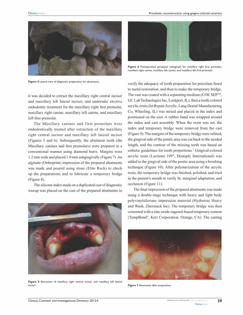
Figure 4 Lateral view of diagnostic preparation for abutments.
Figure 5 Extraction of maxillary right central incisor and maxillary left lateral incisor.
Figure 6 Postoperative periapical radiograph for maxillary right first premolar, maxillary right canine, maxillary left canine, and maxillary left first premolar.
Figure 7 Abutments after preparation.
submit your manuscript | www.dovepress.com
Dovepress
Dovepress
39
Prosthetic reconstruction using gingiva-colored ceramics

Clinical, Cosmetic and Investigational Dentistry 2012:4
try-in was performed one week after the impression was
made, and the completed porcelain fused to the metal fixed
partial denture was cemented one week after the casting
try-in using TempBond (Figure 12). Definitive cementa-
tion was performed using glass ionomer cement after one
month (GC Fuji I®, GC America Inc, Alsop, IL), as shown
in Figure 13. The patient was recalled after 6 months for
clinical evaluation.
DiscussionGingival defects may be treated using a surgical or
prosthetic approach. Alternatives to restore defects and a
deficient edentulous span today should include prosthetic
gingival restoration as an integral part of the overall esthetic
reconstructive options considered.2,4–7
To re-establish natural crown ratios and natural gingival
profiles in complex situations, artificial gingival restoration
can reduce the necessity for technique-sensitive surgical
procedures. It also increases intraoral comfort because of the
smooth uniform interface between the prosthetic gingiva and
the remaining tissues, thereby simplifying and reducing the
cost and duration of treatment.7,8
The diagnostic waxup is critical when the replacement of
missing teeth is esthetically challenging. The waxup provides
a matrix for fabrication of provisional restoration and also a
blueprint for dental positioning and adjustment of the axes
of the teeth.9
Provisional restoration provides a second opportunity for
the dentist to test the restoration design after the diagnostic
waxup. The provisional plays several roles in the treatment
process. It is used to show the patient how this kind of
restoration behaves and to obtain patient approval before
definitive restoration is fabricated. Phonetics are tested with
the provisional, and the patient and dentist can test hygiene
procedures with the provisional in place to check ease of
maintenance.10
Figure 8 Alginate checkup impression after abutment preparation.
Figure 9 Temporary bridge before adding gingival porcelain.
Figure 10 Temporary bridge ready for placement.
Figure 11 Temporary bridge being tried in patient’s mouth.
submit your manuscript | www.dovepress.com
Dovepress
Dovepress
40
Sadaqah and Abu Tair

Clinical, Cosmetic and Investigational Dentistry
Publish your work in this journal
Submit your manuscript here: http://www.dovepress.com/clinical-cosmetic-and-investigational-dentistry-journal
Clinical, Cosmetic and Investigational Dentistry is an international, peer-reviewed, open access, online journal focusing on the latest clini-cal and experimental research in dentistry with specific emphasis on cosmetic interventions. Innovative developments in dental materials, techniques and devices that improve outcomes and patient satisfac-
tion and preference will be highlighted. The manuscript management system is completely online and includes a very quick and fair peer-review system, which is all easy to use. Visit http://www.dovepress.com/testimonials.php to read real quotes from published authors.
Clinical, Cosmetic and Investigational Dentistry 2012:4
In the present case, the final fixed prosthesis was fabricated
with the teeth in their ideal position without following the
current position of the alveolar ridge. Ideal teeth relationship
principles were used to position the upper teeth in a normal
relationship with the lower arch. Guidelines for complete
denture teeth setting, including the ideal distance between
incisive papillae and the labial surface of the central incisors
(7–8 mm), were also used from the waxup procedure through
to the final restoration.5 In this clinical case, use of gingival
porcelain to restore missing tissue volumes met the patient’s
expectations with a noninvasive treatment and a reduced cost
and time needed for the procedure.
DisclosureThe authors report no conflicts of interest in this work.
References 1. Coachman C, Salama M, Garber D, Calamita M, Salama H, Cabral G.
Prosthetic gingival reconstruction in a fixed partial restoration. Part 1: Introduction to artificial gingival as an alternative therapy. Int J Periodontics Restorative Dent. 2009;29:471–477.
2. Hannon SM, Colvin CJ, Zurek DJ. Selective use of gingival-toned ceramics: case reports. Quintessence Int. 1994;25:233–238.
3. Kay HB. Esthetic considerations in the definitive periodontal prosthetic management of the maxillary anterior segment. Int J Periodontics Restorative Dent. 1982;2:45–49.
4. Barzilay I, Irene T. Gingival prosthesis – a review. J Can Dent Assoc. 2003;69:74–78.
5. Priest GF, Lindke L. Gingival-colored porcelain for implant – sup-ported prostheses in the aesthetic zone. Pract Periodontics Aesthet Dent. 1998;10:1231–1240.
6. Tallents RH. Artificial gingival replacements. Oral Health. 1983;73: 37–40.
7. Gracia LT, Verrett RG. Metal-ceramic restorations – custom char-acterization with pink porcelain. Compend Contin Educ Dent. 2004;25:242,244,246.
8. Behrend DA. The design of multiple pontics. J Prosthet Dent. 1981;46:634–638.
9. Salama M, Coachman C, Garber D, Calamita M, Salama H, Cabral G. Prosthetic gingival reconstruction in the fixed partial reconstruction. Part 2: Diagnosis and treatment planning. Int J Periodontics Restorative Dent. 2009;29:573–581.
10. Coachman C, Salama M, Garber D, Calamita M, Salama H, Cabral G. Prosthetic gingival reconstruction in fixed partial restorations. Part 3: Laboratory procedures and maintenance. Int J Periodontics Restorative Dent. 2010;30:19–29.
Figure 12 Definitive restoration is luted with interim luting agent.
Figure 13 Definitive restoration after permanent cementation.
submit your manuscript | www.dovepress.com
Dovepress
Dovepress
Dovepress
41
Prosthetic reconstruction using gingiva-colored ceramics