prospective study of the phenotypic and mutational
TRANSCRIPT

genesG C A T
T A C G
G C A T
Article
Prospective Study of the Phenotypic and Mutational Spectrumof Ocular Albinism and Oculocutaneous Albinism
Hwei Wuen Chan 1,2,3 , Elena R. Schiff 1,2 , Vijay K. Tailor 1,4 , Samantha Malka 1 , Magella M. Neveu 1,2,Maria Theodorou 1 and Mariya Moosajee 1,2,5,6,*
�����������������
Citation: Chan, H.W.; Schiff, E.R.;
Tailor, V.K.; Malka, S.; Neveu, M.M.;
Theodorou, M.; Moosajee, M.
Prospective Study of the Phenotypic
and Mutational Spectrum of Ocular
Albinism and Oculocutaneous
Albinism. Genes 2021, 12, 508.
https://doi.org/10.3390/
genes12040508
Academic Editor: Thomas A. Ciulla
Received: 1 February 2021
Accepted: 26 March 2021
Published: 30 March 2021
Publisher’s Note: MDPI stays neutral
with regard to jurisdictional claims in
published maps and institutional affil-
iations.
Copyright: © 2021 by the authors.
Licensee MDPI, Basel, Switzerland.
This article is an open access article
distributed under the terms and
conditions of the Creative Commons
Attribution (CC BY) license (https://
creativecommons.org/licenses/by/
4.0/).
1 Moorfields Eye Hospital NHS Foundation Trust, London EC1V 2PD, UK;[email protected] (H.W.C.); [email protected] (E.R.S.); [email protected] (V.K.T.);[email protected] (S.M.); [email protected] (M.M.N.); [email protected] (M.T.)
2 Institute of Ophthalmology, University College London, London EC1V 9EL, UK3 Department of Ophthalmology, National University Singapore, Singapore S118177, Singapore4 Experimental Psychology, University College London, London WC1H 0AP, UK5 Great Ormond Street Hospital for Children NHS Foundation Trust, London WC1N 3JH, UK6 The Francis Crick Institute, London NW1 1AT, UK* Correspondence: [email protected]
Abstract: Albinism encompasses a group of hereditary disorders characterized by reduced or ab-sent ocular pigment and variable skin and/or hair involvement, with syndromic forms such asHermansky–Pudlak syndrome and Chédiak–Higashi syndrome. Autosomal recessive oculocuta-neous albinism (OCA) is phenotypically and genetically heterogenous (associated with seven genes).X-linked ocular albinism (OA) is associated with only one gene, GPR143. We report the clinical andgenetic outcomes of 44 patients, from 40 unrelated families of diverse ethnicities, with query albinismpresenting to the ocular genetics service at Moorfields Eye Hospital NHS Foundation Trust betweenNovember 2017 and October 2019. Thirty-six were children (≤ 16 years) with a median age of 31months (range 2–186), and eight adults with a median age of 33 years (range 17–39); 52.3% (n = 23)were male. Genetic testing using whole genome sequencing (WGS, n = 9) or a targeted gene panel(n = 31) gave an overall diagnostic rate of 42.5% (44.4% (4/9) with WGS and 41.9% (13/31) with paneltesting). Seventeen families had confirmed mutations in TYR (n = 9), OCA2, (n = 4), HPS1 (n = 1),HPS3 (n = 1), HPS6 (n = 1), and GPR143 (n = 1). Molecular diagnosis of albinism remains challengingdue to factors such as missing heritability. Differential diagnoses must include SLC38A8-associatedfoveal hypoplasia and syndromic forms of albinism.
Keywords: albinism; foveal hypoplasia; nystagmus; chiasmal misrouting; oculocutaneous albinism;ocular albinism; Hermansky–Pudlak syndrome
1. Introduction
Albinism encompasses a group of rare genetic disorders, frequently but not alwaysassociated with impaired melanosome maturation, melanin pigment synthesis, and distri-bution in melanocytes. Melanocytes are neural crest cells, which can be categorized intocutaneous (hair and skin) or extracutaneous (eye and cochlea). In mammalian eyes, theposterior epithelial surface of the ciliary body (pars plana), sphincter, and dilator musclesof the iris and retinal pigment epithelium (RPE) are derived from the neuroectoderm. Themesenchyme forms the connective tissue and blood vessels of the iris and ciliary bodystroma. The choroid and uveal melanocytes as well as the hair and skin melanocytesare neural crest derived. During embryonic development the RPE is the first pigmentedtissue to be observed due to early expression of melanogenic genes [1,2]. Melanin ex-ists in two forms: eumelanin (black or dark brown) and pheomelanin (red or yellow).Melanin production is initiated by the conversion of tyrosine to L-DOPA by interactingwith tyrosinase. The biosynthetic pathways for the two pigments diverge downstream
Genes 2021, 12, 508. https://doi.org/10.3390/genes12040508 https://www.mdpi.com/journal/genes

Genes 2021, 12, 508 2 of 21
of L-DOPA, determined by the signaling activity of the melanocortin-1 receptor (MC1R),a member of a subgroup of class A G-protein-coupled receptors, responsible for naturalpigment variations in humans [3]. Eumelanogenesis involves two melanogenic enzymes,tyrosinase-related protein 1 (TYRP1) and dopachrome tautomerase (DCT), while pheome-lanogenesis is cysteine dependent. Melanin is deposited within melanosomes. Variationsin melanosome composition and structure determine the degree of pigmentation of theeyes, hair, and skin. In the eye, normal melanin metabolism is important in retinal pigmenthistogenesis, retinal ganglion cell metabolism, the organization and trajectory/projectionof retinal-fugal fibers [4–6]. Impaired melanin biosynthesis, therefore, disrupts the delicateembryological processes of retinal differentiation and optic chiasm decussation, culmi-nating in nystagmus and reduced visual acuity at birth. Ophthalmic manifestations ofabsent or reduced melanin pigment also include iris hypopigmentation with a transillu-mination defect, foveal hypoplasia, hypopigmented fundus, and the hallmark finding ofchiasmal misrouting (increased number of axons crossing the optic chiasm to innervate thecontralateral cortex) on flash and pattern visual evoked potential (VEP).
Oculocutaneous albinism (OCA) is an autosomal recessive disorder affecting all pig-mented tissues in the eyes, hair, and skin to varying degrees. The estimated prevalence ofOCA is 1 in 20,000 individuals [7]. Due to founder effects such as in America, Africa, India,and Japan, the global incidence and distribution of OCA is highly variable between popu-lations. OCA comprises eight clinical subtypes, OCA1-8, with mutations in seven differentgenes encoding either enzymes or membrane transporter proteins that are involved inmelanin synthesis and accumulation of tyrosine. OCA1, caused by mutations in tyrosinase(TYR), can be further categorized into OCA type IA (OMIM #203100), characterized bycomplete lack of tyrosinase activity due to production of an inactive enzyme (“tyrosinasenegative”, complete OCA), and OCA type IB (OMIM #606952), characterized by reducedactivity of tyrosinase (“tyrosinase positive”, partial OCA). OCA1 is the most commonsubtype in the white Caucasian population with an estimated prevalence of 1 in 40,000,accounting for approximately 50% of cases worldwide [8]. OCA2 (OMIM #203200) causedby mutations in the OCA gene, formerly known as the “P” gene, is the most common formof albinism worldwide; the estimated prevalence is 1 in 10,000 in the African Americanpopulation, 1 in 30,000 in Caucasians, and closer to 1 in 3,900 in southern parts of Africa [9–12]. OCA3 (OMIM #203290) has an incidence rate of 1 in 8500 amongst patients of Africanethnicity and is less severe compared to the other OCA subtypes [13,14]. It is caused bymutations in tyrosine-related protein 1 (TYRP1), which encodes an enzyme involved in thedownstream biosynthesis of eumelanin from tyrosine [15]. Mutation in TYRP1 is associ-ated with the early degradation of tyrosinase and late maturation of melanosomes [16,17].OCA4 (OMIM #606574) is caused by mutations in SLC45A2, a membrane-associated trans-porter protein (MATP) gene, and though generally uncommon (estimated prevalence 1 in100,000), it is more prevalent in the Japanese population, accounting for 24% of the OCAcases [18]. OCA5 (OMIM #615312) was mapped to a locus on 4q24, but the specific geneunderlying the pathogenesis remains to be determined [19,20]. OCA6 (OMIM #113750)results from mutations in a protein transporter gene SLC24A5 also involved in melaninsynthesis. OCA7 (OMIM #615179) is associated with mutations in the LRMDA or c10orf11gene, which encodes the leucine-rich melanocyte differentiation-associated protein. OCA8(OMIM #619165) is caused by mutation in the dopachrome tautomerase (DCT) gene, whichcatalyzes dopachrome to produce dihydroxycarboxylic acid in the eumelanin synthesispathway [21]. OCA5-8 are ultra-rare.
Ocular albinism (OA, OMIM #300500) is inherited as an X-linked disorder primarilyaffecting the eyes with relatively normal hair and skin pigmentation. The estimatedprevalence is 1 in 60,000 males [7]. OA is caused by mutations in the G-protein coupledreceptor 143 (GPR143) gene, also known as OA1, expressed in melanocytes. GPR143 trafficsto the membrane of melanosomes and regulates transcription of melanosome genes as wellas melanosome size by disrupting the delivery of melanin-related protein (MRP) to maturemelanosomes [22,23]. Impaired GPR143 signaling causes alterations in the melanosome
Genes 2021, 12, 508 3 of 21
morphology, resulting in the formation of enlarged melanosomes (“macromelanosomes”),disruption in melanosome motility, and overall reduction in melanosome numbers in themelanocytes and RPE [24,25]. GPR143 is located on chromosome Xp22. Female carriers ofGPR143 mutations exhibit mosaicism due to different degrees of lyonization or random Xchromosome inactivation, with the most characteristic finding being bright radial reflex(tapetal reflex) giving rise to a ‘mud-splatter’ appearance on fundus autofluorescence (FAF)imaging [26,27].
Syndromic OCA includes Hermansky–Pudlak syndrome (defective platelet adhe-sion causing easy bruising and severe bleeding), Chédiak–Higashi syndrome (immunedeficiency with increased risk of infection, easy bruising, and bleeding due to thrombo-cytopaenia), and Griscelli syndrome. These are autosomal recessive conditions causedby one or more genes associated with lysosomal protein trafficking. Chédiak–Higashisyndrome (OMIM #214500) is caused by homozygous or compound heterozygous muta-tion in the gene LYST. Three types of Griscelli syndrome (GS) have been reported. GS1(OMIM #214450) is caused by homozygous mutation in the gene MYO5A, resulting in hy-pomelanosis, primary neurological impairment without immunological impairment, andhaemophagocytic syndrome. GS2 (OMIM #607624) is caused by mutation in RAB27A andis associated with immune impairment. GS3 (OMIM #609227) can be caused by mutationin MLPH or MYO5A; it is characterized by hypomelanosis without neurological or immuneimpairment.
Hermansky–Pudlak syndrome (HPS) is a rare, autosomal recessive disorder of im-paired lysosomal related organelles (LROs) synthesis, with an estimated global incidenceof one to nine per 1,000,000 individuals (available online: www.orpha.net; accessed on28 December 2020). The prevalence of each subtype may vary between populations ow-ing to founder effects. HPS comprises a group of 11 multisystem disorders, HPS1-11,characterized by OCA and a combination of bleeding diathesis (secondary to impairedplatelet aggregation), immunodeficiency (neutropaenia), pulmonary fibrosis, and/or in-flammatory bowel disease. The causative genes for HPS1-11: HPS1 (HPS1), HPS2 (AP3B1),HPS3 (HPS3), HPS4 (HPS4), HPS5 (HPS5), HPS6 (HPS6), HPS7 (DTNBP1 or BLOC1S8),HPS8 (BLOC1S3), HPS9 (BLOC1S6), HPS10 (AP3D1), and the recently reported HPS11(BLOC1S5) [28,29]. Though generally rare, it is important to exclude syndromic OCA ascertain systemic associations may present later in life and carry significant morbidity andmortality.
Although OA and OCA are distinct genetic entities, OCA is phenotypically het-erogenous and may overlap with OA. Distinguishing between the two based on clinicalphenotype alone may be challenging. It is important to note that subtle hypopigmentation,especially in individuals with lightly-complected ancestry, may not be reliably appreciatedand coded. Furthermore, considerable phenotypic overlap between albinism and otherconditions such as foveal hypoplasia 2 (FVH2, OMIM #609218, also known as foveal hy-poplasia 2, optic nerve decussation defects, and anterior segment dysgenesis (FHONDA)syndrome) caused by SLC38A8 mutations may also confound the diagnosis. Therefore,molecular confirmation should be sought and correlated with clinical findings before estab-lishing a definitive diagnosis. The purpose of this study is to describe the phenotypic andgenotypic spectrum of a cohort of 44 consecutive patients presenting to the ocular geneticsservice with suspected albinism.
2. Materials and Methods
This was a prospective cohort study of consecutive nystagmus patients with suspectedalbinism presenting to the ocular genetic service at Moorfields Eye Hospital NHS Founda-tion Trust (MEH), London, United Kingdom, between November 2017 and October 2019.Patients who satisfied one of the following inclusion criteria were recruited; (i) positivefamily history of albinism with or without molecular confirmation of the affected familymember(s), (ii) nystagmus with hypopigmentation of the fundus, hair and/or skin, (iii)nystagmus and foveal hypoplasia and/or intracranial chiasmal misrouting.

Genes 2021, 12, 508 4 of 21
Data collected included medical history, family history, pedigree, hair and skin color,best corrected visual acuity (BCVA) and the presence of nystagmus, iris transilluminationdefect on slit lamp biomicroscopy as well as foveal reflex and fundus hypopigmentationon fundoscopy. VA assessment for all preverbal children up to 36 months of age wasperformed using Cardiff cards, for all other patients, BCVA was recorded in LogMAR (andconverted to this if given in Snellen for statistical analysis). Visual acuity of 1/60 or countingfingers, hand movements, light perception, and no perception of light was recorded as 1.98,2.28, 2.6, and 3.0, respectively. Anterior segment imaging of the iris was performed usingthe Haag-Streit slit lamp camera (Haag-Streit Holdings AG, Köniz, Switzerland). Ultra-widefield (UWF) pseudocolor and autofluorescence (AF) fundus imaging were acquiredusing the Optos® California (Optos plc, Dunfermline, UK) and spectral-domain opticalcoherence tomography (SDOCT) was performed using the OCT SPECTRALIS® (HeidelbergEngineering GmbH, Heidelberg, Germany). Chiasmal misrouting was confirmed eitherwith multichannel flash VEP alone (for younger paediatric patients) or a combination ofboth flash and pattern VEP were performed at the Department of Electrophysiology, MEH.
DNA samples extracted from peripheral blood with informed consent was used forgenetic testing. Genetic testing was performed in the clinical and research setting, using thealbinism and nystagmus targeted gene panel (Oculome; available online: http://www.labs.gosh.nhs.uk/media/764794/oculome_v8.pdf; accessed on 28 December 2020) consisting of30 genes (AP3B1, BLOC1S3, BLOC1S6, C10ORF11, CACNA1A, CACNA1F, CASK, DTNBP1,FRMD7, GPR143, HPS1, HPS3, HPS4, HPS5, HPS6, LYST, MANBA, MITF, MLPH, MYO5A,OCA2, PAX6, RAB27A, SACS, SETX, SLC24A5, SLC45A2, TULP1, TYR, TYRP1) through theRare & Inherited Disease Genomic Laboratory at Great Ormond Street Hospital (London,UK) and whole genome sequencing (WGS) as part of the UK Genomics England 100,000Genomes Project, where the results were reviewed by a multidisciplinary team to confirmvariant pathogenicity, prevalence in publicly available genome databases, the clinicalphenotype and mode of inheritance, before the molecular diagnosis was established [30–32].Genomic DNA was processed using an Illumina TruSeq DNA PCR-Free Sample Preparationkit (Illumina Inc., San Diego, CA, USA) and sequenced using an Illumina HiSeq X Ten high-throughput sequencing platform, generating minimum coverage of 15 X for >97% of thecallable autosomal genome. Readings were aligned to either build GRCh37 or GRCH38 ofthe human genome using an Isaac aligner (Illumina Inc.). Single-nucleotide variants (SNVs)and indels (insertions or deletions) were identified using Platypus soft-ware (version 0.8.1;and annotated using Cellbase software (available online: https://github.com/opencb/cellbase; accessed on 28 December 2020). Variant filtering was performed using minorallele frequency (MAF) < 0.001 in publicly available and in-house data sets, predictedprotein effect, and familial segregation. Surviving variants were prioritized using theRetinal disorders version 2.120 (available online: https://panelapp.genomicsengland.co.uk/panels/307/; accessed on 28 December 2020) and the Albinism or congenital nystagmusversion 1.10 (available online: https://panelapp.genomicsengland.co.uk/panels/511/;accessed on 28 December 2020) virtual gene panels [33]. Patients seen between November2017 to September 2018 were recruited into the 100,000 Genomes Project. Upon completionof recruitment to the 100,000 Genomes Project in 2018, all subsequent sequencing wasperformed using the targeted gene panel (Oculome). Affected siblings within the samefamily were all sequenced. Genetic samples of parents and/or additional family memberswere obtained where available.
In silico tools (PolyPhen-2 [34] and SIFT [35]) were used to predict pathogenicity ofthe identified missense variants (Table 1). A missense variant was deemed pathogenicwhen predicted to be “probably damaging” by PolyPhen-2 and “deleterious” by SIFT.Frameshift, splice site, and nonsense mutations were all predicted to be deleterious as aresult of nonsense mediated decay (NMD) or protein truncation.
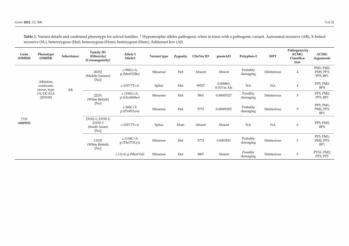
Genes 2021, 12, 508 5 of 21
Table 1. Variant details and confirmed phenotype for solved families. 1 Hypomorphic alleles pathogenic when in trans with a pathogenic variant. Autosomal recessive (AR), X-linkedrecessive (XL), heterozygous (Het), homozygous (Hom), hemizygous (Hem), Ashkenazi Jew (AJ).
Gene(OMIM)
Phenotype(OMIM) Inheritance
Family ID(Ethnicity)
[Consanguinity]
Allele 1Allele2 Variant type Zygosity ClinVar ID gnomAD Polyphen-2 SIFT
PathogenicityACMG
Classifica-tion
ACMGArguments
TYR(606933)
Albinism,oculocuta-
neous, type1A; OCA1A
(203100)
AR
26352(Middle Eastern)
[No]
c.996G>A,p.(Met332Ile) Missense Het Absent Absent Probably
damaging Deleterious 4PM1; PM2;PM5; PP3;PP5; BP1
c.1037-7T>A Splice Het 99527 0.000861,0.015 in AJs NA NA 4 PP5; PM2;
BP4
22151(White British)
[No]
c.1336G>A,p.(Gly446Ser) Missense Het 3801 0.00005327 Possibly
damaging Deleterious 5 PP5; PM2;PP3; BP1
c.242C>T,p.(Pro81Leu) Missense Het 3772 0.00009205 Probably
damaging Deleterious 5PP5; PM1;PM2; PP3;
BP1
23192-1; 23192-2;23192-3
(South Asian)[No]
c.1037-7T>A Splice Hom Absent Absent NA NA 4 PP5; PM2;BP4
13332(White British)
[No]
c.1118C>Ap.(Thr373Lys) Missense Het 3774 0.0003541 Probably
damaging Deleterious 5PP5; PM1;PM2; PP3;
BP1
c.1A>G p.(Met1Val) Missense Het 3807 Absent Possiblydamaging Deleterious 5 PVS1; PM2;
PP3; PP5

Genes 2021, 12, 508 6 of 21
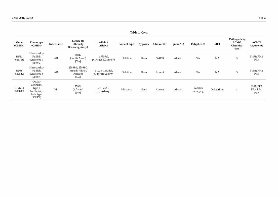
Table 1. Cont.
Gene(OMIM)
Phenotype(OMIM) Inheritance
Family ID(Ethnicity)
[Consanguinity]
Allele 1Allele2 Variant type Zygosity ClinVar ID gnomAD Polyphen-2 SIFT
PathogenicityACMG
Classifica-tion
ACMGArguments
Albinism,oculocuta-
neous, typeIB; OCA1B
(606952)
AR
26680(White British)
[No]
c.575C>A,p.(Ser192Tyr) 1 Missense
Het3778 0.2502 Probably
damaging(both
variants)
Deleterious(both
variants)
Hypomorphicalleles 1 See text
c.1205G>A,p.(Arg402Gln) 1 Missense 3779 0.1765
c.242C>T,p.(Pro81Leu) Missense Het 3772 0.00009205 Probably
damaging Deleterious 5PP5; PM1;PM2; PP3;
BP1
27560(White British)
[No]
c.1217C>T,p.(Pro406Leu) Missense Het 3777 0.003918 Probably
damaging Deleterious 5PP5; PM1;PM2; PP3;
BP1
c.1291C>A,p.(Pro431Thr) Missense Het Absent 0.000007986 Probably
damaging Deleterious 4PM1; PM2;PP3; PP5;
BP1
26903(South Asian)
[Yes]
c.832C>T,p.(Arg278*) Nonsense Hom 99583
0.0001699,0.001274 in S
AsiansNA NA 5 PP5; PVS1;
PM2; PP3
27079(White British)
[No]
c.661G>A,p.(Glu221Lys) Missense Het 212524 0.00000399 Possibly
damaging Deleterious 4PM1; PM2;PP3; PP5;
BP1
c.575C>A,p.(Ser192Tyr) 1 Missense
Het3778 0.2502 Probably
damaging(both
variants)
Deleterious(both
variants)
Hypomorphicalleles 1 See text
c.1205G>A,p.(Arg402Gln) 1 Missense 3779 0.1765
27430(White British)
[No]
c.823G>T,p.(Val275Phe) Missense Het 3773 0.00009916 Possibly
damaging Deleterious 5 PP5; PM2;PP3; BP1
c.575C>A,p.(Ser192Tyr) 1 Missense
Het3778 0.2502 Probably
damaging(both
variants)
Deleterious(both
variants)
Hypomorphicalleles 1 See text
c.1205G>A,p.(Arg402Gln) 1 Missense 3779 0.1765

Genes 2021, 12, 508 7 of 21
Table 1. Cont.
Gene(OMIM)
Phenotype(OMIM) Inheritance
Family ID(Ethnicity)
[Consanguinity]
Allele 1Allele2 Variant type Zygosity ClinVar ID gnomAD Polyphen-2 SIFT
PathogenicityACMG
Classifica-tion
ACMGArguments
OCA2(611409)
Albinism,oculocuta-
neous, type II(203200)
AR
25578(Mixed: White /
African)[No]
c.619_636del,p.(Leu207_Leu212del) Deletion Het 372713
0.0008030 inAfrican /African
Americans
NA NA 4 PP5; PM2;PM4; PP3
c.1327G>A,p.(Val443Ile) Missense Het 955 0.003056 Probably
damaging Deleterious 4PS3; PM3;PP3; PP4;PP5; BS2
25246(Middle Eastern)
[No]
c.1286T>C,p.(Leu429Pro) Missense Het 627601 Absent Probably
damaging Deleterious 4PM1; PM2;PP3; PP5;
BP1
c.1327G>A,p.(Val443Ile) Missense Het 955 0.003056 Probably
damaging Deleterious 4PS3; PM3;PP3; PP4;PP5; BS2
26947(Mixed: White /
African)[No]
c.2177_2181del,p.(Val726Glyfs*13) Deletion Het 498226 Absent NA NA 5 PVS1; PM2;
PP3; PP5
c.1255C>T,p.(Arg419Trp) Missense Het 194160 0.0002659 Probably
damaging Deleterious 4 PM2; PP3;PP5; PM3
27321(Black African)
[No]
c.2228C>T,p.(Pro743Leu) Missense Het 956 0.0001344 Probably
damaging Deleterious 5 PP5; PM2;PP3; BP1
c.1182+1G>A Splice Het 436099 0.0000566 Probablydamaging Deleterious 5 PVS1; PM2;
PP3; PP5
HPS1(604982)
Hermansky-Pudlak
syndrome 1(203300)
AR26677
(South Asian)[No]
c.972dup,p.(Met325Hisfs*128) Duplication Hom Absent 0.000262 NA NA 5 PVS1; PM2;
PP3; PP5
Genes 2021, 12, 508 8 of 21
Table 1. Cont.
Gene(OMIM)
Phenotype(OMIM) Inheritance
Family ID(Ethnicity)
[Consanguinity]
Allele 1Allele2 Variant type Zygosity ClinVar ID gnomAD Polyphen-2 SIFT
PathogenicityACMG
Classifica-tion
ACMGArguments
HPS3(606118)
Hermansky-Pudlak
syndrome 3(614072)
AR26847
(South Asian)[Yes]
c.850del,p.(Arg284Glufs*57) Deletion Hom 664198 Absent NA NA 5 PVS1; PM2;
PP3
HPS6(607522)
Hermansky-Pudlak
syndrome 6(614075)
AR
25888-1; 25888-2(Mixed: White /
African)[No]
c.1228_1252del,p.(Tyr410Valfs*9) Deletion Hom Absent Absent NA NA 5 PVS1; PM2;
PP3
GPR143(300808)
Ocularalbinism,
type I,Nettleship-Falls type(300500)
XL25806
(African)[No]
c.11C>G,p.(Pro4Arg) Missense Hemi Absent Absent Probably
damaging Deleterious 4PM2; PP2;PP3; PP4;
PP5

Genes 2021, 12, 508 9 of 21
This study had relevant local and national research ethics committee approvals (MEHand the Northwest London Research Ethics Committee) and adhered to the tenets ofthe Declaration of Helsinki. Patients and relatives gave written informed consent forparticipation in this study through either the Genetic Study of Inherited Eye Disease (RECreference 12/LO/0141) or Genomics England 100,000 Genomes project (REC reference14/EE/1112).
3. Results3.1. Clinical Findings
This prospective study identified 44 probands from 40 families, of which 47.7% (n = 21)were female and 52.3% (n = 23) were male. The study cohort comprised 36 children (≤ 16years) with a median age of 31 months (range 2–186), and eight adults with a median ageof 33 years (range 17–39). This cohort consisted of families with diverse ethnic origins, 11(27.5%) White British, 8 (20.0%) South Asian, 5 (12.5%) mixed White and Black African, 5(12.5%) White other, 4 (10.0%) African, 4 (10.0%) Black African, 2 (5.0%) Middle Eastern,and 1 (2.5%) mixed White and South Asian. One African family and 3 South Asian familieswere consanguineous (9%, n = 4/44). A positive family history was absent in 81.8% ofprobands (n = 36/44) with the inheritance pattern being unclear.
In the paediatric cohort, BCVA ranged from fixing and following to 1.2 logarithm of theminimal angle of resolution (LogMAR) with a median of 0.8 LogMAR. In the adult group,the range was 0.3 to 1.0 LogMAR with a median of 0.6 LogMAR. Correlation between thechange in BCVA at baseline and most recent follow up visit demonstrated subtle change inBCVA over time (Figure 1A). The change in BCVA of each paediatric patient at baselineand most recent vision as a function of age is illustrated in Figure 1B. The average BCVA atbaseline decreases from 0.6 to 0.48 logMAR for patients between 3–20 years of age.
Genes 2021, 12, x FOR PEER REVIEW 9 of 21
This study had relevant local and national research ethics committee approvals
(MEH and the Northwest London Research Ethics Committee) and adhered to the tenets
of the Declaration of Helsinki. Patients and relatives gave written informed consent for
participation in this study through either the Genetic Study of Inherited Eye Disease (REC
reference 12/LO/0141) or Genomics England 100,000 Genomes project (REC reference
14/EE/1112).
3. Results
3.1. Clinical Findings
This prospective study identified 44 probands from 40 families, of which 47.7% (n =
21) were female and 52.3% (n = 23) were male. The study cohort comprised 36 children (≤
16 years) with a median age of 31 months (range 2–186), and eight adults with a median
age of 33 years (range 17–39). This cohort consisted of families with diverse ethnic origins,
11 (27.5%) White British, 8 (20.0%) South Asian, 5 (12.5%) mixed White and Black African,
5 (12.5%) White other, 4 (10.0%) African, 4 (10.0%) Black African, 2 (5.0%) Middle Eastern,
and 1 (2.5%) mixed White and South Asian. One African family and 3 South Asian families
were consanguineous (9%, n = 4/44). A positive family history was absent in 81.8% of pro-
bands (n = 36/44) with the inheritance pattern being unclear.
In the paediatric cohort, BCVA ranged from fixing and following to 1.2 logarithm of
the minimal angle of resolution (LogMAR) with a median of 0.8 LogMAR. In the adult
group, the range was 0.3 to 1.0 LogMAR with a median of 0.6 LogMAR. Correlation be-
tween the change in BCVA at baseline and most recent follow up visit demonstrated sub-
tle change in BCVA over time (Figure 1A). The change in BCVA of each paediatric patient
at baseline and most recent vision as a function of age is illustrated in Figure 1B. The av-
erage BCVA at baseline decreases from 0.6 to 0.48 logMAR for patients between 3–20 years
of age.
Of the cohort, 89.2% (n = 33/37, 2 were inconclusive, 2 had no chiasmal misrouting; 7
declined electrophysiology testing) had intracranial chiasmal misrouting, 86.4% (n =
38/44) had nystagmus, 75.0% (n = 33/44) had foveal hypoplasia, 72.7% (n = 32/44) had fun-
dus hypopigmentation, 50.0% (n = 22/44) had iris transillumination defect, 38.6% (n =
17/44) had hair and skin (cutaneous) involvement, and 1 patient reported history of easy
bruising (2.3%, n = 1/44) (Figure 2). The demographics and phenotypes are summarized
in Table S1.
Figure 1. Best corrected visual acuity (BCVA) changes in albinism patients. (A) Correlation between the change in BCVA atbaseline and most recent follow up. The black dashed line represents a ‘no change’ scenario, while the grey correlation lineillustrates the subtle change seen in patients. (B) Demonstrates the change in BCVA of each paediatric patient at baselineand most recent vision as a function of age. The black dashed line represents the point at which more accurate visual acuitytesting can be achieved in children 3 years old and upwards.
Of the cohort, 89.2% (n = 33/37, 2 were inconclusive, 2 had no chiasmal misrouting; 7declined electrophysiology testing) had intracranial chiasmal misrouting, 86.4% (n = 38/44)had nystagmus, 75.0% (n = 33/44) had foveal hypoplasia, 72.7% (n = 32/44) had fundus

Genes 2021, 12, 508 10 of 21
hypopigmentation, 50.0% (n = 22/44) had iris transillumination defect, 38.6% (n = 17/44)had hair and skin (cutaneous) involvement, and 1 patient reported history of easy bruising(2.3%, n = 1/44) (Figure 2). The demographics and phenotypes are summarized in Table S1.
Genes 2021, 12, x FOR PEER REVIEW 10 of 21
Figure 1. Best corrected visual acuity (BCVA) changes in albinism patients. (A) Correlation between the change in BCVA
at baseline and most recent follow up. The black dashed line represents a ‘no change’ scenario, while the grey correlation
line illustrates the subtle change seen in patients. (B) Demonstrates the change in BCVA of each paediatric patient at
baseline and most recent vision as a function of age. The black dashed line represents the point at which more accurate
visual acuity testing can be achieved in children 3 years old and upwards.
Figure 2. Overview of the phenotypic spectrum of 44 suspected albinism patients presenting to the
ocular genetics service. For chiasmal misrouting, n = 37, 7 declined electrophysiology visual
evoked potential (VEP) testing.
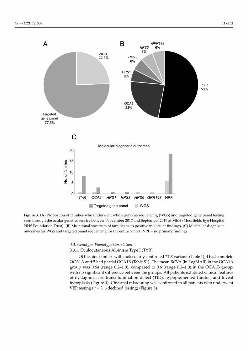
3.2. Genetic Sequencing Outcomes
Initial recruitment comprised 44 families, of which 12 underwent WGS and 32 fami-
lies had targeted gene panel testing. WGS identified biallelic variants in SLC38A8 in three
unrelated families causing FVH2, all had foveal hypoplasia and intracranial chiasmal mis-
routing. Panel testing confirmed a FRMD7 hemizygous mutation causing X-linked con-
genital nystagmus in one family with suspected albinism, where the VEP was inconclu-
sive. All four families were excluded from subsequent analysis of albinism cases, but fur-
ther clinical and genetic details are included under Section 3.3.5. Non-Albinism Cases.
The overall diagnostic yield for albinism was 42.5%; 44.4% (4/9) with WGS, and 41.9%
(13/31) with panel testing. Seventeen families had confirmed mutations (Table 1, Figure
3), of which 13 had OCA. OCA1 was the most common type with 69.2% (9/13) of families
harboring TYR variants, followed by OCA2 in 30.8% (4/13). Three families were diagnosed
with Hermansky–Pudlak syndrome with pathogenic variants in either HPS1 (6.0%; 1/17),
HPS3 (6.0%; 1/17) or HPS6 (6.0%; 1/17). Only one family (6.0%; 1/17) had X-linked OA with
a GPR143 missense variant. Amongst the unsolved cases, a single pathogenic TYR variant
was identified in one White British family (26649) with OCA1A and a single pathogenic
TYRP1 variant causing OCA3 in one Afro-Caribbean family (26948). Unsolved cases from
the targeted panel testing will undergo WGS, and for those unsolved following WGS, fur-
ther data mining will be undertaken to discover novel genes or noncoding variants.
Figure 2. Overview of the phenotypic spectrum of 44 suspected albinism patients presenting to theocular genetics service. For chiasmal misrouting, n = 37, 7 declined electrophysiology visual evokedpotential (VEP) testing.
3.2. Genetic Sequencing Outcomes
Initial recruitment comprised 44 families, of which 12 underwent WGS and 32 familieshad targeted gene panel testing. WGS identified biallelic variants in SLC38A8 in threeunrelated families causing FVH2, all had foveal hypoplasia and intracranial chiasmalmisrouting. Panel testing confirmed a FRMD7 hemizygous mutation causing X-linked con-genital nystagmus in one family with suspected albinism, where the VEP was inconclusive.All four families were excluded from subsequent analysis of albinism cases, but furtherclinical and genetic details are included under Section 3.3.5. Non-Albinism Cases.
The overall diagnostic yield for albinism was 42.5%; 44.4% (4/9) with WGS, and41.9% (13/31) with panel testing. Seventeen families had confirmed mutations (Table 1,Figure 3), of which 13 had OCA. OCA1 was the most common type with 69.2% (9/13)of families harboring TYR variants, followed by OCA2 in 30.8% (4/13). Three familieswere diagnosed with Hermansky–Pudlak syndrome with pathogenic variants in eitherHPS1 (6.0%; 1/17), HPS3 (6.0%; 1/17) or HPS6 (6.0%; 1/17). Only one family (6.0%;1/17) had X-linked OA with a GPR143 missense variant. Amongst the unsolved cases,a single pathogenic TYR variant was identified in one White British family (26649) withOCA1A and a single pathogenic TYRP1 variant causing OCA3 in one Afro-Caribbeanfamily (26948). Unsolved cases from the targeted panel testing will undergo WGS, and forthose unsolved following WGS, further data mining will be undertaken to discover novelgenes or noncoding variants.
Genes 2021, 12, 508 11 of 21Genes 2021, 12, x FOR PEER REVIEW 11 of 21
Figure 3. (A) Proportion of families who underwent whole genome sequencing (WGS) and targeted gene panel testing
seen through the ocular genetics service between November 2017 and September 2019 at MEH (Moorfields Eye Hospital
NHS Foundation Trust). (B) Mutational spectrum of families with positive molecular findings. (C) Molecular diagnostic
outcomes for WGS and targeted panel sequencing for the entire cohort. NPF = no primary findings.
3.3. Genotype-Phenotype Correlation
3.3.1. Oculocutaneous Albinism Type I (TYR)
Of the nine families with molecularly confirmed TYR variants (Table 1), 4 had com-
plete OCA1A and 5 had partial OCA1B (Table S1). The mean BCVA (in LogMAR) in the
OCA1A group was 0.64 (range 0.2–1.0), compared to 0.6 (range 0.2–1.0) in the OCA1B
group, with no significant difference between the groups. All patients exhibited clinical
features of nystagmus, iris transillumination defect (TID), hypopigmented fundus, and
foveal hypoplasia (Figure 4). Chiasmal misrouting was confirmed in all patients who un-
derwent VEP testing (n = 3, 6 declined testing) (Figure 5).
Figure 3. (A) Proportion of families who underwent whole genome sequencing (WGS) and targeted gene panel testingseen through the ocular genetics service between November 2017 and September 2019 at MEH (Moorfields Eye HospitalNHS Foundation Trust). (B) Mutational spectrum of families with positive molecular findings. (C) Molecular diagnosticoutcomes for WGS and targeted panel sequencing for the entire cohort. NPF = no primary findings.
3.3. Genotype-Phenotype Correlation3.3.1. Oculocutaneous Albinism Type I (TYR)
Of the nine families with molecularly confirmed TYR variants (Table 1), 4 had completeOCA1A and 5 had partial OCA1B (Table S1). The mean BCVA (in LogMAR) in the OCA1Agroup was 0.64 (range 0.2–1.0), compared to 0.6 (range 0.2–1.0) in the OCA1B group,with no significant difference between the groups. All patients exhibited clinical featuresof nystagmus, iris transillumination defect (TID), hypopigmented fundus, and fovealhypoplasia (Figure 4). Chiasmal misrouting was confirmed in all patients who underwentVEP testing (n = 3, 6 declined testing) (Figure 5).

Genes 2021, 12, 508 12 of 21Genes 2021, 12, x FOR PEER REVIEW 12 of 21
Figure 4. Iris and multimodal retinal imaging (right and left eye) of a patient (27079) with homozygous TYR mutation. (A,B) Iris
transillumination defects with visualization of nasal lens equator; (C,D) ultra-widefield (UWF) pseudocolor showing hypopigmented
fundi and prominent choroidal vessels; (E,F) UWF fundus autofluorescence (FAF) illustrating the absence of hypoautofluoresence at
the fovea due to decreased luteal pigments, crossing of retinal vessels at the fovea, and prominent choroidal vessels can be appreci-
ated; (G,H) grade 4 foveal hypoplasia on spectral-domain optical coherence tomography (SDOCT).
Figure 4. Iris and multimodal retinal imaging (right and left eye) of a patient (27079) with homozy-gous TYR mutation. (A,B) Iris transillumination defects with visualization of nasal lens equator;(C,D) ultra-widefield (UWF) pseudocolor showing hypopigmented fundi and prominent choroidalvessels; (E,F) UWF fundus autofluorescence (FAF) illustrating the absence of hypoautofluoresence atthe fovea due to decreased luteal pigments, crossing of retinal vessels at the fovea, and prominentchoroidal vessels can be appreciated; (G,H) grade 4 foveal hypoplasia on spectral-domain opticalcoherence tomography (SDOCT).

Genes 2021, 12, 508 13 of 21Genes 2021, 12, x FOR PEER REVIEW 13 of 21
Figure 5. Pattern appearance VEPs (PVEP) from a patient (27079) with TYR mutation. PVEP responses on stimulating the
right eye are displayed on the top row and those on stimulating the left eye are on the bottom row. Column 1 and 2 are
responses from the right hemisphere, column 4 and 5 are responses from the left hemisphere, and column 3 is the com-
bined midline response. PVEP shows contralateral predominance (turquoise arrow) when the opposite eye (dotted red
arrow) is stimulated.
Of the TYR variants identified in this cohort, 11 were missense, 2 were splice-site and
1 was nonsense. The most commonly observed OCA1A variant in patients with OCA,
c.1118C>A p.(Thr373Lys), was identified in the proband of a White British family (13332)
with BCVA of 1.0 LogMAR and the full complement of albinism features. The same vari-
ant was also identified in an unsolved White British family (26649). This variant was re-
ported to occur in approximately 30% of Northern European cases [8,36,37]. The proband
of a consanguineous Pakistani family (26903) had BCVA of 1.0 LogMAR and all ocular
features of albinism were present without obvious cutaneous involvement. She was ho-
mozygous for a loss-of-function TYR variant causing premature translational stop signal
(c.832C>T, p.Arg278*). Several studies reported higher incidence of this causal TYR vari-
ant amongst South Asians (Indian, East Indian/West Bengal, Pakistani) due to consan-
guinity [38,39]. A recurring pair of hypomorphic alleles c.575C>A, p.(Ser192Tyr) and
c.1205G>A, p.(Arg402Gln) was seen across 3 unrelated White British families with
OCA1B, occurring in trans with 3 different pathogenic missense variants. In this OCA1B
subgroup, the mean BCVA (in LogMAR) was 0.74 (range 0.6–1.0), all probands had iris
TID, foveal hypoplasia and fundus hypopigmentation, nystagmus was subtle in one pa-
tient, clearly present in the other two. All 3 probands were of fair complexion that is sim-
ilar to their unaffected relatives. In a French study of 268 patients with OCA1, the
p.(Arg402Gln) variant in trans was identified in 69 patients (25.7%) with variable but gen-
erally mild forms of albinism [40].
3.3.2. Oculocutaneous Albinism Type II (OCA2)
Four families (two mixed White and Black African, one Middle Eastern, one Black
African) had OCA2 mutations, 50% had oculocutaneous involvement. The mean BCVA
(in LogMAR) was 0.5 (range 0.2–1.2). All families had nystagmus and chiasmal misrout-
ing. Other clinical findings of iris TID, foveal hypoplasia, and fundus hypopigmentation
were more variable within this subgroup compared to OCA1. Seven pathogenic OCA2
variants were identified: 4 missense, 1 in-frame deletion, 1 frameshift deletion, 1 non-cod-
ing (splice). The same missense variant c.1327G>A, p.(Val443Ile) was seen in 2 unrelated
families occurring with a different pathogenic variant (1 deletion, 1 missense). Variant
p.Val443Ile is the most common in northern European populations [41,42], but is rare in
RE PVEP
LE PVEP
Figure 5. Pattern appearance VEPs (PVEP) from a patient (27079) with TYR mutation. PVEP responses on stimulating theright eye are displayed on the top row and those on stimulating the left eye are on the bottom row. Column 1 and 2 areresponses from the right hemisphere, column 4 and 5 are responses from the left hemisphere, and column 3 is the combinedmidline response. PVEP shows contralateral predominance (turquoise arrow) when the opposite eye (dotted red arrow)is stimulated.
Of the TYR variants identified in this cohort, 11 were missense, 2 were splice-siteand 1 was nonsense. The most commonly observed OCA1A variant in patients withOCA, c.1118C>A p.(Thr373Lys), was identified in the proband of a White British family(13332) with BCVA of 1.0 LogMAR and the full complement of albinism features. Thesame variant was also identified in an unsolved White British family (26649). This variantwas reported to occur in approximately 30% of Northern European cases [8,36,37]. Theproband of a consanguineous Pakistani family (26903) had BCVA of 1.0 LogMAR and allocular features of albinism were present without obvious cutaneous involvement. Shewas homozygous for a loss-of-function TYR variant causing premature translational stopsignal (c.832C>T, p.Arg278*). Several studies reported higher incidence of this causalTYR variant amongst South Asians (Indian, East Indian/West Bengal, Pakistani) due toconsanguinity [38,39]. A recurring pair of hypomorphic alleles c.575C>A, p.(Ser192Tyr)and c.1205G>A, p.(Arg402Gln) was seen across 3 unrelated White British families withOCA1B, occurring in trans with 3 different pathogenic missense variants. In this OCA1Bsubgroup, the mean BCVA (in LogMAR) was 0.74 (range 0.6–1.0), all probands had iris TID,foveal hypoplasia and fundus hypopigmentation, nystagmus was subtle in one patient,clearly present in the other two. All 3 probands were of fair complexion that is similar totheir unaffected relatives. In a French study of 268 patients with OCA1, the p.(Arg402Gln)variant in trans was identified in 69 patients (25.7%) with variable but generally mild formsof albinism [40].
3.3.2. Oculocutaneous Albinism Type II (OCA2)
Four families (two mixed White and Black African, one Middle Eastern, one BlackAfrican) had OCA2 mutations, 50% had oculocutaneous involvement. The mean BCVA (inLogMAR) was 0.5 (range 0.2–1.2). All families had nystagmus and chiasmal misrouting.Other clinical findings of iris TID, foveal hypoplasia, and fundus hypopigmentation weremore variable within this subgroup compared to OCA1. Seven pathogenic OCA2 variantswere identified: 4 missense, 1 in-frame deletion, 1 frameshift deletion, 1 non-coding (splice).The same missense variant c.1327G>A, p.(Val443Ile) was seen in 2 unrelated familiesoccurring with a different pathogenic variant (1 deletion, 1 missense). Variant p.Val443Ile isthe most common in northern European populations [41,42], but is rare in other populations
Genes 2021, 12, 508 14 of 21
(<1% in the Scandinavian population) [43]. It is associated with residual function of the Pprotein (encoded by OCA2) and development of cutaneous pigment with time in affectedindividuals [44]. Despite impaired or diminished eumelanin synthesis, this progressiveincrease in pigmentation and the propensity to tan is due to the relative preservationof pheomelanin [45,46]. In this study, all probands harboring this variant did not haveobvious cutaneous involvement but displayed fundus hypopigmentation with a meanBCVA (in LogMAR) of 0.3 (range 0.2–0.4). Family 27321 with missense variant c.2228C>T,p.(Pro743Leu) and splice donor variant c.1182+1G>A displayed a more severe phenotypewith 1.2 LogMAR vision and the full complement of albinism features.
3.3.3. Hermansky–Pudlak Syndrome (HPS)
Four patients from three unrelated families were identified with HPS. The mean BCVA(in LogMAR) of this group was 0.6 (range 0.3–0.8). Foveal hypoplasia, iris TID, and chiasmalmisrouting were present in 100% (n = 3), nystagmus and fundus hypopigmentation in 66.7%(n = 2). The lack of clinically apparent nystagmus was associated with relatively bettervisual prognosis in the paediatric cohort [47]. This finding was consistent in our cohortwhere probands from family 25588 without nystagmus recorded vision (in LogMAR) of 0.3versus 0.6 and 0.8, respectively in families 26677 and 26847 with nystagmus. Significantsystemic findings included spontaneous bruising in the proband of family 26677 withhomozygous mutations in HPS1 c.972dup, p.(Met325Hisfs*128). All patients were referredto haematology for further investigation and management.
3.3.4. Ocular Albinism (GPR143)
The proband of Ghanaian family 25806 is a 13-year-old boy with a family historyof maternal great uncles having yellow hair and fair skin. The patient had dark skin,black hair and reduced vision 0.7 and 0.6 LogMAR in the right and left eye, respectively.Examination was significant for nystagmus, although he had previously undergone theAnderson-Kestenbaum procedure in Ghana. He was found to have foveal hypoplasia andVEP confirmed chiasmal misrouting. WGS identified a pathogenic hemizygous mutationin GPR143: c.11C>G, p.(Pro4Arg) confirming the diagnosis of ocular albinism (OA1).
3.3.5. Non-Albinism Cases
All probands with SLC38A8-associated foveal hypoplasia had evidence of nystag-mus, foveal hypoplasia, and intracranial misrouting. Family 23089 proband had bilateralposterior embryotoxon, no signs of anterior segment dysgenesis were seen in the othertwo families (26237 and 26320). Families 23089 and 26320 were consanguineous, carryinghomozygous nonsense mutations in SLC38A8: c.264C>G, p.Tyr88*. Family 26237 had com-pound heterozygous SLC38A8 mutations, c.435G>A, p.Trp145*, and c.632+1G>A. Furtherclinical details of these families were published [48].
The proband of White British family 27561 with a hemizgous FRMD7 variant wasa 3-year-old male with unilateral (left) grade 1b foveal hypoplasia (using the LeicesterGrading System) on OCT [49]. He had blonde hair and fair skin, with 0.4 LogMARvision bilaterally, fine horizontal nystagmus, and hypopigmented fundi. Repeated VEPsconsistently showed equivocal findings on the right and contralateral predominance onthe left. The equivocal VEP findings were partially attributed to maturational factors,but albinism could not be excluded. Targeted gene panel confirmed a novel pathogenicmissense variant in FRMD7 c.790T>G, p.(Cys264Gly). Pathogenic FRMD7 mutations arefully penetrant in males [50]. FRMD7 regulates brain development and neuronal growthby promoting neurite elongation and is expressed in the developing neural retina [51].In patients with FRMD7 variants, OCT imaging has shown abnormal afferent systemdevelopment with foveal hypoplasia [52].

Genes 2021, 12, 508 15 of 21
4. Discussion
This prospective observational study of 44 consecutive patients, from 40 families,presenting with query albinism reported an overall diagnostic yield of 42.5% through WGSor targeted gene panel testing. We defined diagnostic yield as the percentage of individualswith a characteristic clinical phenotype receiving a molecular diagnosis (≥2 pathogenic orlikely pathogenic variants in a gene linked with OCA or ≥1 definite or likely pathogenicvariant in GPR143 for OA). Copy number variations (CNV) may partially account forthe lower rate of diagnosis in this study. The clinical exome (Oculome) does not searchfor CNVs nor have they yet been analyzed by the Genomics England analysis pipeline.Molecular diagnostic rates for albinism are highly variable, ranging from 56–91% in theliterature [32,53,54]. Lasseaux et al. (2018) reported a diagnostic yield of 72% from a largeFrench study of 990 probands with albinism [53]. More recently, Lenassi et al. solved29 out of 32 (91%) preschool children with suspected albinism using a targeted panel of18 (n = 30) or 26 (n = 1) or 40 (n = 1) genes with confirmed mutations in TYR (n = 18),OCA2 (n = 7), TYRP1 (n = 2), HPS5 (n = 1), and GPR143 (n = 1). The authors did notspecify the reason for selecting different gene-specific panels and the patient demographicdetails (aside from age) were not presented [54]. The probands who underwent expandedgene panel testing harbored OCA2 and TYR variants. The OCA2 variant c.1327G>A,p.(Val443Ile) and three TYR variants c.1118C>A, p.(Thr373Lys), c.575C>A, p.(Ser192Tyr),and c.1205G>A, p.(Arg402Gln) were also identified in our cohort through targeted genepanel testing. Comparison of the gene panels in the Lennasi study versus that employedin this study revealed some differences, e.g., they excluded CACNA1A, CASK, HPS1(suboptimal coverage), MANBA, MITF, MLPH, MYO5A, RAB27A, SACS, SETX, and TULP1,whereas our panel did not include ACO2, ATOH7, HMX1, RTN4IP1, SIX6, and SLC4A11.However, none of these genes were identified in the Lenassi cohort, which suggests thatstringency in clinical inclusion criteria is likely the major factor for increased likelihood ofa positive finding.
In our study, the majority of the probands were referred with nystagmus as a pre-dominant clinical feature with equivocal findings of foveal hypoplasia and cutaneousinvolvement; 75% had foveal hypoplasia, 73% had fundus hypopigmentation, and 39%had skin/hair involvement. Ethnic/genetic diversity of the study cohort and possibleunder-ascertainment of cutaneous hypopigmentation may account for smaller percentageof skin/hair involvement reported in this study. Thirty-seven patients underwent VEPtesting (seven declined) and chiasmal misrouting was confirmed in 33 patients (89%). Sixout of the seven patients who declined VEP testing, had confirmed mutations in the TYRgene and one remained unsolved. This highlighted the importance of detailed phenotyp-ing in delineating patients with albinism versus infantile nystagmus. Our diverse patientethnicity may have also contributed to the difference due to rare/unknown ethnic-specificvariants. Of the unsolved cases in this cohort, 45.0% (n = 18/40) underwent targeted paneltesting, these patients will be offered WGS to identify potential novel genes/variants ornon-coding mutations that remain to be determined.
Of interest, a recent genome-wide association study (GWAS) to determine the poly-genic risk score for glaucoma susceptibility and progression identified a significant signalfor TYR [55]. Although the original design of this study did not include glaucoma in thedata collection, retrospective review of the clinical findings did not identify glaucoma inthis cohort.
Several genetic eye disorders falling under infantile nystagmus syndrome (INS)can masquerade as albinism with considerable phenotypic overlap such as dominantPAX6-related oculopathy (OMIM # 136520), SLC38A8-related FVH2 (OMIM #609218), andforms of congenital nystagmus caused by FRMD7 (OMIM #310700) or GPR143 (OMIM#300814) [48,56,57]. Diagnosing albinism based on clinical findings alone is inadequate,particularly in infants and young children, where investigations such as optical coherencetomography (OCT) to assess foveal hypoplasia and VEPs for chiasmal misrouting maybe challenging due to fixation loss and compliance. Candidate genes for INS should be

Genes 2021, 12, 508 16 of 21
grouped together for testing this cohort of patients, which may not yet be included inconventional albinism gene panels for comprehensive screening of patients [32].
In this study, OCA was the most common disease entity with TYR being the mostcommon gene identified. Approximately 480 mutations were identified throughout theTYR gene; up to 77% were missense mutations (67% OCA1A and 33% OCA1B), 15%deletions, and 3% insertions [58,59]. In this cohort, 80% of the OCA1 variants were mis-sense mutations. In cases of missing heritability where only a single pathogenic TYRvariant was identified, studies subsequently identified two hypomorphic alleles, c.575C>A,p.(Ser192Tyr) and c.1205G>A, p.(Arg402Gln) in combination, forming a tri-allelic geno-type [10,60–62]. Human melanocytes with the p.(Arg402Gln) variant retain tyrosinase inthe endoplasmic reticulum (ER) and is associated with moderate thermoinstability resultingin approximately a 75% reduction of enzymatic activity at 37 ◦C [63,64]. The p.(Ser192Tyr)variant was shown to have 60% of wild type activity [65]. Together, the p.(Ser192Tyr) andp.(Arg402Gln) alleles result in the reduction of tyrosinase activity from retention in the ERand the released tyrosinase only having 60% of wild type activity, lowering the tyrosinaseactivity to a pathogenic level. We identified compound heterozygous tri-alleleic genotypein three unrelated families with partial OCA, where the p.(Ser192Tyr) and p.(Arg402Gln)alleles were seen in trans with a rare pathogenic TYR variant (different variant in eachfamily). Various studies reported compound heterozygous tri-alleleic genotype in TYRinvolving both rare (AF <5%) and common (AF 28–36%) functionally damaging vari-ants, which are likely to be on trans alleles [61]. The hypomorphic TYR coding variantsp.(Ser192Tyr) and p.(Arg402Gln) are part of a pathogenic haplotype GYGQ, thought toexplain up to 15% of individuals with albinism [62]. These variants, when in cis with eachother result in a mild phenotype both when in trans with a pathogenic TYR mutation,and when homozygous [61,63]. Less pronounced iris translucency was observed in thesepatients [66]. Albinism should because of this, be considered as a diagnosis in childrenwith subnormal visual acuity and/or otherwise unexplained nystagmus [62].
Two families with likely OCA, remained unsolved through targeted gene panel testingdue to the identification of just one pathogenic variant. Family 26649 with OCA1A had amissense variant in TYR c.1118C>A, p.(Thr373Lys), the White British male proband had 0.54LogMAR vision and all OCA1A features. Cell culture studies of this variant demonstratedprotein misfolding/degradation and retention of mutant tyrosinase in the endoplasmicreticulum accounting for the phenotype [37]. Family 26948 were non-consanguineousWhite African, with two affected siblings with possible OCA3 harboring a heterozygousdeletion in TYRP1 c.1103del; p.(Lys368Serfs*17). The siblings had blonde hair and faircomplexion with good BCVA 0.1 LogMAR, and no nystagmus. Aside from chiasmalmisrouting, there were no ophthalmic features of albinism. OCA3 is common in theAfrican population and known to have a less severe phenotype compared to the otherOCA subtypes. Therefore, the identified pathogenic variant (AF 0.004973 in African orAfrican Americans in gnomAD) [67], and a possible hypomorphic allele that has not yetbeen identified may explain the mild phenotype. The patients with missing heritability,would benefit from WGS to identify a second pathogenic variant in the promoter or otherregulatory regions [68], or possible mutations in undiscovered OCA genes [69,70]. Deeperanalysis may uncover synergistic interactions between known genes [71,72], dominantmutations not recognized due to pigmentation or ethnic background [60,73], hypomorphicmutations in known OCA genes and unrecognized splicing mutations or large deletions.
Three families were found to have homozygous mutations in the HPS gene, onlyone reported a history of spontaneous bruising. Syndromic forms of albinism are ofclinical concern as affected individuals, particularly young children, can be overlooked.Genetic testing for albinism should include genes that cause syndromic forms, as thesehave considerable health implications. Three families (7% of the cohort) had molecularconfirmation of HPS. Our findings of syndromic forms of albinism were consistent withpreviously reported rates of three to five percent [53,54]. Platelet granules and endothelialstorage granules (Weibel–Palade bodies) are members of lysosome-related organelles

Genes 2021, 12, 508 17 of 21
whose formation is regulated by HPS protein-associated complexes such as biogenesis oflysosome-related organelles complex (BLOC-1, -2, and -3).
Genotype-phenotype correlations exist in HPS, where individuals with deficienciesin the biogenesis of lysosome-related organelle complex (BLOC), e.g., BLOC-2 deficiency(caused by HPS3, HPS5, or HPS6) display milder symptoms (such as minimal iris tran-sillumination, fundus hypopigmentation, cutaneous involvement, and visual acuity asgood as 0.4 LogMAR) than those with BLOC-3 deficiency (caused by HPS1 or HPS4) [74].Importantly, in patients harboring HPS1 and HPS4 mutations, pulmonary fibrosis is highlypenetrant and a leading cause of premature death in adulthood, typically around thefourth or fifth decades of life, secondary to respiratory failure [75,76]. Current evidenceindicates that 100% of patients with HPS1 develop pulmonary fibrosis [77]. We identifiedone patient (family 26677) with homozygous HPS1 variants but at four years of age, onlydisplayed oculocutaneous features of albinism. There were no clear phenotypic severitymarkers between any of the HPS families, except that the proband of family 26847 withHPS3 variants reported easy bruising, which is likely linked to their older age. Hence, earlyidentification of syndromic OCA and coordinating the appropriate multidisciplinary careteam, including paediatricians, respiratory specialists, immunologists, and haematologists,is critical to minimize morbidity and mortality of patients.
The correlation of BCVA at presentation and most recent visit was analyzed and didnot show significant change overtime. Accurate documentation of functional vision is oftenchallenging in young children. In this study, more accurate visual assessment could beachieved in children of age three years and above. No clear difference in mean BCVA (inLogMAR) was observed between the OCA1A (0.64), OCA1B (0.60), OCA2 (0.50), and HPS(0.60) subgroups. Refractive errors are common in albinism with myopia, hypermetropiaand high levels of with-the rule astigmatism being reported [78,79]. To what extent thereduced visual acuity reflects the underlying pathology, nystagmus, foveal cone density,and whether these are genetically determined and how these factors influence/arrestnormal emmetropisation in albinism remains debatable.
Although this study had a relatively large sample size for a rare disease cohort,increased numbers of molecularly diagnosed patients will permit more accurate genotype-phenotype analyses. Further limitations of this study include the use of two differentsequencing approaches, where targeted gene panels restrict the choice of genes/regionsscreened and non-coding interrogation.
5. Conclusions
In conclusion, we describe the phenotype and genotype of a series of patients withalbinism. Albinism, in particular OCA, is phenotypically and genetically heterogenousand mild phenotypes may be easily missed. We highlighted the importance of carefulphenotyping to yield a higher molecular diagnostic outcome for definitive diagnosis. Estab-lishing the genetic cause as early as possible has the potential to inform management, allowthe definition of specific care pathways, improve the understanding of developmental eyediseases, and aid future therapeutic development. Nevertheless, a substantial proportionof albinism cases remain genetically undetermined and further research is required tounderstand the genetic aetiology. As more variants and genes become associated withalbinism, the diagnostic yield of genetic testing in these patients is likely to improve.
Supplementary Materials: The following are available online at https://www.mdpi.com/article/10.3390/genes12040508/s1, Table S1: Overview of the patient demographics and clinical phenotype.
Author Contributions: Conceptualization, M.M. and H.W.C.; methodology, M.M. and H.W.C.;validation, M.M., H.W.C., and E.R.S.; formal analysis, H.W.C. and E.R.S.; investigation, M.M., H.W.C.,E.R.S., V.K.T., M.M.N., and M.T.; resources, M.M.; data curation, H.W.C., E.R.S., V.K.T., and S.M.;writing—original draft preparation, H.W.C.; writing—review and editing, M.M., H.W.C., and E.R.S.;visualization, M.M.; supervision, M.M.; project administration, M.M.; funding acquisition, M.M. Allauthors have read and agreed to the published version of the manuscript.
Genes 2021, 12, 508 18 of 21
Funding: The research was funded by the Wellcome Trust (Grant no. 205174/Z/16/Z). We gratefullyacknowledge the support of the National Institute for Health Research (NIHR) Biomedical ResearchCentre based at Moorfields Eye Hospital NHS Foundation Trust and UCL Institute of Ophthalmology.This research was made possible through access to the data and findings generated by the 100,000Genomes Project (http://www.genomicsengland.co.uk; accessed on 28 December 2020). The viewsexpressed are those of the authors and not the funding organizations.
Institutional Review Board Statement: The study was conducted according to the guidelines of theDeclaration of Helsinki and approved by the Institutional Review Board (or Ethics Committee) ofMEH and the Northwest London Research Ethics Committee.
Informed Consent Statement: Informed consent was obtained from all subjects involved in thestudy through either the Genetic Study of Inherited Eye Disease (REC reference 12/LO/0141) orGenomics England 100,000 Genomes project (REC reference 14/EE/1112).
Conflicts of Interest: The authors declare no conflict of interest.
References1. Bharti, K.; Nguyen, M.-T.T.; Skuntz, S.; Bertuzzi, S.; Arnheiter, H. The other pigment cell: Specification and development of the
pigmented epithelium of the vertebrate eye. Pigment. Cell Res. 2006, 19, 380–394. [CrossRef] [PubMed]2. Surace, E.M.; Angeletti, B.; Ballabio, A.; Marigo, V. Expression pattern of the ocular albinism type 1 (Oa1) gene in the murine
retinal pigment epithelium. Investig. Ophthalmol. Vis. Sci. 2000, 41, 4333–4337.3. Sturm, R.A.; Duffy, D.L. Human pigmentation genes under environmental selection. Genome Biol. 2012, 13, 1–15.
[CrossRef] [PubMed]4. Strongin, A.C.; Guillery, R.W. The distribution of melanin in the developing optic cup and stalk and its relation to cellular
degeneration. J. Neurosci. 1981, 1, 1193–1204. [CrossRef]5. Silver, J.; Sapiro, J. Axonal guidance during development of the optic nerve: The role of pigmented epithelia and other extrinsic
factors. J. Comp. Neurol. 1981, 202, 521–538. [CrossRef] [PubMed]6. Silver, J. Studies on the factors that govern directionality of axonal growth in the embryonic optic nerve and at the chiasm of mice.
J. Comp. Neurol. 1984, 223, 238–251. [CrossRef]7. Tsang, S.H.; Sharma, T. X-linked Ocular Albinism. Tissue Eng. 2018, 1085, 49–52. [CrossRef]8. Hutton, S.M.; Spritz, R.A. Comprehensive Analysis of Oculocutaneous Albinism among Non-Hispanic Caucasians Shows that
OCA1 Is the Most Prevalent OCA Type. J. Investig. Dermatol. 2008, 128, 2442–2450. [CrossRef] [PubMed]9. Grønskov, K.; Ek, J.; Brondum-Nielsen, K. Oculocutaneous albinism. Orphanet J. Rare Dis. 2007, 2, 43. [CrossRef]10. Simeonov, D.R.; Wang, X.; Wang, C.; Sergeev, Y.; Dolinska, M.; Bower, M.; Fischer, R.; Winer, D.; Dubrovsky, G.; Balog,
J.Z.; et al. DNA Variations in Oculocutaneous Albinism: An Updated Mutation List and Current Outstanding Issues in MolecularDiagnostics. Hum. Mutat. 2013, 34, 827–835. [CrossRef]
11. Rooryck, C.; Morice-Picard, F.; Elçioglu, N.H.; Lacombe, D.; Taieb, A.; Arveiler, B. Molecular diagnosis of oculocutaneous albinism:New mutations in the OCA1-4 genes and practical aspects. Pigment. Cell Melanoma Res. 2008, 21, 583–587. [CrossRef] [PubMed]
12. Kromberg, J.G.; Jenkins, T. Prevalence of albinism in the South African negro. S. Afr. Med. J. 1982, 61, 383–386.13. Kromberg, J.G.R.; Bothwell, J.; Kidson, S.H.; Manga, P.; Kerr, R.; Jenkins, T. Types of Albinism in the Black Southern Africa
Population. East Afr. Med. J. 2012, 89, 20–27. [PubMed]14. Manga, P.; Kerr, R.; Ramsay, M.; Kromberg, J.G.R. Biology and genetics of oculocutaneous albinism and vitiligo—Common
pigmentation disorders in southern Africa. S. Afr. Med. J. 2013, 103, 984–988. [CrossRef] [PubMed]15. Schiaffino, M.V. Signaling pathways in melanosome biogenesis and pathology. Int. J. Biochem. Cell Biol. 2010, 42, 1094–1104.
[CrossRef] [PubMed]16. David, C.V. Oculocutaneous albinism. Cutis 2013, 91, E1–E4. [PubMed]17. Kobayashi, T.; Imokawa, G.; Bennett, D.C.; Hearing, V.J. Tyrosinase Stabilization by Tyrp1 (the brown Locus Protein). J. Biol.
Chem. 1998, 273, 31801–31805. [CrossRef] [PubMed]18. Suzuki, T.; Tomita, Y. Recent advances in genetic analyses of oculocutaneous albinism types 2 and 4. J. Dermatol. Sci. 2008, 51, 1–9.
[CrossRef] [PubMed]19. Kausar, T.; Bhatti, M.A.; Ali, M.; Shaikh, R.S.; Ahmed, Z.M. OCA5, a novel locus for non-syndromic oculocutaneous albinism,
maps to chromosome 4q24. Clin. Genet. 2013, 84, 91–93. [CrossRef]20. Montoliu, L.; Grønskov, K.; Wei, A.-H.; Martínez-García, M.; Fernández, A.; Arveiler, B.; Morice-Picard, F.; Riazuddin, S.; Suzuki,
T.; Ahmed, Z.M.; et al. Increasing the complexity: New genes and new types of albinism. Pigment. Cell Melanoma Res. 2013, 27,11–18. [CrossRef]
21. Pennamen, P.; Tingaud-Sequeira, A.; Gazova, I.; Keighren, M.; McKie, L.; Marlin, S.; Msc, S.G.H.; Kaplan, J.; Delevoye, C.;Lacombe, D.; et al. Dopachrome tautomerase variants in patients with oculocutaneous albinism. Genet. Med. 2021, 23, 479–487.[CrossRef] [PubMed]
Genes 2021, 12, 508 19 of 21
22. Schiaffino, M.V.; D’Addio, M.; Alloni, A.; Baschirotto, C.; Valetti, C.; Cortese, K.; Puri, C.; Bassi, M.T.; Colla, C.; de Luca, M.; et al.Ocular albinism: Evidence for a defect in an intracellular signal transduction system. Nat. Genet. 1999, 23, 108–112. [CrossRef]
23. Shen, B.; Rosenberg, B.; Orlow, S.J. Intracellular Distribution and Late Endosomal Effects of the Ocular Albinism Type 1 GeneProduct: Consequences of Disease-Causing Mutations and Implications for Melanosome Biogenesis. Traffic 2001, 2, 202–211.[CrossRef] [PubMed]
24. Garner, A.; Jay, B.S. Macromelanosomes in X-linked ocular albinism. Histopathology 1980, 4, 243–254. [CrossRef]25. Palmisano, I.; Bagnato, P.; Palmigiano, A.; Innamorati, G.; Rotondo, G.; Altimare, D.; Venturi, C.; Sviderskaya, E.V.; Piccirillo,
R.; Coppola, M.; et al. The ocular albinism type 1 protein, an intracellular G protein-coupled receptor, regulates melanosometransport in pigment cells. Hum. Mol. Genet. 2008, 17, 3487–3501. [CrossRef]
26. Lam, B.L.; Fingert, J.H.; Shutt, B.C.; Singleton, E.M.; Merin, L.M.; Brown, H.H.; Sheffield, V.C.; Stone, E.M. Clinical andmolecular characterization of a family affected with X-linked ocular albinism (OA1). Ophthalmic Genet. 1997, 18, 175–184.[CrossRef] [PubMed]
27. Wu, A.-L.; Wang, J.-P.; Tseng, Y.-J.; Liu, L.; Kang, Y.-C.; Chen, K.-J.; Chao, A.-N.; Yeh, L.-K.; Chen, T.-L.; Hwang, Y.-S.; et al.Multimodal Imaging of Mosaic Retinopathy in Carriers of Hereditary X-Linked Recessive Diseases. Retina 2018, 38, 1047–1057.[CrossRef] [PubMed]
28. Huizing, M.; Malicdan, M.C.V.; Wang, J.A.; Pri-Chen, H.; Hess, R.A.; Fischer, R.; O’Brien, K.J.; Merideth, M.A.; Gahl, W.A.;Gochuico, B.R. Hermansky–Pudlak syndrome: Mutation update. Hum. Mutat. 2019, 41, 543–580. [CrossRef] [PubMed]
29. Pennamen, P.; Le, L.; Tingaud-Sequeira, A.; Fiore, M.; Bauters, A.; Béatrice, N.V.D.; Coste, V.; Bordet, J.-C.; Plaisant, C.; Diallo,M.; et al. BLOC1S5 pathogenic variants cause a new type of Hermansky–Pudlak syndrome. Genet. Med. 2020, 22, 1–10.[CrossRef] [PubMed]
30. The National Genomics Research and Healthcare Knowledgebase v5; Genomics England: London, UK, 2019.31. Patel, A.; Hayward, J.D.; Tailor, V.; Nyanhete, R.; Ahlfors, H.; Gabriel, C. The Oculome Panel Test: Next-Generation Sequencing to
Diagnose a Diverse Range of Genetic Developmental Eye Disorders. Ophthalmology 2019, 126, 888–907. [CrossRef] [PubMed]32. Jackson, D.; Malka, S.; Harding, P.; Palma, J.; Dunbar, H.; Moosajee, M. Molecular diagnostic challenges for non-retinal
developmental eye disorders in the United Kingdom. Am. J. Med. Genet. Part C Semin. Med. Genet. 2020, 184, 578–589. [CrossRef]33. Taylor, R.L.; Arno, G.; Poulter, J.A.; Khan, K.N.; Morarji, J.; Hull, S.; Pontikos, N.; Martin, A.R.; Smith, K.R.; Ali, M.; et al.
Association of Steroid 5α-Reductase Type 3 Congenital Disorder of Glycosylation with Early-Onset Retinal Dystrophy. JAMAOphthalmol. 2017, 135, 339–347. [CrossRef]
34. Adzhubei, I.A.; Schmidt, S.; Peshkin, L.; Ramensky, V.E.; Gerasimova, A.; Bork, P. A method and server for predicting damagingmissense mutations. Nat. Methods 2010, 7, 248–249. [CrossRef] [PubMed]
35. Ng, P.C.; Henikoff, S. Predicting Deleterious Amino Acid Substitutions. Genome Res. 2001, 11, 863–874. [CrossRef] [PubMed]36. Spritz, R.A.; Ho, L.; Furumura, M.; Hearing, V.J. Mutational Analysis of Copper Binding by Human Tyrosinase. J. Investig.
Dermatol. 1997, 109, 207–212. [CrossRef]37. Halaban, R.; Svedine, S.; Cheng, E.; Smicun, Y.; Aron, R.; Hebert, D.N. Endoplasmic reticulum retention is a common defect
associated with tyrosinase-negative albinism. Proc. Natl. Acad. Sci. USA 2000, 97, 5889–5894. [CrossRef]38. Chaki, M.; Mukhopadhyay, A.; Chatterjee, S.; Das, M.; Samanta, S.; Ray, K. Higher prevalence of OCA1 in an ethnic group of
eastern India is due to a founder mutation in the tyrosinase gene. Mol. Vis. 2005, 11, 531–534. [PubMed]39. Ganguly, K.; Dutta, T.; Saha, A.; Sarkar, D.; Sil, A.; Ray, K.; Sengupta, M. Mapping the TYR gene reveals novel and previously
reported variants in Eastern Indian patients highlighting preponderance of the same changes in multiple unrelated ethnicities.Ann. Hum. Genet. 2020, 84, 303–312. [CrossRef]
40. Monfermé, S.; Lasseaux, E.; Duncombe-Poulet, C.; Hamel, C.; Defoort-Dhellemmes, S.; Drumare, I.; Zanlonghi, X.; Dollfus, H.;Perdomo, Y.; Bonneau, D.; et al. Mild form of oculocutaneous albinism type 1: Phenotypic analysis of compound heterozygouspatients with the R402Q variant of the TYR gene. Br. J. Ophthalmol. 2019, 103, 1239–1247. [CrossRef]
41. Lewis, R.A. Oculocutaneous Albinism Type 2—Retired Chapter, for Historical Reference Only; Adam, M.P., Ardinger, H.H., Pagon, R.A.,Wallace, S.E., Bean, L.J.H., Stephens, K., Eds.; GeneReviews: Seatle, WA, USA, 1993.
42. Oetting, W.S.; Garrett, S.S.; Brott, M.; King, R.A. P gene mutations associated with oculocutaneous albinism type II (OCA2). Hum.Mutat. 2005, 25, 323. [CrossRef]
43. Andersen, J.D.; Pietroni, C.; Johansen, P.; Andersen, M.M.; Pereira, V.A.E.S.; Børsting, C.; Morling, N. Importance of nonsynony-mous OCA 2 variants in human eye color prediction. Mol. Genet. Genom. Med. 2016, 4, 420–430. [CrossRef]
44. Saitoh, S.; Oiso, N.; Wada, T.; Narazaki, O.; Fukai, K. Oculocutaneous albinism type 2 with a P gene missense mutation in apatient with Angelman syndrome. J. Med. Genet. 2000, 37, 392–394. [CrossRef]
45. Hawkes, J.E.; Cassidy, P.B.; Manga, P.; Boissy, R.E.; Goldgar, D.; Cannon-Albright, L.; Florell, S.R.; Leachman, S.A. Report ofa novel OCA2 gene mutation and an investigation of OCA2 variants on melanoma risk in a familial melanoma pedigree. J.Dermatol. Sci. 2013, 69, 30–37. [CrossRef] [PubMed]
46. De Vijlder, H.C.; de Vijlder, J.J.; Neumann, H.A. Oculocutaneous albinism and skin cancer risk. J. Eur. Acad. Dermatol. Venereol.2013, 27, e433–e434. [CrossRef]
47. Gradstein, L.; Fitz-Gibbon, E.J.; Tsilou, E.T.; Rubin, B.I.; Huizing, M.; Gahl, W.A. Eye Movement Abnormalities in Hermansky-Pudlak Syndrome. J. Am. Assoc. Pediatr. Ophthalmol. Strabismus 2005, 9, 369–378. [CrossRef]

Genes 2021, 12, 508 20 of 21
48. Schiff, E.; Tailor, V.; Chan, H.; Theodorou, M.; Webster, A.; Moosajee, M. Novel Biallelic Variants and Phenotypic Features inPatients with SLC38A8-Related Foveal Hypoplasia. Int. J. Mol. Sci. 2021, 22, 1130. [CrossRef] [PubMed]
49. Thomas, M.G.; Kumar, A.; Mohammad, S.; Proudlock, F.A.; Engle, E.C.; Andrews, C. Structural grading of foveal hypopla-sia using spectral-domain optical coherence tomography a predictor of visual acuity? Ophthalmology 2011, 118, 1653–1660.[CrossRef] [PubMed]
50. Thomas, S.; Proudlock, F.A.; Sarvananthan, N.; Roberts, E.O.; Awan, M.; McLean, R.; Surendran, M.; Kumar, A.S.A.; Farooq, S.J.;Degg, C.; et al. Phenotypical characteristics of idiopathic infantile nystagmus with and without mutations in FRMD7. Brain 2008,131, 1259–1267. [CrossRef]
51. Betts-Henderson, J.; Bartesaghi, S.; Crosier, M.; Lindsay, S.; Chen, H.-L.; Salomoni, P.; Gottlob, I.; Nicotera, P. Thenystagmus-associated FRMD7 gene regulates neuronal outgrowth and development. Hum. Mol. Genet. 2009, 19, 342–351. [CrossRef] [PubMed]
52. Thomas, M.G.; Crosier, M.; Lindsay, S.; Kumar, A.; Araki, M.; Leroy, B.P.; McLean, R.J.; Sheth, V.; Maconachie, G.; Thomas, S.; et al.Abnormal retinal development associated with FRMD7 mutations. Hum. Mol. Genet. 2014, 23, 4086–4093. [CrossRef]
53. Lasseaux, E.; Plaisant, C.; Michaud, V.; Pennamen, P.; Trimouille, A.; Gaston, L.; Monfermé, S.; Lacombe, D.; Rooryck, C.;Morice-Picard, F.; et al. Molecular characterization of a series of 990 index patients with albinism. Pigment. Cell Melanoma Res.2018, 31, 466–474. [CrossRef] [PubMed]
54. Lenassi, E.; Clayton-Smith, J.; Douzgou, S.; Ramsden, S.C.; Ingram, S.; Hall, G. Clinical utility of genetic testing in 201 preschoolchildren with inherited eye disorders. Genet. Med. 2020, 22, 745–751. [CrossRef] [PubMed]
55. Craig, J.E.; Neighboarhood Consortium; Han, X.; Qassim, A.; Hassall, M.; Bailey, J.N.C.; Kinzy, T.G.; Khawaja, A.P.; An,J.; Marshall, H.; et al. Multitrait analysis of glaucoma identifies new risk loci and enables polygenic prediction of diseasesusceptibility and progression. Nat. Genet. 2020, 52, 160–166. [CrossRef] [PubMed]
56. Lima-Cunha, D.; Arno, G.; Corton, M.; Moosajee, M. The Spectrum of PAX6 Mutations and Genotype-Phenotype Correlations inthe Eye. Genes 2019, 10, 1050. [CrossRef]
57. Lima-Cunha, D.; Owen, N.; Tailor, V.; Corton, M.; Theodorou, M.; Moosajee, M. PAX6 missense variants in two familieswith isolated foveal hypoplasia and nystagmus: Evidence of paternal postzygotic mosaicism. Eur. J. Hum. Genet. 2020, 29,349–355. [CrossRef]
58. Stenson, P.D.; Mort, M.; Ball, E.V.; Chapman, M.; Evans, K.; Azevedo, L. The Human Gene Mutation Database (HGMD((R))):Optimizing its use in a clinical diagnostic or research setting. Hum. Genet. 2020, 139, 1197–1207. [CrossRef]
59. Dolinska, M.B.; Kus, N.J.; Farney, S.K.; Wingfield, P.T.; Brooks, B.P.; Sergeev, Y.V. Oculocutaneous albinism type 1: Link betweenmutations, tyrosinase conformational stability, and enzymatic activity. Pigment. Cell Melanoma Res. 2017, 30, 41–52. [CrossRef]
60. Chiang, P.-W.; Spector, E.; Tsai, A.C.-H. Oculocutaneous albinism spectrum. Am. J. Med. Genet. Part A 2009, 149A,1590–1591. [CrossRef]
61. Norman, C.S.; O’Gorman, L.; Gibson, J.; Pengelly, R.J.; Baralle, D.; Ratnayaka, J.A.; Griffiths, H.; Rose-Zerilli, M.; Ranger, M.;Bunyan, D.; et al. Identification of a functionally significant tri-allelic genotype in the Tyrosinase gene (TYR) causing hypomorphicoculocutaneous albinism (OCA1B). Sci. Rep. 2017, 7, 1–9. [CrossRef]
62. Grønskov, K.; Jespersgaard, C.; Bruun, G.H.; Harris, P.; Brøndum-Nielsen, K.; Andresen, B.S.; Rosenberg, T. A pathogenichaplotype, common in Europeans, causes autosomal recessive albinism and uncovers missing heritability in OCA1. Sci. Rep.2019, 9, 1–7. [CrossRef]
63. Jagirdar, K.; Smit, D.J.; Ainger, S.A.; Lee, K.J.; Brown, D.L.; Chapman, B. Molecular analysis of common polymorphisms withinthe human Tyrosinase locus and genetic association with pigmentation traits. Pigment. Cell Melanoma Res. 2014, 27, 552–564.[CrossRef] [PubMed]
64. Tripathi, R.K.; Giebel, L.B.; Strunk, K.M.; Spritz, R.A. A polymorphism of the human tyrosinase gene is associated withtemperature-sensitive enzymatic activity. Gene Expr. 2018, 1, 103–110.
65. Chaki, M.; Sengupta, M.; Mondal, M.; Bhattacharya, A.; Mallick, S.; Bhadra, R.; Indian Genome Variation Consortium; Ray, K.Molecular and Functional Studies of Tyrosinase Variants Among Indian Oculocutaneous Albinism Type 1 Patients. J. Investig.Dermatol. 2011, 131, 260–262. [CrossRef]
66. Kessel, L.; Kjer, B.; Lei, U.; Duno, M.; Grønskov, K. Genotype-phenotype associations in Danish patients with ocular andoculocutaneous albinism. Ophthalmic Genet. 2021, 2021, 1–9. [CrossRef]
67. Karczewski, K.J.; Francioli, L.C.; Tiao, G.; Cummings, B.B.; Alföldi, J.; Wang, Q. Variation across 141,456 human exomes andgenomes reveals the spectrum of loss-of-function intolerance across human protein-coding genes. bioRxiv 2019. bioRxiv:531210.
68. Oetting, W.S.; Fryer, J.P.; Shriram, S.; King, R.A. Oculocutaneous Albinism Type 1: The Last 100 Years. Pigment. Cell Res. 2003, 16,307–311. [CrossRef]
69. Lamason, R.L.; Mohideen, M.A.; Mest, J.R.; Wong, A.C.; Norton, H.L.; Aros, M.C. SLC24A5, a putative cation exchanger, affectspigmentation in zebrafish and humans. Science 2005, 310, 1782–1786. [CrossRef] [PubMed]
70. Wasmeier, C.; Romao, M.; Plowright, L.; Bennett, D.C.; Raposo, G.; Seabra, M.C. Rab38 and Rab32 control post-Golgi traffickingof melanogenic enzymes. J. Cell Biol. 2006, 175, 271–281. [CrossRef]
71. Chiang, P.-W.; Fulton, A.B.; Spector, E.; Hisama, F.M. Synergistic interaction of theOCA2andOCA3genes in a family. Am. J. Med.Genet. Part A 2008, 146A, 2427–2430. [CrossRef]

Genes 2021, 12, 508 21 of 21
72. Zuk, O.; Hechter, E.; Sunyaev, S.R.; Lander, E.S. The mystery of missing heritability: Genetic interactions create phantomheritability. Proc. Natl. Acad. Sci. USA 2012, 109, 1193–1198. [CrossRef]
73. Chiang, P.-W.; Drautz, J.M.; Tsai, A.C.-H.; Spector, E.; Clericuzio, C.L. A new hypothesis of OCA1B. Am. J. Med. Genet. Part A2008, 146A, 2968–2970. [CrossRef] [PubMed]
74. Huizing, M.; Helip-Wooley, A.; Westbroek, W.; Gunay-Aygun, M.; Gahl, W.A. Disorders of Lysosome-Related OrganelleBiogenesis: Clinical and Molecular Genetics. Annu. Rev. Genom. Hum. Genet. 2008, 9, 359–386. [CrossRef]
75. Vicary, G.W.; Vergne, Y.; Santiago-Cornier, A.; Young, L.R.; Roman, J. Pulmonary Fibrosis in Hermansky-Pudlak Syndrome. Ann.Am. Thorac. Soc. 2016, 13, 1839–1846. [CrossRef]
76. De Jesus-Rojas, W.; Young, L.R. Hermansky-Pudlak Syndrome. Semin. Respir Crit Care Med. 2020, 41, 238–246. [CrossRef] [PubMed]77. Seward, S.L.; Gahl, W.A. Hermansky-Pudlak Syndrome: Health Care Throughout Life. Pediatrics 2013, 132, 153–160.
[CrossRef] [PubMed]78. Wildsoet, C.F.; Oswald, P.J.; Clark, S. Albinism: Its implications for refractive development. Investig. Ophthalmol. Vis. Sci. 2000, 41,
1–7.79. Yahalom, C.; Tzur, V.; Blumenfeld, A.; Greifner, G.; Eli, D.; Rosenmann, A.; Glanzer, S.; Anteby, I. Refractive profile in
oculocutaneous albinism and its correlation with final visual outcome. Br. J. Ophthalmol. 2011, 96, 537–539. [CrossRef]