proquestâ„¢ two-hybrid system - life technologies
TRANSCRIPT

User Manual
Corporate HeadquartersInvitrogen Corporation1600 Faraday AvenueCarlsbad, CA 92008T: 1 760 603 7200F: 1 760 602 6500E: [email protected]
For country-specific contact information visit our web site at www.invitrogen.com
ProQuest™ Two-Hybrid System A sensitive method for detecting protein- protein interactions Catalog nos. PQ10001-01 and PQ10002-01 Version A 24 October 2005 25-0871

ii
iii
Table of Contents
Table of Contents .................................................................................................................................... iii Kit Contents and Storage..........................................................................................................................v Accessory Products ................................................................................................................................ vii
General Introduction ..................................................................................................... 1 Overview...................................................................................................................................................1 General Description of the Two-Hybrid System........................................................................................3 ProQuest™ Two-Hybrid System................................................................................................................6 Considerations in Designing a Two-Hybrid Screen.................................................................................12 Applications for ProQuest™ Two-Hybrid Screen .....................................................................................14
Part A: Verifying Interaction....................................................................................... 15 Introduction .........................................................................................................................15 Methods ...............................................................................................................................16 Generating Bait and Prey Plasmids ....................................................................... 16
Overview.................................................................................................................................................16 Creating Bait and Prey Plasmids Using the LR Recombination Reaction...............................................21 Transforming Competent Cells with Bait and Prey Plasmids..................................................................23 Analyzing Transformants ........................................................................................................................24
Testing Specific Two-Hybrid Interaction................................................................. 25 Overview.................................................................................................................................................25 Small Scale Yeast Transformation..........................................................................................................27 Characterization of Transformants..........................................................................................................29 Expected Results Testing Specific Two-Hybrid Interaction.....................................................................33
Part B: Forward Two-Hybrid Library Screen............................................................. 34 Introduction .........................................................................................................................34 Methods ...............................................................................................................................36 Testing Bait ............................................................................................................ 36
Overview.................................................................................................................................................36 Small Scale Yeast Transformation..........................................................................................................37 Determining 3AT Sensitivity....................................................................................................................38
Screening Forward Two-Hybrid Library.................................................................. 40 Overview.................................................................................................................................................40 Library Scale Yeast Transformation........................................................................................................41 Characterization of His+ Transformants ..................................................................................................42
What To Do Next.................................................................................................... 45 Overview.................................................................................................................................................45 Prey Plasmid DNA Isolation....................................................................................................................48
Interpretation of Results ......................................................................................... 50
Continued on next page

iv
Table of contents, continued
Part C: ProQuest™ Reverse Two-Hybrid ................................................................... 53 Introduction .........................................................................................................................53
Reverse Two-Hybrid ...............................................................................................................................53 Methods ...............................................................................................................................56
Overview.................................................................................................................................................56 Allele Library Yeast Transformation........................................................................................................58 Characterization of 5FOAR Transformants .............................................................................................61 Example of Expected Results.................................................................................................................64
What To Do Next.................................................................................................... 65
Troubleshooting.......................................................................................................... 66
Troubleshooting.......................................................................................................... 67
Appendix...................................................................................................................... 72
Gateway® Recombination Reactions ..................................................................... 72 Recipes .................................................................................................................. 75 Supplementary Protocols ....................................................................................... 78
Generating a cDNA Library Using Three-Frame.....................................................................................78 Generating a cDNA Library Using Three-Frame, continued ...................................................................79 Library Scale Yeast Transformation (Purchased Competent Cells)........................................................80 Preparing and Transforming Competent Cells (Library Scale) ...............................................................83 Replica Plating/Replica Cleaning............................................................................................................85 Quantitative β-Galactosidase Assays in Liquid Cultures.........................................................................87 Plasmid Shuffling ....................................................................................................................................90
Maps and Features of Vectors ............................................................................... 91 pDEST™32..............................................................................................................................................91 pDEST™22..............................................................................................................................................92 Features of Vectors pDEST™32 and pDEST™22....................................................................................93 pEXP™-AD502........................................................................................................................................94 pEXP™32-Krev1......................................................................................................................................96 pEXP™22-RalGDS-wt, m1, m2 ...............................................................................................................97 Map of pENTR™-gus...............................................................................................................................98
Additional Information ............................................................................................ 99 Technical Service ...................................................................................................................................99 Purchaser Notification...........................................................................................................................100 Gateway® Clone Distribution Policy ......................................................................................................102 Product Qualification.............................................................................................................................103
References........................................................................................................... 104

v
Kit Contents and Storage
Types of Kits This manual is supplied with the products listed below.
Product Catalog no.
ProQuest™ Two-Hybrid System PQ10001-01
ProQuest™ Reverse Two-Hybrid System PQ10002-01
Kit Components The ProQuest™ Two-Hybrid System and ProQuest™ Reverse Two-Hybrid System
include the following components. For a detailed description of the contents of the ProQuest™ Two-Hybrid System, see page vi. For a detailed description of the contents of the SureFrame™ Allele Library Construction Kit reagents, see the SureFrame™ Allele Library Construction Kit manual.
Catalog no. Component
PQ10001-01 PQ10002-01
ProQuest™ Two-Hybrid System √ √
SureFrame™ Allele Library Construction Kit √
Shipping/Storage The ProQuest™ Two-Hybrid System is shipped as described below. Upon receipt,
store each item as detailed below.
Box Component Shipping Storage 1 ProQuest™ Vectors Dry ice -80°C 2 ProQuest™ Control Vectors Dry ice -20°C
3 LR Clonase™ II Dry ice -20°C
Note: For information about the SureFrame™ Allele Library Construction Kit Reagents (Boxes 3-11) supplied with the ProQuest™ Reverse Two-Hybrid System, refer to the SureFrame™ Allele Library Construction Kit manual.
Continued on next page

vi
Kit Contents and Storage, continued
ProQuest™ Two-Hybrid System Reagents
The following reagents are included with the ProQuest™ Two-Hybrid System Reagents.
ProQuest™ Vectors Box (Store the reagents at -80°C)
Reagent Composition Amount
pDEST™22 Lyophilized in TE Buffer, pH 8.0 6 µg
pDEST™32 Lyophilized in TE Buffer, pH 8.0 6 µg
MaV203 glycerol stock YPAD + 20% glycerol 0.5 ml
ProQuest™ Control Vectors Box (Store the reagents at -20°C)
Reagent Composition Amount
pEXP-AD502 Lyophilized in TE Buffer, pH 8.0 1 µg
pEXP32/Krev1 Lyophilized in TE Buffer, pH 8.0 10 µg
pEXP22/RalGDS-wt Lyophilized in TE Buffer, pH 8.0 10 µg
pEXP22/RalGDS-m1 Lyophilized in TE Buffer, pH 8.0 10 µg
pEXP22/RalGDS-m2 Lyophilized in TE Buffer, pH 8.0 10 µg
LR Clonase™ II Box (Store at -20°C for up to 6 months. For long-term storage, store at -80°C.)
Reagent Composition Amount
Gateway® LR Clonase™ II Enzyme Mix
Proprietary 40 µl
Proteinase K Solution 2 µg/µl in: 10 mM Tris-HCl, pH 7.5 20 mM CaCl2 50% glycerol
40 µl
pENTR™-gus Positive Control
50 ng/µl in TE Buffer, pH 8.0 20 µl
Genotype MaV203 The genotype of MaV203 is as follows:
MaV203 (MATα, leu2-3,112, trp1-901, his3∆200, ade2-101, gal4∆, gal80∆, SPAL10::URA3, GAL1::lacZ, HIS3UAS GAL1::HIS3@LYS2, can1R, cyh2R) (Vidal, 1997)

vii
Accessory Products
Introduction The products listed in this section may be used with the ProQuest™ Two-Hybrid System and ProQuest™ Reverse Two-Hybrid System. For more information, refer to our Web site (www.invitrogen.com) or call Technical Service (see page 99).
Pre-made cDNA Libraries
Many pre-made two-hybrid libraries are available from Invitrogen. Order one of the three-frame libraries indicated below, which are enriched for in-frame ORFs. Other ProQuest™ Pre-made cDNA Libraries are available; refer to our Web site (www.invitrogen.com) or call Technical Service (see page 99).
Item Amount Catalog no.
ProQuest™ Three-Frame cDNA Library - Human Spleen
2 x 0.5 ml PL10001-01
ProQuest™ Three-Frame cDNA Library - Human Skeletal Muscle
2 x 0.5 ml PL10001-02
ProQuest™ Three-Frame cDNA Library - Human Heart
2 x 0.5 ml PL10001-03
ProQuest™ Three-Frame cDNA Library - Human Kidney
2 x 0.5 ml PL10001-04
MaV203 Competent Cells
We provide a glycerol stock of the MaV203 yeast strain. A protocol is provided to perform small-scale transformations using this stock. To limit your workload, purchase competent MaV203 cells, subclone scale. Alternatively, prepare competent cells using the S. c. EasyComp™ Kit, which can be frozen for later use. For large-scale applications, such as a forward two-hybrid library screen, we recommend obtaining MaV203 Competent Cells, Library Scale to get the highest transformation efficiency and to limit your workload.
Item Amount Catalog no.
MaV203 Competent Cells, Library Scale 2 x 0.55 ml 11281-011
MaV203 Competent Cells, Subclone Scale 4 x 0.10 ml 11445-012
S. c. EasyComp™
Kit 1 kit K5050-01
Note: For your convenience, we have added a protocol in the Appendix (page 78) to make your own large-scale competent cells using MaV203 cells provided with the kit.
Continued on next page

viii
Accessory Products, Continued
Accessory Products
Some of the reagents supplied in the ProQuest™ Two-Hybrid System and as well as other products suitable for use with the kit are available separately from Invitrogen. Ordering information is provided below.
Item Amount Catalog no.
SureFrame™ Allele Library Construction Kit 1 kit K2005-01
CloneMiner™ cDNA Library Construction Kit
1 kit 18249-029
PureLink™ HQ Mini Plasmid DNA Purification Kit
100 preps K2100-01
PureLink™ HiPure Plasmid Miniprep Kit 25 preps 100 preps
K2100-02 K2100-03
PureLink™ HiPure Plasmid Midiprep Kit 25 preps 50 preps
K2100-04 K2100-05
5-Fluoroorotic Acid (5FOA) 1 g 10836-013
5-bromo-4-chloro-3-indolyl -D-galactopyranoside (X-Gal),
100 mg 1 g
15520-034 15520-018
Denatured Sheared Salmon Sperm DNA 5 x 1 ml 15632-011
One Shot® TOP10 Electrocomp E. coli 10 reactions 20 reactions
C4040-50 C4040-52
E-Shot™ Standard Electroporation Cuvettes 1 pack; 0.1 cm 1 pack; 0.2 cm
P510-50 P520-50
One Shot® ccdB Survival T1R Chemically Competent E. coli
10 transformations C7510-03
Platinum® PCR SuperMix HiFi 100 reactions 12532-016
Platinum® PCR SuperMix 100 reactions 11306-016
Platinum® Taq DNA Polymerase 100 reactions 10966-018
S.N.A.P.™ Gel Purification Kit 25 reactions K1999-25
2.5 mM dNTP Mix 1 ml R725-01
Gateway® LR Clonase™ II Enzyme Mix 20 reactions 100 reactions
11791-020 11791-100
Gateway® BP Clonase™ II Enzyme Mix 20 reactions 100 reactions
11789-020 11789-100
Proteinase K 100 mg 1g
25530-015 25530-031
pCR®8/GW/TOPO® TA Cloning Kit 20 reactions K2500-20
Continued on next page

ix
Accessory Products, Continued
Accessory Products, continued
Continued from previous page
Item Amount Catalog no.
Ampicillin Sodium Salt 200 mg 11593-019
Gentamicin Reagent Solution (10 mg/ml), liquid
10 ml 10 x 10 ml
15710-064 15710-072
SOC Medium 10 x 10 ml 15544-034
LB Agar, powder (Lennox L Agar) 500 g 2.5 kg
22700-025 22700-041
LB Broth Base, powder (Lennox L Broth Base)®
500 g 2.5 kg
12780-052 12780-029
3-Aminotriazole For selection of HIS+ transformants in S. cerevisiae, you will need to obtain 3-
aminotriazole, which is available from Sigma, St. Louis, MO (Catalog No. 09540).
Zymolyase For isolation of yeast DNA you need Zymolyase (1.5 U/µl), which is available
from Genotech, St.Louis, MO (Catalog no. 786-036).
Cycloheximide For Plasmid shuffling (see page 90), you will need to obtain cycloheximide, which
is available from Sigma, St. Louis, MO (Catalog No. C1988).
Yeast Media For yeast selective media, recipes are provided at page 76. Alternatively, pre-
mixed media can be bought from BIO 101, Irvine, CA.

x

1
General Introduction
Overview
Introduction This chapter provides an overview of the ProQuest™ Two-Hybrid System, and what can be found in this manual.
ProQuest™ Two-Hybrid System
The ProQuest™ Two-Hybrid System is a genetic method for detecting interactions between proteins in vivo in the yeast Saccharomyces cerevisiae (Durfee et al., 1993; Fields & Song, 1989; Vojtek et al., 1993). The ProQuest™ Two-Hybrid System draws on modifications by Chevray & Nathans, 1992; Vidal et al., 1996, and Vidal & Legrain, 1999 and incorporates Gateway® Technology.
Supported Applications
The ProQuest™ Two-Hybrid System supports three types of applications:
• Verifying an interaction between two known proteins or protein domains for which there is a prior reason to expect an interaction (testing two-hybrid interactions); see Part A: Verifying Interaction, page 15.
• Screening a library for novel proteins that specifically interact with a known bait (forward two-hybrid library screen); see Part B: Forward Two-Hybrid Library Screen, page 34.
• Identifying mutations that affect complex formation between two proteins known to interact specifically (reverse two-hybrid screen). This procedure is explained in Part C: ProQuest™ Reverse Two-Hybrid, page 53.
Advantages of the ProQuest™ Two-Hybrid System
The ProQuest™ Two-Hybrid System is a system designed to enable detection of protein-protein interactions and has been modified to decrease false positives. The primary modifications include:
• Uses low-copy-number (ARS/CEN) vectors to control over-expression and increase reproducibility
• Contains three different reporter genes with independent promoters to rapidly weed out false positives
• Uses a reporter gene (URA3) that allows both positive and negative selection, which enables advanced two-hybrid techniques such as reverse two-hybrid
• An extended panel of yeast control vectors to aid in setting up the experiments and evaluate results
• Incorporation of the Gateway® Technology to allow rapid and easy generation of bait and prey constructs, and to facilitate down-stream applications
Continued on next page

2
Overview, continued
System Components
The ProQuest™ Two-Hybrid System includes:
• Yeast expression vectors pDEST™22, pDEST™32, and pEXP-AD502 for generation of GAL4 DNA Binding Domain (GAL4 DBD) and GAL4 Activation Domain (GAL4 AD) fusion proteins
• Reagents for production of the expression clones containing GAL4 DBD and GAL4 AD fusion proteins
• A glycerol stock of yeast strain MaV203, which is the two-hybrid yeast strain used
• Positive and negative controls for the two-hybrid assay
Note: The ProQuest™ Reverse Two-Hybrid System additionally includes components for allele library construction. Refer to the SureFrame™ Allele Library Construction Kit manual, supplied with the ProQuest™ Reverse Two-Hybrid System, or available from our Web site (www.invitrogen.com), or from Technical Service (see page 99).
Gateway® Technology
All yeast expression vectors in the ProQuest™ Two-Hybrid System are Gateway®-adapted to allow rapid and easy generation of bait and prey constructs, and to facilitate down-stream applications.
The Gateway® Technology is a universal cloning method that takes advantage of the site-specific recombination properties of bacteriophage lambda (Landy, 1989) to provide a rapid and highly efficient way to move your DNA sequence of interest into multiple vector systems.
For a brief description of the Gateway® Technology, see the Appendix, page 72
Purpose of this Manual
This manual provides the following information:
• An overview of the two-hybrid technology (page 3)
• Instructions to make your bait and prey plasmid (page 15)
• Guidelines for testing the interaction between two proteins (page 25)
• Guidelines for choosing the library you want to screen (page 34)
• Procedures to perform forward two-hybrid library screens (page 40)
• Overview and procedures to perform a reverse two-hybrid screen (page 53)
���������
The ProQuest™ Two-Hybrid System is designed to help you perform your two-hybrid analysis. The system has been designed to help you perform your experiment in the simplest, most direct fashion, but use of the system assumes that users are familiar with manipulating yeast and cloning.
Refer to Molecular Biology handbooks, such as Current Protocols in Molecular Biology (Ausubel et al., 1994), if you are not familiar with the yeast manipulation and cloning steps involved.
3
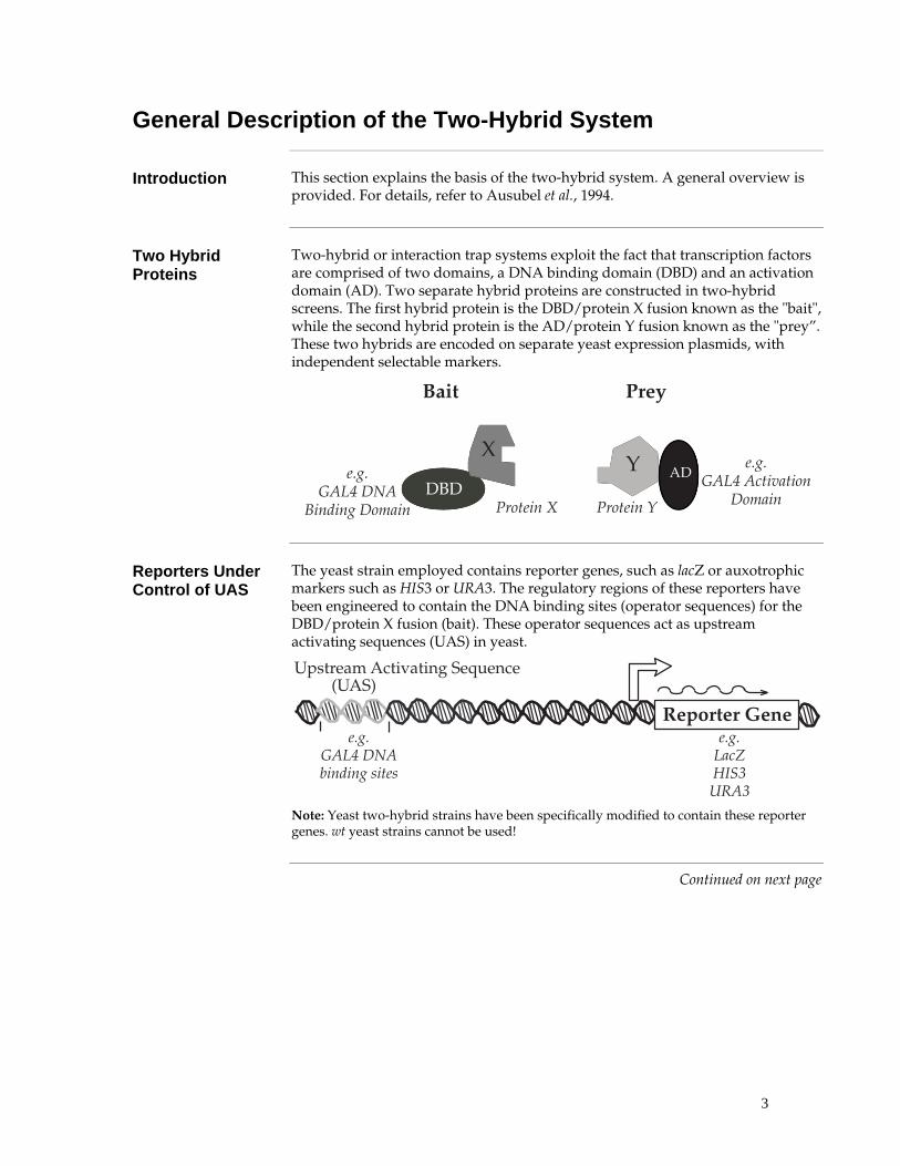
General Description of the Two-Hybrid System
Introduction This section explains the basis of the two-hybrid system. A general overview is provided. For details, refer to Ausubel et al., 1994.
Two Hybrid Proteins
Two-hybrid or interaction trap systems exploit the fact that transcription factors are comprised of two domains, a DNA binding domain (DBD) and an activation domain (AD). Two separate hybrid proteins are constructed in two-hybrid screens. The first hybrid protein is the DBD/protein X fusion known as the "bait", while the second hybrid protein is the AD/protein Y fusion known as the "prey”. These two hybrids are encoded on separate yeast expression plasmids, with independent selectable markers.
���
�
���� ����
� ������������
�� �� ������
������������������
���� ������ �� ������ ��
Reporters Under Control of UAS
The yeast strain employed contains reporter genes, such as lacZ or auxotrophic markers such as HIS3 or URA3. The regulatory regions of these reporters have been engineered to contain the DNA binding sites (operator sequences) for the DBD/protein X fusion (bait). These operator sequences act as upstream activating sequences (UAS) in yeast.
����� ������������������
������� �������������������
���������� � �� ��!���!
�����
Note: Yeast two-hybrid strains have been specifically modified to contain these reporter genes. wt yeast strains cannot be used!
Continued on next page

4
General Description of the Two-Hybrid System, Continued
Interaction Drives Expression of Reporters
The yeast strain used is transformed with the expression plasmids encoding the bait and prey. If protein X interacts with protein Y in the nucleus, this will bring the activation domain together with the DNA-binding domain to reconstitute transcriptional activation and result in expression of the reporter genes.
��������������������������������� �������������
Evaluating Reporter Gene Expression
A
������� ���
������������
�"!������������� ���#$� ��%���&
�
���!�"# ����
���
�����
����
���������������������� ������������$B
���������
�"!������������� ���#$� ��%���&
������
!�"# �������
�����
����
������� ���
There are two main ways to check for positive interactions in yeast strains containing reporter genes:
• Positive interactions are detected by selecting on plates lacking the auxotrophic marker, such as Histidine or Uracil. Yeast cells containing plasmids that express interacting bait and prey proteins will grow and form colonies.
• Positive interactions are detected by assaying for enzyme activity, such as colorimetric assays for β-galactosidase activity. This is used to reduce false positives after selection for auxotrophs, or to measure interaction strength quantitatively.
Note: To select against positive interactions, 5-Fluoroorotic Acid (5FOA) can be used in the appropriate yeast strain, since it kills yeast cells expressing URA3. This feature can be used in reverse two-hybrid screens; see page 53 for explanation.
Continued on next page

5
General Description of the Two-Hybrid System, Continued
Screening Two-Hybrid Libraries
Two-hybrid libraries (i.e. prey libraries) consist of a collection of expression plasmids in which an Activation Domain is fused to individual cDNAs. Screening prey libraries will detect prey proteins that interact with the bait protein of interest. To perform the screen, transform the library and bait expressing plasmids into yeast. Cells containing a prey that interacts with the bait will form colonies on selective plates. Secondary screens, such as for β-galactosidase expression, will confirm the interaction.
False Positives Early two-hybrid systems suffered from false positives - candidate proteins
identified as interacting but which do not truly interact or are biologically irrelevant. False positives can result from:
• Proteins containing regions with surfaces having low affinities for many different proteins, (e.g., large hydrophobic surfaces)
• Proteins that normally interact with a large number of proteins (e.g., heat shock proteins)
• Proteins containing regions functioning as activation domains
• Proteins affecting chromatin structure
• Proteins having low or nonspecific affinities for the promoter regions (or proteins bound there) that drive the expression of reporter genes
Limiting the false positives is essential in successful two-hybrid experiments.
Advanced Two-Hybrid Systems
The ProQuest™ Two-Hybrid System has been extensively improved to limit false positives, as well as to allow performance of more advanced applications, such as reverse two-hybrid. For details about the ProQuest™ Two-Hybrid System and strategies to limit false positives, see page 8; for an explanation on reverse two-hybrid, see page 53.

6
ProQuest™ Two-Hybrid System
Introduction In this section we describe the general properties of the ProQuest™ Two-Hybrid System for use in two-hybrid screens.
Verifying Two-Hybrid Interaction
Using the ProQuest™ Two-Hybrid System to verify an interaction between two known proteins, you will perform the following steps:
1. Construct bait plasmid
2. Construct prey plasmid
3. Transform yeast cells with bait and prey plasmid
4. Test reporter activity
Forward Two-Hybrid Library Screen
Using the ProQuest™ Two-Hybrid System for a forward two-hybrid library screen, you will perform the following steps:
1. Construct and test bait plasmid
2. Construct or obtain two-hybrid library
3. Transform yeast cells with bait plasmid and library
4. Select for reporter activity by growth on auxotrophic plates
5. Confirm interaction of positive prey plasmids
Note: The ProQuest™ Two-Hybrid System comes with a positive interaction control. However, a bait-specific positive interaction control may be an additional useful tool in testing your experiment. Construct a prey plasmid with a known interactor of the bait protein to use as a bait-specific positive control.
Reverse Two-Hybrid Library Screen
Using the ProQuest™ Two-Hybrid System for a reverse two-hybrid library screen, you will perform the following steps:
1. Construct and test bait and wt prey plasmid
2. Construct an allele library of your prey plasmid (see SureFrame™ Allele Library Construction Kit manual)
3. Transform yeast cells with bait plasmid and library
4. Select for reporter activity by growth on selective plates
5. Confirm interaction of positive prey plasmids
Cloning Vectors The ProQuest™ Two-Hybrid System includes these yeast expression vectors:
• pDEST™32 for generation of the bait plasmid
• pDEST™22 for construction of the prey plasmid, or for generation of a two-hybrid library by Gateway® recombination
• pEXP-AD502 for generation of a two-hybrid library by restriction cloning
Continued on next page
7
ProQuest™ Two-Hybrid System, Continued
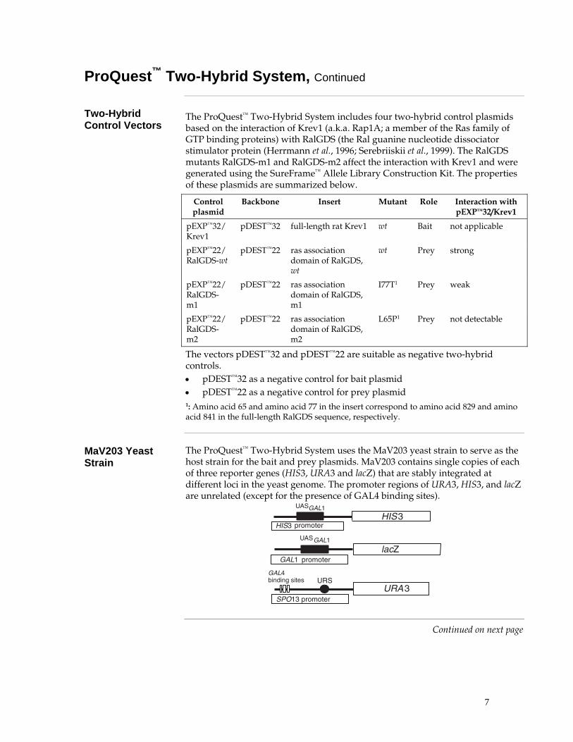
Two-Hybrid Control Vectors
The ProQuest™ Two-Hybrid System includes four two-hybrid control plasmids based on the interaction of Krev1 (a.k.a. Rap1A; a member of the Ras family of GTP binding proteins) with RalGDS (the Ral guanine nucleotide dissociator stimulator protein (Herrmann et al., 1996; Serebriiskii et al., 1999). The RalGDS mutants RalGDS-m1 and RalGDS-m2 affect the interaction with Krev1 and were generated using the SureFrame™ Allele Library Construction Kit. The properties of these plasmids are summarized below.
Control plasmid
Backbone Insert Mutant Role Interaction with pEXP™32/Krev1
pEXP™32/ Krev1
pDEST™32 full-length rat Krev1 wt Bait not applicable
pEXP™22/ RalGDS-wt
pDEST™22 ras association domain of RalGDS, wt
wt Prey strong
pEXP™22/ RalGDS-m1
pDEST™22 ras association domain of RalGDS, m1
I77T1 Prey weak
pEXP™22/ RalGDS-m2
pDEST™22 ras association domain of RalGDS, m2
L65P1 Prey not detectable
The vectors pDEST™32 and pDEST™22 are suitable as negative two-hybrid controls. • pDEST™32 as a negative control for bait plasmid • pDEST™22 as a negative control for prey plasmid 1: Amino acid 65 and amino acid 77 in the insert correspond to amino acid 829 and amino acid 841 in the full-length RalGDS sequence, respectively.
MaV203 Yeast Strain
The ProQuest™ Two-Hybrid System uses the MaV203 yeast strain to serve as the host strain for the bait and prey plasmids. MaV203 contains single copies of each of three reporter genes (HIS3, URA3 and lacZ) that are stably integrated at different loci in the yeast genome. The promoter regions of URA3, HIS3, and lacZ are unrelated (except for the presence of GAL4 binding sites).
����
����
���
�����
�� ����
�� ������
���������������� �
��������
�����
Continued on next page

8
ProQuest™ Two-Hybrid System, Continued
Reducing False Positives
This system reduces false positives by: • Including a third unrelated promoter that facilitates discrimination of
artifactual reporter gene activation. • Providing four phenotypes for assessing true interactors • Using low-copy-number (ARS/CEN) vectors that reduce expression levels
and toxicity
Three Reporter Genes
A major class of false positives is promoter-context dependent, e.g. the prey recognizes promoter sequences or other proteins bound to the promoter (Bartel et al., 1993). In the ProQuest™ Two-Hybrid System, these false positives are reduced because three independent transcription events (from three distinct promoters) must occur at independent chromosomal loci.
Four Phenotypes Induction of the HIS3 and URA3 reporter genes by two-hybrid-dependent transcriptional activation allows cell growth on plates lacking histidine or uracil, respectively. Induction of the lacZ gene results in a blue color when assayed with X-gal (5-bromo-4-chloro-3-indolyl-β-D-galactopyranoside).
Two-hybrid-dependent induction of URA3 results in conversion of the compound 5-fluoroorotic acid (5FOA) to 5-fluorouracil, which is toxic. Hence, cells containing interacting proteins grow when plated on medium lacking uracil, but growth is inhibited when plated on medium containing 5FOA.
��� �� �������������� ��������� ��� � ��
���������� ������ ���
�!"
�#$�!� �
���
���
�����
����
���
�����
Low-Copy-Number Vectors
In the ProQuest™ Two-Hybrid System, the low-copy-number (ARS/CEN) expression vectors express the bait and prey proteins at a relatively low level, which is beneficial for these reasons:
• Overexpression of the bait and prey hybrid proteins increases nonspecific interactions (false positives)
• Many proteins are toxic when overexpressed and interacting proteins may be missed at high expression levels (false negatives)
• ARS/CEN-based vectors provide more consistent plasmid copy numbers (versus the high variability of two micron-based vectors), leading to increased reproducibility of the reporter gene expression levels
• The consistent expression of fusion proteins at levels closer to physiological conditions is particularly valuable for detecting subtle differences, e.g. when characterizing mutations that disrupt known protein:protein interactions
Continued on next page
9
ProQuest™ Two-Hybrid System, Continued
Gateway®
Compatibility Incorporation of the Gateway® Technology into the ProQuest™ System accelerates the cloning of genes into and out of the ProQuest™ Two-Hybrid vectors at several steps:
• Rapidly clone your gene of interest into the bait or prey plasmids using Ultimate™ ORF clones, previously established entry vectors, or a PCR amplification using Gateway® primers
• Transfer libraries into prey plasmid with high efficiency and speed
• Move positive interactors into a variety of expression vectors for downstream protein expression and functional analysis
For more information on Gateway® Recombination, see the Appendix, page 72.
Plasmid Shuffling The ProQuest™ Two-Hybrid System supports plasmid shuffling, which speeds up
rescreening of positives after a library screen.
The yeast strain MaV203 is resistant to cycloheximide (cyhr) due to the recessive cyh2r allele. MaV203 cells containing bait plasmid, which contains the dominant CYH2S gene, are sensitive to cycloheximide. Cells that have spontaneously lost the bait plasmid are selected using cycloheximide The resulting prey-only cells are made competent and re-transformed with the bait. Transformants are then selected and retested for the interaction of bait and prey.
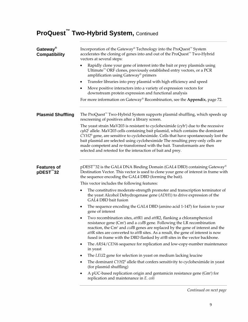
Features of pDEST™32
pDEST™32 is the GAL4 DNA Binding Domain (GAL4 DBD) containing Gateway®
Destination Vector. This vector is used to clone your gene of interest in frame with the sequence encoding the GAL4 DBD (forming the bait).
This vector includes the following features:
• The constitutive moderate-strength promoter and transcription terminator of the yeast Alcohol Dehydrogenase gene (ADH1) to drive expression of the GAL4 DBD bait fusion
• The sequence encoding the GAL4 DBD (amino acid 1-147) for fusion to your gene of interest
• Two recombination sites, attR1 and attR2, flanking a chloramphenicol resistance gene (Cmr) and a ccdB gene. Following the LR recombination reaction, the Cmr and ccdB genes are replaced by the gene of interest and the attR sites are converted to attB sites. As a result, the gene of interest is now fused in frame with the DBD flanked by attB sites in the vector backbone.
• The ARS4/CEN6 sequence for replication and low-copy-number maintenance in yeast
• The LEU2 gene for selection in yeast on medium lacking leucine
• The dominant CYH2S allele that confers sensitivity to cycloheximide in yeast (for plasmid shuffling)
• A pUC-based replication origin and gentamicin resistance gene (Gmr) for replication and maintenance in E. coli
Continued on next page

10
ProQuest™ Two-Hybrid System, Continued
Features of pDEST™22
pDEST™22 is a GAL4 Activation Domain (GAL4 AD) containing Gateway® Destination Vector. This vector is used to clone the second known gene of interest in frame with the sequence encoding the GAL4 AD (generating the prey).
This vector includes the following features:
• The constitutive moderate-strength promoter and transcription terminator of the yeast Alcohol Dehydrogenase gene to drive expression of the GAL4 AD
• The sequence encoding the GAL4 Activation Domain (amino acid 768-881) fused to the nuclear localization signal from SV40 Large T antigen for fusion to your prey gene of interest
• Two recombination sites, attR1 and attR2, flanking a chloramphenicol resistance gene and a ccdB gene. Following the LR recombination reaction, the Cmr and ccdB genes are replaced by the gene of interest and the attR sites are converted to attB sites. As a result, the gene of interest is now fused in frame with AD flanked by attB sites in the Destination Vector backbone.
• The ARS4/CEN6 sequence for replication and maintenance at low-copy- number in yeast
• The TRP1 gene for selection in yeast on medium lacking tryptophan
• A pUC-based replication origin and ampicillin resistance gene for replication and maintenance in E. coli
Features of pEXP™-AD502
pEXP™-AD502 is an Activation Domain (AD) Gateway® Expression Vector. This plasmid is used to construct a cDNA or genomic library for identifying proteins (preys) that interact with the fusion protein (bait).
Features of this vector include:
• The constitutive moderate-strength promoter and transcription terminator of the yeast Alcohol Dehydrogenase gene to drive expression of the GAL4 AD
• The sequence encoding the GAL4 Activation Domain (amino acid 768-881) fused to the nuclear localization signal from SV40 Large T antigen for fusion to your prey gene of interest
• Two recombination sites, attB1 and attB2, flanking a multiple cloning site, including Sal I and Not I sites for generation of cDNA libraries using the SuperScript™ Plasmid System with Gateway® Technology for cDNA Synthesis and Plasmid Cloning.
• The ARS4/CEN6 sequence for replication and maintenance at low-copy- number in yeast
• The TRP1 gene for selection in yeast on medium lacking tryptophan
• A pUC-based replication origin and ampicillin resistance gene for replication and maintenance in E. coli
Note that pEXP™-AD502 is derived from pDEST™22.
Continued on next page

11
ProQuest™ Two-Hybrid System, Continued
Plasmid Isolation Yeast cells containing potentially interacting proteins harbor both bait and prey plasmids. It is desirable to isolate bait and prey plasmids separately in E. coli to confirm the interaction and further characterize the candidate clones. To facilitate the isolation in E. coli, the pDEST™32 vector encodes gentamicin resistance while the pDEST™22 and pEXP™-AD502 vector encodes ampicillin resistance. Plasmid DNA isolated from yeast cells containing bait and prey plasmids is introduced into E. coli by electroporation and transformants containing bait plasmids are selected with ampicillin.
Features of Yeast Strain MaV203
The yeast strain provided in the ProQuest™ System is MaV203 (Vidal et al., 1996; Vidal et al., 1996) and contains the following features:
• A set of non-reverting auxotrophic mutations: leu2 and trp1 to allow selection for the bait and prey fusion vectors, and his3 for growth upon induction of the reporter gene GAL1::HIS3
• Deletions of the GAL4 and GAL80 genes encoding GAL4 and its repressor GAL80, respectively. In the absence of GAL80, galactose is not required for activation of GAL4-inducible promoters
• Three stably integrated single-copy GAL4-inducible reporter genes: SPAL10::URA3 integrated at URA3; HIS3UAS GAL1::HIS3 integrated at LYS2; and GAL1::lacZ integrated at an unknown locus
• The recessive drug resistance marker cyh2R for plasmid shuffling
Genotype MaV203 The genotype of MaV203 is as follows:
MaV203 (MATα, leu2-3,112, trp1-901, his3∆200, ade2-101, gal4∆, gal80∆, SPAL10::URA3, GAL1::lacZ, HIS3UAS GAL1::HIS3@LYS2, can1R, cyh2R) (Vidal, 1997)
���������
The yeast strain MaV203 is unique to the ProQuest™ Two-Hybrid System. Other strains used for two-hybrid analysis cannot be substituted.

12
Considerations in Designing a Two-Hybrid Screen
Introduction Prior to beginning a two-hybrid screen, determine as much information regarding the protein of interest and those interactions that you expect to detect. Several issues that are of particular interest are listed below.
Transcriptional Activator
The fusion of proteins containing domains capable of functioning as transcriptional activators to the GAL4 DBD will induce the reporter genes in the absence of interacting proteins and cannot be used in a typical two-hybrid screen. For example, roughly 0.1% of random E. coli sequences behave as transcriptional activation domains when fused to the GAL4 DBD (Herrmann et al., 1996). Other domain functions should also be considered; e.g., domains exhibiting repressor activity (Vidal, unpublished).
Possible solutions:
• A screen with segments of such proteins that lack these activities can conceivably be constructed and tested.
• Perform a swapped two-hybrid screen where the AD-fusion vector contains the test protein of interest and is used to screen a cDNA library constructed in the DBD-vector (Vidal et al., 1996; Weintraub et al., 1991).
Protein Family It is often useful to anticipate the number of interacting proteins one might expect
to recover from a two-hybrid screen. Test proteins that are members of large protein families may interact with other members at varying degrees, generating a spectrum of reporter gene readout profiles. The prevalence of these proteins should be considered when determining the number of colonies required for a two-hybrid screen and the predicted strength of the reporter gene expression (e.g., strong interactors or weak interactors).
Expression Pattern
The choice of which cDNA library to screen is critical and depends primarily upon the expression pattern of the protein used in the screen. To help in the selection of a cDNA library, verify by PCR the presence of a cDNA corresponding to the bait within the tissue of interest.
Continued on next page

13
Considerations in Designing a Two-Hybrid Screen, Continued
Interaction Artifacts
The interaction of two fusion proteins in a two-hybrid screen is not necessarily an indication that these proteins interact in vivo under native conditions.
• Often only segments of the protein are analyzed, revealing (or masking) domains that might otherwise be unavailable.
• DBD or AD fusion proteins may bear little structural resemblance to the native protein.
• Many posttranslational modifications present in higher eukaryotic cells are absent (or incorrectly modified) in yeast, which may preclude or provide a basis for protein:protein interactions.
• Interactions can be mediated non-specifically (e.g., by large hydrophobic regions).
• Interactions can occur between proteins that are biologically irrelevant (e.g., the proteins exist in different cell types, compartments or at different times during development or cell cycle).
Confirmation of Interaction
It is important to confirm the interactions between protein pairs detected in a two-hybrid screen by biochemical methods. Consider the following options:
• Ideally, the purified protein of interest and antibodies (preferably monoclonal) against the protein of interest are available for immunoprecipitation and Western blot experiments.
• In some cases, antibodies raised against the GAL4 DBD or AD regions, or other epitopes included in the fusion protein, can be used for immunoprecipitation and Western blot experiments.
• Finally, it is important to design in advance a functional test for the biological relevance of the protein:protein interactions.

14
Applications for ProQuest™ Two-Hybrid Screen
Supported Applications
The ProQuest™ Two-Hybrid System supports three types of applications:
• Verifying an interaction between two known proteins or protein domains for which there is a prior reason to expect an interaction
• Screening a library for novel proteins that specifically interact with a known bait (forward two-hybrid library screen)
• Identifying mutations that affect complex formation between two proteins known to interact specifically (reverse two-hybrid screen)
Verifying Interaction
If you want to verify an interaction between two known proteins (testing two-hybrid interactions); see Part A: Verifying Interaction, page 15.
Forward Two-Hybrid Library Screen
If you want to screen a library for novel proteins that specifically interact with your bait (forward two-hybrid library screen); see Part B: Forward Two-Hybrid Library Screen, page 34.
Note: The ProQuest™ Two-Hybrid System comes with a positive interaction control. However, a bait-specific positive interaction control may be an additional useful tool in testing your experiment. Construct a prey plasmid with a known interactor of the bait protein to use as a bait-specific positive control We recommend that you start by verifying the interaction between your bait and this bait-specific positive interaction control, if available. This will allow you to familiarize with the system.
ProQuest™ Reverse Two-Hybrid
If you have already identified a specific two-hybrid interaction and you want to identify mutations that affect complex formation (reverse two-hybrid screen), go to Part C: ProQuest™ Reverse Two-Hybrid, page 53.
Note: Always first verify the interaction as described in Part A: Verifying Interaction before using the SureFrame™ Allele Library Construction Kit to generate your allele library.

15
Part A: Verifying Interaction
Introduction
Introduction This chapter describes how to verify an interaction between two known proteins or protein domains for which there is a prior reason to expect an interaction.
The first part describes the construction of the required bait and prey plasmids (page 16).
The second part describes how to test a specific two-hybrid interaction (page 25).
If you are checking an interaction found in a ProQuest™ forward or reverse two-hybrid screen (a retransformation assay), skip the first part and go directly to Testing Specific Two-Hybrid Interaction, page 25.
Flowchart The figure below illustrates the major steps necessary to verify an interaction
using the ProQuest™ Two-Hybrid System.
������� ������������ ��������������
����������������������
�����������
�������� �
���������������������
���������!�
"������ �
������ #��������� ����#������$%� ������&��� ���������#������$%� �����'�� �����&��� �������
��� #������ �� ������(�)!
� �����������*���
���*�������+�"���� ��������� �
� ��+����#���&��� ����������
���!,����!��� ���-
� ���#� �������� �
�. ��+������� �

16
Methods
Generating Bait and Prey Plasmids Overview
Introduction This section describes how to generate specific bait and prey plasmids. The gene
of interest for the bait plasmid is cloned into pDEST™32, resulting in pEXP32 containing your gene of interest. The gene of interest for the prey plasmid is cloned into pDEST™22, resulting in pEXP™22 containing your gene of interest. pDEST™32 and pDEST™22 are Gateway®-adapted destination vectors.
Gateway® Technology
The Gateway® Technology is a universal cloning method that takes advantage of the site-specific recombination properties of bacteriophage lambda (Landy, 1989) to provide a rapid and highly efficient way to move your DNA sequence of interest into multiple vector systems.
For details about the Gateway® Recombination Reactions, see the Appendix, page 72.
Requirement of Bait and Prey
A specific bait plasmid is required for every application of this manual. A specific prey plasmid is required if you want to test a specific forward two-hybrid interaction or perform a reverse two-hybrid screen. However, a specific prey plasmid of a known interactor of the bait is a valuable positive control when screening a forward two-hybrid library, and you should consider generating one.
���������
If you want to test a specific forward two-hybrid interaction between two proteins, we recommend constructing prey plasmids of each cDNA, and also bait plasmids of each cDNA. This way, you can perform the two-hybrid assay two ways: with protein A as bait and protein B as prey, and with protein B as bait and protein A as prey. This will generate more convincing data regarding the interaction you want to test.
Construction of Plasmids
To construct your bait or prey plasmids, perform the following steps:
1. Identify or generate a suitable entry clone.
2. Perform an LR recombination reaction between entry clone and pDEST™32 or pDEST™22.
3. Transform competent cells.
4. Select the proper expression clone.
Continued on next page
17
Overview, continued
Entry Clone In order to generate the bait plasmid and the prey plasmid, you need an entry
clone with your gene of interest flanked by attL1 and attL2 sites. Use an entry clone you generated for previous Gateway® recombinations, or obtain an Ultimate™ ORF clone of your gene of interest (see orf.invitrogen.com or contact Technical Service, page 99). See the next page for requirements of the entry clone.
Generating a New Entry Clone
If you do not have an entry clone available, and an Ultimate™ ORF clone is not an option, you can generate your entry clone in a number of different ways:
• Perform a BP recombination reaction between an existing expression or cDNA clone with attB1 and attB2 sites and an appropriate donor vector (such as pDONR™221; see the Gateway® Technology with Clonase™ II manual)
• Carry out a PCR on your gene of interest and use TOPO cloning into a suitable entry vector (such as pCR®8/GW/TOPO® TA, Catalog no. K2500-20; see the pCR8®/GW/TOPO®TA Cloning Kit manual, or see the pENTR™ Directional TOPO® Cloning Kits manual for directional cloning)
• Perform PCR with attB primers on your gene of interest and a BP recombination reaction with an appropriate donor vector (such as pDONR™221; see the Gateway® Technology with Clonase™ II manual)
• Use restriction digestion to clone your gene of interest into an entry vector (see the Gateway® pENTR™ Vectors manual)
The indicated manuals are available from www.invitrogen.com or by contacting Technical Service (page 99).
Reading Frame and Stop Codons
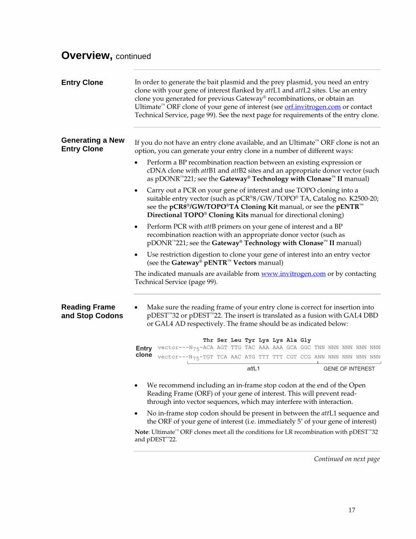
• Make sure the reading frame of your entry clone is correct for insertion into pDEST™32 or pDEST™22. The insert is translated as a fusion with GAL4 DBD or GAL4 AD respectively. The frame should be as indicated below:
��������������������� ����������������� ��� ��� ��� ��� ��� ��� ��� ��� ��� ��� ��� ���
���%�
�������������� ��� ��� ��� ��� ��� ��� ��� ��� ��� ��� ��� ���
$&'&����(')&�&�)
��������
• We recommend including an in-frame stop codon at the end of the Open Reading Frame (ORF) of your gene of interest. This will prevent read-through into vector sequences, which may interfere with interaction.
• No in-frame stop codon should be present in between the attL1 sequence and the ORF of your gene of interest (i.e. immediately 5’ of your gene of interest)
Note: Ultimate™ ORF clones meet all the conditions for LR recombination with pDEST™32 and pDEST™22.
Continued on next page

18
Overview, continued
Cloning Site and Recombination Region of pDEST™32
Use the diagram below to help you clone your gene of interest into pDEST™32. Note the following features in the diagram below:
• The shaded region corresponds to those DNA sequences that will be transferred from the entry vector into pDEST™32 following recombination, forming the bait vector pEXP™32
• The reading frame for the GAL4 DBD is shown; the insert needs to be in frame with GAL4 DBD
• Sequences for suggested forward and reverse sequencing primers are shown
The complete sequence of pDEST™32 is available for downloading from our Web site (www.invitrogen.com) or by contacting Technical Service (see page 99). For a map of pDEST™32, see the Appendix, page 91. ����
����
����
����
����
����
����
����
����
����
����
����
���������� ����������
�������� ������������������������������
��
��
����
���
�������� � ��!�� ����������������������
���������� ���������� ���������� ���������� ���������� ���������� ����������
��� ��� ��� ��� ��� ��� ��� ��� ��� ��� ��� ��� ��� ��� ��� ��� ��� ������ ��� ��� ��� ��� ��� ��� ��� ��� ��� ��� ��� ��� ��� �� ��� ��� ���
��� ��� ��� ��� ��� ��� ��� ��� ��� ��� ��� ��� ��� ��� ��� ��� ��� ������ ��� ��� ��� ��� ��� ��� !�� ��� ��� ��� ��� ��� ��� ��� ��� ��� ���
��� ��� ��� ��� ��� ��� ��� ��� ��� ��� ��� ��� ��� ��� ��� ��� ��� ������ ��� �� ��� ��� !�� ��� �"� ��� �� ��� !�� ��� �"� �� ��� #$� ���
��� ��� ��� ��� ��� ��� ��� ��� ��� ��� ��� ��� ��� ��� ��� ��� ��� ����"� ��� %�� ��� ��� �� ��� ��� �� ��� ��� ��� ��� !"� ��� ��� ��� !"�
��� ��� ��� ��� ��� ��� ��� ��� ��� ��� ��� ��� ��� ��� ��� ��� ��� ���!�� �� ��� ��� ��� ��� &�� ��� ��� ��� &�� ��� ��� ��� ��� ��� ��� ���
��� ��� ��� ��� ��� ��� ��� ��� ��� ��� ��� ��� ��� ��� ��� ��� ��� ������ ��� ��� �"� ��� ��� !"� %�� ��� ��� ��� %�� ��� ��� ��� ��� %�� �"�
��� ��� ��� ��� ��� ��� ��� ��� ��� ��� ��� ��� ��� ��� ��� ��� ��� ������ �� ��� ��� ��� %�� ��� �"� ��� &�� !�� ��� �"� ��� �� ��� #$� ��
��� ��� ��� ��� ��� ��� ��� ��� ��� ��� ��� ��� ��� ��� ��� ��� ��� ������ ��� ��� �"� ��� ��� ��� ��� ��� ��� ��� ��� ��� ��� ��� �� ��� ���
��� ��� ��� ��� ��� ��� ��� ��� ��� ��� ��� ��� ��� ��� ��� ��� ���� ��� ��� ��� ��� ��� ��� ��� ��� ��� ��� ��� ��� ��� ��� ��� � ��������� ��"� %�� ��� ��� �� ��� ��� ��� �"� ��� ��� ��� ��� ��� ��� ���
���������� ���������� ���������� ���������� ���������� ���������� �������������������� ���������� ����������
���������� ���������� ���������� ���������� ���������� ���������� ����������
����������
Continued on next page

19
Overview, continued
Cloning Site and Recombination Region of pDEST™22
Use the diagram below to help you clone your gene of interest into pDEST™22. Note the following features in the diagram below:
• The shaded region corresponds to those DNA sequences that will be transferred from the entry vector into pDEST™22 following recombination, forming the bait vector pEXP™22
• The reading frame for the GAL4 AD is shown; the insert needs to be in frame with GAL4 AD
• Sequences for suggested forward and reverse sequencing primers are shown
The complete sequence of pDEST™22 is available for downloading from our Web site (www.invitrogen.com) or by contacting Technical Service (see page 99). For a map of pDEST™22, see the Appendix, page 92.
���������� ���������� ���������� ���������� ���������� ���������� ����������
��� ��� ��� ��� ��� ��� ��� ��� ��� ��� ��� ��� ��� ��� ��� ��� ��� ��� ���
��� ��� ��� ��� ��� ��� ��� ��� ��� ��� ��� ��� ��� ��� ��� ��� ��� ��� ���
��� ��� ��� ��� ��� ��� ��� ��� ��� ��� ��� ��� ��� ��� ��� ��� ��� ��� ���
��� ��� ��� ��� ��� ��� ��� ��� ��� ��� ��� ��� ��� ��� ��� ��� ��� ��� ���
��� ��� ��� ��� ��� ��� ��� ��� ��� ��� ��� ��� ��� ��� ��� ��� ��� ��� ���
��� ��� ��� ��� ��� ��� ��� ��� ��� ��� ��� ��� ��� ��� ��� ��� ��� ��� ���
��� ��� ��� ��� ��� ��� ��� ��� ��� ��� ��� ��� ��� ��� ��� ��� ��� ���
��� ��� ��� ��� ��� ��� � ���������� ���������� ����������
���������� ���������� ���������� ���������� ���������� ���������� ����������
���������� ���������� ���������� ���������� ���������� ���������� ����������
�� ��
�������� ������������������������������
�������� � ��!�� ����������������������
��"��������"�#�����������"
��� !�� ��� ��� ��� �� ��� %�� ��� ��� ��� ��� ��� !"� ��� ��� ��� ��� ���
��� ��� ��� ��� ��� ��� ��� !"� �"� !"� �"� ��� ��� ��� ��� ��� !�� ��� ���
��� �"� �"� ��� �"� ��� ��� ��� ��� ��� ��� ��� !�� ��� ��� ��� ��� ��� %��
#$� ��� ��� !"� &�� ��� ��� ��� ��� �"� ��� ��� ��� ��� ��� ��� ��� ��� ���
��� ��� !�� ��� ��� !�� ��� ��� �"� ��� ��� �"� ��� ��� ��� ��� !"� ��� ���
�"� �"� ��� &�� !"� ��� �"� �"� �"� &�� ��� ��� %�� ��� ��� ��� ��� !"� ���
��� ��� ��� �"� !�� !�� ��� !�� ��� ��� ��� ��� ��� ��� ��� ��� �"� ���
��� ��� ��� ��� ��� ��� � ���������� � ���������� ���������� ���������� ��� ��� ��� ��� ��� ���
��������������������������
�����
�����
�����
�����
�����
�����
�����
�����
�����
�����
�����
�'(�(��
Continued on next page

20
Overview, continued
Experimental Outline
The experimental outline for generating the bait and prey plasmids is shown below.
�������������#$��� �������
�%������ ���
�"���������&'�'(�������
�����)����!��*��+�'(�������
������� �������������
�%�,����������"�������% "��-� ������"���
������� ���
�"�����������.��/�'+�+
�������
/
�������������������������������!�
��� #����0���������0���
���������1��!���.��� ��������
������������
�����)����!��*��+�'(�������
��������������$��"����
0�����*�������� ��*��������"1
��
���������������������������������
��� #����0���������0���
���������1������.��� ��������
�������� � "������ �
/

21
Creating Bait and Prey Plasmids Using the LR Recombination Reaction
Introduction This section explains how to create specific bait and prey plasmids using an
existing entry clone and the destination vectors pDEST™32 and pDEST™22. To ensure that you obtain the best possible results, we suggest that you read this section and the next section entitled Transforming Competent Cells with Bait and Prey Plasmids before beginning.
Substrates for the LR Recombination Reaction
For most applications, we recommend performing the LR recombination reaction using a:
• Supercoiled attL1 and attL2-containing entry clone
• Supercoiled pDEST™32 or pDEST™22 (contains attR1 and attR2)
Although the Gateway® Technology manual has previously recommended using a linearized destination vector and entry clone for more efficient LR recombination, further testing at Invitrogen has found that linearization of destination vectors and entry clones is generally NOT required to obtain optimal results for any downstream application.
LR Clonase™ II Enzyme Mix
LR Clonase™ II enzyme mix is provided with the kit to catalyze the LR recombination reaction. The LR Clonase™ II enzyme mix combines the proprietary enzyme formulation and 5X LR Clonase Reaction Buffer previously supplied as separate components in LR Clonase™ enzyme mix (Catalog no. 11791-019) into an optimized single tube format to allow easier set-up of the LR recombination reaction. Use the protocol provided on the next page to perform the LR recombination reaction using LR Clonase™ II enzyme mix.
Note: You may perform the LR recombination reaction using LR Clonase™ enzyme mix, if desired. To use LR Clonase™ enzyme mix, follow the protocol provided with the product. Do not use the protocol for LR Clonase™ II enzyme mix provided on the next page.
Resuspending pDEST™32 and pDEST™22
The pDEST™32 and pDEST™22 vectors are supplied as 6 µg of plasmid DNA, lyophilized in TE Buffer, pH 8.0. To use the vector, resuspend in 40 µl of sterile water to obtain a 150 ng/µl stock.
Positive Recombination Control
The pENTR™-gus plasmid is provided with the LR Clonase™ II Enzyme Mix for use as a positive control for recombination and expression. Using the pENTR™-gus entry clone in an LR recombination reaction with a destination vector will allow you to generate an expression clone containing the gene encoding β-glucuronidase (gus) (Kertbundit et al., 1991).
Continued on next page

22
Creating Bait and Prey Plasmids Using the LR Recombination Reaction, continued
Materials Needed You should have the following materials on hand before beginning:
• Purified plasmid DNA of your entry clone for the bait and/or your entry clone for the prey (50-150 ng in TE, pH 8.0)
• pDEST™32 and/or pDEST™22 (both 150 ng/µl in TE, pH 8.0)
• LR Clonase™ II enzyme mix (keep at -20°C until immediately before use)
• TE Buffer, pH 8.0 (10 mM Tris-HCl, pH 8.0, 1 mM EDTA)
• 2 µg/µl Proteinase K solution (supplied with the LR Clonase™ II enzyme mix; thaw and keep on ice until use)
• Positive Recombination Control pENTR-gus, if desired
Setting Up the LR Recombination Reaction
1. Add the following components to 1.5 ml microcentrifuge tubes at room temperature and mix. Note: To include a negative control, set up a second sample reaction and omit the LR Clonase™ II enzyme mix (see Step 4).
Forming Bait Plasmid Forming Prey Plasmid Component
Sample Negative Control
Positive Control
Sample Negative Control
Positive Control
Entry clone for bait (50-150 ng/reaction)
1-7 µl 1-7 µl -- -- -- --
Entry clone for prey (50-150 ng/reaction)
-- -- -- 1-7 µl 1-7 µl --
pDEST™32 (150 ng/µl) 1 µl 1 µl 1µl -- -- --
pDEST™22 (150 ng/µl) -- -- -- 1 µl 1 µl 1µl
pENTR™-gus (50 ng/µl) -- -- 2µl -- -- 2µl
TE Buffer, pH 8.0 to 8 µl to 10 µl 5 µl to 8 µl to 10 µl 5 µl
2. Remove the LR Clonase™ II enzyme mix from -20°C and thaw on ice (~ 2 minutes).
3. Vortex the LR Clonase™ II enzyme mix briefly twice (2 seconds each time). 4. Add 2 µl of LR Clonase™ II enzyme mix to the sample vial. Do not add LR
Clonase™ II enzyme mix to the negative control vial. Mix well by vortexing briefly twice (2 seconds each time). Reminder: Return LR Clonase™ II enzyme mix to -20°C immediately after use.
5. Incubate reactions at 25°C for 1 hour. Note: For most applications, 1 hour will yield a sufficient number of colonies for analysis. Depending on your needs, the length of the recombination reaction can be extended up to 18 hours. For large plasmids (≥ 10 kb), longer incubation times (i.e. overnight incubation) will yield more colonies and are recommended.
6. Add 1 µl Proteinase K solution to each reaction. Incubate 10 minutes at 37°C.
7. Proceed to transform a suitable E. coli host and select for expression clones. Note: You may store the LR reaction at -20°C for up to 1 week before transformation, if desired.

23
Transforming Competent Cells with Bait and Prey Plasmids
Introduction Competent E. coli cells are not provided with the ProQuest™ Two-Hybrid System.
Below the prerequisites of appropriate host strains are indicated. You can order suitable competent cells from Invitrogen or use your standard in-house competent cells if they are compatible.
E. coli Host Strain You may use any recA, endA E. coli strain including OmniMAX™ 2-T1R, TOP10,
DH5α™, DH10B™or equivalent for transformation. Other strains are suitable. Do not use E. coli strains that contain the F′ episome (e.g. TOP10F′) for transformation. These strains contain the ccdA gene and will prevent negative selection with the ccdB gene.
For your convenience, TOP10, DH5α™, and DH10B™ E. coli are available as chemically competent or electrocompetent cells from Invitrogen (see table below).
Item Quantity Catalog No.
Library Efficiency® DH5α™ E. coli 5 x 200 µl 18263-012
One Shot® TOP10 Chemically Competent E. coli 20 x 50 µl C4040-03
One Shot® Max Efficiency® DH10B™ T1 Phage Resistant Chemically Competent E. coli
20 x 50 µl 12331-013
One Shot® TOP10 Electrocomp E. coli 20 x 50 µl C4040-52
ElectroMax™ DH10B™ E. coli 5 x 100 µl 18290-015
Procedure Transform 1 µl of the LR reaction according to the protocol provided with your
competent cells, and plate two concentrations of cells on 10 cm diameter LB agar plates with 10 µg/ml gentamicin (for bait plasmids) or 100 µg/ml ampicillin (for prey plasmids). Let grow overnight at 37°C.
What You Should See
If you use E. coli cells with a transformation efficiency of ≥ 1 x 10⁸ cfu/µg, the LR reaction should give > 5000 colonies if the entire LR reaction is transformed and plated.

24
Analyzing Transformants
Analyzing Transformants
To analyze positive clones, we recommend that you:
1. Pick 5-10 colonies and culture them overnight in LB or SOB medium containing 10 µg/ml gentamicin (for bait plasmids) or 100 µg/ml ampicillin (for prey plasmids).
2. Isolate plasmid DNA using your method of choice. To obtain pure plasmid DNA for automated or manual sequencing, we recommend using the PureLink™ HQ Mini Plasmid Purification Kit (Catalog no. K2100-02).
3. Perform restriction analysis to confirm the presence of the insert.
Note: BsrG I cleaves within all att sites, and can be used to help characterize clones.
Sequencing If you sequenced your entry clone, sequence analysis is not required. However, if
you want to perform sequencing to confirm the reading frame of your expression clone, use primers that anneal 50-300 bp from the junction (either within the vector or the insert). Below are primers you can use to sequence bait and prey junctions.
Plasmid Direction Primer
Bait forward 5´-AACCGAAGTGCGCCAAGTGTCTG-3’
Bait and Prey reverse 5’-AGCCGACAACCTTGATTGGAGAC-3’
Prey forward 5´-TATAACGCGTTTGGAATCACT-3’
If you want to design your own primer, you can download the sequence for pDEST™32 or pDEST™22 from our Web site, www.invitrogen.com. Make sure the 3’ end of the primer is directed towards the junction you want to sequence.
Long-Term Storage
Once you have identified the correct expression clone, be sure to purify the colony and make a glycerol stock for long-term storage. We recommend that you store a stock of plasmid DNA at -20°C.
1. Streak the original colony out for a single colony on an LB plate containing 10 µg/ml gentamicin (for bait plasmids) or 100 µg/ml ampicillin (for prey plasmids).
2. Isolate a single colony and inoculate into 1-2 ml of LB containing 10 µg/ml gentamicin (for bait plasmids) or 100 µg/ml ampicillin (for prey plasmids).
3. Grow until the culture reaches stationary phase.
4. Mix 0.85 ml of culture with 0.15 ml of sterile glycerol and transfer to a cryovial.
5. Store the glycerol stock at -80°C.

25
Testing Specific Two-Hybrid Interaction Overview
Introduction This section describes how to transform your bait and prey plasmid into MaV203
cells, and test activation of the three reporter genes. Use this chapter for three purposes:
• To test a specific interaction between two proteins
• Retransformation assay to confirm the interaction of your bait with a prey identified in the forward two-hybrid screen (page 40)
• Retransformation assay to validate the loss of interaction of a mutant prey as identified in the reverse two-hybrid screen (page 53)
Recommended Controls
The following transformation, and interaction controls are recommended. Bait and prey plasmids are not provided with the system and need to be generated (Generating Bait and Prey Plasmids, page 16); the other vectors are provided with the kit.
LEU2 Plasmid TRP1 Plasmid Purpose
1 none none Negative transformation control
2 pEXP™32/Krev1 pEXP™22/RalGDS-wt Strong positive interaction control
3 pEXP™32/Krev1 pEXP™22/RalGDS-m1 Weak positive interaction control
4 pEXP™32/Krev1 pEXP™22/RalGDS-m2 Negative interaction control
5 pDEST™32 pDEST™22 Negative activation control
6 Bait plasmid pDEST™22 Negative activation control; baseline
If available:
7 pDEST™32 Prey plasmid known to interact with bait
Negative activation control
8 Bait plasmid Prey plasmid known to interact with bait
Bait-specific positive interaction control (if available)
Note: If you are testing multiple bait plasmids, perform a transformation with each bait plasmid for controls 6 and 8; label the controls 6a, 6b, 8a, 8b and so on.
Continued on next page

26
Overview, continued
Experimental Outline
The experimental outline for Testing Specific Two-Hybrid Interaction is shown below.
�������� � "������ �
������ #��������2 �����$%� �����&��� ������
��� #������� � �������(�)!
� �����������*���
��*�������+�"���� ��������� �
� ��+����#���&��� ����������3������ #�������2 ��
���!,����!��� ���-

27
Small Scale Yeast Transformation
Competent Yeast Cells
The MaV203 yeast strain is provided with the kit to serve as the host for your bait and prey plasmids. Below we provide a small-scale protocol for transforming yeast cells. Prepare a new batch of competent cells for every transformation.
Alternatively, to limit your workload and increase the transformation efficiency, you may purchase the following products:
• MaV203 Competent Cells, Subclone Scale from Invitrogen (Catalog no. 11445-012). Use the transformation protocol provided with these cells.
• If you plan to do library scale transformations later on, you can purchase MaV203 Competent Cells, Library Scale from Invitrogen (Catalog no. 11281-011) and use one vial to perform multiple small-scale transformations. Per transformation, scale down to 25 µl competent cells, 1 µg DNA per plasmid, 180 µl PEG/LiAc, 10.8 µl DMSO. Otherwise, follow the transformation protocol provided with these cells.
• If you want to generate a large batch of competent cells that can be frozen, use the S. c. EasyComp™ Kit (Catalog no. K5050-01). Use the transformation protocol provided with this kit.
Note: There are other small-scale transformation methods that can be used; see Gietz et al., 1992; Gietz and Schiestl, 1996; Hill et al., 1991; Schiestl and Gietz, 1989.
Transformation Guide
Perform two transformations per interaction pair you want to test:
• pDEST™32 and prey plasmid to test
• Bait plasmid and prey plasmid to test
Additionally, we recommend generating the controls indicated in the table on page 25.
Select on SC-Leu-Trp plates. Store representative transformants in glycerol at -80°C for future use.
Note: If you want to test a specific forward two-hybrid interaction between two proteins two ways (i.e. with protein A as bait and protein B as prey, and with protein B as bait and protein A as prey), you will perform four transformations per interaction.
���������
You need to generate fresh plates of controls 2-8 (see table on page 25). If you have previously generated the controls and do not want to retransform them, streak a colony from the stored plates onto new SC-Leu-Trp plates and incubate for 48 hours at 30°C. To initiate cultures from frozen yeast stocks, streak a small amount of frozen stock on a YPAD plate. Once growth is established, you may check the phenotype of each strain by streaking the strain on a minimal plate supplemented with the appropriate amino acids. Keep glycerol stocks of all strains including transformed strains. If you use strains or transformants directly from plates be sure the plates are less than 4 days old.
Continued on next page

28
Small Scale Yeast Transformation, continued
Materials Needed Be sure to have the following reagents on hand before starting.
• YPAD
• 1X TE
• 1X LiAc (100mM Lithium Acetate/0.5X TE)
• Denatured sheared salmon sperm DNA (Invitrogen, Catalog no. 15632-011)
• Plasmid DNA to be transformed
• 1X LiAc/40% PEG-3350/1X TE
• DMSO. For best results, use fresh DMSO from an unopened bottle. DMSO that has been stored at -20°C also works well.
• SC-Leu-Trp plates, for selection of yeast cells transformed with both the bait and prey plasmid, or LEU2 and TRP1 plasmid of the controls (see page 25)
• Bait plasmid
• Prey plasmid known to interact with bait in yeast two-hybrid (if available)
• pDEST™32, pDEST™22, pEXP™32/Krev1, pEXP™22/RalGDS-wt, pEXP™22/RalGDS-m1, pEXP™22/RalGDS-m2 (supplied with the kit)
Preparing Competent MaV203 Cells
1. Inoculate 10 ml of YPAD with a colony of MaV203 and shake overnight at 30°C.
2. Determine the OD600 of your overnight culture. Dilute culture to an OD600 of 0.4 in 50 ml of YPAD and grow an additional 2-4 hours.
3. Pellet the cells at 2500 rpm and resuspend the pellet in 40 ml 1X TE.
4. Pellet the cells at 2500 rpm and resuspend pellet in 2 ml of 1X LiAc/0.5X TE.
5. Incubate the cells at room temperature for 10 minutes
6. Proceed immediately to transform the competent MaV203 cells
Transformation of Competent MaV203 Cells
The protocol below describes transformation of MaV203 yeast cells using your own prepared competent cells. To transform MaV203 cells using purchased MaV203 Competent Cells, or the S. c. EasyComp™ Kit, refer to the manual included with each product.
1. For each transformation, mix together 1 µg plasmid DNA and 100 µg denatured sheared salmon sperm DNA with 100 µl of the yeast suspension from Step 5, above.
2. Add 700 µl of 1X LiAc/40% PEG-3350/1X TE and mix well.
3. Incubate solution at 30°C for 30 minutes.
4. Add 88 µl DMSO, mix well, and heat shock at 42°C for 7 minutes.
5. Centrifuge in a microcentrifuge for 10 seconds and remove supernatant.
6. Resuspend the cell pellet in 1 ml 1X TE and re-pellet.
7. Resuspend the pellet in 50-100 µl TE and plate on a selective plate.

29
Characterization of Transformants
Introduction MaV203 cells that contain bait and prey proteins that strongly interact will induce all three reporter genes present in this system (HIS3, URA3, lacZ). Identify these colonies by a series of patching and replica plating steps onto the selection/screen plates, which are described in this section.
��#% "#)�#��
* � ��� � ���� �
��#% "#)�+�������#% "#)�#���+��)
,��*��!�� �������������-�!� -�.#��!������
��� -��.#��!������* � ��� �� ���� �
* � ��� � ���� �
* � ��� � ���� �
�������������� ���� �!�����!�� ������� � � ���� � /� �� ��� ���� �
� �!�����! ��(��"��� ��0123�4��
(��"��� ��0123�4��
� �!�����! ��(��"��� ��0123�4�����
� �!�����! ��(��"��� ��0123�4�����
� �!�����! ��(��"��� ��0123�4�����
2��!�4
2��!��
2��!��
2��!��2��!�5
� �!�����!�� ����� ! ������!�� �6
��� �!�����! ��(��"��� ��0123�4��
(��"��� ��0123�4��
)���-���������2#% "#)�
����������! ��2#% "#)���!�� ����� 7
�����"���!�� ���!�� �8�*&�)9�4�+�� ����� ��
����):����� ��-�2��!��4#;
(��"��� ��0123��;�
2��!�<
�����"���!�� ���!�� �8�����+�� ����� ��
������,"������!��� -��� ="��������> ��'�$��2��$
�����3�� ���� ������ ���� ���/
Note: For an explanation of controls 2-8, see Recommended Controls, page 25
Continued on next page

30
Characterization of Transformants, continued
Required Test Plates
To test a specific interaction, use the plates described below for the assay you perform.
Test HIS3 induction URA3 induction
URA3 induction
β-Galactosidase induction
Assay His auxotrophy 5FOA sensitivity
Uracil auxotrophy
X-gal assay
Plates used SC-Leu-Trp-His+3AT
SC-Leu-Trp+5FOA
SC-Leu-Trp-Ura
YPAD
Concentrations 10 mM 3AT 25 mM 3AT 50 mM 3AT 100 mM 3AT
0.2% 5FOA No Uracil Not applicable
Note: For an explanation about the HIS3 inhibitor 3AT, refer to Testing Bait, page 36
Materials Needed • Plates with transformants and controls (see table, page 25)
• Fresh plates: ∗ SC-Leu-Trp plates, to grow yeast containing the 2 plasmids to be tested ∗ YPAD plates, for X-gal assays to test lacZ induction ∗ SC-Leu-Trp-Ura plates to test URA3 induction ∗ SC-Leu-Trp-His+3AT plates, to test HIS3 induction ∗ SC-Leu-Trp+5FOA plates, to select yeast cells that do not induce URA3
• nitrocellulose or nylon membrane
• 30°C incubator
• Autoclaved velvets for replica plating/cleaning
• X-gal (5-bromo-5-chloro-3-indolyl-β-D-galactoside)
• N,N-dimethyl formamide (DMF)
• Z buffer (60 mM Na2HPO4, 40 mM NaH2PO4 , 10 mM KCl, 1 mM MgSO4, pH 7.0)
• 2-mercaptoethanol
• 125-mm Whatman 541 filter papers
• 15-cm petri dishes
• Forceps
• Liquid nitrogen
Continued on next page

31
Characterization of Transformants, continued
Generating Master Plates
1. Using an autoclaved toothpick or loop, patch onto a single SC-Leu-Trp plate the following:
• Two isolated colonies of yeast controls 2-8 (see table, page 25)
• Four isolated colonies of transformants containing pDEST™32 and prey for an interaction you want to test (up to two clones can be analyzed per plate)
• Four isolated colonies of transformants containing bait plasmid and prey for an interaction you want to test (up to two clones can be analyzed per plate)
2. If more than 2 preys need to be tested, use additional plates. On each plate include the yeast controls 2-8. Put transformants containing the same prey and pDEST™32 or bait plasmid on one plate.
3. Incubate plates for 18 hours at 30°C. Note: If you want to test a specific forward two-hybrid interaction between two proteins two ways (i.e. with protein A as bait and protein B as prey, and with protein B as bait and protein A as prey), put both interaction pairs on the same plate.
���������
If you are unfamiliar with replica plating and replica cleaning, see page 78 before continuing this protocol. Replica cleaning is essential to reduce background.
Testing Reporter Genes
1. Replica plate onto the following plates, in the order listed. Be sure to make asymmetric marks on the plates and membrane to allow for realignment with the master plate. Replica clean where indicated. • YPAD containing a nitrocellulose or nylon membrane for an X-gal Assay • SC-Leu-Trp-Ura • SC-Leu-Trp-His+3AT; replica clean • SC-Leu-Trp+5FOA; replica clean
2. Incubate all plates for ~24 hours at 30°C.
3. After 18 to 24 hours incubation of the YPAD plates containing a membrane, perform an X-gal Assay on the membrane as described below.
4. After incubation of the selection plates for 24 h, replica clean the following plates: • SC-Leu-Trp-Ura • SC-Leu-Trp+5FOA • SC-Leu-Trp-His+3AT
5. Incubate for 2 additional days at 30°C.
6. Compare the phenotypes of the transformants to yeast control 2-8 (see table, page 25), and to the phenotype exhibited in the original screen. Weak phenotypic differences should be considered. A particular prey that activates target genes in the presence of DEST™32 is likely a false positive.
Continued on next page

32
Characterization of Transformants, continued
Factors Influencing Growth Properties
The growth properties of yeast cells on the selection/screen plates can be influenced by several parameters in addition to the induction levels of the reporter genes:
• Cells approaching stationary phase exhibit different expression levels of the hybrid proteins from cells growing in exponential phase.
• As the number of cells transferred by replica plating increases, the phenotypic differences between positive and negative controls decrease. Replica cleaning dilutes and normalizes the number of cells plated.
• The amount of growth of yeast patches on a particular selection plate will vary dramatically between 2 and 6 days of growth. Correct incubation times are critical.
X-gal Assay 1. For each membrane, dissolve 10 mg X-gal in 100 µl DMF. Combine 100 µl
X-gal in DMF, 60 µl 2-mercaptoethanol and 10 ml Z buffer.
2. Stack two round 125-mm Whatman 541 filter papers in a 15-cm petri dish. Saturate with ~8 ml of the X-gal solution. Remove any air bubbles.
3. Using forceps, carefully remove the membrane from the surface of the YPAD plate. Completely immerse the membrane in liquid nitrogen for 20-30 seconds. Place the frozen membrane on top of the soaked Whatman filters colony side up. Remove any air bubbles. Tip the plates slightly and remove excess buffer.
4. Cover the plates and incubate at 37°C. Tip the plates at a slight angle so excess X-gal solution does not accumulate on the membrane. Monitor the appearance of blue color over a 24-h period. Score final results at 24 hours.
Strong interactors show blue color within 1 hour (e.g., yeast control 2; see table, page 25). Weak interactors (e.g., yeast control 3) show blue color within 24 h, but can remain very faint blue to white.
• When scoring the results, the membrane will be a mirror image of the master plate.
• Nitrocellulose membranes are fragile and can crack during freezing; therefore neutrally charged nylon membranes are recommended.
• Handle liquid nitrogen with care. Always wear thick gloves and goggles.
• ONPG will stain. Wear gloves during these procedures.
Quantitative β-Galactosidase Assays
For quantitative measurements of β-galactosidase induction, perform the ONPG assay or the CPRG assay as described in the Appendix (page 87).
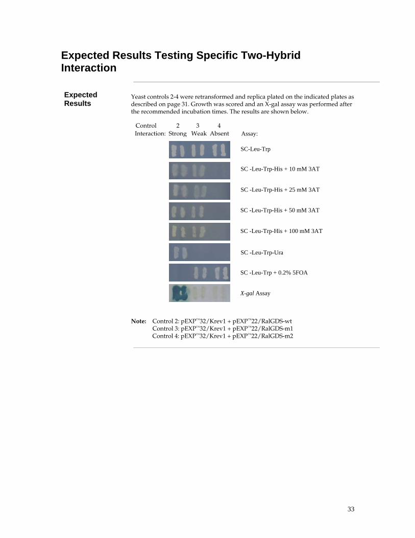
33
Expected Results Testing Specific Two-Hybrid Interaction
Expected Results
Yeast controls 2-4 were retransformed and replica plated on the indicated plates as described on page 31. Growth was scored and an X-gal assay was performed after the recommended incubation times. The results are shown below.
Note: Control 2: pEXP™32/Krev1 + pEXP™22/RalGDS-wt Control 3: pEXP™32/Krev1 + pEXP™22/RalGDS-m1 Control 4: pEXP™32/Krev1 + pEXP™22/RalGDS-m2
Control 2 3 4
SC -Leu-Trp-His + 10 mM 3AT
SC-Leu-Trp
SC -Leu-Trp-His + 25 mM 3AT
SC -Leu-Trp-His + 100 mM 3AT
SC -Leu-Trp-His + 50 mM 3AT
SC -Leu-Trp-Ura
SC -Leu-Trp + 0.2% 5FOA
X-gal Assay
Assay: Interaction: Strong Weak Absent
34
Part B: Forward Two-Hybrid Library Screen
Introduction
Introduction This chapter describes how to perform a forward two-hybrid library screen using the ProQuest™ Two-Hybrid System. This will enable you to identify new putative partners that interact with your bait (your gene of interest cloned into the GAL4 DBD vector). In the first part you will determine the conditions to use for screening the library with your bait plasmids (Testing Bait, page 36). This is absolutely required for all new bait plasmid constructs, since these conditions are impossible to predict. The second part describes how to transform yeast strain MaV203 with bait and library plasmid to identify new putative interactors with your bait (Screening Forward Two-Hybrid Library, page 40).
Required Reagents before Starting
Before you start, you need to have: • A bait plasmid, consisting of your gene of interest (or part thereof) cloned
into pDEST™32 (in frame with the GAL4 DBD). If you do not have such a bait plasmid, generate one as explained in Generating Bait and Prey Plasmids, page 16).
• A two-hybrid library suitable to use with ProQuest™ Two-Hybrid System. See below for available options.
Choosing Two-Hybrid Library
There are a number of different sources for libraries available from Invitrogen:
• Use a ProQuest™ Three-Frame cDNA Library from Invitrogen. These are constructed to enrich for in-frame ORFs, and are especially suited for use in the ProQuest™ Two-Hybrid System.
• Other ProQuest™ Pre-made cDNA Libraries; refer to our Web site (www.invitrogen.com) or call Technical Service (see page 99).
• Generate your own three frame two-hybrid library using a modified method for the CloneMiner™ cDNA Library Construction Kit. See page 78.
• Generate your own two-hybrid library using the regular method for the CloneMiner™ cDNA Library Construction Kit.
• Generate your own two-hybrid library using restriction cloning of your cDNAs into the pEXP™-AD502 plasmid (included in the ProQuest™ Two-Hybrid System). See the SuperScript Plasmid System with Gateway Technology for cDNA Synthesis & Cloning manual for instructions, available for downloading from our Web site (www.invitrogen.com) or by contacting Technical Service (see page 99). A map of the vector pEXP™-AD502 is provided in the Appendix (page 91).
Note: Libraries from sources other than Invitrogen may be used. As a general rule, the library plasmid needs to contain a TRP1 selection marker and an activation domain upstream of the insert. We advise using a ARS4/CEN6 sequence for replication and maintenance at low-copy- number in yeast, and moderate strength promoter such as the yeast Alcohol Dehydrogenase gene (ADH1).
Continued on next page

35
Introduction, continued
Bait-Specific Positive Interaction Control
The ProQuest™ Two-Hybrid System comes with a positive interaction control. However, a bait-specific positive interaction control may be an additional useful tool in testing your experiment. You may construct a prey plasmid with a known interactor of the bait protein to use as a bait-specific positive control and test as explained in Part A: Verifying Interaction, page 15 before starting the two-hybrid library screen. This will also get you familiar with the system.
Flowchart The figure below illustrates the major steps necessary to perform a ProQuest™
Forward Two-Hybrid Library screen.
���&��� ������� "������ �
���&��� ������������$%� �����������������42����������. ��+��������� ���������&������ ��������
��� #������� � �������(�)!
� �����������*���
"������ � ��� ���. ��+�������� � ��*������������������� �
�������+������ ���&��� ������
���!,����!��� ���-
���������!�2������������
#������!�������� ��+"��
� ��+����#���&��� ����������
��5%�� ���+���
� �������������� � ��42
������ #��������2 ��
�������� � �42�� ������
�����"
6�����
�� ��
��&��
� ���������
�����
"������������%��������2 ��
�� ���� ���0��#������

36
Methods
Testing Bait Overview
Introduction To maximize sensitivity of the HIS3 reporter gene, the MaV203 strain already
expresses a basal level of HIS3. Additionally, bait proteins often contain a certain level of transcriptional activity. This is enough to initiate some transcription at the most sensitive reporter in the system, HIS3.
HIS3 encodes an enzyme involved in histidine biosynthesis, which can be specifically inhibited in a dose-dependent manner by 3-Amino-1,2,4-Triazole (3AT). By determining the threshold of resistance to 3AT and including that concentration of 3AT in plates lacking histidine, even slight increases in HIS3 reporter gene expression are detected. This enhances the likelihood of detecting weak protein:protein interactions.
This section describes how to transform your bait plasmid into MaV203 cells, test for self-activation, and determine the proper concentration of 3-amino-triazole (3AT) to suppress self-activation at the HIS3 gene.
Experimental Outline
The experimental outline for Testing Bait is shown below.
�������� �
��� #������� � �������(�)!
"������ �
��������+!2���� �'��
��������!�2�������������
#����##�������!�������
�����������7�� ���� #�������
��5%��
���������0���%����& ������ �8
�������� �
���+���
0��������� �
���' � ����9�
:)����!2�;����!2;)����!2<;����!2
:))����!2

37
Small Scale Yeast Transformation
Transformation Guide
Use this table as a guide for the transformations to perform. Select on SC-Leu-Trp plates, which selects for the presence of both the LEU2 and TRP1 plasmid. Store representative transformants in glycerol at -80°C for future use.
LEU2 Plasmid TRP1 Plasmid Purpose
1 none none Negative transformation control
2 pEXP™32/Krev1 pEXP™22/RalGDS-wt Strong positive interaction control
3 pEXP™32/Krev1 pEXP™22/RalGDS-m1 Weak positive interaction control
4 pEXP™32/Krev1 pEXP™22/RalGDS-m2 Negative interaction control
5 pDEST™32 pDEST™22 Negative self-activation control
6 Bait plasmid pDEST™22 Test of self-activation
If available:
7 pDEST™32 Known prey (if available) Test of self-activation prey
8 Bait plasmid Known prey (if available) Positive interaction control
Note: The bait and prey plasmid have to be generated by the user as described before (Generating Bait and Prey Plasmids, page 16); the other vectors are provided with the kit.
If you plan to screen a two-hybrid library using the Preparing and Transforming Competent Cells (Library Scale) protocol provided in this manual (page 83), we suggest you transfom MaV203 with the bait plasmid without prey plasmid or pDEST™22, and select on an SC-Leu plate. Store this plate at 4°C and make a glycerol stock, as you use it in the Library Scale Yeast Transformation protocol to generate competent yeast cells containing the bait plasmid.
Competent Yeast Cells
The MaV203 yeast strain is provided with the kit to serve as the host for your bait and prey plasmids. Below we provide a small-scale protocol for transforming yeast cells. Prepare a new batch of competent cells for every transformation. Alternatively, to limit your workload and increase the transformation efficiency, you may purchase the following products:
• MaV203 Competent Cells, Subclone Scale from Invitrogen (Catalog no. 11445-012). Use the transformation protocol provided with these cells.
• MaV203 Competent Cells, Library Scale from Invitrogen (Catalog no. 11281-011) and use one vial to perform multiple small-scale transformations (the other vial can be used in a large-scale library transformation). Per transformation, scale down to 25 µl competent cells, 1 µg DNA per plasmid, 180 µl PEG/LiAc, 10.8 µl DMSO. Otherwise, follow the transformation protocol provided with these cells.
• If you want to generate a large batch of competent cells that can be frozen, use the S. c. EasyComp™ Kit (Catalog no. K5050-01). Use the transformation protocol provided with this kit.
Note: There are other small-scale transformation methods that can be used; see Gietz et al., 1992; Gietz and Schiestl, 1996; Hill et al., 1991; Schiestl and Gietz, 1989.

38
Determining 3AT Sensitivity
Introduction In this section you will test the bait (GAL4 DBD fusion) for nonspecific activation. We assess the extent of self-activation on the reporter gene HIS3 by determining the concentration of HIS3 inhibitor 3AT necessary to repress growth. This concentration will later be used in library two-hybrid screening to suppress growth of yeast cells not containing interacting bait and prey.
Self-Activation A suitable bait should not:
• Non-specifically transactivate the reporter constructs in the MaV203 strains.
• Interact with either the nuclear localization signal (NLS) or with the activation domain in pDEST™22.
Materials Required Be sure to have the following reagents and equipment on hand before
proceeding. See page 75 for specific media recipes.
• 30°C incubator
• Plates with strains generated in previous section (page 37)
• 3-aminotriazole (Sigma, St. Louis, MO; Catalog No. 09540)
• SC-Leu-Trp plates containing 0 mM, 10 mM, 25 mM, 50 mM, 75 mM and 100 mM 3AT
• Autoclaved velvets for replica plating/cleaning
���������
If you are unfamiliar with replica plating and replica cleaning, see page 85 before continuing this protocol. Replica cleaning is essential to reduce background.
Testing Bait Plasmid
1. After incubation, patch four different colonies containing bait plasmid and pDEST™22 (transformation 2, previous section, page 37) on a single SC-Leu-Trp plate. On the same plate, patch two colonies each from interaction controls and activation controls (controls 2 to 8, see table in previous section, page 37). Incubate for ~18 hours at 30°C.
2. Replica plate from this SC-Leu-Trp master plate onto SC-Leu-Trp-His plates containing 3AT at concentrations of 0 mM, 10 mM, 25 mM, 50 mM, 75 mM, and 100 mM. Immediately replica clean the plates. Incubate for 24 hours at 30°C.
3. After incubation, replica clean again and incubate for 2 days (40-44 h) at 30°C. The lowest concentration of 3AT that inhibits the growth of the cells from transformation 2 (containing bait and pDEST™22) is the basal amount of 3AT added to all plates lacking histidine.
Continued on next page

39
Determining 3AT Sensitivity, continued
Analysis If cells from transformation 2 (containing bait plasmid and pDEST™22) grow even in the presence of 100 mM 3AT, the bait plasmid likely encodes a protein that directly or indirectly self-activates the reporter genes in this system. Such plasmids are not suitable for use in the two-hybrid screen.
Alternatives for Self-Activating Bait
If your bait is self-activating HIS3, try deletion derivatives of the original bait for a two-hybrid screen. Generate bait plasmids that lack parts of the coding sequence of your gene of interest, and test whether these self-activate. Refer to Molecular Biology handbooks, such as Current Protocols in Molecular Biology (Ausubel et al., 1994), if you are not familiar with the cloning steps involved.
Example of Expected Results
The coding sequence for the cytoplasmic domain of the human DCC gene (deleted in colorectal cancer) was fused to the GAL4 sequence encoding the DNA Binding Domain (GAL4 DBD-DCC) in pDEST™22. Derivatives were constructed that had increasing deletions at the 3’ end (∆ 3, ∆ 2, and ∆ 1, respectively) or 5’ end (∆4 and ∆ 5). Each of these constructs was introduced, along with a non-interacting prey plasmid, into MaV203 (selecting on SC-Leu-Trp plates). Self-activation was tested as described above on SC-Leu-Trp-His at 0 mM, 10 mM, 25 mM, 50 mM, 75 mM and 100 mM 3AT.
The baits were used in subsequent library screens at the indicated concentrations of 3AT except for GAL4 DBD-DCC ∆5. This bait was not used due to self-activation, i.e. growth at 100 mM 3AT.
2��(!�2�������������
����%��%���
34��(42��(�4��(�2��( �22��(
����%��%������
����%��%������
����%��%������
����%��%�����4
� ���������� ���� !�2�������������6
42��(
�2��(
�2��(
�4��(
�+�+'�5�&6
"��6
����%��%�����7 �2��(
Note: The DCC clones were the kind gift of Drs. G. Hu, M. Vidal and E. Fearon (Hu et al., 1997).
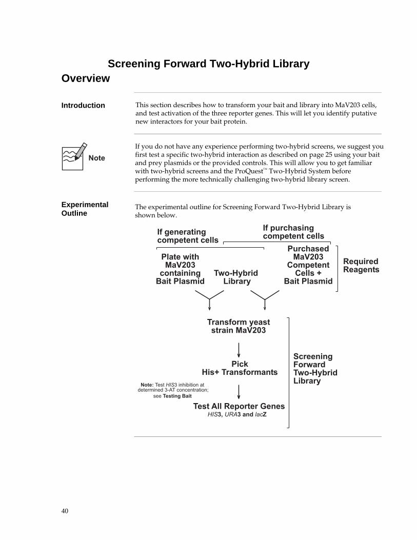
40
Screening Forward Two-Hybrid Library Overview
Introduction This section describes how to transform your bait and library into MaV203 cells,
and test activation of the three reporter genes. This will let you identify putative new interactors for your bait protein.
If you do not have any experience performing two-hybrid screens, we suggest you first test a specific two-hybrid interaction as described on page 25 using your bait and prey plasmids or the provided controls. This will allow you to get familiar with two-hybrid screens and the ProQuest™ Two-Hybrid System before performing the more technically challenging two-hybrid library screen.
Experimental Outline
The experimental outline for Screening Forward Two-Hybrid Library is shown below.
���&��� �������
�����������(�)!
�������+"������ �
��� #������� � �������(�)!
� ��2������������*���
�������+������ ���&��� ������
���!,����!��� ���-
4���6�'�������7���*�)��������� �������� �7%�'��������������8
����� ��+�"��
��9& 8���� #������
��5%�� ���+���
�%���� � ��(�)!
0��������0��� �8
"������ �
�#��%���� �+�������������� �#�+�������+
��������������

41
Library Scale Yeast Transformation
Introduction This section describes how to perform a library transformation. We recommend that you screen >106 yeast transformants for mammalian cDNA libraries. Therefore, use these protocols specifically written for library transformations, since other yeast transformation protocols do not yield enough transformants.
Competent Yeast Cells
To transform your bait and library plasmids into yeast, you need to obtain competent MaV203 cells. We recommend using MaV203 Competent Cells, Library Scale from Invitrogen (Catalog no. 11281-011). Use the transformation protocol provided on page 78. These cells have a very high transformation efficiency (> 2 x 10⁵ transformants/µg library DNA) and are easy to work with.
If you do not want to purchase MaV203 Competent Cells, Library Scale from Invitrogen (Catalog no. 11281-011), use the Preparing and Transforming Competent Cells (Library Scale) protocol for transforming yeast cells described in the Appendix, page 83. Prepare a new batch of competent cells for every transformation. This will take considerably more time and will not yield as high a transformation efficiency. To obtain a decent number of transformants, yeast cells already containing your bait have to be transformed with the library DNA, which will yield an efficiency of > 2 x 10⁵ transformants/µg library DNA.
���������
If you perform the Library Scale Yeast Transformation protocol described in the Appendix, page 83, you need to have a SC-Leu plate with MaV203 transformed with bait plasmid without prey plasmid or pDEST™22. Transform MaV203 with bait plasmid as described on page 27, and select on an SC-Leu plate.
3AT Concentration The optimum concentration of 3AT for your bait must be determined prior to
doing a library transformation; see page 36.
Calculating Transformation Efficiency
The transformation efficiency is represented by the number of colonies per transformation reaction. Count colonies on the 10-cm SC-Leu-Trp plates, preferably on the plates having 20 to 300 colonies. Calculate the transformation efficiency by the following equation:
Number of colonies per transformation reaction = Colonies on a plate x dilution factor × total volume / plated volume
For example, if 150 colonies are counted when 0.1 ml of a 1:100 dilution of the library screen (12 ml total volume) are plated, the calculation would be:
150 × 100 × 12 ml total volume = 1.8 × 10⁶ colonies/reaction 0.1 ml plated
A successful transformation for a library screen should have at least 1×10⁶ transformants.

42
Characterization of His+ Transformants
Introduction You should have identified a number of colonies that grow on plates without histidine. Some of these may be false positives (see page 5); others represent true interactors. MaV203 cells that contain bait and prey proteins that strongly interact will induce all three reporter genes present in this system (HIS3, URA3, lacZ). Identify these colonies by a series of patching and replica plating steps onto the selection/screen plates, which are described in this section.
Controls Use yeast controls 2-8 generated in the previous chapter (page 37). Below is
indicated the purpose of these controls: LEU2 Plasmid TRP1 Plasmid Purpose
2 pEXP™32/Krev1 pEXP™22/RalGDS-wt Strong positive interaction control
3 pEXP™32/Krev1 pEXP™22/RalGDS-m1 Weak positive interaction control
4 pEXP™32/Krev1 pEXP™22/RalGDS-m2 Negative interaction control
5 pDEST™32 pDEST™22 Test of self-activation
6 Bait plasmid pDEST™22 Negative interaction control
If available:
7 pDEST™32 Known prey (if available) Negative interaction control
8 Bait plasmid Known prey (if available) Positive interaction control
Materials Needed • Plates with His+ transfomants
• Yeast controls 2-8 generated in the previous chapter (page 37) • SC-Leu-Trp plates to grow yeast cells containing both bait and prey plasmid • YPAD plates, to grow cells for assaying lacZ activity • SC-Leu-Trp-His+3AT plates, to select for cells expressing HIS3. Use the 3AT
concentration as determined in Testing Bait, page 36 • SC-Leu-Trp-Ura plates, to select for cells expressing URA3 • SC-Leu-Trp+0.2% 5FOA plates, to select against cells expressing URA3 • nitrocellulose or nylon membrane • 30°C incubator • Autoclaved velvets for replica plating/cleaning • X-gal (5-bromo-5-chloro-3-indolyl-β-D-galactoside) • N,N-dimethyl formamide (DMF) • Z buffer (60 mM Na2HPO4, 40 mM NaH2PO4 , 10 mM KCl, 1 mM MgSO4,
pH 7.0) • 2-mercaptoethanol • 125-mm Whatman 541 filter papers and 15-cm petri dishes • Forceps • Liquid nitrogen
Continued on next page

43
Characterization of His+ Transformants, continued
Generating Master Plates
1. Streak transformants that grow on SC-Leu-Trp-His+3AT on SC-Leu-Trp plates to isolate single purified colonies.
2. Prepare fresh colonies of the yeast controls 2-8 generated in the previous chapter (page 37) by streaking a colony from the stored plates onto new SC-Leu-Trp plates.
3. Incubate plates for 48 hours at 30°C.
4. Using an autoclaved toothpick or loop, patch onto a single SC-Leu-Trp plate the following:
• Two isolated colonies of yeast controls 2-8
• Four isolated colonies of each potential positive clone (generally up to four clones can be analyzed per plate)
5. If more than 4 His+ clones were identified, use additional plates. On each plate include the 7 yeast controls.
6. Incubate plates for 18 hours at 30°C.
Note: Store the plates from Step 1 containing the His+ transformants at 4°C.
Testing Reporter Genes
Perform the assays described in Testing Reporter Genes, page 31, and X-gal Assay, page 32, to test for HIS3, URA3 and lacZ expression.
���������
The use of large amounts of DNA used in the library transformation can result in multiple prey clones in a single transformant. Such transformants may show growth on 5FOA plates and care must be taken to identify and retest all candidate prey clones following isolation in E. coli. The use of 5 µg of each vector will reduce the number of transformants containing multiple copies of prey plasmids, but will also reduce transformation efficiency slightly.
Continued on next page

44
Characterization of His+ Transformants, continued
Interpretation of Phenotypes
Interpretation of the four reporter gene readouts is the most critical step in a two-hybrid screen. A summary of likely interpretations of observed phenotypes is provided below. A detailed description of the phenotypes is provided on page 50.
-His lacZ -Ura 0.2% 5FOA Easily-Interpreted Phenotypes
no growth False positive/background
growth blue no growth no growth Interactor, probably weak
growth blue growth no growth Interactor
growth white no growth growth Non-interactor
-His lacZ -Ura 0.2% 5FOA One Inconsistent Phenotype
growth blue growth growth Probable interactorPossible mixed population*
growth white growth no growth Probable interactor (check lacZ expression using CPRG assay)
-His lacZ -Ura 0.2% 5FOA Two Inconsistent Phenotypes
growth blue no growth growth Possible non-interactorPossible weak interactor (look carefully for any inhibition on 5FOA)Possible mixed population*
growth white growth growth Possible interactor (check lacZ expression using CPRG assay)Possible mixed population*; look carefully for inhibition on 5FOA
growth white no growth no growth Probable weak interactor (check lacZ expression using CPRGassay; confirm 5FOA phenotype)
• Intermediate levels of URA3 expression can result in both cell growth inhibition on 0.2% 5FOA and insufficient URA3 gene product to allow growth on plates lacking uracil. The 5FOAS/Ura- phenotype is often indicative of protein pairs that interact weakly.
• The strength of the three different reporter gene read-outs can vary dramatically between different pairs of interacting proteins.
• Colonies that are white in the X-gal assay may be tested in the more sensitive CPRG assay for β-galactosidase activity; see page 87.

45
What To Do Next Overview
Introduction After you have isolated HIS+ transformants, confirm the interaction. Do this
using two independent methods:
1. Repeat the two-hybrid assay with bait and prey plasmids to make sure that no mutations have been generated in the bait plasmid or yeast strain in the primary transformants. The phenotype of the primary transformants (HIS3, URA3 and lacZ expression) should be replicated in this new assay.
2. Perform a biochemical or functional assay to confirm the interaction of bait and prey in an independent assay.
Repeat Two-Hybrid Assay
Several types of “false positives” can result in phenotypes resembling true interactions (e.g., a mutation in bait that converts it to a self activator). If the bait/prey interactions identified above are authentic, the reporter gene phenotype should be reproduced when prey is reintroduced into MaV203 with the original bait plasmid, but not when empty pDEST™32 is introduced. Two methods can be used with the ProQuest™ System:
• Retransformation assay (preferred method)
• Plasmid shuffling (faster method)
Retransformation Assay
The Retransformation assay consists of the following steps:
1. Isolate the prey plasmid in E. coli, as described in Prey Plasmid DNA Isolation, page 48.
2. Perform restriction and sequence analysis.
3. Retransform the prey plasmid in MaV203 together with bait plasmids or controls, and test reporter gene expression (HIS3, URA3 and lacZ) as described in Testing Specific Two-Hybrid Interaction, page 25.
Note: Use 3AT concentration in SC-Leu-Trp plates as determined in Testing Bait, page 36.
Continued on next page

46
Overview, continued
Required Test Plates
To validate potential interacting proteins identified in a two-hybrid screen, use the plates described below for the assay you perform.
Test HIS3 induction URA3 induction
URA3 induction
β-Galactosidase induction
Assay His auxotrophy 5FOA sensitivity
Uracil auxotrophy
X-gal assay
Plates used SC-Leu-Trp-His+3AT
SC-Leu-Trp+5FOA
SC-Leu-Trp-Ura
YPAD
Concentration used
3AT concentration as determined (Testing Bait, page 36)
0.2% 5FOA No Uracil Not applicable
Plasmid Shuffling The plasmid shuffling method (see Appendix, page 90) is fast and easy, but
does not exclude false positives resulting from host cell mutations. Therefore the Retransformation Assay is the preferred method, but plasmid shuffling may be useful if you need to screen large numbers of HIS+ transformants.
Sequence Analysis
If you can repeat the interaction between bait and prey, sequence the insert. Make sure the insert is in frame with the GAL4 AD domain; if not the clone is likely a false positive.
Confirmation of Interaction
If sequence information suggests the interaction may be valid or biologically relevant, perform a biochemical and/or functional assay to validate the interaction in an independent experiment. For suggestions, see Confirmation of Interaction, page 13.
Mutational Analysis of Interaction
If an independent assay supports a valid interaction between bait and prey, you can characterize the interaction further by identifying inhibiting point mutations through reverse two-hybrid. This may give information about the interaction surface, or provide a useful tool in analyzing the biological significance of the interaction. Use the SureFrame™ Allele Library Construction Kit to efficiently generate the necessary allele library; see Part C: ProQuest™ Reverse Two-Hybrid (page 53) for general guidelines.
Continued on next page

47
Overview, continued
Experimental Outline
The experimental outline for the steps to take after HIS+ transformants have been identified is shown below.
�� ������3��5%�����2����
��� � ��%##��+
&��8���� #������
� �����������*��� ���!,����!��� ���-
� ��+����#���&��� ����������
� ������������� � ��42
������ #������(�)!����������� � �� ��������� �
�������� � �42�� ������
"������������%��������2 ��
�� ���� ���0��#������
� ������������� � ��42
� �����������*��� ���!,����!��� ���-
�� ������3��5%�����2����
��� � ��%##��+
� ��+����#���&���
����������
�������� � �42�� ������
�� ���� ���0��#������
��� � ���%##��+������ ������ #��������2 ��0��� ���� �(��*� 1

48
Prey Plasmid DNA Isolation
Introduction Plasmid DNA isolated from yeast is typically not suitable for restriction analysis. Consequently, plasmids isolated from yeast are first introduced into E. coli, then miniprep DNA from the resulting transformants is characterized. Several quick yeast plasmid preparations have been described (Polaina & Adam, 1991). The following method uses the PureLink™ HQ Mini Plasmid DNA Purification Kit, since it is a fast and reliable method for DNA isolation. Other purification kits are suitable.
Materials Needed • 30°C shaker
• SC-Trp medium
• 1 x TE (10 mM Tris-HCl (pH 7.5), 1 mM EDTA)
• PureLink™ HQ Mini Plasmid DNA Purification Kit (Catalog no. K2100-01)
• Zymolyase (1.5 U/µl, Genotech, St.Louis, MO, Catalog no. 786-036)
• 2-mercaptoethanol
• Competent cells, such as One Shot® TOP10 Chemically Competent E. coli cells (Catalog no. C4040-03)
• LB+100 µg/ml ampicillin medium and plates
• 37°C incubator
Plasmid DNA Extraction
Important: This is a modified version of the protocol provided with the kit.
1. Suspend an isolated colony from a fresh plate in 3 ml SC-Trp and incubate at 30°C with shaking for ~24 hours. OD660 should be in between 1.0-2.3.
2. Centrifuge the 3 ml culture at 1500 x g for 15 minutes at room temperature. Remove all residual liquid.
3. Resuspend the cells in 1 ml 1xTE.
4. Centrifuge the 3 ml culture at 1500 x g for 15 minutes at room temperature. Remove all residual liquid.
5. Resuspend the cells in 240 µl Resuspension Buffer containing RNase A (provided with the PureLink™ HQ Mini Plasmid DNA Purification Kit).
6. Add 10 µl Zymolyase (1.5 U/µl) and 5 µl 2-mercaptoethanol.
7. Incubate at 37°C for 30 minutes.
8. Add 240 µl Lysis Buffer (provided with the PureLink™ HQ Mini Plasmid DNA Purification Kit ); mix gently by inverting the tube 4–8 times.
9. Incubate for 3-5 minutes at room temperature (do not exceed 5 minutes).
10. Add 340 µl of Neutralization/Binding Buffer (provided with the PureLink™ HQ Mini Plasmid DNA Purification Kit ), and immediately mix gently by inverting the tube 4-8 times.
Continued on next page

49
Prey Plasmid DNA Isolation, continued
Plasmid DNA Extraction, continued
Continued from previous page
11. Centrifuge for 10 minutes at maximum speed in a tabletop centrifuge to clarify the cell lysates.
12. Place a PureLink™ spin column inside a 2-ml collection tube. Pipette or decant the supernatant into the spin column.
13. Centrifuge the column at room temperature at 10,000–14,000 × g for 30-60 seconds, discard the flow through from the collection tube.
14. Add 650 µl of Wash Buffer prepared with ethanol to the column (provided with the PureLink™ HQ Mini Plasmid DNA Purification Kit).
15. Centrifuge the column at room temperature at 10,000–14,000 × g for 30-60 seconds. Discard the flow through from the collection tube.
16. Repeat the Wash steps 13 and 14.
17. Centrifuge the column at maximum speed for 2.5 minutes to remove the residual wash buffer.
18. Place the spin column in a clean 1.7-ml elution tube.
19. Add 70 µl of Elution Buffer (provided with the PureLink™ HQ Mini Plasmid DNA Purification Kit ), or water to the center of the column.
20. Incubate the column at room temperature for 1 minute.
21. Centrifuge at maximum speed for 2 minutes.
Transforming E. coli
Use 5-10 µl of DNA to transform One Shot® TOP10 Chemically Competent E. coli according to the procedure provided with the cells, or transform your in-house competent cells. Plate ~20% of the transformation mixture on LB agar plate + 100 µg/ml ampicillin. Grow overnight at 37°C.
Note: If you do not get enough colonies, transform 1 µl of DNA into electrocomp cells such as ElectroMAX™ DH10B™ cells (Catalog no. 18290-015).
Analyzing Transformants
To analyze positive clones, we recommend that you:
1. Pick 5-10 colonies and culture them overnight in LB or SOB medium containing 100 µg/ml ampicillin (for prey plasmids).
2. Perform restriction analysis to confirm the presence of the insert. Note: BsrG I cleaves within all att sites, and can be used to help characterize clones.
3. Isolate plasmid DNA using your method of choice. To obtain pure plasmid DNA for automated or manual sequencing, we recommend using the PureLink™ HQ Mini Plasmid Purification Kit and the primers indicated below:
Plasmid Direction Primer
Prey reverse 5’-AGCCGACAACCTTGATTGGAGAC-3’
Prey forward 5´-TATAACGCGTTTGGAATCACT-3’

50
Interpretation of Results
Introduction This section provides help in the interpretation of the results of a two-hybrid experiment.
Interpretation of Growth Is Subjective
The interpretation of growth/slight growth/no growth on the various selection plates is quite subjective. Yeast controls 2-8 (page 25) should show the expected growth profile on the selection plate being examined in order to make valid conclusions. If they do not, confirm that the plates were prepared correctly, that the cells of the controls are fresh and correctly ordered (2-8). If necessary, transform the controls again and use isolated fresh colonies on SC-Leu-Trp plates.
False Positives False positives have been defined as clones containing bait and prey that induce
the reporter genes in a two-hybrid screen, but where bait and prey do not interact; or these interactions are biologically irrelevant. Several recent references have discussed false positives (Bartel et al., 1993; Leanna & Hannink, 1996; Luban & Goff, 1995; Vidal et al., 1996).
False positives in which bait or prey self-activate (e.g., mutations in bait that result in self-activation, or prey clones that activate transcription by binding to the promoters or proteins bound there) are usually eliminated by retesting prey with pDEST™32 (empty GAL4 DBD) and bait (page 25). Swapping the DNA Binding Domain and Activation Domain between bait and prey provides strong evidence confirming the interaction. However, several examples have been described where swapping the DBD and AD impedes legitimate interactions. This is presumably due to steric issues, hence failure to retest in this experiment should not necessarily exclude the interaction.
On occasion, a false positive prey will not induce the reporter genes when tested with DBD alone, but will when an unrelated protein is fused to DBD. Therefore, it is useful to test candidate prey clones against an irrelevant bait, such as pEXP™32/Krev1 (if no interaction is expected with Krev1).
Certain proteins, such as those with low affinities for many different proteins (e.g., containing large hydrophobic domains) or those that may recognize structural features of proteins (e.g., heat shock proteins) may be identified as false positives in a two-hybrid screen. More complex structures, where bait and prey are bridged by a third protein or RNA, may also account for certain false positives.
It is critical to:
• Confirm that bait and prey interact by retesting the interaction and showing that it is specific to the test bait,
• Perform an independent assay such as co-immunoprecipitation, and
• Devise experiments to demonstrate that the interaction is biologically relevant.
Continued on next page
51
Interpretation of Results, continued
Mixed Populations Mixed populations can result from numerous causes. Examples include:
• More than one prey plasmid in the cell resulting in one population that contains prey interacting with bait (no growth on 5FOA but growth on URA- plates), and another population that contains prey that does not interact with bait (giving rise to growth on 5FOA but not on URA- plates)
• Mutations or instability in bait, prey or URA3 reporter or promoter
• Carry over of cells from the -His+3AT plate that do not contain interacting bait and prey
If restriction analysis after Prey Plasmid DNA Isolation (page 45) indicates a mixed population, test all different isolated prey plasmids in the retransformation assay.
HIS3 and 3AT In order to inhibit HIS3 at the threshold level, you should add 3AT to all SC-Leu-
Trp-His plates. Yeast control 2-8 (page 25) will show different growth patterns as the 3AT concentration is increased (see Expected Results Testing Specific Two-Hybrid Interaction, page 33). This must be considered when determining whether cells containing candidate interactors induce the HIS3 reporter gene. It is important to properly replica clean 3AT plates and to interpret the results at the indicated times. As the incubation time is increased beyond the indicated time, growth of cells not inducing HIS3 can become indistinguishable from those inducing the HIS3 reporter gene.
Master Replica Plating/Cleaning
Difficulty in interpreting the reporter gene readouts is very often associated with replica plating and replica cleaning. These procedures take practice. The most important issues have been described in the Appendix, page 85. It is useful to practice with yeast controls 2-8 (page 25) until the expected readouts are obtained consistently. Be sure plates and velvets are very dry. If controls 2-8 do not give the expected results, confirm that the plates were prepared correctly or transform the controls again and use isolated fresh colonies on SC-Leu-Trp plates. Always include controls 2-8 on each master plate (remember to keep the patches away from the edges of the plate) to confirm that the plates are correct, and the replica plating was successful. Alternatives to replica plating and replica cleaning are described in the Appendix, page 78.
Continued on next page

52
Interpretation of Results, continued
URA- Least Sensitive Selection
Growth on the SC-Leu-Trp-Ura plate is the least sensitive selection method and failure of candidate interactors to grow here should not exclude them as true interactors. This is because the URA3 promoter region contains the URS1 sequence that strongly represses transcription (Vidal, 1997). Only strong protein:protein interactions induce this gene sufficiently to allow growth on SC-Ura plates. Furthermore, certain bait fusions, while showing strong induction of the HIS3 and lacZ reporter genes, may show weak induction of URA3.
Controls on 5FOA Plates
Inhibition of growth on 5FOA is more sensitive than growth on SC-Leu-Trp-Ura plates. Compare the amount of growth of the candidate clone with yeast control 2-8 (see table on page 25). Yeast control 3-7 should show good growth (little growth inhibition) on SC-Leu-Trp+0.2% 5FOA; and yeast control 2 and 8 should be completely inhibited. The replica cleaning step is critical for good results on 5FOA. If the controls do not show the expected results, confirm the amount of 5FOA added and the media composition.
X-gal assay X-gal assays for examining induction of the lacZ reporter gene are quite sensitive
if done correctly. Always use a fresh X-gal solution. The addition of excess X-gal/Z-buffer during incubation of the membrane will cause diffusion of the blue color into adjacent colonies. Use the minimal amount (7-8 ml) of buffer to saturate the paper filters. It is useful to tip the plate slightly during incubation to allow any excess buffer to accumulate below the filter papers. Yeast control 2 (see table on page 25) gives a very strong blue reaction. Keep this patch at the bottom of the tipped plate to avoid diffusion into adjacent patches. Efficient cell lysis is critical. Cells are lysed by immersion in liquid nitrogen. Typically 20-30 seconds is sufficient. Be careful with the membrane after lysing the cells. The patches become viscous and touching them will cause smearing. Be certain that the patches are incubated facing up. Nitrocellulose filters often crack in liquid nitrogen. Yeast controls 2-8 must give the expected result to accurately interpret the results. Yeast control 3 should give a very faint blue when lysis is complete. For those candidate clones showing very weak or questionable lacZ activity, it is often useful to use the CPRG assay (Appendix, page 87) to confirm the phenotype.

53
Part C: ProQuest™ Reverse Two-Hybrid
Introduction
Reverse Two-Hybrid
Introduction The ProQuest™ Reverse Two-Hybrid System, which consists of the SureFrame™ Allele Library Construction Kit and the ProQuest™ Two-Hybrid System, offers some clear advantages for performing reverse two-hybrid. In this section we explain reverse two-hybrid and the advantages of using the ProQuest™ Reverse Two-Hybrid System.
We highly recommend using the SureFrame™ Allele Library Construction Kit for generating mutations in the reverse two-hybrid screen. This will minimize production of clones not expressing any product or expressing truncated proteins. The SureFrame™ Allele Library Construction Kit is supplied with the ProQuest™ Reverse Two-Hybrid kit, or can be ordered separately from Invitrogen.
Negative Selection Growth inhibition resulting from induction of the URA3 reporter gene on 5FOA-
containing medium provides a way to rapidly characterize protein:protein interactions. Mutations that inhibit or modulate the protein:protein interaction can be selected by identifying cells that are able to grow on the correct selective plates containing 5-fluoroorotic acid (SC-Leu-Trp+5FOA) (Brachmann & Boeke, 1997; Vidal, 1997; Vidal et al., 1996; Vidal et al., 1996).
Reverse Two-Hybrid
The reverse two-hybrid method is a variation on the yeast two-hybrid system and was developed to identify mutations that disrupt protein interactions. The system can be used to characterize protein-protein interactions by generating an allele library of one of the interacting proteins and selecting for interaction defective alleles.
Continued on next page

54
Reverse Two-Hybrid, Continued
Traditional Methods
Allele libraries are generated by polymerase chain reaction (PCR), such that PCR products are flanked by homologous regions to the prey two-hybrid vector. PCR products are co-transformed into S. cerevisiae with the linearized prey vector and library assembly is mediated through in vivo homologous recombination, or gap repair (Vidal et al., 1996). Protein-protein interactions are evaluated using the counter selectable marker URA3 in the presence of 5FOA. A positive interaction will inhibit growth, whereas disrupted interactions will be resistant to 5FOA (Endoh et al.; Vidal et al., 1996; Vidal et al., 1996; Vidal & Legrain, 1999). Traditional reverse two-hybrid methods are limited by a number of drawbacks:
• Both point mutations and truncated proteins may result in a disrupted interaction, but truncated proteins are less informative and typically represent >97% of 5FOAR colonies (Endoh et al.; Shih et al., 1996; Vidal et al., 1996).
• Selection against truncated proteins can be achieved by incorporating an easily detected C-terminal fusion such as green fluorescent protein (Endoh et al.). However, the allele library produced contains both an N- and C-terminal fusion, which may affect the interaction under study.
• Selection against truncated proteins can be achieved by using an epitope tag at the C-terminus, which may be detected by Western blot (Barr et al., 2004). But this method is not practical for screening out truncated proteins from a library due to its time-consuming nature.
• Identification of full-length proteins is performed after 5FOA selection and less than 3% of 5FOAR colonies are expected to code for full-length proteins.
• Gap repair mediated library assembly limits library complexity due to the low transformation efficiencies typically achieved (~104).
ProQuest™ Reverse Two-Hybrid
The ProQuest™ Reverse Two-Hybrid System is designed to allow production of allele libraries without the drawback of traditional reverse two-hybrid methods discussed above. The system involves producing allele libraries in vitro and selecting for full-length proteins in E. coli prior to analysis in yeast using a vector adapted with Gateway® Technology. It consists of the following steps:
1. Introduce your gene of interest into the Gateway® System by performing PCR and recombining the amplified products with the donor vector pDONR™-Express. This new vector facilitates the expression of entry clones as an N-terminal fusion to the KanR gene.
2. Transform the new construct into E. coli. Only alleles coding for full-length proteins will confer kanamycin resistance and produce colonies for DNA (i.e. allele library) isolation.
3. Transfer the pENTR™–Express allele library to the two-hybrid prey vector, pDEST™22 through a Gateway® LR recombination reaction, which yields a full-length enriched expression library fused to GAL4 AD.
4. Transform the expression library and the bait expressing plasmid into yeast strain MaV203. Select for alleles with defective interaction using the counter selectable marker URA3 in the presence of 5FOA.
Continued on next page
55
Reverse Two-Hybrid, Continued
Advantages The ProQuest™ Reverse Two-Hybrid System has a number of clear advantages
over traditional methods:
• Clones lose the C-terminal fusion used for full-length selection before the two-hybrid step and interactions may be evaluated in the original two-hybrid context.
• The scheme selects against interaction defective truncated proteins prior to yeast transformation, eliminating almost all background normally associated with reverse two-hybrid screens.
• Gateway® recombination combined with the efficiency of E. coli transformation allows for larger (106-107), more complex allele libraries.
See the SureFrame™ Allele Library Construction Kit manual, provided with the ProQuest™ Reverse Two-Hybrid kit, for more information on the SureFrame™ mutagenesis procedure, and for instructions to generate an allele library.

56
Methods
Overview
Introduction In this chapter, you will screen for mutations in the prey that block interaction with the bait in a reverse two-hybrid screen.
Required Bait and Prey Constructs
Before you start you need to generate suitable bait and prey plasmids (see page 16):
• A conventional bait plasmid in pDEST™32
• A prey plasmid in pDEST™22 without stop codons at both the N-terminal or C-terminal side of the insert. Refer to the SureFrame™ Allele Library Construction Kit manual for the required reading frame.
Wild Type Interaction Confirmation
It is important to confirm the interaction of the bait and the wt prey plasmid without stop codons in a two-hybrid assay (see Testing Specific Two-Hybrid Interaction, page 25).
Controls Use this table as a guide for the controls to perform. Plate on SC-Leu-Trp plates,
which selects for the presence of both LEU2 and TRP1 plasmid. Store representative transformants in glycerol at -80°C for future use.
LEU2 Plasmid TRP1 Plasmid Purpose
1 none none Negative transformation control
2 pEXP™32/Krev1 pEXP™22/RalGDS-wt Strong positive interaction control
3 pEXP™32/Krev1 pEXP™22/RalGDS-m1 Weak positive interaction control
4 pEXP™32/Krev1 pEXP™22/RalGDS-m2 Negative interaction control
5 pDEST™32 pDEST™22 Negative activation control
6 Bait plasmid pDEST™22 Negative activation control; baseline
7 pDEST™32 wt prey plasmid Negative activation control
8 Bait plasmid wt prey plasmid wt interaction control
Note: pDEST™32, pDEST™22, pEXP™32/Krev1, pEXP™22/RalGDS-wt, pEXP™22/RalGDS-m1, and pEXP™22/RalGDS-m2 are provided with the kit. Bait and prey plasmids are not provided with the system and need to be generated (see page 16).
5FOA Concentration for Reverse Two-Hybrid Screen
Be sure to evaluate the wt interaction on SC-Leu-Trp-plates containing 0.05%, 0.1% and 0.2% 5FOA. This will also indicate the concentrations of 5FOA to use for the reverse two-hybrid screen: use the two lowest two concentrations that kill MaV203 cells containing bait and wt prey plasmid. If only 0.2% 5FOA causes cell death, use only this concentration for the screen.
Continued on next page
57
Overview, continued
Weak wt Interaction
If the interaction under study is not capable of activating the URA3 reporter (i.e. it is Ura-/5FOAR), but can activate the HIS3 reporter (i.e. His+/3ATR), a reverse two-hybrid analysis is still possible. We suggest performing a genetic screen (as opposed to a selection with 5FOA), whereby an allele library is created under mutagenic conditions, co-transformed with bait and then plated out for plasmid selection first. Patch several thousand colonies onto SC-Leu-Trp-His + 3AT plates to identify His-/3ATS clones.
Allele Library If you have performed the tests described above, construct an allele library of the
prey protein using the SureFrame™ Allele Library Construction Kit. This kit is part of the ProQuest™ Reverse Two-Hybrid System, or can be ordered separately.
Experimental Outline
The outline for performing a reverse two-hybrid assay is shown below.
2�������������������(�����
��� #������� �� �������(�)!
��'�� ����&��� ������
��9�;��2����� #������
��5%�� ���+���
��(�)!0���������0���
�������� � �42�� ������
������ #��������2 ��=� ��+�����#���&��� �����������>
"����� �
� �����!��� %���������������!�" ��$���������"
� �����������*��� ���!,����!��� ���-
� �������������� � ��42
������ #������'�)!����������� � ��� � ����� ��������� �
��5%�����2����
���� ������2 �� 9�������������������������� ���
5���������"����)���*�����"���� ��������"�����$����"$#��)��"�����"������ �������� ������������
�%��������
2������������0�� ��%�����?�
*�������+"����� �������� �

58
Allele Library Yeast Transformation
Introduction In this section, you will introduce your bait plasmid and a two-hybrid library into MaV203.
Competent Yeast Cells
We generally use MaV203 Competent Cells, Library Scale from Invitrogen (Catalog no. 11281-011). This section provides a protocol for transforming MaV203 Competent Cells, Library Scale. Other sources of competent cells may be suitable, but they have not been tested in this application.
Library Scale Competent Yeast Cells
MaV203 Competent Yeast Cells, Library Scale have been developed for use with the ProQuest™ Two Hybrid System to facilitate library-scale transformations. Each tube contains 550 µl competent yeast cells, enough for at least 2×10⁶ colonies.
Materials Needed These materials are needed for a screen:
• MaV203 Competent Cells, Library Scale plus provided transformation reagents (PEG/LiAc Solution)
• Your bait plasmid and your two-hybrid allele library • 10-cm SC-Leu-Trp + 5FOA plates to select against interaction; prepare six
plates for each of the two concentrations of 5FOA decribed in 5FOA Concentration for Reverse Two-Hybrid Screen, page 56. If using only one concentration of 5FOA, prepare 12 plates of that one concentration.
• Two 10-cm SC-Leu-Trp plates to determine the number of transformants • Sterile 1.5 ml microcentrifuge tubes • 30°C incubator • 30°C and 42°C water bath • DMSO. For best results, use fresh DMSO from an unopened bottle. DMSO
that has been stored at -20°C also works well. • Autoclaved dH2O • Autoclaved velvets for replica plating/cleaning
Continued on next page
59
Allele Library Yeast Transformation, continued
Transformation Procedure
One tube of library-scale competent cells (550 µl) is enough for several library screens (25 µl each).
1. Thaw the PEG/LiAc Solution in a beaker containing room temperature water before the assay. Mix the solution well before dispensing.
2. Thaw competent cells by placing in a 30°C water bath for 90 seconds. Do not allow the cells to remain at 30°C longer than 90 seconds. Proceed immediately to Step 3. Steps 3, 4, and 5 can be done at room temperature.
3. Once the cells are completely thawed, invert the cells several times. Do not vortex the cells. Transfer 25 µl cells each per transformation to 1.5 ml microcentrifuge tubes
Note: Freeze the rest of the competent cells at -80°C. Use these cells for retransformation assays; transform as described on this page. Do not use cells already frozen for a library transformation
4. To each aliquot of cells, add 1 µg bait plasmid and 1 µg library DNA.
Note: The total volume of DNA added to the library screen transformations must be ≤10 µl (i.e. bait plasmid and library DNA concentration should be ≥0.2 µg/µl).
5. Add 180 µl of PEG/LiAc Solution to each tube. Invert microcentrifuge tubes gently to mix until all of the components are homogeneous.
6. Incubate for 30 minutes in a 30°C water bath. Swirl the tubes occasionally (every 10 minutes) to resuspend the components.
7. Add 10.8 µl DMSO to each tube. Invert microcentrifuge tubes gently to mix.
8. Heat shock the cells for 20 minutes in a 42°C water bath. Invert the tubes occasionally.
9. Centrifuge 1800 rpm (200-400 × g) for 5 seconds in a microcentrifuge. Carefully discard the supernatant.
10. Suspend each pellet in 750 µl autoclaved dH2O by gently pipetting.
11. Make serial dilutions:
• Dilute 100 µl from each transformation in 900 µl dH2O (1:10 final). • Dilute 100 µl from each 1:10 dilution in 900 µl dH2O (1:100 final).
12. Plate 100 µl of the 1:10 and 1:100 dilution on 10 cm SC-Leu-Trp plates.
13. Plate 100 µl of the undiluted and 1:10 dilution of the screen on 10 cm SC-Leu-Trp- + 5FOA plates until all of the transformation has been plated.
14. Incubate the plates for 3 to 5 days at 30°C.
15. Count the number of colonies on the 10-cm SC-Leu-Trp plates, preferably on a plate having 20-300 colonies. To calculate transformation efficiency, see next page.
Continued on next page

60
Allele Library Yeast Transformation, continued
Calculating Transformation Efficiency
The transformation efficiency is represented by the number of colonies per transformation reaction. Count colonies on the 10-cm SC-Leu-Trp plates, preferably on the plates having 20 to 300 colonies. Calculate the transformation efficiency by the following equation:
Number of colonies per transformation reaction = Colonies on a plate x dilution factor. × total volume / plated volume
For example, if 200 colonies are counted when 100 µl of a 1:100 dilution of the library screen (750 µl total volume) are plated, the calculation would be:
200 × 100 × 750 µl total volume = 1.5 × 10⁵ colonies/reaction 100 µl plated
A successful transformation for a library screen should have at least 1×10⁴ transformants. No transformants should be found on the Negative transformation control plates.

61
Characterization of 5FOAR Transformants
Introduction A source of false positives consists of cells in which the URA3 gene or its promoter has been mutated. These cells are 5FOAR, 3ATR and URA- as long as they contain functional bait and prey proteins. MaV203 cells that contain bait and prey proteins that are interaction impaired will be 5FOAR, 3ATS and URA-. Identify these colonies by a series of patching and replica plating steps onto plates containing increasing amounts of 3AT, which is described in this section.
Materials Needed • Fresh plates with 5FOAR transformants
• Fresh plates with yeast controls 2-8 generated in the previous chapter (page 56)
• SC-Leu-Trp plates
• SC-Leu-Trp-His+3AT plates (10, 25, 50 and 100 mM)
• 30°C incubator
• Autoclaved velvets for replica plating/cleaning
Note: You need to have fresh plates of controls 2-8 (see page 56) and 5FOAR transformants (less than 4 days at 4°C). If you have previously generated the plates and do not want to retransform them, streak a colony from the stored plates onto new SC-Leu-Trp plates and incubate for 48 hours at 30°C.
���������
If you are unfamiliar with replica plating and replica cleaning, see page 85 before continuing this protocol. Replica cleaning is essential to reduce background.
Generating Master Plates
1. Using an autoclaved toothpick or loop, patch onto a single SC-Leu-Trp plate the following:
• One isolated colony each of yeast controls 2-8 (page 56). Make sure to patch your positive and negative controls on every master plate!
• One isolated colony of each potential positive clone (up to fifty clones can be analyzed per plate if spaced as on the template shown on the next page; other templates are suitable)
Note: Two types of colonies will appear on the 5FOA plates; large, distinct colonies and micro-colonies. Only pick the large distinct colonies.
2. Incubate plates for 2 days hours at 30°C.
Note: Store the plates from Step 1 containing the 5FOAR transformants at 4°C.
Continued on next page

62
Characterization of 5FOAR Transformants, continued
Patching Template Below a template is shown to analyze up to fifty clones per plate; other templates are suitable.
��
�
��
��
� ��
����
���
����
����
����
� ��
����
����
�� �
� �
��
�� �
���
� �
���
���
����
����
� ��
����
����
Testing Reporter Genes
1. Replica plate onto SC-Leu-Trp-His+3AT plates; lowest concentration first. Be sure to make asymmetric marks on the plates to allow for realignment with the master plate. Replica clean where indicated.
2. Replica clean until patches are barely visible when held up to the light.
3. Incubate all plates for ~18 hours at 30°C.
4. Replica clean until patches are barely visible (or not visible at al); usually cleaning one or two times.
5. Incubate at 30°C until positive control patch 8 (MaV203 transformed with bait plasmid + prey plasmid containing your wt gene of interest; see table on page 56) is clearly visible (typically 24-48 hours).
6. Compare the phenotypes of the potential positives to yeast control 8. Analyze only the colonies that are more sensitive to 3AT than control 8 (the wt bait and prey containing yeast cells). Weak phenotypic differences should be considered.
Continued on next page

63
Characterization of 5FOAR Transformants, continued
Factors Influencing Growth Properties
The growth properties of yeast cells on the selection/screen plates can be influenced by several parameters in addition to the induction levels of the reporter genes:
• Cells approaching stationary phase exhibit different expression levels of the hybrid proteins from cells growing in exponential phase.
• As the number of cells transferred by replica plating increases, the phenotypic differences between positive and negative controls decrease. Replica cleaning dilutes and normalizes the number of cells plated.
• The amount of growth of yeast patches on a particular selection plate will vary dramatically between 2 and 6 days of growth. Correct incubation times are critical.

64
Example of Expected Results
3AT Titration Below a 3AT titration is shown of an experiment using the ProQuest™ Reverse Two Hybrid System to identify mutations in RalGDS that affect interaction with Krev1. Total colonies screened were 1 x 10⁴; 1% grew on 5FOA. The position of the positive and negative control at the bottom of the plate is indicated with a “+” and a “– “sign respectively. Patches that grew on SC-Leu-Trp-His+100 mM 3AT were discarded (indicated by arrows).
SC
SC-L10
SC-Le50 m
+ -
-Leu-Trp
eu-Trp-HismM 3AT
SC-L10
u-Trp-HisM 3AT
SC-Leu-Trp-His 25 mM 3AT
+
+
-
+ -0
-
+ -eu-Trp-His mM 3AT

65
What To Do Next
Confirming Interactions
The reporter gene phenotype should be reproduced when isolated prey is reintroduced into MaV203 with the original bait plasmid. This is done in a retransformation assay, which consists of the following steps:
1. Isolate the prey plasmid in E. coli (as described in Prey Plasmid DNA Isolation, page 48).
2. Perform digestion of all clones with BsrG1 (along with wt prey plasmid); any clone that displays a different restriction pattern in gel electrophoresis compared to wt prey plasmid should be discarded. They are likely false positives.
3. Retransform the prey plasmid in MaV203 together with bait plasmids or controls, as described in Allele Library Yeast Transformation, page 58. Plate on the following plates as described in Characterization of Transformants, page 29: • 10-cm SC-Leu-Trp plates to generate master plates • Use 10-cm SC-Leu-Trp plates with the minimal concentration of 5FOA to
select against interaction (see page 56) • 10-cm SC-Leu-Trp-His plates with 10 mM, 25 mM, 50 mM and 100 mM
3AT to test for HIS3 induction • 10-cm YPAD plates to perform X-gal assay
Sequence Analysis
1. Perform sequence analysis on pure plasmid DNA (we recommend using the PureLink™ HQ Mini Plasmid Purification Kit). Use the primers indicated below; we generally order unpurified, desalted single-stranded oligos using Invitrogen’s custom primer synthesis service (see www.invitrogen.com for more information).
Plasmid Direction Primer
Prey reverse 5’-AGCCGACAACCTTGATTGGAGAC-3’
Prey forward 5´-TATAACGCGTTTGGAATCACT-3’
2. Perform a multiple sequence alignment with wild type ORF and the interaction defective alleles to map the mutations using sequence analysis software such as Vector NTI Advance™ 10 Sequence Analysis Software (Catalog no. 12605-050). If you see clustering of mutations to a specific region, this region of the protein is most likely the interaction domain.
Continued on next page

66
What To Do Next, continued
Downstream Use of Point Mutants
You now have mutations that affect the interaction of bait and prey. These are Interaction Defective Alleles (IDA). These mutations may be used in the following experiments (but are not limited to):
• If the crystal structure of the protein (or a homologue) is known, it is worthwhile to map the locations of the mutations from IDAs on to the structure. If the mutations cluster to a specific location on the structure, this is mostly the interaction interface.
• Investigating the interaction surface between bait and prey
• Biochemical or functional assays
• Analyzing the biological significance of the interaction
Example of Confirmed Phenotypes
Below a phenotype confirmation is shown to confirm the validity of the 26 interaction deficient mutants. The experiment was performed using the ProQuest™ Reverse Two Hybrid System to identify mutations in RalGDS that affect interaction with Krev1. The example shows 17 IDAs and 9 wt clones.
1 4 3 6 5 8
12 16 14 18 20
22 23 24 30
32
31
44
35 33 36 40
48 C -
41
C+
19
26
1 4 3 6 5 8
12 16 14 18 20
22 23 24 30
32
31
44
35 33 36 40
48 C -
41
C+
19
26
1 4 3 6 5 8
12 16 14 18 20
22 23 24 30
32
31
44
35 33 36 40
48 C -
41
C+
19
26
1 43 65 8
12 1614 18 20
22 23 24 30
32
31
44
3533 36 40
48 C-
41
C+
19
26
1 43 65 8
12 1614 18 20
22 23 24 30
32
31
44
3533 36 40
48 C-
41
C+
19
26
1 4 3 65 8
12 16 14 18 20
22 23 24 30
32
31
44
35 33 36 40
48 C -
41
C+
19
26
1 4 3 65 8
12 16 14 18 20
22 23 24 30
32
31
44
35 33 36 40
48 C -
41
C+
19
26
1 4 3 6 5 8
12 16 14 18 20
22 23 24 30
32
31
44
35 33 36 40
48 C -
41
C+
19
26
1 4 3 6 5 8
12 16 14 18 20
22 23 24 30
32
31
44
35 33 36 40
48 C -
41
C+
19
26
1 4 3 6 5 8
12 16 14 18 20
22 23 24 30
32
31
44
35 33 36 40
48 C -
41
C+
19
26
SC-Leu-Trp-His10 mM 3AT
SC-Leu-Trp-His100 mM 3AT
SC-Leu-Trp-His50 mM 3AT
SC-Leu-Trp-His 25 mM 3AT
SC-Leu-Trp-UraSC-Leu-Trp
1 4 3 6 5 8
12 16 14 18 20
22 23 24 30
32
31
44 35 33
36 40 48 C -
41 C+
19
26
1 4 3 6 5 8
12 16 14 18 20
22 23 24 30
32
31
44 35 33
36 40 48 C -
41 C+
19
26
1 43 65 8
12 1614 1820
22 23 24 30
32
31
44
3533 36 40
48 C-
41
C+
19
26
1 4 3 65 8
12 16 14 18 20
22 23 24 30
32
31
44
35 33 36 40
48 C -
41
C+
19
26
1 4 3 65 8
12 16 14 18 20
22 23 24 30
32
31
44
35 33 36 40
48 C -
41
C+
19
26

67
Troubleshooting
Introduction Use the information in this section to troubleshoot two-hybrid procedures.
Generating Bait and Prey Plasmid
The table below lists some potential problems and possible solutions that may help you troubleshoot the Generating Bait and Prey Plasmid procedures.
Problem Reason Solution
Incorrect antibiotic used to select for transformants
Select for transformants on LB agar plates containing 10 µg/ml gentamicin (for bait plasmids) or 100 µg/ml ampicillin (for prey plasmids).
Didn’t use the suggested LR Clonase™ II enzyme mix or LR Clonase™ II enzyme mix wasinactive
• Make sure to store the LR Clonase™ II enzyme mix at -20°C or -80°C.
• Do not freeze/thaw the LR Clonase™ II enzyme mix more than 10 times.
• Use the recommended amount of LR Clonase™ II enzyme mix
• Test another aliquot of the LRClonase™ II enzyme mix.
Few or no colonies obtained from sample reaction and the transformation control gave colonies
Not enough transformation mixture plated
Increase the amount of E. coli plated.
Plates not replica cleaned Replica clean immediately after replica plating, and again after 24 hours incubation
Inadequate replica cleaning Review Appendix, page 85. Immediately after replica cleaning, plate should contain no remaining visible cells.
Too many cells transferred during replica plating
Review Appendix, page 85. Transfer a minimal number of cells.
Incorrectly prepared 3AT plates Review Appendix, page 75. Confirm that all stock solutions were fresh and prepared correctly. Confirm that the calculation for amount of 3AT addition was correct.
Growth of yeast control 4-7 on SC-Leu-Trp-His+3AT
Incorrect incubation times Incubate plates no longer than 60 hours (40-44 hours is usually best). Colonies arising after 60 hours are not likely to be of interest.
Continued on next page

68
Troubleshooting, Continued
Testing Specific Interaction/ Retransformation Assay
The table below lists some potential problems and possible solutions that may help you troubleshoot testing a specific interaction and retransformation assay.
Problem Reason Solution
Failure to add both bait and prey plasmids during transformation
Use bait and prey plasmids simultaneously in co-transformation procedures.
Failure to obtain transformants of MaV203 with DNA isolated from E. coli Incorrect selection plates Plate co-transformations on SC-Leu-Trp
plates
Candidate clones were false positives Candidate clones could have been mutants of bait that self-activate. See page 50 for additional information on false positives.
Co-transformed pDEST™32 instead of bait plasmid
Retransform MaV203 with bait and prey plasmid.
Failure of candidate clones to reproduce the reporter gene activation observed in the original screen
Multiple prey clones in the original 3ATR transformants
Examine more ampicillinR E. coli transformants for additional prey clones. Test each by reintroduction.
Testing Bait The table below lists some potential problems and possible solutions that may
help you troubleshoot testing the bait experiments. Problem Reason Solution
Incorrectly prepared 3AT plates See above.
Controls are too old or were mixed up
Return to the original DNA stocks provided, retransform on SC-Leu-Trp, and use fresh colonies.
Failure of yeast controls 2, 3 or 8 to grow on SC-Leu-Trp-His+3AT
Uneven replica plating When replica plating, maintain an even pressure across the entire surface of the master and selection plates. Uneven pressure can result in the failure of cells to transfer.
Incorrectly prepared 3AT plates See above.
Strains being tested do not contain bait and prey
Confirm growth on SC-Leu-Trp plates.
Failure of transformants to grow on any of the SC-Leu-Trp-His+3AT plates
Uneven replica plating See above.
Bait self activates Subclone segments of bait into pDEST™32 and retest.
Incorrectly prepared 3AT plates See above.
Improper replica plating or replica cleaning
Review Appendix, page 85. Immediately after replica cleaning, plate should contain no remaining visible cells (although a faint haze may be present on 3AT transformation plates).
Growth of MaV203 transformed with bait and pDEST™22 on all concentrations of 3AT
Incorrect incubation times See above.
Continued on next page

69
Troubleshooting, Continued
Forward Two Hybrid Screen
The table below lists some potential problems and possible solutions that may help you troubleshoot the forward two-hybrid screens.
Problem Reason Solution
Gene of interest not in frame with GAL4 DNA Binding Domain-encoding sequence
Sequence the DBD/test DNA junction.
Poor quality cDNA library Determine the percent of vectors containing inserts and their average size.
Inadequate amount of cDNA library
Confirm concentration of library.
Test DNA cloned into pDEST™32 lacks or masks a domain required for protein:protein interaction
Clone and test alternative segments of the test DNA (bait)
Screen a cDNA library from an alternative tissue, developmental time point, or organism.
cDNA library used does not contain proteins that interact with test protein X
Determine whether the bait protein is expressed in the library.
Some posttranslational modifications cannot be accomplished in yeast.
Prey that interacts with bait may be toxic, unstable or require post-translational modification Make sure a cDNA library is constructed
in pDEST™22 or pEXP-AD502 and not in other high-copy-number AD-vectors.
Some posttranslational modifications cannot be accomplished in yeast.
No transformants on SC-Leu-Trp-His+3AT (candidate interactors)
Bait may be toxic, unstable or require post-translational modification Subclone segments of bait into pDEST™32
and retest.
3AT concentration too low Retest bait on various concentrations of 3AT
Plates made incorrectly Review Recipes, page 75
Improper replica cleaning Review Appendix, page 85. Immediately after replica cleaning, plate should contain no remaining visible cells (although a faint haze may be present on 3AT transformation plates).
High background on SC-Leu-Trp-His+3AT
Improper incubation times Do not incubate plates longer than 60 hours. Colonies arising after 60 hours are not likely to be of interest.
Continued on next page

70
Troubleshooting, Continued
Isolation of Prey Plasmid
The table below lists some potential problems and possible solutions that may help you troubleshoot isolating the prey plasmid.
Problem Reason Solution
E. coli not sufficiently competent Use ElectroMAX™ DH10B™ cells for library.
Too much DNA used Use only 1 µl of DNA. Inhibitory compounds may reduce transformation efficiencies.
Incorrect selection or concentration Select for plasmid on LB+ampicillin (100 µg/ml).
Alternative yeast DNA preparation procedure used
Use the method described on page 45. Other procedures designed for high-copy-number vectors may not work with the ARS/CEN-based vectors used here.
Failure to obtain enough E. coli transformants
DNA suspended in incorrect buffer Electroporation is sensitive to ionic strength. Suspend DNA pellet in TE.
Continued on next page

71
Troubleshooting, Continued
Reverse Two Hybrid Screen
The table below lists some potential problems and possible solutions that may help you troubleshoot the reverse two-hybrid screens.
Problem Reason Solution Bait and prey proteins do not interact
Prior to performing the reverse two-hybrid screen, you must confirm bait and prey proteins (without stop codon) are capable of activating the URA3 reporter and, therefore are 5FOA sensitive.
High background on SC-Leu-Trp + 5FOA
Plates made incorrectly Review recipe, page 75.
Plates made incorrectly Review recipe, page 75.
Inadequate amount of bait vector added
Confirm concentration of bait plasmid.
No transformants on SC-Leu-Trp + 5FOA
Inadequate amount of allele library added
Confirm concentration of allele library.
Allele library does not contain mutants
PCR fidelity is too high. Re-make allele library using a mutagenic PCR protocol.
Plates made incorrectly Review recipe, page 75.
Improper replica cleaning Review Appendix, page 85.
All 5FOAR patches behave like wild type on SC-Leu-Trp-His + 3AT plates (i.e. all are 3ATR)
Improper incubation times Incubate plates as directed.
E. coli not sufficiently competent Use ElectroMAX™ DH10B™ cells for library transformation.
Too much DNA used Use only 1 µl of DNA. Inhibitory compounds may reduce transformation efficiencies
Incorrect selection or concentration
Select for plasmid on LB+ampicillin (100 µg/ml).
Alternative yeast DNA preparation procedure used
Use the method described on page 45. Other procedures designed for high-copy-number vectors may not work with the ARS/CEN-based vectors used here.
Failure to obtain enough E. coli transformants
DNA suspended in incorrect buffer
Electroporation is sensitive to ionic strength. Suspend DNA pellet in TE.
Failure to reproduce initial phenotype (mutant behaves like wt upon phenotype confirmation)
Candidate clone is wt and gave initial mutant phenotype due to background mutation in Mav203 or mutation in the prey protein
Re-check sequence, or disregard = false positive.
Allele that was sequenced and found to be wt produces mutant phenotype upon phenotype confirmation
Prey plasmid may have a mutation in plasmid backbone that is affecting expression
Disregard result = false positive.
Failure to add both bait and prey plasmids during transformation
Use bait and prey plasmids simultaneously in co-transformation procedures.
Failure to obtain transformants of MaV203 with DNA isolated from E. coli Incorrect selection plates Plate co-transformations on SC-Leu-Trp
plates.

72
Appendix
Gateway® Recombination Reactions
Introduction The Gateway® Technology is a universal cloning method that takes advantage of the site-specific recombination properties of bacteriophage lambda (Landy, 1989) to provide a rapid and highly efficient way to move your DNA sequence of interest into multiple vector systems.
Review the information in this section to briefly familiarize yourself with the Gateway® recombination reactions. For details, refer to the Gateway® Technology with Clonase™ II manual available from our web site at www.invitrogen.com or by contacting Technical Service (see page 99).
Recombination Reactions
Two recombination reactions constitute the basis of the Gateway® Technology:
BP Reaction
Facilitates recombination of an attB substrate (attB-PCR product or a linearized attB expression clone) with an attP substrate (donor vector) to create an attL-containing entry clone. This reaction is catalyzed by BP Clonase™ II enzyme mix.
���� ����
� �
����#-!��? ���2���"�������� /� ������!�
���� ����
����
��> ��
���� ����
����
��#��"��
���% ���%
� �
����!�
������������
LR Reaction
Facilitates recombination of an attL substrate (entry clone) with an attR substrate (destination vector) to create an attB-containing expression clone. This reaction is catalyzed by LR Clonase™ II enzyme mix.
���% ���%
� �
����!�
���� ����
����
� ��������> ��
���� ����
� �
/� �����!�
������������
���� ����
����
��#��"��
Characteristics of Modified att Sites
The wild-type λ att recombination sites have been modified in the Gateway® System, thereby ensuring specificity of the recombination reactions to maintain orientation and reading frame. The modified att sites have the following characteristics:
Site Length Found in…
attB 25 bp Expression vector Expression clone
attP 200 bp Donor vector
attL 100 bp Entry vector Entry clone
attR 125 bp Destination vector
Continued on next page

73
Gateway® Recombination Reactions, Continued
Specificity of Modified att Sites
The modified att sites have the following specificity.
• attB1 sites react only with attP1 sites
• attB2 sites react only with attP2 sites
• attL1 sites react only with attR1 sites
• attL2 sites react only with attR2 sites
Vectors in ProQuest™ System
Each of the vectors supplied in the ProQuest™ Two-Hybrid System and ProQuest™ Reverse Two-Hybrid System is Gateway®-adapted, i.e. contains the appropriate att sites that allow site specific recombination to facilitate the transfer of heterologous DNA sequences between vectors.
Gateway® Vectors There are four different types of Gateway®-adapted vectors available from
Invitrogen to generate your desired entry and expression clones:
Gateway® Vector
Vector Characteristics Vectors in ProQuest™Two-Hybrid Systems
Donor vector
Contains attP sites Used to clone attB-flanked PCR products and genes of interest to generate entry clones
pDONR™-Express1
Entry vector
Contains attL sites Used to clone PCR products or restriction fragments that do not contain att sites to generate entry clones
--
Destination vector
Contains attR sites Recombines with the entry clone in an LR reaction to generate an expression clone Contains elements necessary to express the gene of interest in the appropriate system (i.e. E. coli, mammalian, yeast, insect)
pDEST™22 pDEST™32
Expression vector
Contains attB sites Used to clone PCR products or restriction fragments that do not contain att sites to generate expression clones Contains elements necessary to express the gene of interest in the appropriate system (i.e. E. coli, mammalian, yeast, insect)
pEXP™-AD502
1only in the ProQuest™ Reverse Two-Hybrid System
Continued on next page
74
Gateway® Recombination Reactions, Continued
Selection of Gateway® Vectors
To enable recombinational cloning and efficient selection of entry or expression clones, most Gateway® vectors contain two att sites flanking a cassette containing:
• The ccdB gene (see below) for negative selection (present in donor, destination, and supercoiled entry vectors)
• Chloramphenicol resistance gene (CmR) for counterselection (present in donor and destination vectors)
After a BP or LR recombination reaction, this cassette is replaced by the gene of interest to generate the entry clone and expression clone, respectively.
ccdB Gene The presence of the ccdB gene allows negative selection of the donor and
destination (and some entry) vectors in E. coli following recombination and transformation. The CcdB protein interferes with E. coli DNA gyrase (Bernard & Couturier, 1992), thereby inhibiting growth of most E. coli strains (e.g. OmniMAX™ 2-T1R, DH5α™, TOP10). When recombination occurs (i.e. between a destination vector and an entry clone or between a donor vector and an attB-PCR product), the ccdB gene is replaced by the gene of interest. Cells that take up unreacted vectors carrying the ccdB gene or by-product molecules retaining the ccdB gene will fail to grow. This allows high-efficiency recovery of the desired clones.
Propagating Gateway® Vectors
Because of the lethal effects of the CcdB protein, all Gateway® vectors containing the ccdB gene must be propagated in an E. coli strain that is resistant to CcdB effects. We recommend using the ccdB Survival T1R E. coli strain which is resistant to CcdB effects (Bernard & Couturier, 1992; Bernard et al., 1993; Miki et al., 1992).
One Shot® ccdB Survival T1R Chemically Competent E. coli are available from Invitrogen (Catalog no. C7510-03) for transformation.

75
Recipes
SC Medium and Plates
Synthetic Complete Medium: Synthetic Complete medium consists of a nitrogen base, a carbon source, and a “dropout” solution containing essential amino acids, nucleic acids, trace elements and vitamins. For selection purposes, certain amino acids are omitted or “dropped out” (e.g., leucine, tryptophan, histidine) from the dropout solution. For liquid medium, the agar is omitted. Alternative recipes that use yeast nitrogen base with ammonium sulfate are available (Sherman).
1. Prepare the following solutions.
40% glucose 20 mM uracil 100 mM histidine-HCl 100 mM leucine 40 mM tryptophan
2. Autoclave the 40% glucose and filter sterilize the amino acids. Store the amino acids in the dark or wrapped in foil.
3. Prepare an amino acid powder mix of purine and amino acids by mixing equal weights (for example 2-3 g for each compound) of the following: adenine sulfate, alanine, arginine, aspartic acid, asparagine, cysteine, glutamic acid, glutamine, glycine, isoleucine, lysine, methionine, phenylalanine, proline, serine, threonine, tyrosine, and valine.
4. The liquid medium and the agar are autoclaved in two separate 2-L flasks. For 2 L of medium, to one flask add:
Component Amount Yeast nitrogen base without amino acids 13.4 g Amino acid powder mix 2.7 g
5. Add a clean stir bar, suspend in 1 L distilled water and adjust the pH to 5.9 with NaOH. Keep the stir bar in the flask to stir the medium after autoclaving.
6. To the second flask add 40 g of agar in 900 ml distilled water. The agar will be solubilized during autoclaving.
7. Autoclave both flasks for 20 minutes on the liquid setting.
8. After autoclaving, pour the contents of the flask containing agar into the flask containing medium. Cool in a 50°C water bath for about 1 hour. Add 100 ml autoclaved 40% glucose.
9. Depending on the auxotrophies to be tested with the dropout medium, also add the appropriate amino acids (e.g., for SC-Leu, add all except Leucine):
16 ml of 20 mM uracil 16 ml of 100 mM histidine-HCl 16 ml of 100 mM leucine 16 ml of 40 mM tryptophan
Note: Premixed reagents can be purchased from Bio101, Irvine, CA. Follow manufacturer’s instruction when preparing medium.
Continued on next page

76
Recipes, Continued
YPAD Medium and Plates
Rich Medium for the Routine Growth of Yeast:
Component Amount Bacto-yeast extract 10 g Bacto-peptone 20 g Dextrose 20 g Adenine sulfate 100 mg Autoclaved, distilled water to 1 liter
For agar plates, add 20 g bacteriological-grade agar per liter of non-autoclaved YPAD medium. Adjust the pH to 6.0 with HCl. Autoclave at 121°C for 25 minutes. Cool to 55°C and dispense into sterile Petri dishes. Store plates when solidified upside down at 4°C.
LB (Luria-Bertani) Medium and Plates
Composition: 1.0% Tryptone 0.5% Yeast Extract 1.0% NaCl pH 7.0 1. For 1 liter, dissolve 10 g tryptone, 5 g yeast extract, and 10 g NaCl in 950 ml
deionized water. 2. Adjust the pH of the solution to 7.0 with NaOH and bring the volume up to
1 liter. 3. Autoclave on liquid cycle for 20 minutes at 15 psi. Allow solution to cool to
55°C and add antibiotic if needed. 4. Store at room temperature or at +4°C. For LB agar plates: 5. Prepare LB medium as above, but add 15 g/L agar before autoclaving. 6. Autoclave on liquid cycle for 20 minutes at 15 psi. 7. After autoclaving, cool to ~55°C, add antibiotic if needed, and pour into
10 cm plates. 8. Let harden, then invert and store at +4°C.
5FOA Plates Make 2xSC-Leu-Trp medium (SC-Leu-Trp medium with two times the final
concentration of ingredients as described on the previous page) without agar. Cool to approximately 65°C. Add 5FOA as powder at either 0.05%, 0.1%, or 0.2%. Adjust pH to 4.5, filter sterilize. Combine with 4 % agar cooled to 65°C. Mix and pour plates.
3AT Plates Follow the previous recipe for SC-Leu-Trp-His medium. Cool to approximately
65°C. Add 3AT as powder. Stir a few minutes to dissolve, then pour plates without further adjusting the pH.
Continued on next page

77
Recipes, Continued
Cycloheximide Plates
Follow the recipe on page 75 for YPAD or SC medium supplemented with the appropriate amino acids. Cool to approximately 65°C. Add cycloheximide (filter-sterilized stock at 10 mg/ml, stored at -20°C) to a final concentration of 10 µg/ml.
10xTE 100 mM Tris-HCl
10 mM EDTA pH 7.5 Autoclave
10xLiAc 1 M Lithium Acetate
Filter sterilize
1X LiAc/0.5X TE 10xLiAc 10 ml
10xTE 5 ml Sterile water 85 ml Filter sterilize
1X LiAc/1X TE 10xLiAc 10 ml
10xTE 10 ml Sterile water 80 ml Filter sterilize
1X LiAc/40% PEG-3350/1X TE
10xLiAc 10 ml 10xTE 5 ml PEG-3350 40 g Sterile water upto 100 ml Filter sterilize
Z buffer 16.1 g Na2HPO4 · 7H2O (or 8.52 g anhydrous)
5.5 g NaH2PO4 · H2O (or 4.8 g anhydrous) 0.75 g KCl 0.246 g MgSO4 · 7H2O (or 0.12 g anhydrous) Add sterile water up to 1000 ml Adjust pH to 7.0 Filter sterilize

78
Supplementary Protocols Generating a cDNA Library Using Three-Frame
Three-Frame In this section we provide instructions how to construct a three-frame library
using the CloneMiner™ cDNA Library Construction Kit. For detailed instructions, refer to the manual for the CloneMiner™ cDNA Library Construction Kit, which is available for downloading from our Web site at www.invitrogen.com or by contacting Technical Service (see page 99).
Advantages of the Three-Frame Libraries
A modified method is used in the construction of three-frame libraries. This provides the following advantages:
• Enriches for ORFs in all three reading frames
• Allows equal representation of all three frames
• Reduces 5’UTR regions that may contain stop codons
• Limits the 3’ poly-Adenylation sequences
Three Reading Frame Adapters
The Three-Frame cDNA libraries are constructed using three 5’ adapters instead of the single adapter provided in the CloneMiner™ cDNA Library Construction Kit. These adapters differ by one or two nucleotides in length to permit expression of ORFs in all the 3 possible reading frames. This will enrich the cDNA library for in-frame ORFs.
The sequences of the 3 reading frame adapter oligos are as follows:
Reading Frame α (RFα): 5’-TCGTCGGGGACAACTTTGTACAAAAAAGTTGG-3’ 3’- CCCCTGTTGAAACATGTTTTTTCAACCp–5’
Reading Frame β (RFβ): 5’-TCGTCGGGGACAACTTTGTACAAAAAAGTTGGA-3’ 3’- CCCCTGTTGAAACATGTTTTTTCAACCTp–5’
Reading Frame γ (RFγ): 5’-TCGTCGGGGACAACTTTGTACAAAAAAGTTGGAA-3’ 3’- CCCCTGTTGAAACATGTTTTTTCAACCTTp–5’
Note: Reading frame adapter RFα is the same as the adapter from the CloneMiner™ cDNA Library Construction Kit.
Reduced 5’UTRs The CloneMiner™ cDNA Synthesis Kit contains a size fractionation step that
generates cDNA free of adapters and other low molecular weight DNA. In the construction of the Three-Frame cDNA libraries, the largest cDNAs are also excluded, which serves to reduce 5’ UTR regions that may contain stop codons. This is reflected in a smaller average insert size (1-1.5 kb) than the standard CloneMiner™ library construction method.
Continued on next page

79
Generating a cDNA Library Using Three-Frame, continued
Reduced Poly-A Sequences
The CloneMiner™ cDNA Synthesis Kit has been modified to reduce the length of 3’ polyadenylation sequences. Two nucleotides (VN) have been added to the 3’ end of the oligo d(T) primer to anchor the 1st strand cDNA synthesis to the start of the Poly-A tail. The sequence of oligo d(T) 25VN primer is:
5’- Biotin-ACAACTTTGTACAAGAAAGTTGGGTGCGGCCGC (T)25VN –3’
Where V=C,G,A; N=C,G,A,T
Preparation of Three-Frame Libraries
Three-Frame cDNA Libraries are prepared as follows:
1. mRNA is isolated using two steps. First, total RNA is isolated from tissues using the TRIzol® Reagent. Second, mRNA is isolated from total RNA using the FastTrack™ MAG mRNA Isolation Kit (Catalog no. K1580-02).
2. cDNA is synthesized using a modified CloneMiner™ cDNA Library Construction System
• First-strand cDNA is synthesized using Biotin-attB2-Oligo(dT)-VN primer (see page 79)
• Second-strand cDNA is synthesized using E. coli RNase H, E. coli DNA polymerase I and E. coli DNA ligase
• Blunt-end cDNA is created using T4 DNA polymerase
• cDNA is divided into three portions to be adapted with three different reading frame attB1 adapters (α, β and γ). Adapter β and γ contain one and two more base pair respectively at the C-terminal end of the adapter (see page 78).
• Three frame cDNAs are separately size-selected using column chromatography (see page 78)
• Size-selected cDNAs are separately cloned into the pDONR™222 vector through a Gateway® BP recombination reaction
• The BP recombination mix is transformed into ElectroMAX™ DH10B™-T1R E. coli and the number of primary recombinants is determined
3. An equal amount of library DNA from recombinants generated in each reading frame is mixed and transferred into pDEST™22 vector by Gateway® LR recombination
4. The LR recombination reaction is transformed into ElectroMAX™ DH10B™-T1R competent E. coli and number of primary recombinants is determined
5. The cDNA library is amplified once using a semi-solid procedure (Kriegler, 1990) to minimize representational biases

80
Library Scale Yeast Transformation (Purchased Competent Cells)
Introduction In this section, you will introduce your bait plasmid and a two-hybrid library
into MaV203 using co-transformation (both vectors transformed simultaneously) of purchased MaV203 Competent Cells, Library Scale.
Library Scale Competent Yeast Cells
MaV203 Competent Yeast Cells, Library Scale have been developed for use with the ProQuest™ Two Hybrid System to facilitate library-scale transformations. Each tube contains 550 µl competent yeast cells, enough for at least 2×10⁶ colonies.
Transformants per Screen
We recommend that you screen >106 yeast transformants for mammalian cDNA libraries. Therefore, one tube of library-scale competent cells is enough for:
• Two 250-µl aliquots for two independent library screens (over 1×10⁶ transformants each)
• Two 250-µl aliquots for a single large library screen (over 2×10⁶ transformants)
Note: 50 µl of competent cells is used for control purposes.
Materials Needed These materials are needed for a screen with two aliquots of 250-µl of competent
cells: • MaV203 Competent Cells, Library Scale plus provided transformation
reagents (PEG/LiAc Solution ; control plasmid pMAB37 DNA) • Your bait plasmid and your two-hybrid library • Forty 15-cm SC-Leu-Trp-His+3AT plates (the optimum concentration of
3AT for your bait must be determined prior to doing a library transformation; see page 25)
• Eight 10-cm SC-Leu-Trp plates to determine the total number of transformants (six is enough for a single large library screen)
• Sterile 15 or 50 ml polypropylene tubes • Sterile 1.5 ml microcentrifuge tubes • 30°C incubator • 30°C and 42°C water bath • DMSO. For best results, use fresh DMSO from an unopened bottle. DMSO
that has been stored at -20°C also works well. • Autoclaved saline (0.9% NaCl) • Autoclaved velvets for replica plating/cleaning
Continued on next page

81
Library Scale Yeast Transformation (Purchased Competent Cells), continued
���������
• Do not freeze thaw. Competent yeast can only be thawed once without dramatic loss in competency.
• The optimum concentration of 3AT for your bait must be determined prior to doing a library transformation; see page 25.
Transformation Procedure
One tube of library-scale competent cells (550 µl) is enough for 2 library screens (250 µl each) and 1 positive and 1 negative control (25 µl each).
1. Thaw the PEG/LiAc Solution in a beaker containing room temperature water before the assay. Mix the solution well before dispensing.
2. Thaw competent cells by placing in a 30°C water bath for 90 seconds. Do not allow the cells to remain at 30°C longer than 90 seconds. Proceed immediately to Step 3. Steps 3, 4, and 5 can be done at room temperature.
3. Once the cells are completely thawed, invert the cells several times. Do not vortex the cells. Transfer cell volumes to tubes as indicated below:
Screen 1 Screen 2 Positive Control Negative Control
Tube 15 or 50 ml polypropylene
15 or 50 ml polypropylene
1.5 ml microcentrifuge
1.5 ml microcentrifuge
Cells 250 µl 250 µl 25 µl 25 µl
4. To each aliquot of cells, add the following plasmids and libraries.
Screen 1 Screen 2 Positive Control Negative Control
10 µg bait plasmid 10 µg bait plasmid 4 µl pMAB37 DNA no DNA
10 µg library DNA 10 µg library DNA 4 µl pMAB12 DNA no DNA
Note: The total volume of DNA added to the library screen transformations must be ≤100 µl, (i.e. bait plasmid and library DNA concentration should be ≥0.2 µg/µl)
5. Add the indicated volume of PEG/LiAc Solution to each tube. Mix well until all of the components are homogeneous.
Screen 1 Screen 2 Positive Control Negative Control
PEG/LiAc 1.5 ml 1.5 ml 180 µl 180 µl
Mixing Swirl polypropylene tubes Invert microcentrifuge tubes
6. Incubate for 30 minutes in a 30°C water bath. Swirl the tubes occasionally (every 10 minutes) to resuspend the components.
Continued on next page

82
Library Scale Yeast Transformation (Purchased Competent Cells), continued
Transformation Procedure, continued
7. Add the indicated amount of DMSO to each tube. Mix well.
Screen 1 Screen 2 Positive Control Negative Control
DMSO 88 µl 88 µl 10.8 µl 10.8 µl
Mixing Swirl polypropylene tubes Invert microcentrifuge tubes
8. Heat shock the cells for 20 minutes in a 42°C water bath. Swirl the tubes occasionally.
9. Centrifuge each tube as indicated. Carefully discard the supernatant.
Screen 1 Screen 2 Positive Control Negative Control
Centrifuge Tabletop centrifuge Microcentrifuge
Time 5 minutes 5 seconds
Speed 1800 rpm (640 × g) 1800 rpm (200-400 × g)
10. Suspend each pellet in autoclaved saline (0.9% NaCl) by gently pipetting.
Screen 1 Screen 2 Positive Control Negative Control
Saline 8 ml 8 ml 1 ml 1 ml
Note: If you are performing a single large library screen with two 250-µl aliquots you can pool the two library screen transformations.
11. Remove 100 µl from each transformation and dilute 1:100 and 1:1000 in autoclaved saline. Plate 100 µl of each dilution on 10 cm SC-Leu-Trp plates.
12. For each library screen: plate 400 µl aliquots onto twenty 15 cm SC-Leu-Trp-His+3AT plates. Note: Do not plate the controls on the 15-cm SC-Leu-Trp-His+3AT plates!
13. Incubate the plates for 3 days at 30°C.
14. Count the number on the 10-cm SC-Leu-Trp plates, preferably on the plates having 20 to 300 colonies. For calculating transformation efficiency, see below.
15. Replica clean each 15-cm SC-Leu-Trp-His+3AT plate containing the library screens. Note: If you are unfamiliar with replica cleaning, see page 78. Replica cleaning is essential to reduce background.
16. Incubate the plates for 2 to 3 more days at 30°C.

83
Preparing and Transforming Competent Cells (Library Scale)
Introduction This section describes how to perform a library transformation preparing your
own competent cells. We recommend that you screen >106 yeast transformants for mammalian cDNA libraries. This protocol is specifically written for library transformations, since other yeast transformation protocols do not yield enough transformants.
���������
If you perform the Preparing and Transforming Competent Cells (Library Scale) protocol described in this section, you need to have a SC-Leu plate with MaV203 transformed with bait plasmid without prey plasmid or pDEST™22. Transform MaV203 with bait plasmid as described on page 27, and select on an SC-Leu plate.
Materials Needed Be sure to have the following materials and reagents on hand before starting. Pay
close attention to the number and type of plates required as well as the medium.
• Fresh SC-Leu plate of MaV203 transformed with bait plasmid (be sure the plate is less than 4 days old)
• Library DN A (30 µg)
• 50 µg carrier DNA (sheared salmon sperm or yeast tRNA)
• Thirty 15-cm SC-Leu-Trp-His+3AT selection plates to select for HIS+ transformants Note: The optimum concentration of 3AT for your bait must be determined prior to doing a library transformation; see page 36.
• Two 10-cm SC-Leu-Trp plates to determine the number of transformants
• SC-Leu medium
• Sterile water
• 1X TE/1X LiAc
• 40% PEG-3350/1X LiAc/1X TE
• DMSO. For best results, use fresh DMSO from an unopened bottle. DMSO that has been stored at -20°C also works well.
• Autoclaved saline (0.9% NaCl)
• Autoclaved velvets for replica plating/cleaning
• 50 ml conical centrifuge tubes
• 1.5 ml sterile microcentrifuge tubes
• 30°C incubator and shaking incubator; 42°C heat block
Continued on next page
84
Preparing and Transforming Competent Cells (Library Scale), continued
Preparing Competent Cells
1. Inoculate 20 ml of SC-Leu with MaV203 containing your bait plasmid. Grow overnight at 30°C.
2. In the morning, dilute culture into 300 ml SC-Leu to 2 x 10⁶ cells/ml (OD600 = ~0.10). Incubate at 30°C until the culture contains 2 x 10⁷ cells/ml (OD600 = ~0.50).
3. Centrifuge 5 minutes at 1000-1500 x g in a low-speed centrifuge at room temperature to harvest cells. Resuspend in 30 ml sterile water and transfer to a 50 ml conical tube.
4. Centrifuge 5 minutes. at 1000-1500 x g. Decant supernatant and resuspend cells in 1.5 ml 1X TE/1X LiAc.
5. Proceed immediately to transform competent cells.
Transforming Competent Cells
1. Add 1 µg library DNA and 50 µg high-quality sheared salmon sperm carrier DNA to each of 30 sterile 1.5 ml microcentrifuge tubes. Add 50 µl of the resuspended yeast solution from Step 4 to each tube. Note: The total volume of library and salmon sperm DNA added should be <20 µl and preferably <10 µl.
2. Add 300 µl of sterile 40% PEG-3350/1X LiAc/1X TE to each tube, and invert to mix thoroughly. Incubate 30 minutes at 30°C.
3. Add DMSO to 10% (~40 µl per tube) and invert to mix. Heat shock 10 minutes in 42°C heating block.
4. Remove 100 µl from one transformation tube and dilute 1:100 and 1:1000 in autoclaved saline. Plate 100 µl of each dilution on 10-cm SC-Leu-Trp plates.
5. Plate each transformation tube on a separate 15-cm selection plate. 6. Incubate the plates for 3 days at 30°C. 7. Count the number on the 10-cm SC-Leu-Trp plates, preferably on the plates
having 20 to 300 colonies. For calculating transformation efficiency, see below.
8. Replica clean each 15-cm selection plate containing the library screens. Note: If you are unfamiliar with replica cleaning, see page 78. Replica cleaning is essential to reduce background.
9. Incubate the plates for 2 to 3 more days at 30°C.

85
Replica Plating/Replica Cleaning
Introduction Replica plating is performed by gently pressing a master plate onto an autoclaved velvet to transfer the colonies or patches to a selection plate. Replica cleaning serves to remove excess cell material transferred to the selection plates. The figure below shows plates that have not been replica cleaned and plates that have been replica cleaned properly.
������"����"����
���"����"����
��%���%'��:�2;�<�4=+�
��%���%'��%>��:��2��(�7�'
�������������
�������������
������������������
/��-������������
/��-������������
������"����"����
���"����"����
Essential Tips • It is crucial to transfer a minimal number of cells to the selection plates.
• The time of incubation of the master plate can affect the results. This is probably due to yeast cells approaching stationary phase, which can result in changing expression levels of the hybrid proteins.
• During replica plating, do not press the plate onto the replica velvet too hard. This smears the cells and increases surface moisture.
• Replica cleaning requires a fair amount of pressure in order to remove all excess cell material. After replica cleaning, the recipient plate should not contain visible cell material. At most, a shadow of cells should be observed when held to the light. If substantial cell material is present, repeat the replica clean procedure with a fresh velvet. It is often necessary to replica clean a plate 2-3 times to remove all of the cell material. A successfully replica cleaned plate will typically contain a faint imprint of the velvet on the surface of the agar.
• Too much moisture causes smearing of the patches. Be sure plates are dry. Allow the freshly poured agar plates to dry for 3 or 4 days before use.
• Prewarm plates at 30°C to helps keep surface moisture to a minimum.
• Be sure the replica velvets are clean and very dry. Replica cleaning works poorly with moist velvets.
• If suitable, patch controls on every master plate, and verify the phenotypes on the selection plates to control the efficiency of the different steps.
Continued on next page
86
Replica Plating/Replica Cleaning, continued
Procedure for Replica Plating
After incubation of the master plate (18 h, 30°C), gently press the master plate with a light, consistent pressure onto an autoclaved velvet. Be sure to transfer only a slight haze of cells and avoid cell clumps on the velvet. This will make subsequent replica cleaning easier. The selection plate(s) of interest are then gently pressed onto this “inoculated” velvet to transfer the colonies or patches. A single “inoculated” velvet can be used to inoculate up to five selection plates. Place asymmetric marks on the master plate and selection plates to allow realignment.
Procedure for Replica Cleaning
Immediately following replica plating or following incubation, replica clean (“dilute”) the cells on the selection plate by pressing a new autoclaved velvet onto the surface. A greater amount of pressure is required in this procedure than in replica plating.
Note: Where indicated, the plates should be replica cleaned again after an initial 24-h incubation.
Cleaning Velvets After replica plating or cleaning, remove the cell material from the velvets by light
brushing with a bottle brush and immerse them in water. Autoclave them in the water with a 25-min cycle. Air dry the velvets by hanging them. Pack the velvets flat in aluminum foil, approximately 30/pack, and autoclave with a 20-min cycle and a 99-min drying cycle (if available). After several uses, the velvets can be machine washed using cold water. It is very important not to add soap. Machine dry using low heat. Pack the velvets as before and autoclave.
Alternatives to Replica Plating/Cleaning
Replica plating/replica cleaning are the fastest, easiest and most reproducible methods for identifying candidate clones; however, alternatives can be used:
• Suspend candidate yeast colonies in sterile saline, and prepare 1:10 dilutions. Spot Samples from each of these dilutions (i.e., 10 µl) onto each of the selection plates (include SC-Leu-Trp), and following incubation, determine the amount of growth for each dilution. By comparing growth on SC-Leu-Trp to the same dilution on each selection plate, the extent to which the reporter gene is induced can be determined. With care, several candidate clones can be examined on each selection plate.
• A similar option is to streak candidate yeast colonies (using a sterile loop or toothpick) on each of the selection plates and, following incubation, determine the extent of growth.

87
Quantitative β-Galactosidase Assays in Liquid Cultures
Introduction Quantitative assays for β-galactosidase (β-gal) activity in liquid cultures can be performed using either o-nitrophenyl-β-D-galactopyranoside (ONPG) or Chlorophenol red-β-D-galactopyranoside (CPRG) as a substrate. As a substrate, CPRG is more sensitive and faster than ONPG and is therefore particularly useful for clones exhibiting weak or moderate expression levels of β-galactosidase.
• It may be necessary to dilute or concentrate the cells to remain within the linear range of the assay.
• Both ONPG and CPRG will stain. Wear gloves during these procedures.
ONPG Assay For each strain, test 3 to 5 independent isolated colonies. Assay each sample
extract in triplicate to reduce variability. The ONPG assay incubation requires 5 minutes to 24 hours. ONPG may not be appropriate for analysis of weak interactions.
1. Inoculate an isolated single colony into 2.5 ml SC-Leu-Trp using an entire yeast colony/tube. Incubate overnight with shaking (230-250 rpm) at 30°C.
2. Inoculate 5 ml YPAD medium with 1 ml culture giving a starting OD600 of ~0.5. Incubate at 30°C with shaking (230-250 rpm) until the OD600 = 1.0-1.5.
3. Dissolve ONPG at 4 mg/ml in Z buffer with shaking for 1 to 2 hours.
4. Determine and record the final OD600. Be sure the cells are well suspended (no clumps) by vortexing or by gently pipetting up and down. Accurate readings require OD600 < 1.0.
5. Place 1.5 ml culture in each of three 1.5-ml microcentrifuge tubes. Centrifuge at 14,000 x g for 30 seconds. Carefully remove and discard supernatant (avoid cell loss in all steps as this will affect the final activity calculations).
6. Resuspend each cell pellet in 1.5 ml Z buffer. Centrifuge at 14,000 x g for 30 seconds. Carefully remove and discard supernatant.
7. Resuspend each cell pellet in 300 µl Z buffer. Transfer 100 µl of the cell suspension to a fresh graduated microcentrifuge tube. Add autoclaved, acid-washed 0.5-mm glass beads to a final volume of 200 µl. Place the remaining 200 µl on ice for repeat assays if necessary.
8. Vortex 1-2 minutes.
9. Prepare a stock of 700 µl Z buffer + 1.9 µl 2-mercaptoethanol per sample to be assayed.
10. Set up a blank tube with 100 µl Z buffer.
11. Add 700 µl Z buffer + 2-mercaptoethanol to each extract and the blank.
12. Start timer. Immediately add 160 µl of ONPG in Z buffer to the reaction and blank tubes and place tubes in a 30°C water bath.
Continued on next page

88
Quantitative β-Galactosidase Assays in Liquid Cultures, continued
ONPG Assay, continued
13. Monitor color development. After a medium-yellow color develops, stop the reaction by addition of 400 µl 1 M Na2CO3 to each reaction and the blank. Record elapsed time. Reaction times vary from 5 minutes to 24 hours.
14. Centrifuge reaction tubes for 5 minutes at 14,000 x g. Note: Cells can also be lysed by three cycles of immersion in liquid nitrogen until the cells are frozen (10 seconds), followed by a brief (90 seconds) incubation in a 37°C water
15. Carefully transfer supernatants to clean cuvettes, avoiding all cellular debris.
16. Calibrate the spectrophotometer against the blank at OD420.
17. Measure the OD420 of each sample. The linear range of this assay at OD420 is 0.02-1.0. For accuracy, the OD420 is best read between 0.3-0.7.
18. Calculate β-gal units, where 1 unit of β-gal is defined as the amount that hydrolyzes 1 mmol of ONPG to o-nitrophenol and D-galactose per minute:
β-gal units = 1,000 x OD420/(t x V x OD600); where
t = elapsed time (in minutes) of incubation V = volume of culture used in the assay (ml) OD420 = absorbance by o-nitrophenol (and light scattering by cell debris) OD600 = cell density at the start of the assay
CPRG Assay For each strain, test 3 to 5 independent isolated colonies. Assay each sample
extract in triplicate to reduce variability. The CPRG assay typically requires 3 minutes to 24 hours. The CPRG assay is the preferred substrate for weak interactors.
1. Prepare Buffer 1, Buffer 2, and 6 mM ZnCl2.
• Buffer 1: For 100 ml, dissolve 2.38 g HEPES, 0.9 g NaCl, 0.065 g L-aspartate (hemi-Mg salt), 1.0 g BSA, and 50 µl Tween 20® in 75 ml distilled water and adjust the pH to 7.25-7.3. Adjust volume to 100 ml. Filter sterilize and store at 4°C for up to 3 months.
• Buffer 2: Dissolve 27.1 mg CPRG in 20 ml Buffer 1 (to give 2.23 mM CPRG). Filter sterilize and store in the dark at 4°C for up to 3 months.
2. Inoculate an isolated single-colony into 2.5 ml SC-Leu-Trp using an entire yeast colony per tube. Grow overnight with shaking (230-250 rpm) at 30°C.
3. Inoculate 5 ml YPAD medium with 1.0 ml overnight culture giving a starting OD600 of ~0.5. Incubate at 30°C with shaking (230-250 rpm) until the OD600= 1.0-1.5.
4. Determine and record the final OD600. Be sure the cells are well suspended (no clumps) by vortexing or by gently pipetting up and down. Accurate readings require OD600 <1.0.
Continued on next page

89
Quantitative β-Galactosidase Assays in Liquid Cultures, continued
CPRG Assay, continued
5. Place 1.5 ml culture in each of three graduated 1.5-ml microcentrifuge tubes. Centrifuge at 14,000 x g for 30 seconds. Carefully remove and discard supernatant (avoid cell loss in all steps as this will effect the final activity calculations).
6. Suspend each cell pellet in 1.0 ml Buffer 1. Centrifuge at 14,000 g for 30 seconds. Carefully remove and discard supernatant.
7. Suspend each cell pellet in a final volume of 100 µl Buffer 1. Add autoclaved, acid-washed 0.5-mm glass beads to a final volume of 200 µl.
8. Vortex 1-2 min.
9. Prepare a buffer blank by combining 100 µl Buffer 1 and 900 µl Buffer 2.
10. Add 900 µl Buffer 2 to each sample. Vortex to mix thoroughly. Start timer/record time.
11. Monitor color development. After a rusty yellow to a red-brown color develops, stop the reaction by addition of 250 µl of 6 mM ZnCl2 to the sample and the buffer blank. Record elapsed time. Reaction times can vary from seconds to 24 hours.
12. Centrifuge samples at 14,000 x g for 1 minutes to pellet cell debris.
13. Carefully transfer supernatants to clean cuvettes, avoiding all cellular debris.
14. Calibrate the spectrophotometer against the blank at OD574.
15. Measure the OD574 of each sample. The linear range of this assay at OD574 is 0.25-1.8.
16. Calculate β-gal units, where 1 unit of β-gal is defined as the amount that hydrolyzes 1 mmol of CPRG to chloramphenicol red and D-galactose per minute:
β-gal units = 1,000 x OD574/(t x V x OD600); where t = elapsed time (in minutes) of incubation V = volume of culture used in the assay (ml) OD574 = absorbance by chloramphenicol red (and light scattering by cell debris) OD600 = cell density at the start of the assay

90
Plasmid Shuffling
Introduction The pDEST™32 plasmid contains the CYH2S gene. Cells harboring this plasmid do not grow when plated on medium containing cycloheximide. When MaV203cells transformed with bait and prey plasmid are plated on SC-Trp+cycloheximide, surviving cells will have spontaneously lost the single-copy bait plasmid and will contain only the prey plasmid; thus the cured cells will not grow when plated on SC-Leu. A transformation assay is then used to reintroduce bait into prey-containing cells. The reporter gene phenotype of the resulting transformants is determined.
Procedure for Plasmid Shuffling
1. From an SC-Leu-Trp master plate containing the patched cells with candidate interacting fusion proteins, streak colonies onto SC-Trp+cyh (10 µg/ml). Incubate 3-5 days at 30°C.
2. Suspend a good-sized isolated colony from the SC-Trp+cyh plate in ~50 µl autoclaved, distilled water and spread onto the center (~5 cm2) of a 10-cm YPAD plate using an autoclaved loop or toothpick. Incubate overnight at 30°C. Repeat for each candidate clone.
3. For each clone, scrape the cells from the YPAD plate and completely suspend (by brief vortexing and pipetting up and down) in 5 ml autoclaved, distilled water. Add a sufficient amount of this cell suspension to 100 ml YPAD broth in a 500-ml flask to produce a final OD600 of 0.1. Reserve approximately 10 ml YPAD medium to use as a blank in the spectrophotometer. Note: Perform serial 1:10 dilutions in water of the 5-ml cell suspension then determine the OD600 of each dilution to allow an estimate of the amount of cell suspension required to produce the desired OD of 0.1. Accurate cell densities require that the measured OD < 1.0. Use plastic cuvettes.
4. Follow the sequential transformation procedure described on page 83. The method can be scaled down ten-fold.
5. For each transformation, combine 50 µl cells, 5 µl freshly boiled herring or salmon sperm carrier DNA and 100 ng of each plasmid DNA in an autoclaved 1.5-ml microcentrifuge tube. Mix gently by pipetting up and down. Add 300 µl PEG/LiAc solution and mix gently.
6. Incubate for 30 minutes in a 30°C water bath.
7. Heat shock for 15 minutes in a 42°C water bath.
8. Centrifuge in a microcentrifuge (6,000-8,000 x g) for 20-30 seconds at room temperature. Carefully remove the supernatant.
9. Suspend the cell pellet in 500 µl autoclaved, distilled water.
10. Plate 100 µl onto a SC-Leu-Trp selection plate. Incubate for 48-72 hours at 30°C.
11. Patch at least three transformants from each transformation onto an SC-Leu-Trp plate. Also patch yeast controls 2-8. Incubate for 24 hours at 30°C.
12. Replica plate this master plate onto SC-Leu-Trp-His+3AT, SC-Leu-Trp+0.2% 5FOA, SC-Leu-Trp-Ura, and YPAD+nylon or nitrocellulose membrane (for X-gal Assay) and treat as described on page 29.

91
Maps and Features of Vectors pDEST™32
Map of pDEST™32 The figure below shows the map of the pDEST™32 vector. The complete sequence
of pDEST™32 is available for downloading from our Web site (www.invitrogen.com) or by contacting Technical Service (see page 99).
� !�""
�#�
$%��&
� ���� �������� �����'���� �
$ �("")�
�������*$
��(+��,
-��&
'���.
&�&�
�� !�
�/!�
0������ �#���������!�:��@@��%����� �
��>����������?��)������27%�443��������)�� ���� �����?�)������4.�%�2����������� 7���� ��40�<#4�5�2 !��� ���!� ������� �@2��A�� � 7���� ��4���#�0<0������ � 7���� ������#�<�5����4���� 7���� ���<�<#�;;���>����������������������������������?��)��������@%��3A ��������?��)������A27%424.���������?�)�����43A3%A.A�������&A�������?�)�����34.@%.�23��������������������������?��)�����.�4�%[email protected]�0�1�5��������?��)�����@.77%�242A�$�"�*�,��� ������������$�0�B>�1?�)���������4%��.@��0�1
@�A�B����! � ����������
����.�
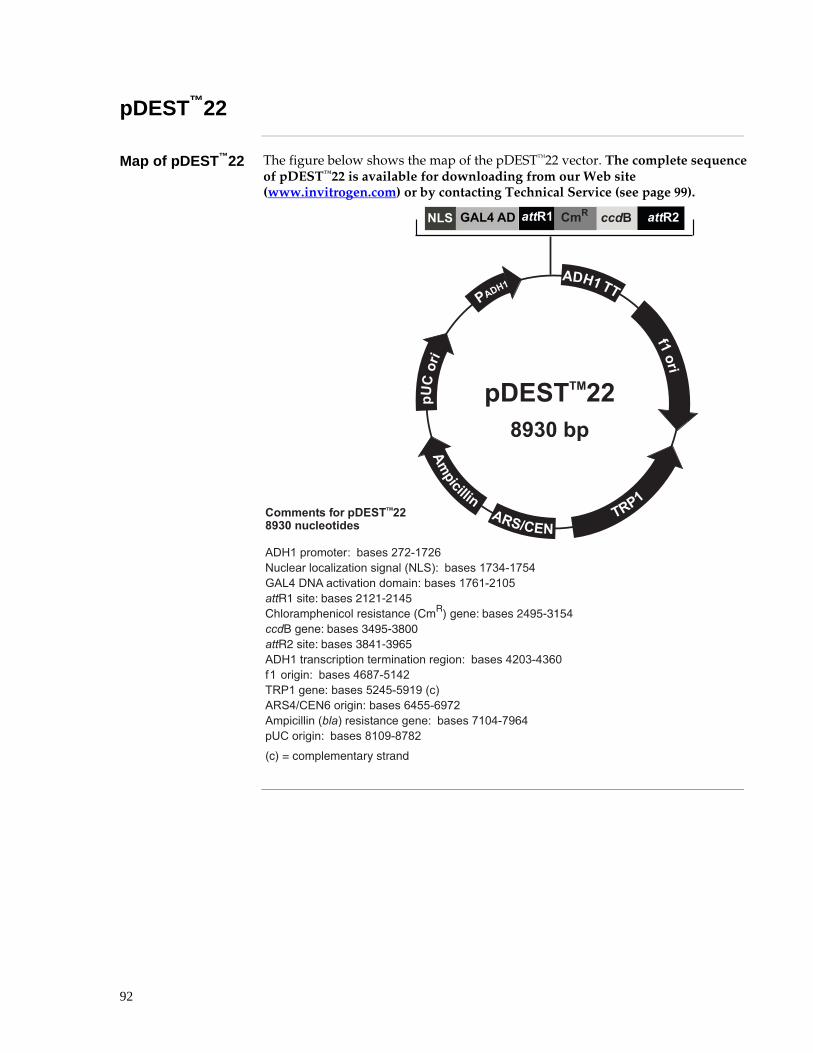
92
pDEST™22
Map of pDEST™22 The figure below shows the map of the pDEST™22 vector. The complete sequence of pDEST™22 is available for downloading from our Web site (www.invitrogen.com) or by contacting Technical Service (see page 99).
� !�""
"���
$%��&
�� !�
0������� �����'����� ,�(
$ �("")���� ��*$
0������ �#����������AB!)��%����� �
��>����������?��)������3�%�3�A��"����"���"�#�����������"�0��1?��)������37�%�34�������������������� �����?�)������3A�%��24��������� 7���� ��4�4�#4���2 !��� ���!� ������� �@2��A�� � 7���� ��4�C�#���������� � 7���� ����C�#�;00����4���� 7���� ���;��#�C5���>����������������������������������?��)�������27%�7A2 ��������?��)������A.3%4���'��������?�)�����4��4%4@�@�0�1������&A�������?�)�����A�44%A@3�������""���0���1����������������?��)�����3�2�%3@A��5��������?��)�����.�2@%.3.�
@�A�B����! � ����������
��(+��,
-��&
�.$&�&��&�
���"

93
Features of Vectors pDEST™32 and pDEST™22
Features of Vectors
The table below shows the features of the pDEST™32 and pDEST™22 vectors.
Feature Vector Benefit
ADH1 promoter pDEST™32, pDEST™22 Permits medium-level, constitutive expression of the gene of interest
GAL4 DBD coding sequence
pDEST™32 Allows bait protein to bind to GAL4 binding sites in promoters of reporter genes and activate reporters after prey recruitment
GAL4 AD coding sequence
pDEST™22 Allows prey protein to activate reporters after binding to bait protein
attR1 and attR2 sites pDEST™32, pDEST™22 Bacteriophage λ-derived recombination sequences that allow recombinational cloning of a gene of interest in the expression construct with a Gateway® destination vector (Landy, 1989).
Chloramphenicol resistance gene (CmR)
pDEST™32, pDEST™22 Allows counterselection of the plasmid.
ccdB gene pDEST™32, pDEST™22 Permits negative selection of the plasmid.
ADH1 TT pDEST™32, pDEST™22 Allows transcription termination and polyadenylation of mRNA.
f1 origin pDEST™32, pDEST™22 Allows rescue of single-stranded DNA
LEU2 gene pDEST™32 Permits selection for plasmid in transformed yeast cells
TRP1 gene pDEST™22 Permits selection for plasmid in transformed yeast cells
ARS/CEN pDEST™32, pDEST™22 Yeast centromere and replication origin: allows replication and maintenance of plasmid in yeast
Gentamicin resistance gene
pDEST™32 Permits selection for the plasmid in E. coli
Ampicillin resistance gene
pDEST™22 Permits selection for the plasmid in E. coli
pUC origin pDEST™32, pDEST™22 Permits high-copy replication and maintenance in E. coli.
Cycloheximide Sensitivity Gene (CYH2S)
pDEST™32 Lets you counter select against bait plasmid through plasmid shuffling

94
pEXP™-AD502
Map of pEXP™-AD502
The figure below shows the map of the pEXP™-AD502 vector. The complete sequence of pEXP™-AD502 is available for downloading from our Web site (www.invitrogen.com) or by contacting Technical Service (see page 99).
��1��2�;)�<C:�9�
���>�
��> ''
����
�&A����>�
�5�
���
����2�4�� ��": ��42������ ��"�
�..����-��$�0�1� ����2��&����3������#���&4�
�������� 7����� ��5�#���<(����������)$7���� ����4�#��4��D�0��"�! ��!��!�E����������!�@'%�A7���� ����45#���;�������>����������7���� �����5#�C0������7���� ���C�0#�C������47���� ���C<C#400������������������ �������� ���7���� ��40��#4���-������7����� ��4<0�#���;� ���� � �@�A7���� ���4��#�C4C� ������ �@�A7���� ���C�0#�0��2&'58������@�A7���� ����5�#�C<C������� 7����� ���0�4#���5������!!���@���A� ������� �� � 7����� ������#�C<���2�����7����� ��5��5#5<;C@�A�B����! � ����������
'��
�
������""��
Continued on next page

95
pEXP™-AD502, continued
MCS of pEXP™-AD502
The figure below shows the multiple cloning site of the pEXP™-AD502 vector. Use the diagram below to help you clone your gene of interest into pDEST™32. Note the following features in the diagram below:
• The reading frame for the GAL4 AD is shown; the insert needs to be in frame with that
• Sequences for suggested forward and reverse sequencing primers are shown below ���������� ���������� ���������� ���������� ���������� ���������� ����������
�� ��� ��� ��� ��� ��� ��� ��� ��� ��� ��� ��� ��� ��� ��� ��� ��� ��� ��� ���
��� ��� ��� ��� ��� ��� ��� ��� ��� ��� ��� ��� ��� ��� ��� ��� ��� ��� ���
��� ��� ��� ��� ��� ��� ��� ��� ��� ��� ��� ��� ��� ��� ��� ��� ��� ��� ���
��� ��� ��� ��� ��� ��� ��� ��� ��� ��� ��� ��� ��� ��� ��� ��� ��� ��� ���
��� ��� ��� ��� ��� ��� ��� ��� ��� ��� ��� ��� ��� ��� ��� ��� ��� ��� ���
��� ��� ��� ��� ��� ��� ��� ��� ��� ��� ��� ��� ��� ��� ��� ��� ��� ��� ���
��� ��� ��� ��� ��� ��� ��� ��� ��� ��� ��� ��� ��� ��� ��� ��� ��� ��� ���
��� ��� ��� ��� ��� ��� ��� ��� ��� ��� ��� ��� ��� ��� �������� ����������
���������� ���������� ���������� ���������� ���������� ���������� ����������
���������� ���������� ���������� ���������� ���������� ���������� ����������
���������� ��������
�������� ������������������������������
�������� � ��!�� ����������������������
�C�2����"����"���"�#�����������"�0��1
��� !�� ��� ��� ��� �� ��� %�� ��� ��� ��� ��� ��� !"� ��� ��� ��� ��� ���
��� ��� ��� ��� ��� ��� ��� !"� �"� !"� �"� ��� ��� ��� ��� ��� !�� ��� ���
��� �"� �"� ��� �"� ��� ��� ��� ��� ��� ��� ��� !�� ��� ��� ��� ��� ��� %��
#$� ��� ��� !"� &�� ��� ��� ��� ��� �"� ��� ��� ��� ��� ��� ��� ��� ��� ���
��� ��� !�� ��� ��� !�� ��� ��� �"� ��� ��� �"� ��� ��� ��� ��� !"� ��� ���
�"� �"� ��� &�� !"� ��� �"� �"� �"� &�� ��� ��� %�� ��� ��� ��� ��� !"� ���
��� ��� ��� �"� !�� !�� ��� !�� ��� ��� ��� ��� ��� ��� ��� �"� ��� ��� ���
��� ��� ��� ��� ��� ��� �"�
����9 ��9
���9 ���99
������������������� �����
(�(
(�(
(��
(��
(�'�
((
(���
(��
('��
('��
���
�(��
���9 ����9 ���99 ���9��
��
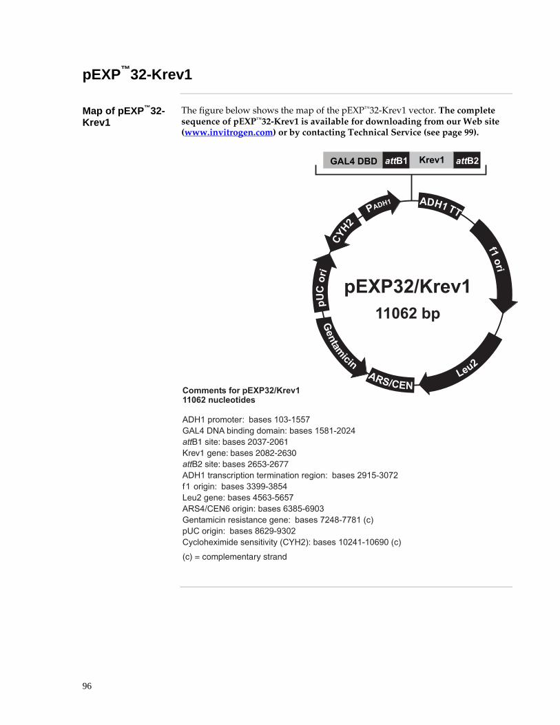
96
pEXP™32-Krev1
Map of pEXP™32-Krev1
The figure below shows the map of the pEXP™32-Krev1 vector. The complete sequence of pEXP™32-Krev1 is available for downloading from our Web site (www.invitrogen.com) or by contacting Technical Service (see page 99).
� !�""
�#�
$%��&
� ���� �������� �����'���� �
$�0� �+5�6�������*$
��(+��,
-��&
'���.
&�&�
�� !�
�/!�
0������ �#�����1�!�3?��':::)@���%����� �
��>����������?��)������27%�443��������)�� ���� �����?�)������4.�%�2����������� 7���� ��40�<#405�F >��� � 7���� ��40;4#45�0����4���� 7���� ��45��#45<<��>����������������������������������?��)������@�4%723� ��������?��)�����77@@%7.4����������?�)������4A7%4A43������&A�������?�)�����A7.4%A@27��������������������������?��)�����3��.%33.��0�1�5��������?��)�����.A�@%@72��$�"�*�,��� ������������$�0�B>�1?�)������2���%�2A@2�0�1
@�A�B����! � ����������
5�6�

97
pEXP™22-RalGDS-wt, m1, m2
Maps of pEXP™22-RalGDS-wt, m1, m2
The figure below shows the map of the pEXP™22-RalGDS-wt, pEXP™22-RalGDS m1, m2 vector. The complete sequence of pEXP™22/RalGDS-wt, pEXP™22/RalGDS-m1 and pEXP™22/RalGDS-m2 are available for downloading from our Web site (www.invitrogen.com) or by contacting Technical Service (see page 99).
D�=�����"��������E�!������*��!�" �$��E���*���933'E��� ����*����A4�
� !�""
"���
$%��&
�� !�
���*�����3�:3������� �����'����� ,�(
$�0���+���' (7�+.�+.������*$
0������ �#�����1���3���*����3�:3��<D@A��%����� �
��>����������?��)������3�%�3�A��"����"���"�#�����������"�0��1?��)������37�%�34�������������������� �����?�)������3A�%��24��������� 7���� ��4�4�#4�����!$*��� � G7���� ��4�55#4�54����4���� 7���� ��4�<C#4�0���>����������������������������������?��)������3��%�.@. ��������?��)�����7��4%7A.2'��������?�)�����73.7%��43�0�1������&A�������?�)������@@7%44�2������""���0���1����������������?��)�����4A��%A42��5��������?��)�����AA�3%37�@
@�A�B����! � ����������
��(+��,
-��&
�.$&�&��&�

98
Map of pENTR™-gus
Description pENTR™-gus is a 3841 bp entry clone containing the Arabidopsis thaliana gene for β-glucuronidase (gus) (Kertbundit et al., 1991). The gus gene was amplified using PCR primers containing attB recombination sites. The amplified PCR product was then used in a BP recombination reaction with pDONR201™ to generate the entry clone. For more information about the BP recombination reaction, refer to the Gateway® Technology with Clonase™ II manual which is available for downloading from our Web site or by contacting Technical Service.
Map of Control Vector
The figure below summarizes the features of the pENTR™-gus vector. The complete sequence for pENTR™-gus is available from our Web site (www.invitrogen.com) or by contacting Technical Service (see page 99).
�..����-��$�,"�18#�")
�����#���&4�
���%�7���� ��CC#�C;�@���! � ����������A����� � 7���� ��44;#40�C���%47���� ��40��#4��0��2�����7���� ��4400#4;<��@2AF��������� ������� �� � 7���� ��4CC0#�;0��@2A
2�B����! � ����������
$�,"�18#�")
����*$
���%�
���%4
��2 ����
F��������
���
99
Additional Information Technical Service
Web Resources
Visit the Invitrogen Web site at www.invitrogen.com for:
• Technical resources, including manuals, vector maps and sequences, application notes, MSDSs, FAQs, formulations, citations, handbooks, etc.
• Complete technical service contact information
• Access to the Invitrogen Online Catalog
• Additional product information and special offers
Contact Us For more information or technical assistance, call, write, fax, or email. Additional
international offices are listed on our Web page (www.invitrogen.com). Corporate Headquarters: Invitrogen Corporation 1600 Faraday Avenue Carlsbad, CA 92008 USA Tel: 1 760 603 7200 Tel (Toll Free): 1 800 955 6288 Fax: 1 760 602 6500 E-mail: [email protected]
Japanese Headquarters: Invitrogen Japan LOOP-X Bldg. 6F 3-9-15, Kaigan Minato-ku, Tokyo 108-0022 Tel: 81 3 5730 6509 Fax: 81 3 5730 6519 E-mail: [email protected]
European Headquarters: Invitrogen Ltd Inchinnan Business Park 3 Fountain Drive Paisley PA4 9RF, UK Tel: +44 (0) 141 814 6100 Tech Fax: +44 (0) 141 814 6117 E-mail: [email protected]
Material Data Safety Sheets (MSDSs)
MSDSs are available on our Web site at www.invitrogen.com. On the home page, click on Technical Resources and follow instructions on the page to download the MSDS for your product.
Limited Warranty Invitrogen is committed to providing our customers with high-quality goods and services.
Our goal is to ensure that every customer is 100% satisfied with our products and our service. If you should have any questions or concerns about an Invitrogen product or service, contact our Technical Service Representatives.
Invitrogen warrants that all of its products will perform according to specifications stated on the certificate of analysis. The company will replace, free of charge, any product that does not meet those specifications. This warranty limits Invitrogen Corporation’s liability only to the cost of the product. No warranty is granted for products beyond their listed expiration date. No warranty is applicable unless all product components are stored in accordance with instructions. Invitrogen reserves the right to select the method(s) used to analyze a product unless Invitrogen agrees to a specified method in writing prior to acceptance of the order.
Invitrogen makes every effort to ensure the accuracy of its publications, but realizes that the occasional typographical or other error is inevitable. Therefore Invitrogen makes no warranty of any kind regarding the contents of any publications or documentation. If you discover an error in any of our publications, please report it to our Technical Service Representatives.
Invitrogen assumes no responsibility or liability for any special, incidental, indirect or consequential loss or damage whatsoever. The above limited warranty is sole and exclusive. No other warranty is made, whether expressed or implied, including any warranty of merchantability or fitness for a particular purpose.

100
Purchaser Notification
Introduction Use of the ProQuest™ Two-Hybrid System is covered under the licenses detailed below.
Information for European Customers
The S. cerevisiae strains MaV203 is genetically modified and contains three GAL4 inducible reporter genes stably integrated into the genome. S. As a condition of sale, this product must only be used in accordance with all applicable local legislation and guidelines including EC directive 90/219/EEC on the contained use of genetically modified organisms.
Limited Use Label License No: 11 ProQuest™ Reverse Two-Hybrid System
The ProQuest™ Reverse Two-Hybrid System is the subject of one or more patents licensed to Invitrogen Corporation by Massachusetts General Hospital and Johns Hopkins University (including U.S. Patent Nos. 5,955,280 and/or 5,965,368 and foreign equivalents) ("Reverse Two-Hybrid”). The purchase of this product conveys to the buyer the non-transferable right to use the purchased amount of the product and components of the product in research conducted by the buyer (whether the buyer is an academic or for-profit entity. The buyer cannot sell or otherwise transfer (a) this product (b) its components or (c) materials made using this product or its components to a third party or otherwise use this product or its components or materials made using this product or its components for Commercial Purposes. The buyer may transfer information or materials made through the use of this product to a scientific collaborator, provided that such transfer is not for any Commercial Purpose, and that such collaborator agrees in writing (a) not to transfer such materials to any third party, and (b) to use such transferred materials and/or information solely for research and not for Commercial Purposes. Commercial Purposes means any activity by a party for consideration and may include, but is not limited to: (1) use of the product or its components in manufacturing; (2) use of the product or its components to provide a service, information, or data; (3) use of the product or its components for therapeutic, diagnostic or prophylactic purposes; or (4) resale of the product or its components, whether or not such product or its components are resold for use in research. Invitrogen Corporation will not assert a claim against the buyer of infringement of the above patents based upon the manufacture, use or sale of a therapeutic, clinical diagnostic, vaccine or prophylactic product developed in research by the buyer in which this product or its components was employed, provided that neither this product nor any of its components was used in the manufacture of such product. If the purchaser is not willing to accept the limitations of this limited use statement, Invitrogen is willing to accept return of the product with a full refund. For information on purchasing a license to this product for purposes other than research, contact Licensing Department, Invitrogen Corporation, 1600 Faraday Avenue, Carlsbad, California 92008. Phone (760) 603-7200. Fax (760) 602-6500.
Limited Use Label License No: 141 Expression of Polypeptides in Yeast
This product is the subject of one or more of U.S. Patent Nos. 5,618,676, 5,854,018, 5,856,123, 5,919,651 and foreign equivalents. Rights to use this product are limited to academic research use only. Non-academic entities are required to obtain a separate license from Washington Research Foundation to utilize this product for any use. Washington Research Foundation, 2815 Eastlake Avenue East, Suite 300, Seattle, Washington 98102. Tel: 206-336-5600. Fax: 206-336-5615.
Continued on next page

101
Purchaser Notification, continued
Limited Use Label License No: 19 Gateway® Cloning Products
The purchase of this product conveys to the buyer the non-transferable right to use the purchased amount of the product and components of the product in research conducted by the buyer (whether the buyer is an academic or for profit entity). The purchase of this product does not convey a license under any method claims in the foregoing patents or patent applications, or to use this product with any recombination sites other than those purchased from Invitrogen Corporation or its authorized distributor. The right to use methods claimed in the foregoing patents or patent applications with this product for research purposes only can only be acquired by the use of ClonaseTM purchased from Invitrogen Corporation or its authorized distributors. The buyer cannot modify the recombination sequence(s) contained in this product for any purpose. The buyer cannot sell or otherwise transfer (a) this product, (b) its components, or (c) materials made by the employment of this product or its components to a third party or otherwise use this product or its components or materials made by the employment of this product or its components for Commercial Purposes. The buyer may transfer information or materials made through the employment of this product to a scientific collaborator, provided that such transfer is not for any Commercial Purpose, and that such collaborator agrees in writing (a) not to transfer such materials to any third party, and (b) to use such transferred materials and/or information solely for research and not for Commercial Purposes. Notwithstanding the preceding, any buyer who is employed in an academic or government institution may transfer materials made with this product to a third party who has a license from Invitrogen under the patents identified above to distribute such materials. Transfer of such materials and/or information to collaborators does not convey rights to practice any methods claimed in the foregoing patents or patent applications. Commercial Purposes means any activity by a party for consideration and may include, but is not limited to: (1) use of the product or its components in manufacturing; (2) use of the product or its components to provide a service, information, or data; (3) use of the product or its components for therapeutic, diagnostic or prophylactic purposes; or (4) resale of the product or its components, whether or not such product or its components are resold for use in research. Invitrogen Corporation will not assert a claim against the buyer of infringement of the above patents based upon the manufacture, use or sale of a therapeutic, clinical diagnostic, vaccine or prophylactic product developed in research by the buyer in which this product or its components was employed, provided that none of (i) this product, (ii) any of its components, or (iii) a method claim of the foregoing patents, was used in the manufacture of such product. Invitrogen Corporation will not assert a claim against the buyer of infringement of the above patents based upon the use of this product to manufacture a protein for sale, provided that no method claim in the above patents was used in the manufacture of such protein. If the purchaser is not willing to accept the limitations of this limited use statement, Invitrogen is willing to accept return of the product with a full refund. For information on purchasing a license to use this product for purposes other than those permitted above, contact Licensing Department, Invitrogen Corporation, 1600 Faraday Avenue, Carlsbad, California 92008. Phone (760) 603-7200.
Limited Use Label License No: 67 Two Hybrid Technology
Practice of the two-hybrid system is covered by U.S. Patents Nos. 5,283,173, 5,468,614 and 5,667,973 assigned to The Research Foundation of the State University of New York (SUNY). Purchase of any Invitrogen two-hybrid reagents does not imply or convey a license to practice the two-hybrid system covered by these patents, beyond use of the enclosed kit for non-commercial research. Commercial entities purchasing these reagents must obtain a license from The Research Foundation of the State University of New York before using them. For license information, please contact: The Office of Technology Licensing and Industry Relations, The Research Foundation of SUNY at Stony Brook, W5530 Melville Memorial Library, Stony Brook, NY 11794-3368, Phone: 631-632-9009; Fax: 631-632-1505; You may not distribute the System or the vectors or host strains contained in it to others. You may not transfer modified, altered, or original material from the system to a third party without written notification to, and written approval from Invitrogen. You may not assign, sub-license, rent, lease or otherwise transfer any of the rights or obligations set forth herein, except as expressly permitted by Invitrogen.

102
Gateway® Clone Distribution Policy
Introduction The information supplied in this section is intended to provide clarity concerning Invitrogen’s policy for the use and distribution of cloned nucleic acid fragments, including open reading frames, created using Invitrogen’s commercially available Gateway® Technology.
Gateway® Entry Clones
Invitrogen understands that Gateway® entry clones, containing attL1 and attL2 sites, may be generated by academic and government researchers for the purpose of scientific research. Invitrogen agrees that such clones may be distributed for scientific research by non-profit organizations and by for-profit organizations without royalty payment to Invitrogen.
Gateway® Expression Clones
Invitrogen also understands that Gateway® expression clones, containing attB1 and attB2 sites, may be generated by academic and government researchers for the purpose of scientific research. Invitrogen agrees that such clones may be distributed for scientific research by academic and government organizations without royalty payment to Invitrogen. Organizations other than academia and government may also distribute such Gateway® expression clones for a nominal fee ($10 per clone) payable to Invitrogen.
Additional Terms and Conditions
We would ask that such distributors of Gateway® entry and expression clones indicate that such clones may be used only for research purposes, that such clones incorporate the Gateway® Technology, and that the purchase of Gateway® Clonase™ from Invitrogen is required for carrying out the Gateway® recombinational cloning reaction. This should allow researchers to readily identify Gateway® containing clones and facilitate their use of this powerful technology in their research. Use of Invitrogen’s Gateway® Technology, including Gateway® clones, for purposes other than scientific research may require a license and questions concerning such commercial use should be directed to Invitrogen’s licensing department at 760-603-7200.

103
Product Qualification
Introduction This section describes the criteria used to qualify the components of the ProQuest™ Two-Hybrid System.
Vectors The structure of each vector is verified by restriction enzyme digestion. For
pEXP™32-Krev1 and pEXP™22-RalGDS-wt, m1, m2 the insert is sequenced.
MaV203 Contamination Check
100ul of MaV203 is streaked out on one LB plate and one YPAD plate. The LB plate is incubated at 37oC for 2 days and the YPAD plate at 30oC for 3 days. No colony type should be present on the LB plate, and no more than one colony type should be observed on the YPAD plate.
Viability
100µl of serial dilutions of MaV203 (10-3, 10-4 and 10-5) are plated onto duplicate YPAD plates and duplicate SC-Leu-Trp plates. Plates are incubated for 2 days at 30oC. Viable cell count must be >2x10⁵ for YPAD. No growth should be observed on SC-Leu-Trp.
Mating type PCR
Mating type is verified by PCR on DNA purified from MaV203 using mating type specific primers.
Gateway® Clonase™
II Enzyme Mix Gateway® BP and LR Clonase™ II Enzyme Mixes are functionally tested in a one hour recombination reaction followed by a transformation assay.

104
References
Ausubel, F. M., Brent, R., Kingston, R. E., Moore, D. D., Seidman, J. G., Smith, J. A., and Struhl, K. (1994) Current Protocols in Molecular Biology, Greene Publishing Associates and Wiley-Interscience, New York
Barr, R. K., Hopkins, R. M., Watt, P. M., and Bogoyevitch, M. A. (2004) Reverse two-hybrid screening identifies residues of JNK required for interaction with the kinase interaction motif of JNK-interacting protein-1. J Biol Chem 279, 43178-43189. Epub 42004 Jul 43122.
Bartel, P. L., Chien, C.-T., Sternglanz, R., and Fields, S. (1993) Elimination of False Positives That Arise in Using the Two-Hybrid System. BioTechniques 14, 920-924
Bernard, P., and Couturier, M. (1992) Cell Killing by the F Plasmid CcdB Protein Involves Poisoning of DNA-Topoisomerase II Complexes. J. Mol. Biol. 226, 735-745
Bernard, P., Kezdy, K. E., Melderen, L. V., Steyaert, J., Wyns, L., Pato, M. L., Higgins, P. N., and Couturier, M. (1993) The F Plasmid CcdB Protein Induces Efficient ATP-dependent DNA Cleavage by Gyrase. J. Mol. Biol. 234, 534-541
Brachmann, R. K., and Boeke, J. D. (1997) Tag Games in Yeast: The Two-Hybrid System and Beyond. Current Opin. Biotech. 8, 561-568
Chevray, P. M., and Nathans, D. (1992) Protein Interaction Cloning in Yeast: Identification of Mammalian Proteins that React with the Leucine Zipper of Jun. Proc. Nat. Acad. Sci. 89, 5789-5793
Durfee, T., Becherer, K., Chen, P. L., Yeh, S. H., Yang, Y., Kilburn, A. E., Lee, W. H., and Elledge, S. J. (1993) The retinoblastoma protein associates with the protein phosphatase type 1 catalytic subunit. Genes Dev 7, 555-569.
Endoh, H., Walhout, A. J., and Vidal, M. A green fluorescent protein-based reverse two-hybrid system: application to the characterization of large numbers of potential protein-protein interactions. 74-88.
Fields, S., and Song, O. (1989) A Novel Genetic System to Detect Protein-Protein Interaction. Nature 340, 245-246
Herrmann, C., Horn, G., Spaargaren, M., and Wittinghofer, A. (1996) Differential interaction of the ras family GTP-binding proteins H-Ras, Rap1A, and R-Ras with the putative effector molecules Raf kinase and Ral-guanine nucleotide exchange factor. J Biol Chem 271, 6794-6800.
Hu, G., Zhang, S., Vidal, M., Baer, J. L., Xu, T., and Fearon, E. R. (1997) Mammalian homologs of seven in absentia regulate DCC via the ubiquitin-proteasome pathway. Genes Dev 11, 2701-2714.
Kertbundit, S., Greve, H. d., Deboeck, F., Montagu, M. V., and Hernalsteens, J. P. (1991) In vivo Random β-glucuronidase Gene Fusions in Arabidopsis thaliana. Proc. Natl. Acad. Sci. USA 88, 5212-5216
Landy, A. (1989) Dynamic, Structural, and Regulatory Aspects of Lambda Site-specific Recombination. Ann. Rev. Biochem. 58, 913-949
Continued on next page

105
References, continued
Leanna, C. A., and Hannink, M. (1996) The reverse two-hybrid system: a genetic scheme for selection against specific protein/protein interactions. Nucleic Acids Res 24, 3341-3347.
Luban, J., and Goff, S. P. (1995) The Yeast Two-Hybrid System for Studying Protein-Protein Interactions. Curr. Opinion Biotechnol. 6, 59-64
Miki, T., Park, J. A., Nagao, K., Murayama, N., and Horiuchi, T. (1992) Control of Segregation of Chromosomal DNA by Sex Factor F in Escherichia coli. Mutants of DNA Gyrase Subunit A Suppress letD (ccdB) Product Growth Inhibition. J. Mol. Biol. 225, 39-52
Polaina, J., and Adam, A. C. (1991) A fast procedure for yeast DNA purification. Nucleic Acids Res 19, 5443.
Serebriiskii, I., Khazak, V., and Golemis, E. A. (1999) A Two-Hybrid Dual Bait System to Discriminate Specificity of Protein Interactions. J. Biol. Chem. 274, 17080-17087
Sherman, F. Getting started with yeast. 3-21.
Shih, H. M., Goldman, P. S., DeMaggio, A. J., Hollenberg, S. M., Goodman, R. H., and Hoekstra, M. F. (1996) A positive genetic selection for disrupting protein-protein interactions: identification of CREB mutations that prevent association with the coactivator CBP. Proc Natl Acad Sci U S A 93, 13896-13901.
Vidal, M. (1997) The Reverse Two-Hybrid System. The Two-Hybrid System, Oxford University Press, New York
Vidal, M. (1997) The Reverse Two-Hybrid System. The Two-Hybrid System 109
Vidal, M., Brachmann, R. K., Fattaey, A., Harlow, E., and Boeke, J. D. (1996) Reverse two-hybrid and one-hybrid systems to detect dissociation of protein-protein and DNA-protein interactions. Proc Natl Acad Sci U S A 93, 10315-10320.
Vidal, M., Braun, P., Chen, E., Boeke, J. D., and Harlow, E. (1996) Genetic characterization of a mammalian protein-protein interaction domain by using a yeast reverse two-hybrid system. Proc Natl Acad Sci U S A 93, 10321-10326.
Vidal, M., and Legrain, P. (1999) Yeast forward and reverse 'n'-hybrid systems. Nucleic Acids Res 27, 919-929.
Vojtek, A. B., Hollenberg, S. M., and Cooper, J. A. (1993) Mammalian Ras Interacts Directly with the Serine/Threonine Kinase Raf. Cell 74, 205-214
Weintraub, H., Davis, R., Tapscott, S., Thayer, M., Krause, M., Benezra, R., Blackwell, T. K., Turner, D., Rupp, R., Hollenberg, S., and et al. (1991) The myoD gene family: nodal point during specification of the muscle cell lineage. Science 251, 761-766.
For research use only. Not intended for any animal or human therapeutic or diagnostic use ©2005 Invitrogen Corporation. All rights reserved.

106