programming membrane permeability using integrated
TRANSCRIPT

12282 | Chem. Commun., 2017, 53, 12282--12285 This journal is©The Royal Society of Chemistry 2017
Cite this:Chem. Commun., 2017,
53, 12282
Programming membrane permeability usingintegrated membrane pores and blockersas molecular regulators†
Julia M. Thomas,a Mark S. Friddin, a Oscar Ces ab and Yuval Elani *ab
We report a bottom-up synthetic biology approach to engi-
neering vesicles with programmable permeabilities. Exploiting the
concentration-dependent relationship between constitutively active
pores (alpha-hemolysin) and blockers allows blockers to behave as
molecular regulators for tuning permeability, enabling us to system-
atically modulate cargo release kinetics without changing the lipid
fabric of the system.
The use of biologically derived molecular components hasproved to be a powerful strategy for the design and assemblyof a rich and diverse range of lipid-bilayer encased micro-systems. They enable a repertoire of biomimetic features tobe incorporated into cell-like architectures1 including motility,2
protein synthesis,3 and enzymatic reaction cascades,4 in additionto mechanosensitive behaviours,5 active molecular transport,6
and light-activated responses.7
This repurposing of biological machinery to impart func-tionality into artificial systems is one of the core tenants ofbottom-up synthetic biology, which has shown promise indeveloping a new generation of liposomal drug deliverysystems,8,9 vesicle-based microreactors,4,10,11 and membrane-bound artificial cells.3,12–15 Here, we exploit this strategy toconstruct Giant Unilamellar Vesicles (GUVs) with user-definedpermeabilities. Crucially our approach does not rely uponchemical modification or breakdown of the lipid chassis andinstead exploits the interactions between embedded membraneprotein pores and chemical blockers.
Fundamental to successful applications for these techno-logies is the ability to precisely control the exchange of materialsbetween individual compartments and the external medium.This is critical for controlling drug release rates, modulatingreaction kinetics, mediating communication between artificial
cells and neighbouring biological cells,13 and regulating theinterchange of nutrients and waste products.3 To date, this hasbeen achieved by triggering the free-diffusion of materialsthrough protein channels inserted into the membrane orby releasing the entire contents of the compartment by lysis.Alternative approaches involve generating defects in the membranefabric, e.g. through UV irradiation,16 photocleavage,17–19 photo-isomerisation,20 molecular absorbers,21,22 by taking themembrane through a phase transition,23,24 and applicationof shear forces,25 electric fields,11,26 osmotic gradients andchanges in pH.27
These approaches rely on the application of external forcesand the extent of permeability cannot be controlled system-atically. They are not only binary in nature but also offer nosustainable control over bilayer permeability, especially if thecompartment is destroyed in the process. Herein we addressthis problem by demonstrating that membrane permeabilitycan be finely tuned by exploiting the relationship between aconstitutively active membrane pore, alpha-hemolysin (a-HL),and the reversible blocker TRIMEB (heptakis(2,3,6-tri-O-methyl)-b-cyclodextrin).
a-HL is a heptameric transmembrane pore from Staphylococcusaureus that has a water-accessible channel lumen that is 1.4 nmwide at its narrowest point through which molecules can passivelydiffuse.28,29 It can thus be used as a molecular sieve: it is perme-able to globular molecules up to 2000 Da in size30 (a categorywhich most drugs and metabolites fall under), and longerelongated polymers,31 yet is impermeable to biological macro-molecules (including DNA and proteins). This makes it suitablefor use in vesicle-based enzymatic reactors and artificial cells.TRIMEB is a cyclic oligosaccharide that reversibly and non-covalently binds to the a-HL pore lumen on the cis side of thebarrel near Met 113,32 restricting the passage of molecules.32,33
The extent of blockage and the proportion of time the blocker isin the bound state contributes to total leakage through thepore, a process previously quantified using optical ion flux.34
Due to the larger size of the blocker compared to the pore, it isunlikely that TRIMEB itself can fully translocate through a-HL.
a Department of Chemistry, Imperial College London, Exhibition Road, London,
SW7 2AZ, UK. E-mail: [email protected] Institute of Chemical Biology, Imperial College London, Exhibition Road, London,
SW7 2AZ, UK
† Electronic supplementary information (ESI) available: Experimental details. SeeDOI: 10.1039/c7cc05423h
Received 13th July 2017,Accepted 16th October 2017
DOI: 10.1039/c7cc05423h
rsc.li/chemcomm
ChemComm
COMMUNICATION
Ope
n A
cces
s A
rtic
le. P
ublis
hed
on 0
1 N
ovem
ber
2017
. Dow
nloa
ded
on 3
/19/
2022
3:0
9:49
AM
. T
his
artic
le is
lice
nsed
und
er a
Cre
ativ
e C
omm
ons
Attr
ibut
ion
3.0
Unp
orte
d L
icen
ce.
View Article OnlineView Journal | View Issue

This journal is©The Royal Society of Chemistry 2017 Chem. Commun., 2017, 53, 12282--12285 | 12283
Supported by single-channel electrical measurements indroplet interface bilayers (DIBs), we show that this relationshipcan be leveraged to carefully control the rate of release of afluorescent dye contained within a-HL functionalised GUVs.We achieve this by regulating the concentration of TRIMEBco-encapsulated inside the GUVs, which acts as an adaptermolecule, enabling the permeability of the GUV to be preciselypre-determined (Fig. 1A). Our approach does not rely uponchemical modification to the vesicles or the application ofexternally applied forces, and does not lead to the destructionof the membrane chassis.
1-Palmitoyl-2-oleoyl-sn-glycero-3-phosphocholine (POPC) GUVsin TRIS buffer (25 mM Tris–HCl; 500 mM KCl; pH 8.0) wereprepared by the emulsion phase transfer technique.35 The effectof blocker concentration was studied by encapsulating 1 mMcalcein with TRIMEB concentrations ranging from 1–50 mM.An inverted fluorescence microscope (FITC filter; 200 msexposure) was used to quantify dye leakage, with vesicle contoursand mean fluorescence extracted using a thresholding function ofan image analysis software (Image J). Data from a minimumof 10 GUVs were taken for each experimental condition (fullexperimental details are available in the ESI†). a-HL was addedto the external solution as water soluble monomers (final concen-tration 50 ng mL�1) which proceeded to bind to the membraneand assemble into the pore complex.
After a variable lag phase of between 5 and 60 minutes(Fig. 1B and C), GUV fluorescence was found to decay exponen-tially following the addition of the protein. We attribute this lagto the time taken for a-HL monomers to bind to the membraneand to oligomerize into a functional pore. Depending onblocker concentration, the GUV interior was indistinguishablefrom background after 5–80 minutes. The contrast of the GUVunder phase contrast microscopy also faded over time as thecomposition of the interior approached that of the exterior(as observed elsewhere).36 The smaller the concentration ofblocker, the quicker the fluorescence decay (Fig. 1D).
The permeability of our system (Peff) can be defined by theequation below:
dNp
dt¼ Peff
Np
V� cp0
� �(1)
Here, Np is the number of encapsulated fluorescent particles,V is vesicle volume, and cp0 is the concentration of calcein outsidethe vesicle. As external calcein is removed during GUV preparation,and as the volume of the GUV is negligible compared to that of theexternal solution we can assume that cp0 = 0 at all times.
Only GUVs where r 4 5 mm were analysed as thesecould easily be resolved using our setup. By fitting total GUVfluorescence per unit volume (which is proportional to dyeconcentration)37 to the equation above we extract Peff for GUVscontaining embedded pores and variable amounts of blocker(Fig. 2). We note that in this case, it is not the intrinsicmembrane permeability that is being measured, but the perme-ability of the entire vesicle/a-HL/TRIMEB assembly, eliminatingthe dependence on membrane area.
Fig. 1 Vesicle permeability fluorescence assay. (A) Schematic of thevesicle/a-HL/TRIMEB system. The presence of blocker reduces calceinflux through the pore. (B) Phase contrast and fluorescence microscopyimages after a pore insertion event. As the internal and external contentof the vesicle equilibrates the contrast of the membrane contour reduces.(C) Typical trace of vesicle fluorescence over time. Following a lag phase(blue) an exponential decay is seen (red) due to the formation of a pore andefflux of calcein. (D) Average vesicle fluorescence levels over time withdifferent TRIMEB concentrations (lag phase removed). The larger theconcentration of blocker the slower the decay.
Communication ChemComm
Ope
n A
cces
s A
rtic
le. P
ublis
hed
on 0
1 N
ovem
ber
2017
. Dow
nloa
ded
on 3
/19/
2022
3:0
9:49
AM
. T
his
artic
le is
lice
nsed
und
er a
Cre
ativ
e C
omm
ons
Attr
ibut
ion
3.0
Unp
orte
d L
icen
ce.
View Article Online
12284 | Chem. Commun., 2017, 53, 12282--12285 This journal is©The Royal Society of Chemistry 2017
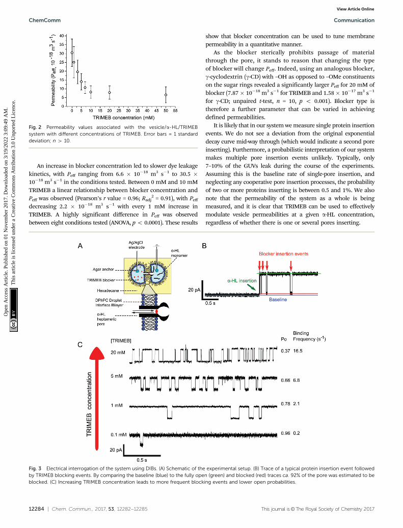
An increase in blocker concentration led to slower dye leakagekinetics, with Peff ranging from 6.6 � 10�18 m3 s�1 to 30.5 �10�18 m3 s�1 in the conditions tested. Between 0 mM and 10 mMTRIMEB a linear relationship between blocker concentration andPeff was observed (Pearson’s r value = 0.96; Radj
2 = 0.91), with Peff
decreasing 2.2 � 10�18 m3 s�1 with every 1 mM increase inTRIMEB. A highly significant difference in Peff was observedbetween eight conditions tested (ANOVA, p o 0.0001). These results
show that blocker concentration can be used to tune membranepermeability in a quantitative manner.
As the blocker sterically prohibits passage of materialthrough the pore, it stands to reason that changing the typeof blocker will change Peff. Indeed, using an analogous blocker,g-cyclodextrin (g-CD) with –OH as opposed to –OMe constituentson the sugar rings revealed a significantly larger Peff for 20 mM ofblocker (7.87� 10�18 m3 s�1 for TRIMEB and 1.58� 10�17 m3 s�1
for g-CD; unpaired t-test, n = 10, p o 0.001). Blocker type istherefore a further parameter that can be varied in achievingdefined permeabilities.
It is likely that in our system we measure single protein insertionevents. We do not see a deviation from the original exponentialdecay curve mid-way through (which would indicate a second poreinserting). Furthermore, a probabilistic interpretation of our systemmakes multiple pore insertion events unlikely. Typically, only7–10% of the GUVs leak during the course of the experiments.Assuming this is the baseline rate of single-pore insertion, andneglecting any cooperative pore insertion processes, the probabilityof two or more proteins inserting is between 0.5 and 1%. We alsonote that the permeability of the system as a whole is beingmeasured, and it is clear that TRIMEB can be used to effectivelymodulate vesicle permeabilities at a given a-HL concentration,regardless of whether there is one or several pores inserting.
Fig. 2 Permeability values associated with the vesicle/a-HL/TRIMEBsystem with different concentrations of TRIMEB. Error bars = 1 standarddeviation; n 4 10.
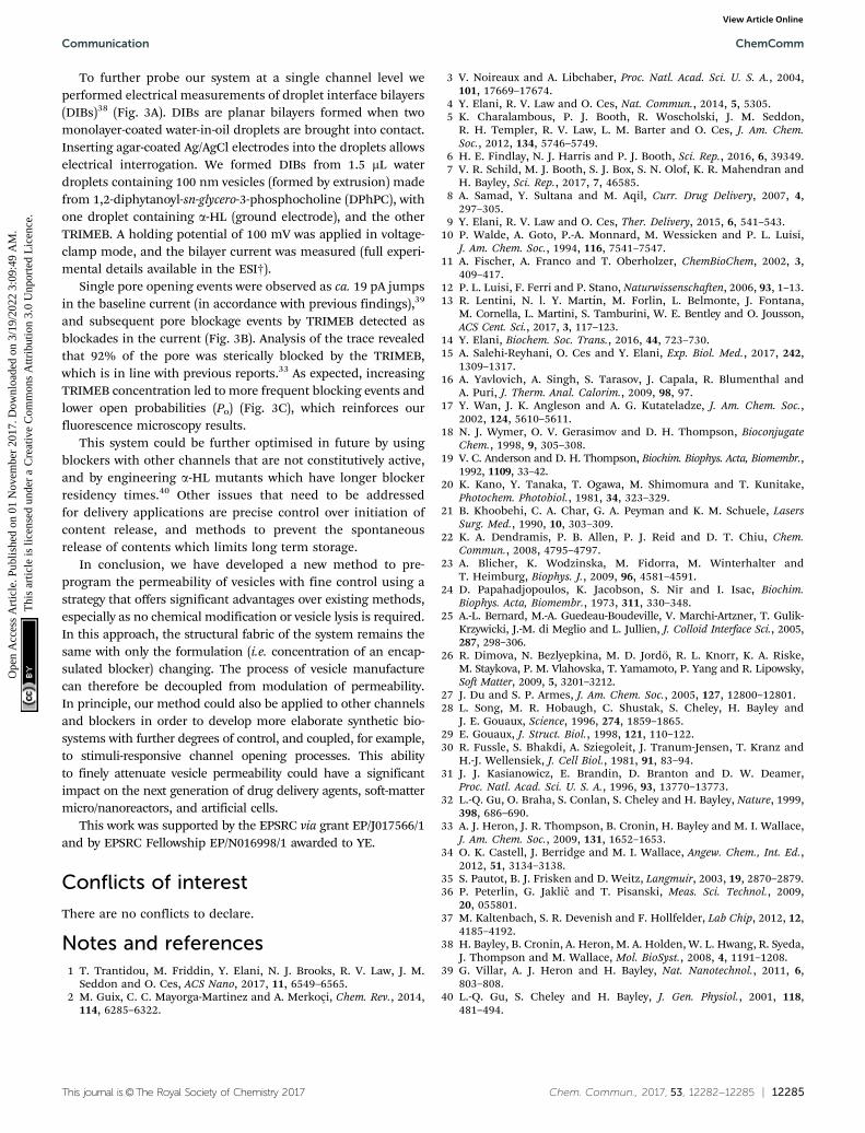
Fig. 3 Electrical interrogation of the system using DIBs. (A) Schematic of the experimental setup. (B) Trace of a typical protein insertion event followedby TRIMEB blocking events. By comparing the baseline (blue) to the fully open (green) and blocked (red) traces ca. 92% of the pore was estimated to beblocked. (C) Increasing TRIMEB concentration leads to more frequent blocking events and lower open probabilities.
ChemComm Communication
Ope
n A
cces
s A
rtic
le. P
ublis
hed
on 0
1 N
ovem
ber
2017
. Dow
nloa
ded
on 3
/19/
2022
3:0
9:49
AM
. T
his
artic
le is
lice
nsed
und
er a
Cre
ativ
e C
omm
ons
Attr
ibut
ion
3.0
Unp
orte
d L
icen
ce.
View Article Online
This journal is©The Royal Society of Chemistry 2017 Chem. Commun., 2017, 53, 12282--12285 | 12285
To further probe our system at a single channel level weperformed electrical measurements of droplet interface bilayers(DIBs)38 (Fig. 3A). DIBs are planar bilayers formed when twomonolayer-coated water-in-oil droplets are brought into contact.Inserting agar-coated Ag/AgCl electrodes into the droplets allowselectrical interrogation. We formed DIBs from 1.5 mL waterdroplets containing 100 nm vesicles (formed by extrusion) madefrom 1,2-diphytanoyl-sn-glycero-3-phosphocholine (DPhPC), withone droplet containing a-HL (ground electrode), and the otherTRIMEB. A holding potential of 100 mV was applied in voltage-clamp mode, and the bilayer current was measured (full experi-mental details available in the ESI†).
Single pore opening events were observed as ca. 19 pA jumpsin the baseline current (in accordance with previous findings),39
and subsequent pore blockage events by TRIMEB detected asblockades in the current (Fig. 3B). Analysis of the trace revealedthat 92% of the pore was sterically blocked by the TRIMEB,which is in line with previous reports.33 As expected, increasingTRIMEB concentration led to more frequent blocking events andlower open probabilities (Po) (Fig. 3C), which reinforces ourfluorescence microscopy results.
This system could be further optimised in future by usingblockers with other channels that are not constitutively active,and by engineering a-HL mutants which have longer blockerresidency times.40 Other issues that need to be addressedfor delivery applications are precise control over initiation ofcontent release, and methods to prevent the spontaneousrelease of contents which limits long term storage.
In conclusion, we have developed a new method to pre-program the permeability of vesicles with fine control using astrategy that offers significant advantages over existing methods,especially as no chemical modification or vesicle lysis is required.In this approach, the structural fabric of the system remains thesame with only the formulation (i.e. concentration of an encap-sulated blocker) changing. The process of vesicle manufacturecan therefore be decoupled from modulation of permeability.In principle, our method could also be applied to other channelsand blockers in order to develop more elaborate synthetic bio-systems with further degrees of control, and coupled, for example,to stimuli-responsive channel opening processes. This abilityto finely attenuate vesicle permeability could have a significantimpact on the next generation of drug delivery agents, soft-mattermicro/nanoreactors, and artificial cells.
This work was supported by the EPSRC via grant EP/J017566/1and by EPSRC Fellowship EP/N016998/1 awarded to YE.
Conflicts of interest
There are no conflicts to declare.
Notes and references1 T. Trantidou, M. Friddin, Y. Elani, N. J. Brooks, R. V. Law, J. M.
Seddon and O. Ces, ACS Nano, 2017, 11, 6549–6565.2 M. Guix, C. C. Mayorga-Martinez and A. Merkoçi, Chem. Rev., 2014,
114, 6285–6322.
3 V. Noireaux and A. Libchaber, Proc. Natl. Acad. Sci. U. S. A., 2004,101, 17669–17674.
4 Y. Elani, R. V. Law and O. Ces, Nat. Commun., 2014, 5, 5305.5 K. Charalambous, P. J. Booth, R. Woscholski, J. M. Seddon,
R. H. Templer, R. V. Law, L. M. Barter and O. Ces, J. Am. Chem.Soc., 2012, 134, 5746–5749.
6 H. E. Findlay, N. J. Harris and P. J. Booth, Sci. Rep., 2016, 6, 39349.7 V. R. Schild, M. J. Booth, S. J. Box, S. N. Olof, K. R. Mahendran and
H. Bayley, Sci. Rep., 2017, 7, 46585.8 A. Samad, Y. Sultana and M. Aqil, Curr. Drug Delivery, 2007, 4,
297–305.9 Y. Elani, R. V. Law and O. Ces, Ther. Delivery, 2015, 6, 541–543.
10 P. Walde, A. Goto, P.-A. Monnard, M. Wessicken and P. L. Luisi,J. Am. Chem. Soc., 1994, 116, 7541–7547.
11 A. Fischer, A. Franco and T. Oberholzer, ChemBioChem, 2002, 3,409–417.
12 P. L. Luisi, F. Ferri and P. Stano, Naturwissenschaften, 2006, 93, 1–13.13 R. Lentini, N. l. Y. Martın, M. Forlin, L. Belmonte, J. Fontana,
M. Cornella, L. Martini, S. Tamburini, W. E. Bentley and O. Jousson,ACS Cent. Sci., 2017, 3, 117–123.
14 Y. Elani, Biochem. Soc. Trans., 2016, 44, 723–730.15 A. Salehi-Reyhani, O. Ces and Y. Elani, Exp. Biol. Med., 2017, 242,
1309–1317.16 A. Yavlovich, A. Singh, S. Tarasov, J. Capala, R. Blumenthal and
A. Puri, J. Therm. Anal. Calorim., 2009, 98, 97.17 Y. Wan, J. K. Angleson and A. G. Kutateladze, J. Am. Chem. Soc.,
2002, 124, 5610–5611.18 N. J. Wymer, O. V. Gerasimov and D. H. Thompson, Bioconjugate
Chem., 1998, 9, 305–308.19 V. C. Anderson and D. H. Thompson, Biochim. Biophys. Acta, Biomembr.,
1992, 1109, 33–42.20 K. Kano, Y. Tanaka, T. Ogawa, M. Shimomura and T. Kunitake,
Photochem. Photobiol., 1981, 34, 323–329.21 B. Khoobehi, C. A. Char, G. A. Peyman and K. M. Schuele, Lasers
Surg. Med., 1990, 10, 303–309.22 K. A. Dendramis, P. B. Allen, P. J. Reid and D. T. Chiu, Chem.
Commun., 2008, 4795–4797.23 A. Blicher, K. Wodzinska, M. Fidorra, M. Winterhalter and
T. Heimburg, Biophys. J., 2009, 96, 4581–4591.24 D. Papahadjopoulos, K. Jacobson, S. Nir and I. Isac, Biochim.
Biophys. Acta, Biomembr., 1973, 311, 330–348.25 A.-L. Bernard, M.-A. Guedeau-Boudeville, V. Marchi-Artzner, T. Gulik-
Krzywicki, J.-M. di Meglio and L. Jullien, J. Colloid Interface Sci., 2005,287, 298–306.
26 R. Dimova, N. Bezlyepkina, M. D. Jordo, R. L. Knorr, K. A. Riske,M. Staykova, P. M. Vlahovska, T. Yamamoto, P. Yang and R. Lipowsky,Soft Matter, 2009, 5, 3201–3212.
27 J. Du and S. P. Armes, J. Am. Chem. Soc., 2005, 127, 12800–12801.28 L. Song, M. R. Hobaugh, C. Shustak, S. Cheley, H. Bayley and
J. E. Gouaux, Science, 1996, 274, 1859–1865.29 E. Gouaux, J. Struct. Biol., 1998, 121, 110–122.30 R. Fussle, S. Bhakdi, A. Sziegoleit, J. Tranum-Jensen, T. Kranz and
H.-J. Wellensiek, J. Cell Biol., 1981, 91, 83–94.31 J. J. Kasianowicz, E. Brandin, D. Branton and D. W. Deamer,
Proc. Natl. Acad. Sci. U. S. A., 1996, 93, 13770–13773.32 L.-Q. Gu, O. Braha, S. Conlan, S. Cheley and H. Bayley, Nature, 1999,
398, 686–690.33 A. J. Heron, J. R. Thompson, B. Cronin, H. Bayley and M. I. Wallace,
J. Am. Chem. Soc., 2009, 131, 1652–1653.34 O. K. Castell, J. Berridge and M. I. Wallace, Angew. Chem., Int. Ed.,
2012, 51, 3134–3138.35 S. Pautot, B. J. Frisken and D. Weitz, Langmuir, 2003, 19, 2870–2879.36 P. Peterlin, G. Jaklic and T. Pisanski, Meas. Sci. Technol., 2009,
20, 055801.37 M. Kaltenbach, S. R. Devenish and F. Hollfelder, Lab Chip, 2012, 12,
4185–4192.38 H. Bayley, B. Cronin, A. Heron, M. A. Holden, W. L. Hwang, R. Syeda,
J. Thompson and M. Wallace, Mol. BioSyst., 2008, 4, 1191–1208.39 G. Villar, A. J. Heron and H. Bayley, Nat. Nanotechnol., 2011, 6,
803–808.40 L.-Q. Gu, S. Cheley and H. Bayley, J. Gen. Physiol., 2001, 118,
481–494.
Communication ChemComm
Ope
n A
cces
s A
rtic
le. P
ublis
hed
on 0
1 N
ovem
ber
2017
. Dow
nloa
ded
on 3
/19/
2022
3:0
9:49
AM
. T
his
artic
le is
lice
nsed
und
er a
Cre
ativ
e C
omm
ons
Attr
ibut
ion
3.0
Unp
orte
d L
icen
ce.
View Article Online