program - eoaprof. abdel-rahman amer 2003 – 2004 prof. nabil khalifa 2004 - 2005 prof. mohamed...
TRANSCRIPT
14th International Congress of
Skeletal Deformities Correction
4-6 July, 2018
Intercontinental- City Stars Hotel, Cairo - Egypt
PROGRAM
& ABSTRACTS
2018

Dear International Congress of Skeletal Deformities Correction (ICSDC) participants,
It’s our honor to invite you to the 14th International Congress of Skeletal Deformities
Correction of the Egyptian Orthopaedic Association on July 4 - 6, 2018. The long &
successful history of the Association is worldwide known as rich, productive and true
international gathering of the most famous names in the world of orthopaedic
surgery. The most productive & effective way to acquire knowledge is through exchange of
knowledge and experience among different orthopaedic experts. We are sure you will enjoy
staying in Cairo, where the glory and history of the ancient Egypt merges with the beauty
and wonders of the modern state.
We look forward to welcoming you to the 14th International Congress of Skeletal
Deformities Correction.
Congress Board
Congress President Congress General Secretaries Congress Treasurer Prof. Anis Shiha Prof. Gamal Hosny Prof. Abdel Mohsen
Arafa
Prof. Hani El Mowafi

EGYPTIAN ORTHOPAEDIC ASSOCIATION BOARD
Prof. Anis Shiha President
Prof. Abdelsalam Eid Secretary General
Prof. Hani El Mowafi Treasurer
Prof. Adel Adawy Member
Prof. Ahmed Kholeif Member
Prof. Alaa El Zoheiry Member
Prof. Bahaa Kornah Member
Prof. Hassan El-Zaher Member
Prof. Hazem Abd El-Azeem Member
Prof. Gamal A. Hosny Member
Prof. Mahmoud El Rosasy Member
Prof. Mohamed Bahy El Shafie Member
Prof. Mohamed Fadel Member
Prof. Mohamed Osama Hegazy Member
Prof. Talaat Ezz El-Din Member

EGYPTIAN ORTHOPAEDIC ASSOCIATION PREVIOUS PRESIDENTS
Prof. Mohamed Kamel Hussein 1948 - 1967
Prof. Gawad Hamada 1968 - 1969
Prof. Abdel-Hay El-Sharkawy 1970 - 1971
Prof. Hussein K. Hassab 1972 - 1973
Prof. Mohamed S. Mehrez 1974 - 1975
Prof. Ahmus K. El-Hamamsy 1976 – 1977
Prof. Mansour Shawky 1978 - 1979
Prof. Mohamed Abdalla 1980 - 1981
Prof. Abdou Sallam 1982 - 1983
Prof. Amin Reda 1984 - 1985
Prof. Mahmoud Akl 1986 - 1987
Prof. Hussein Abdel-Fattah 1988 – 1989
Prof. Abdel-Hay Mashhour 1989 – 1990
Prof. El-Sayed Wahb 1990 – 1991
Prof. Fawzy Moustafa 1991 – 1992
Prof. Wael Mansour 1992 – 1993
Prof. Abdel-Salam Goumaa 1993 – 1994
Prof. Galal Zaki 1994 – 1995
Prof. Hassan El-Zaher 1995 – 1997
Prof. Farouk Youssef 1997 – 1998
Prof. Raafat H. Badawi 1998 – 2001
Prof. Mamdouh Zaki 2001 – 2003
Prof. Abdel-Rahman Amer 2003 – 2004
Prof. Nabil Khalifa 2004 - 2005
Prof. Mohamed Shafik 2005 - 2006
Prof. M. Osama Hegazy 2006 - 2007
Prof. Hazem Abdel Azeem 2007 - 2008
Prof. Khamis El Deeb 2008 - 2009
Prof. Samir Zaki Kotb 2009 - 2010
Prof. Abdel Mohsen Arafa 2010 - 2011
Prof. Gamal A. Hosny 2011 - 2013
Prof. Adel Adawy 2013 - 2015
Prof. Alaa El Zoheiry 2015 - 2016

Scientific Committee
Prof. Kamal El Gaafary
Prof. Abdel Mohsen Arafa
Prof. Bahaa Koranh
Prof. Mahmoud El Rosasy
Workshop Committee
Chairman: Prof. Hani El Mowafi
Co-Chairman: Prof. Mohamed Fadel
Co-Chairman: Prof. Hesham El Ashry
Organizing Committee
Ahmad Allam
Hatem Kotb
Barakt El Alfy
Nabil El Moghazy
Abdel Khalik El Zalabani
Abdel Salam Abdel Aleem
Ahmed Ekram
Ahmed A. Elsheikh
Amr El Battoty
Ashraf Khanfour
Ayman Hussein
Gamal El Mashad
Mohamed Abdel Aal
Mohamed Abdel Aal Hussein
Mohamed Azmy
Mohamed El Kersh
Mohamed Laklouk
Mohamed Zaki
Ossam Metwaly
Saad Gaballah
Sameh El Safti
Sherief Nasseef
Wael Shaaban

WEDNESDAY,
JULY 4, 2018
08:00 am
Registration

WEDNESDAY, JULY 4, 2018
Papers
Session
(1) 09:00-10:00
Chairpersons Prof. Abdelsalam Eid Prof. Bahaa Kornah Prof. Sherif Naseef Bishay
09:00 09:07 External fixator-assisted corrections of deformity in the femur: Outcomes and complications.
Ahmed A. Elsheikh Egypt
09:07 09:14 Subtrochanteric pediatric fractures, external fixation plus limitted internal fixation
Saad Gaballah Egypt
09:14 09:21 knee multi-planner deformity correction using TSF. frame
Sameh Al-Safty Egypt
09:21 09:28 Over growth of the tibia after complete healing of Congenital pseudarthrosis of the tibia
Ahmed Ibrahim Zayda Egypt
09:28 09:35 Management of Complex Deformities Around the Knee Joint Using the Taylor Spatial Frame
Osam Metwally Egypt
09:35 09:42 External fixator-assisted corrections of deformity in the tibia: Outcomes and complications.
Ahmed A. Elsheikh Egypt
09:42 09:49 prediction of timing of temporary hemiepiphysiodesis for coronal angular deformity by multiplier method . Is it a reliable tool? Review Article
Mohamed A. Abdel-Aal Egypt
09:49 09:56 Acute and gradual correction of adolescent tibia vara; obstacles and outcome
Abdel-Salam Abdel-Aleem Ahmed Egypt
09:56 10:00 Discussion

WEDNESDAY, JULY 4, 2018
Symposium
Blount Disease
Session
(2) 10:00-11:00
Moderator
Prof. Hatem Kotb Prof. Mohamed Lotfy Fahmy Prof. Saleh Alsaifi
10:00 10:10 Mohamed Lotfy Fahmy Kuwait
10:10 10:20 Hatem Kotb Egypt
10:20 10:30 Saleh Alsaifi Kuwait
10:30 10:40 Reggi Hamdi Canada
10:40 10:50 Ashraf Khanfour Egypt
10:50 11:00 Discussion
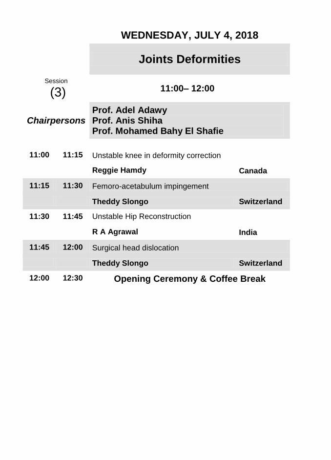
WEDNESDAY, JULY 4, 2018
Joints Deformities
Session
(3) 11:00– 12:00
Chairpersons
Prof. Adel Adawy Prof. Anis Shiha Prof. Mohamed Bahy El Shafie
11:00 11:15 Unstable knee in deformity correction
Reggie Hamdy Canada
11:15 11:30 Femoro-acetabulum impingement
Theddy Slongo Switzerland
11:30 11:45 Unstable Hip Reconstruction
R A Agrawal India
11:45 12:00 Surgical head dislocation
Theddy Slongo Switzerland
12:00 12:30 Opening Ceremony & Coffee Break

WEDNESDAY, JULY 4, 2018
Lectures
Session
(4) 12:30– 01:30
Chairpersons
Prof. Nabil El Moghazy Prof. Nehad El Mahboub Prof. R A Agrawal
12:30 12:45 Ilizarov technology in China
Sihe Qin China
12:45 01:00 Management of lower limb deformities in Rickets
Saw Aik Malaysia
01:00 01:15 Anterolateral bowing of the tibia
Gamal Hosny Egypt
01:15 01:30 Management of Cubitus Varus by Ilizarov
R A Agrawal India
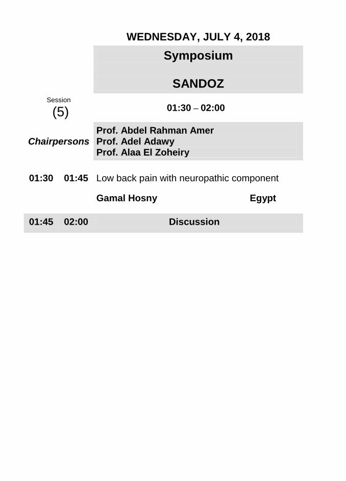
WEDNESDAY, JULY 4, 2018
Symposium
SANDOZ
Session
(5) 01:30 – 02:00
Chairpersons
Prof. Abdel Rahman Amer Prof. Adel Adawy Prof. Alaa El Zoheiry
01:30 01:45 Low back pain with neuropathic component
Gamal Hosny Egypt
01:45 02:00 Discussion

WEDNESDAY, JULY 4, 2018
Symposium
MSD
Session
(6) 02:00 – 02:30
Chairpersons Prof. Alaa El Zoheiry Prof. Sherin Khalil
02:00 02:15 Role of Etoricoxib in Chronic Pain Management.
Ashraf El Nahal Egypt
02:15 02:30 Discussion
02:30 03:30 Lunch
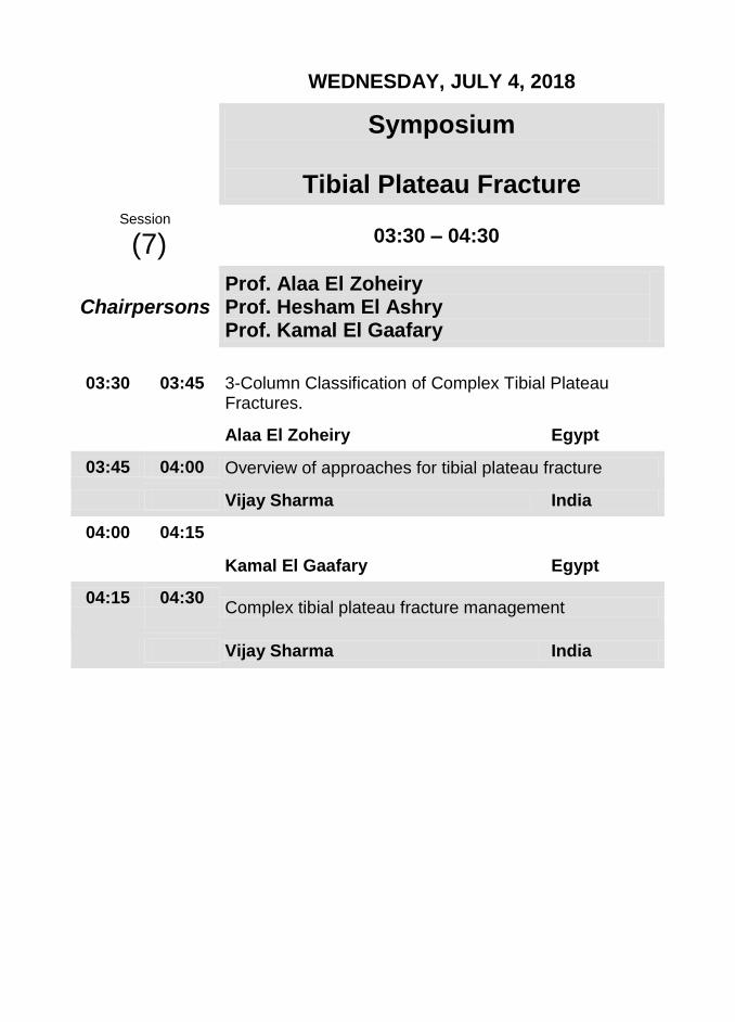
WEDNESDAY, JULY 4, 2018
Symposium
Tibial Plateau Fracture
Session
(7) 03:30 – 04:30
Chairpersons
Prof. Alaa El Zoheiry Prof. Hesham El Ashry Prof. Kamal El Gaafary
03:30 03:45 3-Column Classification of Complex Tibial Plateau Fractures.
Alaa El Zoheiry Egypt
03:45 04:00 Overview of approaches for tibial plateau fracture
Vijay Sharma India
04:00 04:15
Kamal El Gaafary Egypt
04:15 04:30 Complex tibial plateau fracture management
Vijay Sharma India
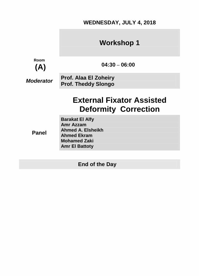
WEDNESDAY, JULY 4, 2018
Workshop 1
Room
(A) 04:30 – 06:00
Moderator Prof. Alaa El Zoheiry Prof. Theddy Slongo
External Fixator Assisted Deformity Correction
Panel
Barakat El Alfy Amr Azzam Ahmed A. Elsheikh Ahmed Ekram Mohamed Zaki Amr El Battoty
End of the Day

WEDNESDAY, JULY 4, 2018
Workshop 2
Room
(B) 04:30 – 06:00
Moderator
Prof. Reggie Hamdy Prof. Osama Hegazy Prof. Bahaa Kornah
Pelvic Osteotomies
Panel
Sherief Nasseef Mahmoud El Rosasy Mohamed El Kersh
Mohamed Abdel Aal Hussein
End of the Day
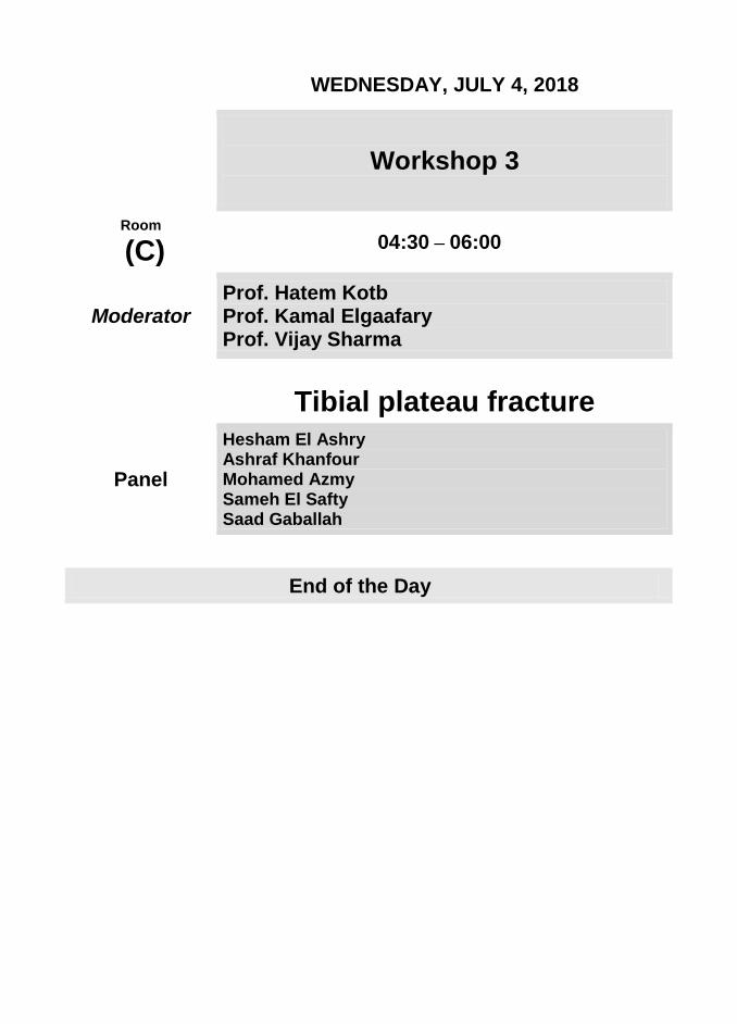
WEDNESDAY, JULY 4, 2018
Workshop 3
Room
(C) 04:30 – 06:00
Moderator
Prof. Hatem Kotb Prof. Kamal Elgaafary Prof. Vijay Sharma
Tibial plateau fracture
Panel
Hesham El Ashry Ashraf Khanfour Mohamed Azmy Sameh El Safty Saad Gaballah
End of the Day

THURSDAY, JULY 5, 2018
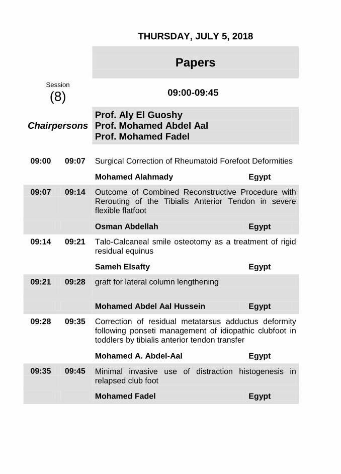
THURSDAY, JULY 5, 2018
Papers
Session
(8) 09:00-09:45
Chairpersons
Prof. Aly El Guoshy Prof. Mohamed Abdel Aal Prof. Mohamed Fadel
09:00 09:07 Surgical Correction of Rheumatoid Forefoot Deformities
Mohamed Alahmady Egypt
09:07 09:14 Outcome of Combined Reconstructive Procedure with Rerouting of the Tibialis Anterior Tendon in severe flexible flatfoot
Osman Abdellah Egypt
09:14 09:21 Talo-Calcaneal smile osteotomy as a treatment of rigid residual equinus
Sameh Elsafty Egypt
09:21 09:28 graft for lateral column lengthening
Mohamed Abdel Aal Hussein Egypt
09:28 09:35 Correction of residual metatarsus adductus deformity following ponseti management of idiopathic clubfoot in toddlers by tibialis anterior tendon transfer
Mohamed A. Abdel-Aal Egypt
09:35 09:45 Minimal invasive use of distraction histogenesis in relapsed club foot
Mohamed Fadel Egypt

THURSDAY, JULY 5, 2018
Foot and Ankle Deformities
Session
(9) 09:45–11:00
Chairpersons
Prof. Ahmed Kholeif Prof. Hani El Mowafi Prof. Wagih Mousa
09:45 10:00 Correction of post Traumatic Foot Deformity.
Wagih Mousa UK
10:00 10:15 Gradual correction of stiff Clubfoot deformity using Hinged Ilizarov frame
Saw Aik Malaysia
10:15 10:30 Ilizarov technique for treatment of complex ankle malformation.
Sihe Qin China
10:30 10:45 Distraction histogenesis, the hope for Diabetic foot.
Yong Hong Zhang China
10:45 11:00 Correction of deformity in Charcot foot
Hani El Mowafi Egypt
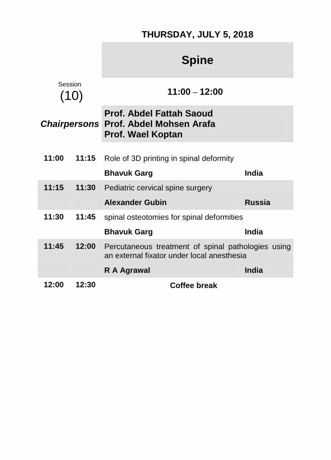
THURSDAY, JULY 5, 2018
Spine
Session
(10) 11:00 – 12:00
Chairpersons
Prof. Abdel Fattah Saoud Prof. Abdel Mohsen Arafa Prof. Wael Koptan
11:00 11:15 Role of 3D printing in spinal deformity
Bhavuk Garg India
11:15 11:30 Pediatric cervical spine surgery
Alexander Gubin Russia
11:30 11:45 spinal osteotomies for spinal deformities
Bhavuk Garg India
11:45 12:00 Percutaneous treatment of spinal pathologies using an external fixator under local anesthesia
R A Agrawal India
12:00 12:30 Coffee break

THURSDAY, JULY 5, 2018
Lengthening and Reconstruction
Session
(11) 12:30 – 01:30
Chairpersons
Prof. Ahmad Allam Prof. Lotfy El Adwar Prof. Mahmoud El Rosasy
12:30 12:45 Corticotomy technique using guided double barrel drill sleeve
Saw Aik Malaysia
12:45 01:00 Cellular advances & limb reconstruction
Reggie Hamdy Canada
01:00 01:15 The organizational specifics of the limb lengthening and reconstruction center.
Alexander Gubin Russia
01:15 01:30 Lengthening nails
Reggie Hamdy Canada
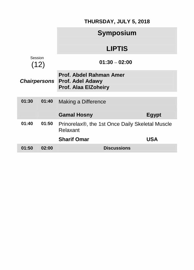
THURSDAY, JULY 5, 2018
Symposium
LIPTIS
Session
(12) 01:30 – 02:00
Chairpersons Prof. Abdel Rahman Amer Prof. Adel Adawy Prof. Alaa ElZoheiry
01:30 01:40 Making a Difference
Gamal Hosny Egypt
01:40 01:50 Prinorelax®, the 1st Once Daily Skeletal Muscle Relaxant
Sharif Omar USA
01:50 02:00 Discussions
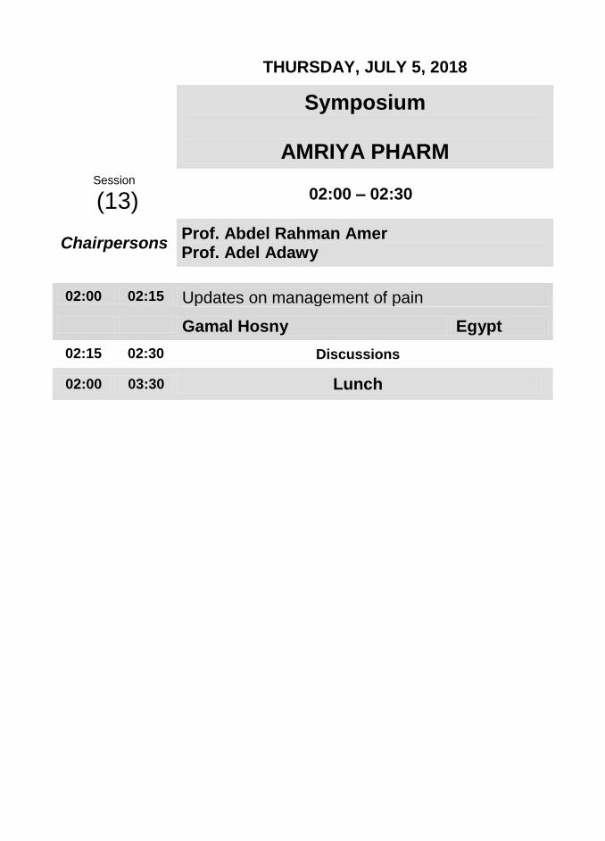
THURSDAY, JULY 5, 2018
Symposium
AMRIYA PHARM
Session
(13) 02:00 – 02:30
Chairpersons Prof. Abdel Rahman Amer Prof. Adel Adawy
02:00 02:15 Updates on management of pain
Gamal Hosny Egypt
02:15 02:30 Discussions
02:00 03:30 Lunch
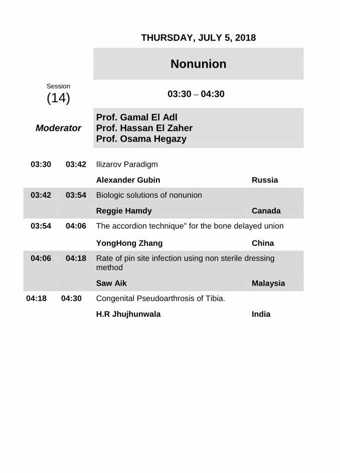
THURSDAY, JULY 5, 2018
Nonunion
Session
(14) 03:30 – 04:30
Moderator
Prof. Gamal El Adl Prof. Hassan El Zaher
Prof. Osama Hegazy
03:30 03:42 Ilizarov Paradigm
Alexander Gubin Russia
03:42 03:54 Biologic solutions of nonunion
Reggie Hamdy Canada
03:54 04:06 The accordion technique" for the bone delayed union
YongHong Zhang China
04:06 04:18 Rate of pin site infection using non sterile dressing method
Saw Aik Malaysia
04:18 04:30 Congenital Pseudoarthrosis of Tibia.
H.R Jhujhunwala India
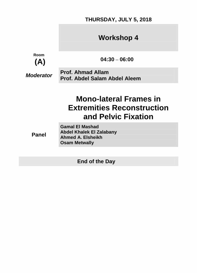
THURSDAY, JULY 5, 2018
Workshop 4
Room
(A) 04:30 – 06:00
Moderator Prof. Ahmad Allam Prof. Abdel Salam Abdel Aleem
Mono-lateral Frames in Extremities Reconstruction
and Pelvic Fixation
Panel
Gamal El Mashad Abdel Khalek El Zalabany Ahmed A. Elsheikh Osam Metwally
End of the Day

THURSDAY, JULY 5, 2018
Workshop 5
Room
(B) 04:30 – 06:00
Moderator
Prof. H.R Jhujhunwala Prof. Mahmoud El Rosasy Prof. Mohamed Fadel Prof. R A Agrawal
Pelvic Support Osteotomy
Panel Ashraf Khanfour Mohamed Azmy Mohamed Abdel Aal Hussein
End of the Day
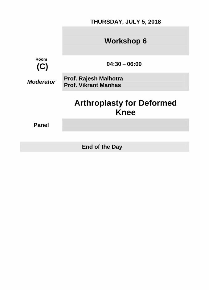
THURSDAY, JULY 5, 2018
Workshop 6
Room
(C) 04:30 – 06:00
Moderator Prof. Rajesh Malhotra Prof. Vikrant Manhas
Arthroplasty for Deformed Knee
Panel
End of the Day
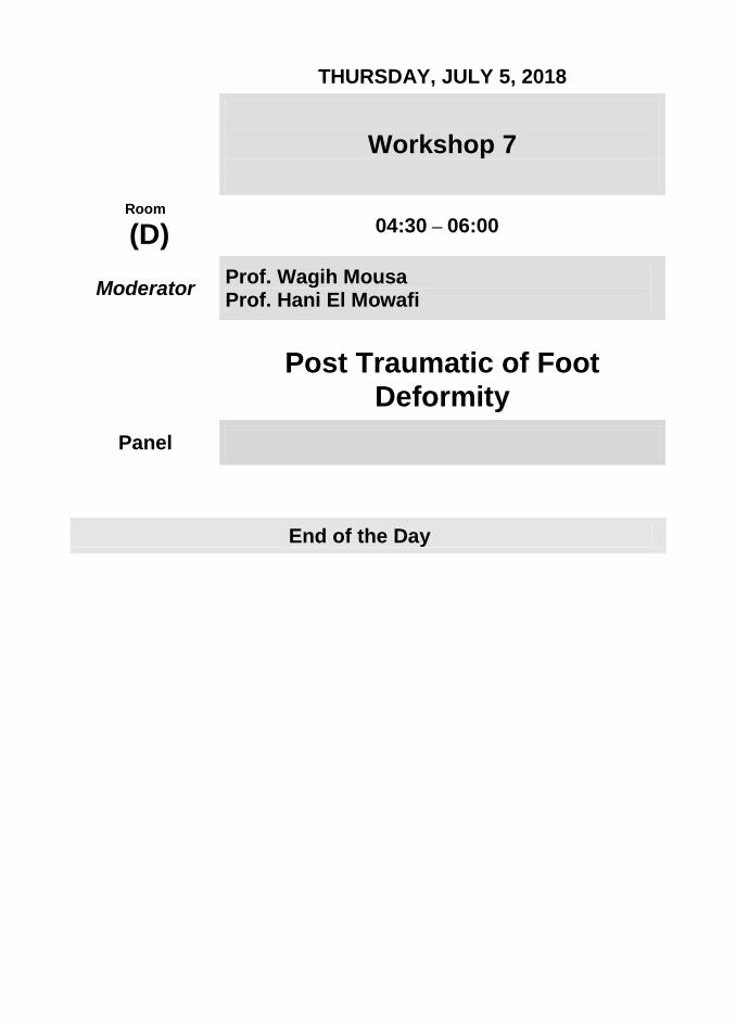
THURSDAY, JULY 5, 2018
Workshop 7
Room
(D) 04:30 – 06:00
Moderator Prof. Wagih Mousa Prof. Hani El Mowafi
Post Traumatic of Foot Deformity
Panel
End of the Day

FRIDAY,
JULY 6, 2018
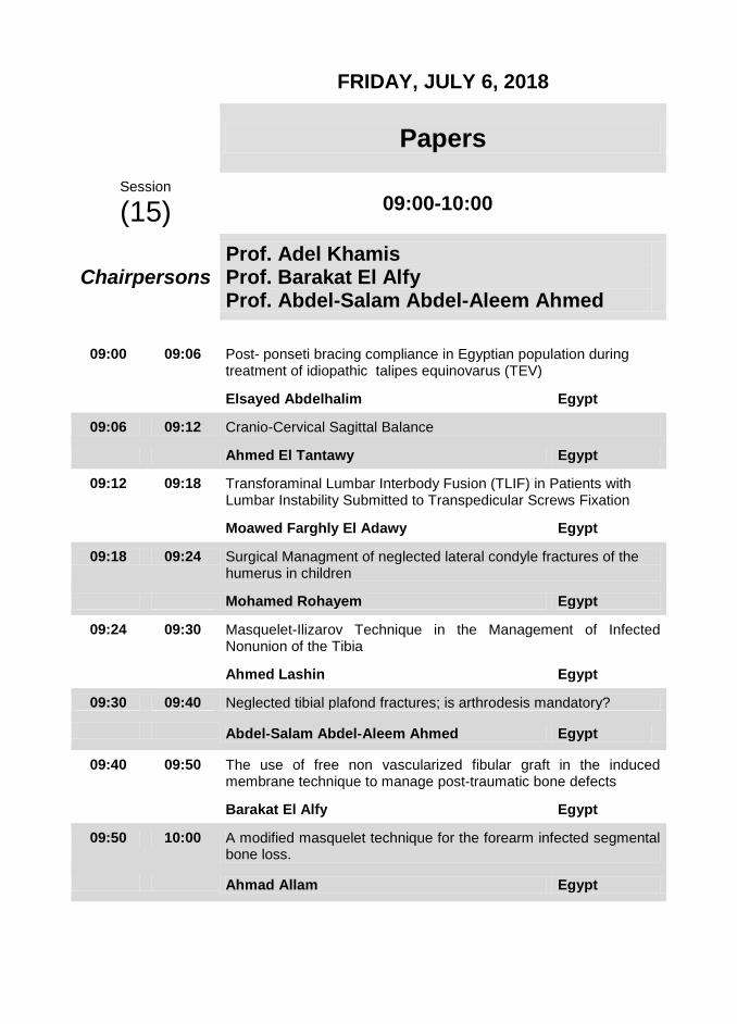
FRIDAY, JULY 6, 2018
Papers
Session
(15) 09:00-10:00
Chairpersons
Prof. Adel Khamis Prof. Barakat El Alfy Prof. Abdel-Salam Abdel-Aleem Ahmed
09:00 09:06 Post- ponseti bracing compliance in Egyptian population during treatment of idiopathic talipes equinovarus (TEV)
Elsayed Abdelhalim Egypt
09:06 09:12 Cranio-Cervical Sagittal Balance
Ahmed El Tantawy Egypt
09:12 09:18 Transforaminal Lumbar Interbody Fusion (TLIF) in Patients with Lumbar Instability Submitted to Transpedicular Screws Fixation
Moawed Farghly El Adawy Egypt
09:18 09:24 Surgical Managment of neglected lateral condyle fractures of the humerus in children
Mohamed Rohayem Egypt
09:24 09:30 Masquelet-Ilizarov Technique in the Management of Infected Nonunion of the Tibia
Ahmed Lashin Egypt
09:30 09:40 Neglected tibial plafond fractures; is arthrodesis mandatory?
Abdel-Salam Abdel-Aleem Ahmed Egypt
09:40 09:50 The use of free non vascularized fibular graft in the induced membrane technique to manage post-traumatic bone defects
Barakat El Alfy Egypt
09:50 10:00 A modified masquelet technique for the forearm infected segmental bone loss.
Ahmad Allam Egypt

FRIDAY, JULY 6, 2018
Symposium Deformity Correction
Internal fixation vs External fixation
Session
(16) 10:00 – 11:15
Moderator
Prof. Alaa El Zoheiry Prof. Essam El Sherief Prof. Theddy Slongo
10:00 10:15 Essam El Sherief Egypt
10:15 10:25 Alaa El Zoheiry Egypt
10:25 10:35 Theddy Slongo Switzerland
10:35 10:45 Mahmoud El Rosasy Egypt
10:45 10:55 Ihab Badawy Egypt
10:55 10:05 Amin Abd Elrazik Egypt
10:05 11:15 Discussion

FRIDAY, JULY 6, 2018
Symposium
OCTOBER PHARMA
Session
(17) 11:15 – 11:45
Chairpersons Prof. Alaa El Zoheiry Prof. Hani El Mowafi
11:15 11:45 Gout Management Update
Gamal Hosny Egypt
12:00 02:00 Friday Prayer + Lunch
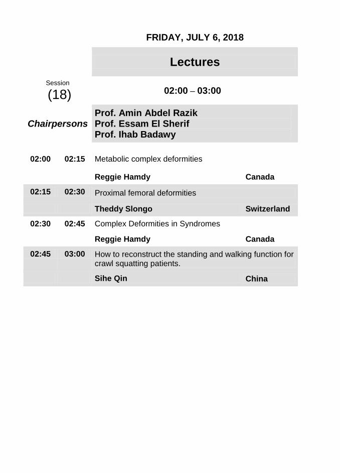
FRIDAY, JULY 6, 2018
Lectures
Session
(18) 02:00 – 03:00
Chairpersons
Prof. Amin Abdel Razik Prof. Essam El Sherif Prof. Ihab Badawy
02:00 02:15 Metabolic complex deformities
Reggie Hamdy Canada
02:15 02:30 Proximal femoral deformities
Theddy Slongo Switzerland
02:30 02:45 Complex Deformities in Syndromes
Reggie Hamdy Canada
02:45 03:00 How to reconstruct the standing and walking function for crawl squatting patients.
Sihe Qin China
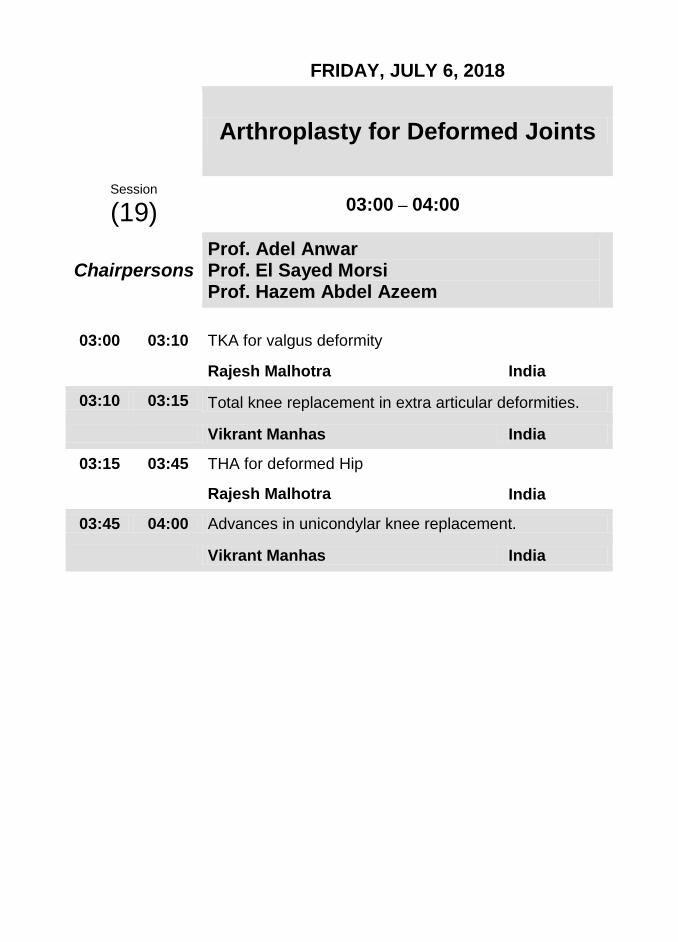
FRIDAY, JULY 6, 2018
Arthroplasty for Deformed Joints
Session
(19) 03:00 – 04:00
Chairpersons
Prof. Adel Anwar Prof. El Sayed Morsi Prof. Hazem Abdel Azeem
03:00 03:10 TKA for valgus deformity
Rajesh Malhotra India
03:10 03:15 Total knee replacement in extra articular deformities.
Vikrant Manhas India
03:15 03:45 THA for deformed Hip
Rajesh Malhotra India
03:45 04:00 Advances in unicondylar knee replacement.
Vikrant Manhas India
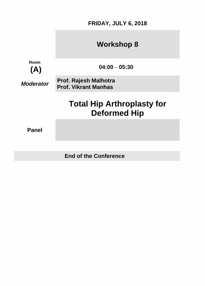
FRIDAY, JULY 6, 2018
Workshop 8
Room
(A) 04:00 – 05:30
Moderator Prof. Rajesh Malhotra Prof. Vikrant Manhas
Total Hip Arthroplasty for Deformed Hip
Panel
End of the Conference

FRIDAY, JULY 6, 2018
Workshop 9
Room
(B) 04:00 – 05:30
Moderator
Prof. Reggie Hamdy Prof. Osama Hegazy Prof. Bahaa Kornah Prof. Mahmoud El Rosasy
Pelvic Osteotomies
Panel Sherief Nasseef Mohamed El Kersh
End of the Conference
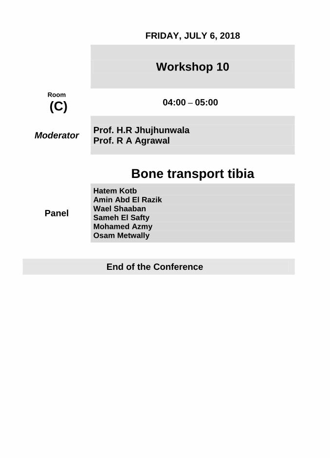
FRIDAY, JULY 6, 2018
Workshop 10
Room
(C) 04:00 – 05:00
Moderator Prof. H.R Jhujhunwala Prof. R A Agrawal
Bone transport tibia
Panel
Hatem Kotb Amin Abd El Razik Wael Shaaban Sameh El Safty Mohamed Azmy Osam Metwally
End of the Conference

ABSTRACTS

1. EXTERNAL FIXATOR-ASSISTED CORRECTIONS OF DEFORMITY IN THE FEMUR: OUTCOMES AND
COMPLICATIONS.
Ahmed A. Elsheikh, Philippa Thorpe, Nikolaos Giotakis, Badri Narayan, Selvadurai Nayagam
Egypt
Introduction: We report the outcome of external fixator-assisted correction of deformities in the femur. Methods: 35 patients (39 limbs) were included. The deformities arose from malunion in 41 %, were idiopathic in 15 %, and from hypophosphataemic rickets in 15%. Correction was undertaken acutely using a rail external and performed after preoperative plans and software-based simulations. Definitive stabilisation used submuscular plate fixation in 16 limbs, intramedullary nailing in 21 limbs, and both in two femurs to achieve optimum stability. The outcomes of interest were the quality of correction and complications thereafter. Results: Patients’ mean age at surgery was 40 years (range 17 - 84). The mean deformities were: 30° internal rotation; 11° Valgus; 24° procurvatum; 15° Varus; 20° recurvatum and 30° external rotation. Full deformity correction was achieved without intraoperative complications. Statistically significant changes in the mechanical axis, aMPFA and aLDFA were produced. One superficial infection, one deep infection and two delayed unions occurred. There were no nerve palsies. We used radiological consolidation as an outcome endpoint. Radiological consolidation (absence of a visible osteotomy line on plain x-ray views) was evident for 28 limbs at a mean of 6.8 ±4.8 months (range 2.6 to 25). Eleven limbs remain under review with the most recent radiologic findings satisfactory. Discussion & Conclusion: Acute deformity correction using external fixator assistance can be a reliable alternative to methods based only on external fixation.
2. SUBTROCHANTERIC PEDIATRIC FRACTURES, EXTERNAL FIXATION PLUS LIMITTED INTERNAL FIXATION
Saad Gaballah
Egypt
Introduction: Subtrochanteric pediatric fractures are uncommon. Differrent treatment options are available: traction, spica cast, internal fixation and external fixation. Methods:
Between January 2014 and March 2016, 10 patients with closed subtrochanteric pediatric fractures were treated in Damanhour teaching hospital. Fractures were reduced either by closed manipulations under C arm or by limitted open approach and fixed prelimenery by k wire or elastic intramedullary nail according to fracture configuration. Simple modular fixator( 2 side plates connected by 2 rods) was applied with 2 to 3 schanz screws proximal and distal to the fracture. Results: The main age at the time of injury was 7.11 years( range 5.2- 11.3 years). 2 fractures were pathological and 8 were traumatic.one case has associated splenic rupture and one has humeral fracture. All fractures united within a main of 8.5 weeks (range 7- 13). Main follow up was 22.4 months(range 12- 36) Discussion & Conclusion: There is no general ageement regarding the method of treatment of pediatric subtrochanteric fractures. External fixation plus limitted internal fixation is a good option as it requires no massive soft tissue dissection, no blood loss and no cases of nonunion or deep infection were encountered
3. KNEE MULTI-PLANNER DEFORMITY CORRECTION USING TSF. FRAME
Sameh Al-Safty
Egypt
Introduction: Multi-planner knee deformity need correction in different axes.this can be done by ilizarov system utilize hings and translation mechanism this need an effort for orientation TSF .frame solve these problems. it consists of 2 rings connecting by 6 telescopic struts at special universal joints . Methods: 6 patients with complex post traumatic knee deformities . they genuvarus , recurvatum and shortening. 2 females and 4 males . age from 16 : 20 Ys .utilising taylor spatial frame (TSF) . all cases were assisted clinically , radiologically , by Lysholm and Xford knee scores . Results: all patients were corrected by TSF frame . all patients are very satisfied . Lysholm knee score improved from 55% : 90% . no limb length discripancies . one patient have 5 degrees recurvatum . Discussion & Conclusion: TSF fixator is able to correct a 6 axes deformities and also leg length discripancies by adjusting only strut lengths one ring can be repositioned with respect to the other ring.
4. OVER GROWTH OF THE TIBIA AFTER COMPLETE HEALING OF CONGENITAL PSEUDARTHROSIS OF THE
TIBIA
Ahmed Ibrahim Zayda
Egypt
Introduction: Congenital pseudarthrosis of the tibia (CPT) is a rare pathology and bilateral forms are extremely rare. The tibia shows area of segmental dysplasia resulting in anterolateral bowing of the bone, reduced growth in the distal tibial epiphysis and, shortening of the limb usually occurs. Once a fracture occurs, there is a little or no tendency for the lesion to heal spontaneously. Treatment is mainly surgical. The key to get primary union is to excise hamartomatous tissue and pathological periosteum. Surgical options such as segment transport with Ilizarov technique , intramedullary nailing, and vascularized fibula graft, have shown equivocal success rate in achieving primary union although they are often associated with acceptable results. Amputation should be reserved for failed reconstruction, severe limb length discrepancy and gross deformities of leg and ankle. Methods: 8 patients had (CPT) treated with an Ilizarov bone transport in Menoufia university hospitals. The treatment consisted of extensive debridement of the pseudarthrosis (a segmental resection of the pseudarthrosis site) and Ilizarov bone transport. Results: A total of 8 patients were studied. Age at start of treatment ranged from 3 to 15 years . The mean follow-up was 8 years (range 3–14 years). Primary consolidation was seen in all patients (100 %). Refracture occurred in 2 cases, one of them was retreated with Ilizarov technique with revision surgery and (complete excision of the surrounding periosteum and talking a periosteal graft from the lateral surface of the iliac bone and making it as a tube around the nonunion site and bone grafting of the docking site) and the other one treated with interlocking nail tibia and bone graft. At final follow-up, all patients experienced union. Over growth after complete union occurred in two cases by about 4 cm in one patient and one cm in the other one with associated valgus deformity at the knee, tibia and ankle joints with no recurrence of tibial sclerosis in all patients. Discussion & Conclusion: The present data confirm a good primary healing rate in 100% of cases. However refracture occurred in 2 cases that was finally healed with revision surgery. The incidence of tibial over growth after complete union is not widely documented in the literature. Also the valgus deformity at the tibia and both knee and ankle joints with recurrence of anterior bowing occurred in two cases that didnot easily treateted with temporary epiphysiodesis and is a real problem that need further discussion and investiagtions. Each child with CPT must be followed up till skeletal maturity to identify and rectify residual problems after primary healing. Have a Comment?: causes of over growth and valgus deformity after complete union is matter of discussion?
5. MANAGEMENT OF COMPLEX DEFORMITIES AROUND THE KNEE JOINT USING THE TAYLOR SPATIAL FRAME
Gamal Ahmed Hosny, Mahmoud Mohamed Ibrahem Matter, Ahmed Mohamed Ahmed Nahla, Osam Mohamed Metwally Hassan.
Egypt
Objective: The aim of this study is to evaluate the results of using the TSF in treatment of complex lower limb deformities around the knee joint. Subjects and Methods: This study is prospective study was conducted involving 21patients who underwent correction of the mechanical axis using TSF and bone osteotomy in a complex knee deformity. Results: Using Schoenecker’s criteria there were 95.23% good result in term of good results in terms of pain and radiological criteria in our patients, 0 % fair results and there were 4.76% poor results. Conclusion: TSF is an excellent tool for the correction of multiple plane deformity around the knee joint in children and adolescents and significantly expands our ability to correct precisely the most difficult deformities. Key words: Taylor spatial frame, complex lower limb deformity, varus, valgus.
6. EXTERNAL FIXATOR-ASSISTED CORRECTIONS OF DEFORMITY IN THE TIBIA: OUTCOMES AND
COMPLICATIONS.
Ahmed A. Elsheikh, Philippa Thorpe, Nikolaos Giotakis, Badri Narayan, Selvadurai Nayagam
Egypt
Introduction: We report the outcome of external fixator-assisted correction of deformities in the tibia. Methods: 21 patients (23 limbs) were included. The deformities were malunions in 17%, idiopathic in 17 %, from hypophosphatemic rickets in 13% and were residual to previous lengthenings in 17%. Correction was undertaken acutely using a rail external fixator and performed after preoperative plans and software-based simulations. Definitive stabilisation used submuscular plate fixation in 21 limbs and intramedullary nailing in two. The outcomes of interest were the quality of correction and complications thereafter. Results: Patients’ mean age at surgery was 32 years (range 18 - 60). The mean deformities were: 16° Valgus, 9° Varus, 26° procurvatum, 27° recurvatum and 26° external rotation. Full deformity correction was achieved without intraoperative complications with only one patient retaining 5° degrees of distal procurvatum to allow sucessful plate fixation on the tibia. Statistically significant changes in the mechanical axis, aMPTA and aLDTA were produced. Three had local irritation from the plate which was removed after consolidation and one
had irritation from a fibula osteotomy. There were no infections or nerve palsies. We used radiological consolidation as an outcome endpoint. Radiological consolidation (absence of a visible osteotomy line on plain x-ray views) was evident for 17 limbs at a mean of 5 ±2.7 months (range 2.4 to 14). Six limbs remain under review with the most recent radiologic findings satisfactory. Discussion & Conclusion: Acute deformity correction using external fixator assistance is a reliable alternative to methods based only on external fixation.
7. PREDICTION OF TIMING OF TEMPORARY HEMIEPIPHYSIODESIS FOR CORONAL
ANGULAR DEFORMITY BY MULTIPLIER METHOD . IS IT A RELIABLE TOOL? REVIEW ARTICLE
Mohamed A. Abdel-Aal
Egypt
Introduction: Timing of temporary epiphysiodesis was predicted according to Paley via multiplier method. This method and uptill nowis not emphasised clinically for its reliabilityregarding clinical outcomes.The review aims at predicting timing of epiphyseodesis using multiplier method and testing its reliability based on the data from the published studies Methods: the following electronic databases: Cochrane, PubMed, andEMBASE had been searched A total of 35 studies met the inclusion criteria. The associations of patient characteristics with Deviation of prediction were assessed with the use of univariatelogistic regression Results: 30 articles wereconsideredas poor quality while 5 studies were evaluatedasmoderate evidence. The rate of deviation of prediction in the involved studies 49.8 Discussion & Conclusion: The multiplier method is not firmly reliable in predicting the timing for temporary hemiepiphysiodesis, however it is prone to be more reliable for the younger patients with idiopathic coronal deformity.
8. ACUTE AND GRADUAL CORRECTION OF ADOLESCENT TIBIA VARA; OBSTACLES AND OUTCOME
Abdel-Salam Abdel-Aleem Ahmed
Egypt
Introduction:
Adolescent tibia vara is usually characterized by combined marked genu varum, procurvatum, and internal tibial torsion. Corrective tibial osteotomy is the standard treatment protocol when no growth remains. This study the obstacles and the outcomes

following acute and gradual corrections of these deformities. Patients and Methods: The study included 29 patients with 43 tibias and a mean age of 16.7 years (range, 15 to 20 y). Eleven patients were females, and 20 were males. All patients underwent correction Ilizarov external fixator. Acute correction was done for 19 tibias in 17 patients (Group A) with up to 35° of angular deformity, and gradual correction and lengthening was done for 24 tibias in 12 with severe deformities (Group B). Femoral deformity was corrected in an earlier session in five patients. After analysis of the medial proximal tibial angle and posterior proximal tibial angle, the oblique plane deformity with the antero-lateral apex averaged 31.1° in Group A, and 67.4° in Group B. Results: The mean follow-up period was 43 (Range 24-61) months. All cases were corrected with sound osteotomy union without secondary procedure or bone grafting. The mean External Fixation Period (EFP) was 17.14 weeks in Group A, and 42.85 weeks in Group B. The average lengthening was 1.75 cm in group A, and 6.27 cm in group B. Complications included delayed union (one case), superficial pin-tract infection (18 patients) and a broken Schanz screw (two cases). There was no neurovascular affection. Conclusion: Ilizarov fixator is a viable treatment option for adolescent tibia vara, with the ability of both acute and gradual correction. It is a versatile device with correction of different elements of the deformity. Bilateral cases had the opportunity of simultaneous limb lengthening.
9. UNSTABLE KNEE IN DEFORMITY CORRECTION
Reggie Hamdy Canada
10. FEMORO-ACETABULUM IMPINGEMENT
Theddy Slongo Switzerland
11. UNSTABLE HIP RECONSTRUCTION
R A Agrawal India
12. SURGICAL HEAD DISLOCATION
Theddy Slongo Switzerland

13. ILIZAROV TECHNOLOGY IN CHINA
Sihe Qin China
14. MANAGEMENT OF LOWER LIMB DEFORMITIES IN RICKETS
Saw Aik Malaysia
15. ANTEROLATERAL BOWING OF THE TIBIA
Gamal Hosny Egypt
16. MANAGEMENT OF CUBITUS VARUS BY ILIZAROV
R A Agrawal India
17. 3-COLUMN CLASSIFICATION OF COMPLEX TIBIAL PLATEAU FRACTURES.
Alaa El Zoheiry
Egypt
18. OVERVIEW OF APPROACHES FOR TIBIAL PLATEAU FRACTURE
Vijay Sharma
India
19. ????????????
Kamal El Gaafary Egypt
20. COMPLEX TIBIAL PLATEAU FRACTURE MANAGEMENT
Vijay Sharma
India
21. SURGICAL CORRECTION OF RHEUMATOID FOREFOOT DEFORMITIES
Mohamed Alahmady
Egypt
Introduction: Surgical correction of severe rheumatoid forefoot deformities with resection arthroplasties of the lesser metatarsal phalangeal joints and arthrodesis of the first metatarsal phalangeal joint resulted in a significant long-term improvement with respect to shoe wear, pain, and the ability to stand and walk in 95% of the patients. Ninety percent had a good or excellent functional result. There was minimal recurrence of deformity. Modifications of the procedure with maintenance of the proximal phalangeal bases and K-wire fixation of the metatarsal phalangeal arthroplasty and interphalangeal joints resulted in an improved cosmetic result and simplified postoperative management with an equal functional result and no increase in recurrence of deformity or complications. Methods: Twenty patients with severe forefoot deformities secondary to inflammatory arthritis rheumatoid patients and l psoriatic patient) underwent surgical correction of 28 feet by first metatarsal phalangeal arthrodesis and resection arthroplasties of the lesser metatarsal phalangeal joints between January 1982 and January 1988. There were 15 women and five men. Their age ranged from 30 to 69 years. They all had longstanding inflammatory arthritis and many were on prednisone or methotrexate therapy. All had failed conservative management of their deformity. Eight patients had undergone previous surgery consisting of hallux valgus corrections, lesser metatarsal head resections, or hammertoe corrections. The patients’ charts and radiographs were reviewed retrospectively. Each patient completed a question12 MANN AND SCHAKEL Foot & Ankle International/Vol. 16, No. 1IJanuary 1995 saw in such a manner that good bony coaptation is obtained when the arthrodesis is placed in its desired position of 15-20" of valgus and 20-30" of dorsiflexion with respect to the metatarsal shaft. Any medial prominence of the metatarsal is resected. The arthrodesis site is not transfixed at this time. The surgical technique with respect to the lesser toes utilized on the first 10 patients (1 2 feet) has been described previosly. The technique currently used, which was utilized on the last 12 patients (16 feet) in this series, is described below. Two dorsal longitudinal incisions are made, the first centered between the second and third metatarsals and the second between the fourth and fifth metatarsals. The extensor tendons, dorsal capsule, and collateral ligaments are divided and sharply dissected away from the metatarsal head, neck, and distal shaft region. Circumferential dissection is performed around the base of the proximal phalanx to release the plantar plate and plantar aponeurosis. Distraction is placed on the toes and the plantar plate and volar capsule are dissected away from the metatarsal head and neck with either a scalpel or Freer elevator, staying close to bone to prevent neurovascular damage.
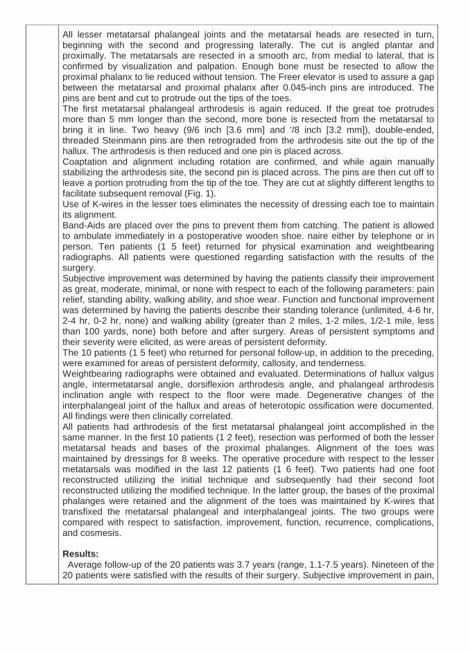
All lesser metatarsal phalangeal joints and the metatarsal heads are resected in turn, beginning with the second and progressing laterally. The cut is angled plantar and proximally. The metatarsals are resected in a smooth arc, from medial to lateral, that is confirmed by visualization and palpation. Enough bone must be resected to allow the proximal phalanx to lie reduced without tension. The Freer elevator is used to assure a gap between the metatarsal and proximal phalanx after 0.045-inch pins are introduced. The pins are bent and cut to protrude out the tips of the toes. The first metatarsal phalangeal arthrodesis is again reduced. If the great toe protrudes more than 5 mm longer than the second, more bone is resected from the metatarsal to bring it in line. Two heavy (9/6 inch [3.6 mm] and '/8 inch [3.2 mm]), double-ended, threaded Steinmann pins are then retrograded from the arthrodesis site out the tip of the hallux. The arthrodesis is then reduced and one pin is placed across. Coaptation and alignment including rotation are confirmed, and while again manually stabilizing the arthrodesis site, the second pin is placed across. The pins are then cut off to leave a portion protruding from the tip of the toe. They are cut at slightly different lengths to facilitate subsequent removal (Fig. 1). Use of K-wires in the lesser toes eliminates the necessity of dressing each toe to maintain its alignment. Band-Aids are placed over the pins to prevent them from catching. The patient is allowed to ambulate immediately in a postoperative wooden shoe. naire either by telephone or in person. Ten patients (1 5 feet) returned for physical examination and weightbearing radiographs. All patients were questioned regarding satisfaction with the results of the surgery. Subjective improvement was determined by having the patients classify their improvement as great, moderate, minimal, or none with respect to each of the following parameters: pain relief, standing ability, walking ability, and shoe wear. Function and functional improvement was determined by having the patients describe their standing tolerance (unlimited, 4-6 hr, 2-4 hr, 0-2 hr, none) and walking ability (greater than 2 miles, 1-2 miles, 1/2-1 mile, less than 100 yards, none) both before and after surgery. Areas of persistent symptoms and their severity were elicited, as were areas of persistent deformity. The 10 patients (1 5 feet) who returned for personal follow-up, in addition to the preceding, were examined for areas of persistent deformity, callosity, and tenderness. Weightbearing radiographs were obtained and evaluated. Determinations of hallux valgus angle, intermetatarsal angle, dorsiflexion arthrodesis angle, and phalangeal arthrodesis inclination angle with respect to the floor were made. Degenerative changes of the interphalangeal joint of the hallux and areas of heterotopic ossification were documented. All findings were then clinically correlated. All patients had arthrodesis of the first metatarsal phalangeal joint accomplished in the same manner. In the first 10 patients (1 2 feet), resection was performed of both the lesser metatarsal heads and bases of the proximal phalanges. Alignment of the toes was maintained by dressings for 8 weeks. The operative procedure with respect to the lesser metatarsals was modified in the last 12 patients (1 6 feet). Two patients had one foot reconstructed utilizing the initial technique and subsequently had their second foot reconstructed utilizing the modified technique. In the latter group, the bases of the proximal phalanges were retained and the alignment of the toes was maintained by K-wires that transfixed the metatarsal phalangeal and interphalangeal joints. The two groups were compared with respect to satisfaction, improvement, function, recurrence, complications, and cosmesis. Results: Average follow-up of the 20 patients was 3.7 years (range, 1.1-7.5 years). Nineteen of the 20 patients were satisfied with the results of their surgery. Subjective improvement in pain,

standing and walking ability, and shoe wear was excellent in 15 (75%) patients, good in four (20%), and poor in one (5%) patient. There were no fair results. Functional improvement was also significant. Before surgery, 17 patients’ function (85%) was rated as poor. After surgery, 12 patients’ function (60%) was rated as excellent and six patients’ function (30%) was rated as good (Table 1). There was one fair rating and one poor rating. No patient believed they were worse or had a decreased functional level. Shoe wear was also significantly improved. Before surgery, only one man was able to wear dress shoes. After surgery, all five were able to do so. Before surgery, 11 women (73%) could wear only custommolded shoes, open-toe sandals, or tennis shoes. Only one could wear heels. After surgery, nine (60%) could wear heels of lV2 inches in height. Three (20%) could wear flat dress shoes. Two wore tennis shoes and one wore sandals (poor result). No patient wore custom-molded shoes or orthoses. No correlation between the position of first metatarsal phalangeal joint, arthrodesis with respect to either dorsiflexion or valgus positioning, and the ability to wear heels could be demonstrated. Before surgery, 19 (95%) patients complained of painful plantar callosities. Sixteen patients (80%) complained of a painful medial eminence and seven patients (35%) complained of painful hammertoes. After surgery, 18 patients (90%) felt that their pain was greatly improved. The postoperative pain was predominantly rated as mild. The majority of residual pain. Three patients (1 5%) had symptoms under the tibial sesamoid. Three patients (1 5%) had symptoms at the interphalangeal joint of the hallux. No patient had pain over the medial eminence. Two patients (1 0%) had isolated painful plantar callosities under lesser metatarsals and two (1 0%) had isolated symptomatic hammertoes. Mild callosities were present under the tibial sesamoid in seven of the 28 feet. As noted above, three were symptomatic. All were minimally or only intermittently painful. Isolated plantar callosities under the lesser metatarsals were present in two feet and were mildly symptomatic. Both appeared to be due to heterotopic bone formation. One patient developed a symptomatic callosity under the second and third metatarsal heads secondary to heterotopic ossification and underwent resection of this bone with resolution of the calluses and symptoms. Before surgery, the average hallux valgus angle was 43.5" (range 27-90"). The postoperative hallux valgus fusion angle was an average 20.5" (range 7-30"). Before surgery, the average intermetatarsal angle was 12.9" (range 5-20"). Afterward, the average intermetatarsal angle was 10.3" (range 4-1 8"). This represents an average improvement of 2.6". Following surgery, no patient had symptoms over the medial aspect of the first metatarsal head. Bony arthrodesis occurred in 27 of 28 feet (96%) at an average of 11.0 weeks. There was one fibrous ankylosis that was asymptomatic. The average angle of dorsiflexion at the arthrodesis with respect to the first metatarsal shaft (dorsiflexion angle) was 20.5" (range 10-37"). The average angle of dorsiflexion of the proximal phalanx with respect to the floor (angle of inclination) was 0.1 " (range - 15" to + 15") (Fig. 2). No significant rotational deformity was noted after surgery. Of the 10 patients (15 feet) seen back in long-term follow-up, degenerative arthritis of the interphalangeal joint of the hallux was noted in two feet on preoperative radiographs. Radiographs at the time of follow-up showed degenerative arthritis in nine of 15 feet (60%). The one patient with psoriatic arthritis had significant preoperative changes at the interphalangeal joint; after surgery, spontaneous arthrodesis that was asymptomatic developed in this patient. Three of the nine feet (20% of total feet) were symptomatic. No patient had symptoms at the interphalangeal joint without evidence of degenerative changes. There was no correlation between interphalangeal joint degenerative arthritis and the position of dorsiflexion or valgus in which the first metatarsal phalangeal joint was

fused. Complications of the procedure were three instances of delayed wound healing, one superficial wound infection, and one pin tract infection due to one of the 0.045-inch K-wires in a lesser toe. There were no pin tract infections involving the Steinmann pins in the hallux. There were no deep infections. All infections resolved with use of an oral cephalosporin and local wound care. There were no ischemic toes. Comparison of the two different surgical techniques utilized for the lesser toes demonstrated minimal difference. Subjective and functional results were equal, with 90% or greater good and excellent results in each group. There was no increased rate of recurrence of deformity with maintenance of the proximal phalangeal bases. No difference was noted in the wound infection rate and only one pin tract infection developed in the second group (it resolved with oral antibiotics). It was noted, however, that the feet treated with maintenance of the phalangeal bases and K-wire fixation had a more cosmetically pleasing appearance. Discussion & Conclusion: In 1912, Hoffman" described resection of the metatarsal heads for correction of severe forefoot dorsi flexion deformities. Clayton4 modified this procedure by adding resection of the bases of the proximal phalanges and advocating a dorsal rather than plantar incision. These procedures, or modifications of them entailing some type of resection arthroplasty of the metatarsal phalangeal joints, including the first metatarsal phalangeal joint, have become the standard of treatment. Good results with 80% or greater patient satisfaction has been reported by many au A number of authors, however, have noted persistent or recurrent deformity with resection arthroplasties. Recurrent hallux valgus has been noted in 50% or and many of these are symptomatic.10,20,26 Recurrent plantar callosities have been noted in 30-60% of patient McGarvey and Johnson2' found that 20% of their patients had continued pain on weight bearing and only 59% had an increase in their ambulatory function. Fowlerg or Clayton435 procedures decreased to 55% at 4%- to 8%-year follow-up. Several series showed an increase in the use of custom shoe wear after resection arthroplasty. In several series, there has been a higher rate of good results in those patients who had a fusion or ankylosis of the first metatarsal phalangeal joint when compared with others in the series who had a resection arthroplasty..In patients with hallux valgus, Henry and Waugh" demonstrated an increase in weightbearing of the hallux with a resultant decrease in metatarsalgia and plantar callosities after metatarsal phalangeal joint arthrodesis when compared with Keller arthroplasty. At the end of stance phase, after a resection arthroplasty, the metatarsal phalangeal joints are forced into dorsiflexion and lateral deviation, which frequently results in a recurrence of the hallux valgus deformity and cocking-up of the lesser metatarsal phalangeal joints. This can lead to a recurrence of plantar callosities and pain.' Arthrodesis of the first metatarsal phalangeal joint results in a functionally longer first ray. Patients with arthrodesis of the first metatarsal phalangeal joint have a 41 % decrease in great toe contact time. This results in an earlier lift-off, which diminishes the dorsiflexion forces across the lesser metatarsal phalangeal joints. The significant functional improvement noted in 90% of the patients in this series substantiates the above observations. Similar good results have been reported previosly. Our results did not deteriorate over time. Recurrence of the lesser metatarsal phalan, geal joint deformities did not occur. None of the patients required custom shoes or orthoses and all of the men and 80% of the women could wear dress shoes The patients in this series experienced a significant ' improvement in shoe wear. All of the men were able to wear dress shoes. Sixty percent of the women could i wear heels of up to 1% inch in height and
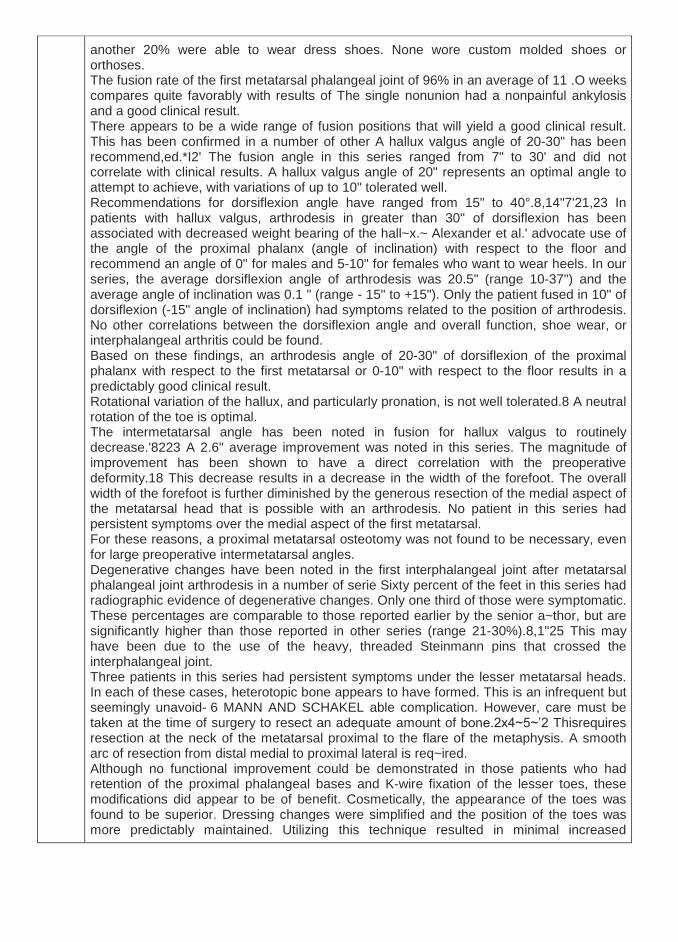
another 20% were able to wear dress shoes. None wore custom molded shoes or orthoses. The fusion rate of the first metatarsal phalangeal joint of 96% in an average of 11 .O weeks compares quite favorably with results of The single nonunion had a nonpainful ankylosis and a good clinical result. There appears to be a wide range of fusion positions that will yield a good clinical result. This has been confirmed in a number of other A hallux valgus angle of 20-30" has been recommend,ed.*I2' The fusion angle in this series ranged from 7" to 30' and did not correlate with clinical results. A hallux valgus angle of 20" represents an optimal angle to attempt to achieve, with variations of up to 10" tolerated well. Recommendations for dorsiflexion angle have ranged from 15" to 40°.8,14"7'21,23 In patients with hallux valgus, arthrodesis in greater than 30" of dorsiflexion has been associated with decreased weight bearing of the hall~x.~ Alexander et al.' advocate use of the angle of the proximal phalanx (angle of inclination) with respect to the floor and recommend an angle of 0" for males and 5-10" for females who want to wear heels. In our series, the average dorsiflexion angle of arthrodesis was 20.5" (range 10-37") and the average angle of inclination was 0.1 " (range - 15" to +15"). Only the patient fused in 10" of dorsiflexion (-15" angle of inclination) had symptoms related to the position of arthrodesis. No other correlations between the dorsiflexion angle and overall function, shoe wear, or interphalangeal arthritis could be found. Based on these findings, an arthrodesis angle of 20-30" of dorsiflexion of the proximal phalanx with respect to the first metatarsal or 0-10" with respect to the floor results in a predictably good clinical result. Rotational variation of the hallux, and particularly pronation, is not well tolerated.8 A neutral rotation of the toe is optimal. The intermetatarsal angle has been noted in fusion for hallux valgus to routinely decrease.'8223 A 2.6" average improvement was noted in this series. The magnitude of improvement has been shown to have a direct correlation with the preoperative deformity.18 This decrease results in a decrease in the width of the forefoot. The overall width of the forefoot is further diminished by the generous resection of the medial aspect of the metatarsal head that is possible with an arthrodesis. No patient in this series had persistent symptoms over the medial aspect of the first metatarsal. For these reasons, a proximal metatarsal osteotomy was not found to be necessary, even for large preoperative intermetatarsal angles. Degenerative changes have been noted in the first interphalangeal joint after metatarsal phalangeal joint arthrodesis in a number of serie Sixty percent of the feet in this series had radiographic evidence of degenerative changes. Only one third of those were symptomatic. These percentages are comparable to those reported earlier by the senior a~thor, but are significantly higher than those reported in other series (range 21-30%).8,1"25 This may have been due to the use of the heavy, threaded Steinmann pins that crossed the interphalangeal joint. Three patients in this series had persistent symptoms under the lesser metatarsal heads. In each of these cases, heterotopic bone appears to have formed. This is an infrequent but seemingly unavoid- 6 MANN AND SCHAKEL able complication. However, care must be taken at the time of surgery to resect an adequate amount of bone.2x4~5~’2 Thisrequires resection at the neck of the metatarsal proximal to the flare of the metaphysis. A smooth arc of resection from distal medial to proximal lateral is req~ired. Although no functional improvement could be demonstrated in those patients who had retention of the proximal phalangeal bases and K-wire fixation of the lesser toes, these modifications did appear to be of benefit. Cosmetically, the appearance of the toes was found to be superior. Dressing changes were simplified and the position of the toes was more predictably maintained. Utilizing this technique resulted in minimal increased

morbidity (one pin tract infection). Adequate bone resection was possible in all cases after a satisfactory soft tissue release of the metatarsal phalangeal joint had been accomplished. The manipulative correction of the lesser toe interphalangeal joint deformities, when combined with the metatarsal head resections, resulted in excellent clinical correction of these deformities; only two patients (1 0%) had residual symptomatic hammertoes. Therefore, it would seem that a resection-type correction is not necessary primarily and should be reserved for residual or recurrent symptomatic deformities. In conclusion, resection arthroplasty of the lesser metatarsal phalangeal joints combined with arthrodesis of the first metatarsal phalangeal joint results in a long-lasting correction of forefoot deformities due to inflammatory arthropathies, with 90% good and excellent results. Maintenance of the proximal phalangeal bases and K-wire fixation of the lesser toes improve the cosmetic results and simplifies the postoperative management .
22. OUTCOME OF COMBINED RECONSTRUCTIVE PROCEDURE WITH REROUTING OF THE TIBIALIS ANTERIOR TENDON IN
SEVERE FLEXIBLE FLATFOOT
Osman Abd Ellah Mohamed Egypt
Introduction: The clinical appearance of the severe flexible flat –foot consists of A collapsed medial longitudinal arch. Heel valgus. Fore foot abduction heel cord tightness, and forefoot supinated or varus. Methods: A combined procedure is described that addresses all the components at fault in the severely flexible flatfoot deformity children. The Evans calcaneal distraction wedge osteotomy will lengthen the lateral column, correcting the heel valgus and forefoot abduction. A naviculo –first cuneiform wedge resection (medial and planter) and fusion will shorten and reshape the collapsed medial arch .this is augmented by reconstruction and placation of the lengthened planter ligaments with planter rerouting of the tibialis anterior tendon to act as a strong planter ligament. In addition shifting the tibialis anterior pull proximally acts as a sling to the talus head. z- Plasty of the tight tendo Achilles is always needed nineteen feet in11 patients were the subject of this study .the period of follow –up ranged from 8 to 42 months. Results: : were assessed to the relief of the relief of foot strain and calf pains, improvement in shoe wear. General activity and foot shape. To evaluate foot shape, reconstruction of the medial arch and heel posture were assessed. The children and parents were satisfied with the results in17 feet (89.5%). improvement of the radiological measurements was evident and was statistically significant. Discussion & Conclusion: Combined procedure proved effectively at reconstruction of all factors at fault in one sitting. A useful correction of a significant deformity in relatively young children will prevent more serious deformity later in life. Have a Comment?:
good procedures for flate foot
23. TALO-CALCANEAL SMILE OSTEOTOMY AS A TREATMENT OF RIGID RESIDUAL EQUINUS
Sameh Al-safty
Egypt
Introduction: Gradual correction by means of external fixation is established as a powerful technique for the resistant and recurrent clubfoot. this method appears to preserve suppleness and can be combined with foot osteotomies asan alternative to triple joint fusion in the salvage of stiff recurrent deformities in older and adolescent children. in the relapsed and stiff variants , the ilizarov method of gradual correction by external fixation is remarkable at salvaging these problems : full correction and some restoration of foot and ankle suppleness is possible . in older children and adolescents this technique can be combined with calcaneal and midfoot osteotomies to create a plantigrade foot around a stiff ankle and sub-talar joint . Methods: 15 cases in 13 patients were presented by rigid equinus with stiff ankles . all patient had a history of surgically correction : 6 patients (2 operations) , 7 patients (3 operations). age range from 4-10 Ys . we had 5 females , 8 males . ilizarov was applied after small lateral incision at the level of sub-talar joint. smile osteotomiy was done through talus and calcaneus then gradual correction . Results: 10 patient had full corrected with plantigrade food . 2 patients have 10 degrees planter flexion. 1 patient underwent a revision surgery . gait improved from tiptoing to plantigrsde . Discussion & Conclusion: recurrent club-foot or residual stiff equinus deformity can be treated by many salvage methods as talar osteotomy , talar de-cancellation , talectomy or tripple arthrodesis . our technique based on ilizarov metnhod appear to provide a similar or better result without diminution in foot size .
24. GRAFT FOR LATERAL COLUMN LENGTHENING
Mohamed Abdel Aal Hussein Egypt
25. CORRECTION OF RESIDUAL METATARSUS ADDUCTUS DEFORMITY FOLLOWING PONSETI MANAGEMENT OF

IDIOPATHIC CLUBFOOT IN TODDLERS BY TIBIALIS ANTERIOR TENDON TRANSFER
Mohamed A. Abdel-Aal
Egypt Introduction: Ponseti technique becomes a gold standard treatment for correction of idiopathic clubfoot and widely reports to provide reliable results. However, a relapsed deformity may occur owing to imbalance of strong tibialis anterior and weak its antagonist resulting in dynamic metatarsus adductus deformity of forefoot. Methods: In this prospective case series study, 21 children (9 females) with residual dynamic metatarsus adductus following treatment of idiopathic congenital talipes equinovarus utilizing Ponseti technique in the orthopedic department of Al-Azher University Hospital, Mapera Zagazig Hospital and El-Bakry General Hospital treated by transfer of tibialis anterior tendon into third cuneiform bone. Evaluation comprised clinical, appearance, radiological assesment and complications Results: Mean follow-up 23.4 months and mean age of patients 35.3 months. All transferred tendons healed properly without major complications. Clinically; Dimeglio score improved from 5.8 preoperative to 1.3 postoperative and the clinical appearance according to Garceau and Palmer improved from 2.8 to 3.4. No major complications or correction loss developed in any case. Discussion & Conclusion: Residual dynamic metatarsus adductus deformity following Ponseti management of congenital talipes equinovarus can be corrected efficiently and simply by tibialis anterior tendon transfer.
26. MINIMAL INVASIVE USE OF DISTRACTION HISTOGENESIS IN RELAPSED CLUB FOOT
Mohamed Fadel
Egypt
Introduction: Conventional surgical treatment of relapsed club foot deformities is not always successful or easy to apply. In this study we evaluate the use of the distraction histogenesis technique for management of relapsed club foot deformities. Methods: fifty three cases 2- 6 years old with relapsed club foot deformities with history of average 3 previous operations (range, 1-8 operations).This thesis based on 50 consecutive cases (61 feet), of average age 4 years and 3 months (range, 2- 6 years). We used preoperative assembly of the leg construct of the apparatus but ankle and foot construct was designed according to the condition of deformity. Twenty patients were discharged from the hospital the same day of the operation.

Results: The range of operative time was 1 – 2.5 hours (average of 1.5 hours). Average time in the fixator was 18weeks (range, 10 weeks - 30 weeks). After fixator removal cast was applied for one month, followed by night splint and special shoes for their daily activities.The average follow-up period was 42 months (range,36 - 84 months) after fixator removal.The results were: good in 50 feet, fair in 7, bad in 4 Discussion & Conclusion: Ilizarov Treatment is lengthy, difficult, fraught with complications, and a technically demanding procedure. However, we believe that Minimal invasive use of distraction histogenesis in relapsed club foot using Ilizarov external fixator in a closed management method in treating relapsed club foot deformities in the gray old age zone is an effective.
27. CORRECTION OF POST TRAUMATIC FOOT DEFORMITY.
Wagih Mousa UK
28. GRADUAL CORRECTION OF STIFF CLUBFOOT DEFORMITY USING HINGED ILIZAROV FRAME
Saw Aik Malaysia
29. ILIZAROV TECHNIQUE FOR TREATMENT OF COMPLEX ANKLE MALFORMATION.
Sihe Qin
China
30. DISTRACTION HISTOGENESIS, THE HOPE FOR DIABETIC FOOT.
Yong Hong Zhang
China
31. CORRECTION OF DEFORMITY IN CHARCOT FOOT
Hani El Mowafi Egypt

32. ROLE OF 3D PRINTING IN SPINAL DEFORMITY
Bhavuk Garg India
33. PEDIATRIC CERVICAL SPINE SURGERY
Alexander Gubin Russia
34. SPINAL OSTEOTOMIES FOR SPINAL DEFORMITIES
Bhavuk Garg India
35. PERCUTANEOUS TREATMENT OF SPINAL PATHOLOGIES USING AN EXTERNAL FIXATOR UNDER LOCAL
ANESTHESIA
R A Agrawal India
36. CORTICOTOMY TECHNIQUE USING GUIDED DOUBLE BARREL DRILL SLEEVE
Saw Aik Malaysia
37. Cellular advances & limb reconstruction
Reggie Hamdy Canada
38. THE ORGANIZATIONAL SPECIFICS OF THE LIMB LENGTHENING AND RECONSTRUCTION CENTER.
Alexander Gubin
Russia
39. LENGTHENING NAILS
Reggie Hamdy Canada
40. ILIZAROV PARADIGM
Alexander Gubin Russia
41. BIOLOGIC SOLUTIONS OF NONUNION
Reggie Hamdy Canada
42. THE ACCORDION TECHNIQUE" FOR THE BONE DELAYED UNION
Yong Hong Zhang
China
43. RATE OF PIN SITE INFECTION USING NON STERILE DRESSING METHOD
Saw Aik Malaysia
44. CONGENITAL PSEUDOARTHROSIS OF TIBIA.
H.R Jhujhunwala India
45. POST- PONSETI BRACING COMPLIANCE IN EGYPTIAN POPULATION DURING TREATMENT OF IDIOPATHIC
TALIPES EQUINOVARUS (TEV)

Elsayed Abdelhalim Egypt
46. CRANIO-CERVICAL SAGITTAL BALANCE
Ahmed El Tantawy Egypt
Introduction: There is an increasing evidence of the close association between sagittal plane deformity and self-reported disability. So, it is vital for spine surgeons to have consistent and reproducible parameters to evaluate the cranio-cervical sagittal balance (CCSB). Methods: We conducted an electronic-based search in the literature for identifying the different parameters that were used or evolved in the last 10 years for assessing CCSB. Results: Conventionally used parameters, among different series, are C2-C7 lordosis and cervical sagittal vertical axis. More recently, the head sagittal balance was assessed based on sella turcica and McGregor line and includes: cranial tilt (CT), cranial slope (CS) and cranial incidence (CI). Additionally, CCSB was evaluated by using one of the two vertebrae located at the base of the cervical spine as a reference landmark. One reference depends on C7 vertebra and defines: C7 Slope, Sella turcica tilt and spino-cranial angle (SCA). The other reference is based on T1 vertebra and includes: T1 slope, T1 tilt, T1 incidence, and thoracic inlet angle (TIA). Discussion & Conclusion: Different tools have been introduced, in the literature, for assessing CCSB and to distinguish between abnormal parameters which contribute to deformity from ones which compensate for it. CI and TIA are morphological parameters that could be used as a reference and constant parameters, to assess cervical compensation. CT and Neck tilt are positional parameters of head /neck version that act as compensatory mechanisms in order to maintain an upright posture with age-related changes in sagittal alignment.
47. TRANSFORAMINAL LUMBAR INTERBODY FUSION (TLIF) IN PATIENTS WITH LUMBAR INSTABILITY SUBMITTED TO
TRANSPEDICULAR SCREWS FIXATION
Moawed Farghly Eladawy, M.D.; Samir Mahmoud El Ghandour, M.D.; Ahmed Abdelaty, M.D.
Egypt
Introduction: This study is conducted to evaluate the clinical and radiological outcomes of Transforaminal lumbar interbody fusion (TLIF), with pediclular screws fixation, in the treatment of patients with lumbar instability.
Methods: A prospective study of 20 patients with lumbar instability treated with transforaminal lumbar interbody fusion (TLIF) technique. All patients have been operated upon with such technique of fusion with one cage (carbon fiber) and pediclular screws fixation. We included patients between 18-65 years, without previous lumbar surgery, grades 1 and 2 spondylolisthesis. Patients with previous lumbar surgery or with higher grades of spondylolisthesis were excluded. Visual Analogue Scale (VAS) for pain and Oswestry Disability Index (ODI) were used to assess the patients pre-operative and at post-operative intervals at 1, 3, 6 months till two year duration. Pre-operative and post-operative radiographs were done using static and dynamic X-rays, computerized tomography, and magnetic resonance imaging. Results: : Patients were assessed for severity of low back pain in a percentage value by VAS score pre-operatively, after surgery by 48 hours, one month, three months, and six months until two year. The maximal score pre-operative was nine and minimal was six with a mean of 7.7 (±0.864), While six months till two year postoperatively, the maximal score was 6 and minimal was 1 with a mean of 2.2 (±1.76). Regarding the percentage of pre-operative severity of pain, 9 patients (45%), suffered from moderate pain while 11 patients (55%) had severe pain. After one year follow up the percentage changed to 85% with mild pain and only 15 % with moderate one respectively with nearly same results after two years. The ODI maximal score pre-operative was 80 and minimal was 40. While from six months till two year duration postoperatively, the maximal score was 35 and minimal was 10 with statistical significance. While radiologically, 17 patients, (85%) have showed good fusion results while the rest 3 patients, (15%) have showed delayed or no signs of fusion. Discussion & Conclusion: TLIF provides good clinical and radiological outcomes in management of lumbar instability after follow-up period of two year duration. Operative complications are less in TLIF technique, and risk of dural tear is diminished significantly. TLIF can save much operative time and blood loss.
48. SURGICAL MANAGMENT OF NEGLECTED LATERAL CONDYLE FRACTURES OF THE HUMERUS IN CHILDREN
Mohamed Rohayem
Egypt
Introduction: Delayed presentation of lateral condylar fracturesof the humerus in children is relatively common in the developingregions of the world.However,little have been published about this subject. These fractures are difficult to managedue to the action of the common extensor origin which renders any fixation insecure of such osteoporotic fragment. Methods: nine children with non-united fractures of the lateral humeral condylewith/without deformity were included in this prospective study.The main presenting complaint was deformity with limitation of elbow range of motion.Fixation and correction were done through the fracture site via lateral approach by tension band and concomitant bone graft when indicated
Results: The results were evaluated by Hardacre criteria.Three cases have full range of movements .The rest have functional range of motion with subtle deformity,no arthritic or neurological symptoms and complete healing of fracture.One patient had unsightly scar at the graft site which needed scar revision Discussion & Conclusion: Open reduction and internal fixation is the method of choice for the management of sequale of neglectedlateral condylar fractures of the humerus.
49. MASQUELET-ILIZAROV TECHNIQUE IN THE MANAGEMENT OF INFECTED NONUNION OF THE TIBIA
Ahmed M. Lashin, MSc. Osama El-Gebaly, MD. Mahmoud El-Rosasy,
MD. Ashraf Atef, MD. Egypt
Introduction: Reconstruction of large segmental bone defects is a challenging situation. Such defects may result from direct trauma, post-osteomyelitis debridement or tumour resection. Oncological debridement with wide resection of infected tissues decreases the recurrence rate of infection in osteomyelitis. The combination of Masquelet and Ilizarov techniques may boost the odds of a successful reconstruction by summation of the advantages of both techniques. Methods: This prospective case series included twenty patients with infected non-united fractures of the tibia managed by Ilizarov bone transport through an induced membrane. The mean size of the defect after debridement was 6.2 cm (range 4-11 cm). The mean age of the patients was 29 years (13 to 54). The average follow up period was 29 months (range 12-48 months). Results: Successful reconstruction with no recurrence of infection was achieved in all cases without the need of soft tissue coverage. Bone graft was needed in two cases. The mean time to union was 7.3 months. Unsatisfactory end results occurred in three cases (preoperative ankle stiffness in two and residual leg length discrepancy in one). Discussion & Conclusion: Spacer insertion in post-debridement defects in infected nonunion allows for temporization until eradication of infection. Moreover, it permits local delivery of antibiotics and provides
a tunnel for bone transport. Bone transport overcomes bone graft shortage when using Masquelet technique alone in large defects. This decreases the donor site morbidity and allows for radical debridement.
50. NEGLECTED TIBIAL PLAFOND FRACTURES; IS ARTHRODESIS MANDATORY?
Abdel-Salam Abdel-Aleem Ahmed
Egypt
Introduction: Tibial pilon fractures are controversial and potentially devastating injuries. Reports of neglected are scarce; however, such reports for pilon fractures are yet lacking. The aim of this study was to report the outcomes of the management of neglected pilon fractures by Ilizarov fixator, and to evaluate whether this technique was successful to avoid arthrodesis. Patients and Methods: This retrospective study included 18 patients with a mean age of 42.17 years. Twelve patients were males and six were females. The mean duration from trauma to management by Ilizarov fixator was 11.13 weeks (Range: 7 to 15). All fractures were AO-OTA type 43 C. Four cases were open fractures. The radiographs were evaluated for tibial alignment, quality of reduction, and development of arthrosis. The American Orthopaedic Foot and Ankle Society (AOFAS) Ankle-Hind foot Scale was used for functional assessment. Results: The follow-up period ranged from 18 to 168 (mean; 38.00) months. Bone grafting was done in three cases. The quality of reduction was excellent in two cases, satisfactory in 13 cases, and poor in three cases. The ankle-spanning frame was removed after a mean of 15.27 weeks (Range: 12 to 26). The external fixator period averaged 29.8 weeks (Range: 26 to 36). All fracture healed without deep infection. Ankle dorsiflexion and plantar-flexion averaged 8.67° and 25.67° respectively. The mean AOFAS Ankle-hindfoot score was 82.53. One case had mild anterior translation, and another one had a procurvatum of about 5°. Arthrodesis was done for three cases. Arthrosis developed in 6 ankles. Conclusion: A satisfactory outcome was achieved after management by Ilizarov fixator with avoiding arthrodesis in most cases.
51. THE USE OF FREE NON VASCULARIZED FIBULAR GRAFT IN THE INDUCED MEMBRANE TECHNIQUE TO MANAGE
POST-TRAUMATIC BONE DEFECTS
Barakat El Alfy Egypt
Introduction:
The induced membrane technique is increasingly used in management of segmental skeletal defects with promising results. The aim of the present study is to assess the results of free non vascularized fibular graft in the induced membrane technique to manage bone defects. Methods: : Fifteen patients with segmental skeletal defects were treated by the induced membrane technique using the free non vascularized fibular graft. The ages ranged from 20 to 48 years with an average of 32 years. The cause of the defects was post-traumatic bone loss in all cases. The defects were located in the distal femur in 9 cases, proximal tibia in 2 cases and middle third of the tibia in 4 cases. The defects ranged from 5 cm to 14 cm with an average of 8 cm. All cases were treated by the induced membrane technique in two stages. Autogenous cancellous bone graft and free non vascularized fibular graft were used to fill the defect in the second stage of surgery. Results: All cases healed without additional procedures after the second stage except in two cases. The time of healing ranged from 4- 13 months with an average of 7 months. After physiotherapy all cases regained full range of ankle and knee movements except one case. The complications included nonunion of the graft in two cases, deep wound infection in one case, and chronic pain along the iliac crest in one case. No cases were complicated by implant failure or refracture. Discussion & Conclusion: The use of free non vascularized fibular graft in the induced membrane technique reduces the amount of iliac bone graft required to fell the defect. It also provides a mechanical support that prevents refracture and implant failure. The combination of autogenous cancellus bone graft, free nonvascularized fibular graft and stable fixation by locked plate improves the results of the induced membrane technique.
52. A MODIFIED MASQUELET TECHNIQUE FOR THE FOREARM INFECTED SEGMENTAL BONE LOSS.
Ahmad Allam
Egypt
53. METABOLIC COMPLEX DEFORMITIES
Reggie Hamdy Canada
54. PROXIMAL FEMORAL DEFORMITIES
Theddy Slongo Switzerland

55. COMPLEX DEFORMITIES IN SYNDROMES
Reggie Hamdy Canada
56. HOW TO RECONSTRUCT THE STANDING AND WALKING FUNCTION FOR CRAWL SQUATTING PATIENTS.
Sihe Qin
China
57. TKA FOR VALGUS DEFORMITY Rajesh Malhotra
India
58. TOTAL KNEE REPLACEMENT IN EXTRA ARTICULAR DEFORMITIES.
Vikrant Manhas
India
59. THA FOR DEFORMED HIP
Rajesh Malhotra India
60. ADVANCES IN UNICONDYLAR KNEE REPLACEMENT.
Vikrant Manhas India